Abstract
Objective
In patients with acute chest pain who have had myocardial infarction excluded, plasma cardiac troponin I concentrations ≥5 ng/L are associated with risk of future adverse cardiovascular events. We aim to evaluate the association between cardiac troponin and coronary plaque composition in such patients.
Methods
In a prespecified secondary analysis of a prospective cohort study, blinded quantitative plaque analysis was performed on 242 CT coronary angiograms of patients with acute chest pain in whom myocardial infarction was excluded. Patients were stratified by peak plasma cardiac troponin I concentration ≥5 ng/L or <5 ng/L. Associations were assessed using univariable and multivariable logistic regression analyses.
Results
The cohort was predominantly middle-aged (62±12 years) men (69%). Patients with plasma cardiac troponin I concentration ≥5 ng/L (n=161) had a higher total (median 33% (IQR 0–47) vs 0% (IQR 0–33)), non-calcified (27% (IQR 0–37) vs 0% (IQR 0–28)), calcified (2% (IQR 0–8) vs 0% (IQR 0–3)) and low-attenuation (1% (IQR 0–3) vs 0% (IQR 0–1)) coronary plaque burden compared with those with concentrations <5 ng/L (n=81; p≤0.001 for all). Low-attenuation plaque burden was independently associated with plasma cardiac troponin I concentration ≥5 ng/L after adjustment for clinical characteristics (adjusted OR per doubling 1.62 (95% CI 1.17 to 2.32), p=0.005) or presence of any visible coronary artery disease (adjusted OR per doubling 1.57 (95% CI 1.07 to 2.37), p=0.026).
Conclusion
In patients with acute chest pain but without myocardial infarction, plasma cardiac troponin I concentrations ≥5 ng/L are associated with greater burden of low-attenuation coronary plaque.
INTRODUCTION
High-sensitivity cardiac troponin assays have transformed the assessment of patients with acute chest pain through the development and implementation of accelerated diagnostic pathways to ‘rule in’ and ‘rule out’ myocardial infarction in the emergency department.1–6 Yet a substantial proportion of patients have intermediate cardiac troponin concentrations that fall between these ‘rule in’ and ‘rule out’ thresholds. These patients are 5–10 times more likely to have a major adverse cardiac event at 1 year compared with those below the ‘rule out’ threshold of 5 ng/L.1 7 8 The underlying pathophysiological mechanism for this increased risk remains uncertain.
The PRECISE-CTCA study (Troponin Within the Normal Reference Range to Risk Stratify Patients With Acute Chest Pain for Computed Tomography Coronary Angiography; ClinicalTrials.gov number NCT04549805) demonstrated that coronary artery disease was three times more likely in patients without myocardial infarction who had intermediate cardiac troponin compared with those with low concentrations.9 However, newer diagnostic approaches can go beyond the assessment of coronary stenosis to define plaque characteristics and identify high-risk atherosclerosis associated with acute coronary syndromes.10 In particular, low-attenuation plaque correlates with the lipid-rich necrotic core of high-risk atherosclerotic plaque and is a strong independent predictor of future cardiovascular events.11 In this prespecified secondary analysis of the PRECISE-CTCA study, we aimed to evaluate the association between cardiac troponin concentrations within the normal reference range and coronary plaque composition in patients with acute chest pain who have had myocardial infarction excluded.
METHODS
Study population
The PRECISE-CTCA study was a single-centre prospective cohort study enrolling 250 patients with suspected acute coronary syndrome presenting to the Royal Infirmary of Edinburgh, Edinburgh, Scotland.9 Smoking history was categorised into any history of smoking versus never smoked. Estimated glomerular filtration rates were baseline values. History of hypertension was based on clinical history taken from the patient on admission to hospital.
High-sensitivity cardiac troponin testing
All participants had myocardial infarction excluded with a maximal plasma high-sensitivity cardiac troponin I concentration (ARCHITECT STAT Troponin I Assay; Abbott Laboratories, Abbott Park, Illinois) below the 99th centile upper reference limit (34 ng/L for men and 16 ng/L for women). A second sample was sent for testing in all patients with intermediate values and in those below the rule-out threshold where the presentation was within 2 hours of symptom onset according to clinical practice guideline recommendations.12–14 Participants were then divided into two groups based on their peak cardiac troponin concentrations: low concentration (<5 ng/L, below the risk stratification threshold) and intermediate concentration (between 5 ng/L and the sex-specific 99th centile).
CT coronary angiography
All recruited participants underwent CT coronary angiography using a 128-slice scanner (Biotech mCT, Siemens Healthcare) as soon as possible after discharge. Scans were performed after administration of rate-limiting medication if the heart rate was ≥60 beats per minute and sublingual glyceryl trinitrate given unless contraindicated. A tube voltage of 100 kVp was used for patients with a body mass index of <25 kg/m2 and 120 kVp for those over 25 kg/m2. Iodine-based contrast agent was injected at a rate of 4–5 mL/s. Scans were performed during breath-hold using prospective ECG gating in diastole unless heart rate control was suboptimal, in which case images were acquired in the systolic phase. Images were reconstructed using 180° rotation, filtered back projection, 512×512 matrix, medium smooth reconstruction kernel (B26f) and 0.75 mm slice thickness at 0.5 mm increments.
Obstructive coronary artery disease was defined as luminal stenosis of ≥70% in one or more major epicardial artery or ≥50% in the left main stem. The presence of any coronary artery disease was defined as any visually observed luminal stenosis ≥10% due to the presence of atherosclerosis.
Quantitative plaque analysis
Scans were anonymised and exported in DICOM (Digital Imaging and Communications in Medicine). Quantitative plaque analysis was performed using semiautomated software (Auto-Plaque V.2.5; Cedars-Sinai Medical Center, Los Angeles, USA). This method has been validated against intravascular ultrasound and has excellent observer agreement.15 16
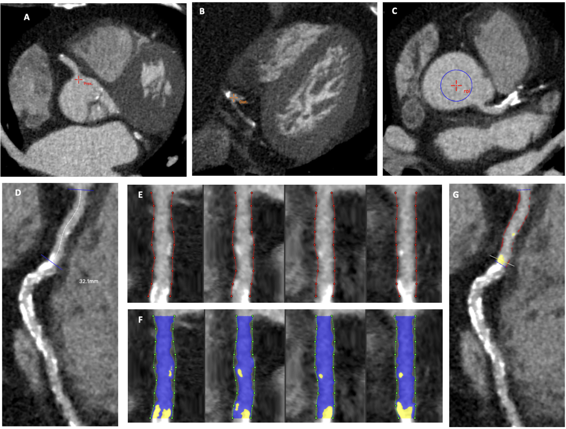
A region of interest was placed in the proximal aorta to define blood pool attenuation. Coronary centrelines were then extracted in vessels with visible coronary artery disease, including side branches ≥2 mm in diameter. Coronary segments were defined manually using side branches to mark progression from proximal to distal vessel in accordance with the Society of Cardiovascular Computed Tomography guidelines.17 To avoid introducing noise, stented segments, coronary artery bypass graft insertion points and coronary segments with no visually observed coronary artery disease did not undergo quantitative plaque analysis. Plaque constituents were automatically deter-mined using scan-specific Hounsfield unit (HU) thresholds based on blood pool attenuation. Manual adjustments were made after careful assessment of each segment to ensure pericoronary adipose tissue and vessel walls were not included as plaque.15 Low-attenuation plaque was defined by a fixed HU threshold of <30 HU, as defined previously.18 If image quality was deemed too poor to complete quantitative plaque analysis, scans were excluded. Scans were analysed by a single trained observer, blinded to patients’ results and demographics. Figure 1 delineates a step-by-step approach to demonstrate how the plaque analysis was performed.
Figure 1.

Process of conducting the plaque analysis. Proximal and distal points of the right coronary artery were placed (A and B) to extract coronary centreline. Region of interest was placed in the aortic root (C) to define blood pool attenuation. The proximal segment of the right coronary artery was manually selected (D). Vessel wall was delineated (E) and lumen was defined (in blue) using scan-specific Hounsfield unit thresholds for non-calcified and calcified plaque (F), which were manually adjusted where required to match visual review of the plaque. Completed analysis of proximal right coronary artery (G) demonstrates a large burden of non-calcified plaque (in red; 33%) and a small burden of calcified plaque (in yellow; 3%).
The total volume of coronary plaque was measured in mm3. The volume of plaque subtypes was also measured, including non-calcified, calcified and low-attenuation plaques. To account for differences in vessel volume between patients, plaque burden was calculated by dividing plaque volume by the analysed vessel volume (ie, total vessel volume of all analysed segments) on a per-patient level and multiplying by 100. To determine the relative utility of quantitative plaque analysis, comparisons were made with existing semiquantitative assessments of coronary artery plaque. These included the segment-involved score, segment severity score and CT-Leaman score.
Statistical analysis
Continuous variables are presented as mean±SD when data were normally distributed or median (IQR) when not. Statistical significance was assessed using Pearson’s χ2 or Fisher’s exact test for categorical variables (depending on the number of observations) and Wilcoxon rank-sum test or Student’s t-test for non-normal or normally distributed continuous data as appro-priate. Univariable logistic regression analysis was performed on clinical variables including age, sex, relevant medical history, presence of any coronary disease, presence of obstructive coronary disease, semiquantitative coronary CT scores (segment-involved score, segment severity score and CT-Leaman score) and different plaque subtypes to determine the OR with 95% CI of an intermediate cardiac troponin concentration. To further account for clinically relevant risk factors, a multivariable model was also created using a priori selection, adjusting for age (per 10-year increase), sex, smoking history, hypertension, diabetes mellitus, estimated glomerular filtration rate, plaque volume and burden of different plaque subtypes (total, non-calcified, calcified and low-attenuation). The model was also used to determine whether traditional markers of coronary artery disease severity, such as the presence of obstructive disease and semiquantitative coronary CT scores, were independently associated with intermediate cardiac troponin concentrations. For all logistic regression analyses, semiquantitative coronary CT scores, plaque volumes and plaque burden were log-transformed (log2 of 1 plus the plaque variable). Statistical significance was defined as a two-sided p value <0.05. All statistical analyses were performed using R (V.4.0.2; R Foundation for Statistical Computing, Vienna, Austria).
Patient and public involvement
Patients or the public were not involved in the design, conduct, reporting or dissemination plans of our research.
RESULTS
Study population
Of the 250 study participants, 242 scans were of sufficient quality to undertake plaque analysis. The study population comprised mostly male (69%) participants with a mean age of 62±12 years. A total of 161 (67%) patients had an intermediate (between 5 ng/L and the sex-specific 99th centile) and 81 (33%) had a low (<5 ng/L) cardiac troponin concentration. Patients with intermediate cardiac troponin concentrations were older, more likely to have diabetes mellitus and to have lower estimated glomerular filtration rates. All other comorbidities were equally distributed between the two groups. Patients with an intermediate cardiac troponin concentration were more likely to have coronary artery disease and higher semiquantitative CT scores (table 1).
Table 1.
Baseline demographics and CT characteristics
| Characteristics | Low cardiac troponin (<5 ng/L) n=81* | Intermediate cardiac troponin (5 ng/L-99th centile) n=161* | P value † |
|---|---|---|---|
| Age | 57+11 | 64+12 | <0.001 |
| Male sex | 51 (63) | 116 (72) | 0.15 |
| Smoking history | 46 (57) | 86 (53) | 0.6 |
| Diabetes mellitus | 4 (4.9) | 28 (17) | 0.007 |
| Hypertension | 29 (36) | 75 (47) | 0.11 |
| Hyperlipidaemia | 20 (25) | 31 (19) | 0.6 |
| Family history of coronary artery disease | 27 (33) | 62 (39) | 0.4 |
| Previous myocardial infarction | 12 (15) | 36 (22) | 0.2 |
| GRACE score | 87+25 | 97+25 | 0.005 |
| Estimated glomerular filtration rate (mL/min/1.73 m2) | 89+14 | 82+18 | <0.001 |
| Any coronary artery disease | 35 (43) | 116 (72) | <0.001 |
| Obstructive coronary artery disease | 16 (20) | 51 (32) | 0.050 |
| Segment-involved score | 0 (0–3) | 2 (0–6) | <0.001 |
| Segment severity score | 0 (0–4) | 3 (0–9) | <0.001 |
| CT-Leaman score | 0 (0–5) | 5 (0–10) | <0.001 |
| Quantitative plaque analysis | |||
| Total plaque burden (%) | 0 (0–32) | 33 (0–47) | <0.001 |
| Non-calcified plaque burden (%) | 0 (0–28) | 26 (0–37) | <0.001 |
| Calcified plaque burden (%) | 0 (0–3.1) | 2.0 (0–7.5) | <0.001 |
| Low-attenuation plaque burden (%) | 0 (0–0.97) | 0.92 (0–3.16) | <0.001 |
Median (IQR), mean±SD or n (%).
Wilcoxon rank-sum test, Pearson’s χ2 test, Fisher’s exact test or Student’s t-test.
GRACE, Global Registry of Acute Coronary Events.
Plaque quantification
Overall, participants had a median total plaque burden of 27% (0–42), which consisted of non-calcified (23%, IQR 0–34), calcified (1.2%, IQR 0–6.1) and low-attenuation (0.62%, IQR 0–2.50) plaque burden. Patients with intermediate cardiac troponin concentrations had higher burden of total (33% (IQR 0–47) vs 0% (IQR 0–33)), non-calcified (27% (IQR 0–37) vs 0% (IQR 0–28)), calcified (2% (IQR 0–8) vs 0% (IQR 0–3)) and low-attenuation (1% (IQR 0–3) vs 0% (IQR 0–1)) plaque compared with those with a low cardiac troponin concentration (table 1). A similar pattern of findings was seen for plaque volumes (table 2). Density plots demonstrated the proportionate differences between those with low and intermediate cardiac troponin concentrations were most pronounced for low-attenuation plaque burden (figure 2).
Table 2.
Plaque volume and cardiac troponin
| Plaque subtype | Overall n=242* | Low cardiac troponin (<5 ng/L) n=81* | Intermediate cardiac troponin (5 ng/L-99th centile) n=161* | P value † |
|---|---|---|---|---|
| Total plaque volume (mm3) | 146 (0–527) | 0 (0–184) | 233 (0–653) | <0.001 |
| Non-calcified plaque volume (mm3) | 127 (0–440) | 0 (0–167) | 201 (0–553) | <0.001 |
| Calcified plaque volume (mm3) | 7 (0–69) | 0 (0–23) | 15 (0–101) | <0.001 |
| Low-attenuation plaque volume (mm3) | 4 (0–29) | 0 (0–10) | 7 (0–41) | <0.001 |
| Univariate regression analysis of the association between plaque volume and cardiac troponin | ||||
| Characteristics | n | OR‡ | 95% CI | P value |
| Total plaque volume§ | 242 | 1.14 | 1.07 to 1.22 | <0.001 |
| Non-calcified plaque volume§ | 242 | 1.15 | 1.07 to 1.23 | <0.001 |
| Calcified plaque volume§ | 242 | 1.20 | 1.09 to 1.32 | <0.001 |
| Low-attenuation plaque volume§ | 242 | 1.32 | 1.17 to 1.49 | <0.001 |
| Multivariate regression analysis of the association between plaque volume and cardiac troponin¶ | ||||
| Characteristics | n | OR‡ | 95% CI | P value |
| Total plaque volume§ | 242 | 1.07 | 0.98 to 1.16 | 0.14 |
| Non-calcified plaque volume§ | 242 | 1.07 | 0.98 to 1.16 | 0.14 |
| Calcified plaque volume§ | 242 | 1.07 | 0.95 to 1.21 | 0.2 |
| Low-attenuation plaque volume§ | 242 | 1.20 | 1.05 to 1.39 | 0.011 |
Median (IQR).
Wilcoxon rank-sum test.
OR of intermediate versus low troponin concentration.
Log-transformed, OR per doubling.
Results adjusted for clinical factors (age, sex, smoking history, hypertension, diabetes, estimated glomerular filtration rate and individual plaque subtypes).
Figure 2.

Density plot comparing the proportion of patients with low (<5 ng/L) and intermediate (≥5 ng/L–99th centile) cardiac troponin concentrations for each plaque burden subtype. Proportionally, low-attenuation plaque burden appears lowest in patients with a high-sensitivity troponin of <5 ng/L compared with other plaque subtypes. *Plaque subtype log-transformed.
Associations with intermediate cardiac troponin concentration
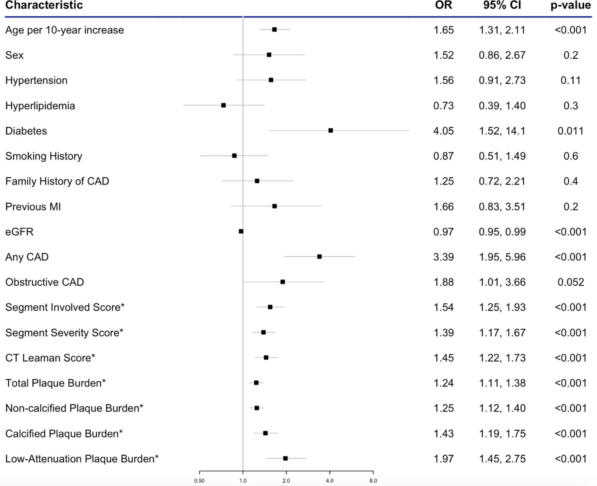
On univariable logistic regression analysis, we found that age (OR 1.65 (95% CI 1.31 to 2.11), p<0.001) and diabetes mellitus (OR 4.05 (95% CI 1.52 to 14.1), p=0.011) were associated with intermediate cardiac troponin concentrations. As previously demonstrated, patients were three times more likely to have any coronary artery disease if they had an intermediate compared with low cardiac troponin concentration.9 Higher segment involvement score, segment stenosis score and CT-Leaman score were all associated with intermediate compared with low cardiac troponin concentrations. All plaque burden subtypes were associated with intermediate cardiac troponin concentrations, with low-attenuation plaque burden appearing to have the strongest association (OR per doubling 1.97 (95% CI 1.45 to 2.75), p<0.001; figure 3). This was also the case for all plaque volume subtypes, with low-attenuation plaque volume again demon-strating the strongest association (OR per doubling 1.32 (95% CI 1.17 to 1.49), p<0.001; table 2).
Figure 3.

Univariable logistic regression analysis to determine the association of clinical and CT characteristics with an intermediate cardiac troponin concentration (≥5 ng/L–99th centile). *Log-transformed, OR per doubling of variable. CAD, coronary artery disease; eGFR, estimated glomerular infiltration rate; MI, myocardial infarction
On multivariable logistic regression analysis adjusting for known clinical risk factors, the association between presence of any coronary artery disease and intermediate cardiac troponin concentrations was of borderline significance (OR 1.95 (95% CI 0.99 to 3.88), p=0.055). This was also the case for all semiquantitative CT scores. Except for low-attenuation plaque burden (adjusted OR per doubling 1.55 (95% CI 1.13 to 2.20), p<0.009), none of the other quantitative plaque metrics was associated with intermediate cardiac troponin concentrations (table 3). Again, there was a similar pattern of association with plaque volume subtypes, with only low-attenuation plaque volume being independently associated with intermediate cardiac troponin concentrations (adjusted OR per doubling 1.20 (95% CI 1.05 to 1.39), p=0.011; table 2).
Table 3.
Multivariable logistic regression analysis evaluating the association between intermediate troponin concentrations and CT findings
| Characteristics | n | OR* | 95% CI* | P value |
|---|---|---|---|---|
| Results adjusted for clinical factors† | ||||
| Any coronary artery disease | 242 | 1.95 | 0.99 to 3.88 | 0.055 |
| Obstructive coronary artery disease | 242 | 0.83 | 0.38 to 1.82 | 0.6 |
| Segment-involved score‡ | 242 | 1.22 | 0.93 to 1.62 | 0.2 |
| Segment severity score‡ | 242 | 1.13 | 0.90 to 1.42 | 0.3 |
| CT-Leaman score‡ | 242 | 1.22 | 0.98 to 1.52 | 0.072 |
| Total plaque burden‡ | 242 | 1.11 | 0.97 to 1.26 | 0.14 |
| Non-calcified plaque burden‡ | 242 | 1.11 | 0.97 to 1.27 | 0.14 |
| Calcified plaque burden‡ | 242 | 1.13 | 0.88 to 1.46 | 0.3 |
| Low-attenuation plaque burden‡ | 242 | 1.62 | 1.17 to 2.32 | 0.005 |
| Results adjusted for presence of any coronary artery disease | ||||
| Total plaque burden‡ | 242 | 1.07 | 0.89 to 1.29 | 0.4 |
| Non-calcified plaque burden‡ | 242 | 1.07 | 0.88 to 1.29 | 0.5 |
| Calcified plaque burden‡ | 242 | 1.15 | 0.87 to 1.51 | 0.3 |
| Low-attenuation plaque burden‡ | 242 | 1.57 | 1.07 to 2.37 | 0.026 |
OR of intermediate versus low troponin concentration.
Clinical factors: age, sex, smoking history, hypertension, diabetes, estimated glomerular filtration rate and individual CT findings/plaque subtypes.
Log-transformed, OR per doubling.
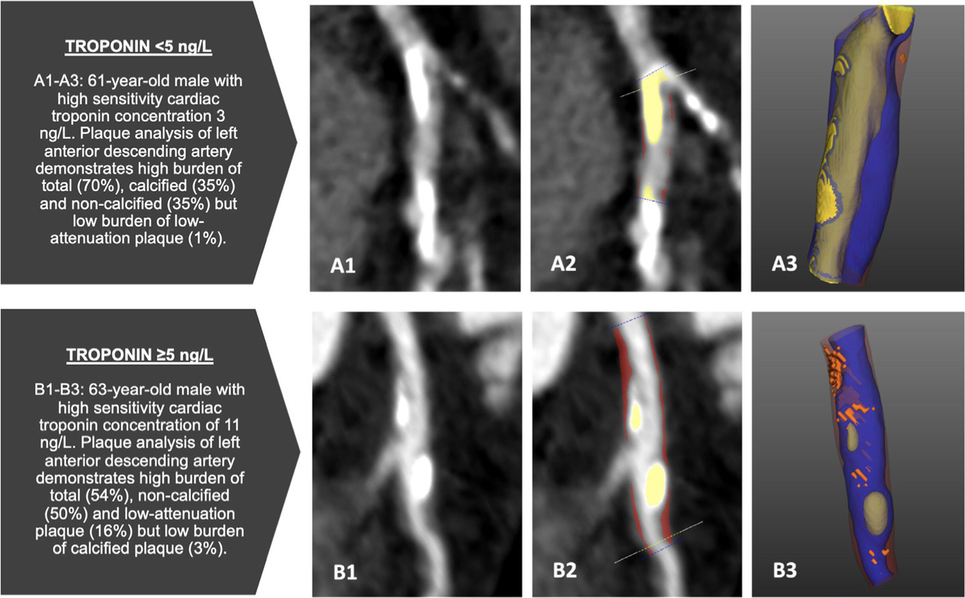
In a separate multivariable model evaluating the association between plaque burden subtypes and intermediate cardiac troponin concentration, we adjusted for the presence of any coronary artery disease. Again, only low-attenuation plaque burden was independently associated with an intermediate cardiac troponin concentration (OR per doubling 1.57 (95% CI 1.07 to 2.37), p=0.026; figure 4, table 3).
Figure 4.

Comparative cases of plaque burden in patients with troponin concentration <5 ng/L (A1–A3) and ≥5 ng/L (B1–B3). Blue, lumen; yellow, calcified plaque; red, non-calcified plaque; orange, low-attenuation plaque.
DISCUSSION
In this prespecified secondary analysis of the PRECISE-CTCA study, we evaluated the association between quantitative plaque characteristics and plasma high-sensitivity cardiac troponin concentrations within the normal reference range in patients with acute chest pain who have had myocardial infarction excluded. Patients with intermediate cardiac troponin concentrations had a higher burden of coronary plaque compared with those with low cardiac troponin concentrations, and this was most pronounced for low-attenuation plaque burden. These observations could explain the adverse prognosis in these patients.
We have previously demonstrated that in patients with acute chest pain but without myocardial infarction, those with intermediate cardiac troponin concentrations had a 5–10 times higher risk of subsequent myocardial infarction and cardiac death at 1 year compared with those with low concentrations.1 9 Although those with intermediate troponin concentrations were found to have three times higher odds of having coronary artery disease on CT coronary angiography compared with those with low concentrations, as many as one-third of patients with low troponin concentrations had some evidence of coronary artery disease. Here, we present data on the burden of plaque subtypes in such patients. We show that there is a higher burden of low-attenuation plaque in those with intermediate cardiac troponin concentrations. Low-attenuation plaque is increasingly recognised as a marker of plaque instability and a major predictor of risk.11 19 Defined as plaque with an HU threshold below 30, it correlates with the lipid-rich necrotic core of high-risk atheroma that drives plaque rupture and acute myocardial infarction.18 20 Our findings therefore provide a mechanistic link that potentially explains why patients with intermediate cardiac troponin concentrations are at a substantially increased risk of future cardiovascular events.
The strength of association between low-attenuation plaque burden and intermediate cardiac troponin concentration was notably greater than that of any other quantitative or semiquantitative measure. Indeed, even when adjusting for the presence of any coronary artery disease, low-attenuation plaque was the only metric which was associated with an intermediate cardiac troponin concentration. However, cardiac troponin is not released directly from low-attenuation plaque. Why then do we observe a high burden of low-attenuation plaque in patients with an intermediate cardiac troponin concentration? Cardiac troponin is a biomarker for myocardial injury measured on a continuous spectrum, and thus intuitively some individuals may have suffered plaque rupture or erosion, even when troponin concentrations fall below the accepted diagnostic threshold of myocardial infarction.21 Lowering the threshold for diagnosis of myocardial infarction from the numerically arbitrary 99th centile would identify more patients with myocardial infarction (improved sensitivity), but would increase the risk of overdiagnosing or misdiagnosing myocardial infarction (worse specificity).22 Indeed, some have suggested a probabilistic approach to the diagnosis of acute myocardial infarction with varying thresholds of cardiac troponin depending on a range of clinical factors.3 23 In this setting, plaque quantification and in particular the burden of low-attenuation plaque may help reduce misclassification by identifying patients who are more likely to have had an acute coronary syndrome due to a dynamic or progressive atherosclerotic process.
There is an overlap between different CT coronary angiography (CTCA) metrics to assess coronary artery disease, including stenosis, quantitative plaque analysis, semiquantitative CTCA scores, visually assessed high-risk plaque characteristics and biomechanical assessments, such as CT-derived fractional flow reserve.11 24 In patients with stable chest pain, severity of stenosis is important to quantify as it correlates with impaired coronary blood flow as calculated by fractional flow reserve, and this relates to symptoms of angina. Previous studies have demonstrated that combining plaque burden and stenosis severity provides a better marker of abnormal invasive fractional flow reserve than either metric alone.25 However, our population and endpoints of interest differ from such analyses since we studied patients with acute chest pain and wished to explore the mechanism of the future risk of myocardial infarction rather than reversible ischaemia or angina pectoris. These distinctions are important because rupture of non-obstructive coronary plaques is the principal underlying cause of acute myocardial infarction.26 Moreover, in our study, the prevalence of obstructive coronary artery disease was no different between those with low or intermediate cardiac troponin concentrations. The mechanisms underlying intermediate cardiac troponin concentrations in patients with acute chest pain remain uncertain, but we provide evidence of an association with quantitative plaque characteristics, and in particular with low-attenuation plaque.
Patients who present with symptoms or evidence of myocardial ischaemia at rest but without a detectable rise or fall in cardiac troponin are often diagnosed with unstable angina.27 Despite its declining incidence, unstable angina remains a major cause of hospitalisation28 and has been associated with improved clinical outcomes in trials of therapeutic interventions for acute coronary syndromes.29 30 It therefore seems likely that there may have been some patients in our cohort who had unstable angina, especially in those with intermediate cardiac troponin concentrations. As such, the association with low-attenuation plaque may represent a useful method of detecting those with high-risk or unstable plaque who have unstable angina. These findings have important potential sequelae for future clinical practice and could assist in the discrimination between those with or without unstable angina, impacting on their subsequent management.
Our study has some limitations which we should acknowledge. First, this was designed as a cross-sectional study, and we do not have outcome data in this cohort. However, the clinical outcomes of such patients have been reported previously and our study population is representative of this prior work.1 4 8 Second, we would also acknowledge the need for external validation of our findings in a larger and more ethnically diverse population. Our data are hypothesis-generating and future studies should focus on whether CT and quantitative plaque analyses can guide treatments to improve outcomes in this patient population. Indeed, this will be addressed in the ongoing TARGET-CTCA trial (Troponin in Acute chest pain to Risk stratify and Guide EffecTive use of Computed Tomography Coronary Angiography; NCT03952351), where patients with intermediate cardiac troponin concentrations who have been discharged from hospital are randomised to CT coronary angiography or standard of care. Assay precision was not measured daily in our clinical laboratory at the thresholds used to identify low-risk patients, and although this threshold is above the limit of quantification for this assay it is possible a small number of patients may have been misclassified. Finally, although the process of plaque analysis is semiautomated, it can still be time-consuming, particularly when there is a large burden of disease distributed throughout the coronary tree. Adoption of further automation and machine learning would help facilitate its more widespread clinical use.
In conclusion, we present data that demonstrate the strong association between high-sensitivity cardiac troponin concentrations within the normal reference range and low-attenuation plaque burden, independent of clinical risk factors or the presence of coronary artery disease. These findings provide a potential mechanistic explanation for the observed increase in risk of adverse cardiac outcomes in these patients, supporting the use of high-sensitivity cardiac troponin concentrations within the normal reference range to risk-stratify patients with acute chest pain and identify those who would benefit most from treatment.
WHAT IS ALREADY KNOWN ON THIS TOPIC
In patients with acute chest pain who have had myocardial infarction excluded, plasma cardiac troponin I concentrations ≥5 ng/L are strongly associated with risk of future adverse cardiovascular events.
WHAT THIS STUDY ADDS
In a prespecified analysis of a prospective cohort study of patients who had myocardial infarction excluded, low-attenuation plaque burden was associated with high-sensitivity cardiac troponin concentrations above 5 ng/L, independent of clinical risk factors.
HOW THIS STUDY MIGHT AFFECT RESEARCH, PRACTICE OR POLICY
Quantitative plaque analysis provides mechanistic insights into the worse prognosis of patients with troponin concentrations above 5 ng/L.
These observations may help risk-stratify patients with acute chest pain without myocardial infarction.
Acknowledgements
We would like to thank all the PRECISE-CTCA investigators.
Funding
MNM (FS/19/46/34445), MCW (FS/ICRF/20/26002, FS/11/014, CH/09/002), NLM (CH/F/21/90010, RG/20/10/34966, RE/18/5/34216) and DEN (FS/19/15/34155, CH/09/002, RG/16/10/32375, RE/18/5/34216) are supported by the British Heart Foundation. DEN is also a recipient of a Wellcome Trust Senior Investigator Award (WT103782AIA). DD is supported by National Institutes of Health/National Heart, Lung, and Blood Institute grants (1R01HL148787-01A1 and 1R01HL151266). KKL is supported by a British Heart Foundation (BHF) Clinical Research Training Fellowship (FS/18/25/33454).
Footnotes
Competing interests Outside the current study, DD received software royalties from Cedars-Sinai Medical Center and has a patent. MCW has spoken at meetings sponsored by Canon Medical Systems and Siemens Healthineers. DEN is on the Editorial Board of Heart. MRD, MCW and KKL are members of Heart’s Editorial Board.
Patient and public involvement Patients and/or the public were not involved in the design, or conduct, or reporting, or dissemination plans of this research.
Ethics approval This study involves human participants and was approved by South East Scotland Research Ethics Committee (0118/SS/0114). Participants gave informed consent to participate in the study before taking part.
Data availability statement
Data are available upon reasonable request.
REFERENCES
- 1.Shah ASV, Anand A, Sandoval Y, et al. High-sensitivity cardiac troponin I at presentation in patients with suspected acute coronary syndrome: a cohort study. Lancet 2015;386:2481–8. [DOI] [PMC free article] [PubMed] [Google Scholar]
- 2.Chapman AR, Lee KK, McAllister DA, et al. Association of high-sensitivity cardiac troponin I concentration with cardiac outcomes in patients with suspected acute coronary syndrome. JAMA 2017;318:1913–24. [DOI] [PMC free article] [PubMed] [Google Scholar]
- 3.Neumann JT, Twerenbold R, Ojeda F, et al. Application of high-sensitivity troponin in suspected myocardial infarction. N Engl J Med 2019;380:2529–40. [DOI] [PubMed] [Google Scholar]
- 4.Twerenbold R, Costabel JP, Nestelberger T, et al. Outcome of applying the ESC 0/1-hour algorithm in patients with suspected myocardial infarction. J Am Coll Cardiol 2019;74:483–94. [DOI] [PubMed] [Google Scholar]
- 5.Anand A, Lee KK, Chapman AR, et al. High-sensitivity cardiac troponin on presentation to rule out myocardial infarction: a Stepped-Wedge cluster randomized controlled trial. Circulation 2021;143:2214–24. [DOI] [PMC free article] [PubMed] [Google Scholar]
- 6.Lambrakis K, Papendick C, French JK, et al. Late outcomes of the RAPID-TnT randomized controlled trial: 0/1-Hour high-sensitivity troponin T protocol in suspected ACS. Circulation 2021;144:113–25. [DOI] [PubMed] [Google Scholar]
- 7.Bularga A, Lee KK, Stewart S, et al. High-sensitivity troponin and the application of risk stratification thresholds in patients with suspected acute coronary syndrome. Circulation 2019;140:1557–68. [DOI] [PMC free article] [PubMed] [Google Scholar]
- 8.Than MP, Aldous SJ, Troughton RW, et al. Detectable high-sensitivity cardiac troponin within the population reference interval conveys high 5-year cardiovascular risk: an observational study. Clin Chem 2018;64:1044–53. [DOI] [PubMed] [Google Scholar]
- 9.Lee KK, Bularga A, O’Brien R, et al. Troponin-guided coronary computed tomographic angiography after exclusion of myocardial infarction. J Am Coll Cardiol 2021;78:1407–17. [DOI] [PMC free article] [PubMed] [Google Scholar]
- 10.de Knegt MC, Linde JJ, Fuchs A, et al. Relationship between patient presentation and morphology of coronary atherosclerosis by quantitative multidetector computed tomography. Eur Heart J Cardiovasc Imaging 2019;20:1221–30. [DOI] [PubMed] [Google Scholar]
- 11.Williams MC, Kwiecinski J, Doris M, et al. Low-attenuation Noncalcified plaque on coronary computed tomography angiography predicts myocardial infarction: results from the multicenter SCOT-HEART trial (Scottish computed tomography of the heart). Circulation 2020;141:1452–62. [DOI] [PMC free article] [PubMed] [Google Scholar]
- 12.Collet J-P, Thiele H, Barbato E, et al. 2020 ESC guidelines for the management of acute coronary syndromes in patients presenting without persistent ST-segment elevation. Eur Heart J 2021;42:1289–367. [DOI] [PubMed] [Google Scholar]
- 13.National-Institute-for-Health-and-Care-Excellence. High-sensitivity troponin tests for the early rule out of NSTEMI. NICE guidelines 2020;DG40:1–44. [Google Scholar]
- 14.Kontos MC, de Lemos JA, Deitelzweig SB, et al. 2022 ACC Expert Consensus Decision Pathway on the Evaluation and Disposition of Acute Chest Pain in the Emergency Department. J Am Coll Cardiol 2022;80:1925–60. [DOI] [PMC free article] [PubMed] [Google Scholar]
- 15.Meah MN, Singh T, Williams MC, et al. Reproducibility of quantitative plaque measurement in advanced coronary artery disease. J Cardiovasc Comput Tomogr 2021;15:333–8. [DOI] [PMC free article] [PubMed] [Google Scholar]
- 16.Dey D, Schepis T, Marwan M, et al. Automated three-dimensional quantification of noncalcified coronary plaque from coronary CT angiography: comparison with intravascular us. Radiology 2010;257:516–22. [DOI] [PubMed] [Google Scholar]
- 17.Raff GL, Abidov A, Achenbach S, et al. SCCT guidelines for the interpretation and reporting of coronary computed tomographic angiography. J Cardiovasc Comput Tomogr 2009;3:122–36. [DOI] [PubMed] [Google Scholar]
- 18.Matsumoto H, Watanabe S, Kyo E, et al. Standardized volumetric plaque quantification and characterization from coronary CT angiography: a head-to-head comparison with invasive intravascular ultrasound. Eur Radiol 2019;29:6129–39. [DOI] [PMC free article] [PubMed] [Google Scholar]
- 19.Meah MN, Tzolos E, Wang K-L, et al. Plaque burden and 1-year outcomes inacute chest pain: results from the multicenter RAPID-CTCA trial. JACC Cardiovasc Imaging 2022;15:1916–25. [DOI] [PubMed] [Google Scholar]
- 20.Motoyama S, Sarai M, Harigaya H, et al. Computed tomographic angiography characteristics of atherosclerotic plaques subsequently resulting in acute coronary syndrome. J Am Coll Cardiol 2009;54:49–57. [DOI] [PubMed] [Google Scholar]
- 21.Collinson PO, Stubbs PJ. Are troponins confusing? Heart 2003;89:1285–7. [DOI] [PMC free article] [PubMed] [Google Scholar]
- 22.Mills NL, Lee KK, McAllister DA, et al. Implications of lowering threshold of plasma troponin concentration in diagnosis of myocardial infarction: cohort study. BMJ 2012;344:e1533. [DOI] [PMC free article] [PubMed] [Google Scholar]
- 23.Than MP, Pickering JW, Sandoval Y, et al. Machine learning to predict the likelihood of acute myocardial infarction. Circulation 2019;140:899–909. [DOI] [PMC free article] [PubMed] [Google Scholar]
- 24.Griffin WF, Choi AD, Riess JS, et al. Ai evaluation of stenosis on coronary CT angiography, comparison with quantitative coronary angiography and fractional flow reserve: a CREDENCE trial substudy. JACC Cardiovasc Imaging 2022. doi: 10.1016/j.jcmg.2021.10.020. [Epub ahead of print: 15 Feb 2022]. [DOI] [PubMed] [Google Scholar]
- 25.Lin A, van Diemen PA, Motwani M, et al. Machine learning from quantitative coronary computed tomography angiography predicts fractional flow Reserve-Defined ischemia and impaired myocardial blood flow. Circ Cardiovasc Imaging 2022;15:e014369. [DOI] [PMC free article] [PubMed] [Google Scholar]
- 26.Maddox TM, Stanislawski MA, Grunwald GK, et al. Nonobstructive coronary artery disease and risk of myocardial infarction. JAMA 2014;312:1754–63. [DOI] [PMC free article] [PubMed] [Google Scholar]
- 27.Braunwald E, Morrow DA. Unstable angina: is it time for a requiem? Circulation 2013;127:2452–7. [DOI] [PubMed] [Google Scholar]
- 28.Puelacher C, Gugala M, Adamson PD, et al. Incidence and outcomes of unstable angina compared with non-ST-elevation myocardial infarction. Heart 2019;105:1423–31. [DOI] [PubMed] [Google Scholar]
- 29.Lewis HD, Davis JW, Archibald DG, et al. Protective effects of aspirin against acute myocardial infarction and death in men with unstable angina. N Engl J Med Overseas Ed 1983;309:396–403. [DOI] [PubMed] [Google Scholar]
- 30.Fox KAA, Mehta SR, Peters R, et al. Benefits and risks of the combination of clopidogrel and aspirin in patients undergoing surgical revascularization for non-ST-elevation acute coronary syndrome: the clopidogrel in unstable angina to prevent recurrent ischemic events (cure) trial. Circulation 2004;110:1202–8. [DOI] [PubMed] [Google Scholar]
Associated Data
This section collects any data citations, data availability statements, or supplementary materials included in this article.
Data Availability Statement
Data are available upon reasonable request.


