Abstract
In order to generate recombinant bovine respiratory syncytial virus (BRSV), the genome of BRSV strain A51908, variant ATue51908, was cloned as cDNA. We provide here the sequence of the BRSV genome ends and of the entire L gene. This completes the sequence of the BRSV genome, which comprises a total of 15,140 nucleotides. To establish a vaccinia virus-free recovery system, a BHK-derived cell line stably expressing T7 RNA polymerase was generated (BSR T7/5). Recombinant BRSV was reproducibly recovered from cDNA constructs after T7 RNA polymerase-driven expression of antigenome sense RNA and of BRSV N, P, M2, and L proteins from transfected plasmids. Chimeric viruses in which the BRSV leader region was replaced by the human respiratory syncytial virus (HRSV) leader region replicated in cell culture as efficiently as their nonchimeric counterparts, demonstrating that all cis-acting sequences of the HRSV promoter are faithfully recognized by the BRSV polymerase complex. In addition, we report the successful recovery of a BRSV mutant lacking the complete NS2 gene, which encodes a nonstructural protein of unknown function. The NS2-deficient BRSV replicated autonomously and could be passaged, demonstrating that NS2 is not essential for virus replication in cell culture. However, growth of the mutant was considerably slower than and final infectious titers were reduced by a factor of at least 10 compared to wild-type BRSV, indicating that NS2 provides a supporting factor required for full replication capacity.
Bovine respiratory syncytial virus (BRSV) is a major etiological agent of respiratory tract disease in calves (43). Together with human respiratory syncytial virus (HRSV) and pneumonia virus of mice, it belongs to the genus Pneumovirus within the family Paramyxoviridae (31). The BRSV genome encodes at least 10 proteins which are expressed by transcription of 10 mRNAs (24, 27). They include two nonstructural proteins (NS1 and NS2); four RNA-associated proteins, namely, the nucleoprotein N, the phosphoprotein P, the large, catalytic subunit L of the RNA polymerase, and a transcription elongation factor encoded by the first of two overlapping open reading frames of the M2 gene (36); and three envelope-associated proteins, namely, the fusion protein F, the attachment protein G, and the small hydrophobic protein SH. Only a little is known about the function of the NS1 and NS2 genes, the presence of which distinguishes the members of the genus Pneumovirus from all other paramyxoviruses. The NS1 protein of HRSV was recently found to strongly inhibit transcription and replication in an HRSV minigenome system (1), whereas the function of NS2 is still unclear.
As for all members of the order Mononegavirales, the genomic RNA of respiratory syncytial viruses is contained in a ribonucleoprotein (RNP) complex, in which it is tightly encapsidated by N and associated with P and L. Only RNA that is contained within an RNP complex may serve as a template for the viral RNA polymerase (14, 46). Transcription and replication are directed by extragenic promoters which are located at the RNA ends. The genome promoter or leader region is located at the 3′ end of the viral RNA and directs successive transcription of a leader RNA (6) and free subgenomic mRNAs in the order 3′-le-NS1-NS2-N-P-M-SH-G-F-M2-L-5′, as well as synthesis of a full-length antigenomic RNA. The latter appears to involve cotranscriptional encapsidation into the RNP complex. The 3′ end of the antigenome RNA (the complement of the extragenic 5′ region of the genome, which is known as the “trailer”) provides cis-acting signals that solely direct replicative synthesis of full-length genomic RNAs.
In recent years, protocols that allow intracellular reconstitution of RNP complexes of negative-strand RNA viruses entirely from plasmid-derived components have been made available. Simultaneous expression of full-length antigenome RNA and of individual RNP-associated proteins resulted in the initiation of an infectious cycle and the recovery of recombinant rhabdoviruses (23, 40, 45), paramyxoviruses (2, 4, 10, 13, 17, 18, 20, 32), and a bunyavirus (3). In the case of rhabdoviruses and most paramyxoviruses, N, P, and L proteins were found to be sufficient to render full-length antigenome RNAs infectious. In the case of HRSV, however, the M2 gene had to be expressed in addition to the N, P, and L proteins for successful recovery of recombinant HRSV (4).
We report here the cDNA cloning of the entire BRSV genome and the establishment of a system allowing genetic manipulation of BRSV in order to provide tools for experimental analysis of pneumovirus molecular biology and for development of defined, attenuated vaccines. The integrity of the newly determined nucleotide sequences of the BRSV L gene and of the terminal BRSV promoters was confirmed by successful recovery of recombinant BRSV. Interestingly, the HRSV genomic promoter was able to completely substitute for the BRSV sequences. Moreover, the established vaccinia virus-free recovery system allowed us to isolate considerably attenuated virus mutants and to provide direct evidence that the NS2 gene is not essential for BRSV replication.
MATERIALS AND METHODS
Cells and virus.
MDBK cells were infected at a multiplicity of infection (MOI) of 0.01 with cell culture supernatant of BRSV, strain A51908 (American Type Culture Collection [ATCC]) (29). After 90 min of adsorption, the inoculum was removed and cells were incubated at 37°C in minimal essential medium supplemented with 3% fetal calf serum (FCS) in a 5% CO2 atmosphere. After 8 days postinfection, when an extensive cytopathic effect (CPE) was observed, the medium was adjusted to 100 mM MgSO4 and 50 mM HEPES (pH 7.5) (12), and the highly cell-associated virus was released by freezing and thawing. BRSV was also grown on BHK-21 cells (clone BSR T7/5), yielding lower titers. When propagated on BSR T7/5 cells, the virus was harvested after 5 days postinfection, when the CPE was maximal. Titrations were carried out in duplicate in microwell plates by the limiting dilution method. To 0.1 ml of serial 10-fold virus dilutions per well, 104 BSR T7/5 cells were added in a 0.1-ml volume. After 48 h, cells were fixed in 80% acetone. An indirect immunofluorescence assay using a bovine serum specific to BRSV was done, and foci of infected cells were counted.
cDNA synthesis and cloning of genome ends.
Five days postinfection, total RNA was prepared from MDBK cells infected with BRSV (RNeasy; Qiagen). cDNA clones covering the BRSV genome were generated by specifically primed preparative cDNA syntheses according to Gubler and Hofmann (15). The first-strand reaction was primed with an NS1 gene-specific synthetic oligonucleotide primer (ATue51908 nucleotides [nt] 59 to 79) or with an oligonucleotide derived from the M2/L gene overlap (ATue51908 nt 8376 to 8393). Second-strand synthesis was done with a cDNA synthesis kit (Pharmacia); after NotI/EcoRI adapter ligation and size selection, the DNA was cloned into Lambda ZAP II phages (Stratagene). Clones containing virus cDNA were identified by plaque hybridization with [α-32P]dCTP (3,000 Ci/mmol; ICN)-labeled (nick translation kit; Amersham) reverse transcription (RT)-PCR fragments derived from the N, G, or M2 gene, or restriction fragments from L gene cDNA clones. From positive phages, recombinant pBluescript SK− was excised in vivo as recommended by the supplier. At least three independent cDNA clones were sequenced to generate the ATue51908 consensus sequence. The NS1 and NS2 gene sequences (ATue51908 nt 1 to 1146) and the terminal 1.4 kb of the L gene sequence (ATue51908 nt 13701 to 15140) were verified by sequencing of cloned RT-PCR products.
The terminal sequences of genome and antigenome RNAs were determined by polyadenylation and subsequent RT-PCR. Briefly, 5 μg of total RNA from infected cells was incubated for 30 min at 37°C in a 50-μl reaction mix containing 5 units of poly(A) polymerase (Pharmacia Biotech), 40 mM Tris (pH 8.0), 10 mM MgCl2, 2.5 mM MnCl2, 250 mM NaCl, 250 μM ATP, and 50 μg of bovine serum albumin per ml. A one-tube RT-PCR containing a proofreading polymerase (Titan RT-PCR Kit; Boehringer Mannheim) was done on 1 μg of polyadenylated RNA. For RT-PCR of the leader region, an oligo(dT) primer containing a HindIII site and a genome sense primer from the NS1 gene (ATue51908 nt 306 to 286) were used. A fragment of about 300 bp was cloned into pBluescript SK−, and the leader consensus sequence was determined from six individual clones. To determine the trailer end, an additional RT-PCR from the polyadenylated RNA was done, assuming that polyadenylated antigenome template was present. The RT-PCR was primed by the oligo(dT) primer and an antigenome sense oligonucleotide which was derived from the L gene/trailer consensus sequence previously determined (ATue51908 nt 14980 to 15000). The expected fragment of 160 bp was purified and cloned into pBluescript SKII− prior to sequencing. All sequences were determined by an ABI Prism 377 automated sequencer; data were analyzed with the Genetics Computer Group (GCG) Wisconsin Package, version 9.1 (GCG, Madison, Wis.).
Construction of BRSV full-length plasmids.
As a backbone for the construction of transcription plasmids yielding antigenomic full-length RNA, the vector pX12ΔT, which was derived from pX8ΔT (40) by eliminating more restriction sites from the multiple cloning site, was used. The plasmid contains the 84 hepatitis delta virus (HDV) antigenome ribozyme sequence in the SmaI site, followed by a T7 RNA polymerase transcription termination sequence in the BamHI site (40).
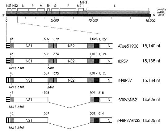
By a multiple-step cloning procedure, a synthetic T7 RNA polymerase promoter sequence followed by three nonviral G residues and a synthetic leader sequence derived from HRSV strain A2 (nt 1 to 44) (28), the BRSV NS1 and N genes, and the last 0.7 kb of the L gene and adjacent trailer were assembled in pX12ΔT so that the last nucleotide of the trailer is directly followed by the HDV ribozyme sequence. The NS1 noncoding region preceding the translation start codon was modified to contain a synthetic NotI tag (see Results). Subsequently, the HRSV leader sequence was replaced by the BRSV leader sequence by site-directed mutagenesis (22) in order to generate nonchimeric recombinants. Both the BRSV construct and the chimeric HRSV-BRSV construct were used as backbone for the assembly of the complete BRSV genome from cDNA. The first plasmids used for recovery of infectious recombinant virus (rH/BRSVΔNS2, rBRSVΔNS2 [see Fig. 1]) were termed pH/BRSVΔNS2, encoding a chimera with HRSV leader sequence and a deletion of the complete NS2 gene, and pBRSVΔNS2, containing the homologous BRSV leader sequence.
FIG. 1.
Schematic presentation of the BRSV ATue51908 genome (drawn to scale). The locations of transcripts (shaded bars) and protein-encoding frames (open bars) are shown relative to the viral RNA (vRNA) (solid bar). In the enlargements, the organizations of recombinant viruses and ATue51908 are compared. The 3′-terminal 45-nt BRSV leader regions are marked by horizontal stripes, and the 44-nt HRSV A2 leader regions are marked by vertical stripes. Genetic tags (NotI) and nucleotide deletions in the NS1 noncoding regions (shaded) of the recombinant viruses are indicated. The relative positions of corresponding nucleotides are given for the gene starts of NS1, NS2, and N and nucleotides flanking the NS2 deletion. The overall lengths of the vRNAs are shown on the right.
The last step for generation of a recombinant full-length construct consisted of the insertion of the NS2 gene. The NS2 gene was obtained by RT-PCR, cloned in pBluescriptSK− and sequenced, and transferred into the singular KpnI site of the full-length clones (ATue51908 nt 1,026) after restriction of the PCR fragment with Acc65I and BanII and Klenow treatment of all ends, resulting in a deletion of 4 nt (positions 510 to 513) from the NS1 noncoding region following the NS1 translation stop codon. The resulting full-length plasmids were termed pBRSV and pH/BRSV, the latter containing the HRSV derived leader region. Figure 1 gives an overview of the four different recombinants recovered from cDNA.
Construction of expression plasmids pN, pP, pL, and pM2.
To allow cap-independent translation of mRNAs in T7 RNA polymerase-expressing cells, the encephalomyocarditis virus (EMCV) internal ribosomal entry site (IRES) (11) was cloned downstream of the T7 RNA polymerase promoter of plasmid pT7T (9). PCR fragments in which the respective translation start codon is contained in an NcoI or AflIII restriction site were used to clone the open reading frame of the BRSV N (ATue51908 nt 1144 to 2605), P (ATue51908 nt 2351 to 3249), M2 (ATue51908 nt 7523 to 8436), or L (ATue51908 nt 8414 to 15140) gene into the NcoI site of the IRES specifying the translation start codon. Inserts generated by PCR were sequenced completely.
Establishment of a cell line stably expressing phage T7 RNA polymerase.
To obtain a cell line stably expressing T7 RNA polymerase, 106 BHK-21 cells (clone BSR-CL13) were transfected with 1 μg of pSC6-T7-NEO encoding the T7 RNA polymerase gene under control of the cytomegalovirus promoter and the neomycin resistance gene (32) (kindly provided by M. Billeter). Resistant clones were selected after addition of geneticin (1 mg/ml) to the cell culture medium. T7 RNA polymerase expression of selected cell clones was monitored after transfection of 5 μg of pT7T-N, which encodes the rabies virus N protein downstream of the T7 RNA polymerase promoter (9). Two days after transfection, cells were stained with a fluorescein isothiocyanate conjugate recognizing rabies virus N protein (Centocor) and analyzed by immunofluorescence microscopy (not shown). After two further cloning steps, a cell clone which constitutively expressed T7 RNA polymerase (BSR T7/5) was selected for further use. Even after 100 successive passages (with every second passage involving geneticin selection), no decrease in T7 RNA polymerase-directed gene expression was observed in BSR T7/5 cells.
Transfection experiments and recovery of recombinant BRSV (rBRSV).
BSR T7/5 cells were grown overnight to 80% confluency in 32-mm-diameter dishes in Eagle’s medium supplemented with 10% FCS. One hour before transfection, cells were washed twice with medium without FCS. Cells were transfected with a plasmid mixture containing 10 μg of full-length plasmid (pBRSV, pH/BRSV, pBRSVΔNS2, or pH/BRSVΔNS2), 4 μg of pN, 4 μg of pP, 2 μg of pM2, and 2 μg of pL. Transfection experiments were carried out with a mammalian transfection kit (CaPO4 transfection protocol; Stratagene). The transfection medium was removed at 4 h posttransfection; cells were washed and maintained in Eagle’s medium containing 3% FCS.
Five days after transfection, cells were split at a ratio of 1:3. Between 7 and 10 days posttransfection, a typical CPE was observed, yielding several foci per dish. The cells were split every 4 to 5 days, until between days 21 and 28 posttransfection a total CPE was observed. The virus was released by freezing and thawing. Cellular debris was removed by pelleting at 800 × g, and the supernatant was used for production of high-titer material in MDBK cells and for further experiments.
Northern hybridization of viral RNA.
Total RNA from BSR T7/5 cells was isolated at 2 to 5 days postinfection (RNeasy; Qiagen). The RNA was analyzed by denaturing gel electrophoresis (39), blotted onto nitrocellulose, cross-linked to membranes by UV, and hybridized with DNA fragments labeled (nick translation kit; Amersham) with [α-32P]dCTP (3,000 Ci/mmol; ICN). The N gene-specific probe was generated by RT-PCR and tested for specificity by sequencing analysis before use; the NS2-specific probe consisted of an AseI (ATue51908 nt 574)/KpnI (ATue51908 nt 1026) fragment of a cloned and sequenced RT-PCR product which contains the complete NS2 open reading frame. Northern blots were exposed to Kodak X-Omat AR films at −70°C with an intensifying screen.
RT-PCR.
To generate DNA probes for hybridization, for cloning of expression plasmids, and for RNA analysis of recombinant virus, RT-PCR was done with avian myeloblastosis virus reverse transcriptase (Life Sciences) for first-strand synthesis, as described above, and a proofreading thermostable polymerase (Pfu; Stratagene) for PCR under conditions recommended by the supplier. The RNA of the recombinant viruses was analyzed by two sets of RT-PCR. The first was designed to demonstrate the synthetic NotI restriction site following the NS1 gene start. First-strand primers were selected to hybridize to the 3′ end of genomic RNA. The reverse primer (ATue51908 nt 306 to 286) was derived from the NS1 gene sequence. The second PCR was designed to demonstrate the absence or presence of the NS2 gene. First-strand synthesis was done with the same set of leader-specific primers, whereas a primer derived from the N gene sequence (ATue51908 nt 1165 to 1146) served as reverse primer.
Nucleotide sequence accession number.
The nucleotide sequence of BRSV strain ATue51908 has been deposited in the GenBank database under accession no. AF092942.
RESULTS
cDNA cloning and sequence analysis of BRSV strain ATue51908.
We generated cDNA clones covering the complete genome of BRSV strain A51908, as obtained from the ATCC, after 12 passages in MDBK cells. The sequence of the 3′ half of the BRSV genome was available, covering nine genes from the NS1 gene to the M2-L gene overlap (25, 26, 30, 37, 38, 47, 48) (see Fig. 1). Two primers were selected for specific priming of first-strand cDNA synthesis, the first hybridizing to the 3′ proximal NS1 noncoding region and the second hybridizing to the M2-L gene overlap. Total RNA of BRSV-infected cells was used as starting material. Virus-specific cDNA clones were detected by hybridization with PCR probes derived from the N, G, and M2 genes. Each probe was tested for specificity by sequence analysis and by hybridization with virus RNA before use. Clones covering the entire L gene were obtained by genome walking. Besides the correctly primed cDNA clones, we found a considerable amount of BRSV-specific clones that were random primed, together covering the entire genome except for the NS2 gene. A consensus sequence was derived from at least three independent cDNA clones. The last 1.4 kb of the genomic sequence and the NS1 and NS2 gene sequences were, in addition, confirmed by sequencing of cloned RT-PCR fragments. In this case, at least six different PCR clones were analyzed to generate a consensus sequence.
Nucleotides 46 to 8454 of the consensus sequence, spanning the NS1, NS2, N, P, M, SH, G, F, and M2 genes, were compared to the previously published sequences of BRSV strain A51908 (GenBank accession no. U15937 [NS1], U15938 [NS2], M35076 [N], M93127 [P], D01012 [M and SH], and M82816 [F and M2] [26, 48]), revealing considerable divergence. The total sequence identity was found to be 96.2%, owing to 319 differences and 10 gaps in 8,454 nt. For the NS1, NS2, N, P, M, F, and M2 genes, sequence identities were in the range of 95 to 98%, and the amino acid identity ranged between 100 and 97%. Both the SH and the G genes exhibited nucleotide identity of merely 93% with the published A51908 sequences. In the deduced SH and G proteins, only 86 and 88% of the amino acids are identical, respectively. Together, the predicted amino acid sequences of the nine genes contain 97 differences compared to the A51908 sequence, over a total of 2,263 amino acids. Since the sequence data obtained differed substantially from the published A51908 sequence, we designated our starting virus BRSV strain ATue51908. The total sequence of BRSV strain ATue51908 (GenBank accession no. AF092942) comprises 15,140 nt and codes for 10 mRNAs (Fig. 1). As described for other strains, the G and M2 mRNAs each contain two open reading frames.
Sequence of the ATue51908 L gene.
Until now, the sequence of the largest BRSV gene coding for the catalytic subunit of the viral RNP-dependent RNA polymerase (L) was not available. The BRSV L gene has a length of 6,573 nt, including the gene start signal (5′-GGACAAAA-3′ [DNA positive strand]) and the transcription stop and polyadenylation signal (5′-AGTTATTTAAAAA-3′ [positive strand]). The first ATG located in a favorable context for initiation of translation is partially contained in the gene start signal (5′-GGACAAAATG-3′), resulting in an extremely short 5′ noncoding region of 7 nt. The translation stop codon, TAA, is followed by a noncoding region of 64 nt and the transcription stop/polyadenylation signal. The predicted open reading frame codes for a protein of 2,162 amino acids. No additional open reading frames longer than 30 amino acids were identified.
Compared to the L gene of HRSV strain A2 (41), the BRSV L gene has identity of 77% and encodes a protein with amino acid identity of 84%. The L proteins of BRSV, HRSV, and turkey rhinotracheitis virus (TRTV) (34) share an amino-terminal extension of about 20 amino acids and a carboxy-terminal truncation of about 100 amino acids, compared to L proteins of other nonsegmented negative-strand RNA viruses. This feature therefore appears to be common in the Pneumovirinae subfamily.
Sequence analysis of the BRSV leader and trailer regions.
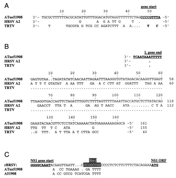
The leader region of BRSV (Fig. 2A) is 45 nt in length, 1 nt longer than that of HRSV A2 (28). The terminal regions are highly conserved. Compared to HRSV, the first 17 nt are identical and the first 28 nt contain two differences. Overall, the two RSV leader regions have identity of 82%. Alignment of the BRSV and HRSV leader regions to that of TRTV (33) revealed identities of 61 and 67%, respectively.
FIG. 2.
Alignment of the BRSV ATue51908 3′ leader region (A) and 5′ trailer (B) sequences with those from other pneumoviruses. The DNA sequences are shown in 3′-to-5′ viral RNA sense. For HRSV strain A2 (28) and TRTV (33), only deviations from the ATue51908 sequence are indicated. Gaps are represented by dots, and the start signal of the first gene (NS1 in BRSV and HRSV, N in TRTV) and the L gene end signal are underlined. Numbering of the trailer sequences starts with the first nucleotide downstream of the L gene transcription stop signal. (C) Alignment of the NS1 noncoding region of the recombinant BRSVs containing the NotI tag (boxed) with the ATue51908 consensus sequence and the published A51908 sequence. The sequences are shown as DNA positive strands. Only nucleotides differing from the recombinant sequence are indicated. Gaps are indicated by dots, and the NS1 gene start signal and translation start codon are underlined.
The 5′ extragenic region of the BRSV genome (Fig. 2B) is composed of 161 nt, which is 6 nt longer than the HRSV A2 trailer region (28). This is thus far the longest trailer sequence of a member of the Paramyxoviridae family. The BRSV leader and trailer regions themselves exhibit a terminal complementarity of the extreme 11 nt which is interrupted at position 4, as is also found in TRTV. The 5′-terminal 14 nt of BRSV and HRSV are identical, and the 5′-terminal 25 nt contain only one different nucleotide. The L-proximal two-thirds of the trailer regions show a very low degree of homology, and the overall identity is 51%. The TRTV trailer region (33), which is composed of only 40 nt, is more similar to the aligned region of HRSV than of BRSV (60 and 50% identity, respectively).
Construction of full-length BRSV cDNA clones.
A cDNA copy of the BRSV genome was assembled in pX12ΔT downstream of a T7 RNA polymerase promoter such that antigenomic RNA would be transcribed. Three G residues were introduced between the promoter and the leader sequence to facilitate initiation of transcription. It was previously shown that additional nucleotides at the 5′ ends of HRSV genomes do not interfere with encapsidation and replication (35). To achieve correct cleavage of the RNA transcript at the 3′ end of the antigenome-like RNA, the HDV antigenome ribozyme sequence was joined directly to the last nucleotide of the trailer sequence (40).
At first, an NS2 deletion mutant was assembled in a plasmid containing the HRSV A2 leader region (nt 1 to 44) (28) instead of the BRSV leader region and in which the NS1 gene end signal, the NS2 gene start signal, and the complete NS2 coding region (514 nt) are absent (rH/BRSVΔNS2 [Fig. 1]). Thus, the NS1 translation stop codon is followed by the gene end signal derived from the NS2 gene. Subsequently, an RT-PCR-generated NS2 gene sequence was introduced into the NS2-deficient cDNA to generate a nondeficient, chimeric full-length genome (rH/BRSV [Fig. 1]). Due to the cloning procedure, 4 nt from the wild-type noncoding sequence between the NS1 translation stop codon and the NS1 gene end signal (nt 510 to 513) were deleted. To finally replace the HRSV leader sequence by the original BRSV leader sequence, site-directed mutagenesis was done, giving rise to rBRSVΔNS2 and rBRSV (for details, see Materials and Methods).
All four cDNA constructs contain three common differences from the standard ATue51908 sequence: a 16-nt difference, involving deletion of one nucleotide, in the NS1 5′ noncoding region, creating a singular NotI site and allowing easy discrimination of recombinant virus and wild-type ATue51908 virus (Fig. 2C), as well as two single nucleotide exchanges which are derived from one of the cDNA clones used for cloning (pL23). One of the exchanges is located in the BRSV trailer sequence (nt 15049, T to C) and the other in the L coding sequence (nt 14312, A to C), resulting in an amino acid change from isoleucine to leucine. Overall, the genome of the full-length BRSV recombinant is 5 nt shorter than the standard ATue51908 virus genome, and the chimeric rH/BRSV sequence is 6 nt shorter, due to the shorter HRSV leader. The genomes of the NS2 deletion mutants rBRSVΔNS2 and rH/BRSVΔNS2 are lacking 514 internal nucleotides (Fig. 1).
Recovery of infectious BRSV from cDNA in a vaccinia virus-free cell system.
Most systems for recovery of negative-strand RNA viruses make use of vaccinia helper virus providing T7 RNA polymerase for intracellular transcription of full-length RNAs and expression of support proteins. However, to prevent possible interference with the notoriously very slow BRSV replication or virus assembly and to avoid the necessity of separating vaccinia virus from BRSV, we decided to establish a vaccinia virus-free system. A cell line which stably expresses phage T7 RNA polymerase (BSR T7/5) was generated by transfection of BHK-21 cells, clone BSR-CL13, with a plasmid expressing phage T7 RNA polymerase under control of the cytomegalovirus promoter (pSC6-T7-NEO, kindly provided by M. Billeter). BSR T7/5 cells were transfected with plasmids expressing antigenomic BRSV RNA and support plasmids expressing the BRSV N, P, L, and M2 proteins. The support plasmids contained the consensus sequence of the respective ATue51908 open reading frame, downstream of the ECMV IRES sequence, to allow cap-independent translation. Five days after transfection, cells were split in a 1:3 ratio. Between days 7 and 10 after transfection, a typical CPE was observed, in the form of several foci of fusing cells per dish. When any of the support plasmids was omitted, no foci were observed.
All transfection experiments were done in quadruplicate and repeated at least three times. Each of the four different full-length constructs yielded recombinant virus in all dishes. The cells were split every 4 to 5 days, and the recovered virus was released by freezing and thawing when the CPE was maximal. Clarified supernatant was used for three rounds of amplification in the case of the full-length recombinant. In order to amplify the NS2 deletion mutant, at least five passages were necessary to produce stock material suitable for further characterizations.
Identification of genetic tags and transcription analysis of recombinant virus.
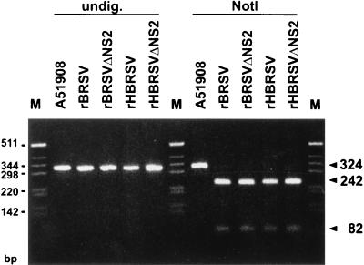
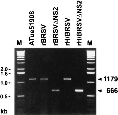
Genetic tags were used to confirm the identity of the recombinant viruses. Total RNA of virus-infected BSR T7/5 cells was isolated when the CPE was maximal, and RT-PCR was performed. One assay was done to demonstrate the novel NotI restriction site contained in the NS1 5′ noncoding region of all recombinant viruses. Primers LeBRSV or LeH/BRSV and NS1r, which are specific for the leader regions and the NS1 gene, respectively, were used to amplify a 300-bp fragment which was then digested with NotI. The RT-PCR products obtained from recombinant viruses were cleaved by NotI, whereas the fragment obtained from standard ATue51908 virus was not (Fig. 3). A second RT-PCR with the leader-specific primer and the primer Nr, specific for the N gene, was designed to reveal the absence or presence of the NS2 gene. A PCR product of 1.2 kb was obtained in the case of standard ATue51908 virus or of full-length recombinant viruses (Fig. 4). In the case of rBRSVΔNS2 and rH/BRSVΔNS2, a fragment of about 0.7 kb was amplified by the same primers, demonstrating the deletion of the NS2 gene. No PCR product was detected when the RT step was omitted (not shown), demonstrating that the PCR products originate from viral RNA rather than from plasmid contaminations.
FIG. 3.
Demonstration of the NotI tag in the genome RNA of recombinant BRSVs. RT-PCR was performed on total RNA of BSR T7/5 cells infected with standard BRSV ATue51908 or with recombinant virus by using positive-sense primers annealing to the genome 3′ ends and a negative-sense primer binding in the NS1 gene. No PCR product was detected when the RT step was omitted. Digestion with NotI of the 324-bp (calculated size) RT-PCR product originating from recombinant viruses yielded two bands of calculated sizes of 82 and 242 bp, whereas the RT-PCR product from standard BRSV strain ATue51908 was not cleaved.
FIG. 4.
Demonstration of NS2 gene deletion by RT-PCR. Total RNA of BSR T7/5 cells infected with standard BRSV ATue51908 or with recombinant virus was used for RT-PCR with a positive-sense primer hybridizing to the 3′ end of the BRSV genome RNA and a negative-sense primer binding in the N gene. Products from full-length recombinants and wild-type virus yielded a product of 1,179 bp (calculated size) spanning the NS2 gene, whereas deletion mutants gave rise to a 666-bp (calculated) fragment, reflecting the 513-nt deletion. No PCR product was detected when the RT step was omitted.
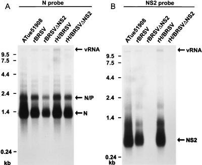
In order to demonstrate transcripts from the recombinant viruses and from standard ATue51908, RNA of infected cells was analyzed by Northern hybridization. Total cellular RNA was isolated 5 days after infection of BSR T7/5 cells at an MOI of 0.01. After hybridization with an N-specific probe, genome RNAs and monocistronic N mRNAs were detected in cells infected with the recombinant BRSVs and standard ATue51908, demonstrating efficient transcription and replication of the recombinant BRSVs (Fig. 5). In addition, a prominent band representing bicistronic N/P readthrough mRNA was present in all lanes. RNA from cells infected with the NS2 deletion mutants did not hybridize with an NS2 probe, whereas a prominent band corresponding to NS2 mRNA was present in cells infected with ATue51908 or the full-length recombinants (Fig. 5). These results again confirmed the absence of the complete NS2 gene in the genome of deletion mutants, as well as the absence of contaminating helper virus. The NS2 deletion mutants thus replicate autonomously in cell culture.
FIG. 5.
Demonstration of virus transcripts by Northern hybridization. Total RNA of BSR T7/5 cells infected with rBRSV or standard ATue51908 was isolated 2 to 4 days after infection and separated on a 2% denaturing agarose gel. The blot was hybridized with PCR-derived probes specific for the BRSV N gene (nt 1429 to 2277) (A) and for the NS2 gene (nt 574 to 1026) (B). Transcripts corresponding to N mRNA (vRNA), bicistronic N/P readthrough transcripts, and NS2 mRNA are indicated.
Recombinant viruses lacking the NS2 gene are attenuated in cell culture.
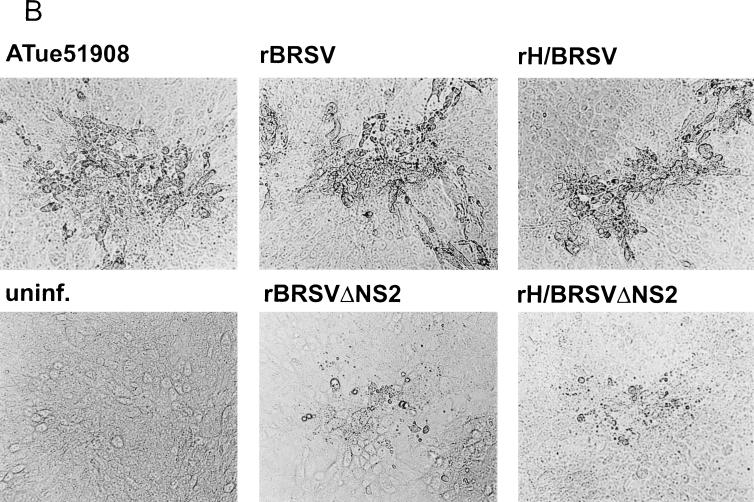
Growth characteristics of recombinant BRSV were studied with both the BHK-derived BSR T7/5 cell line and MDBK cells. The two full-length recombinants did not differ from standard ATue51908 virus in their final titers of infectious virus nor in their growth kinetics or phenotype. In MDBK cells, ATue51908 and the full-length recombinants yielded maximal titers of 106 PFU/ml after 8 days of infection at an MOI of 0.01, while in BSR T7/5 cells maximum titers of 105 PFU/ml were achieved at 4 days after infection at the same MOI. The chimeric viruses containing the HRSV leader region did not differ in any respect from the recombinants containing the autologous BRSV leader sequence. Compared to the full-length viruses, the NS2 deletion mutants showed slower growth and reduced final titers in both BSR T7/5 and MDBK cells. In BSR T7/5 cells, a similar type of CPE, consisting of large syncytia detaching from the monolayer, developed after infection with both full-length and NS2-deficient viruses (Fig. 6A). When grown on MDBK cells, the full-length viruses caused typical foci of enlarged cells, indistinguishable from standard ATue51908 virus foci (Fig. 6B). The NS2 deletion mutants, however, yielded only small pinpoint foci in MDBK monolayers, consisting of a small number of infected cells (Fig. 6B). Due to their very slow growth in MDBK, cells had to be split every three days in a 1:3 ratio to keep the infection going on. At 15 days after infection of MDBK cells at an MOI of 0.01, antigen was detected in less than 60% of cells. In contrast, in the case of standard ATue51908 or of full-length recombinant virus, it was not possible to split MDBK cells infected with an MOI of 0.01 more than once due to an extensive CPE. When BSR T7/5 cells were infected with the NS2 deletion mutants at an MOI of 0.01, CPE was maximal at 5 days after infection. In these cultures, antigen was detected in more than 80% of cells. Still, compared to full-length virus, maximum titers of the NS2 deletion mutants were 10-fold lower in both cell lines, with maximum titers reaching 104 PFU per ml in BSR T7/5 cells at 5 days after infection and 105 PFU per ml in MDBK cells at 15 days after infection and four passages of infected cells. These features demonstrate that the NS2 gene is nonessential, though in its absence the replication capacity of BRSV is reduced and the CPE in MDBK cells is less pronounced.
FIG. 6.
CPE of recombinant BRSVs in BSR T7/5 cells (A) and MDBK cells (B). (A) In BSR T7/5 cultures, all recombinants induced syncytia of cells detaching from the monolayer, which were indistinguishable from those of standard ATue51908, at 56 h postinfection at an MOI of 0.01. (B) In MDBK cells, both full-length recombinant viruses rBRSV and rH/BRSV and standard ATue51908 virus produced similar foci of enlarged cells at 5 days postinfection, whereas the NS2 deletion mutants produced only small foci of degenerating cells.
DISCUSSION
In this work, we describe the cDNA cloning of the entire BRSV genome and the recovery of recombinant BRSV from cDNA constructs. To our knowledge, this is the second species of the genus Pneumovirus for which the entire genome sequence has been determined and which has been made amenable to genetic manipulation. The viruses we have recovered are derived from a parental virus obtained after 12 passages in MDBK cells of BRSV strain A51908, obtained from the ATCC. Compared to the published sequences of strain A51908, a marked divergence was observed. The most striking deviations are located in the SH and G genes, which displayed 86 and 88% amino acid identity, respectively. The parental virus was therefore designated ATue51908 (GenBank accession no. AF092942).
The approach used to rescue cDNA derived from ATue51908 into infectious BRSV follows the principles shown to be successful for recovery first of recombinant rabies virus (40) and then of a variety of negative-strand RNA viruses from the rhabdovirus, paramyxovirus, and bunyavirus families (8). It involves simultaneous expression of viral antigenome RNA and of RNP proteins in order to promote “illegitimate” encapsidation of the RNA in such a way that the novel RNP structure may serve as a template for the viral RNA polymerase. The expression systems most widely used rely on infection of cells with recombinant vaccinia viruses (vTF7-3 or MVA) (2–4, 10, 13, 17, 18, 23, 45) providing T7 RNA polymerase, which is needed for expression of proteins and RNAs from transfected plasmids. This, however, requires coping with vaccinia virus-induced CPEs which limit the window for recovery, vaccinia virus-induced recombination of transfected plasmid DNA (13, 18), and separation of different viruses by biophysical (40), biochemical (45), or biological (13) means. We describe here the establishment of a BHK-derived cell line stably expressing T7 RNA polymerase, as first reported for measles virus (32), obviating the use of vaccinia helper virus. Especially for recovery of BRSV, the availability of a vaccinia virus-free system was regarded as crucial, since BRSV replicates very slowly and to low titers in cell culture and is highly cell associated, like vaccinia virus, and since virions are pleomorphic in size and shape, with some particles comparable in size to vaccinia virus. Moreover, a general advantage of the T7 polymerase-expressing cell line in combination with IRES-containing support plasmids (11) was confirmed in the rabies virus recovery system. Compared to the vaccinia virus-driven expression system, the recovery rates were increased at least 10-fold (12a).
In contrast to other paramyxoviruses or rhabdoviruses, where N, P, and L proteins are sufficient to support encapsidation of antigenome RNA and to initiate an infectious cycle, recovery of the pneumovirus HRSV has been shown to require the additional expression of the M2 gene (ORF 1), which encodes a transcription elongation factor (5, 16). We therefore included a BRSV M2-expressing plasmid in the transfection experiments. Both viruses corresponding to ATue51908, chimeric viruses possessing the leader region of HRSV, and considerably attenuated NS2-deletion mutants could be isolated from every transfected cell culture dish. The recovery rate thus exceeds by far 1 in 106 transfected cells.
The successful recovery of recombinant BRSV indistinguishable in phenotype from the parental virus ATue51908 confirmed the authenticity and the functionality of the determined nucleotide sequences. A particular focus was laid on the functional characterization of BRSV sequences previously not available, namely, the gene ends and the L gene. The deduced BRSV L and HRSV L proteins showed amino acid identity of 84% and a similarity of 89%. The amino acid differences were distributed over the entire protein, indicating rather identical functions of the two proteins. This finding, and the high degree of similarity of the HRSV and BRSV genome ends, prompted us to see whether the BRSV polymerase would be able to recognize HRSV promoter structures.
The 3′ end of the pneumovirus genome (the genome promoter) contains cis-acting signals directing both transcription of subgenomic, free mRNAs and synthesis of full-length RNPs, as well as sequences specifying an encapsidation signal. In the case of HRSV minigenomes, the promoter function was highly sensitive to insertion of nucleotides into the terminal 10 residues and into the stretch of residues 21 to 25 (6, 7). Interestingly, the eight nucleotide differences of the BRSV leader region are all located outside of these functionally critical regions. To determine whether the published HRSV sequence can substitute for the BRSV genome promoter, chimeras which contain the heterologous HRSV leader region were designed. The chimeric viruses could be rescued with the same efficiency as rBRSV and replicated with the same speed to the same titers and produced the same type of CPE. The same applied to the chimeric and authentic NS2 deletion mutants (see below). Therefore, the heterologous HRSV leader region is faithfully recognized by the BRSV polymerase and provides functionally identical signals for RNA transcription and replication and for encapsidation of RNAs. The observed nucleotide differences apparently do not specify genetic information that may account for species specificity, further confirming the close relationship of BRSV and HRSV.
Members of the genus Pneumovirus show particular features that distinguish them from other paramyxovirus genera, such as the lack of P gene RNA editing. HRSV does not appear to obey the “rule of six” (35), as do most paramyxoviruses which are able to edit their P gene RNAs (21). Apparently, this is also true for BRSV. Neither the parental virus ATue51908 nor any of the recombinants have a genome consisting of multiples of 6 nt. The recombinant BRSV genome lacks five residues compared to the ATue51908 genome but replicated with the same efficiency. This applies also to the chimeric H/BRSV, which lacks six residues due to the shorter HRSV leader region. This 1-nt difference was also maintained in the NS2 deletion mutants BRSVΔNS2 and H/BRSVΔNS2. Again, they were found to replicate at identical rates.
Another peculiarity of pneumoviruses is the high number of encoded genes, some of which do not have counterparts in other paramyxoviruses, such as SH, M2, and the nonstructural genes NS1 and NS2. It was recently shown that G and SH of HRSV are not essential for viral replication in vitro but may enhance membrane fusion (19). A role for the M2 protein as a transcription elongation factor has also been established (5, 16), but the function of the two nonstructural proteins NS1 and NS2 has remained rather obscure. In an artificial minigenome assay, HRSV NS1 and, to a lesser degree, NS2, showed inhibitory effects on transcription and replication (1). Only revertants of HRSV NS2 knockout viruses, in which tandem stop codons were introduced in such a way that the NS2 protein would not be expressed, could be isolated from plaques observed in transfection experiments (42), indicating an important function of HRSV NS2 in the virus life cycle. Strikingly, in the avian pneumovirus TRTV, both nonstructural genes are absent, emphasizing the question of whether they represent essential genes in mammalian pneumoviruses such as BRSV.
To address possible functions of NS2 in the virus life cycle, we assembled a BRSV cDNA copy lacking the entire NS2 gene. The successful recovery of a virus autonomously propagating in BSR cells demonstrated that the NS2 gene is not essential for replication of BRSV in vitro. The pattern and the relative amounts of mRNAs and full-length virus RNA produced in infected cells were not markedly changed, except for the lack of an NS2 transcript. Interestingly, the effect of the NS2 deficiency appeared to be more pronounced in the bovine MDBK cell line than in BSR cells. Syncytia caused by the NS2-deficient mutants in BSR T7/5 cell monolayers were phenotypically indistinguishable from those caused by standard ATue51908. In contrast, BRSVΔNS2-infected MDBK cells were not enlarged, unlike ATue51908-infected cells. Moreover, maximum titers were reached in MDBK cells infected with NS2 deletion mutants by 15 days after infection, compared to 8 days after infection with standard ATue51908. In BSR T7/5 cells, maximal final infectious titers were reached by 5 days after infection, which is comparable to nondeficient viruses. In both cell lines, however, the maximum virus yield of the NS2 deletion mutants was reduced by a factor of 10 (105 and 104 in BSR T7/5 cells for rBRSV and rBRSVΔNS2, respectively, and 106 and 105 in MDBK cells, respectively). As the cDNA constructs used for recovery of nondeficient rBRSV were made by completion of the NS2 deletion cDNA, the observed slower growth and the reduced virus titers are due to the lack of NS2 rather than to putative differences in other parts of the genome. Thus, although not essential, NS2 is an accessory factor able to substantially support virus growth, by a thus far unknown mechanism. Further experiments using modified virus mutants are now feasible and should help to reveal the mechanisms of NS2 involved in facilitating virus growth in different cell types.
The successful recovery of the first BRSV strain from which an entire gene has been deleted is important not only for studying the molecular biology and genetics of the virus but also because it provides a severely attenuated virus with an unequivocal serological marker. Due to their 3′-proximal locations, NS1 and NS2 are expressed at high levels and induce antibodies in infected calves (44). Further manipulation of NS2-deficient viruses may lead to the development of attenuated marker vaccines, easily distinguishable from wild-type virus. For the prevention of respiratory diseases, live vaccines appear to be best suited due to their ability to confer local immunity in addition to humoral immune response. It will also be of interest to determine whether foreign epitopes or proteins can be incorporated into the virion in order to design vaccines for other respiratory pathogens or vectors for transient gene therapy.
ACKNOWLEDGMENTS
We thank Veronika Schlatt and Karin Kegreiss for perfect technical assistance.
This work was supported by Intervet International B.V.
REFERENCES
- 1.Atreya P L, Peeples M E, Collins P L. The NS1 protein of human respiratory syncytial virus is a potent inhibitor of minigenome transcription and RNA replication. J Virol. 1998;72:1452–1461. doi: 10.1128/jvi.72.2.1452-1461.1998. [DOI] [PMC free article] [PubMed] [Google Scholar]
- 2.Baron M D, Barrett T. Rescue of rinderpest virus from cloned cDNA. J Virol. 1997;71:1265–1271. doi: 10.1128/jvi.71.2.1265-1271.1997. [DOI] [PMC free article] [PubMed] [Google Scholar]
- 3.Bridgen A, Elliott R M. Rescue of a segmented negative-strand RNA virus entirely from cloned complementary cDNAs. Proc Natl Acad Sci USA. 1996;93:15400–15404. doi: 10.1073/pnas.93.26.15400. [DOI] [PMC free article] [PubMed] [Google Scholar]
- 4.Collins P L, Hill M G, Camargo E, Grosfeld H, Chanock R M, Murphy B R. Production of infectious human respiratory syncytial virus from cloned cDNA confirms an essential role for the transcription elongation factor from the 5′ proximal open reading frame of the M2 mRNA in gene expression and provides a capability for vaccine development. Proc Natl Acad Sci USA. 1995;92:11563–11567. doi: 10.1073/pnas.92.25.11563. [DOI] [PMC free article] [PubMed] [Google Scholar]
- 5.Collins P L, Hill M G, Cristina J, Grosfeld H. Transcription elongation factor of respiratory syncytial virus, a nonsegmented negative-strand RNA virus. Proc Natl Acad Sci USA. 1996;93:81–85. doi: 10.1073/pnas.93.1.81. [DOI] [PMC free article] [PubMed] [Google Scholar]
- 6.Collins P L, Mink M A, Stec D S. Rescue of synthetic analogs of respiratory syncytial virus genomic RNA and effect of truncations and mutations on the expression of a foreign reporter gene. Proc Natl Acad Sci USA. 1991;88:9663–9667. doi: 10.1073/pnas.88.21.9663. [DOI] [PMC free article] [PubMed] [Google Scholar]
- 7.Collins P L, Stec D S, Kuo L, Hill III M G, Camargo E, Dimock K, Grosfeld H, Mink M A. Rescue of synthetic helper-dependent analogs of the genomic RNAs of respiratory syncytial virus and parainfluenza virus type 3. In: Brown F, Chanock R M, Ginsberg H S, Lerner R A, editors. Vaccines 1993: modern approaches to new vaccines including prevention of AIDS. Cold Spring Harbor, N.Y: Cold Spring Harbor Laboratory Press; 1993. pp. 259–264. [Google Scholar]
- 8.Conzelmann K-K. Genetic manipulation of non-segmented negative-strand RNA viruses. J Gen Virol. 1996;77:381–389. doi: 10.1099/0022-1317-77-3-381. [DOI] [PubMed] [Google Scholar]
- 9.Conzelmann K-K, Schnell M J. Rescue of synthetic genomic RNA analogs of rabies virus by plasmid-encoded proteins. J Virol. 1994;68:713–719. doi: 10.1128/jvi.68.2.713-719.1994. [DOI] [PMC free article] [PubMed] [Google Scholar]
- 10.Durbin A P, Hall S L, Siew J W, Whitehead S S, Collins P L, Murphy B R. Recovery of infectious human parainfluenza virus type 3 from cDNA. Virology. 1997;235:323–332. doi: 10.1006/viro.1997.8697. [DOI] [PubMed] [Google Scholar]
- 11.Elroy-Stein O, Fuerst T R, Moss B. Cap-independent translation of mRNA conferred by encephalomyocarditis virus 5′ sequence improves the performance of the VV/bacteriophage T7 hybrid expression system. Proc Natl Acad Sci USA. 1989;86:6126–6130. doi: 10.1073/pnas.86.16.6126. [DOI] [PMC free article] [PubMed] [Google Scholar]
- 12.Fernie B F, Gerin J L. The stabilization and purification of respiratory syncytial virus using MgSO4. Virology. 1980;106:141–144. doi: 10.1016/0042-6822(80)90229-9. [DOI] [PubMed] [Google Scholar]
- 12a.Finke, S. Unpublished data.
- 13.Garcin D, Pelet T, Calain P, Roux L, Curran J, Kolakofsky D. A highly recombinogenic system for the recovery of infectious Sendai paramyxovirus from cDNA: generation of a novel copy-back nondefective interfering virus. EMBO J. 1995;14:6087–6094. doi: 10.1002/j.1460-2075.1995.tb00299.x. [DOI] [PMC free article] [PubMed] [Google Scholar]
- 14.Grosfeld H, Hill M G, Collins P L. RNA replication by respiratory syncytial virus (RSV) is directed by the N, P, and L proteins; transcription also occurs under these conditions but requires RSV superinfection for efficient synthesis of full-length mRNA. J Virol. 1995;69:5677–5686. doi: 10.1128/jvi.69.9.5677-5686.1995. [DOI] [PMC free article] [PubMed] [Google Scholar]
- 15.Gubler U, Hofmann B J. A simple and very efficient method of generating cDNA libraries. Gene. 1983;25:263–269. doi: 10.1016/0378-1119(83)90230-5. [DOI] [PubMed] [Google Scholar]
- 16.Hardy R W, Wertz G W. The product of the respiratory syncytial virus M2 gene ORF1 enhances readthrough of intergenic junctions during viral transcription. J Virol. 1998;72:520–526. doi: 10.1128/jvi.72.1.520-526.1998. [DOI] [PMC free article] [PubMed] [Google Scholar]
- 17.He B, Paterson R G, Ward C D, Lamb R A. Recovery of infectious SV5 from cloned DNA and expression of a foreign gene. Virology. 1997;237:249–260. doi: 10.1006/viro.1997.8801. [DOI] [PubMed] [Google Scholar]
- 18.Hoffman M A, Banerjee A K. An infectious clone of human parainfluenza virus type 3. J Virol. 1997;71:4272–4277. doi: 10.1128/jvi.71.6.4272-4277.1997. [DOI] [PMC free article] [PubMed] [Google Scholar]
- 19.Karron R A, Buonagurio D A, Georgiu A G, Whitehead S S, Adamus J E, Clements-Mann M L, Harris D O, Randolph V B, Udem S A, Murphy B R, Sidhu M S. Respiratory syncytial virus (RSV) SH and G proteins are not essential for viral replication in vitro: clinical evaluation and molecular characterization of a cold-passaged, attenuated RSV subgroup B mutant. Proc Natl Acad Sci USA. 1997;94:13961–13966. doi: 10.1073/pnas.94.25.13961. [DOI] [PMC free article] [PubMed] [Google Scholar]
- 20.Kato A, Sakai Y, Shioda T, Kondo T, Nakanishi M, Nagai Y. Initiation of Sendai virus multiplication from transfected cDNA of RNA with negative or positive sense. Genes Cells. 1996;1:569–899. doi: 10.1046/j.1365-2443.1996.d01-261.x. [DOI] [PubMed] [Google Scholar]
- 21.Kolakofsky D, Pelet T, Garcin D, Hausmann S, Curran J, Roux L. Paramyxovirus RNA synthesis and the requirement for hexamer genome length: the rule of six revisited. J Virol. 1998;72:891–899. doi: 10.1128/jvi.72.2.891-899.1998. [DOI] [PMC free article] [PubMed] [Google Scholar]
- 22.Kunkel T A, Roberts J D, Zakour R A. Rapid and efficient site-specific mutagenesis without phenotypic selection. Methods Enzymol. 1987;54:367–382. doi: 10.1016/0076-6879(87)54085-x. [DOI] [PubMed] [Google Scholar]
- 23.Lawson N D, Stillman E A, Whitt M A, Rose J K. Recombinant vesicular stomatitis viruses from DNA. Proc Natl Acad Sci USA. 1995;92:4477–4481. doi: 10.1073/pnas.92.10.4477. [DOI] [PMC free article] [PubMed] [Google Scholar]
- 24.Lerch R A, Stott E J, Wertz G W. Characterization of bovine respiratory syncytial virus proteins and mRNAs and generation of cDNA clones to the viral mRNAs. J Virol. 1989;63:833–840. doi: 10.1128/jvi.63.2.833-840.1989. [DOI] [PMC free article] [PubMed] [Google Scholar]
- 25.Mallipeddi S K, Samal S K. Sequence comparison between the phosphoprotein mRNAs of human and bovine respiratory syncytial viruses identifies a divergent domain in the predicted protein. J Gen Virol. 1992;73:2441–2444. doi: 10.1099/0022-1317-73-9-2441. [DOI] [PubMed] [Google Scholar]
- 26.Mallipeddi S K, Samal S K. Sequence variability of the glycoprotein gene of bovine respiratory syncytial virus. J Gen Virol. 1993;74:2001–2004. doi: 10.1099/0022-1317-74-9-2001. [DOI] [PubMed] [Google Scholar]
- 27.Mallipeddi S K, Samal S K, Mohanty S B. Analysis of polypeptides synthesized in bovine respiratory syncytial virus-infected cells. Arch Virol. 1990;115:23–36. doi: 10.1007/BF01310620. [DOI] [PubMed] [Google Scholar]
- 28.Mink M A, Stec D S, Collins P L. Nucleotide sequences of the 3′ leader and 5′ trailer regions of human respiratory syncytial virus genomic RNA. Virology. 1991;185:615–624. doi: 10.1016/0042-6822(91)90532-g. [DOI] [PubMed] [Google Scholar]
- 29.Mohanty S B, Ingling A L, Lillie M G. Experimentally induced respiratory syncytial viral infection in calves. Am J Vet Res. 1975;36:417–419. [PubMed] [Google Scholar]
- 30.Pastey M K, Samal S K. Nucleotide sequence analysis of the non-structural NS1 (1C) and NS2 (1B) protein genes of bovine respiratory syncytial virus. J Gen Virol. 1995;76:193–197. doi: 10.1099/0022-1317-76-1-193. [DOI] [PubMed] [Google Scholar]
- 31.Pringle C R. Virus taxonomy 1996—a bulletin from the Xth International Congress of Virology in Jerusalem. Arch Virol. 1996;141:2251–2256. doi: 10.1007/BF01718231. [DOI] [PMC free article] [PubMed] [Google Scholar]
- 32.Radecke F, Spielhofer P, Schneider H, Kaelin K, Huber M, Doetsch C, Christiansen G, Billeter M A. Rescue of measles viruses from cloned cDNA. EMBO J. 1995;14:5773–5784. doi: 10.1002/j.1460-2075.1995.tb00266.x. [DOI] [PMC free article] [PubMed] [Google Scholar]
- 33.Randhawa J S, Marriott A C, Pringle C R, Easton A J. Rescue of synthetic minireplicons establishes the absence of the NS1 and NS2 genes from avian pneumovirus. J Virol. 1997;71:9849–9854. doi: 10.1128/jvi.71.12.9849-9854.1997. [DOI] [PMC free article] [PubMed] [Google Scholar]
- 34.Rhandawa J S, Wilson S D, Tolley K P, Cavanagh D, Pringle C R, Easton A J. Nucleotide sequence of the gene encoding the viral polymerase of avian pneumovirus. J Gen Virol. 1996;77:3047–3051. doi: 10.1099/0022-1317-77-12-3047. [DOI] [PubMed] [Google Scholar]
- 35.Samal S K, Collins P L. RNA replication by a respiratory syncytial virus RNA analog does not obey the rule of six and retains a nonviral trinucleotide extension at the leader end. J Virol. 1996;70:5075–5082. doi: 10.1128/jvi.70.8.5075-5082.1996. [DOI] [PMC free article] [PubMed] [Google Scholar]
- 36.Samal S K, Pastey M K, McPhillips T H, Mohanty S B. Bovine respiratory syncytial virus nucleocapsid protein expressed in insect cells specifically interacts with the phosphoprotein and the M2 protein. Virology. 1993;193:470–473. doi: 10.1006/viro.1993.1148. [DOI] [PubMed] [Google Scholar]
- 37.Samal S K, Zamora M. Nucleotide sequence analysis of a matrix and small hydrophobic protein dicistronic mRNA of bovine respiratory syncytial virus demonstrates extensive sequence divergence of the small hydrophobic protein from that of human respiratory syncytial virus. J Gen Virol. 1991;72:1715–1720. doi: 10.1099/0022-1317-72-7-1715. [DOI] [PubMed] [Google Scholar]
- 38.Samal S K, Zamora M, McPhillips T H, Mohanty S B. Molecular cloning and sequence analysis of bovine respiratory syncytial virus mRNA encoding the major nucleocapsid protein. Virology. 1991;180:453–456. doi: 10.1016/0042-6822(91)90057-i. [DOI] [PubMed] [Google Scholar]
- 39.Sambrook J, Fritsch E F, Maniatis T. Molecular cloning: a laboratory manual. 2nd ed. Cold Spring Harbor, N.Y: Cold Spring Harbor Laboratory Press; 1989. [Google Scholar]
- 40.Schnell M J, Mebatsion T, Conzelmann K-K. Infectious rabies viruses from cloned cDNA. EMBO J. 1994;13:4195–4203. doi: 10.1002/j.1460-2075.1994.tb06739.x. [DOI] [PMC free article] [PubMed] [Google Scholar]
- 41.Stec D S, Hill III M G, Collins P L. Sequence analysis of the polymerase L gene of human respiratory syncytial virus and predicted phylogeny of nonsegmented negative-strand viruses. Virology. 1991;183:273–287. doi: 10.1016/0042-6822(91)90140-7. [DOI] [PubMed] [Google Scholar]
- 42.Teng M N, Collins P L. Abstracts of the 10th International Conference on Negative Strand Viruses, Dublin, Ireland, 21 to 26 September 1997. 1997. Recombinant RSV which does not express the NS2 protein has altered growth characteristics in tissue culture, abstr. 249; p. 174. [Google Scholar]
- 43.Van der Poel W H, Brand A, Kramps J A, van Oirschot J T. Respiratory syncytial virus infections in human beings and in cattle. An epidemiological review. J Infect. 1994;29:215–228. doi: 10.1016/s0163-4453(94)90866-4. [DOI] [PubMed] [Google Scholar]
- 44.Westenbrink F, Kimman T G, Brinkhof J M A. Analysis of the antibody response to bovine respiratory syncytial virus proteins in calves. J Gen Virol. 1989;70:591–601. doi: 10.1099/0022-1317-70-3-591. [DOI] [PubMed] [Google Scholar]
- 45.Whelan S P J, Ball L A, Barr J N, Wertz G T. Efficient recovery of infectious vesicular stomatitis virus entirely from cDNA clones. Proc Natl Acad Sci USA. 1995;92:8388–8392. doi: 10.1073/pnas.92.18.8388. [DOI] [PMC free article] [PubMed] [Google Scholar]
- 46.Yu Q, Hardy R W, Wertz G W. Functional cDNA clones of the human respiratory syncytial (RS) virus N, P, and L proteins support replication of RS virus genomic RNA analogs and define minimal trans-acting requirements for RNA replication. J Virol. 1995;69:2412–2419. doi: 10.1128/jvi.69.4.2412-2419.1995. [DOI] [PMC free article] [PubMed] [Google Scholar]
- 47.Zamora M, Samal S K. Gene junction sequences of bovine respiratory syncytial virus. Virus Res. 1992;24:115–121. doi: 10.1016/0168-1702(92)90035-8. [DOI] [PubMed] [Google Scholar]
- 48.Zamora M, Samal S K. Sequence analysis of M2 mRNA of bovine respiratory syncytial virus obtained from an F-M2 dicistronic mRNA suggests structural homology with that of human respiratory syncytial virus. J Gen Virol. 1992;73:737–741. doi: 10.1099/0022-1317-73-3-737. [DOI] [PubMed] [Google Scholar]


