

Abstract
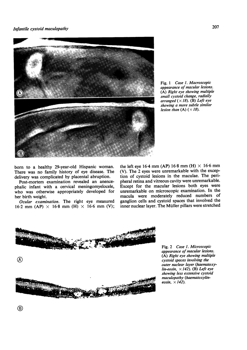
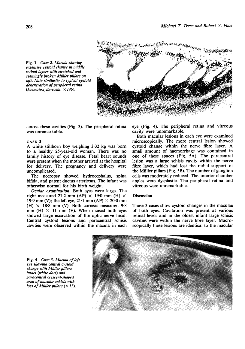
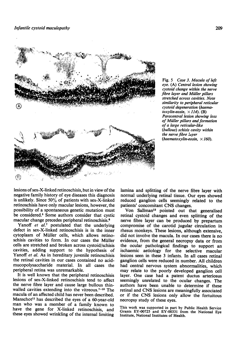
Three premature infants with no family history of eye disease showed bilateral cystoid maculopathy on post-mortem examination. These macular lesions are indistinguishable macroscopically from the macular lesions of sex-linked juvenile retinoschisis. Cystoid change was observed at various vertical retinal levels. The oldest child had a schisis cavity within the nerve fibre layer. All cases had reduced numbers of retinal ganglion cells and central nervous system (CNS) abnormalities. The authors believe these are the first reported cases of a cystoid macular lesion present at birth and the first description of the pathological features of a cystoid macular lesion in infants.
Full text
PDF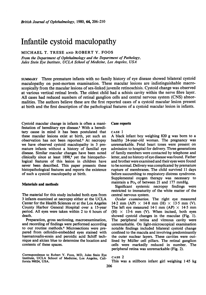
Images in this article
Selected References
These references are in PubMed. This may not be the complete list of references from this article.
- Harris G. S. Juvenile sex-linked retinoschisis. Bibl Ophthalmol. 1969;79:363–370. [PubMed] [Google Scholar]
- Manschot W. A. Pathology of hereditary juvenile retinoschisis. Arch Ophthalmol. 1972 Aug;88(2):131–138. doi: 10.1001/archopht.1972.01000030133002. [DOI] [PubMed] [Google Scholar]
- Roth A. M., Foos R. Y. A system for the macroexamination of eyes in the laboratory. Am J Clin Pathol. 1973 May;59(5):674–683. doi: 10.1093/ajcp/59.5.674. [DOI] [PubMed] [Google Scholar]
- Von Sallmann L. The effect of intrauterine surgical procedures on the development of the primate eye. Invest Ophthalmol. 1969 Feb;8(1):51–60. [PubMed] [Google Scholar]
- Yanoff M., Kertesz Rahn E., Zimmerman L. E. Histopathology of juvenile retinoschisis. Arch Ophthalmol. 1968 Jan;79(1):49–53. doi: 10.1001/archopht.1968.03850040051014. [DOI] [PubMed] [Google Scholar]