Abstract
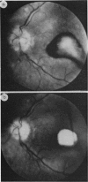
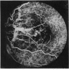
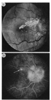
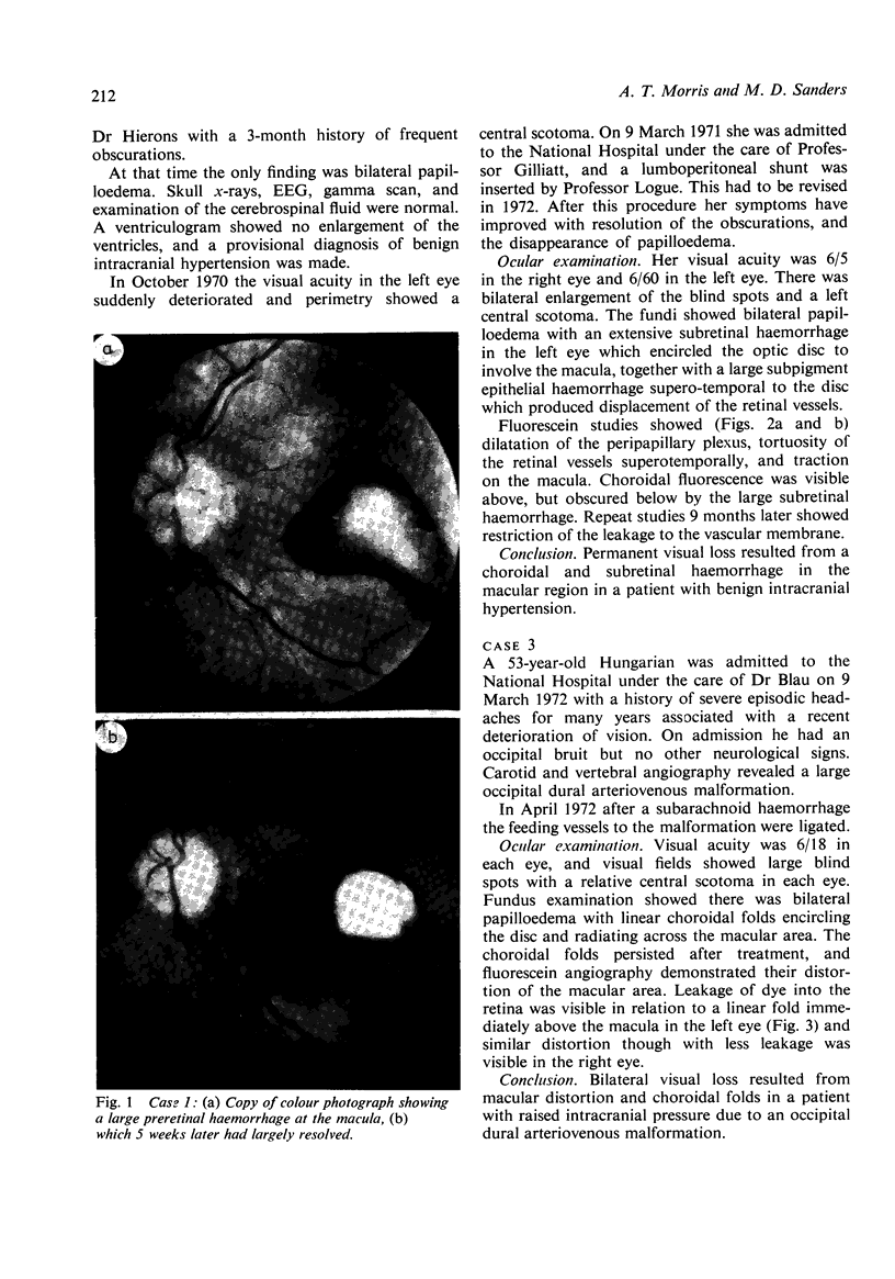
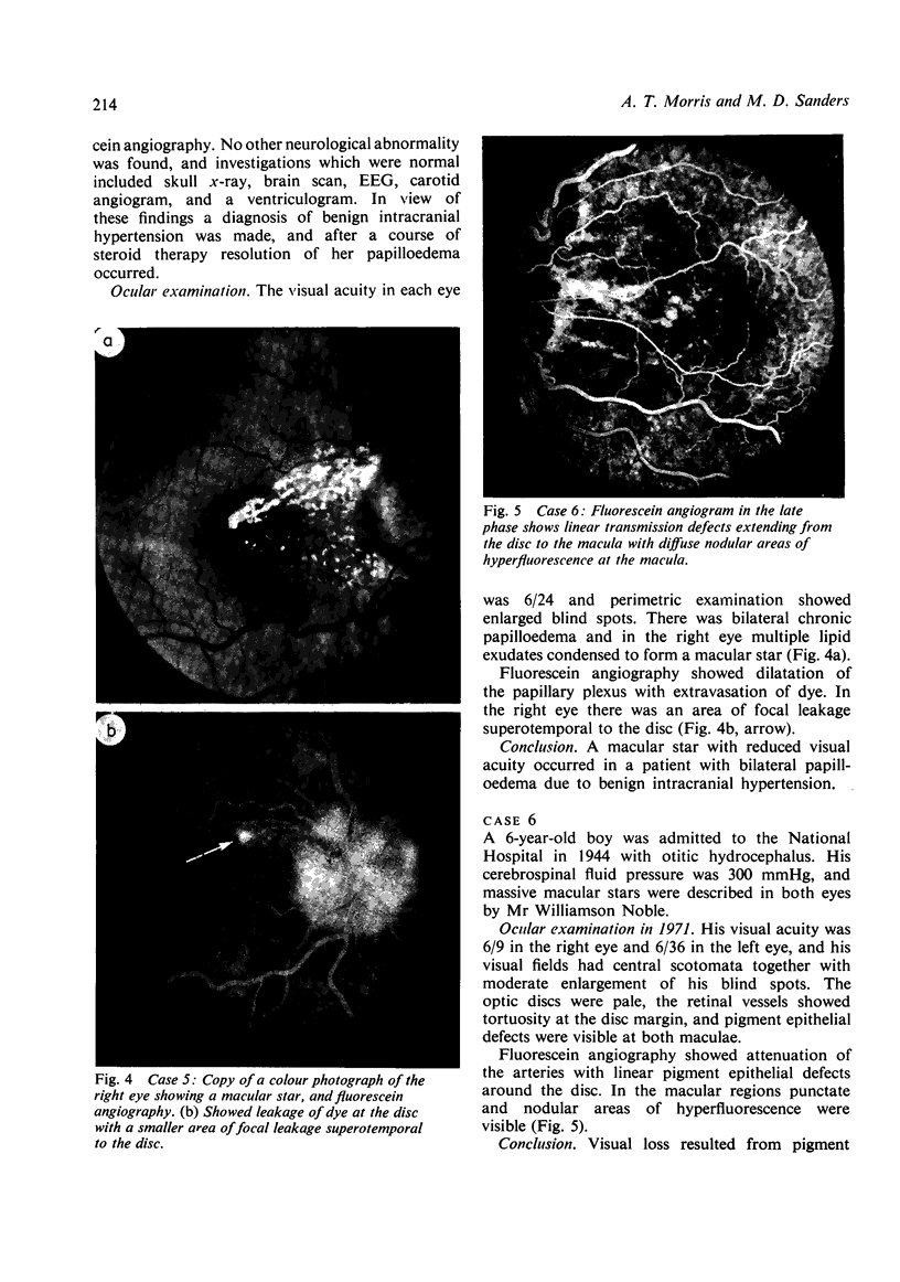
Six cases are presented with macular changes in association with papilloedema; 4 suffered permanent visual loss. The present paper emphasises this previously infrequent finding and discusses the haemodynamic and mechanical factors responsible. The macular changes consisted of haemorrhages situated in front, within, or behind the retina, and occasionally the results of neovascular membrane formation produced secondary visual loss. Changes in the pigment epithelium were seen in 3 cases associated with choroidal folds. Macular stars rarely produce visual loss. Recognition of these changes is important in the assessment of the visual loss in papilloedema.
Full text
PDF


Images in this article
Selected References
These references are in PubMed. This may not be the complete list of references from this article.
- Bird A. C., Sanders M. D. Choroidal folds in association with papilloedema. Br J Ophthalmol. 1973 Feb;57(2):89–97. doi: 10.1136/bjo.57.2.89. [DOI] [PMC free article] [PubMed] [Google Scholar]
- Davidson S. I. A surgical approach to plerocephalic disc oedema. Trans Ophthalmol Soc U K. 1970;89:669–690. [PubMed] [Google Scholar]
- Galbraith J. E., Sullivan J. H. Decompression of the perioptic meninges for relief of papilledema. Am J Ophthalmol. 1973 Nov;76(5):687–692. doi: 10.1016/0002-9394(73)90564-3. [DOI] [PubMed] [Google Scholar]
- Smith J. L., Hoyt W. F., Newton T. H. Optic nerve sheath decompression for relief of chronic monocular choked disc. Am J Ophthalmol. 1969 Oct;68(4):633–639. doi: 10.1016/0002-9394(69)91243-4. [DOI] [PubMed] [Google Scholar]


