Abstract
Methane is a potent greenhouse gas, which likely enabled the evolution of life by keeping the early Earth warm. Here, we demonstrate routes towards abiotic methane and ethane formation under early-earth conditions from methylated sulfur and nitrogen compounds with prebiotic origin. These compounds are demethylated in Fenton reactions governed by ferrous iron and reactive oxygen species (ROS) produced by light and heat in aqueous environments. After the emergence of life, this phenomenon would have greatly intensified in the anoxic Archean by providing methylated sulfur and nitrogen substrates. This ROS-driven Fenton chemistry can occur delocalized from serpentinization across Earth’s humid realm and thereby substantially differs from previously suggested methane formation routes that are spatially restricted. Here, we report that Fenton reactions driven by light and heat release methane and ethane and might have shaped the chemical evolution of the atmosphere prior to the origin of life and beyond.
Subject terms: Carbon cycle, Carbon cycle
Abiotic methane and ethane formation routes in aqueous environments driven by light and heat are identified. The released hydrocarbons may have contributed to the chemical evolution of the atmosphere from prior to the origin of life until today.
Introduction
Methane (CH4) is a potent greenhouse gas which has in the past and is still today contributing to climate change1. Atmospherically accumulated CH4 and ethane (C2H6) might also explain the “faint young sun paradox”, which describes the apparent contradiction of a fainter sun (70 – 83% of the current solar energy output) but a climate that was at least as warm as today during early Earth (4.5–2.5 Ga ago)2–4. Although these CH4 levels would be essential to keep the Earth a liquid hydrosphere to allow the evolution of life during the Archean (4.0–2.5 Ga), the source of CH4 prior to the origin of life is still under debate5. While CH4 was released by submarine volcanism, most CH4 is suggested to be formed as side product of serpentinization5. After the evolution of microbial methanogenesis latest by 3.5 Ga6, methanogenesis could have been responsible for a CH4 flux comparable to today7. Thus, methanogenesis is expected to be the main source of CH4 during the Archean, supported by light carbon isotope values in sedimentary deposits8. However, isotope signals can only manifest upon reoxidation and CH4 itself does not leave much of a signature in the geological record. Thus, the actual CH4 concentrations and the potential abiotic sources during early Earth remain elusive. Based on mass-independent fractionation of sulfur, at least 20 ppmv CH4 was present around 2.4 Ga ago9. A more recent study analyzing the fractionation of xenon isotopes suggests CH4 levels of >5000 ppmv around 3.5 Ga ago10. Catling et al. expect even higher CH4 levels at the beginning of the Archean (4 Ga)3 before methanogenesis evolved. Yet, the processes responsible for these high CH4 levels and their relative contributions remain controversial.
Recently, we discovered a non-enzymatic CH4 formation mechanism expected to occur in all living organisms11. The mechanism has been demonstrated to be active in over 30 very diverse organisms11 and suggested to explain previously observed CH4 formation by cyanobacteria12, freshwater and marine algae13,14, saprotrophic fungi15 and plants16. The CH4 formation is driven by a cascade of radical reactions, governed by the interplay of reactive oxygen species (ROS) and ferrous iron (Fe2+), methylated sulfur (S)- and nitrogen (N)-compounds are oxidatively demethylated by hydroxyl radicals (∙OH) and oxo-iron(IV) complexes ([FeIV=O]2+) to yield methyl radicals (·CH3)11.
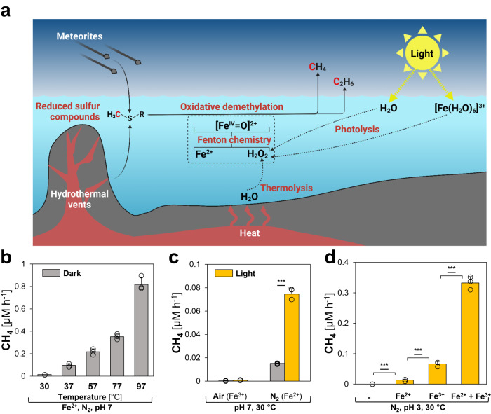
Here we show that this abiotic mechanism occurs also outside living cells and might have contributed to CH4 levels before life emerged. All needed components: (i) methylated S- and N-compounds, (ii) Fe2+ and (iii) ROS are found under early-earth conditions. (i) In a prebiotic world, methylated S-compounds like methanethiol, dimethyl sulfide (DMS) or dimethyl sulfoxide (DMSO) were formed abiotically under the reducing conditions of hydrothermal vents17–19 or transported to Earth by carbonaceous meteorites during early Earth meteorite bombardment20,21. Upon the emergence of life, more methylated S-/N-compounds were produced by cells and organisms, i.e. methionine, dimethylsulfoniopropionate or trimethylamine22. (ii) Under the anoxic conditions of the early Earth, oceans were rather ferruginous, i.e. rich in Fe2+ required for Fenton chemistry23,24, nonetheless ferric iron (Fe3+) also occurred in Archean seawater25. Additionally, the mechanism driven by Fe2+ can be enhanced by Fenton-promoting Fe2+-chelators, e.g. ATP or citrate26. Under anoxic conditions, Fe(III)-carboxylate complexes are photochemically reduced via ligand-to-metal charge transfer (LMCT)27, resulting in Fe2+ and organic radicals28. (iii) Under ambient temperatures, low ROS levels exist in water that increase with heat29, or can be generated by photolysis or radiolysis30–33. Under acidic conditions, i.e. in volcanic lakes34, illumination of Fe(III)-aqua complexes ([Fe(H2O)6]3+) forms Fe2+ and ROS35,36. Thus, we hypothesized that the Fenton reaction of Fe2+ with H2O2, generated by heat and light, could have driven the formation of CH4 from methylated S-/N-compounds independent of temperatures and pressures occurring at hydrothermal vents but at ambient conditions as early as the prebiotic world of the Hadean (4.5–4.0 Ga, Fig. 1a). To identify critical components of such a mechanism, we used aqueous model systems to determine the influence of heat, light, and (bio)molecules on CH4 formation in abiotic and biotic environments.
Fig. 1. Heat and light drive CH4 formation under abiotic conditions.
a Reduced, methylated S-/N-compounds are formed abiotically in hydrothermal vents or transported to Earth by carbonaceous meteorites. Under anoxic conditions, H2O2 is formed by thermolysis and photolysis of water and [Fe(H2O)6]3+ complexes, reacting with dissolved ferrous iron (Fe2+) to hydroxyl radicals (∙OH) and [FeIV= O]2+ compounds that drive the oxidative demethylation of methylated S-/N-compounds, thereby facilitating CH4 and C2H6 formation. b Thermolysis: CH4 is formed from DMSO under high temperatures. c Water photolysis: The formation of CH4 is increased by light. d [Fe(H2O)6]3+ photolysis: Under acidic conditions, light-driven CH4 formation is enhanced by [Fe(H2O)6]3+ photochemistry. All experiments were conducted in closed glass vials containing buffered solutions (pH 7 or pH 3) supplemented with DMSO and Fe2+ or Fe3+ at 30 °C (b, c) under a N2 or air atmosphere. Statistical analysis was performed using paired two-tailed t tests, ***p ≤ 0.001. The bars are the mean + standard deviation of triplicates, shown as circles. a Was created with BioRender.com.
Results
Methane is formed under abiotic conditions
To investigate CH4 formation under abiotic conditions (Fig. 1a), we designed a chemical model system consisting of a nitrogen atmosphere, a potassium phosphate-buffered solution (pH 7, expected during the Archean at 4.0 Ga37) supplemented with Fe2+ and the abiotically formed DMSO which serves as methyl donor for ROS-driven CH4 formation. Over the course of the experiments, no pH change was observed, while low amounts of Fe(OH)2 precipitated. In this model system, CH4 was consistently formed from DMSO in the dark (Fig. 1b). CH4 formation rates increased with rising temperatures from 30 to 97 °C, consistent with the previously reported temperature-dependency of ROS levels in water29. While only marginal CH4 formation rates derived from DMSO were observed at 30 °C (~0.02 μM h−1), rates increased 41-fold to ~0.82 μM h−1 at 97 °C. In addition, low C2H6 amounts were formed (Supplementary Fig. 1), most likely resulting from the recombination of two methyl radicals23. At 37 °C, the CH4:C2H6 ratio was ~110, with an increasing trend towards higher temperatures. As the ROS-driven CH4:C2H6 ratios are substantially lower than those observed for archaeal methanogenesis38, the CH4:C2H6 ratios could serve as indicator to distinguish microbial from abiotic processes.
Light enhanced the abiotic CH4 formation rates (Fig. 1c) by photolysis of water and generation of H2O2 at 30 °C (Supplementary Fig. 2). Notably, CH4 amounts increased ~4-fold from ~0.02 μM h−1 to ~0.08 μM h−1 upon broad-spectrum illumination (~ 350 nm < λ < ~1010 nm at 82 ± 4 µmol photons m−2s−1, Supplementary Fig. 3). This data provides evidence that light-driven CH4 formation from methylated S-compounds can occur even in the absence of biomolecules. The addition of oxygen to the samples stopped the formation of CH4 in this pH-neutral model system supplemented with Fe3+ (Fig. 1c). In contrast, under acidic (pH 3), illuminated conditions CH4 formation rates increased ~5-fold upon Fe3+-supplementation in comparison to Fe2+-addition, indicating light-driven ROS and Fe2+ formation from [Fe(H2O)6]3+ complexes (Fig. 1d)35. Upon supplementation of 1 mM Fe3+ and 1 mM Fe2+, keeping the overall iron concentration unchanged at 2 mM, CH4 formation rates increased to ~0.33 μM h−1. This 5-fold rate increase is driven by both ROS-inducing Fe3+ and Fenton-driving Fe2+. Under pH-neutral conditions, mixing Fe2+ and Fe3+ only increased CH4 formation rates by ~1.3-fold in comparison to Fe2+-supplemented samples, while only trace amounts of CH4 were obtained from Fe3+-supplemented samples (Supplementary Fig. 4). Thus, illuminated [Fe(H2O)6]3+ complexes generate both Fe2+ and ROS, thereby contributing to the ROS-driven CH4 formation under acidic conditions.
Taken together, we demonstrated that heat and light drive the formation of CH4 and C2H6 in an anoxic, abiotic environment under ambient temperatures and pressures. These results establish a ROS-driven mechanism based on Fenton chemistry that can occur delocalized from serpentinization across Earth’s humid realm and thereby substantially differs from previously suggested mechanisms that are spatially restricted. Thus, this non-enzymatic hydrocarbon formation mechanism could have released CH4 and C2H6 into the atmosphere of the Hadean and Archean. Besides CH4, C2H6 is considered an important factor in keeping the early Earth warm, since C2H6 absorbs from 11 to 13 µm in an atmospheric window (roughly 8–13 µm) where H2O and CO2 do not absorb strongly2. Together, the hydrocarbons produced by these pathways might offer a solution to the “faint young sun paradox”3,4.
(Bio)molecules enhance the heat-driven CH4 formation
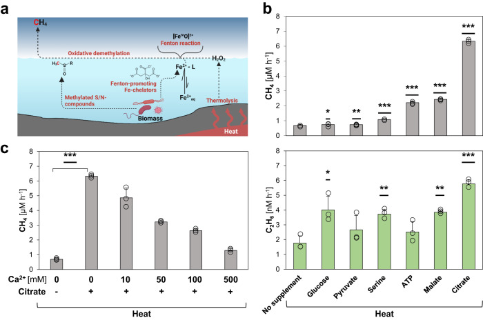
Even before life emerged, several metabolites, e.g. citrate and malate, could have been formed via an ancient, non-enzymatic TCA cycle predecessor driven by ROS39,40. Catalyzed by iron particles, the formation of pyruvate from CO2 was recently reported41. Intriguingly, citrate and malate, as well as other primordial (bio)molecules with a putative prebiotic origin, including ATP42 or serine43, have been reported to act as Fenton-promoting Fe2+-chelators26. We therefore investigated if these hydroxylated and carboxylated (bio)molecules enhance the ROS-driven CH4 formation rates (Fig. 2a).
Fig. 2. (Bio)molecules enhance heat-driven CH4 formation.
a Overview of CH4 formation driven by heat. Living organisms produce S-/N-methylated compounds that serve as substrates for CH4 formation and Fe2+-chelators that promote Fenton chemistry and enhance CH4 formation. b Heat-driven CH4 (upper panel) and C2H6 (lower panel) formation is enhanced upon supplementation with (bio)molecules. c Citrate enhances heat-driven CH4 formation acting as iron-chelator. Upon the addition of Ca2+, CH4 levels decrease due to the replacement of Fenton-promoting Fe2+-citrate complexes with Ca2+-citrate complexes. All experiments were conducted in closed glass vials containing a buffered solution (pH 7) supplemented with DMSO, Fe2+ and, optionally, citrate and Ca2+ under a pure nitrogen atmosphere at 97 °C (heat). Statistical analysis was performed using paired two-tailed t tests, *p ≤ 0.05, **p ≤ 0.01, ***p ≤ 0.001. The bars are the mean + standard deviation of triplicates, shown as circles. a Was created with BioRender.com.
Indeed, the addition of pyruvate, glucose, serine, ATP, malate or citrate to the heat-driven (97 °C) model system increased the abiotic CH4 formation rate, e.g. more than 11-fold for citrate (Fig. 2b). Corresponding C2H6 rates significantly increased for glucose, serine, malate and citrate, resulting in CH4:C2H6 ratios between ~190 (glucose) and ~1100 (citrate, Fig. 2b). To test if these enhancing effects were indeed driven by Fe2+ chelation, we supplemented the assays with the Fe2+-competitor Ca2+ (Fig. 2c). Since (bio)molecules like citrate can alternatively chelate Ca2+ ions, we expected that increasing Ca2+ concentrations result in decreasing CH4 formation rates by replacing Fenton-promoting Fe2+-citrate complexes with Ca2+-citrate complexes. Upon addition of 10 mM and 500 mM Ca2+, CH4 formation rates significantly decreased from ~6.32 μM h−1 to ~4.86 μM h−1 and ~1.29 μM h−1, respectively. Thus, 500 mM Ca2+ suppressed ~90% of the Fenton-promoting effect of citrate supplementation. The Ca2+ concentration-dependent decrease of the heat-driven CH4 formation rate supports the role of citrate as a Fenton-promoting Fe2+-chelator, which is further indicated by citrate dissolving any ferruginous precipitate.
Together, ROS generated by heat interact with iron and thereby drive the formation of methyl radicals from S-/N-methylated compounds, resulting in CH4 and C2H6. Moreover, several hydroxylated or carboxylated (bio)molecules with a putative prebiotic origin were shown to act as Fenton-promoting Fe2+-chelators, indicating that ROS-driven CH4 formation may have already been widespread within the timeframe of the transition from prebiotic chemistry to the origin of life. The rise of life would have fostered the abiotic, non-enzymatic CH4 formation due to the consequential formation and release of biomolecules serving as chelators and substrates.
A light-driven iron redox cycle sustains CH4 formation
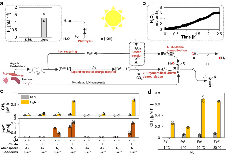
During Fenton chemistry, Fe2+ is either oxidized to [FeIV= O]2+ or ferric iron (Fe3+). As Fe3+ cannot drive Fenton reactions23,24, CH4 formation rates decrease with increasing reaction time and increasing concentrations of Fe3+. While this effect may have been minor in the ferruginous Archean oceans, Fe3+ likely dominated the iron pool in the photic zone of the oceans latest by the rise of photoferrotrophy and was also prevalent in several ecological niches, e.g. volcanic lakes34. The evolution of photosynthesis and the subsequent biological production of O2 oxidized the majority of the available Fe2+ to Fe3+. Thus, abiotic ROS-driven CH4 formation would have been hindered in the sunlit realm by the late Archean in the absence of an iron redox cycle at neutral pH. Intriguingly, besides acting as Fenton-promoting Fe2+-chelators26, (bio)molecules like citrate were reported to reduce Fe3+ to Fe2+ via LMCT under oxic and anoxic conditions27. Therefore, (bio)molecules may have facilitated widespread iron redox cycling, e.g. by forming Fe(III)-carboxylate complexes. Furthermore, previous studies showed that, upon illumination of water hydroxyl radicals (·OH) and hydrogen atoms are generated, forming H2O2 and H230–33. Thus, we hypothesized that light could drive CH4 formation in the absence of Fe2+ by simultaneously (i) generating ROS from water and (ii) reducing Fe3+ to Fe2+ via LMCT, thereby recycling Fe3+ and keeping the Fenton reaction running (Fig. 3).
Fig. 3. A light-driven iron redox cycle drives and enhances CH4 formation.
Upon illumination, water is photolytically split into hydroxyl radicals (·OH) and hydrogen forming H2 and H2O2. Organic Fe3+-complexes (Fe3+-[L1]) are converted into Fe2+ and organic radicals (·L1) via ligand-to-metal charge transfer (LMCT). The generated Fe2+ reacts with H2O2 to ·OH or [FeIV= O]2+ and thereby drives the generation of methyl radicals (·CH3) from S-/N-methylated compounds. The LMCT-generated ·L1 decomposes into CO2 and another organic radical (·L2) that additionally facilitates CH4 formation upon reacting with S-/N-methylated compounds. Under light, (a) H2 (gray bars) and (b) H2O2 is formed in pure buffer. c Upon illumination, CH4 formation rates (yellow bars) are increased. Fe2+ formation (brown bars) depends on anoxic conditions and is driven by LMCT induced by the addition of citrate. d Light and heat have synergistic effects on CH4 formation. While heat drives CH4 formation upon Fe2+-supplementation, light increases CH4 formation upon Fe3+- and Fe2+-addition. All experiments were conducted in closed glass vials containing a buffered solution (pH 7) supplemented with DMSO, Fe2+ or Fe3+, N2 or air atmosphere in the presence or absence of citrate incubated under light or in the dark at 4 °C or 30 °C. The bars are the mean + standard deviation of triplicates, shown as circles. a, b Was created with BioRender.com.
To verify our hypothesis, we first confirmed light-dependent ROS production in our model system in the absence of substrate, iron and organic ligands by measuring final reaction products of photolysis: H2 and H2O2 (Fig. 3a, b). We measured H2 production at a rate of ~1.3 nM h−1 in anoxic samples under broad-spectrum illumination but not in samples kept in the dark (Fig. 3a). A continuous formation of H2O2 was measured online using microsensors, which confirmed light-dependent production dynamics in pure buffer (Fig. 3b). Via fluorescence-based H2O2 endpoint measurements, we found that both iron and DMSO reduced the H2O2 concentrations. The decrease in H2O2 levels can be attributed to Fenton reactions between H2O2, Fe2+ and the radical scavenger DMSO (Supplementary Fig. 5).
Building on this, we closely investigated the interplay of LMCT and iron photochemistry on CH4 formation. For this purpose, we analyzed our chemical model system containing a buffered solution (pH 7), Fe2+ or Fe3+, DMSO, in the presence or absence of citrate for the formation of CH4 and the concentration of available Fe2+ (Fig. 3c). The influence of the following parameters on the formation of CH4 was tested: (i) O2 ( ~ 21% in air), (ii) oxidation state of the supplemented iron species (Fe2+ vs. Fe3), (iii) light and (iv) presence/absence of citrate. (i) CH4 formation rates under anoxic conditions always exceeded rates under oxic conditions. (ii) Without citrate, initial Fe2+-supplementation was required to form significant CH4 levels. (iii) CH4 formation always increased with light. (iv) Upon citrate addition, CH4 formation was enhanced in illuminated and anoxic samples containing DMSO and Fe2+ or Fe3+. Besides elevated CH4 formation rates, citrate addition also increased the final Fe2+ concentrations, e.g. from ~0 mM Fe2+ to ~1.5 mM Fe2+ in illuminated and anoxic samples.
After determining the influence of the four parameters (i) O2, (ii) iron (iii) light and (iv) (bio)molecules, we further investigated them individually to gain a better understanding of their contribution and role in the light-driven CH4 formation.
(i) O2: The influence of O2 on LMCT and CH4 formation was studied in citrate-supplemented samples by adding various amounts of air. Fe2+ concentrations and CH4 formation rates decreased with increasing O2 levels (Supplementary Fig. 6). In comparison to 0 % O2, the Fe2+ concentration dropped drastically already at 0.2 % O2 and was ~96 % lower at 2 % O2, while CH4 formation rates decreased approximately linearly with the O2 level. This indicates the presence of a Fe-cycle, in which most LMCT-formed Fe2+ is instantly re-oxidized, either by O2 or Fenton reactions. The balance between these Fe2+ sinks depend on O2 availability and governs CH4 formation rates. In the presence of O2, we also detected methanol (CH3OH) formation rates ranging from ~0.003 μM h−1 (0.2 % O2) to ~0.07 μM h−1 (21 % O2). CH3OH is preferentially formed through the reaction of ·CH3 with O223,44. Without the addition of O2, no CH3OH was detected, indicating anoxic conditions in our standard assays.
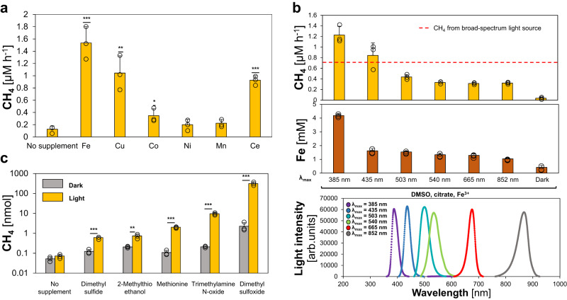
(ii) Iron: The role of the LMCT-rate and the corresponding Fe2+ availability for CH4 formation was tested by supplementing the assays with various Fe3+ concentrations (Supplementary Fig. 7). At lower Fe3+ concentrations, CH4 formation rates increased steeper than the measured Fe2+ concentrations. At high Fe3+ concentrations, CH4 formation rates leveled off, while Fe2+ concentrations continued to increase. This indicates that Fe2+ is limiting the demethylation rates at low iron concentrations, because it is immediately re-oxidized, while light-dependent ROS production is limiting CH4 formation at high iron concentrations. Most importantly, these data highlight that a light- and ROS-driven iron cycle can facilitate high rates of CH4 formation, even in the presence of O2 and the absence of detectable Fe2+, which opens the possibility of widespread abiotic CH4 production after the great oxidation event as well as in diverse modern habitats. Next, we investigated the role of the alkali metal magnesium (Mg2+) due to its high environmental abundance and found that Mg2+ does not facilitate CH4 formation in illuminated buffer containing DMSO and citrate (Supplementary Fig. 8). Upon additional Fe3+ supplementation, Mg2+ also decreased CH4 formation rates by replacing Fenton-promoting Fe3+-citrate complexes by Mg2+-citrate complexes, thereby acting similar to Ca2+ that was demonstrated to decrease heat-driven CH4 formation (Fig. 2c). Besides iron, the transition metals copper, cerium, cobalt, nickel and manganese were reported to drive Fenton chemistry45,46, resulting in the release of CH4. Thus, we tested different transition metals in our chemical model system, containing DMSO as substrate and ascorbate as a strong metal reductant47,48. We observed that copper, cobalt and cerium also enhanced CH4 formation rates (Fig. 4a). However, the activity of copper, cobalt and cerium was lower than iron. The high activity of iron combined with its ubiquitous abundance in the Precambrian highlights the global distribution and importance of this mechanism.
Fig. 4. Transition metals, wavelengths and methylated sulfur- and nitrogen compounds mediate light-driven CH4 formation.
a Iron, cobalt and cerium enhance light-driven CH4 formation. No significant CH4 increase was observed for cobalt, nickel and manganese supplementation. b Light-driven CH4 formation and Fe2+ generation increases in the near-UV spectrum. c Light-driven formation of CH4 from methylated S-/N-compounds (logarithmic scale). Upon illumination, significant increases in CH4 levels were measured for dimethyl sulfide, methionine, 2-methylthioethanol, trimethylamine N-oxide and dimethyl sulfoxide (DMSO). All experiments were conducted in closed glass vials containing a buffered solution (pH 7), N2 and either Fe3+ or other transition metals (a), DMSO or other substrates (c) and either ascorbate (a) or citrate (b, c). Samples were incubated under broad-spectrum light (a, c), specific wavelengths (b) or in the dark at 30 °C. The dashed red line depicts the average CH4 amounts obtained from samples illuminated by a broad-spectrum light source. Statistical analysis was performed using paired two-tailed t tests, *p ≤ 0.05, **p ≤ 0.01, ***p ≤ 0.001. The bars are the mean + standard deviation of triplicates, shown as circles.
(iii) Light: It is established that light quality has an important influence on photolysis. Short wavelength light in the ultraviolet spectrum was reported to drive water photolysis and LMCT more efficiently than longer wavelengths49. We expected that shorter wavelength light would increase both CH4 formation rates and Fe2+ levels. Indeed, CH4 formation rates surged from ~0.3 μM h−1 (λmax = 534 nm) to ~1.23 μM h−1 (λmax = 388 nm, Fig. 4b) and Fe2+ concentrations almost tripled from ~1.3 mM (λmax = 534 nm) to ~4.2 mM (λmax = 388 nm). Although the broad-spectrum light had a 1.5-fold higher energy flux (57 ± 2 kJ m−2 h−1) compared to the 388 nm-LED light (37 ± 2 kJ m−2 h−1), the CH4 formation rate under the broad-spectrum light was only half (0.7 μM h−1). Given that the stratospheric ozone layer was absent during the Hadean and Archaean, higher fluxes of short wavelength light (i.e. ultraviolet light), reached aqueous environments and may have further enhanced the ROS-driven CH4 formation.
(iv) (Bio)molecules: After illumination of Fe3+-ligand complexes, one electron is transferred via LMCT from a carboxylated ligand (L1) to Fe3+, an organic radical (·L1), i.e. citrate radical, is generated. As described in the literature28, we observed the subsequent CO2 disassembly from citrate radicals (Supplementary Fig. 9). We speculated that the remaining organic radical (·L2) could react with DMSO, resulting in ·CH3 and the formation of CH4 (Fig. 3). Since we cannot directly detect organic radicals, we mimicked the proposed reaction in an anoxic model system only containing DMSO and the radical-generating 2,2’-azobis(2-amidinopropane) dihydrochloride (APPH) that readily decomposes into carbon-centered organic radicals at 40 °C (Supplementary Fig. 10). Indeed, we observed CH4 formation in a mixture of DMSO and APPH, while only trace amounts of CH4 were observed from either DMSO or AAPH alone, suggesting an organic radical-driven CH4 formation mechanism. In short, carboxylates like citrate facilitate LMCT, thereby reducing Fe3+ to Fe2+ and forming organic radicals. Both resulting compounds drive CH4 formation. Overall, CH4 can be formed under anoxic conditions via (i) water thermolysis, (ii) water photolysis, (iii) [Fe(H2O)6]3+ photolysis and (iv) LMCT-induced carbon-centered radicals. Apart from serving as chelators, some (bio)molecules could also serve as substrates for Fenton reactions. Thus, we investigated four S-/N-methylated compounds in the presence of the chelator citrate. Upon illumination, CH4 was formed from dimethyl sulfide, methionine, 2-methylthioethanol and trimethylamine (Fig. 4c). These observations indicate that ROS-driven CH4 formation significantly increased after the origin of life by providing biomolecules as chelators and substrates.
Finally, synergistic effects between light and heat were observed (Fig. 3d). For Fe2+-supplemented samples, CH4 rates at 4 °C increased from ~0.056 μM h−1 in the dark over ~0.19 μM h−1 under light to ~0.65 μM h−1 in illuminated samples at 30 °C. For Fe3+-supplemented samples, only CH4 rates below 0.03 μM h−1 were obtained in the dark, while CH4 formation rates were slightly above Fe2+-supplemented samples in the light, again demonstrating the effects of LMCT and LMCT-induced carbon-centered radicals. Thus, the two factors heat and light synergistically combine for a stable and enhanced ROS and CH4 formation.
Biomass-derived CH4 with an abiotic isotope fractionation
Considering the impact of (bio)molecules on the LMCT-driven Fenton reaction, organic radical generation and the role of biomolecules as substrates, we expect the discussed mechanisms to have played and still play the most important role in the vicinity of decaying biomass. To demonstrate that CH4 is indeed formed from dead biomass in the presence of a variety of biomolecules and not just in our well-defined model systems, we conducted deuterium labeling experiments. For this purpose, we grew the bacterium B. subtilis in Luria-Bertani medium supplemented with 10% D2O and inactivated the cells by sonication and freezing (see Methods).
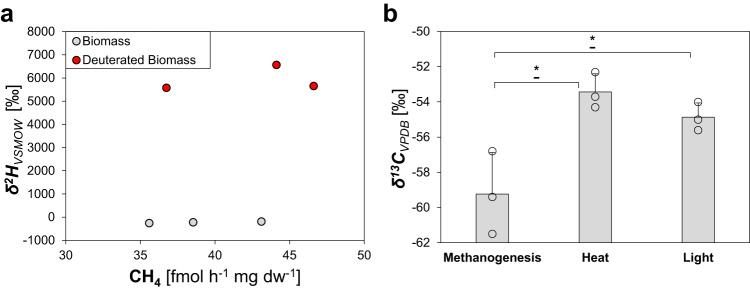
The obtained dead biomass was supplemented with Fe3+ and ascorbate and incubated under broad-spectrum light. Around 40 fmol CH4 h−1 mg−1 dry weight was obtained from labeled and unlabeled biomass (Fig. 5a). In addition, stable hydrogen isotope values (δ2H) of CH4 from D2O-treated biomass showed strong enrichment in deuterium (~5900 ‰) in comparison to unlabeled biomass (~−225 ‰), demonstrating a direct conversion of isotopically labeled biomass to CH4. This suggests that the availability of biomass, upon the emergence of life, has increased the CH4 formation by delivering both (i) S-/N-methylated compounds and (ii) Fenton-promoting iron chelators. The presence of CH4 has been suggested to be crucial for the evolution of life, since it could serve as life´s first carbon source via methanotrophy50–52. Following this line of thought, we could demonstrate that methanotrophic Methylocystis hirsuta grew on CH4 generated by our light-driven model system, transferred to the headspace of the M. hirsuta culture (Supplementary Fig. 11). In fact, the “last methane-metabolizing ancestor” had likely the genes to perform methanogenesis and anaerobic methane oxidation53, suggesting that, under high CH4 concentrations, methanotrophy could have emerged prior to methanogenesis.
Fig. 5. Isotope labeling studies confirm dead biomass as substrate and show an abiotic isotope fractionation for ROS-driven CH4 formation.
a Unlabeled or deuterium-enriched CH4 is formed from unlabeled biomass (gray dots) or deuterated biomass (red dots), respectively. b Stable carbon isotope values of cultures from the methanogen Methanothermobacter marburgensis, heat-, or light-generated CH4. All experiments were conducted in closed glass vials containing a buffered solution (a, b—heat, light) or culture medium (b—methanogenesis), supplemented with Fe3+ and ascorbate (a) or Fe2+ and citrate (b—heat, light) under a nitrogen atmosphere, incubated under light at 30 °C or in the dark at 97 °C. Statistical analysis was performed using paired two-tailed t tests, *p ≤ 0.05. The bars are the mean + standard deviation of triplicates, shown as circles.
Finally, we speculated that ROS-driven CH4 formation leads to different stable carbon isotope values (δ13C) compared to biological processes, i.e., methanogenesis. The observed δ13C values for CH4 generated by heat or light were less negative (~−54 ± 1.1‰) compared to the δ13C value of the methanogen Methanothermobacter marburgensis (~−59.2 ± 2.3‰, Fig. 5b). While the isotopic fractionation during abiotic ROS-driven CH4 formation remains to be studied in depth, these results suggest a lower carbon isotope fractionation for ROS-driven CH4 formation than for enzymatic methanogenesis. Together with the observed CH4:C2H6 ratios, isotopic signatures may therefore serve to differentiate between CH4 formed enzymatically or abiotically on Earth and extraterrestrial planets.
Discussion
In this work, we demonstrated that the interplay of Fe2+ and H2O2, generated by heat and light, drives CH4 and C2H6 formation from methylated S-/N-compounds via Fenton chemistry under conditions that were globally prevalent in the Hadean and Archean. As we observed CH4 formation under suboxic and oxic conditions, these mechanisms could, in principle, also contribute to extant CH4 emissions from aqueous environments that were recently shown to correlate with light instead of specific enzymatic pathways54. The here described pathways allow CH4 and C2H6 formation in many aqueous environments including oceans, lakes, rivers, and ponds, delocalized from restricted hotspots for (bio)molecule formation such as hydrothermal vents or ultramafic rocks, in superficial water layers driven by light and throughout the entire water column driven by heat. After the emergence of life, this phenomenon would have greatly intensified in the anoxic Archaean and the subsequent “boring billion”55,56. The increasing amounts of biomass provided methylated S-/N-substrates, Fe-chelating biomolecules reducing Fe3+ to Fe2+ and releasing organic radicals and thus enhance ROS-driven CH4 formation. Possibly, these reactions facilitated elevated CH4 and C2H6 levels during the Hadean and Archean. These hydrocarbons would have contributed to atmospheric temperatures on Earth and allowed the evolution of life in a liquid hydrosphere which could have influenced the evolution of metabolism by allowing the rise of methanotrophy prior to methanogenesis. This work lays the foundation to explore further the mechanism’s role in shaping the evolution of the atmosphere on Earth and other planets and its influence on the current climate change.
Methods
General assay conditions
Unless otherwise indicated, 4 mL samples were incubated in closed 20 mL glass vials at 30 °C under a pure nitrogen (N2) atmosphere and subsequently analyzed via gas chromatography (GC).
Heat assays
In total, 500 mM DMSO and 10 mM FeSO4 were added to 20 mM degassed potassium phosphate buffer (pH 7) in an anaerobic tent. The headspace of the closed vials was then cycled three times with vacuum and N2. Samples were incubated at 37 °C, 57 °C, 77 °C and 97 °C for 6 h in an incubator in the dark. Optionally, 20 mM citrate, malate, ATP, serine, glucose or pyruvate were also supplemented. Ca2+ was added in the form of CaCl2. Samples were measured within the linear range of CH4 formation rate via gas chromatography.
Light assays
In total, 500 mM DMSO, 2 mM of either FeCl3 or FeSO4 and, optionally, 10 mM citrate were added to 20 mM degassed potassium phosphate buffer (pH 7). Anoxic conditions were generated by drawing vacuum eight times for 1 min and a subsequent filling with N2. For experiments investigating [Fe(H2O)6]3+ complexes, samples were incubated under anoxic, acidic conditions (20 mM Tris · HCl buffer, pH 3) and supplemented with 500 mM DMSO and either 2 mM FeCl3, 2 mM FeSO4 or 1 mM FeCl3 and 1 mM FeSO4, each. For the investigation of transition metals (Fig. 4a), 2 mM cerium (CeNH4SO4), manganese (MnSO4), cobalt (CoNO3), nickel (NiSO4), copper (CuCl2) or iron (FeCl3) and 10 mM pH-neutral ascorbate were added to 500 mM DMSO and 20 mM potassium phosphate buffer with an incubation for 1 day. The effect of different wavelengths on CH4 formation (Fig. 4b) was investigated by adding 5 mM FeCl3, 10 mM citrate and 500 mM DMSO to 20 mM potassium phosphate buffer with an incubation for 1 day. For the determination of substrates for CH4 formation (Fig. 4c), 500 mM DMS, methionine, 2-methylthioethanol, Trimethylamine N-oxide or DMSO were added to 10 mM FeCl3 and 100 mM citrate in 20 mM potassium phosphate buffer with an incubation for 3 days. Samples were incubated under air or N2 in the dark or under constant broad-spectrum illumination from light bulbs (Osram, Superlux, Super E SIL 60; Φ = 82 ± 4 µmol photons m−2 s−1, H = 52 ± 2 kJ m−2 h−1; Supplementary Fig. 3) for 1 day. Samples were measured within the linear range of the CH4 formation via gas chromatography. Specific wavelengths were provided by diodes (H2A1 series, Roithner Lasertechnik, Austria) emitting UV-A, blue, cyan, green, red or near-infrared light (λmax = 388 nm, Φ = 35 ± 1 µmol photons m−2 s−1, H = 36 ± 2 kJ m−2 h−1; λmax = 436 nm, Φ = 45 ± 1 µmol photons m−2 s−1, H = 45 ± 1 kJ m−2 h−1; λmax = 500 nm, Φ = 64 ± 4 µmol photons m−2 s−1, H = 55 ± 3 kJ m−2 h−1; λmax = 534 nm, Φ = 63 ± 1 µmol photons m−2 s−1, H = 50 ± 1 kJ m−2 h−1; λmax = 675 nm, Φ = 45 ± 4 µmol photons m−2 s−1, H = 29 ± 3 kJ m−2 h−1; or λmax = 868 nm, Φ = 69 ± 7 µmol photons m−2 s−1, H = 35 ± 3 kJ m−2 h−1) Light intensity was determined using a fiber optic scalar irradiance microsensor57 connected to a spectrometer (USB4000; Ocean Optics, USA) placed in the center of the incubation vials and calibrated using a spherical light probe (Walz) connected to a LI-250A light meter (Li-Cor Biosciences GmbH, Germany)58. Concentration of Fe2+ was quantified with the colorimetric ferrozine method59.
Bacillus subtilis biomass assays
B. subtilis was grown in 500 mL LB media, supplemented with 10 % H2O or D2O, grown for 36 h at 37 °C and 180 rpm. The obtained culture was collected by three cycles of centrifugation (10 min, 4743 × g) and resuspended in 35 mL 20 mM potassium phosphate buffer (pH 7) in order to remove the excess D2O. Biomass was then generated by sonication (4-times, 1 min) and freezing of the samples. Subsequently, 80 mL buffer was supplemented with 10 mL biomass, 20 mM FeCl3 and 50 mM ascorbic acid, saturated with N2 for 30 min and incubated in 100 mL closed glass vials under N2 and constant broad-spectrum illumination for 3 days. The gas headspace was extracted with a syringe and analyzed with regard to CH4 content and δ2H values.
Methylocystis hirsuta and Methanothermobacter marburgensis cultivation
M. hirsuta growth media contained 0.5 g Na2HPO4 · 2H2O, 0.22 g KH2PO4, 1 g KNO3, 0.4 mg CaCl2 · 2H2O, 2 mg MgSO4 · 7H2O per liter, supplemented with 5 mg Na2EDTA, 0.06 mg CuCl2 · 5H2O, 2 mg FeSO4 · 7H2O, 0.1 mg ZnSO4 · 7H2O, 0.03 mg MnCl4 · 4H2O, 0.05 mg H3BO3, 0.2 mg CoCl2 · 6H2O, 0.02 mg NiCl2 · 6H2O and 0.03 mg Na2MoO4 · 2H2O per liter. M. hirsuta was cultivated in 100 mL closed glass vials containing 30 mL culture and was incubated at 25 °C and 150 rpm under an air atmosphere. Methane was produced by supplementing 2 L degassed 20 mM potassium phosphate buffer with 1 M DMSO, 25 mM FeSO4 and 50 mM ascorbic acid, incubating the solution under constant illumination in 1 L flasks and collecting the formed CH4 with syringes. M. hirsuta cultures were either supplemented with 25 mL light-generated CH4 or 25 mL pure N2. M. marburgensis was cultivated as previously described60.
Continuous H2O2 measurements using microsensors
To visualize H2O2 production in the illuminated anoxic model system, an H2O2 microsensor was positioned in the solution. The H2O2 microsensors were built, calibrated and used as described previously61. We sealed the vial opening with self-adhesive tape, rigorously bubbled the liquid with N2 and then adjusted a gentle flow of N2 through the headspace to minimize oxygen input from the atmosphere. Light was provided from halogen lamps (KL2500, Schott) at an intensity of 1027 µmol photons m−2 s−1. We did not attempt to calculate light-dependent H2O2 production rates due to the open design of the system, which allowed for the exchange of H2O2 with the headspace across the water interface.
End-point H2O2 measurements
After illumination, 290 µL sample was mixed anaerobically with 9 µL Amplex Ultrared (Thermofisher, A36006, 30 µM final concentration) and 1 µL recombinant APEX2 (0.23 µM final concentration). Fluorescence was then measured with a plate reader (BMG ClarioStar™) at 568 nm excitation / 581 nm emission. A calibration curve was established with H2O2 following the same procedure. To prevent O2-driven H2O2 generation while sample preparation, all buffers were saturated with N2 and the plate reader was kept at a partial oxygen pressure of 0.1% with an atmospheric control unit (Clariostar, BMG). Before sample preparation, all sample components (20 mM potassium phosphate buffer, DMSO, 1 M citrate and 100 mM FeCl3) were degassed and kept in an anoxic tent overnight.
Quantification of CH4, C2H6, CO2, and H2 (GC-FID)
Amounts of formed CH4, C2H6, CO2 and H2 were determined via headspace analysis using a PerkinElmer® Clarus®690 GC system (GC–FID/TCD) with a custom-made column circuit (ARNL6743). The headspace samples were injected by a TurboMatrixX110 (PerkinElmer Inc, Waltham, USA) autosampler, heating the samples to 45 °C for 15 min prior to injection. The samples were then separated on a HayeSep column (7’ HayeSep N 1/8” Sf; PerkinElmer®), followed by molecular sieve (9’ Molecular Sieve 13×1/8” Sf; PerkinElmer®) kept at 60 °C. Subsequently, the gases were detected with a flame ionization detector (FID, at 250 °C) and a thermal conductivity detector (TCD, at 200 °C). The quantification of CH4, C2H6, CO2 and H2 was based on linear standard curves that were derived from measuring varying amounts of these gases.
CH3OH measurements (GC-FID)
CH3OH was quantified with a GC-FID (Shimadzu GC-2010 Plus, FID-2010 Plus, 280 °C) containing an AOC 20i autosampler and a ZB-WAXplus (Zebron) column (30 m x ⌀ = 0.25 mm, df, 0.25 µm). A H2O sample (1 μL) was injected in the split liner (250 °C, split 5,15,50). The temperature program was kept at 35 °C for 5 min and then increased by 50 °C min−1 until 200 °C which was kept for 3 min. Helium served as carrier gas (flow rate: 1.95 ml min−1) and the FID was operated with 400 ml min−1 synthetic air, 40 ml min−1 H2 and 30 ml min−1 N2, serving as a makeup gas. For Split 5, a calibration curve (R2 = 0.9931) was generated by diluting CH3OH (99.9% purity), while an R2 = 0.9981 for split 15 and an R2 = 0.9997 for split 50 was determined.
δ 13C stable isotope measurements (GC-C-IRMS)
δ13C values of CH4 were determined by gas chromatography-combustion-isotope ratio mass spectrometry (GC-C-IRMS). Aliquots of headspace gas were transferred to an evacuated sample loop (40 mL) and a cryogenic pre-concentration unit to trap CH4. CH4 was trapped on HayeSep D, separated from interfering compounds by GC and transferred to the GC-C-IRMS. The system consists of a cryogenic pre-concentration unit directly connected to an HP 6890 N GC (He flow rate: 1.8 mL min−1; Agilent Technologies, Santa Clara, USA) fitted with a GS-Carbonplot capillary column (30 m * 0.32 mm i.d., df 1.5 µm; Agilent Technologies) and a PoraPlot capillary column (25 m * 0.25 mm (i.d.), df 8 µm; Varian, Lake Forest, USA). The GC flow was coupled using a press-fit connector to a combustion reactor comprised of an oxidation reactor (ceramic tube (Al2O3), length 320 mm, inner diameter 0.5 mm, with oxygen-activated Cu/Ni/Pt wires inside; reactor temperature 960 °C) and a GC Combustion III Interface (ThermoQuest Finnigan) to decompose CH4 into CO2. 13C/12C ratios were determined with a DeltaPLUSXL mass spectrometer (ThermoQuest Finnigan, Bremen, Germany). High-purity CO2 (Messer Griesheim, Frankfurt, Germany) was used as the working monitoring gas. 13C/12C ratios (δ13C values) are expressed in the conventional δ notation in per mil versus VPDB, calculated as:
| 1 |
δ13C values were corrected using three reference standards of high-purity CH4 with δ13C values of –54.5 ± 0.2 ‰ (Isometric Instruments, Victoria, Canada), –66.5 ± 0.2 ‰ (Isometric Instruments) and –42.3 ± 0.2 ‰ (in-house), calibrated against International Atomic Energy Agency and NIST reference substances.
δ2H stable isotope measurements (GC-TC-IRMS)
δ2H values for CH4 were determined using GC-temperature conversion-isotope ratio mass spectrometry (GC-TC-IRMS). The analytical set-up was the same as the one used for δ13C stable isotope measurements except that the He flow rate was changed to 0.6 ml min−1 and, instead of combustion to CO2 and H2O, CH4 was thermolytically converted (at 1450 °C) to hydrogen and carbon. After IRMS measurements, the obtained δ2H values were corrected by using two reference standards of high-purity CH4 with δ2H values of –149.9‰ ± 0.2‰ (T-iso2, Isometric Instruments) and –190.6‰ ± 0.2‰ (in house). All δ2H values are expressed in the conventional δ notation in per mil versus Vienna Standard Mean Ocean Water (VSMOW), calculated as
| 2 |
Statistics
Unless indicated otherwise, all experiments were performed with N = 3 replicates (3 biological replicates). To test for significant differences in CH4 formation between two samples, single-factor analysis (two-tailed students t test) of variance (ANOVA) was used.
Supplementary information
Acknowledgements
We thank N. Oehlmann, F. Schmidt, H. Addison, A. Lago Maciel, G. Marijan, A. Goldman, K. Guo, F. Arriaza-Gallardo, M. Schneider, C. Hoyer, M. Schroll and M. Greule for practical and theoretical support; I. Bischofs, S. Shima and W. Liesack for providing strains; G. Hochberg, M. Preiner and T. Erb for providing critical comments and feedback. Figures 1A, 2A and 3A, B were created with BioRender.com. This work was supported by German Research Foundation (DFG) grant 446841743 (JGR), SPP2306 (UB, TPD) and KE884 19-1 (FK). J.G.R., L.E. and J.M.K are grateful for generous support from the Max Planck Society. L.E thanks the Friedrich Naumann Foundation for support. J.M.K. is grateful for the support from the state of Hesse and the UMR.
Author contributions
J.G.R. and L.E. conceived the project. J.G.R. supervised and administered the project. J.G.R., F.K., J.M.K., T.D. and L.E. acquired funding. L.E. and J.G.R designed and analyzed the experiments. L.E. performed the experiments. J.M.K. was involved in H2O2 microsensor and LED experiments (Figs. 3B, 4B, Supplementary Fig. 3). U.B. measured H2O2 formation (Supplementary Figs. 2, 5 and 10). J.H. measured methanol formation (Supplementary Fig. 6). L.E. and J.G.R. conceptualized, visualized and wrote the original draft. J.G.R., LE, FK and JMK edited the draft. All authors read and reviewed the manuscript.
Peer review
Peer review information
Nature Communications thanks the anonymous reviewer(s) for their contribution to the peer review of this work. A peer review file is available.
Funding
Open Access funding enabled and organized by Projekt DEAL.
Data availability
All data are available in the main text or the supplementary information. The data generated in this study have been deposited on the Edmond database62, the open repository of the Max Planck Society, under 10.17617/3.6X6JXR.
Competing interests
The authors declare no competing interests.
Footnotes
Publisher’s note Springer Nature remains neutral with regard to jurisdictional claims in published maps and institutional affiliations.
Contributor Information
Leonard Ernst, Email: leonard.ernst@mpi-marburg.mpg.de.
Johannes G. Rebelein, Email: johannes.rebelein@mpi-marburg.mpg.de
Supplementary information
The online version contains supplementary material available at 10.1038/s41467-023-39917-0.
References
- 1.Badr O, Probert D, O’callaghan PW. Atmospheric methane: Its contribution to global warming. Appl. Energy. 1991;40:273–313. doi: 10.1016/0306-2619(91)90021-O. [DOI] [Google Scholar]
- 2.Haqq-Misra JD, Domagal-Goldman SD, Kasting PJ, Kasting JF. A revised, hazy methane greenhouse for the Archean Earth. Astrobiology. 2009;8:1127–1137. doi: 10.1089/ast.2007.0197. [DOI] [PubMed] [Google Scholar]
- 3.Catling DC, Zahnle KJ. The Archean atmosphere. Sci. Adv. 2020;6:eaax1420. doi: 10.1126/sciadv.aax1420. [DOI] [PMC free article] [PubMed] [Google Scholar]
- 4.Feulner G. The faint young Sun problem. Rev. Geophys. 2012;50:RG2006. doi: 10.1029/2011RG000375. [DOI] [Google Scholar]
- 5.Kasting JF. Atmospheric composition of Hadean–early Archean Earth: The importance of CO. Geol. Soc. Am. Spec. Pap. 2014;504:19–28. [Google Scholar]
- 6.Wolfe JM, Fournier GP. Horizontal gene transfer constrains the timing of methanogen evolution. Nat. Ecol. Evol. 2018;2:897–903. doi: 10.1038/s41559-018-0513-7. [DOI] [PubMed] [Google Scholar]
- 7.Kharecha P, Kasting J, Siefert J. A coupled atmosphere-ecosystem model of the early Archean earth. Geobiology. 2005;3:53–76. doi: 10.1111/j.1472-4669.2005.00049.x. [DOI] [Google Scholar]
- 8.Stüeken EE, Buick R. Environmental control on microbial diversification and methane production in the Mesoarchean. Precambrian Res. 2018;304:64–72. doi: 10.1016/j.precamres.2017.11.003. [DOI] [Google Scholar]
- 9.Zahnle K, Claire M, Catling AD. The loss of mass-independent fractionation in sulfur due to a Palaeoproterozoic collapse of atmospheric methane. Geobiology. 2006;4:271–283. doi: 10.1111/j.1472-4669.2006.00085.x. [DOI] [Google Scholar]
- 10.Zahnle KJ, Gacesa M, Catling DC. Strange messenger: A new history of hydrogen on Earth, as told by Xenon. Geochim. Cosmochim. Acta. 2019;244:56–85. doi: 10.1016/j.gca.2018.09.017. [DOI] [Google Scholar]
- 11.Ernst L, et al. Methane formation driven by reactive oxygen species across all living organisms. Nature. 2022;603:482–487. doi: 10.1038/s41586-022-04511-9. [DOI] [PubMed] [Google Scholar]
- 12.Bižić M, et al. Aquatic and terrestrial cyanobacteria produce methane. Sci. Adv. 2020;6:eaax5343. doi: 10.1126/sciadv.aax5343. [DOI] [PMC free article] [PubMed] [Google Scholar]
- 13.Klintzsch T, et al. Methane production by three widespread marine phytoplankton species: Release rates, precursor compounds, and potential relevance for the environment. Biogeosciences. 2019;16:4129–4144. doi: 10.5194/bg-16-4129-2019. [DOI] [Google Scholar]
- 14.Hartmann JF, et al. High spatiotemporal dynamics of methane production and emission in oxic surface water. Environ. Sci. Technol. 2020;54:1451–1463. doi: 10.1021/acs.est.9b03182. [DOI] [PubMed] [Google Scholar]
- 15.Lenhart K, et al. Evidence for methane production by saprotrophic fungi. Nat. Commun. 2012;3:1–8. doi: 10.1038/ncomms2049. [DOI] [PubMed] [Google Scholar]
- 16.Keppler F, Hamilton JTG, Braß M, Röckmann T. Methane emissions from terrestrial plants under aerobic conditions. Nature. 2006;439:187–191. doi: 10.1038/nature04420. [DOI] [PubMed] [Google Scholar]
- 17.Rogers KL, Schulte MD. Organic sulfur metabolisms in hydrothermal environments. Geobiology. 2012;10:320–332. doi: 10.1111/j.1472-4669.2012.00324.x. [DOI] [PubMed] [Google Scholar]
- 18.Zeng X, Alain K, Shao Z. Microorganisms from deep-sea hydrothermal vents. Mar. Life sci. Technol. 2021;3:204–230. doi: 10.1007/s42995-020-00086-4. [DOI] [PMC free article] [PubMed] [Google Scholar]
- 19.Duperron S, et al. The bacterial symbionts of closely related hydrothermal vent snails with distinct geochemical habitats show broad similarity in chemoautotrophic gene content. Front. Microbiol. 2019;10:1818. doi: 10.3389/fmicb.2019.01818. [DOI] [PMC free article] [PubMed] [Google Scholar]
- 20.Zherebker A, et al. Speciation of organosulfur compounds in carbonaceous chondrites. Sci. Rep. 2021;11:1–13. doi: 10.1038/s41598-021-86576-6. [DOI] [PMC free article] [PubMed] [Google Scholar]
- 21.Vogt M, Hopp J, Gail HP, Ott U, Trieloff M. Acquisition of terrestrial neon during accretion—A mixture of solar wind and planetary components. Geochim. Cosmochim. Acta. 2019;264:141–164. doi: 10.1016/j.gca.2019.08.016. [DOI] [Google Scholar]
- 22.Dunbar KL, Scharf DH, Litomska A, Hertweck C. Enzymatic carbon-sulfur bond formation in natural product biosynthesis. Chem. Rev. 2017;117:5521–5577. doi: 10.1021/acs.chemrev.6b00697. [DOI] [PubMed] [Google Scholar]
- 23.Althoff F, et al. Abiotic methanogenesis from organosulphur compounds under ambient conditions. Nat. Commun. 2014;5:4205. doi: 10.1038/ncomms5205. [DOI] [PubMed] [Google Scholar]
- 24.Enami S, Sakamoto Y, Colussi AJ. Fenton chemistry at aqueous interfaces. Proc. Natl. Acad. Sci. USA. 2014;111:623–628. doi: 10.1073/pnas.1314885111. [DOI] [PMC free article] [PubMed] [Google Scholar]
- 25.Dodd MS, et al. Abiotic anoxic iron oxidation, formation of Archean banded iron formations, and the oxidation of early Earth. Earth Planet Sci. Lett. 2022;584:117469. doi: 10.1016/j.epsl.2022.117469. [DOI] [Google Scholar]
- 26.Rush JD, Koppenol WH. Reactions of Fe(II)-ATP and Fe(II)-citrate complexes with t-butyl hydroperoxide and cumyl hydroperoxide. FEBS Lett. 1990;275:114–116. doi: 10.1016/0014-5793(90)81452-T. [DOI] [PubMed] [Google Scholar]
- 27.Lueder U, Jørgensen BB, Kappler A, Schmidt C. Photochemistry of iron in aquatic environments. Environ. Sci. Process. Impacts. 2020;22:12–24. doi: 10.1039/C9EM00415G. [DOI] [PubMed] [Google Scholar]
- 28.Glebov EM, et al. Intermediates in photochemistry of Fe(III) complexes with carboxylic acids in aqueous solutions. Photochem. Photobiol. Sci. 2011;10:425–430. doi: 10.1039/c0pp00151a. [DOI] [PubMed] [Google Scholar]
- 29.Bruskov VI, Masalimov ZK, Chernikov AV. Heat-induced generation of reactive oxygen species in water. Dokl. Biochem. Biophys. 2002;384:181–184. doi: 10.1023/A:1016036617585. [DOI] [PubMed] [Google Scholar]
- 30.Chang Y, et al. Water photolysis and its contributions to the hydroxyl dayglow emissions in the atmospheres of Earth and Mars. J. Phys. Chem. 2020;11:9086–9092. doi: 10.1021/acs.jpclett.0c02803. [DOI] [PubMed] [Google Scholar]
- 31.Jin F, Wei M, Liu C, Ma Y. The mechanism for the formation of OH radicals in condensed-phase water under ultraviolet irradiation. Phys. Chem. Chem. Phys. 2017;19:21453–21460. doi: 10.1039/C7CP01798G. [DOI] [PubMed] [Google Scholar]
- 32.Azrague K, et al. Hydrogen peroxide evolution during V-UV photolysis of water. Photochem. Photobiol. Sci. 2005;4:406–408. doi: 10.1039/b500162e. [DOI] [PubMed] [Google Scholar]
- 33.Boyle JW, Ghormley JA, Hochanadel CJ, Riley JF. Production of hydrated electrons by flash photolysis of liquid water with light in the first continuum. J. Phys. Chem. 1969;73:2886–2890. doi: 10.1021/j100843a017. [DOI] [Google Scholar]
- 34.Agangi A, Hofmann A, Ossa Ossa F, Paprika D, Bekker A. Mesoarchaean acidic volcanic lakes: A critical ecological niche in early land colonisation. Earth Planet Sci Lett. 2021;556:116725. doi: 10.1016/j.epsl.2020.116725. [DOI] [Google Scholar]
- 35.Timoshnikov VA, Kobzeva TV, Polyakov NE, Kontoghiorghes GJ. Inhibition of Fe2+- and Fe3+- induced hydroxyl radical production by the iron-chelating drug deferiprone. Free Radic. Biol. Med. 2015;78:118–122. doi: 10.1016/j.freeradbiomed.2014.10.513. [DOI] [PubMed] [Google Scholar]
- 36.Benckelberg H, Warneck P. Photodecomposition of Iron(III) hydroxo and sulfato complexes in aqueous solution: wavelength dependence of OH and SO4-Quantum Yields. J. Phys. Chem. 1995;99:5214–5221. doi: 10.1021/j100014a049. [DOI] [Google Scholar]
- 37.Krissansen-Totton J, Arney GN, Catling DC. Constraining the climate and ocean pH of the early Earth with a geological carbon cycle model. Proc. Natl. Acad. Sci. U.S.A. 2018;115:4105–4110. doi: 10.1073/pnas.1721296115. [DOI] [PMC free article] [PubMed] [Google Scholar]
- 38.Bernard BB, Brooks JM, Sackett WM. Natural gas seepage in the Gulf of Mexico. Earth Planet. Sci. Lett. 1976;31:48–54. doi: 10.1016/0012-821X(76)90095-9. [DOI] [Google Scholar]
- 39.Keller MA, Kampjut D, Harrison SA, Ralser M. Sulfate radicals enable a non-enzymatic Krebs cycle precursor. Nat. Ecol. Evol. 2017;1:83. doi: 10.1038/s41559-017-0083. [DOI] [PMC free article] [PubMed] [Google Scholar]
- 40.Kitadai N, Kameya M, Fujishima K. Origin of the reductive tricarboxylic acid (rTCA) cycle-type CO2 fixation: A Perspective. Life. 2017;7:39–54. doi: 10.3390/life7040039. [DOI] [Google Scholar]
- 41.Varma SJ, Muchowska KB, Chatelain P, Moran J. Native iron reduces CO2 to intermediates and end-products of the acetyl-CoA pathway. Nat. Ecol. Evol. 2018;2:1019–1024. doi: 10.1038/s41559-018-0542-2. [DOI] [PMC free article] [PubMed] [Google Scholar]
- 42.Chu XY, Xu YY, Tong XY, Wang G, Zhang HY. The legend of ATP: From origin of life to precision medicine. Metabolites. 2022;12:461. doi: 10.3390/metabo12050461. [DOI] [PMC free article] [PubMed] [Google Scholar]
- 43.Kebukawa Y, Asano S, Tani A, Yoda I, Kobayashi K. Gamma-ray-induced amino acid formation in aqueous small bodies in the early solar system. ACS Cent. Sci. 2022;2022:1664–1671. doi: 10.1021/acscentsci.2c00588. [DOI] [PMC free article] [PubMed] [Google Scholar]
- 44.Benzing K, Comba P, Martin B, Pokrandt B. bF. Keppler, Nonheme iron‐oxo‐catalyzed methane formation from methyl thioethers: Scope, mechanism, and relevance for natural systems. Chem. Eur. J. 2017;23:10465–10472. doi: 10.1002/chem.201701986. [DOI] [PubMed] [Google Scholar]
- 45.Bokare AD, Choi W. Review of iron-free Fenton-like systems for activating H2O2 in advanced oxidation processes. J Hazard. Mater. 2014;275:121–135. doi: 10.1016/j.jhazmat.2014.04.054. [DOI] [PubMed] [Google Scholar]
- 46.Hussain S, Aneggi E, Goi D. Catalytic activity of metals in heterogeneous Fenton-like oxidation of wastewater contaminants: a review. Environ. Chem. Lett. 2021;19:2405–2424. doi: 10.1007/s10311-021-01185-z. [DOI] [Google Scholar]
- 47.Foyer CH, Noctor G. Ascorbate and glutathione: The heart of the redox hub. Plant Physiol. 2011;155:2–18. doi: 10.1104/pp.110.167569. [DOI] [PMC free article] [PubMed] [Google Scholar]
- 48.Oikawa S, Kawanishi S. Distinct mechanisms of site-specific DNA damage induced by endogenous reductants in the presence of iron(III) and copper(II) Biochim. Biophys. Acta Gene Regul. Mech. 1998;1399:19–30. doi: 10.1016/s0167-4781(98)00092-x. [DOI] [PubMed] [Google Scholar]
- 49.Lueder U, Jorgensen B, Maisch M, Schmidt C, Kappler A. Influence of Fe(III) source, light quality, photon flux and presence of oxygen on photoreduction of Fe(III)-organic complexes – Implications for light-influenced coastal freshwater and marine sediments. Sci. Total Environ. 2022;814:152767. doi: 10.1016/j.scitotenv.2021.152767. [DOI] [PubMed] [Google Scholar]
- 50.Russell MJ, Nitschke W. Methane: Fuel or exhaust at the emergence of life? Astrobiology. 2017;17:1053–1066. doi: 10.1089/ast.2016.1599. [DOI] [PMC free article] [PubMed] [Google Scholar]
- 51.Nitschke W, Russell MJ. Beating the acetyl coenzyme A-pathway to the origin of life. Philos. Trans. R. Soc. Lond. B, Biol. Sci. 2013;368:20120258. doi: 10.1098/rstb.2012.0258. [DOI] [PMC free article] [PubMed] [Google Scholar]
- 52.Ettwig KF, et al. Nitrite-driven anaerobic methane oxidation by oxygenic bacteria. Nature. 2010;464:543–548. doi: 10.1038/nature08883. [DOI] [PubMed] [Google Scholar]
- 53.Adam PS, Kolyfetis GE, Bornemann TLV, Vorgias CE, Probst AJ. Genomic remnants of ancestral methanogenesis and hydrogenotrophy in Archaea drive anaerobic carbon cycling. Sci. Adv. 2022;8:eabm9651. doi: 10.1126/sciadv.abm9651. [DOI] [PMC free article] [PubMed] [Google Scholar]
- 54.Ordóñez C, et al. Evaluation of the methane paradox in four adjacent pre-alpine lakes across a trophic gradient. Nat. Commun. 2023;14:2165. doi: 10.1038/s41467-023-37861-7. [DOI] [PMC free article] [PubMed] [Google Scholar]
- 55.Canfield DE. A new model for Proterozoic ocean chemistry. Nature. 1998;396:450–453. doi: 10.1038/24839. [DOI] [Google Scholar]
- 56.Canfield DE, et al. Ferruginous conditions dominated later neoproterozoic deep-water chemistry. Science. 2008;321:949–952. doi: 10.1126/science.1154499. [DOI] [PubMed] [Google Scholar]
- 57.Lassen C, Ploug H, Jørgensen BB. A fibre-optic scalar irradiance microsensor: application for spectral light measurements in sediments. FEMS Microbiol. Lett. 1992;86:247–254. doi: 10.1111/j.1574-6968.1992.tb04816.x. [DOI] [Google Scholar]
- 58.Klatt JM, et al. Anoxygenic photosynthesis controls oxygenic photosynthesis in a cyanobacterium from a sulfidic spring. Appl. Environ. Microbiol. 2015;81:2025–2031. doi: 10.1128/AEM.03579-14. [DOI] [PMC free article] [PubMed] [Google Scholar]
- 59.S. S. Nielsen, C. E. Carpenter, R. E. Ward Eds., Iron Determination by Ferrozine Method. Food analysis laboratory manual. 157–159 (Springer, 2017).
- 60.Vitt S, et al. The F420-reducing [NiFe]-Hydrogenase complex from Methanothermobacter marburgensis, the first X-ray structure of a group 3 family member. J. Mol. Biol. 2014;426:2813–2826. doi: 10.1016/j.jmb.2014.05.024. [DOI] [PubMed] [Google Scholar]
- 61.Ousley S, de Beer D, Bejarano S, Chennu A. High-resolution dynamics of hydrogen peroxide on the surface of scleractinian corals in relation to photosynthesis and feeding. Front Mar. Sci. 2022;9:812–839. doi: 10.3389/fmars.2022.812839. [DOI] [Google Scholar]
- 62.Rebelein, J. G. Methane formation driven by light and heat prior to the origin of life and beyond. Edmond, 10.17617/3.6X6JXR. (2023). [DOI] [PMC free article] [PubMed]
Associated Data
This section collects any data citations, data availability statements, or supplementary materials included in this article.
Supplementary Materials
Data Availability Statement
All data are available in the main text or the supplementary information. The data generated in this study have been deposited on the Edmond database62, the open repository of the Max Planck Society, under 10.17617/3.6X6JXR.


