Abstract
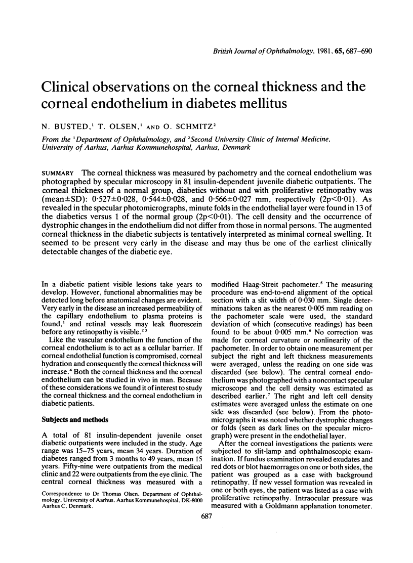
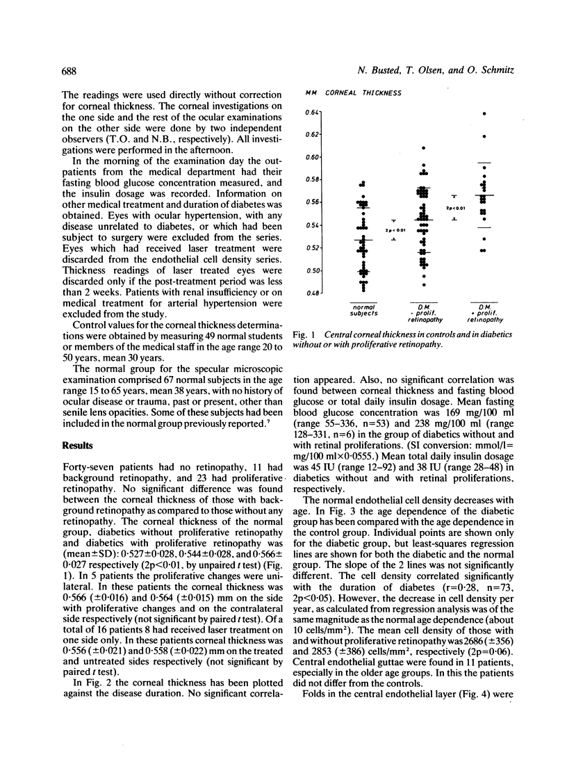
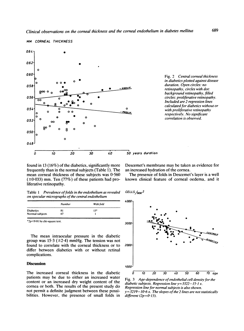
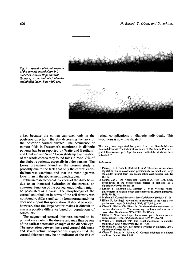
The corneal thickness was measured by pachometry and the corneal endothelium was photographed by specular microscopy in 81 insulin-dependent juvenile diabetic outpatients. The corneal thickness of a normal group, diabetics without and with proliferative retinopathy was (mean +/- SD): 0.527 +/- 0.028, 0.544 +/- 0.028, and 0.566 +/- 0.027 mm, respectively (2p less than 0.01). As revealed in the specular photomicrographs, minute folds in the endothelial layer were found in 13 of the diabetics versus 1 of the normal group (2p less than 0.01). The cell density and the occurrence of dystrophic changes in the endothelium did not differ from those in normal persons. The augmented corneal thickness in the diabetic subjects is tentatively interpreted as minimal corneal swelling. It seemed to be present very early in the disease and may thus be one of the earliest clinically detectable changes off the diabetic eye.
Full text
PDF


Images in this article
Selected References
These references are in PubMed. This may not be the complete list of references from this article.
- Cunha-Vaz J., Faria de Abreu J. R., Campos A. J. Early breakdown of the blood-retinal barrier in diabetes. Br J Ophthalmol. 1975 Nov;59(11):649–656. doi: 10.1136/bjo.59.11.649. [DOI] [PMC free article] [PubMed] [Google Scholar]
- Ehlers N., Sperling S. A technical improvement of the Haag-Streit pachometer. Short communication. Acta Ophthalmol (Copenh) 1977 Apr;55(2):333–336. doi: 10.1111/j.1755-3768.1977.tb01314.x. [DOI] [PubMed] [Google Scholar]
- HENKIND P., WISE G. N. Descemet's wrinkles in diabetes. Am J Ophthalmol. 1961 Sep;52:371–374. doi: 10.1016/0002-9394(61)90736-x. [DOI] [PubMed] [Google Scholar]
- Krupin T., Waltman S. R., Oestrich C., Santiago J., Ratzan S., Kilo C., Becker B. Vitreous fluorophotometry in juvenile-onset diabetes mellitus. Arch Ophthalmol. 1978 May;96(5):812–814. doi: 10.1001/archopht.1978.03910050418002. [DOI] [PubMed] [Google Scholar]
- Mishima S. Corneal thickness. Surv Ophthalmol. 1968 Sep;13(2):57–96. [PubMed] [Google Scholar]
- Olsen T., Busted N., Schmitz O. Corneal thickness in diabetes mellitus. Lancet. 1980 Apr 19;1(8173):883–883. doi: 10.1016/s0140-6736(80)91389-6. [DOI] [PubMed] [Google Scholar]
- Olsen T., Nielsen C. B., Ehlers N. On the optical measurement of corneal thickness. II. The measuring conditions and sources of error. Acta Ophthalmol (Copenh) 1980 Dec;58(6):975–984. doi: 10.1111/j.1755-3768.1980.tb08325.x. [DOI] [PubMed] [Google Scholar]
- Olsen T. Non-contact specular microscopy of human corneal endothelium. Acta Ophthalmol (Copenh) 1979;57(6):986–998. doi: 10.1111/j.1755-3768.1979.tb00529.x. [DOI] [PubMed] [Google Scholar]
- Parving H. H., Noer I., Deckert T., Evrin P. E., Nielsen S. L., Lyngsoe J., Mogensen C. E., Rorth M., Svendsen P. A., Trap-Jensen J. The effect of metabolic regulation on microvascular permeability to small and large molecules in short-term juvenile diabetics. Diabetologia. 1976 May;12(2):161–166. doi: 10.1007/BF00428983. [DOI] [PubMed] [Google Scholar]


