Abstract
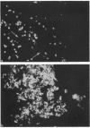
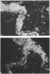
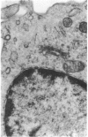
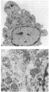
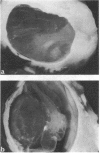
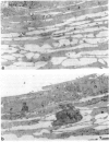
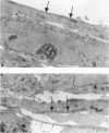
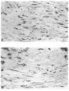
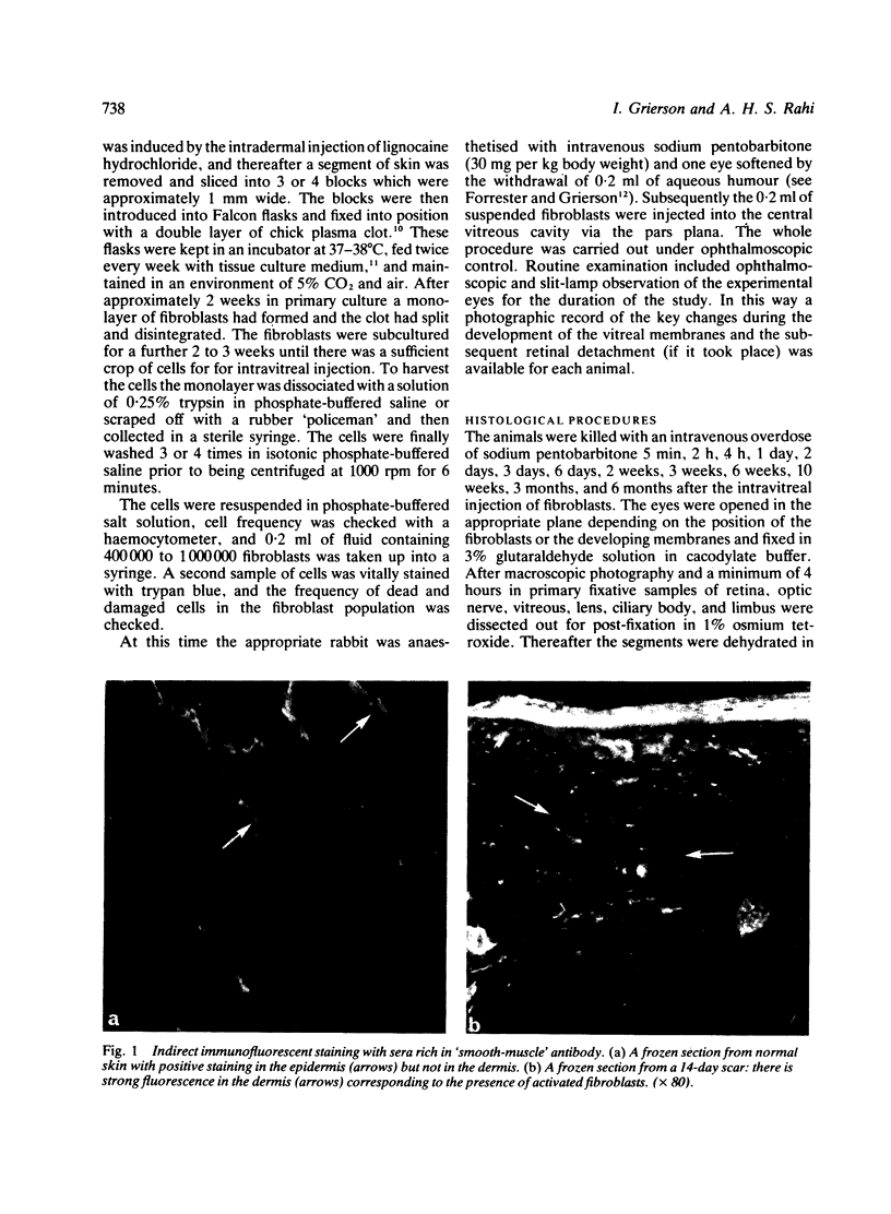
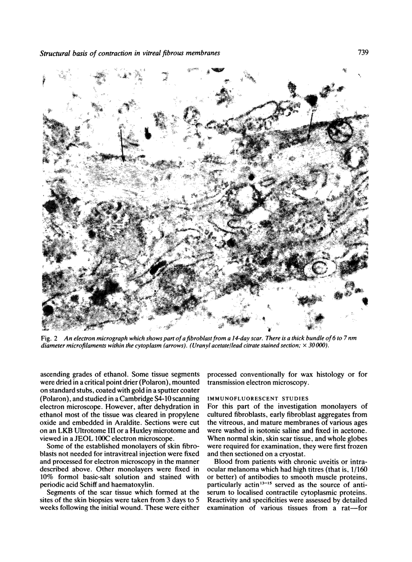
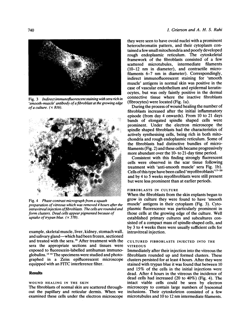
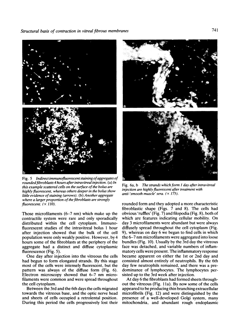
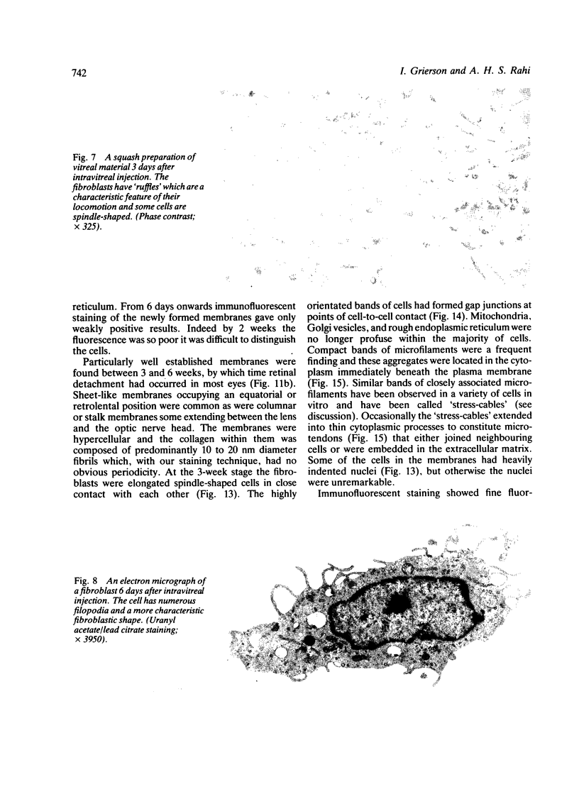
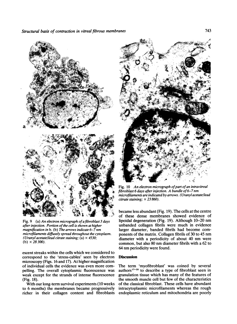
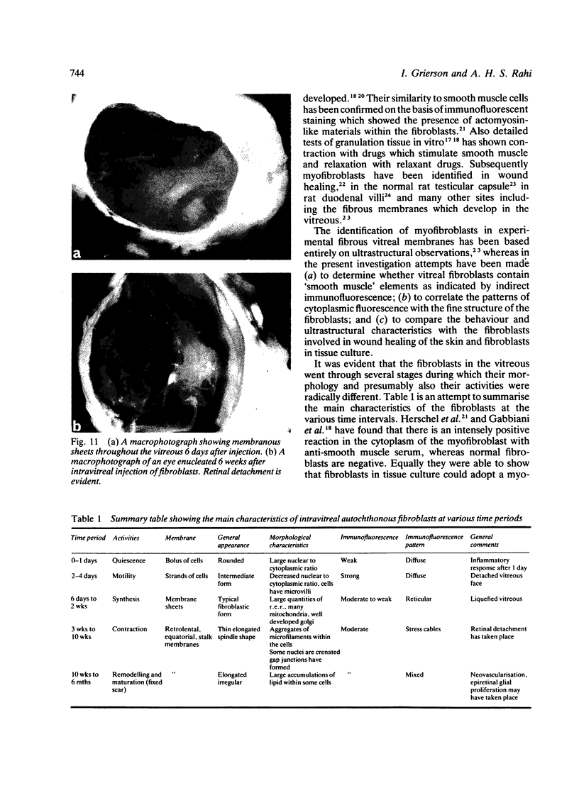
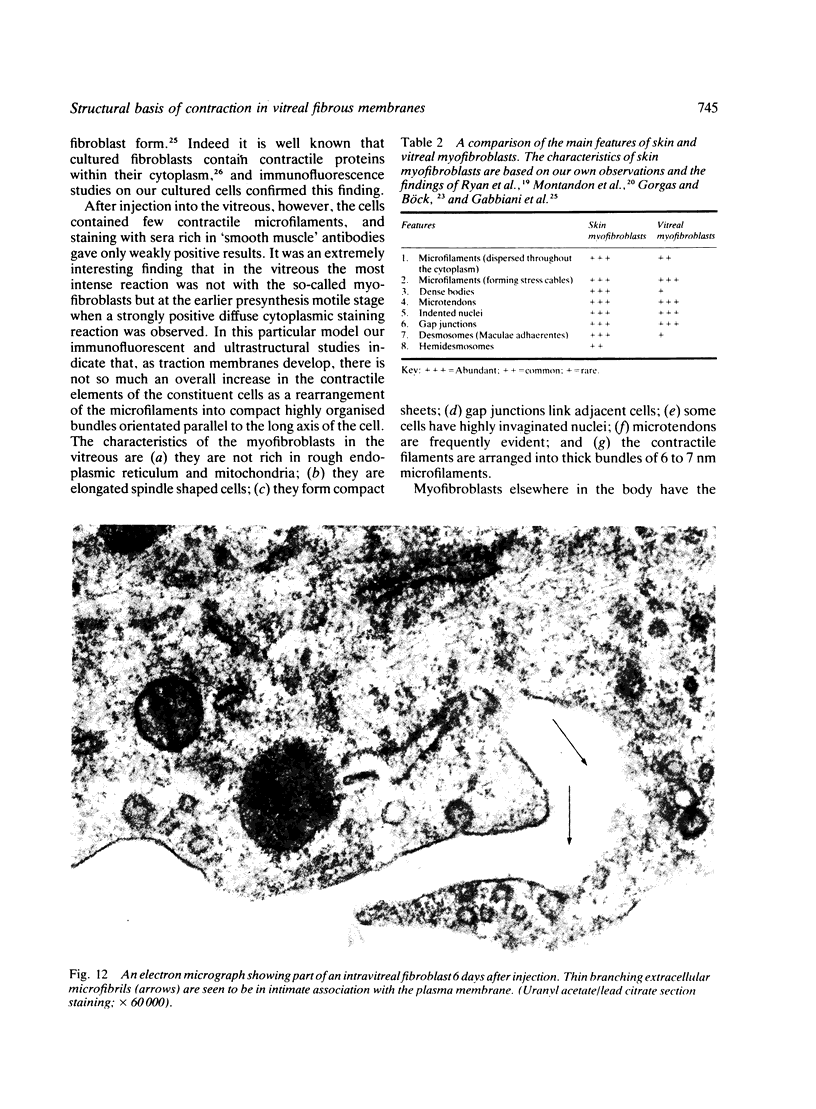
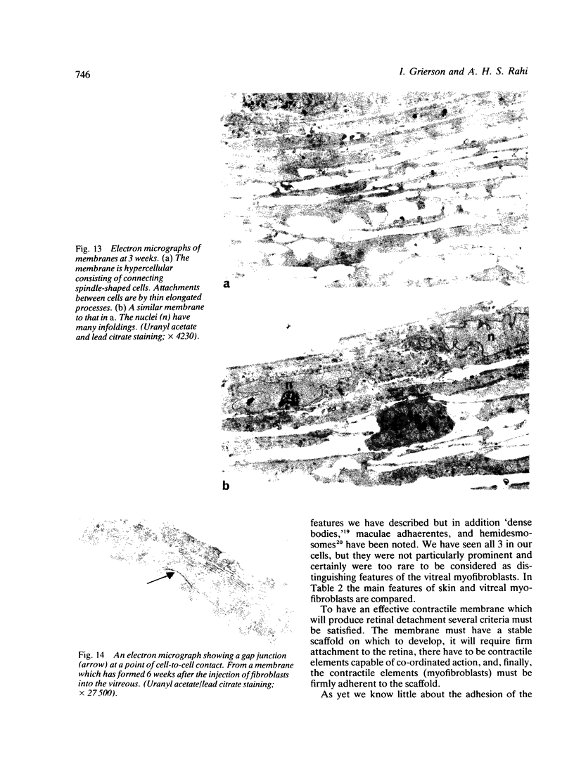
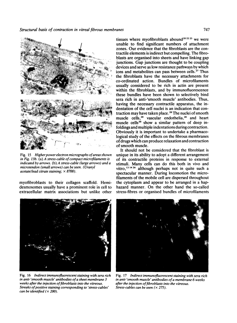
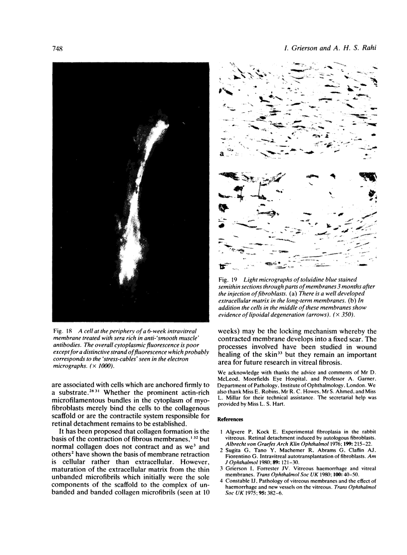
A combined ultrastructural and immunofluorescent study was conducted on experimentally induced fibrous membranes in the vitreous of adult rabbits. Autochthonous tissue cultured fibroblasts were injected into the mid-vitreous of one eye of each of 25 rabbits. The animals were monitored routinely with an ophthalmoscope and slit-lamp and were killed at various time periods between 5 minutes and 6 months. Appropriate tissue was taken for light microscopy, transmission electron microscopy, scanning electron microscopy, and indirect immunofluorescence. With this model we were able to show that the contractile elements in fibrous membranes are probably modified fibroblasts called myofibroblasts which are most abundant 3 to 6 weeks after injection. This is the time when retinal detachment usually occurs. It is our impression that, as traction membranes develop, there is not so much an increase in the contractile elements of the constituent cells as a rearrangement of the existing cytoplasmic microfilaments into compact highly organised bundles called stress cables. The behaviour and ultrastructural characteristics of intravitreal fibroblasts compare with the action of fibroblasts in the healing of wounds.
Full text
PDF
Images in this article
Selected References
These references are in PubMed. This may not be the complete list of references from this article.
- Algvere P., Kock E. Experimental Fibroplasia in the rabbit vitreous. Retinal detachment induced by autologous fibroblasts. Albrecht Von Graefes Arch Klin Exp Ophthalmol. 1976 May 26;199(3):215–222. doi: 10.1007/BF00417290. [DOI] [PubMed] [Google Scholar]
- Bloom S., Cancilla P. A. Conformational changes in myocardial nuclei of rats. Circ Res. 1969 Feb;24(2):189–196. doi: 10.1161/01.res.24.2.189. [DOI] [PubMed] [Google Scholar]
- Coles W. H., Haik G. M. Vitrectomy in intraocular trauma. Its rationale and its indications and limitations. Arch Ophthalmol. 1972 Jun;87(6):621–628. doi: 10.1001/archopht.1972.01000020623002. [DOI] [PubMed] [Google Scholar]
- Constable I. J. Pathology of vitreous membranes and the effect of haemorrhage and new vessels on the vitreous. Trans Ophthalmol Soc U K. 1975;95(3):382–386. [PubMed] [Google Scholar]
- Forrester J. V., Grierson I. The cellular response to blood in the vitreous: an ultrastructural study. J Pathol. 1979 Sep;129(1):43–52. doi: 10.1002/path.1711290108. [DOI] [PubMed] [Google Scholar]
- Gabbiani G., Hirschel B. J., Ryan G. B., Statkov P. R., Majno G. Granulation tissue as a contractile organ. A study of structure and function. J Exp Med. 1972 Apr 1;135(4):719–734. doi: 10.1084/jem.135.4.719. [DOI] [PMC free article] [PubMed] [Google Scholar]
- Gilula N. B., Reeves O. R., Steinbach A. Metabolic coupling, ionic coupling and cell contacts. Nature. 1972 Feb 4;235(5336):262–265. doi: 10.1038/235262a0. [DOI] [PubMed] [Google Scholar]
- Gorgas K., Böck P. Myofibroblasts in the rat testicular capsule. Cell Tissue Res. 1974;154(4):533–541. doi: 10.1007/BF00219672. [DOI] [PubMed] [Google Scholar]
- Grierson I., Rahi A. H. Microfilaments in the cells of the human trabecular meshwork. Br J Ophthalmol. 1979 Jan;63(1):3–8. doi: 10.1136/bjo.63.1.3. [DOI] [PMC free article] [PubMed] [Google Scholar]
- Grierson I., Robins E., Howes R. C. Preliminary observations on human trabecular meshwork cells in vitro. Albrecht Von Graefes Arch Klin Exp Ophthalmol. 1980;212(3-4):173–186. doi: 10.1007/BF00410513. [DOI] [PubMed] [Google Scholar]
- Güldner F. H., Wolff J. R., Keyserlingk D. G. Fibroblasts as a part of the contractile system in duodenal villi of rat. Z Zellforsch Mikrosk Anat. 1972;135(3):349–360. doi: 10.1007/BF00307181. [DOI] [PubMed] [Google Scholar]
- Hirschel B. J., Gabbiani G., Ryan G. B., Majno G. Fibroblasts of granulation tissue: immunofluorescent staining with antismooth muscle serum. Proc Soc Exp Biol Med. 1971 Nov;138(2):466–469. doi: 10.3181/00379727-138-35920. [DOI] [PubMed] [Google Scholar]
- Lane B. P. Alterations in the cytologic detail of intestinal smooth muscle cells in various stages of contraction. J Cell Biol. 1965 Oct;27(1):199–213. doi: 10.1083/jcb.27.1.199. [DOI] [PMC free article] [PubMed] [Google Scholar]
- Lazarides E. Immunofluorescence studies on the structure of actin filaments in tissue culture cells. J Histochem Cytochem. 1975 Jul;23(7):507–528. doi: 10.1177/23.7.1095651. [DOI] [PubMed] [Google Scholar]
- Machemer R. Massive periretinal proliferation: a logical approach to therapy. Trans Am Ophthalmol Soc. 1977;75:556–586. [PMC free article] [PubMed] [Google Scholar]
- Majno G., Gabbiani G., Hirschel B. J., Ryan G. B., Statkov P. R. Contraction of granulation tissue in vitro: similarity to smooth muscle. Science. 1971 Aug 6;173(3996):548–550. doi: 10.1126/science.173.3996.548. [DOI] [PubMed] [Google Scholar]
- Majno G., Shea S. M., Leventhal M. Endothelial contraction induced by histamine-type mediators: an electron microscopic study. J Cell Biol. 1969 Sep;42(3):647–672. doi: 10.1083/jcb.42.3.647. [DOI] [PMC free article] [PubMed] [Google Scholar]
- Montandon D., Gabbiani G., Ryan G. B., Majno G. The contractile fibroblast. Its relevance in plastic surgery. Plast Reconstr Surg. 1973 Sep;52(3):286–290. [PubMed] [Google Scholar]
- Rahi A. H., Grierson I., Ashton N. Contractile proteins in ocular tissues. Their role in health and disease. Trans Ophthalmol Soc U K. 1980 Apr;100(Pt 1):155–161. [PubMed] [Google Scholar]
- Rahi A., Ashton N. Contractile proteins in retinal endothelium and other non-muscle tissues of the eye. Br J Ophthalmol. 1978 Sep;62(9):627–643. doi: 10.1136/bjo.62.9.627. [DOI] [PMC free article] [PubMed] [Google Scholar]
- Rudolph R., Guber S., Suzuki M., Woodward M. The life cycle of the myofibroblast. Surg Gynecol Obstet. 1977 Sep;145(3):389–394. [PubMed] [Google Scholar]
- Ryan G. B., Cliff W. J., Gabbiani G., Irlé C., Montandon D., Statkov P. R., Majno G. Myofibroblasts in human granulation tissue. Hum Pathol. 1974 Jan;5(1):55–67. doi: 10.1016/s0046-8177(74)80100-0. [DOI] [PubMed] [Google Scholar]
- Squier C. A., Kremenak C. R. Myofibroblasts in healing palatal wounds of the beagle dog. J Anat. 1980 May;130(Pt 3):585–594. [PMC free article] [PubMed] [Google Scholar]
- Sugita G., Tano Y., Machemer R., Abrams G., Claflin A., Fiorentino G. Intravitreal autotransplantation of fibroblasts. Am J Ophthalmol. 1980 Jan;89(1):121–130. doi: 10.1016/0002-9394(80)90238-x. [DOI] [PubMed] [Google Scholar]
- Trenchev P., Holborow E. J. The specificity of anti-actin serum. Immunology. 1976 Oct;31(4):509–517. [PMC free article] [PubMed] [Google Scholar]
- Winthrop S. R., Cleary P. E., Minckler D. S., Ryan S. J. Penetrating eye injuries: a histopathological review. Br J Ophthalmol. 1980 Nov;64(11):809–817. doi: 10.1136/bjo.64.11.809. [DOI] [PMC free article] [PubMed] [Google Scholar]


