Abstract
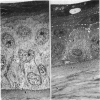
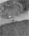
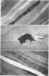
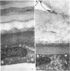
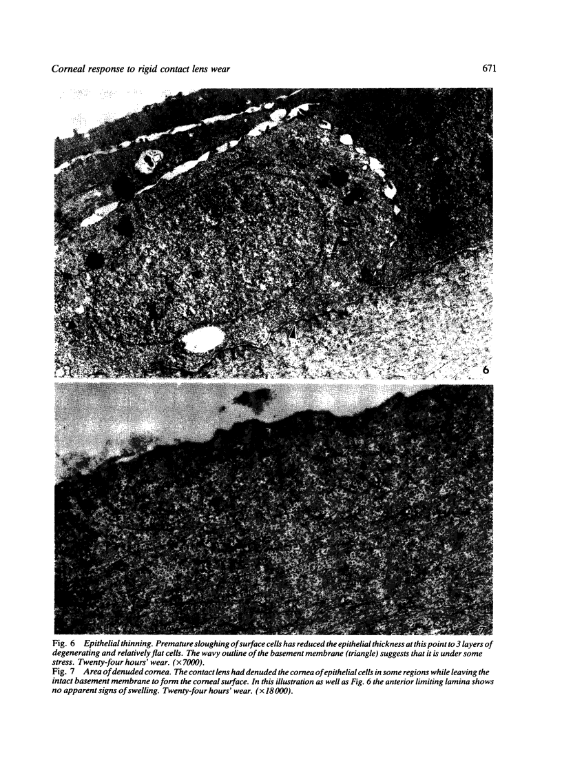
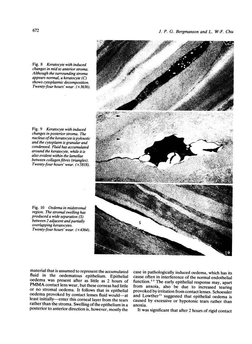
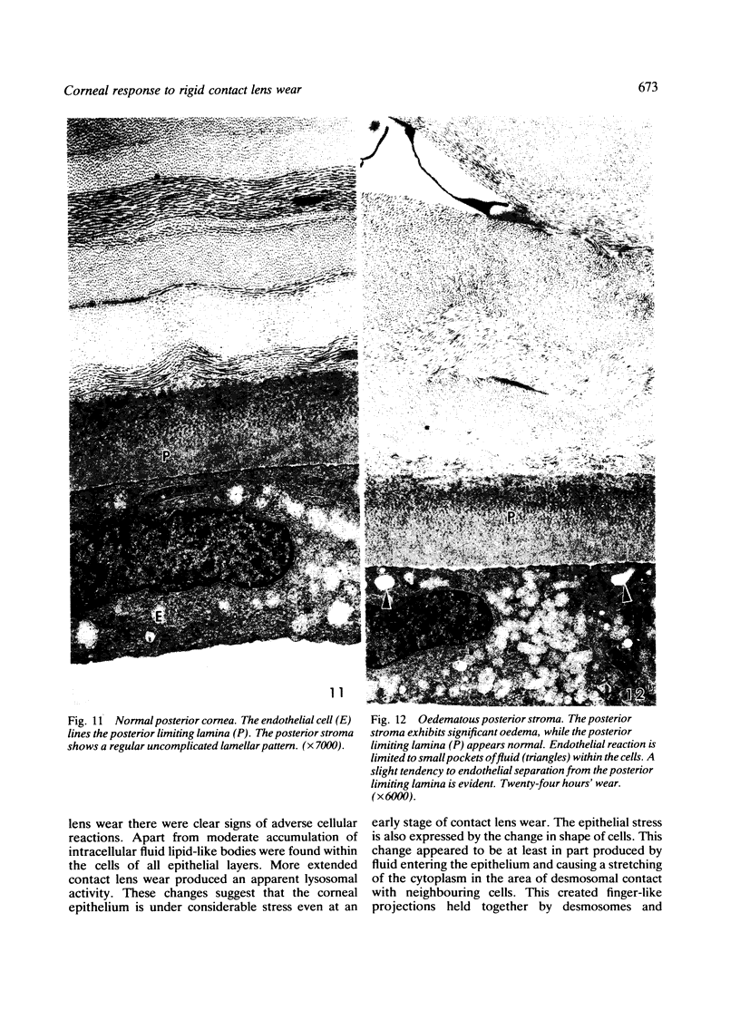
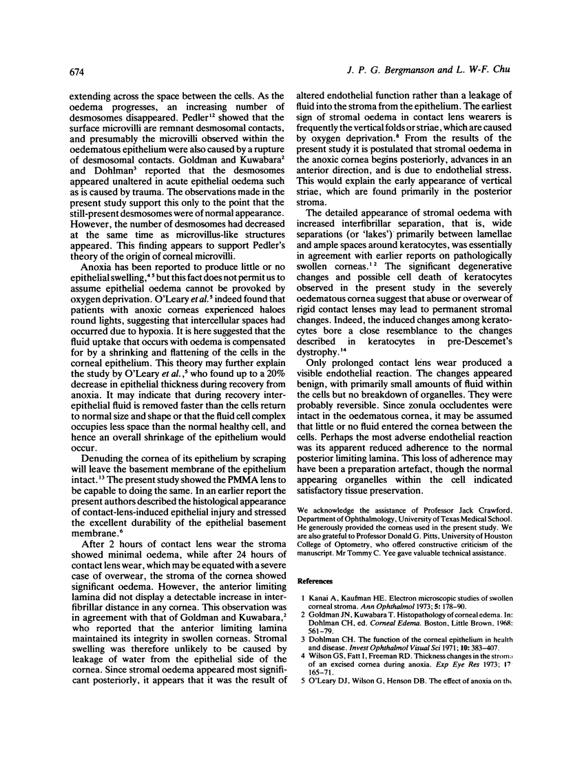
Three adult rhesus monkeys were subjected to 2 and 24 hours of polymethylmethacrylate (PMMA) contact lens wear. The induced corneal changes were examined with the electron microscope. Mild epithelial oedema as well as early degenerative cell changes was present already after 2 hours' wear. Rigid lens wear for 24 hours produced more severe oedema and cell alterations together with premature cell loss and ultimately, in areas of lens bearing, corneal denuding. Only the monkeys wearing contact lenses for 24 hours had significant stromal swelling, which was primarily evident in the posterior region, while the anterior limiting lamina remained unaffected. The stromal swelling was patchy and mainly around keratocytes and between lamellae, while fluid within the lamellae was evident only occasionally in posterior stroma. Changes among keratocytes were evident, especially posteriorly, where reaction was frequently severe. Endothelial reaction was restricted to a limited fluid uptake in the 24-hour-wear experiment. In addition there was in these monkeys an apparent loosening of the endothelial adhesion to the posterior limiting lamina. It is concluded that the oedematous epithelium undergoes cell shrinkage and flattening, which is compensated for by an uptake of fluid. The uptake of fluid maintains the overall normal thickness of the epithelium. The conclusion is supported by other studies, where the normal thickness of oedematous epithelium has been shown by pachometry. The results in the present study further suggest that stromal oedema in the contact lens wearer is a result of a relative loss of endothelial function, leading to a swelling that moves in a posterior to anterior direction.
Full text
PDF
Images in this article
Selected References
These references are in PubMed. This may not be the complete list of references from this article.
- Curran R. E., Kenyon K. R., Green W. R. Pre-Descemet's membrane corneal dystrophy. Am J Ophthalmol. 1974 May;77(5):711–716. doi: 10.1016/0002-9394(74)90536-4. [DOI] [PubMed] [Google Scholar]
- Dohlman C. H. The function of the corneal epithelium in health and disease. The Jonas S. Friedenwald Memorial Lecture. Invest Ophthalmol. 1971 Jun;10(6):383–407. [PubMed] [Google Scholar]
- Hirst L. W., Kenyon K. R., Fogle J., Stark W. J. Corneal epithelial adhesion studies. Aust J Ophthalmol. 1981 May;9(2):103–111. doi: 10.1111/j.1442-9071.1981.tb01497.x. [DOI] [PubMed] [Google Scholar]
- Kanai A., Kaufman H. E. Electron microscopic studies of swollen corneal stroma. Ann Ophthalmol. 1973 Feb;5(2):178–190. [PubMed] [Google Scholar]
- O'Leary D. J., Wilson G., Henson D. B. The effect of anoxia on the human corneal epithelium. Am J Optom Physiol Opt. 1981 Jun;58(6):472–476. [PubMed] [Google Scholar]
- PEDLER C. The fine structure of the corneal epithelium. Exp Eye Res. 1962 Mar;1:286–289. doi: 10.1016/s0014-4835(62)80012-8. [DOI] [PubMed] [Google Scholar]
- Polse K. A., Mandell R. B. Etiology of corneal striae accompanying hydrogel lens wear. Invest Ophthalmol. 1976 Jul;15(7):553–556. [PubMed] [Google Scholar]
- Schoessler J. P., Lowther G. E. Slit lamp observations of corneal edema. Am J Optom Arch Am Acad Optom. 1971 Aug;48(8):666–671. doi: 10.1097/00006324-197108000-00007. [DOI] [PubMed] [Google Scholar]
- Wilson G. S., Fatt I., Freeman R. D. Thickness changes in the stroma of an excised cornea during anoxia. Exp Eye Res. 1973 Oct 24;17(2):165–171. doi: 10.1016/0014-4835(73)90206-6. [DOI] [PubMed] [Google Scholar]


