Abstract
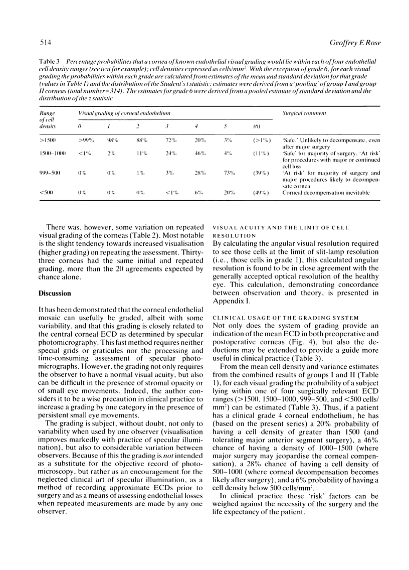
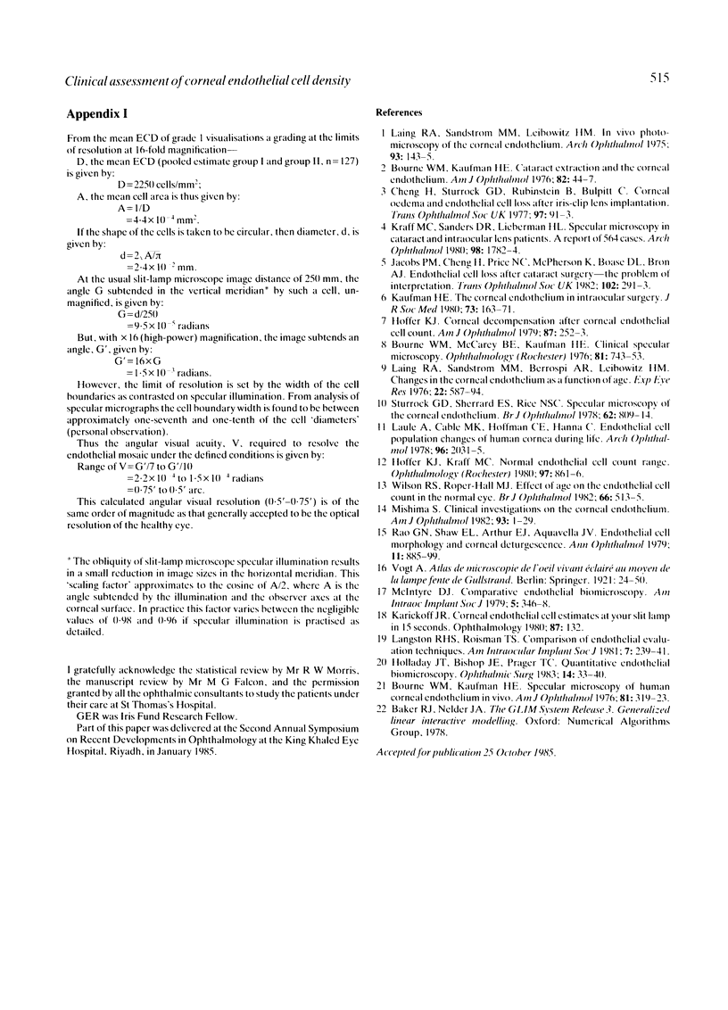
An original system of grading of the corneal endothelial specular reflection, as assessed with a Haag-Streit 900 slit-lamp biomicroscope, has been shown to have a very highly significant relation to the endothelial cell density measured by contact specular photomicroscopy. The grading, though subjective and therefore not a substitute for the detailed record of photomicroscopy, is readily applicable to clinical practice and is a useful method of comparing and recording endothelial cell densities when assessed by the same observer. The intraobserver, but not the interobserver, variability has been examined, and the theoretical aspects of the grading system and a clinically applicable interpretation of the grading are presented.
Full text
PDF
Selected References
These references are in PubMed. This may not be the complete list of references from this article.
- Bourne W. M., Kaufman H. E. Specular microscopy of human corneal endothelium in vivo. Am J Ophthalmol. 1976 Mar;81(3):319–323. doi: 10.1016/0002-9394(76)90247-6. [DOI] [PubMed] [Google Scholar]
- Bourne W. M., McCarey B. E., Kaufman H. E. Clinical specular microscopy. Trans Sect Ophthalmol Am Acad Ophthalmol Otolaryngol. 1976 Sep-Oct;81(5):743–753. [PubMed] [Google Scholar]
- Kraff M. C., Sanders D. R., Lieberman H. L. Specular microscopy in cataract and intraocular lens patients. A report of 564 cases. Arch Ophthalmol. 1980 Oct;98(10):1782–1784. doi: 10.1001/archopht.1980.01020040634009. [DOI] [PubMed] [Google Scholar]
- Laing R. A., Sandstrom M. M., Leibowitz H. M. In vivo photomicrography of the corneal endothelium. Arch Ophthalmol. 1975 Feb;93(2):143–145. doi: 10.1001/archopht.1975.01010020149013. [DOI] [PubMed] [Google Scholar]
- Laing R. A., Sanstrom M. M., Berrospi A. R., Leibowitz H. M. Changes in the corneal endothelium as a function of age. Exp Eye Res. 1976 Jun;22(6):587–594. doi: 10.1016/0014-4835(76)90003-8. [DOI] [PubMed] [Google Scholar]
- Langston R. H., Roisman T. S. Comparison of endothelial evaluation techniques. J Am Intraocul Implant Soc. 1981 Summer;7(3):239–241. doi: 10.1016/s0146-2776(81)80004-3. [DOI] [PubMed] [Google Scholar]
- McIntyre D. J. Comparative endothelial biomicroscopy. J Am Intraocul Implant Soc. 1979 Oct;5(4):346–348. doi: 10.1016/s0146-2776(79)80101-9. [DOI] [PubMed] [Google Scholar]
- Mishima S. Clinical investigations on the corneal endothelium-XXXVIII Edward Jackson Memorial Lecture. Am J Ophthalmol. 1982 Jan;93(1):1–29. doi: 10.1016/0002-9394(82)90693-6. [DOI] [PubMed] [Google Scholar]
- Rao G. N., Shaw E. L., Arthur E. J., Aquavella J. V. Endothelial cell morphology and corneal deturgescence. Ann Ophthalmol. 1979 Jun;11(6):885–899. [PubMed] [Google Scholar]
- Wilson R. S., Roper-Hall M. J. Effect of age on the endothelial cell count in the normal eye. Br J Ophthalmol. 1982 Aug;66(8):513–515. doi: 10.1136/bjo.66.8.513. [DOI] [PMC free article] [PubMed] [Google Scholar]


