Abstract
The literature suggests that posterior polymorphous dystrophy (PPD) may show features such as iridocorneal adhesions, glassy membranes, and pupillary ectropion which are typically ascribed to the iridocorneal endothelial (ICE) syndrome. This complicates diagnosis. PPD, unlike ICE, is familial, and ICE, unlike PPD, is usually progressive and frequently complicated by glaucoma: thus it is important to distinguish between them. To determine whether this could be achieved by specular microscopy, since the posterior corneal surface is abnormal in both conditions, 57 cases of ICE and 44 of PPD were repeatedly examined and photographed with the specular microscope. Progressive and/or static morphological features of the corneal endothelium and Descemet's membrane were found that were specific for each condition. Specular microscopy can thus provide a definitive diagnosis of ICE or PPD even in uncertain cases.
Full text
PDF
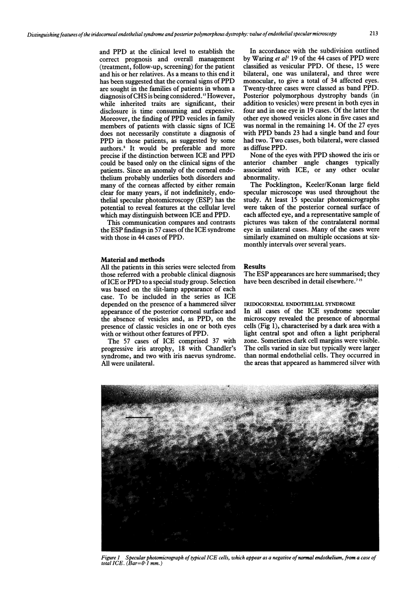
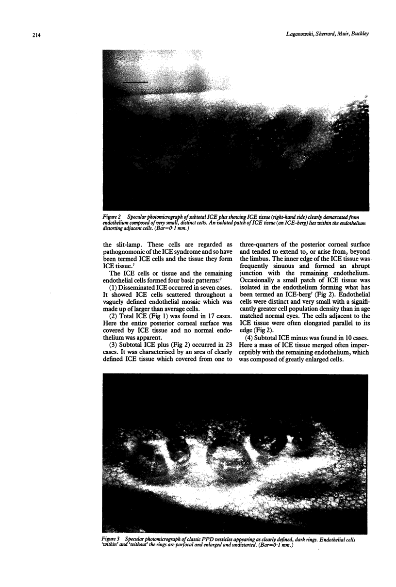
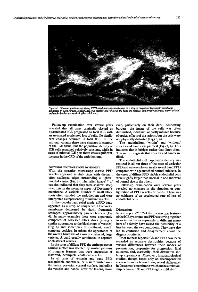
Images in this article
Selected References
These references are in PubMed. This may not be the complete list of references from this article.
- Alvarado J. A., Murphy C. G., Juster R. P., Hetherington J. Pathogenesis of Chandler's syndrome, essential iris atrophy and the Cogan-Reese syndrome. II. Estimated age at disease onset. Invest Ophthalmol Vis Sci. 1986 Jun;27(6):873–882. [PubMed] [Google Scholar]
- Boruchoff S. A., Kuwabara T. Electron microscopy of posterior polymorphous degeneration. Am J Ophthalmol. 1971 Nov;72(5):879–887. doi: 10.1016/0002-9394(71)91683-7. [DOI] [PubMed] [Google Scholar]
- Boruchoff S. A., Weiner M. J., Albert D. M. Recurrence of posterior polymorphous corneal dystrophy after penetrating keratoplasty. Am J Ophthalmol. 1990 Mar 15;109(3):323–328. doi: 10.1016/s0002-9394(14)74559-3. [DOI] [PubMed] [Google Scholar]
- Bourne W. M. Partial corneal involvement in the iridocorneal endothelial syndrome. Am J Ophthalmol. 1982 Dec;94(6):774–781. doi: 10.1016/0002-9394(82)90302-6. [DOI] [PubMed] [Google Scholar]
- CHANDLER P. A. Atrophy of the stroma of the iris; endothelial dystrophy, corneal edema, and glaucoma. Am J Ophthalmol. 1956 Apr;41(4):607–615. [PubMed] [Google Scholar]
- Cibis G. W., Krachmer J. A., Phelps C. D., Weingeist T. A. The clinical spectrum of posterior polymorphous dystrophy. Arch Ophthalmol. 1977 Sep;95(9):1529–1537. doi: 10.1001/archopht.1977.04450090051002. [DOI] [PubMed] [Google Scholar]
- Cibis G. W., Krachmer J. H., Phelps C. D., Weingeist T. A. Iridocorneal adhesions in posterior polymorphous dystrophy. Trans Sect Ophthalmol Am Acad Ophthalmol Otolaryngol. 1976 Sep-Oct;81(5):770–777. [PubMed] [Google Scholar]
- Hartmann C. Die "indirekte"Spiegelmikroskopie. Eine neue Technik zur In-vivo-Untersuchung der Hornhautrückfläche. Ophthalmologica. 1981;183(4):177–186. doi: 10.1159/000309163. [DOI] [PubMed] [Google Scholar]
- Hirst L. W., Quigley H. A., Stark W. J., Shields M. B. Specular microscopy of iridocorneal endothelia syndrome. Am J Ophthalmol. 1980 Jan;89(1):11–21. doi: 10.1016/0002-9394(80)90223-8. [DOI] [PubMed] [Google Scholar]
- Hirst L. W., Waring G. O., 3rd Clinical specular microscopy of posterior polymorphous endothelial dystrophy. Am J Ophthalmol. 1983 Feb;95(2):143–155. doi: 10.1016/0002-9394(83)90007-7. [DOI] [PubMed] [Google Scholar]
- Neubauer L., Lund O. E., Leibowitz H. M. Specular microscopic appearance of the corneal endothelium in iridocorneal endothelial syndrome. Arch Ophthalmol. 1983 Jun;101(6):916–918. doi: 10.1001/archopht.1983.01040010916012. [DOI] [PubMed] [Google Scholar]
- Sherrard E. S., Buckley R. J. The relief mode. New application of the corneal specular microscope. Arch Ophthalmol. 1982 Feb;100(2):296–300. doi: 10.1001/archopht.1982.01030030298014. [DOI] [PubMed] [Google Scholar]
- Sherrard E. S., Frangoulis M. A., Muir M. G., Buckley R. J. The posterior surface of the cornea in the irido-corneal endothelial syndrome: a specular microscopical study. Trans Ophthalmol Soc U K. 1985;104(Pt 7):766–774. [PubMed] [Google Scholar]
- Shields M. B. Progressive essential iris atrophy, Chandler's syndrome, and the iris nevus (Cogan-Reese) syndrome: a spectrum of disease. Surv Ophthalmol. 1979 Jul-Aug;24(1):3–20. doi: 10.1016/0039-6257(79)90143-7. [DOI] [PubMed] [Google Scholar]
- Waring G. O., 3rd, Bourne W. M., Edelhauser H. F., Kenyon K. R. The corneal endothelium. Normal and pathologic structure and function. Ophthalmology. 1982 Jun;89(6):531–590. [PubMed] [Google Scholar]
- Waring G. O., 3rd, Rodrigues M. M., Laibson P. R. Corneal dystrophies. II. Endothelial dystrophies. Surv Ophthalmol. 1978 Nov-Dec;23(3):147–168. doi: 10.1016/0039-6257(78)90151-0. [DOI] [PubMed] [Google Scholar]


