Abstract
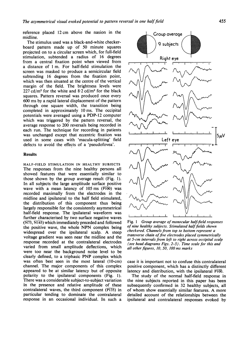
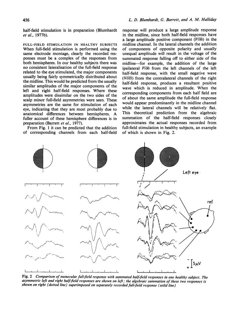
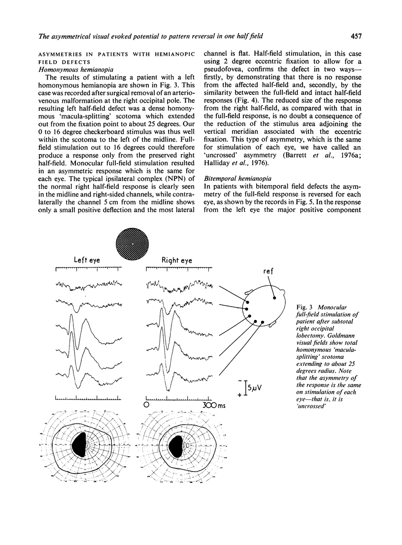
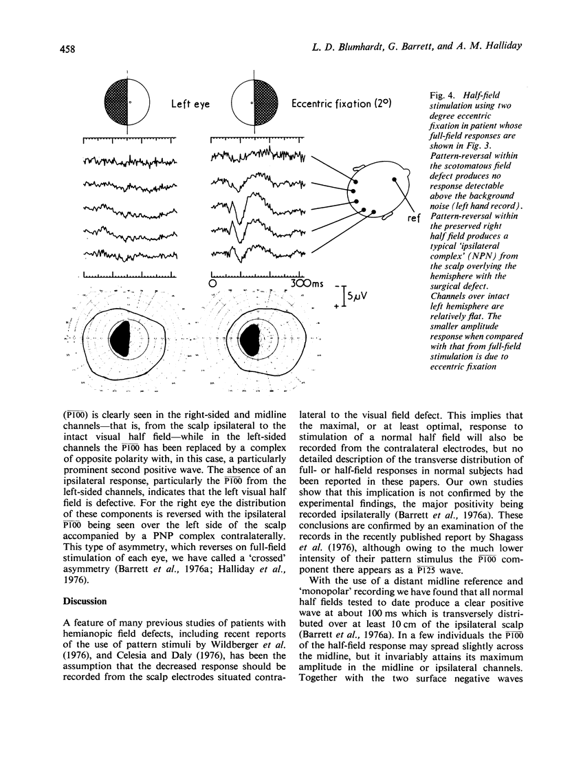
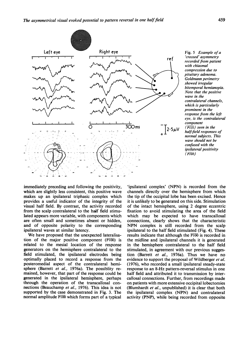
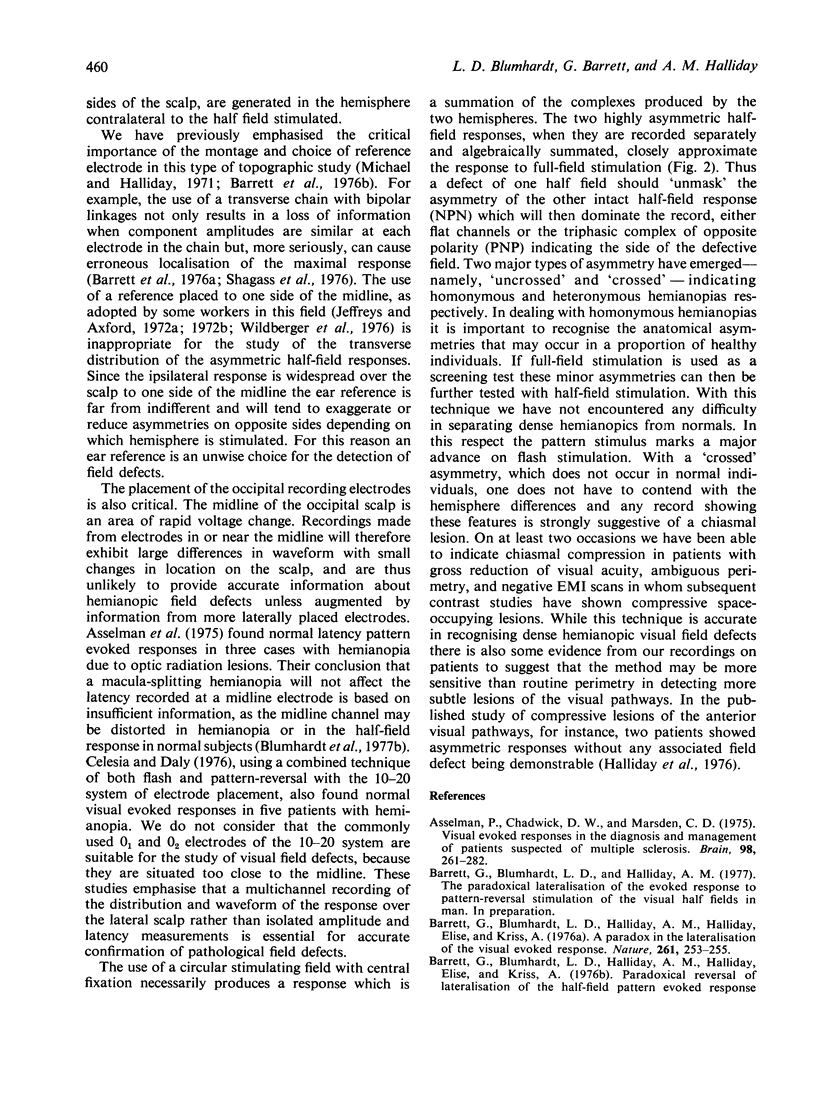
The consistency of the major positive component (P100) of the full-field pattern-reversal response provides a clinically valuable and objective means of detecting visual field defects. Its normally symmetrical distribution about the midline of the occipital scalp results from the summation of two highly asymmetric half-field responses, each of which shows the positive component well lateralised with a widespread distribution on the ipsilateral side. Stimulation of each eye in patients with bitemporal and homonymous hemianopias results in two characteristic patterns of asymmetry, named 'crossed' and 'uncrossed' respectively, in which the major positivity is consistently recorded on the side ipsilateral to the preserved half field. Recordings from a patient after occipital lobectomy confirm the authors' previous suggestion that although the major positive component is recorded from the ipsilateral scalp the typical asymmetric half-field response is generated in the contralateral hemisphere.
Full text
PDF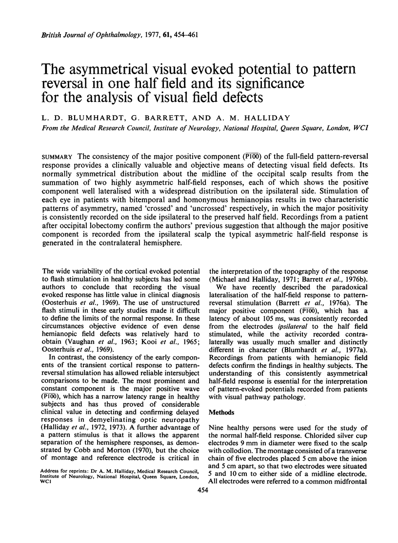
Selected References
These references are in PubMed. This may not be the complete list of references from this article.
- Asselman P., Chadwick D. W., Marsden D. C. Visual evoked responses in the diagnosis and management of patients suspected of multiple sclerosis. Brain. 1975 Jun;98(2):261–282. doi: 10.1093/brain/98.2.261. [DOI] [PubMed] [Google Scholar]
- Barett G., Blumhardt L., Halliday A. M., Halliday E., Kriss A. A paradox in the lateralisation of the visual evoked response. Nature. 1976 May 20;261(5557):253–255. doi: 10.1038/261253a0. [DOI] [PubMed] [Google Scholar]
- Barett G., Blumhardt L., Halliday A. M., Halliday E., Kriss A. A paradox in the lateralisation of the visual evoked response. Nature. 1976 May 20;261(5557):253–255. doi: 10.1038/261253a0. [DOI] [PubMed] [Google Scholar]
- Beauchamp M., Matthews W. B., Small D., Stein J. F. The topography of the visual evoked responses to half field stimulation [proceedings]. J Physiol. 1976 Sep;260(2):46P–47P. [PubMed] [Google Scholar]
- Cobb W. A., Morton H. B. Evoked potentials from the human scalp to visual half-field stimulation. J Physiol. 1970 Jun;208(2):39P–40P. [PMC free article] [PubMed] [Google Scholar]
- Halliday A. M., Halliday E., Kriss A., McDonald W. I., Mushin J. The pattern-evoked potential in compression of the anterior visual pathways. Brain. 1976 Jun;99(2):357–374. doi: 10.1093/brain/99.2.357. [DOI] [PubMed] [Google Scholar]
- Halliday A. M., McDonald W. I., Mushin J. Delayed visual evoked response in optic neuritis. Lancet. 1972 May 6;1(7758):982–985. doi: 10.1016/s0140-6736(72)91155-5. [DOI] [PubMed] [Google Scholar]
- Halliday A. M., McDonald W. I., Mushin J. Visual evoked response in diagnosis of multiple sclerosis. Br Med J. 1973 Dec 15;4(5893):661–664. doi: 10.1136/bmj.4.5893.661. [DOI] [PMC free article] [PubMed] [Google Scholar]
- Jeffreys D. A., Axford J. G. Source locations of pattern-specific components of human visual evoked potentials. I. Component of striate cortical origin. Exp Brain Res. 1972;16(1):1–21. doi: 10.1007/BF00233371. [DOI] [PubMed] [Google Scholar]
- Jeffreys D. A., Axford J. G. Source locations of pattern-specific components of human visual evoked potentials. II. Component of extrastriate cortical origin. Exp Brain Res. 1972;16(1):22–40. doi: 10.1007/BF00233372. [DOI] [PubMed] [Google Scholar]
- KOOI K. A., GUEVENER A. M., BAGCHI B. K. VISUAL EVOKED RESPONSES IN LESIONS OF THE HIGHER OPTIC PATHWAYS. Neurology. 1965 Sep;15:841–854. doi: 10.1212/wnl.15.9.841. [DOI] [PubMed] [Google Scholar]
- Michael W. F., Halliday A. M. Differences between the occipital distribution of upper and lower field pattern-evoked responses in man. Brain Res. 1971 Sep 24;32(2):311–324. doi: 10.1016/0006-8993(71)90327-1. [DOI] [PubMed] [Google Scholar]
- Oosterhuis H. J., Ponsen L., Jonkman E. J., Magnus O. The average visual response in patients with cerebrovascular disease. Electroencephalogr Clin Neurophysiol. 1969 Jul;27(1):23–34. doi: 10.1016/0013-4694(69)90105-9. [DOI] [PubMed] [Google Scholar]
- Shagass C., Amadeo M., Roemer R. A. Spatial distribution of potentials evoked by half-field pattern-reversal and pattern-onset stimuli. Electroencephalogr Clin Neurophysiol. 1976 Dec;41(6):609–622. doi: 10.1016/0013-4694(76)90006-7. [DOI] [PubMed] [Google Scholar]
- VAUGHAN H. G., Jr, KATZMAN R., TAYLOR J. ALTERATIONS OF VISUAL EVOKED RESPONSE IN THE PRESENCE OF HOMONYMOUS VISUAL DEFECTS. Electroencephalogr Clin Neurophysiol. 1963 Oct;15:737–746. doi: 10.1016/0013-4694(63)90164-0. [DOI] [PubMed] [Google Scholar]
- Wildberger H. G., Van Lith G. H., Wijngaarde R., Mak G. T. Visually evoked cortical potentials in the evaluation of homonymous and bitemporal visual field defects. Br J Ophthalmol. 1976 Apr;60(4):273–278. doi: 10.1136/bjo.60.4.273. [DOI] [PMC free article] [PubMed] [Google Scholar]


