Abstract
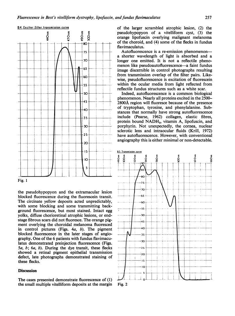
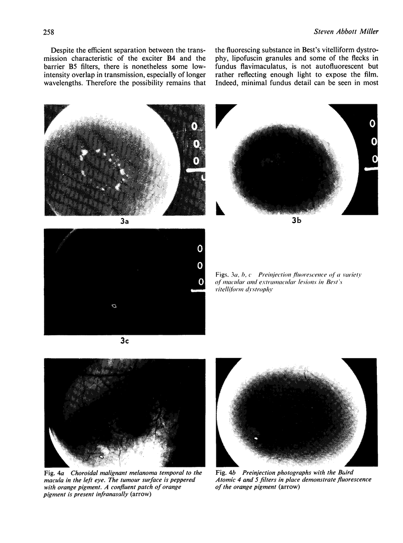
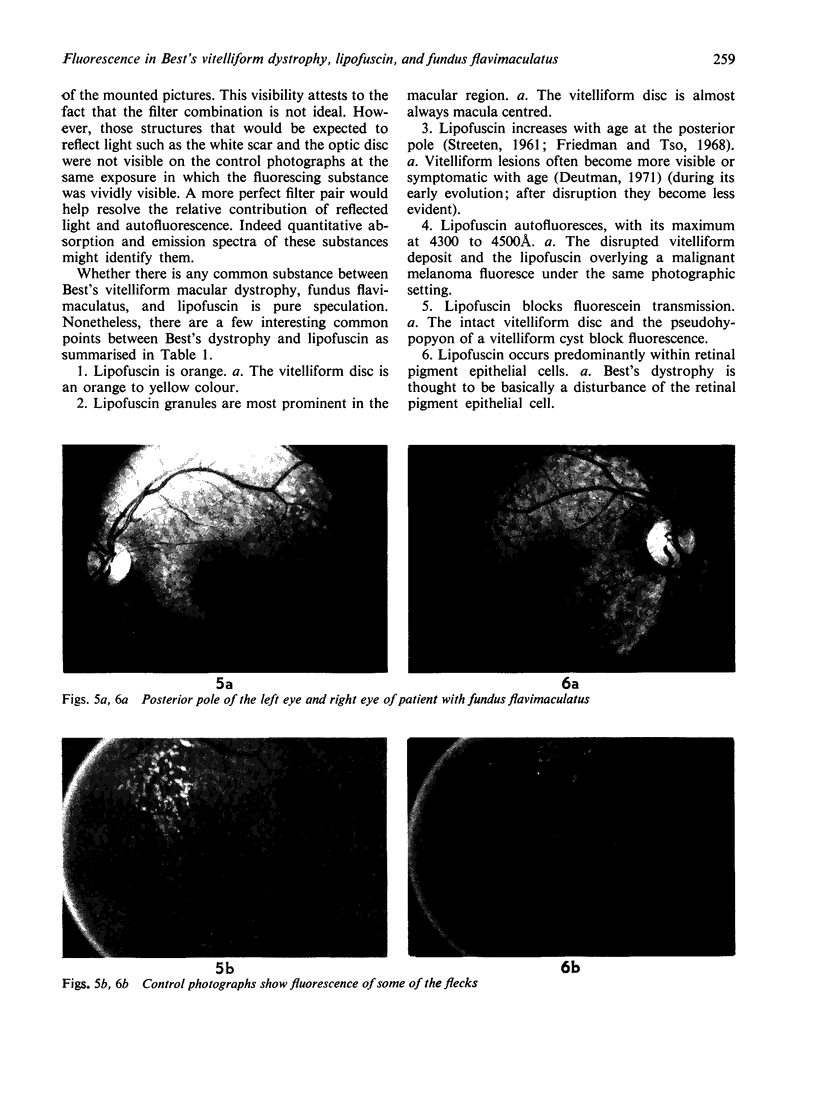
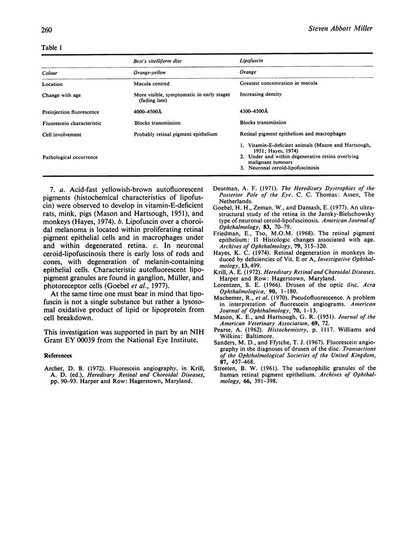
Control photographs, with the Baird Atomic B4 and B5 filters in place prior to fluorescein injection, show exposure of the film corresponding to (1) the small yellow vitelliform lesions at the edge of a disrupted disc, (2) the pseudohpopyon in a vitelliform cyst, (3) orange lipofuscin overlying a malignant melanoma, and (4) some of the flecks in a case of funds flavimaculatus. Because of transmission overlap between the filters, the relative contribtution reflected light and true autofluorescence is difficult to quantitate. Reflectile structures such as the optic nerve or a white scar were essentially unexposed, but minimal fundus detail was seen. Some parallels exist between lipofuscin and the content of a disrupted vitelliform lesion.
Full text
PDF
Images in this article
Selected References
These references are in PubMed. This may not be the complete list of references from this article.
- Friedman E., Ts'o M. O. The retinal pigment epithelium. II. Histologic changes associated with age. Arch Ophthalmol. 1968 Mar;79(3):315–320. doi: 10.1001/archopht.1968.03850040317017. [DOI] [PubMed] [Google Scholar]
- Goebel H. H., Zeman W., Damaske E. An ultrastructural study of the retina in the Jansky-Bielschowsky type of neuronal ceroid-lipofuscinosis. Am J Ophthalmol. 1977 Jan;83(1):70–79. doi: 10.1016/0002-9394(77)90194-5. [DOI] [PubMed] [Google Scholar]
- Machemer R., Norton E. W., Gass J. D., Choromokos E. Pseudofluorescence--a problem in interpretation of fluorescein angiograms. Am J Ophthalmol. 1970 Jul;70(1):1–10. doi: 10.1016/0002-9394(70)90658-6. [DOI] [PubMed] [Google Scholar]
- Sanders M. D., Ffytche T. J. Fluorescein angiography in the diagnosis of drusen of the disc. Trans Ophthalmol Soc U K. 1967;87:457–468. [PubMed] [Google Scholar]


