Abstract
Microbial species often occur in complex communities and exhibit intricate synergistic and antagonistic interactions. To avoid predation and compete for favorable niches, bacteria have evolved specialized protein secretion systems. The type VI secretion system (T6SS) is a versatile secretion system widely distributed among Gram-negative bacteria that translocates effectors into target cells or the extracellular milieu via various physiological processes. Pseudomonas aeruginosa is an opportunistic pathogen responsible for many diseases, and it has three independent T6SSs (H1-, H2-, and H3-T6SS). In this study, we found that the H3-T6SS of highly virulent P. aeruginosa PA14 is negatively regulated by OxyR and OmpR, which are global regulatory proteins of bacterial oxidative and acid stress. In addition, we identified a H3-T6SS effector PA14_33970, which is located upstream of VgrG3. PA14_33970 interacted directly with VgrG3 and translocated into host cells. Moreover, we found that H3-T6SS and PA14_33970 play crucial roles in oxidative, acid, and osmotic stress resistance, as well as in motility and biofilm formation. PA14_33970 was identified as a new T6SS effector promoting biofilm formation and thus named TepB. Furthermore, we found that TepB contributes to the virulence of P. aeruginosa PA14 toward Caenorhabditis elegans. Overall, our study indicates that H3-T6SS and its biofilm-promoting effector TepB are regulated by OxyR and OmpR, both of which are important for adaptation of P. aeruginosa PA14 to multiple stressors, providing insights into the regulatory mechanisms and roles of T6SSs in P. aeruginosa.
Supplementary Information
The online version contains supplementary material available at 10.1007/s44154-022-00078-7.
Keywords: P. aeruginosa PA14, Regulation, H3-T6SS, TepB, Stress resistance, Virulence
Introduction
Bacteria have evolved specialized protein secretion systems to deliver proteins into the extracellular space or to neighboring cells, and these systems play key roles in interactions with the environment, competitor bacteria, and host organisms (Cianfanelli et al. 2016). The type VI secretion system (T6SS) is a widely distributed type of proteinaceous machinery that delivers effector molecules directly into the inside of target cells via a one-step process (Zoued et al. 2014). T6SS is structurally homologous to contractile phage tails (Filloux 2009), with a complex structure consisting of a VipA/B outer sheath comprising a valine glycine repeat G (VgrG) trimer, PAAR domain-containing protein, Hcp inner tube, and transmembrane-baseplate complex formed of 13 essential core proteins along with additional accessory proteins (Leiman et al. 2009; Zoued et al. 2014). In the current model, T6SS features dynamic firing cycles including assembly, contraction, and disassembly of a sheath-like structure, followed by expulsion of a cell-puncturing device loaded with multiple effectors (Cianfanelli et al. 2016). ClpV and IcmF, two conserved T6SS components with ATPase activity, are crucial to the reassembly of T6SS structures and the secretion of Hcp, VgrG and substrates (Bonemann et al. 2009; Records 2011). T6SS effectors are transported by interacting with a core component or designated cargo effectors, or by fusing to structural components, known as specialised effectors. Furthermore, VgrG, Hcp, and PAAR play important roles in effector delivery (Durand et al. 2014).
As a tool for protein secretion, T6SS is involved in nutrition uptake, toxin delivery, cell-to-cell communication, interspecies competition, and virulence. T6SS and its effectors play various physiological roles improving the adaptability of bacteria to adverse environmental conditions (Lin et al. 2021; Yu et al. 2021). However, T6SS is an energetically expensive machine that is tightly regulated according to environmental conditions. T6SS is controlled by multiple transcriptional regulators in response to a wide variety of signals including salinity, iron concentration, temperature, and other stressors (Yang et al. 2021). For example, T6SS4 is regulated by OxyR under oxidative stress, triggering secretion of the effectors YezP and TssS, in Yersinia pseudotuberculosis (Wang et al. 2015; Zhu et al. 2021). In Burkholderia thailandensis, the regulators OxyR and Zur induce T6SS4 to secrete the effectors TseM and TseZ in response to environmental stresses (Si et al. 2017a, b). In Vibrio anguillarum, RpoS-regulated T6SS is involved in resistance to hydrogen peroxide (H2O2) and low-pH stress (Weber et al. 2009).
Pseudomonas aeruginosa is a common opportunistic Gram-negative pathogen that is widely distributed in the environment. P. aeruginosa has been the focus of intense research due to its prominent roles in several diseases, including septicemia and pneumonia (Chen et al. 2015). The P. aeruginosa genome encodes various virulence factors including secretion systems that contribute to its pathogenicity toward several hosts (Bleves et al. 2010). P. aeruginosa has three distinct and conserved T6SSs (H1-, H2-, and H3-T6SS), which play crucial roles in competition and pathogenicity by secreting multiple effectors (Mougous et al. 2006; Russell et al. 2014; Sana et al. 2016). The expression and functions of P. aeruginosa T6SSs are fine-tuned by regulators of various pathways in response to the environment. For example, in P. aeruginosa PAO1, H1-T6SS is upregulated by LadS and downregulated by RetS (Mougous et al. 2006); H2-T6SS is negatively regulated by RpoN, Fur, and CueR during bacterial competition and virulence (Sana et al. 2012, 2013; Han et al. 2019); and H3-T6SS is negatively regulated by Fur in response to the extracellular iron concentration (Lin et al. 2017). In P. aeruginosa PAK, H1-T6SS is negatively regulated by RsmA and RetS, impacting bacterial biofilm formation and virulence (Brencic and Lory 2009; Moscoso et al. 2011). All three T6SS types are regulated by RsmA and AmrZ in the highly virulent P. aeruginosa PA14 (Allsopp et al. 2017). Given the functional diversity of T6SSs in P. aeruginosa, their regulatory mechanisms and effectors must be identified, especially in the relatively less studied but highly virulent P. aeruginosa strain PA14.
In this study, we explored the regulation of H3-T6SS in P. aeruginosa PA14 and found that H3-T6SS is negatively regulated by OxyR and OmpR. Furthermore, we investigated the functions of H3-T6SS and its effector PA14_33970 (hereafter referred to as TepB) in P. aeruginosa PA14. Our results suggest that H3-T6SS and TepB play crucial roles in resistance to oxidative, acid and osmotic stresses, as well as motility, biofilm formation, and virulence in P. aeruginosa PA14.
Results
OxyR and OmpR negatively regulate H3-T6SS expression in P. aeruginosa PA14
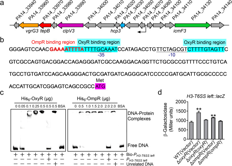
To investigate the function of H3-T6SS (genes PA14_33940 to PA14_34140) in P. aeruginosa PA14, we first analyzed the H3-T6SS gene cluster. We found that H3-T6SS genes are orientated in different directions (Fig. 1a), and that most genes are distributed in the left gene cluster. Then, we analyzed the left H3-T6SS promoter using Virtual Footprint software and identified two OxyR-binding sites (ATTTTATTTTGCAAAT and CTTTTGTAGTT) and an OmpR binding site (GAAAATTTTA) upstream of the gene PA14_34070 (Fig. 1b). We then examined the interactions of the left H3-T6SS promoter with OxyR and OmpR using electrophoretic mobility shift assay (EMSA). We generated a probe containing the left H3-T6SS promoter sequence (PH3-T6SS left), which was amplified from base − 207 to − 1 relative to the ATG start codon of the first open reading frame of the left H3-T6SS operon. This probe was incubated with His6-OxyR or His6-OmpR, leading to the formation of DNA − protein complexes (Fig. 1c). These DNA − protein complexes were completely disrupted by the addition of excess unlabeled probe, but not an unrelated control probe. This pattern indicates that the H3-T6SS promoter interacts with OxyR and OmpR, suggesting that H3-T6SS expression is regulated directly by OxyR and OmpR.
Fig. 1.
H3-T6SS is negatively regulated by OxyR and OmpR in P. aeruginosa PA14. a Gene organization of H3-T6SS gene cluster in P. aeruginosa PA14. b The sequence of the H3-T6SS-left promoter. The cyan part is the OxyR binding site predicted by software; the bold red letters is the predicted OmpR binding site; the ATG start codon of the first ORF of the H3-T6SS operon is marked in purple, and the − 35 and − 10 elements of the H3-T6SS promoter are underlined. c EMSA experiments verify the binding of His6-OxyR and OmpR to the H3-T6SS promoter. PH3-T6SS left is the unlabeled H3-T6SS promoter probe, Bio-PH3-T6SS left is labeled by biotin, and Unrelated DNA is the unrelated DNA fragment in same length. d β-galactosidase analyses of H3-T6SS promoter activities by using the transcriptional PH3-T6SS left::lacZ chromosomal fusion reporter expressed in the P. aeruginosa PA14 strains grown to stationary phase in TSB medium. Data represent the mean ± SEM of three biological replicates, each of which was performed with three technical replicates. **P < 0.01
To further investigate the roles of OxyR and OmpR in regulating the H3-T6SS operon, the single-copy fusion reporter plasmid PT6SS3 left::lacZ was introduced into the genomes of the wild-type (WT), ΔoxyR mutant, ΔompR mutant, and complemented ΔoxyR(oxyR) and ΔompR(ompR) strains. Quantitative analysis of the LacZ activity of the resulting strains revealed that oxyR and ompR deletion significantly increased in the activity of the H3-T6SS promoter, which was fully restored to the WT level by complementation with corresponding plasmids expressing oxyR (pME6032-oxyR) or ompR (pME6032-ompR) (Fig. 1d). This result confirms that OxyR and OmpR negatively regulate H3-T6SS expression. As OxyR and OmpR regulate gene expression in response to stresses, the same experiment was performed under 1 mM H2O2 oxidative stress, and a similar result was obtained (Fig. S1). Taken together, our findings suggest that both OxyR and OmpR negatively regulate H3-T6SS expression by binding to its promoter in P. aeruginosa PA14.
TepB is a substrate of H3-T6SS in P. aeruginosa PA14
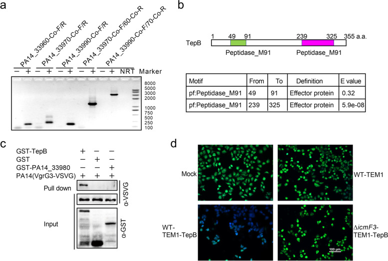
T6SS is critical to several bacterial processes that involve the secretion of effectors. Genes encoding T6SS substrates of are often located proximally to structural genes such as VgrG and Hcp (Durand et al. 2014; Bondage et al. 2016). Therefore, we examined the putative T6SS effector TepB (PA14_33970), which is located upstream of VgrG3 (PA14_33990) in the H3-T6SS gene cluster of P. aeruginosa PA14 (Fig. 1a). We performed reverse-transcription polymerase chain reaction to examine the expression profiles of TepB and the left H3-T6SS gene cluster. Two primer pairs (PA14_33990-Co-F and PA14_33970-Co-R; PA14_33970-Co-F and PA14_33960-Co-R) were designed to produce overlapping fragments, designated PA14_33990-PA14_33970 and PA14_33970-PA14_33960. The DNA fragment located between the two focal genes was amplified in reactions containing cDNA, but not in those containing double-distilled water (negative control) (Fig. 2a). This finding indicates that the genes PA14_33990, tepB (PA14_33970), and PA14_33960 are co-transcribed in the same operon.
Fig. 2.
TepB (PA14_33970) is a substrate of H3-T6SS. a Cotranscription analysis of PA14_33970–90 in P. aeruginosa PA14 by RT-PCR. -: PCR product with dd H2O as template. +: PCR product with P. aeruginosa PA14 cDNA as template. NRT: PCR product with no reverse transcriptase sample of P. aeruginosa PA14 RNA. b Protein motif prediction of TepB. c GST pull-down assay to detect the interaction between TepB and VgrG3. VgrG3-VSVG was incubated with GST, GST-TepB or GST-PA14_33980, and the protein complexes captured with glutathione beads were detected by Western blotting. d Translocation of TepB proteins into HeLa cells. HeLa cells were mock-infected or infected with indicated PA14 strains expressing TEM1-TepB at an MOI of 100 for 3 h and loaded with CCF2-AM. Visualization of the translocation of TepB using fluorescence microscopy. Scale bar, 100 μm
TepB is predicted to be an effector protein containing two peptidase M91 motifs (Fig. 2b). Notably, several effector-encoding genes are located in close proximity to the vgrG3, hcp3, or paar gene, and the associated effectors are secreted during interactions with the corresponding protein (VgrG3, Hcp3, or PAAR). Therefore, we performed a glutathione-S-transferase (GST) pull-down assay to examine the interaction of TepB with VgrG3, a H3-T6SS core component that transports secreted effector proteins via direct binding. We found that VgrG3-VSVG was retained by GST- TepB (Fig. 2c). In contrast, no interaction was detected between VgrG3-VSVG and GST or GST-PA14_33980 (Fig. 2c). As reported previously (Jiang et al. 2014; Zhu et al. 2021), translocation of TepB was detected using a TEM1-TepB fusion protein in HeLa cells treated with fusion proteins from WT P. aeruginosa PA14, a T6SS-deficient strain or mock treatment. TEM1-TepB was observed in cells infected with the fusion protein-expressing strain but not the T6SS-deficient strain (Fig. 2d), indicating that TepB was secreted into HeLa cells via T6SS. Our results suggest that TepB is a substrate of H3-T6SS in P. aeruginosa PA14.
H3-T6SS and TepB are required for resistance to oxidative, acid and osmotic stresses in P. aeruginosa PA14
In addition to its roles in bacterial competition, host infection, and virulence (Xu et al. 2014; Ho et al. 2017; Song et al. 2021), T6SS has important functions in resistance to various environmental stresses including acid, heat, antibiotic, and oxidative stresses (Wang et al. 2015; Yu et al. 2021). This resistance is achieved via the secretion of effectors, which is typically regulated by transcription factors (Yang et al. 2018). For example, T6SS1 in Cupriavidus necator, which is regulated by the transcription factor Fur, secretes the lipopolysaccharide-binding effector TeoL to construct outer membrane vesicles in response to oxidative stress (Li et al. 2021). The OxyR-regulated T6SS4 secretes the Zn2+-binding effector YezP, which plays an important role in protection against oxidative stress in Y. pseudotuberculosis (Zhang et al. 2013; Wang et al. 2015).
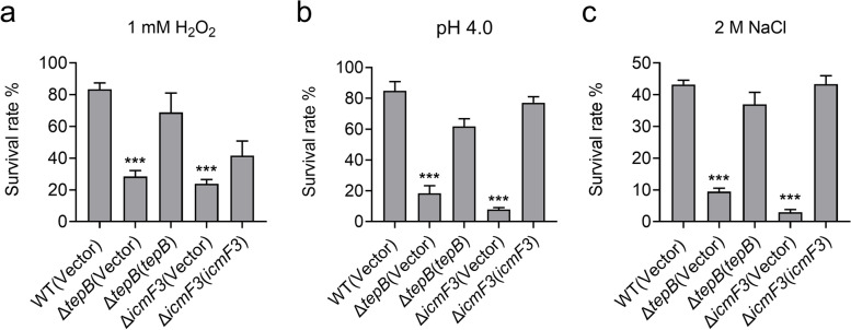
Our results indicate that TepB is a substrate of H3-T6SS, which is negatively regulated by OxyR and OmpR (Figs. 1 and 2), suggesting that the function of TepB is related to environmental cues sensed by these regulatory proteins. OxyR is a global regulator of the oxidative stress response; therefore, we investigated whether H3-T6SS and TepB play roles in protection against oxidative stress in P. aeruginosa PA14. We found that H2O2 tolerance was reduced in the H3-T6SS mutant ΔicmF3 and ΔtepB compared with the WT strain, and that resistance was restored to WT levels through complementation of the icmF3 and tepB genes (Fig. 3a). Our data indicate that H3-T6SS and TepB contribute to the survival of P. aeruginosa PA14 cells under oxidative stress conditions. OmpR regulates the expression of genes in response to changes in osmolarity and pH. The direct regulation of H3-T6SS by OmpR prompted us to examine whether H3-T6SS and TepB are involved in pH and osmotic stress resistance. We assessed the viability of the P. aeruginosa PA14 H3-T6SS mutants ΔicmF3 and ΔtepB following incubation at pH 4.0 for 30 min. The ΔicmF3 and ΔtepB mutants showed much lower survival rates than that of the WT after treatment at pH 4.0, and the WT survival phenotype was restored after complementation of the icmF3 and tepB genes (Fig. 3b). Similar results were obtained in these strains after treatment with 2 M NaCl (Fig. 3c). Our results indicate that H3-T6SS and TepB contribute to the survival of P. aeruginosa PA14 under oxidative, pH, and osmotic stress conditions.
Fig. 3.
H3-T6SS and TepB are involved in oxidative, acid and osmotic stress resistance. a, b and c The viability of mid-exponential phase P. aeruginosa PA14 strains was determined after challenge with 1 mM H2O2 (a), pH 4.0 (b) or 2 M NaCl (c) for 30 min. Statistical analyses for the rest of the assays were performed using unpaired two-tailed Student’s t-test. Data represent the mean ± SEM of three biological replicates, each of which was performed with three technical replicates. ***P < 0.001
H3-T6SS and TepB influence the motility of P. aeruginosa PA14
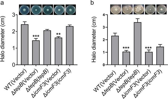
For many bacteria, motility is crucial to survival, growth, biofilm formation, and virulence. Motility enables bacteria to move toward resources and supports the dispersal of progeny (Nan and Zusman 2016). Bacteria have developed several motility mechanisms to exploit available environments (Wadhwa and Berg 2021), including swimming and swarming, which are the most common motility styles (Rashid and Kornberg 2000; Burrows 2012). As T6SS is involved in motility in Citrobacter freundii and Xanthomonas phaseoli (Liu et al. 2015; Montenegro Benavides et al. 2021), we investigated whether H3-T6SS and TepB are also involved in motility in P. aeruginosa PA14. The mutants ΔicmF3 and ΔtepB were significantly less motile than the WT strain, and this motility defect was fully restored upon complementation (Fig. 4a). We compared swarming motility among the WT, ΔicmF3, ΔtepB, and complemented ΔicmF3(icmF3) and ΔtepB(tepB) strains. Swarming motility was weaker in the ΔicmF3 and ΔtepB mutants than in the WT strain, and the motile phenotype was restored in the mutants via complementation of the icmF3 and tepB genes (Fig. 4b). Collectively, these results suggest that H3-T6SS and TepB influence the motility of P. aeruginosa PA14.
Fig. 4.
The motility is influenced by H3-T6SS and TepB. a The swimming motility of P. aeruginosa PA14 strains on motility agar plates containing 1 mM IPTG and 100 μg/mL tetracycline after 30 h of incubation at 30 °C. b The swarming motility of P. aeruginosa PA14 strains on the swarm plates containing 1 mM IPTG and 100 μg/mL tetracycline after 36 h of incubation at 30 °C. Data represent the mean ± SEM of three biological replicates, each of which was performed with three technical replicates. **P < 0.01; ***P < 0.001
H3-T6SS and TepB influence biofilm formation by P. aeruginosa PA14
Biofilm formation is governed by adaptive responses to microenvironmental cues, and involves motility (Tolker-Nielsen 2015). For the pathogen P. aeruginosa, biofilms represent an important virulence factor that plays a role in infection and the avoidance of host immunity (Al-Wrafy et al. 2017). Therefore, we examined the effects of H3-T6SS and TepB on biofilm formation using the crystal violet biofilm assay. We found that biofilm formation was defective in the strains ΔicmF3 and ΔtepB compared with the WT (Fig. 5a and b). Biofilm formation was restored to the WT level in the mutant lines following complementation of the icmF3 and tepB genes (Fig. 5a and b). These results suggest that H3-T6SS and TepB play pivotal roles in biofilm formation, and we newly identify TepB as a biofilm-promoting T6SS effector.
Fig. 5.
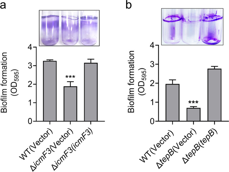
H3-T6SS and TepB influence biofilm formation. a and b Saturated bacterial cultures were diluted 100-fold in fresh TSB medium. After vertical incubation for 2 days with shaking at 120 rpm at 37 °C, biofilm formation of the strains was determined by crystal violet staining and quantified using optical density measurement. Data represent the mean ± SEM of three biological replicates, each of which was performed with three technical replicates. ***P < 0.001
TepB is involved in the virulence of P. aeruginosa PA14 toward Caenorhabditis elegans
Our results suggest that the H3-T6SS effector TepB influences motility and biofilm formation, which are crucial to virulence in P. aeruginosa PA14. The P. aeruginosa PAO1 H3-T6SS mutant strains ΔclpV3 and ΔicmF3 exhibited reduced virulence in a worm model (Sana et al. 2013; Lin et al. 2015). Therefore, we investigated whether the H3-T6SS effector TepB is involved in the pathogenicity of P. aeruginosa PA14 using a C. elegans infection model. The worms were infected with the WT, ΔtepB mutant, and complemented ΔtepB(tepB) strains. Infection with the WT strain resulted in a 27% survival rate for C. elegans within 48 h of infection, and the survival rate was increased to 57% after infection with the ΔtepB mutant, which was restored to the WT level when complemented with tepB gene. We also found that the survival rate of C. elegans was significantly higher at each time point after infection with the ΔtepB mutant compared with the WT and complemented ΔtepB(tepB) strains (Fig. 6). Together, these data suggest that TepB is required for virulence in P. aeruginosa PA14.
Fig. 6.
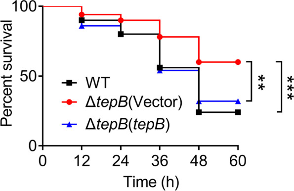
TepB is involved in virulence toward C. elegans. C. elegans were infected with P. aeruginosa PA14 wild-type, ΔtepB mutant and the complemented ΔtepB(tepB) strains. The survival rate of C. elegans after infection by P. aeruginosa PA14 was shown in 48 h. Data represent the mean of three biological replicates, each of which was performed with three technical replicates. Statistical analysis was performed by Log-Rank test. **P < 0.002; ***P < 0.001
Discussion
T6SS of P. aeruginosa plays important roles in pathogenicity to host cells and adaptation to various environments (Chen et al. 2015). Regulation of T6SS allows P. aeruginosa to respond to its environment (Chen et al. 2015). In P. aeruginosa PA14, RsmA downregulates the expression of all three T6SS loci. AmrZ regulates H2-T6SS negatively and H1- and H3-T6SS positively, via direct binding to their promoters (Allsopp et al. 2017). LasR and MvfR suppress the expression of H1-T6SS during P. aeruginosa pathogenesis but activate H2- and H3-T6SS (Lesic et al. 2009; Maura et al. 2016). RetS negatively controls the expression of H1- and H3-T6SS. In this study, we found that two transcriptional regulators, OxyR and OmpR, negatively regulate the expression of H3-T6SS via direct binding to its promoter. OxyR and OmpR control the expression of H3-T6SS and an effector protein involved in oxidative stress resistance, pH and osmotic stress tolerance, and biofilm formation. These findings clarify the regulation and function of H3-T6SS in P. aeruginosa PA14.
OxyR is a master regulator of oxidative stress in bacteria. In P. aeruginosa, OxyR is the most important regulator of the responses to H2O2 and organic peroxide stresses (da Cruz Nizer et al. 2021). OxyR has been reported to regulate over 100 genes involved in oxidative stress resistance, swarming, virulence, and other biological processes in P. aeruginosa (Vinckx et al. 2010; Wei et al. 2012; Panmanee et al. 2017). Furthermore, OxyR controls the secretion of potent cytotoxic factors in a manner partially dependent on the type III secretion system (Melstrom Jr. et al. 2007). OxyR regulation of SecA and other secreted proteins, but not T6SS proteins, has been reported previously (Wei et al. 2012; Panmanee et al. 2017). In the present study, we found that T6SS and TepB, which play roles in tolerance to pH and oxidative stresses, biofilm formation, motility, and virulence in P. aeruginosa PA14, were downregulated by OxyR. As a versatile bacterial weapon, T6SS secretes various cytotoxic effectors to facilitate bacterial competition and virulence (Coulthurst 2019). This study provides insights into the mechanism by which OxyR controls the secretion of cytotoxic factors.
OmpR is a global regulator of the responses to pH and osmotic stresses, and is widely distributed among bacteria (Gerken et al. 2020; Kenney and Anand 2020). OmpR regulates the expression of T6SS in Y. pseudotuberculosis in response to pH and osmotic stresses (Gueguen et al. 2013; Zhang et al. 2013). However, the function of OmpR in P. aeruginosa remains unclear. AlgR1, a homolog of OmpR, activates the expression of algD under osmotic stress by binding to its promoter in P. aeruginosa (Kato and Chakrabarty 1991). Another homolog, PhoB, is involved in crosstalk in P. aeruginosa, affecting bacterial behavior (Bielecki et al. 2015). In this study, we identified OmpR in P. aeruginosa PA14 and found that it suppresses the expression of H3-T6SS. Furthermore, our results indicate that H3-T6SS and its effector protein are involved in acid and osmotic stress resistance. Our results are consistent with the reported functions of OmpR in other bacteria (Gueguen et al. 2013; Zhang et al. 2013).
The tepB gene is located within the H3-T6SS gene cluster and in the same operon as clpV3 and vgrG3 (Fig. 2a). tepB was predicted to encode a T6SS effector and to contribute to the virulence of P. aeruginosa PA14 (Lesic et al. 2009). In this study, we demonstrated that TepB interacts with VgrG3 and translocates into host cells (Fig. 2c and d). Proteins encoded by genes in close proximity to vgrG and that interact with VgrG are typically effectors secreted by T6SS (Hachani et al. 2014; Wettstadt 2020; Wu et al. 2020). Therefore, TepB was considered a T6SS effector. T6SS secretes effectors in two ways: transporting the substrate into the environment or injecting the effector into other bacteria or host cells via direct contact (Lin et al. 2021). We detected no TepB in the supernatant but observed it in host cells (Fig. 2d), suggesting that TepB is an injected effector. TepB may be secreted by T6SS in a contact-dependent manner as reported previously. We found that TepB is also involved in motility, biofilm formation, stress tolerance, and pathogenicity in P. aeruginosa PA14. TepB is absent in P. aeruginosa PAO1, which is less virulent than P. aeruginosa PA14. This effector may contribute to the superior virulence of P. aeruginosa PA14 compared with P. aeruginosa PAO1, similar to the function of the H2-T6SS effector PldA in the virulence of clinically isolated infectious P. aeruginosa (Boulant et al. 2018). Although we revealed the primary functions of TepB in this work, the molecular mechanisms underlying these functions require further investigation.
T6SS has versatile functions in stress resistance, biofilm formation, metal acquisition, and pathogenicity (Lin et al. 2017, 2021; Coulthurst 2019). The functions of H3-T6SS in P. aeruginosa PA14 include tolerance to H2O2, acid and osmotic stresses, motility, biofilm formation, and pathogenicity, consistent with reported T6SS functions in other bacterial species. The involvement of T6SS in oxidative and acid stress resistance has not been reported previously in P. aeruginosa. The pathogens P. aeruginosa, Mycobacterium tuberculosis, and Yersinia pestis form biofilms, enhancing their ability to survive and defend themselves within a host rather than as individual planktonic cells (Darby et al. 2002; Kumar et al. 2017). Multiple factors including regulatory proteins, membrane proteins, secretion systems, motility, and attachment, as well as environmental conditions such as temperature, affect biofilm formation in P. aeruginosa during persistent infections (Whiteley et al. 2001; Kim et al. 2020). The expression levels of the three T6SS types are relatively high in P. aeruginosa PAO1 biofilm cells. H1-T6SS is not involved in biofilm formation, but it affects swarming (Chen et al. 2020). IcmF3, a component of H3-T6SS, decreases biofilm formation, and increases swarming in P. aeruginosa PAO1 (Lin et al. 2015). We found that H3-T6SS and its effector TepB increases swimming, swarming, and biofilm formation in P. aeruginosa PA14. H3-T6SS plays a different role in biofilm formation in P. aeruginosa PA14 than that in P. aeruginosa PAO1 (Lin et al. 2015), and this may be responsible for the difference in biofilm invasion strategies between these two strains (Kasetty et al. 2021). Motility and biofilm formation contribute to acute and chronic infections, respectively (Balasubramanian et al. 2013). Our results suggest that H3-T6SS and TepB play roles in both acute and chronic P. aeruginosa PA14 infections.
TepB is a newly identified biofilm-promoting protease effector secreted by T6SS. Extracellular enzymes, such as polysaccharide-degrading hydrolases, esterases, nucleases, proteases, and lyases, are crucial for matrix turnover during biofilm formation, detachment, and dispersal (Flemming et al. 2022). For example, the serine protease autotransporter family protein SepA can promote biofilm formation by processing Aap and AtlE extracellularly in Staphylococcus epidermidis (Martinez-Garcia et al. 2018). The PAO1 extracellular elastase LasB has been reported to promote biofilm formation partly via rhamnolipid-mediated regulation (Yu et al. 2014). PAO1 secretes the DNA-specific endonuclease EndA to degrade extracellular DNA in biofilms, leading to the dispersal of PAO1 from the biofilm (Cherny and Sauer 2019). Membrane proteins and secretion systems, including TolA, OmlA, the twin arginine translocation pathway, type II secretion system, type III secretion system, and T6SS, are involved in the translocation of the biofilm matrix and extracellular enzymes (Whiteley et al. 2001; Lin et al. 2021; Flemming et al. 2022). However, few effectors associated with secretion systems, especially in T6SS, have been found to promote biofilm formation. Only VgrG and Hcp, which are both components and effectors of the T6SS, have been found to play positive roles in biofilm formation in bacteria (Sha et al. 2013; Fei et al. 2022; Pan et al. 2022). TepB is the first identified enzymatic effector of T6SS that promotes biofilm formation during acute and chronic infections of P. aeruginosa PA14. The mechanism by which TepB promotes biofilm formation via metalloprotease activity requires further study.
In conclusion, we found that the transcriptional regulators OxyR and OmpR downregulate the expression of H3-T6SS in pathogenic P. aeruginosa PA14 via direct binding to its promoter region. H3-T6SS and its biofilm-promoting effector TepB improve tolerance to oxidative, acid, and osmotic stresses, motility, biofilm formation, and virulence in P. aeruginosa PA14. This study elucidated the regulation and functions of H3-T6SS in P. aeruginosa PA14, although details of the underlying mechanisms requires further investigation.
Materials and methods
Bacterial strains and growth conditions
Bacterial strains and plasmids used in this study are listed in Supplementary Table S1. Escherichia coli strains were grown at 37 °C in either Luria-Bertani (LB) broth or agar. P. aeruginosa PA14 strains were grown at 37 °C in tryptic soy broth (TSB) medium or M9 minimal medium (6 g/L Na2HPO4, 3 g/L KH2PO4, 0.5 g/L NaCl, 1 g/L NH4Cl, 2 mM MgSO4, 0.1 mM CaCl2, 0.2% glucose, pH 7.0). The P. aeruginosa PA14 strain was the parent of all derivatives used in this study. To generate in-frame deletion mutants, the pK18mobsacB derivatives were transformed into relevant P. aeruginosa PA14 strains through E. coli S17–1-mediated conjugation and screened as described previously (Lin et al. 2017; Li et al. 2021). Antibiotics were used at the following concentrations for E. coli: kanamycin, 50 μg/mL; tetracycline, 15 μg/mL; gentamicin, 10 μg/mL; and for P. aeruginosa PA14: kanamycin, 50 μg/mL; chloramphenicol, 30 μg/mL; gentamicin, 200 μg/mL; tetracycline, 200 μg/mL for plates or 160 μg/mL for liquid growth. All chemicals were of Analytical Reagent Grade purity or higher.
Plasmid construction
Primers used in this study are listed in Supplementary Table S2. The plasmid pK18-Gm-ΔtepB (PA14_33970) was used to construct the ΔtepB in-frame deletion mutant of P. aeruginosa PA14. A 679-bp upstream fragment and an 866-bp downstream fragment of tepB were amplified using the primer pairs PA14_33970-Up-F-BamHI/PA14_33970-Up-R and PA14_33970-Down-F/PA14_33970-Down-R-HindIII, respectively. The upstream and downstream PCR fragments were ligated by overlapping PCR, and the resulting PCR product was digested with BamHI/HindIII and inserted into the BamHI/HindIII sites of the suicide vector pK18-Gm to produce pK18-Gm-ΔtepB. The knock-out plasmids pK18-Gm-icmF3 (PA14_34130), pK18-Gm-oxyR (PA14_70560) and pK18-Gm-ompR (PA14_68700) were constructed in a similar manner by using primers list in Supplementary Table S2. To complement the ΔtepB mutant, primers PA14_33970-F-EcoRI/PA14_33970-R-BglII were used to amplify the tepB gene from the P. aeruginosa PA14 genome DNA. The PCR product of tepB was digested with EcoRI/BglII and cloned into the EcoRI/BglII sites of plasmid pME6032 to produce pME6032-tepB. The complementation plasmids pME6032-icmF3, pME6032-oxyR, and pME6032-ompR were similarly constructed by using primers list in Supplementary Table S2. To construct pME6032-tepB-vsvg, primers PA14_33970-F-EcoRI/PA14_33970-R-vsvg-BglII was used to amplify the tepB gene and the PCR product was digested with EcoRI/BglII and cloned into similarly digested pME6032 to generate pME6032-tepB-VSVG. The plasmid pME6032-vgrG3-vsvg was constructed in a similar method by using primers list in Supplementary Table S2. For constructing expression plasmids, the genes encoding P. aeruginosa TepB, PA14_33980, OxyR and OmpR were amplified by PCR. The obtained DNA fragments were digested and inserted into similar digested pGEX-6p-1 and pET28a, yielding corresponding plasmids, respectively. For complementation, complementary plasmids pME6032-oxyR, pME6032-ompR, pME6032-icmF3 and pME6032-tepB were introduced into respective mutants by electroporation. The integrity of the insert in all constructs was confirmed by DNA sequencing.
Purification of recombinant proteins and Western blotting
His6- and GST-tagged recombinant proteins were expressed and purified from E. coli as describe (Shen et al. 2009). In short, the pET28a and pGEX-6p-1 derivatives were transformed into BL21(DE3) and XL1-Blue host strains, respectively. Bacteria were cultured at 37 °C in LB medium to an OD600 of 0.6, shifted to 18 °C and induced with 0.5 mM IPTG for 16 h. Harvested cells were disrupted by sonication, and His6- or GST-tagged proteins were purified with the His•Bind Ni-NTA resin (Novagen, Madison, WI) or the GST•Bind resin (Novagen, Madison, WI) according to manufacturer’s instructions. Purified recombinant proteins were dialyzed against the appropriate buffer overnight at 4 °C and stored at − 80 °C until use. Protein concentrations were measured using the Bradford assay according to the manufacturer’s instructions (Bio-Rad, Hercules, CA) with bovine serum albumin as standard.
For Western blotting, samples resolved by SDS–PAGE were transferred onto polyvinylidene difluoride membranes. After blocking with 5% (w/v) BSA in TBST buffer (50 mM Tris pH 7.4, 150 mM NaCl, 0.05% Tween 20), membranes were incubated with the appropriate primary antibody: anti-VSVG (Santa Cruz Biotechnology, USA), 1:5000 and anti-GST (Zhongshan Golden Bridge Biotechnology, Beijing, China), 1:2000. The membrane was washed three times in TBST buffer and incubated with 1:10,000 dilution of horseradish peroxidase-conjugated secondary antibodies (Shanghai Genomics, Shanghai, China) for 2 h. Signals were detected using the ECL plus kit following the manufacturer’s specified protocol.
Electrophoretic mobility shift assay (EMSA)
Electrophoretic mobility shift assay was performed as previously described (Si et al. 2017a). Briefly, Bio-PH3-T6SS left was amplified from the P. aeruginosa PA14 genome DNA with primers PH3-T6SS left-F/PH3-T6SS left-R (labeled with biotin). The unlabeled PH3-T6SS left competitor probe was amplified from the P. aeruginosa PA14 genome DNA with primers PH3-T6SS left-F/PH3-T6SS left-R. All PCR products were purified by EasyPure Quick Gel Extraction Kit (TransGen Biotech, Beijing, China). Each 20-μL EMSA reaction solution was prepared by adding the following components according to the manufacturer’s protocol (LightShift Chemiluminescent EMSA Kit, Thermo Fisher Scientific, CA, USA): 1× binding buffer, 2.5% glycerol, 5 mM MgCl2, 0.05% NP-40, 5 mM EDTA, 20 ng probe and 0–0.5 ng protein. Reaction solutions were incubated for 20 min at 26 °C. The protein-probes mixtures were separated by using a 6% polyacrylamide native gel and transferred to a Biodyne B Nylon membrane (Thermo Fisher Scientific, CA, USA). Migration of biotin-labeled probe was detected by streptavidin-horseradish peroxidase conjugates that bind to biotin and chemiluminescent substrate according to the manufacture’s protocol.
Construction of chromosomal fusion reporters and β-galactosidase assays
The lacZ fusion reporter vector pMini-CTX-PH3-T6SS left::lacZ and pMini-CTX-PH3-T6SS3 right::lacZ were transformed into E. coli S17–1 λpir and mated with P. aeruginosa PA14 strains as described previously (Hoang et al. 2000). Promoter fragments were integrated at the CTX phage attachment site (attB) in strain P. aeruginosa PA14 and the interrelated mutant strains, and the pFLP2 plasmid expressing Flp recombinase was used to excision of the Tcr marker following the protocol to obtain the unmarked transcriptional fusion strains (Hoang et al. 2000). The lacZ fusion reporter strains were grown in TSB medium with or without 1 mM H2O2 at 37 °C. The β-galactosidase activity was assayed using ONPG (o-Nitrophenyl β-D-galactopyranoside) as the substrate and expressed in Miller units.
Analysis of cotranscription by reverse transcription-PCR (RT-PCR)
Gene cotranscription assay was performed as previously described (Zheng et al. 2014). Mid-exponential phase P. aeruginosa PA14 strains grown in M9 medium were harvested and RNA was extracted using the SteadyPure Universal RNA Extraction Kit AG21017 (Accurate Biotechnology, Hunan, China), and treated with RNase-free DNase I according to the manufacturer’s protocol after its integrity was checked by agarose electrophoresis. First-strand cDNA was reverse transcribed from 1 μg DNase I-digested RNA using the Evo M-MLV RT Kit with gDNA Clean for qPCR AG11705 (Accurate Biotechnology, Hunan, China) according to the manufacturer’s protocol. The resulting cDNA was used as the template to amplify the intragenic regions of PA14_33990, tepB and PA14_33960 genes with 2 × Accurate Master Mix AG1107 (Accurate Biotechnology, Hunan, China). ddH2O and No Reverse Transcriptase (NRT) sample were used as negative controls, respectively. The specific primers used for amplification are list in Supplementary Table S2.
GST pull-down assay
The GST pull-down assay was performed as previously described with minor modifications (Xu et al. 2010). To verify the interaction between TepB with VgrG3, stationary phase P. aeruginosa PA14 cells expressing VgrG3-VSVG protein were lysed in Bugbuster solution (Novagen, Madison, WI). Cleared cell lysates were incubated with 10 μg purified GST-TepB on a rotator at 4 °C overnight, and 40 μL prewashed glutathione-Sepharose beads (Novagen, Madison, WI) were added to the reactions. After another 4 h of incubation at 4 °C, the beads were washed six times with TEN buffer (100 mM Tris pH 8.0, 10 mM EDTA, 500 mM NaCl). Retained proteins were detected by immunoblotting after SDS–PAGE.
Bacterial survival assay
Mid-logarithmic phase P. aeruginosa strains grown in TSB medium were collected, washed and diluted 100-fold into M9 medium, and then treated with or without H2O2 (1.0 mM), 2 M NaCl or pH 4.0 for 30 min at 37 °C. After treatment, the cultures were serially diluted and plated onto LB agar plates, and colonies were counted after 24 h growth at 37 °C. Percentage survival was calculated by dividing the CFU number of stressed cells by the CFU number of unstressed cells (Song et al. 2015). All these assays were performed in triplicate at least three times.
Motility assay
Swimming motility assay was performed as previously described (Inoue et al. 2008; Li et al. 2019). Briefly, 1 μL bacterium solution was injected into semi-solid agar medium (1% tryptone, 0.5% NaCl, 0.3% Difco Bacto agar) and incubated for 30 h under 30 °C before observation. Motility halos were measured after 30 h of incubation. The swarming motility assay was performed as previously described (Rashid and Kornberg 2000). Briefly, a single colony selected from TSB plates was touched slightly on soft agar medium (8 g/L Nutrient Broth, 5 g/L glucose, 0.5% Difco Bacto agar) and incubated for 36 h under 30 °C before observation, and then motility halos were measured.
Biofilm formation assay
Biofilm formation was determined following the methods of O’Toole and Zhang (O’Toole and Kolter 1998; Zhang et al. 2020). Briefly, overnight bacterial cultures were diluted 100-fold in fresh 4 mL TSB medium with appropriate antibiotics when necessary. After vertical incubation for 2 days with the shake of 120 rpm at 37 °C, the bacterial cultures were removed and the test tubes were washed twice with fresh phosphate buffered saline (PBS). The cells that adhered to the tubes were stained with 0.1% crystal violet for 30 min and then washed twice with PBS. The cell-bound dye was dissolved in 5 mL of 95% ethanol, and the absorbance of the eluted solution was measured using a microplate reader at 595 nm.
Caenorhabditis elegans killing assay
P. aeruginosa strains were grown overnight at 37 °C and supplemented with nematode growth medium (NGM) following the published method (Tan et al. 1999). The NGM plates were incubated firstly at 37 °C for 24 h and then at 25 °C for 24 h before seeding with adult hermaphrodite worms. Before solidification, all experimental plates were added to 200 μM 5-fluorodeoxyuridine, which was used to prevent the development of progeny. In each assay, 40–50 adult nematodes were transferred to a single plate. Plates were incubated at 25 °C and scored for live worms every 12 h for total time of 48 h. The experiments were conducted in triplicate and E. coli OP50 was used as the negative control. A worm was considered dead when it no longer responded to touch. Any worms that died as a result of getting stuck to the wall of the plate were excluded from the analyses.
Translocation assay for TEM1 fusion protein
The translocation assay for TEM1-TepB fusion protein was performed as previously described (Jiang et al. 2014; Zhu et al. 2021). HeLa cells were grown in 96-well black-wall, clear-bottom plates and infected with PA14 WT or T6SS deficient mutant strains with TEM1-TepB (at an MOI of 100) for 3 h. Host cells were then washed with PBS for three times and treated with CCF2-AM (LiveBLAzer FRET-B/G Loading Kit, Invitrogen) for 90 min at room temperature. Samples were examined with a Nikon fluorescence microscope (Nikon, Japan).
Statistical analysis
All experiments were performed at least in triplicate and repeated on two different occasions. Data are expressed as mean ± SEM. Differences between frequencies were assessed by the Student’s t-test (bilateral and unpaired). Statistical analysis of results was conducted by using GraphPad Prism 8 (GraphPad Software, San Diego California, USA), using a P value of < 0.05 as statistically significant. The survival times of Caenorhabditis elegans were analyzed using Kaplan-Meyer curves and the comparisons were performed using the Log-Rank test. P value < 0.033 was used as statistically significant.
Supplementary Information
Additional file 1: Fig. S1. Promoter activity analysis under oxidative stress. Table S1. Bacterial strains and plasmids used in this study. Table S2. Primers used in this study. Supplementary References.
Acknowledgements
We thank Dr. Herbert P. Schweizer at Colorado State University for kindly providing the plasmid pMini-CTX::lacZ. We thank the Biology Teaching and Research Core Facility at College of Life Sciences, NWAFU (Ningjuan Fan, Hui Duan and Xiyan Chen) for their technical support.
Authors’ contributions
Y.Y., D.P., P.C. and C.L. designed the research. Y.Y., D.P., Y.T., J.L., K.Z., and Z.Y. performed the experimental work. Y.Y., D.P., C.L. and L.Z. analyzed the data. Y.Y., D.P. and C.L. drafted the manuscript. Y.W. and P.C. revised the manuscript. The author(s) read and approved the final manuscript.
Funding
This work was supported by grants of the National Key R&D Program of China (Grants 2021YFA0909600), National Natural Science Foundation of China (Grants 31970114, 32100034 and 32100149).
Availability of data and materials
All datasets generated for this study are included in the article/Supplementary Information.
Declarations
Ethics approval and consent to participate
Not applicable.
Consent for publication
Not applicable.
Competing interests
The authors declare no competing interests.
Footnotes
Publisher’s Note
Springer Nature remains neutral with regard to jurisdictional claims in published maps and institutional affiliations.
Yantao Yang and Damin Pan contributed equally to this work.
Contributor Information
Peng Chen, Email: pengchen@nwsuaf.edu.cn.
Changfu Li, Email: lierfu@nwafu.edu.cn.
References
- Allsopp LP, Wood TE, Howard SA, Maggiorelli F, Nolan LM, Wettstadt S, Filloux A. RsmA and AmrZ orchestrate the assembly of all three type VI secretion systems in Pseudomonas aeruginosa. Proc Natl Acad Sci U S A. 2017;114:7707–7712. doi: 10.1073/pnas.1700286114. [DOI] [PMC free article] [PubMed] [Google Scholar]
- Al-Wrafy F, Brzozowska E, Gorska S, Gamian A. Pathogenic factors of Pseudomonas aeruginosa - the role of biofilm in pathogenicity and as a target for phage therapy. Postepy Hig Med Dosw (Online) 2017;71:78–91. doi: 10.5604/01.3001.0010.3792. [DOI] [PubMed] [Google Scholar]
- Balasubramanian D, Schneper L, Kumari H, Mathee K. A dynamic and intricate regulatory network determines Pseudomonas aeruginosa virulence. Nucleic Acids Res. 2013;41:1–20. doi: 10.1093/nar/gks1039. [DOI] [PMC free article] [PubMed] [Google Scholar]
- Bielecki P, Jensen V, Schulze W, Godeke J, Strehmel J, Eckweiler D, et al. Cross talk between the response regulators PhoB and TctD allows for the integration of diverse environmental signals in Pseudomonas aeruginosa. Nucleic Acids Res. 2015;43:6413–6425. doi: 10.1093/nar/gkv599. [DOI] [PMC free article] [PubMed] [Google Scholar]
- Bleves S, Viarre V, Salacha R, Michel GP, Filloux A, Voulhoux R. Protein secretion systems in Pseudomonas aeruginosa: a wealth of pathogenic weapons. Int J Med Microbiol. 2010;300:534–543. doi: 10.1016/j.ijmm.2010.08.005. [DOI] [PubMed] [Google Scholar]
- Bondage DD, Lin JS, Ma LS, Kuo CH, Lai EM. VgrG C terminus confers the type VI effector transport specificity and is required for binding with PAAR and adaptor-effector complex. Proc Natl Acad Sci U S A. 2016;113:E3931–E3940. doi: 10.1073/pnas.1600428113. [DOI] [PMC free article] [PubMed] [Google Scholar]
- Bonemann G, Pietrosiuk A, Diemand A, Zentgraf H, Mogk A. Remodelling of VipA/VipB tubules by ClpV-mediated threading is crucial for type VI protein secretion. EMBO J. 2009;28:315–325. doi: 10.1038/emboj.2008.269. [DOI] [PMC free article] [PubMed] [Google Scholar]
- Boulant T, Boudehen YM, Filloux A, Plesiat P, Naas T, Dortet L. Higher prevalence of PldA, a Pseudomonas aeruginosa trans-kingdom H2-type VI secretion system effector, in clinical isolates responsible for acute infections and in multidrug resistant strains. Front Microbiol. 2018;9:2578. doi: 10.3389/fmicb.2018.02578. [DOI] [PMC free article] [PubMed] [Google Scholar]
- Brencic A, Lory S. Determination of the regulon and identification of novel mRNA targets of Pseudomonas aeruginosa RsmA. Mol Microbiol. 2009;72:612–632. doi: 10.1111/j.1365-2958.2009.06670.x. [DOI] [PMC free article] [PubMed] [Google Scholar]
- Burrows LL. Pseudomonas aeruginosa twitching motility: type IV pili in action. Annu Rev Microbiol. 2012;66:493–520. doi: 10.1146/annurev-micro-092611-150055. [DOI] [PubMed] [Google Scholar]
- Chen L, Zou Y, Kronfl AA, Wu Y. Type VI secretion system of Pseudomonas aeruginosa is associated with biofilm formation but not environmental adaptation. Microbiologyopen. 2020;9:e991. doi: 10.1002/mbo3.991. [DOI] [PMC free article] [PubMed] [Google Scholar]
- Chen L, Zou Y, She P, Wu Y. Composition, function, and regulation of T6SS in Pseudomonas aeruginosa. Microbiol Res. 2015;172:19–25. doi: 10.1016/j.micres.2015.01.004. [DOI] [PubMed] [Google Scholar]
- Cherny KE, Sauer K (2019) Pseudomonas aeruginosa requires the DNA-specific endonuclease EndA to degrade extracellular genomic DNA to disperse from the biofilm. J Bacteriol 201. 10.1128/JB.00059-19 [DOI] [PMC free article] [PubMed]
- Cianfanelli FR, Monlezun L, Coulthurst SJ. Aim, load, fire: the type VI secretion system, a bacterial nanoweapon. Trends Microbiol. 2016;24:51–62. doi: 10.1016/j.tim.2015.10.005. [DOI] [PubMed] [Google Scholar]
- Coulthurst S. The type VI secretion system: a versatile bacterial weapon. Microbiology (Reading) 2019;165:503–515. doi: 10.1099/mic.0.000789. [DOI] [PubMed] [Google Scholar]
- da Cruz Nizer WS, Inkovskiy V, Versey Z, Strempel N, Cassol E, Overhage J (2021) Oxidative stress response in Pseudomonas aeruginosa. Pathogens 10. 10.3390/pathogens10091187 [DOI] [PMC free article] [PubMed]
- Darby C, Hsu JW, Ghori N, Falkow S. Caenorhabditis elegans: plague bacteria biofilm blocks food intake. Nature. 2002;417:243–244. doi: 10.1038/417243a. [DOI] [PubMed] [Google Scholar]
- Durand E, Cambillau C, Cascales E, Journet L. VgrG, Tae, Tle, and beyond: the versatile arsenal of type VI secretion effectors. Trends Microbiol. 2014;22:498–507. doi: 10.1016/j.tim.2014.06.004. [DOI] [PubMed] [Google Scholar]
- Fei N, Ji W, Yang L, Yu C, Qiao P, Yan J et al (2022) Hcp of the type VI secretion system (T6SS) in Acidovorax citrulli group II strain Aac5 has a dual role as a core structural protein and an effector protein in colonization, growth ability, competition, biofilm formation, and ferric Iron absorption. Int J Mol Sci 23. 10.3390/ijms23179632 [DOI] [PMC free article] [PubMed]
- Filloux A. The type VI secretion system: a tubular story. EMBO J. 2009;28:309–310. doi: 10.1038/emboj.2008.301. [DOI] [PMC free article] [PubMed] [Google Scholar]
- Flemming H-C, van Hullebusch ED, Neu TR, Nielsen PH, Seviour T, Stoodley P et al (2022) The biofilm matrix: multitasking in a shared space. Nat Rev Microbiol. 10.1038/s41579-022-00791-0 [DOI] [PubMed]
- Gerken H, Vuong P, Soparkar K, Misra R (2020) Roles of the EnvZ/OmpR two-component system and porins in iron acquisition in Escherichia coli. mBio 11. 10.1128/mBio.01192-20 [DOI] [PMC free article] [PubMed]
- Gueguen E, Durand E, Zhang XY, d'Amalric Q, Journet L, Cascales E. Expression of a Yersinia pseudotuberculosis type VI secretion system is responsive to envelope stresses through the OmpR transcriptional activator. PLoS One. 2013;8:e66615. doi: 10.1371/journal.pone.0066615. [DOI] [PMC free article] [PubMed] [Google Scholar]
- Hachani A, Allsopp LP, Oduko Y, Filloux A. The VgrG proteins are “à la Carte” delivery systems for bacterial type VI effectors. J Biol Chem. 2014;289:17872–17884. doi: 10.1074/jbc.M114.563429. [DOI] [PMC free article] [PubMed] [Google Scholar]
- Han Y, Wang T, Chen G, Pu Q, Liu Q, Zhang Y, et al. A Pseudomonas aeruginosa type VI secretion system regulated by CueR facilitates copper acquisition. PLoS Pathog. 2019;15:e1008198. doi: 10.1371/journal.ppat.1008198. [DOI] [PMC free article] [PubMed] [Google Scholar]
- Ho BT, Fu Y, Dong TG, Mekalanos JJ. Vibrio cholerae type 6 secretion system effector trafficking in target bacterial cells. Proc Natl Acad Sci U S A. 2017;114:9427–9432. doi: 10.1073/pnas.1711219114. [DOI] [PMC free article] [PubMed] [Google Scholar]
- Hoang TT, Kutchma AJ, Becher A, Schweizer HP. Integration-proficient plasmids for Pseudomonas aeruginosa: site-specific integration and use for engineering of reporter and expression strains. Plasmid. 2000;43:59–72. doi: 10.1006/plas.1999.1441. [DOI] [PubMed] [Google Scholar]
- Inoue T, Shingaki R, Fukui K. Inhibition of swarming motility of Pseudomonas aeruginosa by branched-chain fatty acids. FEMS Microbiol Lett. 2008;281:81–86. doi: 10.1111/j.1574-6968.2008.01089.x. [DOI] [PubMed] [Google Scholar]
- Jiang F, Waterfield NR, Yang J, Yang G, Jin Q. A Pseudomonas aeruginosa type VI secretion phospholipase D effector targets both prokaryotic and eukaryotic cells. Cell Host Microbe. 2014;15:600–610. doi: 10.1016/j.chom.2014.04.010. [DOI] [PubMed] [Google Scholar]
- Kasetty S, Katharios-Lanwermeyer S, O’Toole GA, Nadell CD. Differential surface competition and biofilm invasion strategies of Pseudomonas aeruginosa PA14 and PAO1. J Bacteriol. 2021;203:e0026521. doi: 10.1128/JB.00265-21. [DOI] [PMC free article] [PubMed] [Google Scholar]
- Kato J, Chakrabarty AM. Purification of the regulatory protein AlgR1 and its binding in the far upstream region of the algD promoter in Pseudomonas aeruginosa. Proc Natl Acad Sci U S A. 1991;88:1760–1764. doi: 10.1073/pnas.88.5.1760. [DOI] [PMC free article] [PubMed] [Google Scholar]
- Kenney LJ, Anand GS (2020) EnvZ/OmpR two-component signaling: an archetype system that can function noncanonically. EcoSal Plus 9. 10.1128/ecosalplus.ESP-0001-2019 [DOI] [PMC free article] [PubMed]
- Kim S, Li XH, Hwang HJ, Lee JH (2020) Thermoregulation of Pseudomonas aeruginosa biofilm formation. Appl Environ Microbiol 86. 10.1128/AEM.01584-20 [DOI] [PMC free article] [PubMed]
- Kumar A, Alam A, Rani M, Ehtesham NZ, Hasnain SE. Biofilms: survival and defense strategy for pathogens. Int J Med Microbiol. 2017;307:481–489. doi: 10.1016/j.ijmm.2017.09.016. [DOI] [PubMed] [Google Scholar]
- Leiman PG, Basler M, Ramagopal UA, Bonanno JB, Sauder JM, Pukatzki S, et al. Type VI secretion apparatus and phage tail-associated protein complexes share a common evolutionary origin. Proc Natl Acad Sci U S A. 2009;106:4154–4159. doi: 10.1073/pnas.0813360106. [DOI] [PMC free article] [PubMed] [Google Scholar]
- Lesic B, Starkey M, He J, Hazan R, Rahme LG. Quorum sensing differentially regulates Pseudomonas aeruginosa type VI secretion locus I and homologous loci II and III, which are required for pathogenesis. Microbiology (Reading) 2009;155:2845–2855. doi: 10.1099/mic.0.029082-0. [DOI] [PMC free article] [PubMed] [Google Scholar]
- Li C, Zhu L, Pan D, Li S, Xiao H, Zhang Z et al (2019) Siderophore-mediated Iron acquisition enhances resistance to oxidative and aromatic compound stress in Cupriavidus necator JMP134. Appl Environ Microbiol 85. 10.1128/AEM.01938-18 [DOI] [PMC free article] [PubMed]
- Li C, Zhu L, Wang D, Wei Z, Hao X, Wang Z et al (2021) T6SS secretes an LPS-binding effector to recruit OMVs for exploitative competition and horizontal gene transfer. ISME J. 10.1038/s41396-021-01093-8 [DOI] [PMC free article] [PubMed]
- Lin J, Cheng J, Chen K, Guo C, Zhang W, Yang X, et al. The icmF3 locus is involved in multiple adaptation- and virulence-related characteristics in Pseudomonas aeruginosa PAO1. Front Cell Infect Microbiol. 2015;5:70. doi: 10.3389/fcimb.2015.00070. [DOI] [PMC free article] [PubMed] [Google Scholar]
- Lin J, Xu L, Yang J, Wang Z, Shen X (2021) Beyond dueling: roles of the type VI secretion system in microbiome modulation, pathogenesis and stress resistance. Stress Biol 1. 10.1007/s44154-021-00008-z [DOI] [PMC free article] [PubMed]
- Lin J, Zhang W, Cheng J, Yang X, Zhu K, Wang Y, et al. A Pseudomonas T6SS effector recruits PQS-containing outer membrane vesicles for iron acquisition. Nat Commun. 2017;8:14888. doi: 10.1038/ncomms14888. [DOI] [PMC free article] [PubMed] [Google Scholar]
- Liu L, Hao S, Lan R, Wang G, Xiao D, Sun H, Xu J. The type VI secretion system modulates flagellar gene expression and secretion in Citrobacter freundii and contributes to adhesion and cytotoxicity to host cells. Infect Immun. 2015;83:2596–2604. doi: 10.1128/IAI.03071-14. [DOI] [PMC free article] [PubMed] [Google Scholar]
- Martinez-Garcia S, Rodriguez-Martinez S, Cancino-Diaz ME, Cancino-Diaz JC. Extracellular proteases of Staphylococcus epidermidis: roles as virulence factors and their participation in biofilm. APMIS. 2018;126:177–185. doi: 10.1111/apm.12805. [DOI] [PubMed] [Google Scholar]
- Maura D, Hazan R, Kitao T, Ballok AE, Rahme LG. Evidence for direct control of virulence and defense gene circuits by the Pseudomonas aeruginosa quorum sensing regulator, MvfR. Sci Rep. 2016;6:34083. doi: 10.1038/srep34083. [DOI] [PMC free article] [PubMed] [Google Scholar]
- Melstrom KA, Jr, Kozlowski R, Hassett DJ, Suzuki H, Bates DM, Gamelli RL, Shankar R. Cytotoxicity of Pseudomonas secreted exotoxins requires OxyR expression. J Surg Res. 2007;143:50–57. doi: 10.1016/j.jss.2007.02.046. [DOI] [PMC free article] [PubMed] [Google Scholar]
- Montenegro Benavides NA, Alvarez BA, Arrieta-Ortiz ML, Rodriguez RL, Botero D, Tabima JF, et al. The type VI secretion system of Xanthomonas phaseoli pv. manihotis is involved in virulence and in vitro motility. BMC Microbiol. 2021;21:14. doi: 10.1186/s12866-020-02066-1. [DOI] [PMC free article] [PubMed] [Google Scholar]
- Moscoso JA, Mikkelsen H, Heeb S, Williams P, Filloux A. The Pseudomonas aeruginosa sensor RetS switches type III and type VI secretion via c-di-GMP signalling. Environ Microbiol. 2011;13:3128–3138. doi: 10.1111/j.1462-2920.2011.02595.x. [DOI] [PubMed] [Google Scholar]
- Mougous JD, Cuff ME, Raunser S, Shen A, Zhou M, Gifford CA, et al. A virulence locus of Pseudomonas aeruginosa encodes a protein secretion apparatus. Science. 2006;312:1526–1530. doi: 10.1126/science.1128393. [DOI] [PMC free article] [PubMed] [Google Scholar]
- Nan B, Zusman DR. Novel mechanisms power bacterial gliding motility. Mol Microbiol. 2016;101:186–193. doi: 10.1111/mmi.13389. [DOI] [PMC free article] [PubMed] [Google Scholar]
- O’Toole GA, Kolter R. Flagellar and twitching motility are necessary for Pseudomonas aeruginosa biofilm development. Mol Microbiol. 1998;30:295–304. doi: 10.1046/j.1365-2958.1998.01062.x. [DOI] [PubMed] [Google Scholar]
- Pan P, Wang X, Chen Y, Chen Q, Yang Y, Wei C, et al. Effect of Hcp iron ion regulation on the interaction between Acinetobacter baumannii with human pulmonary alveolar epithelial cells and biofilm formation. Front Cell Infect Microbiol. 2022;12:761604. doi: 10.3389/fcimb.2022.761604. [DOI] [PMC free article] [PubMed] [Google Scholar]
- Panmanee W, Charoenlap N, Atichartpongkul S, Mahavihakanont A, Whiteside MD, Winsor G, et al. The OxyR-regulated phnW gene encoding 2-aminoethylphosphonate:pyruvate aminotransferase helps protect Pseudomonas aeruginosa from tert-butyl hydroperoxide. PLoS One. 2017;12:e0189066. doi: 10.1371/journal.pone.0189066. [DOI] [PMC free article] [PubMed] [Google Scholar]
- Rashid MH, Kornberg A. Inorganic polyphosphate is needed for swimming, swarming, and twitching motilities of Pseudomonas aeruginosa. Proc Natl Acad Sci U S A. 2000;97:4885–4890. doi: 10.1073/pnas.060030097. [DOI] [PMC free article] [PubMed] [Google Scholar]
- Records AR. The type VI secretion system: a multipurpose delivery system with a phage-like machinery. Mol Plant-Microbe Interact. 2011;24:751–757. doi: 10.1094/MPMI-11-10-0262. [DOI] [PubMed] [Google Scholar]
- Russell AB, Peterson SB, Mougous JD. Type VI secretion system effectors: poisons with a purpose. Nat Rev Microbiol. 2014;12:137–148. doi: 10.1038/nrmicro3185. [DOI] [PMC free article] [PubMed] [Google Scholar]
- Sana TG, Berni B, Bleves S. The T6SSs of Pseudomonas aeruginosa strain PAO1 and their effectors: beyond bacterial-cell targeting. Front Cell Infect Microbiol. 2016;6:61. doi: 10.3389/fcimb.2016.00061. [DOI] [PMC free article] [PubMed] [Google Scholar]
- Sana TG, Hachani A, Bucior I, Soscia C, Garvis S, Termine E, et al. The second type VI secretion system of Pseudomonas aeruginosa strain PAO1 is regulated by quorum sensing and Fur and modulates internalization in epithelial cells. J Biol Chem. 2012;287:27095–27105. doi: 10.1074/jbc.M112.376368. [DOI] [PMC free article] [PubMed] [Google Scholar]
- Sana TG, Soscia C, Tonglet CM, Garvis S, Bleves S. Divergent control of two type VI secretion systems by RpoN in Pseudomonas aeruginosa. PLoS One. 2013;8:e76030. doi: 10.1371/journal.pone.0076030. [DOI] [PMC free article] [PubMed] [Google Scholar]
- Sha J, Rosenzweig JA, Kozlova EV, Wang S, Erova TE, Kirtley ML, et al. Evaluation of the roles played by Hcp and VgrG type 6 secretion system effectors in Aeromonas hydrophila SSU pathogenesis. Microbiology (Reading) 2013;159:1120–1135. doi: 10.1099/mic.0.063495-0. [DOI] [PMC free article] [PubMed] [Google Scholar]
- Shen X, Banga S, Liu Y, Xu L, Gao P, Shamovsky I, et al. Targeting eEF1A by a Legionella pneumophila effector leads to inhibition of protein synthesis and induction of host stress response. Cell Microbiol. 2009;11:911–926. doi: 10.1111/j.1462-5822.2009.01301.x. [DOI] [PMC free article] [PubMed] [Google Scholar]
- Si M, Wang Y, Zhang B, Zhao C, Kang Y, Bai H, et al. The type VI secretion system engages a redox-regulated dual-functional heme transporter for zinc acquisition. Cell Rep. 2017;20:949–959. doi: 10.1016/j.celrep.2017.06.081. [DOI] [PubMed] [Google Scholar]
- Si M, Zhao C, Burkinshaw B, Zhang B, Wei D, Wang Y, et al. Manganese scavenging and oxidative stress response mediated by type VI secretion system in Burkholderia thailandensis. Proc Natl Acad Sci U S A. 2017;114:E2233–E2242. doi: 10.1073/pnas.1614902114. [DOI] [PMC free article] [PubMed] [Google Scholar]
- Song L, Pan J, Yang Y, Zhang Z, Cui R, Jia S, et al. Contact-independent killing mediated by a T6SS effector with intrinsic cell-entry properties. Nat Commun. 2021;12:423. doi: 10.1038/s41467-020-20726-8. [DOI] [PMC free article] [PubMed] [Google Scholar]
- Song Y, Xiao X, Li C, Wang T, Zhao R, Zhang W, et al. The dual transcriptional regulator RovM regulates the expression of AR3- and T6SS4-dependent acid survival systems in response to nutritional status in Yersinia pseudotuberculosis. Environ Microbiol. 2015;17:4631–4645. doi: 10.1111/1462-2920.12996. [DOI] [PubMed] [Google Scholar]
- Tan MW, Mahajan-Miklos S, Ausubel FM. Killing of Caenorhabditis elegans by Pseudomonas aeruginosa used to model mammalian bacterial pathogenesis. Proc Natl Acad Sci U S A. 1999;96:715–720. doi: 10.1073/pnas.96.2.715. [DOI] [PMC free article] [PubMed] [Google Scholar]
- Tolker-Nielsen T. Biofilm development. Microbiol Spectr. 2015;3:MB-0001-2014. doi: 10.1128/microbiolspec.MB-0001-2014. [DOI] [PubMed] [Google Scholar]
- Vinckx T, Wei Q, Matthijs S, Cornelis P. The Pseudomonas aeruginosa oxidative stress regulator OxyR influences production of pyocyanin and rhamnolipids: protective role of pyocyanin. Microbiology (Reading) 2010;156:678–686. doi: 10.1099/mic.0.031971-0. [DOI] [PubMed] [Google Scholar]
- Wadhwa N, Berg HC (2021) Bacterial motility: machinery and mechanisms. Nat Rev Microbiol. 10.1038/s41579-021-00626-4 [DOI] [PubMed]
- Wang T, Si M, Song Y, Zhu W, Gao F, Wang Y, et al. Type VI secretion system transports Zn2+ to combat multiple stresses and host immunity. PLoS Pathog. 2015;11:e1005020. doi: 10.1371/journal.ppat.1005020. [DOI] [PMC free article] [PubMed] [Google Scholar]
- Weber B, Hasic M, Chen C, Wai SN, Milton DL. Type VI secretion modulates quorum sensing and stress response in Vibrio anguillarum. Environ Microbiol. 2009;11:3018–3028. doi: 10.1111/j.1462-2920.2009.02005.x. [DOI] [PubMed] [Google Scholar]
- Wei Q, Minh PN, Dotsch A, Hildebrand F, Panmanee W, Elfarash A, et al. Global regulation of gene expression by OxyR in an important human opportunistic pathogen. Nucleic Acids Res. 2012;40:4320–4333. doi: 10.1093/nar/gks017. [DOI] [PMC free article] [PubMed] [Google Scholar]
- Wettstadt S. Should I kill or should I go: T6SS regulation networks in Vibrio. Environ Microbiol. 2020;22:1–4. doi: 10.1111/1462-2920.14830. [DOI] [PubMed] [Google Scholar]
- Whiteley M, Bangera MG, Bumgarner RE, Parsek MR, Teitzel GM, Lory S, Greenberg EP. Gene expression in Pseudomonas aeruginosa biofilms. Nature. 2001;413:860–864. doi: 10.1038/35101627. [DOI] [PubMed] [Google Scholar]
- Wu CF, Lien YW, Bondage D, Lin JS, Pilhofer M, Shih YL, et al. Effector loading onto the VgrG carrier activates type VI secretion system assembly. EMBO Rep. 2020;21:e47961. doi: 10.15252/embr.201947961. [DOI] [PMC free article] [PubMed] [Google Scholar]
- Xu H, Yang J, Gao W, Li L, Li P, Zhang L, et al. Innate immune sensing of bacterial modifications of Rho GTPases by the Pyrin inflammasome. Nature. 2014;513:237–241. doi: 10.1038/nature13449. [DOI] [PubMed] [Google Scholar]
- Xu L, Shen X, Bryan A, Banga S, Swanson MS, Luo ZQ. Inhibition of host vacuolar H+-ATPase activity by a Legionella pneumophila effector. PLoS Pathog. 2010;6:e1000822. doi: 10.1371/journal.ppat.1000822. [DOI] [PMC free article] [PubMed] [Google Scholar]
- Yang X, Liu H, Zhang Y, Shen X (2021) Roles of type VI secretion system in transport of metal ions. Front Microbiol 12. 10.3389/fmicb.2021.756136 [DOI] [PMC free article] [PubMed]
- Yang X, Pan J, Wang Y, Shen X. Type VI secretion systems present new insights on pathogenic Yersinia. Front Cell Infect Microbiol. 2018;8:260. doi: 10.3389/fcimb.2018.00260. [DOI] [PMC free article] [PubMed] [Google Scholar]
- Yu H, He X, Xie W, Xiong J, Sheng H, Guo S, et al. Elastase LasB of Pseudomonas aeruginosa promotes biofilm formation partly through rhamnolipid-mediated regulation. Can J Microbiol. 2014;60:227–235. doi: 10.1139/cjm-2013-0667. [DOI] [PubMed] [Google Scholar]
- Yu KW, Xue P, Fu Y, Yang L (2021) T6SS mediated stress responses for bacterial environmental survival and host adaptation. Int J Mol Sci 22. 10.3390/ijms22020478 [DOI] [PMC free article] [PubMed]
- Zhang L, Li S, Liu X, Wang Z, Jiang M, Wang R, et al. Sensing of autoinducer-2 by functionally distinct receptors in prokaryotes. Nat Commun. 2020;11:5371. doi: 10.1038/s41467-020-19243-5. [DOI] [PMC free article] [PubMed] [Google Scholar]
- Zhang W, Wang Y, Song Y, Wang T, Xu S, Peng Z, et al. A type VI secretion system regulated by OmpR in Yersinia pseudotuberculosis functions to maintain intracellular pH homeostasis. Environ Microbiol. 2013;15:557–569. doi: 10.1111/1462-2920.12005. [DOI] [PubMed] [Google Scholar]
- Zheng Y, Kahnt J, Kwon IH, Mackie RI, Thauer RK. Hydrogen formation and its regulation in Ruminococcus albus: involvement of an electron-bifurcating [FeFe]-hydrogenase, of a non-electron-bifurcating [FeFe]-hydrogenase, and of a putative hydrogen-sensing [FeFe]-hydrogenase. J Bacteriol. 2014;196:3840–3852. doi: 10.1128/JB.02070-14. [DOI] [PMC free article] [PubMed] [Google Scholar]
- Zhu L, Xu L, Wang C, Li C, Li M, Liu Q et al (2021) T6SS translocates a micropeptide to suppress STING-mediated innate immunity by sequestering manganese. Proc Natl Acad Sci U S A 118. 10.1073/pnas.2103526118 [DOI] [PMC free article] [PubMed]
- Zoued A, Brunet YR, Durand E, Aschtgen MS, Logger L, Douzi B, et al. Architecture and assembly of the type VI secretion system. Biochim Biophys Acta. 2014;1843:1664–1673. doi: 10.1016/j.bbamcr.2014.03.018. [DOI] [PubMed] [Google Scholar]
Associated Data
This section collects any data citations, data availability statements, or supplementary materials included in this article.
Supplementary Materials
Additional file 1: Fig. S1. Promoter activity analysis under oxidative stress. Table S1. Bacterial strains and plasmids used in this study. Table S2. Primers used in this study. Supplementary References.
Data Availability Statement
All datasets generated for this study are included in the article/Supplementary Information.


