Abstract
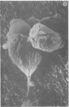
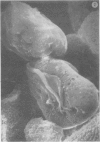
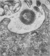
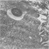
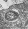
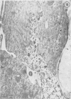
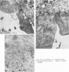
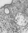
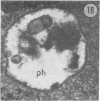
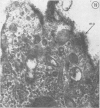
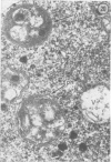
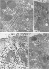
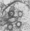
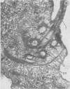
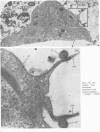
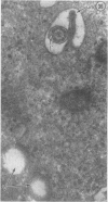
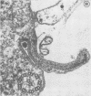
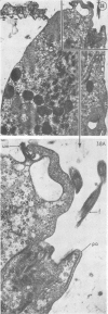
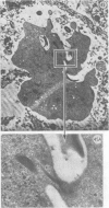
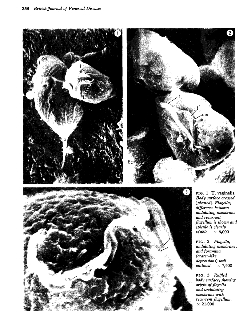
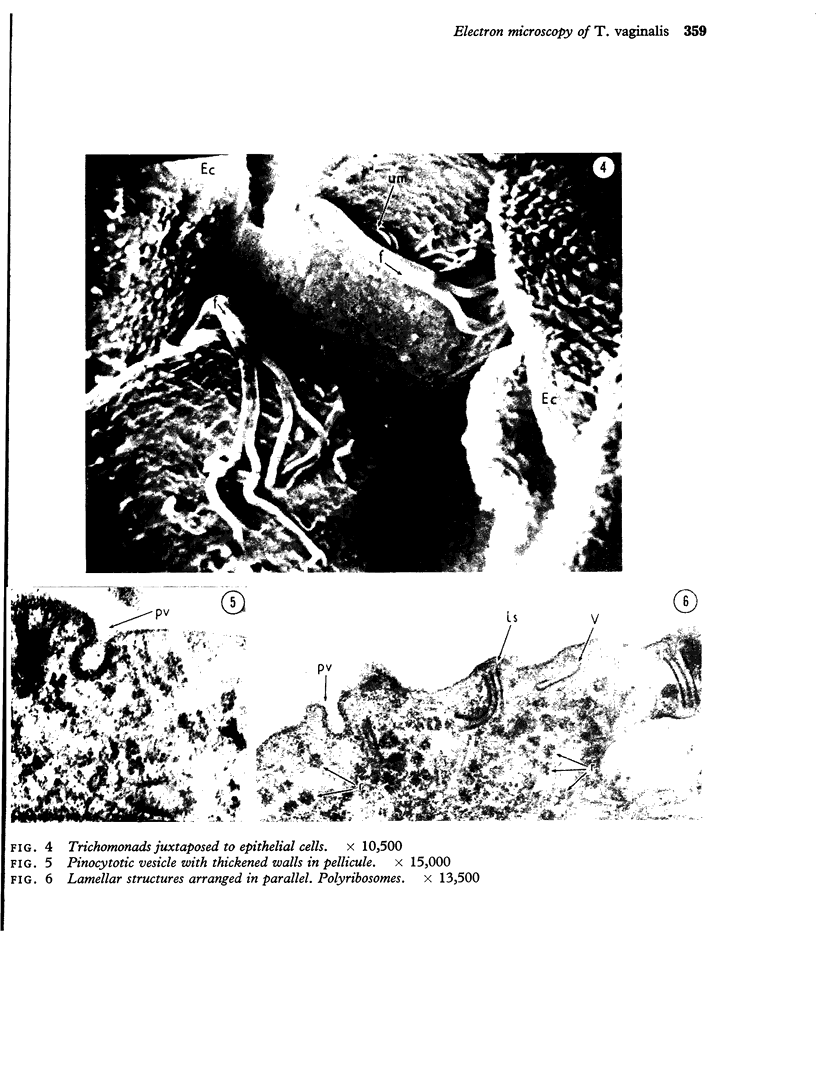
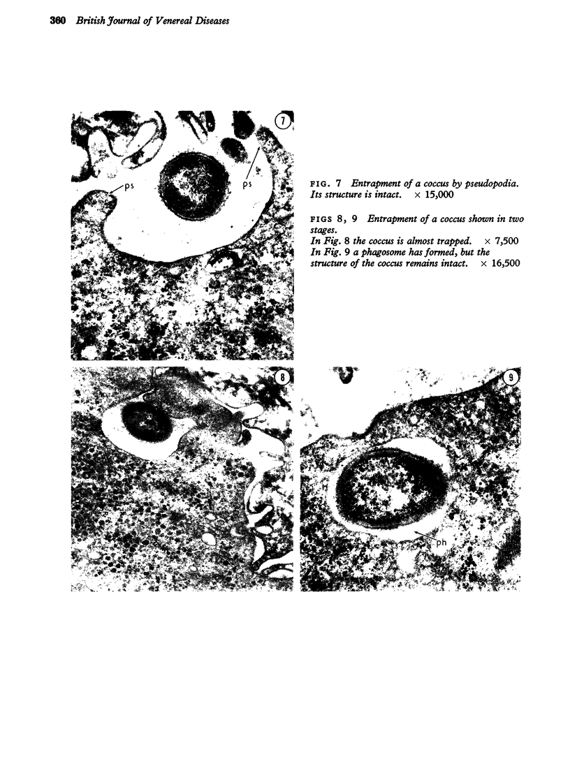
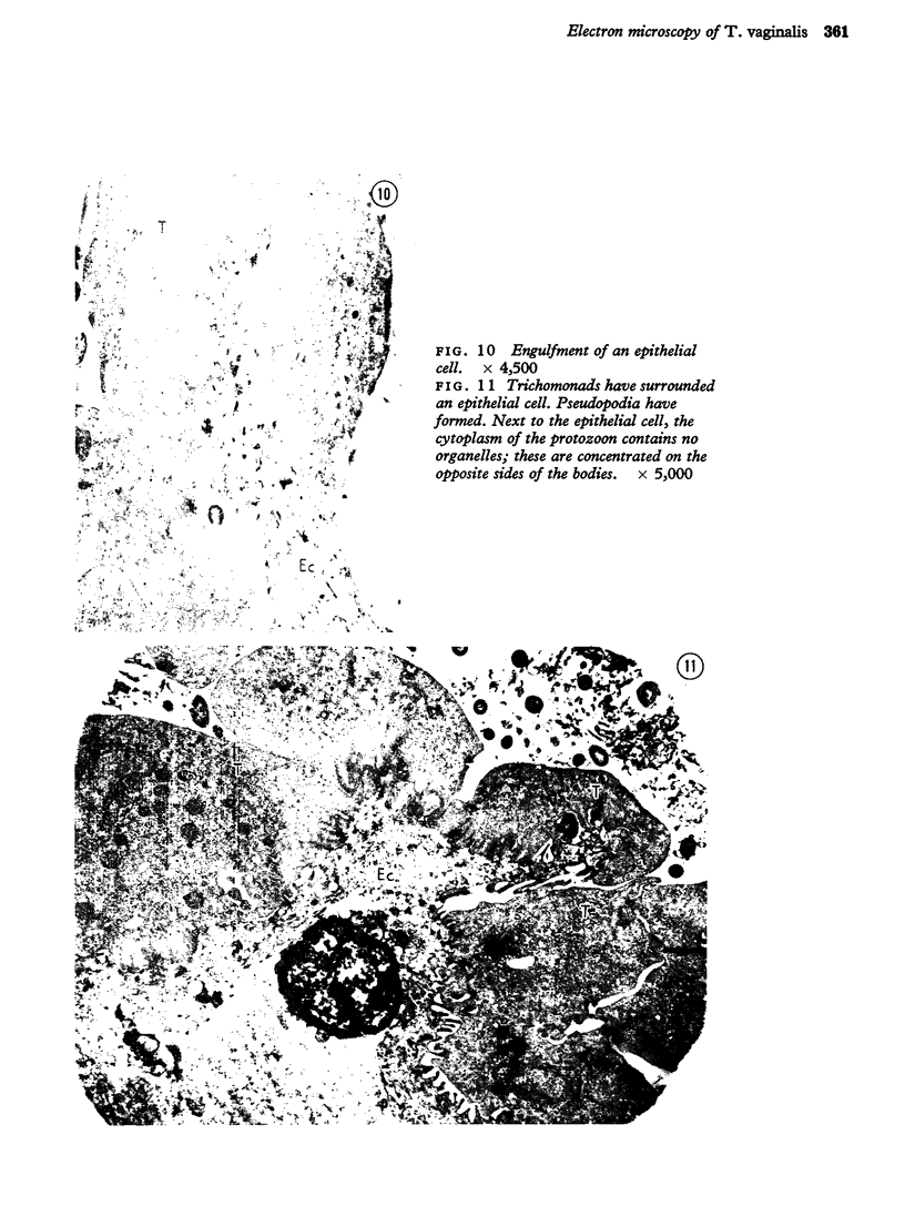
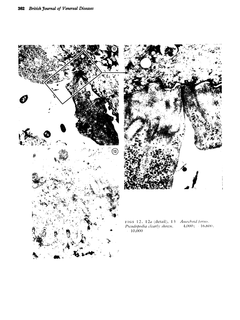
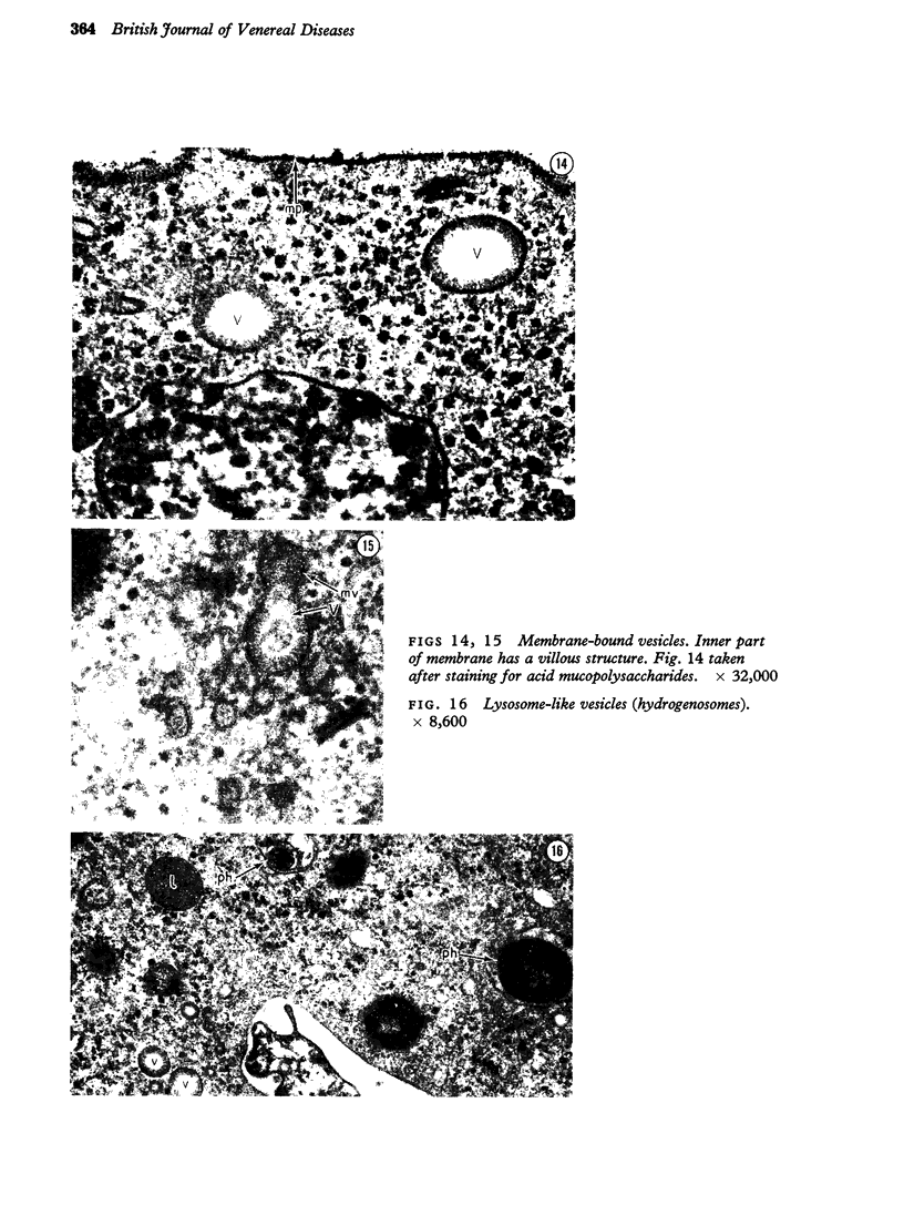
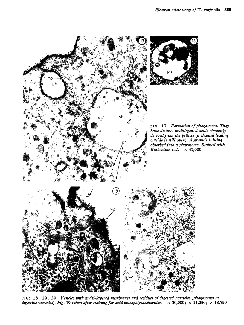
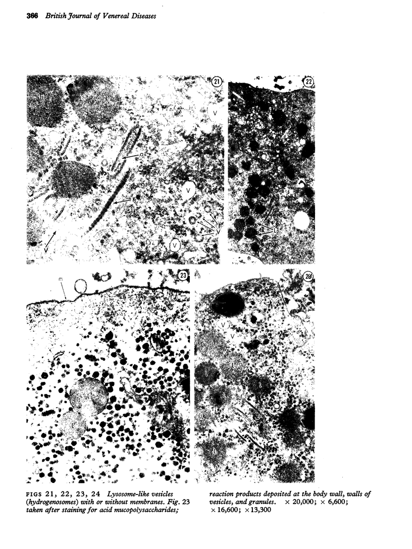
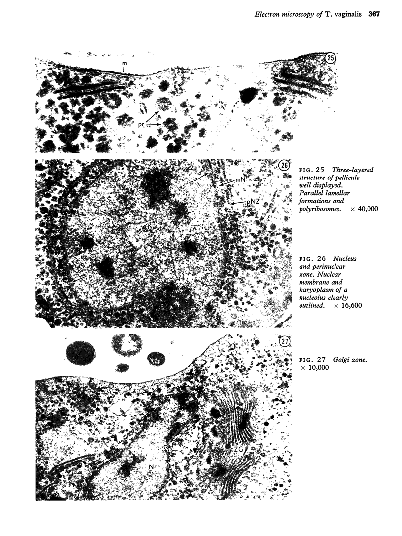
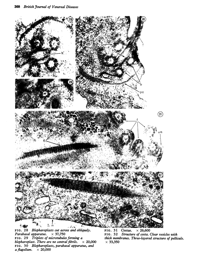
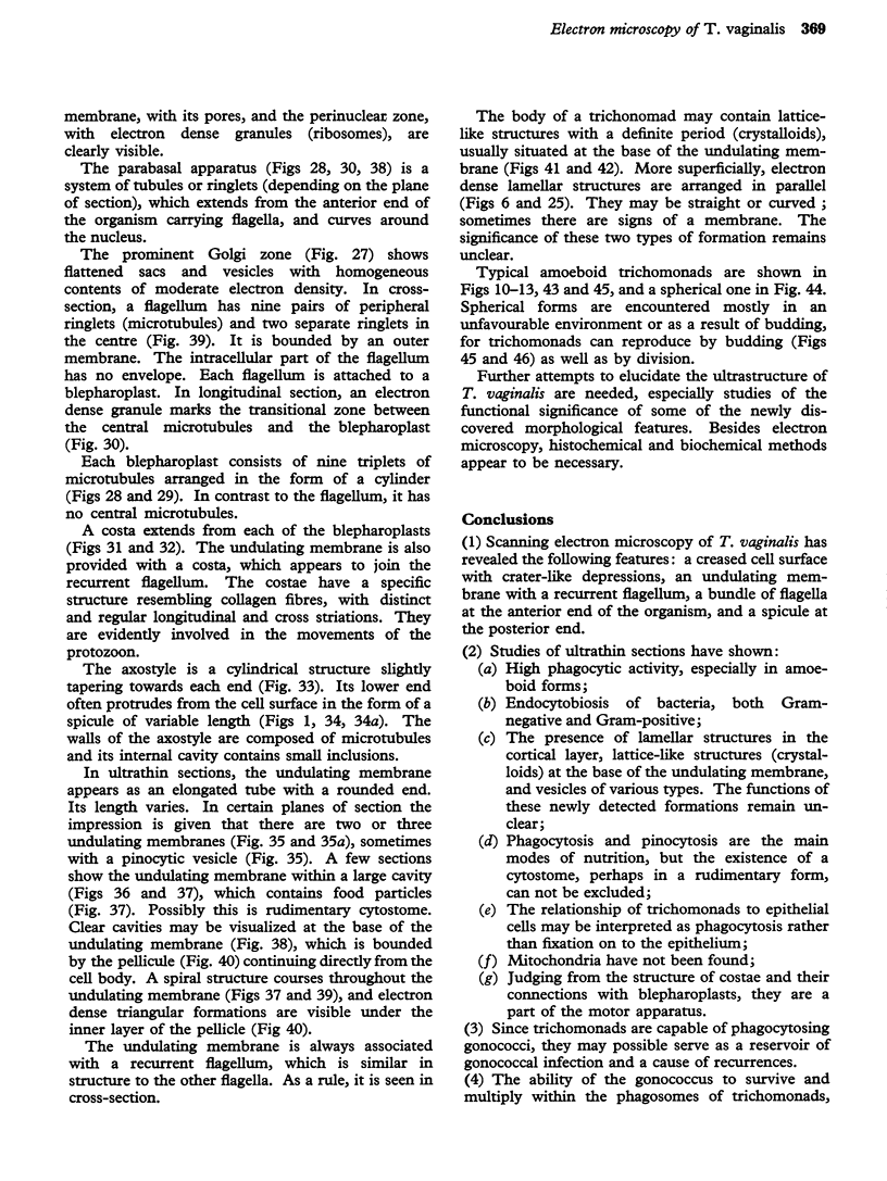
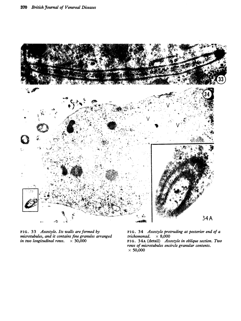
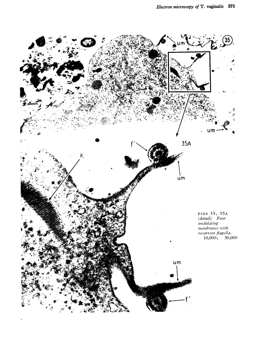
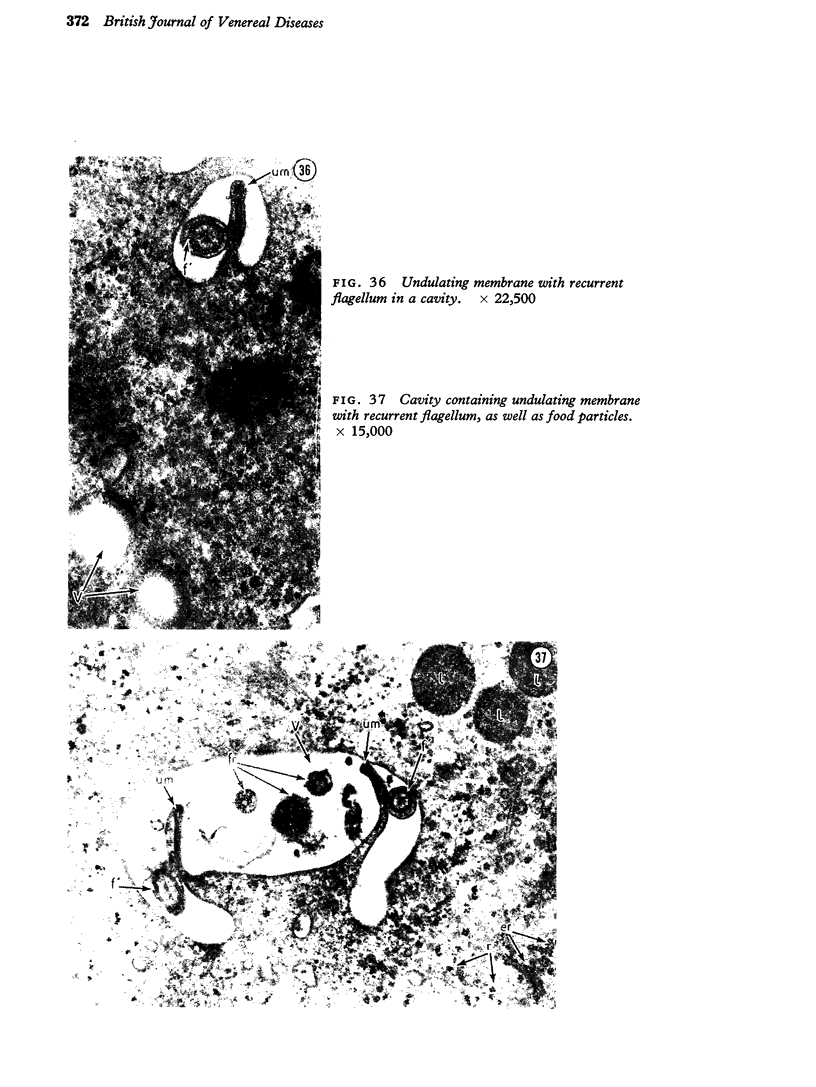
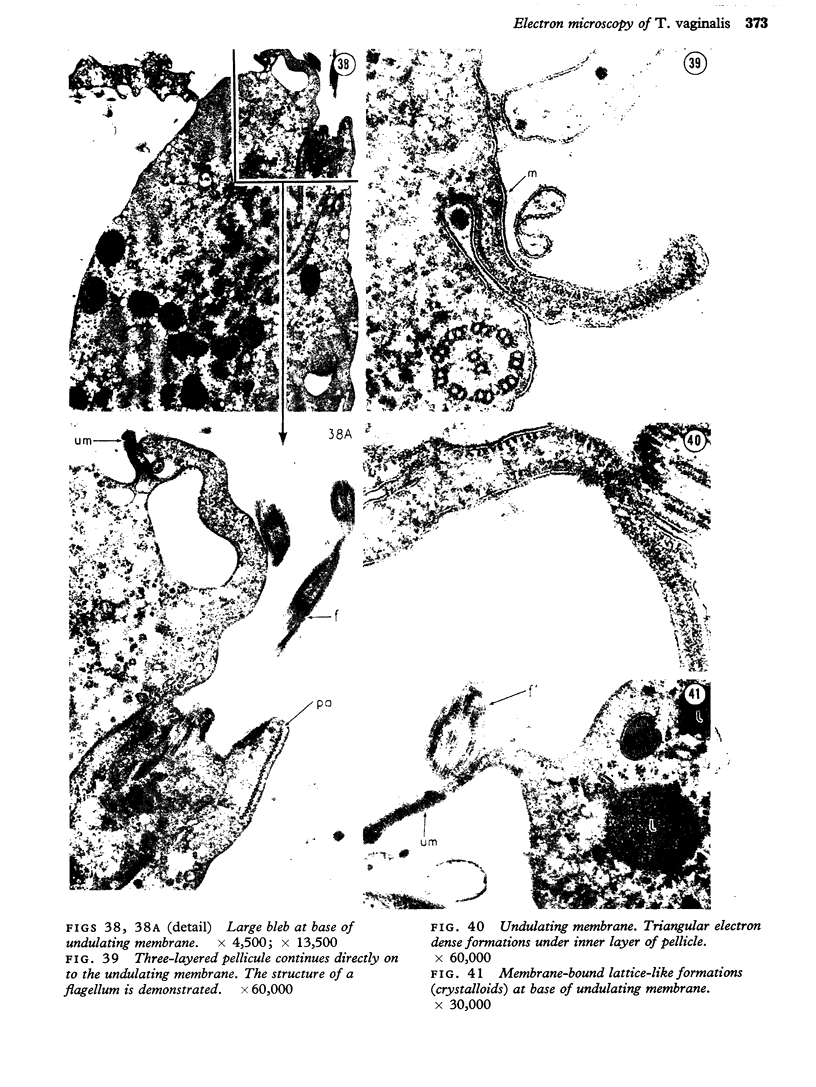
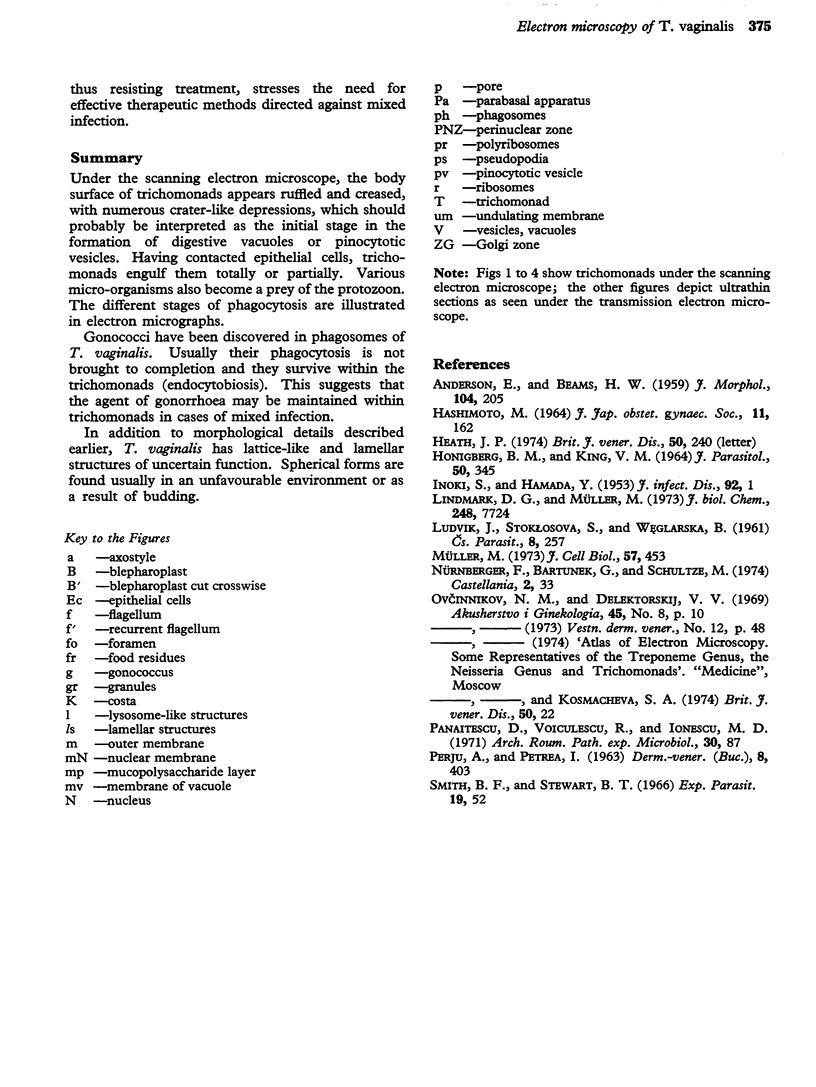
Under the scanning electron microscope, the body surface of trichomonads appears ruffled and creased, with numerous crater-like depressions, which should probably be interpreted as the initial stage in the formation of digestive vacuoles or pinocytotic vesicles. Having contacted epithelial cells, trichomonads engulf them totally or partially. Various micro-organisms also become a prey of the protozoon. The different stages of phagocytosis are illustrated in electron micrographs. Gonococci have been discovered in phagosomes of T. vaginalis. Usually their phagocytosis is not brought to completion and they survive within the trichomonads (endocytobiosis). This suggests that the agent of gonorrhoea may be maintained within trichomonads in cases of mixed infection. In addition to morphological details described earlier, T. vaginalis has lattice-like and lamellar structures of uncertain function. Spherical forms are found usually in an unfavourable environment or as a result of budding.
Full text
PDF
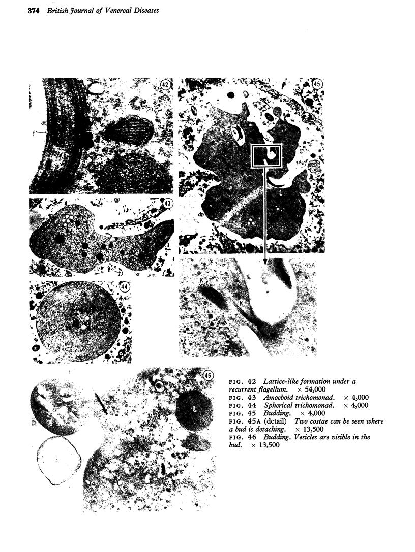
Images in this article
Selected References
These references are in PubMed. This may not be the complete list of references from this article.
- ANDERSON E., BEAMS H. W. The cytology of Tritrichomonas as revealed by the electon microscope. J Morphol. 1959 Mar;104:205–235. doi: 10.1002/jmor.1051040203. [DOI] [PubMed] [Google Scholar]
- HONIGBERG B. M., KING V. M. STRUCTURE OF TRICHOMONAS VAGINALIS DONN'E. J Parasitol. 1964 Jun;50:345–364. [PubMed] [Google Scholar]
- Heath J. P. Letter: Ultrastructure of T. vaginalis. Br J Vener Dis. 1974 Jun;50(3):240–241. doi: 10.1136/sti.50.3.240. [DOI] [PMC free article] [PubMed] [Google Scholar]
- INOKI S., HAMADA Y. Experimental transmission of Trichomonas vaginalis (pure culture) into mice. J Infect Dis. 1953 Jan-Feb;92(1):1–3. doi: 10.1093/infdis/92.1.1. [DOI] [PubMed] [Google Scholar]
- Lindmark D. G., Müller M. Hydrogenosome, a cytoplasmic organelle of the anaerobic flagellate Tritrichomonas foetus, and its role in pyruvate metabolism. J Biol Chem. 1973 Nov 25;248(22):7724–7728. [PubMed] [Google Scholar]
- Müller M. Biochemical cytology of trichomonad flagellates. I. Subcellular localization of hydrolases, dehydrogenases, and catalase in Tritrichomonas foetus. J Cell Biol. 1973 May;57(2):453–474. doi: 10.1083/jcb.57.2.453. [DOI] [PMC free article] [PubMed] [Google Scholar]
- Panaitescu D., Voiculescu R., Ionescu M. D. Sur l'ultrastructure de Trichomonas vaginalis. Arch Roum Pathol Exp Microbiol. 1971 Mar;30(1):87–106. [PubMed] [Google Scholar]


