ABSTRACT
A conspicuous roadblock to studying marine bacteria for fundamental research and biotechnology is a lack of modular synthetic biology tools for their genetic manipulation. Here, we applied, and generated new parts for, a modular plasmid toolkit to study marine bacteria in the context of symbioses and host-microbe interactions. To demonstrate the utility of this plasmid system, we genetically manipulated the marine bacterium Pseudoalteromonas luteoviolacea, which stimulates the metamorphosis of the model tubeworm, Hydroides elegans. Using these tools, we quantified constitutive and native promoter expression, developed reporter strains that enable the imaging of host-bacteria interactions, and used CRISPR interference (CRISPRi) to knock down a secondary metabolite and a host-associated gene. We demonstrate the broader utility of this modular system for testing the genetic tractability of marine bacteria that are known to be associated with diverse host-microbe symbioses. These efforts resulted in the successful conjugation of 12 marine strains from the Alphaproteobacteria and Gammaproteobacteria classes. Altogether, the present study demonstrates how synthetic biology strategies enable the investigation of marine microbes and marine host-microbe symbioses with potential implications for environmental restoration and biotechnology.
IMPORTANCE
Marine Proteobacteria are attractive targets for genetic engineering due to their ability to produce a diversity of bioactive metabolites and their involvement in host-microbe symbioses. Modular cloning toolkits have become a standard for engineering model microbes, such as Escherichia coli, because they enable innumerable mix-and-match DNA assembly and engineering options. However, such modular tools have not yet been applied to most marine bacterial species. In this work, we adapt a modular plasmid toolkit for use in a set of 12 marine bacteria from the Gammaproteobacteria and Alphaproteobacteria classes. We demonstrate the utility of this genetic toolkit by engineering a marine Pseudoalteromonas bacterium to study their association with its host animal Hydroides elegans. This work provides a proof of concept that modular genetic tools can be applied to diverse marine bacteria to address basic science questions and for biotechnology innovations.
KEYWORDS: CRISPRi, golden gate, violacein, metamorphosis, tubeworm, Hydroides, modular, marine, symbiosis
INTRODUCTION
Marine bacteria are a valuable and currently under-utilized resource for environmental restoration (1 - 6) and bioprospecting (7, 8), especially considering their influence on biogeochemical cycles (9) and their vital role in evolution through symbioses with eukaryotes (10). While advances in metagenomic sequencing have enabled a deeper exploration of microbial diversity and gene content (11, 12), genetic tools to explore functions in marine bacteria remain scarce.
Effective genetic engineering approaches in model microbial species, such as Escherichia coli, utilize standardized and modular cloning toolkits (13 - 19), which leverage aligned plasmid parts based on the ordered pairings of restriction site overhangs to enable innumerable mix-and-match plasmid assembly options. However, such modular genetic tools have not yet been applied to most marine bacterial species. Thus, adapting and applying standardized molecular cloning tools for studying marine bacteria can provide a framework for addressing functional questions for basic science and biotechnology.
Marine bacteria are of specific interest as targets for genetic tool development due to their ability to produce diverse bioactive metabolites (20), their prominent associations in aquatic microbiomes, and their involvement in host-microbe symbioses (21 - 23). Alphaproteobacteria and Gammaproteobacteria, in particular, are the most abundant orders in the ocean (12) and are prominent members of the microbiomes of animals such as phytoplankton (12), tubeworms (21), and corals (24).
Of particular interest as targets for genetic manipulation are marine Pseudoalteromonas species because they produce a number of bioactive secondary metabolites (8, 25 - 29) and are often found in association with marine invertebrates (30 - 36). Pseudoalteromonas species are known to engage in a transient symbiosis called bacteria-stimulated metamorphosis, whereby surface-bound bacteria promote the larval-to-juvenile life cycle transition in invertebrates such as tubeworms and corals (37, 38). Pseudoalteromonas luteoviolacea stimulates the metamorphosis of the tubeworm Hydroides elegans (39, 40) by producing syringe-like protein complexes called Metamorphosis-Associated Contractile structures (MACs). MACs stimulate tubeworm metamorphosis by injecting an effector protein termed Mif1 into tubeworm larvae (40 - 42). Genes encoding the MACs structure are found in the P. luteoviolacea genome as a gene cluster encoding structural components, such as the macB baseplate and macS sheath, as well as the protein effector gene mif1 (41). Despite the significant insights gained by using genetics in P. luteoviolacea, new genetic tools are needed to further dissect the function of MACs and their stimulation of tubeworm metamorphosis.
In this work, we utilize a modular plasmid toolkit, and contribute new Marine Modification Kit (MMK) plasmid parts, to study bacteria-stimulated metamorphosis in the Gammaproteobacterium, P. luteoviolacea. We demonstrate the broader utility of this approach by conjugating MMK plasmids into marine Alphaproteobacteria and Gammaproteobacteria that have been shown previously to be involved in diverse host-microbe interactions.
RESULTS
Toolkit-enabled quantitative promoter expression in P. luteoviolacea
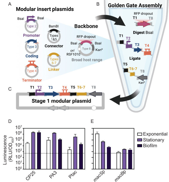
To test the application of modular genetic tools in marine bacteria, we identified a set of preexisting parts from the Yeast Toolkit and Bee Toolkit platforms (17, 18) and used Golden Gate Assembly (14) for rapid, modular construction of plasmids (Fig. 1A through C). Each type of part is defined by its functional role (e.g., promoter and coding sequence [CDS]) and directional 4 bp overhangs generated by flanking Type IIS (BsaI) restriction sites. The modular parts include Type-1 and Type-5 stage-2 connectors with BsmBI recognition sites (17, 18), a Type-2 promoter with ribosome binding site (RBS), a Type-3 protein CDS (e.g., gfp and Nanoluciferase), a Type-4 terminator, an optional Type-6 repressor and Type-7 promoter with RBS, and a Type-8 backbone. Preexisting Type-8 backbones are available with different origins of replication (ColE1 and RSF1010) and antibiotic resistance markers (ampicillin, kanamycin, or spectinomycin resistance) (17, 18). For this work, we selected a broad-host-range (BHR) plasmid backbone containing a kanamycin resistance gene, a reporter CDS (fluorescent gfp-optim1, mRuby, or Nanoluciferase [NLuc]), T7 terminator, and a stage-2 assembly connector. The backbone selected has an RSF1010 origin of replication which is known to replicate in a broad range of Gram-positive and Gram-negative bacterial hosts at a copy number of 10–12 per chromosome and also contains a promiscuous origin of transfer and the plasmid-encoded mobilization genes repA, repB, repC, and mobC (43, 44). An auxotrophic MFDλpir strain was used as the E. coli donor, thus obviating the need to generate antibiotic-resistant recipient strains to counter select E. coli donor cells after conjugation (45).
Fig 1.
Schematic overview of the modular plasmid system and quantitative promoter measurements. (A) Schematic representation of the modular golden gate assembly plasmid parts with flanking BsaI cut sites (dashed lines). Overlapping 4 bp overhangs are color coordinated. The modular broad-host-range (BHR) backbone (pBTK402) contains inverted BsaI cut sites and an RFP dropout. (B) Golden Gate Assembly is performed in a one-tube reaction by digesting the backbone and insert part plasmids with BsaI and ligating with T4 ligase. (C) A modular stage-1 plasmid is complete when all overlapping inserts are successfully assembled in order. (D and E) Luciferase assays of P. luteoviolacea strains expressing plasmids with different promoters during exponential, stationary, or biofilm growth driving a Nanoluciferase (NLuc) gene where (D) shows CP25-NLuc-T7, PA3-NLuc-T7, Ptac-NLuc-T7 and (E) compares native MACs macS and macB promoters. Luminescence, as relative luminescence units (RLUs), is normalized to optical density at 600 nm (OD600) and plotted on a log base 10 scale. The dashed line indicates P. luteoviolacea cells expressing a non-luminescent plasmid as represented by the dotted line (Y = 524 RLU/OD600). Plotted is the mean of three biological replicates. Error bars indicate standard deviations.
To apply the modular genetic tools in a marine symbiosis model, we tested the expression of five promoters in P. luteoviolacea. We assembled plasmids with each promoter fused to NLuc and conjugated the plasmids into P. luteoviolacea. We utilized two existing BHR promoters, PA3 and CP25, previously shown to work in diverse bee gut microbes (17, 46, 47). We also created a Ptac lacO promoter part (pMMK201), which is a hybrid of the lac and trp promoters amplified from the pANT4 plasmid (48). When P. luteoviolacea with the plasmids were grown in exponential, stationary, or biofilm growth phases, we observed at least 10-fold more luminescence signal compared to the background with all BHR promoters tested (Fig. 1D).
Previous observations have shown that the production of MACs is greatest during the exponential phase of growth when P. luteoviolacea is cultured in rich media (40). However, the expression of mac genes in live cultures has not been previously quantified. To observe the expression of two native mac promoters, we constructed two plasmids with P. luteoviolacea promoters driving the expression of the MACs structural genes; promoters from the MACs sheath (macS promoter, pMMK203) and baseplate (macB promoter, pMMK202) genes. The macSp luciferase reporter strain was elevated 1,000-fold in exponential growth as compared to 100-fold in stationary and 10-fold in biofilm phase, when compared to the detection limit (Fig. 1E). In contrast, the macB, baseplate promoter exhibited similar levels of luminescence among each phase, approximately 10-fold higher than the detection limit (Fig. 1E).
Functional CRISPRi knockdown of secondary metabolite biosynthesis in P. luteoviolacea
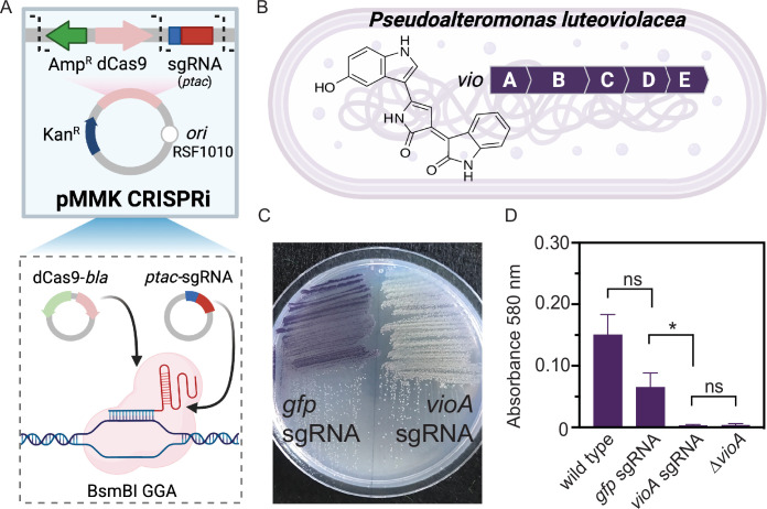
While previous studies in P. luteoviolacea have used gene knockouts to interrogate gene function, these approaches are time-consuming and low-throughput. We therefore tested whether P. luteoviolacea is amenable to gene knockdown via CRISPR interference (CRISPRi) (Fig. 2A and B) (49, 50). As a proof of concept, we targeted the vioA gene that encodes a key enzyme in the biosynthesis of violacein (51), which gives P. luteoviolacea its characteristic purple pigment (Fig. 2B). An assembled plasmid containing dCas9 and a single-guide RNA (sgRNA) targeting vioA (pMMK603) was conjugated into P. luteoviolacea resulting in the visible absence of the purple pigment associated with violacein production on the plate (Fig. 2C). We also created a plasmid containing dCas9 and a sgRNA targeting gfp to test whether the presence of the CRISPRi machinery adversely affected wild-type (WT) P. luteoviolacea or violacein production. No difference was observed between the growth and cell morphology of P. luteoviolacea containing gfp or vioA sgRNA CRISPRi plasmids compared to WT (Fig. S1). WT P. luteoviolacea produced violacein as expected, while P. luteoviolacea with CRISPRi with the gfp sgRNA produced a statistically comparable amount of violacein (adjusted P = 0.26, n = 8, Dunn’s multiple comparison test). A significant reduction of violacein production was observed between cultures of P. luteoviolacea strains expressing the vioA and gfp targeting CRISPRi plasmids (adjusted P = 0.02, n = 8, Dunn’s multiple comparison test) (Fig. 2D). The lack of violacein in the vioA knockdown strain was comparable to that of a P. luteoviolacea strain with an in-frame deletion of vioA (adjusted P = 0.26, n = 8, Dunn’s multiple comparison test) (Fig. 2D). These results demonstrate the successful implementation of CRISPRi for gene knockdown in P. luteoviolacea.
Fig 2.
CRISPRi knockdown of secondary metabolite production in P. luteoviolacea. (A) Schematic representation of modular CRISPRi parts adapted to include dCas9-bla and Ptac sgRNA parts, pMMK601, and pMMK602, respectively. Part plasmids are combined, and a BsmBI Golden Gate Assembly was performed. (B) Schematic representation of the violacein gene cluster vioABCD in P. luteoviolacea and the violacein molecular structure. The CRISPRi system was assembled with an sgRNA targeting the vioA gene (pMMK603) and employed to knock down violacein production in P. luteoviolacea. (C) P. luteoviolacea with gfp (pMMK815) or vioA (pMMK816) sgRNA plasmids grown on marine agar plates. (D) Quantification of violacein production (measured at 580 nm) between P. luteoviolacea containing gfp or vioA sgRNA plasmids. Asterisks indicate significant differences (*P = 0.02, Dunn’s multiple comparisons test). Bars represent the mean of eight total replicates and error bars indicate standard deviations.
Functional CRISPRi knockdown and visualization of P. luteoviolacea during a tubeworm-microbe interaction
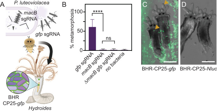
We next tested whether CRISPRi would be functional in the context of a marine host-microbe interaction by targeting the macB gene, which encodes the MACs baseplate, an essential component of the MACs complex that induces tubeworm metamorphosis (39, 40) (Fig. 3A). Biofilm metamorphosis assays were performed comparing P. luteoviolacea strains with sgRNAs targeting macB (pMMK604) or the sgRNA targeting gfp control (Fig. 3B). The strain with sgRNA targeting macB exhibited significantly reduced levels of tubeworm metamorphosis compared to the gfp-sgRNA control (adjusted P < 0.0001, Dunn’s multiple comparisons test, n = 12) (Fig. 3B). The reduction of metamorphosis stimulation in the macB-sgRNA knockdown strain was comparable to that of a P. luteoviolacea strain with an in-frame deletion of macB carrying the gfp-sgRNA control plasmid (adjusted P ≥ 0.99, Dunn’s multiple comparison test, n = 12) (Fig. 3B). These results demonstrate that CRISPRi paired with a modular plasmid system is a viable tool for interrogating gene function during a marine host-microbe interaction.
Fig 3.
Functional knockdown of MACs and visualization of P. luteoviolacea during the tubeworm-microbe interaction. (A) Schematic depicting P. luteoviolacea and the production of MACs, which induce tubeworm metamorphosis. CRISPRi single-guide RNA (sgRNA) targeting the macB MACs baseplate gene prevents MACs from assembling, rendering the bacterium unable to induce metamorphosis. Cells that produce intact MACs are able to induce tubeworm metamorphosis. A strong fluorescent reporter strain (BHR-CP25-gfp) enabled visualization of live tubeworm-bacteria interactions. (B) Bar graph representing biofilm metamorphosis assays with P. luteoviolacea carrying a CRISPRi plasmid targeting macB or gfp and Hydroides tubeworms. A P. luteoviolacea ∆macB strain with a sgRNA targeting gfp and a treatment without bacteria (no bacteria) were included as controls. Biofilm concentrations were made with cells at OD600 0.1. Bars plotted show the average of 12 replicates, performed across three independent experiments. Each well contained 20–40 worms. Error bars indicate standard deviations. Statistical significance between treatments was determined by Dunn’s multiple comparisons test (N = 12). (C and D) Merged fluorescence and DIC micrographs of Hydroides elegans juveniles imaged 24 h after the competent larvae were exposed to inductive biofilms of P. luteoviolacea containing plasmids with (C) CP25-gfp or (D) CP25-NLuc. Strains containing NLuc plasmids were used as a negative control to account for autofluorescence. Yellow arrows show accumulation of fluorescent bacteria in the Hydroides juvenile pharynx. Scale bar is 100 µm.
To date, bacteria have not been visualized during or after the stimulation of metamorphosis in Hydroides. To test whether marine bacteria harboring a toolkit plasmid are amenable to live-cell imaging when in association with juvenile tubeworms, we created biofilms of P. luteoviolacea containing plasmids encoding CP25-gfp-T7 (gfp) or CP25-Nanoluc-T7 (NLuc) and added competent Hydroides larvae. After incubation for 24 h, biofilms of gfp-expressing P. luteoviolacea were clearly observed when visualized by fluorescence microscopy (Fig. 3C). P. luteoviolacea stimulated Hydroides metamorphosis while carrying a modular plasmid and fluorescent bacteria were observed being ingested by the Hydroides juveniles. Bacteria can be seen collecting in the pharynx (Fig. 3C, yellow arrows), then moving in a peristaltic fashion toward the gut (Movie S1). In contrast, bacteria containing a CP25-NLuc-T7 plasmid were difficult to visualize by light microscopy, in the absence of the gfp fluorescent marker (Fig. 3D). Taken together, the modular plasmid system enables live imaging and experimentation during a marine host-microbe interaction.
Applying the modular toolkit in marine Alphaproteobacteria and Gammaproteobacteria
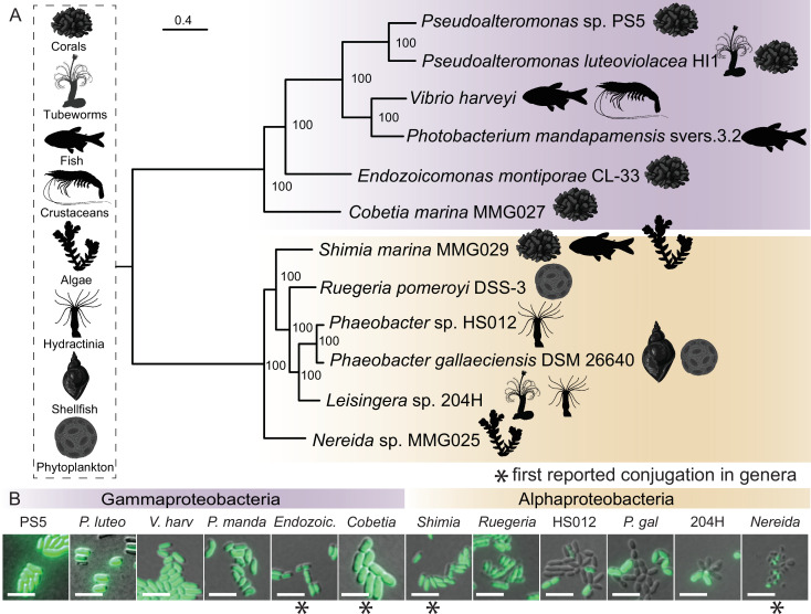
Given the success of genetic manipulation of P. luteoviolacea, we tested whether other marine Proteobacteria might be amenable to conjugation and retention of a modular genetic toolkit plasmid. To this end, we isolated or acquired representative bacteria that are known to engage in symbioses with marine plants or animals in the ocean (Fig. 4A; Tables S1 and S2). To enable genetic selection using antibiotics, we determined the minimum inhibitory concentration for each bacterial strain tested against kanamycin (Table S1). When conjugation was performed using the BHR (RSF1010) plasmid backbone, CP25 promoter, gfp reporter, and T7 terminator, we observed the expression of gfp in 12 marine strains across two proteobacterial classes, four orders, and 10 genera (Fig. 4B). Adaptations to the conjugation protocol and the use of constitutive promoters driving gfp enabled visual confirmation of successful conjugation (Fig. 4B, Materials and Methods).
Fig 4.
Marine Proteobacteria are amenable to plasmid uptake and stable replication of toolkit plasmids. (A) Maximum likelihood phylogeny built using the whole genomes of 12 strains selected for manipulation and successfully conjugated in this study (52, 53). All strains used in this study are known for their interaction with a range of marine biota and the icons depicting their associated host are shown in the vertical box. Gammaproteobacteria strains are highlighted in purple and Alphaproteobacteria strains are shown in gold. Scale bar is 0.4 and bootstraps were generated using the rapid-bootstrapping method (54). The tree was rooted at the midpoint with FigTree (v1.4.4). (B) Fluorescence and DIC overlay micrographs of overnight cultures containing constitutively expressed RSF1010 ori fluorescence vector (CP25-gfp-T7). Scale bar is 5 µm. Stars denote environmental strains that serve as the first reported conjugation for that genera.
DISCUSSION
Modular genetic tools provide insights about bacteria-stimulated metamorphosis
We tested a modular plasmid toolkit on a genetically tractable marine bacterium, P. luteoviolacea, that promotes the metamorphosis of the tubeworm Hydroides elegans (40, 41, 55) and produces several bioactive secondary metabolites (26, 29, 56, 57). We expand the tools available for functional interrogation of bacteria-stimulated metamorphosis in P. luteoviolacea by quantifying gene expression by a luminescence assay (Fig. 1D and E), and using CRISPRi to knock down the secondary metabolite, violacein (Fig. 2C and D), as well as a metamorphosis-associated gene, macB (Fig. 3B) during the bacteria-tubeworm interaction. Distinct patterns of sheath (macSp) (41, 58) and baseplate (macBp) promoter induction suggest distinct mechanisms of gene regulation within the MACs gene cluster. Expression of the sheath gene was sensitive to bacterial mode of growth, while baseplate gene expression appeared static across the growth conditions tested. Although MACs are known to produce two effectors that stimulate tubeworm metamorphosis and kill eukaryotic cells (41, 58), the environmental conditions that promote MACs production remain poorly characterized. The tools developed here could help to characterize the conditions under which P. luteoviolacea MACs are produced or assembled and could help in the development of MACs or other contractile injection systems for use in biotechnology (59, 60). The modular tools in this work open new capabilities for interrogating bacteriology, including the ability to quantify gene expression in live cultures, knock down gene expression for rapid functional testing, and visualize bacteria during an in vivo interaction.
Whether, and how, bacteria and the animal are harmed or benefit from the interaction during bacteria-stimulated metamorphosis remains a prominent question in the field (38, 61, 62). Swimming Hydroides larvae initially encounter and are stimulated to undergo metamorphosis by the bacterial biofilm. And MACs were previously visualized within P. luteoviolacea biofilms by tagging the MACs baseplate with super-folder GFP (40). However, less attention has been put on the interaction between Hydroides and the bacteria after metamorphosis. Previous work by Gosselin et al. has shown that Hydroides is able to feed on bacteria as the sole food source (63). In the present work, we visualize live bacteria surrounding and being ingested by Hydroides juveniles (Fig. 3C) (21). The visualization of transgenic bacteria in Hydroides will enable future lines of research that can help dissect the role of microbiome seeding in bacteria-stimulated metamorphosis. More broadly, our results showcase the feasibility of using a modular plasmid toolkit to test hypotheses about bacteria-stimulated metamorphosis and provide a framework for the interrogation of other bacteria and their products that promote host-microbe symbioses (36, 64, 65).
Toolkit compatibility in marine bacteria
In this work, we explore genetic tractability in 12 ecologically relevant marine bacteria that belong to two Proteobacterial classes (Fig. 4). The Gammaproteobacteria strains conjugated successfully in this study are a selection of symbiosis-associated strains representing five genera (Fig. 4A) (66 - 72). To our knowledge, this is the first report of genetic tractability in strains from the genera Endozoicomonas, Nereida, and Cobetia (Fig. 4B). Endozoicomonas species are among the most abundant bacterial symbionts in some corals and other marine hosts (73 - 75). Related strains of Cobetia have been implicated in thermotolerance against bleaching in coral experiments with probiotic consortium treatments (76). The conjugation of the representative Endozoicomonas and Cobetia strains in this study is a considerable step toward exploring function in coral host-microbiome interactions at a critical time to encourage the restoration of coral reefs (6, 77, 78). The genetic conjugation of Pseudoalteromonas sp. PS5 in this study presents an opportunity to explore secondary metabolite production, including the coral metamorphosis-inducing compound, tetrabromopyrrole (Fig. 4) (36, 79). The Alphaproteobacteria strains tested for compatibility with MMK plasmids fall within the Roseobacter group (Fig. 4A), an ecologically important group of bacteria known to play a role in sulfur and carbon cycling on marine phytoplankton (80 - 82). Roseobacter strains have also been explored as probiotics for the aquaculture industry (83 - 85). We tested the toolkit with the tractable, phytoplankton-associated species of Phaeobacter gallaeciensis (86), and Ruegeria pomeroyi (87), and demonstrated conjugation with invertebrate microbiome-associated strains Phaeobacter sp. HS012 (88) and Leisingera sp. 204H (89) (Fig. 4). Using modified Shimia sp. may be of interest for future coral microbiome studies (90 - 93). Species in the Nereida genus have been isolated from kelp (94) and are associated with gall formations (95, 96). Tractability in this strain could help guide further understanding of microbe-seaweed interactions (97, 98), kelp aquaculture, and the development of kelp probiotics (99). In summary, the development of methods and established tractability of several new strains and genera have significant implications for the future of bacterial genetic development in established and emerging symbiosis systems.
Future modifications
The modularity of the plasmid toolkit enables the potential for creating new plasmids that are compatible with the existing system to boost functionality. For example, the addition of backbone plasmid parts (Type-8) with different origins of replication and selectable markers could allow utilization in bacteria that are naturally resistant to the antibiotics used in this and prior works (17, 18). We have created a Type-8 Tn10 transposon backbone for stable integration of toolkit parts into the genomes of marine bacteria and used this part to integrate a fluorescent gfp marker into the genome of Pseudoalteromonas sp. PS5 (79). Type-8 parts like this could be used to tag and track marine bacteria for studying host-microbe interactions in the future.
The current promoter driving dCas9 is constitutive. However, adding an inducible promoter driving dCas9 (e.g., PBAD for arabinose induction) would allow the CRISPRi system to be controllable. The expression of gfp was not uniformly observed in the Phaeobacter, Leisingera, and Nereida strains (Fig. 4B). However, the plasmid toolkit could be used to identify plasmid components that would produce uniform expression (e.g., different origins of replication, selectable markers, promoters, etc.). In the future, more strains may be tested for manipulation with the present toolkit plasmids for applying genetics in a broader array of bacteria types.
Conclusion
The modular plasmid toolkit described here provides a basis for streamlining the genetic manipulation of marine bacteria for basic and applied purposes. These tools reveal new possibilities to study marine microbes in the context of plant and animal interactions, or with challenging and diverse non-model bacteria, ultimately helping us harness marine microbes for research, bioproduction, and biotechnology.
MATERIALS AND METHODS
Bacterial culture
A list of strains used in this study, isolation sources, accession numbers, and minimum inhibitory concentration can be found in Table S1. Environmental strains of marine bacteria were isolated and cultured on Marine Broth (MB) 2216 (BD Difco) and or natural seawater tryptone (NSWT) media (1 L 0.2 µm filtered natural seawater from Scripps Pier, La Jolla, CA, 2.5 g tryptone, 1.5 g yeast extract, 1.5 mL glycerol). MB and NSWT media are used interchangeably throughout the study; however, the experiments were always conducted using only one media type. Marine bacteria were incubated between 25°C and 30°C, and cultures were shaken at 200 rpm. All liquid cultures were inoculated with a single colony and incubated between 16 and 18 h, unless otherwise indicated. E. coli SM10λpir and S17-1λpir were cultured in LB (Miller, BD Difco) at 37°C, shaking at 200 rpm. E. coli MFDλpir (45) was cultured in LB supplemented with 0.3 mM Diaminopimelic acid (DAP). For E. coli, antibiotic selections with ampicillin, kanamycin, and chloramphenicol were performed using a concentration of 12.5 µg mL−1.
Plasmid construction and assembly
Golden Gate Assembly-compatible parts from the BTK, YTK (17, 18), and MMK used in this work can be found in Table S3. New plasmid parts were made by PCR amplifying insert and backbone fragments and combining them with Gibson Assembly with a 2:1 ratio (20 fmol insert: 10 fmol backbone) (100). PCR amplification was performed with custom primers (Table S4), a high-fidelity DNA polymerase (PrimeSTAR GXL, Takara), and purified using a DNA Clean and Concentrator kit (Zymo Research). Part plasmids were assembled to make a stage 1 plasmid using Golden Gate Assembly, with T4 DNA ligase (Promega) and either BsaI or BsmBI (New England Biolabs), depending on the construct. Single-tube assembly was performed by running the following thermocycler program (BsaI/BsmBI): 37/42°C for 5 min, 16°C for 5 min, repeat 30×, 37/55°C for 10 min, and 80°C for 10 min. The assemblies were directly electroporated into S17-1λpir cells, confirmed by colony PCR (EconoTaq PLUS Green, LGC Biosearch) with internal primers, and then electroporated into MFDλpir cells for conjugation. To facilitate assembly for and expression of CRISPRi parts in P. luteoviolacea, we moved the BsmBI cut site in the dCas9 part plasmid (pBTK614) to a location where the existing bla gene will be retained in the assembled plasmid (pMMK601), and thus also conferring resistance to ampicillin. In the sgRNA plasmid (pBTK615), we replaced the existing PA1 promoter with the Ptac promoter (including −35 and −10 sequences but excluding lacO), which drives the sgRNA expression (pMMK602). The CRISPRi assemblies were electroporated directly into SM10λpir cells and shuttled to MFDλpir cells for conjugation.
Biparental conjugation in marine bacteria
E. coli donor strains (MFDλpir or SM10λpir) containing the mobilizable plasmids were grown under antibiotic selection in LB with the appropriate supplements (including 0.3 mM DAP for E. coli MFDλpir). Conjugations were performed as previously described (17) with modifications for culturing marine bacteria. Briefly, several colonies of the recipient strains were inoculated and grown overnight in liquid culture. Recipient and donor cultures were spun down (4,000 × g for 2 min) in a 1:1 OD600 ratio. All donor supernatant was removed leaving only the cell pellet. All but 100 µL of the recipient supernatant is removed, and the cell pellet is resuspended. The recipient suspension was transferred to the donor pellet, which was resuspended with the recipient cells. Two 50 µL spots are plated onto NSWT (supplemented with 0.3 mM DAP for MFDλpir-mediated conjugations) and incubated overnight at 25°C with the lids facing up. The next day, spots were scraped up with a pipette tip and resuspended in 500 µL of liquid marine media and 100 µL was plated onto marine media containing antibiotic selection, according to the minimum inhibitory concentration (Table S1). Streptomycin-resistant P. luteoviolacea (Table S1) were conjugated with E. coli SM10λpir, and counterselection was performed with 100–200 µg/mL streptomycin. All other marine bacteria (Table S1) were conjugated with E. coli MFDλpir, and transconjugant selection was performed in the absence of DAP. Several of the bacteria take longer to grow or do not reach a high optical density (i.e., Endozoicomonas, Ruegeria, and Nereida) in culture. Slower-growing marine bacteria were conjugated by growing larger 50 mL initial volumes of culture and spinning down the entire culture to reach 1:1 (donor:host) ratios.
Phylogeny
Strains or close representative strains used in this study were compiled into a genome group on PATRIC v3.6.12 (101). A whole genome phylogenetic codon tree composed of 100 single-copy genes (102) was performed using the Phylogenetic Tree Service (103 - 105). A maximum likelihood phylogeny was generated using the best protein model found by RaxMLv8.2.11 (106), which was LG. Bootstraps were generated using the rapid bootstrapping algorithm with the default of 100 resamples (54). The tree was visualized with FigTree v1.4.4. and was rooted at the mid-line.
Growth curve
Pseudoalteromonas luteoviolacea ∆vioA and WT were grown on MB agar plates and incubated overnight at 25°C. P. luteoviolacea strains expressing CRISPRi plasmids were grown on MB agar plates with 200 µg mL−1 of kanamycin and grown overnight at 25˚C. Single colonies were picked and inoculated into 5 mL of MB liquid media with the respective antibiotics listed above. Two biological replicate cultures were inoculated for each strain by picking different colonies from the agar plate and inoculating separate 5 mL cultures. Cultures were incubated at 25°C for 18 h shaking at 200 rpm. From the initial cultures, a subculture was created by performing a 1:25 dilution into the subculture. The subculture consisted of 25 mL of MB liquid media and 1 mL of original culture along with the respective antibiotics into a 125-mL flask. Subcultures were incubated at 25°C shaking at 200 rpm throughout the growth curve experiment. Optical density (OD) at a wavelength of 600 nm was measured from the subculture every half hour for the first 5 h and then measured every hour until 10 h with a final measurement at 24 h.
Luciferase culture and assay
P. luteoviolacea containing plasmids with constitutive or native promoters driving Nanoluciferase (NLuc) were inoculated into 5 mL of MB or NSWT media with appropriate antibiotics and grown at 25°C at 200 rpm for 24 h. Each biological replicate was represented by a separate culture. Cultures used for the growth phase assay were inoculated as a 1:100 dilution with the appropriate antibiotic, and then incubated at 25°C and shaking at 200 rpm. The luminescence of cultures was measured at exponential (OD600 0.35–1.0), early stationary (OD600 1.0–1.45), or late stationary (OD600 2.38–2.54) phases. For biofilm cultures, 1.5 mL of stationary-phase culture was pelleted and plated as a single spot on NSWT or MB plates. Biofilm plates were incubated at 20–25°C for 24–28 h. Each spot was scraped with a pipette tip and resuspended in 200 µL of NSWT or MB media before being resuspended in NSWT or MB. Luciferase reactions were performed with 100 µL of bacterial culture or biofilm resuspension aliquoted into opaque white walled 96-well plates (Corning #3642), with a modified protocol as written for Promega Nano-Glo Live Cell Assay System (Promega, catalog #N2011). Briefly, bacteria and the final reagent mix (2.5 µL of Nano-Glo LCS dilution buffer, 0.5 µL of Nano-Glo live cell substrate, and 17.5 µL of water) were read at a 1:1 ratio. Luminescence was measured on a Molecular Devices Microplate FilterMax F5 reader with a custom program on the Softmax Pro 7 software. Readings were done on the kinetic luminescence mode at 2 min intervals for 20 min in total, using a 400-ms integration time, a 1-mm height read, and no other optimization or shaking settings. The detection limit is defined as the average expression of P. luteoviolacea cells expressing a non-luminescent plasmid across growth conditions. Raw data were normalized to the OD600 of the culture used and plotted with an N = 3 biological replicates.
Violacein extraction
The specified P. luteoviolacea strains were struck onto NSWT media containing 200 µg mL−1 of streptomycin and kanamycin and incubated overnight at 25°C. Single colonies were inoculated into 5 mL of liquid media containing the same antibiotic concentrations. Cultures were incubated at 25°C, shaking at 200 rpm between 18 and 20 h. Cultures were removed from the incubator and standardized to an OD600 of 1.5. The cells were pelleted, and the supernatant was removed. The cell pellet was resuspended in 200 µL of 100% ethanol. The resuspended cells were pelleted and the supernatant containing the crude extract was recorded on a BioTek Synergy HT plate reader (Vermont, USA) using the Gen5 program (v2.00.18) with an endpoint reading at 580 nm.
Microscopy
Microscopy was performed using a Zeiss Axio Observer.Z1 inverted microscope equipped with an Axiocam 506 mono camera and Neofluar10x/0.3 Ph1/DICI (Hydroides co-cultures) or Apochromat 100×/1.4 Oil DICIII (bacteria only) objectives. The Zeiss HE eGFP filter set 38 was used to capture GFPoptim-1 expression and Zeiss HE mRFP filter set 63 was used to capture mRuby2 expression. For Nanoluciferase controls, images were captured using the same fluorescence exposure times as the gfp optim-1 and mRuby2 labeled strains of the same species.
Bacterial culture (2 µL) was added to freshly prepared 1% saltwater low-melt agarose (Apex catalog #20-103, Bioresearch products) pads on glass slides and coverslips were placed on top. Hydroides elegans were prepared in visualization chambers (Lab-Tek Chambered Coverglasses catalog #155411PK) with bacteria and imaged.
Hydroides elegans culture
Hydroides elegans adults were collected from Quivira Basin, San Diego, CA, USA. The larvae were cultured and reared as previously described (40, 107). Larvae were maintained in beakers containing filtered artificial seawater (35 PSU) and were given new beakers with water changes daily. The larvae were fed live Isochrysis galbana and cultures were maintained as described previously. The larvae were used for metamorphosis assays once they reached competency (between 5 and 7 d old) (108).
Hydroides elegans metamorphosis assays
Biofilm metamorphosis assays were performed using previously described methods (39, 40, 109). Briefly, bacteria were struck onto MB plates with 300 µg mL−1 kanamycin as appropriate and were incubated overnight at 25°C. Up to three single colonies were inoculated into liquid broth and incubated overnight (between 15 and 18 h), shaking at 200 rpm. Cultures were pelleted at 4,000 × g for 2 min, the spent media were removed, and the cell pellets were washed twice with filtered artificial sea water (ASW). The concentration of the cells was diluted to OD600 of 0.1, and four 100 µL aliquots of the cell concentrate were added to 96-well plates. The cells were given between 2 and 3 h to form biofilms, then the planktonic cells were removed and the adhered cells were washed twice with filtered ASW. Between 20 and 40 larvae were added to each well in 100 µL of filtered ASW. Metamorphosis was scored after 24 h. Three biological replicates were performed on different days using separate Hydroides larvae originating from different male and female animals.
Chambered metamorphosis assays were performed using the same preparation principles as described above with the following modifications. Visualization chambers (Lab-Tek, catalog # 155411) were used for setting up the metamorphosis assay, then subsequently imaged. Inductive strains containing constitutively expressed gfp/mRuby/NLuc plasmids were struck out onto MB media containing 300 µg mL−1 kanamycin. Several colonies were inoculated into 5 mL MB media with antibiotics. Cultures were grown for 18 h and cells were washed and allowed to form biofilms as described above. Cell concentrations ranging between OD600 0.1 and 0.5 were used to elicit optimal metamorphosis. Larvae were concentrated and the resident filtered ASW was treated with 300 µg mL−1 kanamycin. Larvae were imaged 24 h later.
Online protocols
Selected protocols used in this study can be accessed on the Shikuma Lab protocols.io page: https://www.protocols.io/workspaces/shikuma-lab-sdsu (110 - 112).https://www.protocols.io/workspaces/shikuma-lab-sdsu https://www.protocols.io/workspaces/shikuma-lab-sdsu
ACKNOWLEDGMENTS
We thank current and former Shikuma Lab members who helped with cloning and providing feedback for this paper, including Taylor Darby, Milagros Esmerode, Nicole Jacobson, and Dr. Kate Nesbit. We thank Dr. Alison Gould, Dr. Stanley Maloy, and Dr. Kristen Marhaver for their contribution of strains or samples for this study. We thank Dr. Alyssa Demko, Dr. Jennifer Sneed, Dr. Christian Voolstra, Dr. Claudia Pogoreutz, Dr. Anny Càrdenas, Dr. Jennifer Doudna, Dr. Valerie Paul, and Dr. Benjamin Rubin for their feedback on the project and manuscript. Schematic figures were created in part using Biorender.com (license agreement numbers: QO25BCR8B5, PU25BCRD05, OH25BCRM4P, and CM25BCRQ3B).
This work was supported by the National Science Foundation (2017232404, A.T.A.; 1942251, N.J.S.; and OCE-1837116, B.S.M.), the Gordon and Betty Moore Foundation (GBMF9344 to N.J.S.; https://doi.org/10.37807/GBMF9344), Office of Naval Research (N00014-20-1-2120 to N.J.S.), the National Institutes of Health, NIGMS (R35GM146722 to N.J.S. and R01ES030316 to B.S.M.), and the Alfred P. Sloan Foundation, Sloan Research Fellowship (N.J.S.).
A.T.A. and N.J.S. are coinventors on provisional U.S. patent application serial number 63/323653, entitled "Genetic Engineering of Marine Bacteria for Biomaterial Production, Probiotic Use in Aquaculture and Marine Environmental Restoration" and assigned to San Diego State University Research Foundation.
Contributor Information
Nicholas J. Shikuma, Email: nshikuma@sdsu.edu.
Edward G. Ruby, University of Hawaii at Manoa, Honolulu, Hawaii, USA
DIRECT CONTRIBUTION
This article is a direct contribution from Bradley S. Moore, a Fellow of the American Academy of Microbiology, who arranged for and secured reviews by Blake Ushijima, University of North Carolina Wilmington, and Shengda Zhang, Danmarks Tekniske Universitet.
SUPPLEMENTAL MATERIAL
The following material is available online at https://doi.org/10.1128/mbio.01502-23.
Tables S1 to S4, Fig. S1, and Movie S1 legend.
Hydroides feeding.
ASM does not own the copyrights to Supplemental Material that may be linked to, or accessed through, an article. The authors have granted ASM a non-exclusive, world-wide license to publish the Supplemental Material files. Please contact the corresponding author directly for reuse.
REFERENCES
- 1. Prado S, Romalde JL, Barja JL. 2010. Review of probiotics for use in bivalve hatcheries. Vet Microbiol 145:187–197. doi: 10.1016/j.vetmic.2010.08.021 [DOI] [PubMed] [Google Scholar]
- 2. D’Alvise PW, Lillebø S, Prol-Garcia MJ, Wergeland HI, Nielsen KF, Bergh Ø, Gram L. 2012. Phaeobacter gallaeciensis reduces Vibrio anguillarum in cultures of microalgae and rotifers, and prevents vibriosis in cod larvae. PLoS One 7:e43996. doi: 10.1371/journal.pone.0043996 [DOI] [PMC free article] [PubMed] [Google Scholar]
- 3. Peixoto RS, Rosado PM, Leite D de A, Rosado AS, Bourne DG. 2017. Beneficial microorganisms for corals (BMC): proposed mechanisms for coral health and resilience. Front Microbiol 8:341. doi: 10.3389/fmicb.2017.00341 [DOI] [PMC free article] [PubMed] [Google Scholar]
- 4. Kracke F, Vassilev I, Krömer JO. 2015. Microbial electron transport and energy conservation - the foundation for optimizing bioelectrochemical systems. Front Microbiol 6:575. doi: 10.3389/fmicb.2015.00575 [DOI] [PMC free article] [PubMed] [Google Scholar]
- 5. Nozzi NE, Oliver JWK, Atsumi S. 2013. Cyanobacteria as a platform for biofuel production. Front Bioeng Biotechnol 1:7. doi: 10.3389/fbioe.2013.00007 [DOI] [PMC free article] [PubMed] [Google Scholar]
- 6. Peixoto RS, Voolstra CR, Sweet M, Duarte CM, Carvalho S, Villela H, Lunshof JE, Gram L, Woodhams DC, Walter J, Roik A, Hentschel U, Thurber RV, Daisley B, Ushijima B, Daffonchio D, Costa R, Keller-Costa T, Bowman JS, Rosado AS, Reid G, Mason CE, Walke JB, Thomas T, Berg G. 2022. Harnessing the microbiome to prevent global biodiversity loss. Nat Microbiol 7:1726–1735. doi: 10.1038/s41564-022-01173-1 [DOI] [PubMed] [Google Scholar]
- 7. Lozada M, Dionisi HM. 2015. Microbial bioprospecting in marine environments, p 307–326. In Springer handbook of marine biotechnology. doi: 10.1007/978-3-642-53971-8 [DOI] [Google Scholar]
- 8. Paulsen SS, Strube ML, Bech PK, Gram L, Sonnenschein EC, Hallam SJ. 2019. Marine chitinolytic Pseudoalteromonas represents an untapped reservoir of bioactive potential. mSystems 4:e00060-19. doi: 10.1128/mSystems.00060-19 [DOI] [PMC free article] [PubMed] [Google Scholar]
- 9. Madsen EL. 2011. Microorganisms and their roles in fundamental biogeochemical cycles. Curr Opin Biotechnol 22:456–464. doi: 10.1016/j.copbio.2011.01.008 [DOI] [PubMed] [Google Scholar]
- 10. McFall-Ngai M, Hadfield MG, Bosch TCG, Carey HV, Domazet-Lošo T, Douglas AE, Dubilier N, Eberl G, Fukami T, Gilbert SF, Hentschel U, King N, Kjelleberg S, Knoll AH, Kremer N, Mazmanian SK, Metcalf JL, Nealson K, Pierce NE, Rawls JF, Reid A, Ruby EG, Rumpho M, Sanders JG, Tautz D, Wernegreen JJ. 2013. Animals in a bacterial world, a new imperative for the life sciences. Proc Natl Acad Sci USA 110:3229–3236. doi: 10.1073/pnas.1218525110 [DOI] [PMC free article] [PubMed] [Google Scholar]
- 11. Paoli L, Ruscheweyh H-J, Forneris CC, Hubrich F, Kautsar S, Bhushan A, Lotti A, Clayssen Q, Salazar G, Milanese A, Carlström CI, Papadopoulou C, Gehrig D, Karasikov M, Mustafa H, Larralde M, Carroll LM, Sánchez P, Zayed AA, Cronin DR, Acinas SG, Bork P, Bowler C, Delmont TO, Gasol JM, Gossert AD, Kahles A, Sullivan MB, Wincker P, Zeller G, Robinson SL, Piel J, Sunagawa S. 2022. Biosynthetic potential of the global ocean microbiome. Nature 607:111–118. doi: 10.1038/s41586-022-04862-3 [DOI] [PMC free article] [PubMed] [Google Scholar]
- 12. Sunagawa S, Coelho LP, Chaffron S, Kultima JR, Labadie K, Salazar G, Djahanschiri B, Zeller G, Mende DR, Alberti A, Cornejo-Castillo FM, Costea PI, Cruaud C, d’Ovidio F, Engelen S, Ferrera I, Gasol JM, Guidi L, Hildebrand F, Kokoszka F, Lepoivre C, Lima-Mendez G, Poulain J, Poulos BT, Royo-Llonch M, Sarmento H, Vieira-Silva S, Dimier C, Picheral M, Searson S, Kandels-Lewis S, Bowler C, de Vargas C, Gorsky G, Grimsley N, Hingamp P, Iudicone D, Jaillon O, Not F, Ogata H, Pesant S, Speich S, Stemmann L, Sullivan MB, Weissenbach J, Wincker P, Karsenti E, Raes J, Acinas SG, Bork P, Tara Oceans coordinators . 2015. Structure and function of the global ocean microbiome. Science 348:1261359. doi: 10.1126/science.1261359 [DOI] [PubMed] [Google Scholar]
- 13. Shetty RP, Endy D, Knight TF. 2008. Engineering biobrick vectors from biobrick parts. J Biol Eng 2:5. doi: 10.1186/1754-1611-2-5 [DOI] [PMC free article] [PubMed] [Google Scholar]
- 14. Engler C, Kandzia R, Marillonnet S. 2008. A one pot, one step, precision cloning method with high throughput capability. PLoS One 3:e3647. doi: 10.1371/journal.pone.0003647 [DOI] [PMC free article] [PubMed] [Google Scholar]
- 15. Wiles TJ, Wall ES, Schlomann BH, Hay EA, Parthasarathy R, Guillemin K. 2018. Modernized tools for streamlined genetic manipulation and comparative study of wild and diverse proteobacterial lineages. mBio 9:e01877-18. doi: 10.1128/mBio.01877-18 [DOI] [PMC free article] [PubMed] [Google Scholar]
- 16. Vasudevan R, Gale GAR, Schiavon AA, Puzorjov A, Malin J, Gillespie MD, Vavitsas K, Zulkower V, Wang B, Howe CJ, Lea-Smith DJ, McCormick AJ. 2019. Cyanogate: a modular cloning suite for engineering cyanobacteria based on the plant moclo syntax. Plant Physiol 180:39–55. doi: 10.1104/pp.18.01401 [DOI] [PMC free article] [PubMed] [Google Scholar]
- 17. Leonard SP, Perutka J, Powell JE, Geng P, Richhart DD, Byrom M, Kar S, Davies BW, Ellington AD, Moran NA, Barrick JE. 2018. Genetic engineering of bee gut microbiome bacteria with a toolkit for modular assembly of broad-host-range plasmids. ACS Synth Biol 7:1279–1290. doi: 10.1021/acssynbio.7b00399 [DOI] [PMC free article] [PubMed] [Google Scholar]
- 18. Lee ME, DeLoache WC, Cervantes B, Dueber JE. 2015. A highly characterized yeast toolkit for modular, multipart assembly. ACS Synth Biol 4:975–986. doi: 10.1021/sb500366v [DOI] [PubMed] [Google Scholar]
- 19. Whitaker WR, Shepherd ES, Sonnenburg JL. 2017. Tunable expression tools enable single-cell strain distinction in the gut microbiome. Cell 169:538–546. doi: 10.1016/j.cell.2017.03.041 [DOI] [PMC free article] [PubMed] [Google Scholar]
- 20. Buijs Y, Bech PK, Vazquez-Albacete D, Bentzon-Tilia M, Sonnenschein EC, Gram L, Zhang S-D. 2019. Marine proteobacteria as a source of natural products: advances in molecular tools and strategies. Nat Prod Rep 36:1333–1350. doi: 10.1039/c9np00020h [DOI] [PubMed] [Google Scholar]
- 21. Vijayan N, Lema KA, Nedved BT, Hadfield MG. 2019. Microbiomes of the polychaete Hydroides elegans (Polychaeta: Serpulidae) across its life-history stages. Mar Biol 166:19. doi: 10.1007/s00227-019-3465-9 [DOI] [Google Scholar]
- 22. Bourne DG, Dennis PG, Uthicke S, Soo RM, Tyson GW, Webster N. 2013. Coral reef invertebrate microbiomes correlate with the presence of photosymbionts. ISME J 7:1452–1458. doi: 10.1038/ismej.2012.172 [DOI] [PMC free article] [PubMed] [Google Scholar]
- 23. Stephens WZ, Burns AR, Stagaman K, Wong S, Rawls JF, Guillemin K, Bohannan BJM. 2016. The composition of the zebrafish intestinal microbial community varies across development. ISME J 10:644–654. doi: 10.1038/ismej.2015.140 [DOI] [PMC free article] [PubMed] [Google Scholar]
- 24. Bourne DG, Morrow KM, Webster NS. 2016. Insights into the coral microbiome: underpinning the health and resilience of reef ecosystems. Annu Rev Microbiol 70:317–340. doi: 10.1146/annurev-micro-102215-095440 [DOI] [PubMed] [Google Scholar]
- 25. Bowman JP. 2007. Bioactive compound synthetic capacity and ecological significance of marine bacterial genus Pseudoalteromonas. Mar Drugs 5:220–241. doi: 10.3390/md504220 [DOI] [PMC free article] [PubMed] [Google Scholar]
- 26. Maansson M, Vynne NG, Klitgaard A, Nybo JL, Melchiorsen J, Nguyen DD, Sanchez LM, Ziemert N, Dorrestein PC, Andersen MR, Gram L, Turnbaugh PJ. 2016. An integrated metabolomic and genomic mining workflow to uncover the biosynthetic potential of bacteria. mSystems 1:e00028-15. doi: 10.1128/mSystems.00028-15 [DOI] [PMC free article] [PubMed] [Google Scholar]
- 27. Offret C, Desriac F, Le Chevalier P, Mounier J, Jégou C, Fleury Y. 2016. Spotlight on antimicrobial metabolites from the marine bacteria Pseudoalteromonas: chemodiversity and ecological significance. Mar Drugs 14:129. doi: 10.3390/md14070129 [DOI] [PMC free article] [PubMed] [Google Scholar]
- 28. Chau R, Pearson LA, Cain J, Kalaitzis JA, Neilan BA, Atomi H. 2021. A Pseudoalteromonas clade with remarkable biosynthetic potential. Appl Environ Microbiol 87:1–16. doi: 10.1128/AEM.02604-20 [DOI] [PMC free article] [PubMed] [Google Scholar]
- 29. Thøgersen MS, Delpin MW, Melchiorsen J, Kilstrup M, Månsson M, Bunk B, Spröer C, Overmann J, Nielsen KF, Gram L. 2016. Production of the bioactive compounds violacein and indolmycin is conditional in a maeA mutant of Pseudoalteromonas luteoviolacea S4054 lacking the malic enzyme. Front Microbiol 7:1461. doi: 10.3389/fmicb.2016.01461 [DOI] [PMC free article] [PubMed] [Google Scholar]
- 30. Carpizo-Ituarte E, Hadfield MG. 1998. Stimulation of metamorphosis in the polychaete Hydroides elegans haswell (Serpulidae). Biol Bull 194:14–24. doi: 10.2307/1542509 [DOI] [PubMed] [Google Scholar]
- 31. Huang S, Hadfield MG. 2003. Composition and density of bacterial biofilms determine larval settlement of the polychaete Hydroides elegans. Mar Ecol Prog Ser 260:161–172. doi: 10.3354/meps260161 [DOI] [Google Scholar]
- 32. Unabia CRC, Hadfield MG. 1999. Role of bacteria in larval settlement and metamorphosis of the polychaete Hydroides elegans. Marine Biology 133:55–64. doi: 10.1007/s002270050442 [DOI] [Google Scholar]
- 33. Tran C, Hadfield MG. 2011. Larvae of Pocillopora damicornis (Anthozoa) settle and metamorphose in response to surface-biofilm bacteria. Mar Ecol Prog Ser 433:85–96. doi: 10.3354/meps09192 [DOI] [Google Scholar]
- 34. Negri AP, Webster NS, Hill RT, Heyward AJ. 2001. Metamorphosis of broadcast spawning corals in response to bacteria isolated from crustose algae. Mar Ecol Prog Ser 223:121–131. doi: 10.3354/meps223121 [DOI] [Google Scholar]
- 35. Tebben J, Tapiolas DM, Motti CA, Abrego D, Negri AP, Blackall LL, Steinberg PD, Harder T. 2011. Induction of larval metamorphosis of the coral Acropora millepora by tetrabromopyrrole isolated from a Pseudoalteromonas bacterium. PLoS One 6:e19082. doi: 10.1371/journal.pone.0019082 [DOI] [PMC free article] [PubMed] [Google Scholar]
- 36. Sneed JM, Sharp KH, Ritchie KB, Paul VJ. 2014. The chemical cue tetrabromopyrrole from a biofilm bacterium induces settlement of multiple Caribbean corals. Proc Biol Sci 281:20133086. doi: 10.1098/rspb.2013.3086 [DOI] [PMC free article] [PubMed] [Google Scholar]
- 37. Cavalcanti GS, Alker AT, Delherbe N, Malter KE, Shikuma NJ. 2020. The influence of bacteria on animal metamorphosis. Annu Rev Microbiol 74:137–158. doi: 10.1146/annurev-micro-011320-012753 [DOI] [PubMed] [Google Scholar]
- 38. Shikuma NJ. 2021. Bacteria-stimulated metamorphosis: an ocean of insights from investigating a transient host-microbe interaction. mSystems:e0075421. doi: 10.1128/mSystems.00754-21 [DOI] [PubMed] [Google Scholar]
- 39. Huang Y, Callahan S, Hadfield MG. 2012. Recruitment in the sea: bacterial genes required for inducing larval settlement in a polychaete worm. Sci Rep 2:228. doi: 10.1038/srep00228 [DOI] [PMC free article] [PubMed] [Google Scholar]
- 40. Shikuma NJ, Pilhofer M, Weiss GL, Hadfield MG, Jensen GJ, Newman DK. 2014. Marine tubeworm metamorphosis induced by arrays of bacterial phage tail-like structures. Science 343:529–533. doi: 10.1126/science.1246794 [DOI] [PMC free article] [PubMed] [Google Scholar]
- 41. Ericson CF, Eisenstein F, Medeiros JM, Malter KE, Cavalcanti GS, Zeller RW, Newman DK, Pilhofer M, Shikuma NJ. 2019. A contractile injection system stimulates tubeworm metamorphosis by translocating a proteinaceous effector. Elife 8:e46845. doi: 10.7554/eLife.46845 [DOI] [PMC free article] [PubMed] [Google Scholar]
- 42. Malter KE, Esmerode M, Damba M, Alker AT, Forsberg EM, Shikuma NJ. 2022. Diacylglycerol, PKC and MAPK signaling initiate tubeworm metamorphosis in response to bacteria. Dev Biol 487:99–109. doi: 10.1016/j.ydbio.2022.04.009 [DOI] [PubMed] [Google Scholar]
- 43. Fürste JP, Pansegrau W, Frank R, Blöcker H, Scholz P, Bagdasarian M, Lanka E. 1986. Molecular cloning of the plasmid RP4 primase region in a multi-host-range tacP expression vector. Gene 48:119–131. doi: 10.1016/0378-1119(86)90358-6 [DOI] [PubMed] [Google Scholar]
- 44. Meyer R. 2009. Replication and conjugative mobilization of broad host-range IncQ plasmids. Plasmid 62:57–70. doi: 10.1016/j.plasmid.2009.05.001 [DOI] [PMC free article] [PubMed] [Google Scholar]
- 45. Ferrières L, Hémery G, Nham T, Guérout A-M, Mazel D, Beloin C, Ghigo J-M. 2010. Silent mischief: bacteriophage Mu insertions contaminate products of Escherichia coli random mutagenesis performed using suicidal transposon delivery plasmids mobilized by broad-host-range RP4 conjugative machinery. J Bacteriol 192:6418–6427. doi: 10.1128/JB.00621-10 [DOI] [PMC free article] [PubMed] [Google Scholar]
- 46. Siebenlist U. 1979. Nucleotide sequence of the three major early promoters of bacteriophage T7. Nucleic Acids Res 6:1895–1907. doi: 10.1093/nar/6.5.1895 [DOI] [PMC free article] [PubMed] [Google Scholar]
- 47. Jensen PR, Hammer K. 1998. The sequence of spacers between the consensus sequences modulates the strength of prokaryotic promoters. Appl Environ Microbiol 64:82–87. doi: 10.1128/AEM.64.1.82-87.1998 [DOI] [PMC free article] [PubMed] [Google Scholar]
- 48. Lee AK, Falkow S. 1998. Constitutive and inducible green fluorescent protein expression in Bartonella henselae. Infect Immun 66:3964–3967. doi: 10.1128/IAI.66.8.3964-3967.1998 [DOI] [PMC free article] [PubMed] [Google Scholar]
- 49. Qi LS, Larson MH, Gilbert LA, Doudna JA, Weissman JS, Arkin AP, Lim WA. 2013. Repurposing CRISPR as an RNA-guided platform for sequence-specific control of gene expression. Cell 152:1173–1183. doi: 10.1016/j.cell.2013.02.022 [DOI] [PMC free article] [PubMed] [Google Scholar]
- 50. Larson MH, Gilbert LA, Wang X, Lim WA, Weissman JS, Qi LS. 2013. CRISPR interference (CRISPRi) for sequence-specific control of gene expression. Nat Protoc 8:2180–2196. doi: 10.1038/nprot.2013.132 [DOI] [PMC free article] [PubMed] [Google Scholar]
- 51. Balibar CJ, Walsh CT. 2006. In vitro biosynthesis of violacein from L-tryptophan by the enzymes VioA-E from Chromobacterium violaceum. Biochemistry 45:15444–15457. doi: 10.1021/bi061998z [DOI] [PubMed] [Google Scholar]
- 52. Davis JJ, Wattam AR, Aziz RK, Brettin T, Butler R, Butler RM, Chlenski P, Conrad N, Dickerman A, Dietrich EM, Gabbard JL, Gerdes S, Guard A, Kenyon RW, Machi D, Mao C, Murphy-Olson D, Nguyen M, Nordberg EK, Olsen GJ, Olson RD, Overbeek JC, Overbeek R, Parrello B, Pusch GD, Shukla M, Thomas C, VanOeffelen M, Vonstein V, Warren AS, Xia F, Xie D, Yoo H, Stevens R. 2020. The PATRIC bioinformatics resource center: expanding data and analysis capabilities. Nucleic Acids Res 48:D606–D612. doi: 10.1093/nar/gkz943 [DOI] [PMC free article] [PubMed] [Google Scholar]
- 53. Guindon S, Gascuel O. 2003. A simple, fast, and accurate algorithm to estimate large phylogenies by maximum likelihood. Syst Biol 52:696–704. doi: 10.1080/10635150390235520 [DOI] [PubMed] [Google Scholar]
- 54. Stamatakis A, Hoover P, Rougemont J. 2008. A rapid bootstrap algorithm for the RAxML web servers. Syst Biol 57:758–771. doi: 10.1080/10635150802429642 [DOI] [PubMed] [Google Scholar]
- 55. Shikuma NJ, Antoshechkin I, Medeiros JM, Pilhofer M, Newman DK, Medeiros JM, Pilhofer M, Newman DK. 2016. Stepwise metamorphosis of the tubeworm Hydroides elegans is mediated by a bacterial inducer and MAPK signaling. Proc Natl Acad Sci U S A 113:10097–10102. doi: 10.1073/pnas.1603142113 [DOI] [PMC free article] [PubMed] [Google Scholar]
- 56. Busch J, Agarwal V, Schorn M, Machado H, Moore BS, Rouse GW, Gram L, Jensen PR. 2019. Diversity and distribution of the bmp gene cluster and its polybrominated products in the genus Pseudoalteromonas. Environ Microbiol 21:1575–1585. doi: 10.1111/1462-2920.14532 [DOI] [PMC free article] [PubMed] [Google Scholar]
- 57. Agarwal V, El Gamal AA, Yamanaka K, Poth D, Kersten RD, Schorn M, Allen EE, Moore BS. 2014. Biosynthesis of polybrominated aromatic organic compounds by marine bacteria. Nat Chem Biol 10:640–647. doi: 10.1038/nchembio.1564 [DOI] [PMC free article] [PubMed] [Google Scholar]
- 58. Rocchi I, Ericson CF, Malter KE, Zargar S, Eisenstein F, Pilhofer M, Beyhan S, Shikuma NJ. 2019. A bacterial phage tail-like structure kills eukaryotic cells by injecting a nuclease effector. Cell Rep 28:295–301. doi: 10.1016/j.celrep.2019.06.019 [DOI] [PubMed] [Google Scholar]
- 59. Jiang F, Shen J, Cheng J, Wang X, Yang J, Li N, Gao N, Jin Q. 2022. N-terminal signal peptides facilitate the engineering of PVC complex as a potent protein delivery system. Sci Adv 8:eabm2343. doi: 10.1126/sciadv.abm2343 [DOI] [PMC free article] [PubMed] [Google Scholar]
- 60. Xu J, Ericson CF, Lien Y-W, Rutaganira FUN, Eisenstein F, Feldmüller M, King N, Pilhofer M. 2022. Identification and structure of an extracellular contractile injection system from the marine bacterium Algoriphagus machipongonensis. Nat Microbiol 7:397–410. doi: 10.1038/s41564-022-01059-2 [DOI] [PMC free article] [PubMed] [Google Scholar]
- 61. Freckelton M, Nedved BT. 2020. When does symbiosis begin? bacterial cues necessary for metamorphosis in the marine polychaete Hydroides elegans. In Cellular dialogues in the Holobiont. doi: 10.1201/9780429277375 [DOI] [Google Scholar]
- 62. Aldred N, Nelson A. 2019. Microbiome acquisition during larval settlement of the barnacle Semibalanus balanoides. Biol Lett 15:20180763. doi: 10.1098/rsbl.2018.0763 [DOI] [PMC free article] [PubMed] [Google Scholar]
- 63. Gosselin LA, Qian PY. 1997. Can bacterivory alone sustain larval development in the polychaete Hydroides elegans and the barnacle Balanus amphitrite. Mar Ecol Prog Ser 161:93–101. doi: 10.3354/meps161093 [DOI] [Google Scholar]
- 64. Freckelton ML, Nedved BT, Hadfield MG. 2017. Induction of invertebrate larval settlement; different bacteria, different mechanisms? Sci Rep 7:42557. doi: 10.1038/srep42557 [DOI] [PMC free article] [PubMed] [Google Scholar]
- 65. Petersen LE, Kellermann MY, Nietzer S, Schupp PJ. 2021. Photosensitivity of the bacterial pigment cycloprodigiosin enables settlement in coral larvae—light as an understudied environmental factor. Front Mar Sci 8. doi: 10.3389/fmars.2021.749070 [DOI] [Google Scholar]
- 66. Nyholm SV, McFall-Ngai MJ. 2021. A lasting symbiosis: how the Hawaiian bobtail squid finds and keeps its bioluminescent bacterial partner. Nat Rev Microbiol 19:666–679. doi: 10.1038/s41579-021-00567-y [DOI] [PMC free article] [PubMed] [Google Scholar]
- 67. Visick KL, Stabb EV, Ruby EG. 2021. A lasting symbiosis: how Vibrio fischeri finds a squid partner and persists within its natural host. Nat Rev Microbiol 19:654–665. doi: 10.1038/s41579-021-00557-0 [DOI] [PMC free article] [PubMed] [Google Scholar]
- 68. Gould AL, Dunlap PV. 2019. Shedding light on specificity: population genomic structure of a symbiosis between a coral reef fish and luminous bacterium. Front Microbiol 10:2670. doi: 10.3389/fmicb.2019.02670 [DOI] [PMC free article] [PubMed] [Google Scholar]
- 69. Zhang XH, He X, Austin B. 2020. Vibrio harveyi: a serious pathogen of fish and Invertebrates in mariculture. Mar Life Sci Technol 2:231–245. doi: 10.1007/s42995-020-00037-z [DOI] [PMC free article] [PubMed] [Google Scholar]
- 70. King RK, Flick GJ, Pierson D, Smith SA, Boardman GD, Coale CW. 2004. Identification of bacterial pathogens in biofilms of recirculating aquaculture systems. J Aquat Food Prod Technol 13:125–133. doi: 10.1300/J030v13n01_11 [DOI] [Google Scholar]
- 71. Ushijima B, Videau P, Burger AH, Shore-Maggio A, Runyon CM, Sudek M, Aeby GS, Callahan SM. 2014. Vibrio coralliilyticus strain OCN008 is an etiological agent of acute montipora white syndrome. Appl Environ Microbiol 80:2102–2109. doi: 10.1128/AEM.03463-13 [DOI] [PMC free article] [PubMed] [Google Scholar]
- 72. Ushijima Blake, Richards GP, Watson MA, Schubiger CB, Häse CC, Fernández Robledo JA. 2018. Factors affecting infection of corals and larval oysters by Vibrio coralliilyticus. PLoS ONE 13:e0199475. doi: 10.1371/journal.pone.0199475 [DOI] [PMC free article] [PubMed] [Google Scholar]
- 73. Neave MJ, Apprill A, Ferrier-Pagès C, Voolstra CR. 2016. Diversity and function of prevalent symbiotic marine bacteria in the genus Endozoicomonas. Appl Microbiol Biotechnol 100:8315–8324. doi: 10.1007/s00253-016-7777-0 [DOI] [PMC free article] [PubMed] [Google Scholar]
- 74. Neave MJ, Michell CT, Apprill A, Voolstra CR. 2017. Endozoicomonas genomes reveal functional adaptation and plasticity in bacterial strains symbiotically associated with diverse marine hosts. Sci Rep 7:1–12. doi: 10.1038/srep40579 [DOI] [PMC free article] [PubMed] [Google Scholar]
- 75. Pogoreutz C, Rädecker N, Cárdenas A, Gärdes A, Wild C, Voolstra CR. 2018. Dominance of Endozoicomonas bacteria throughout coral bleaching and mortality suggests structural inflexibility of the Pocillopora verrucosa microbiome. Ecol Evol 8:2240–2252. doi: 10.1002/ece3.3830 [DOI] [PMC free article] [PubMed] [Google Scholar]
- 76. Rosado PM, Leite DCA, Duarte GAS, Chaloub RM, Jospin G, Nunes da Rocha U, P. Saraiva J, Dini-Andreote F, Eisen JA, Bourne DG, Peixoto RS. 2019. Marine probiotics: increasing coral resistance to bleaching through microbiome manipulation. ISME J 13:921–936. doi: 10.1038/s41396-018-0323-6 [DOI] [PMC free article] [PubMed] [Google Scholar]
- 77. Li J, Yang Q, Dong J, Sweet M, Zhang Y, Liu C, Zhang Y, Tang X, Zhang W, Zhang S. 2022. Microbiome engineering: a promising approach to improve coral health. Engineering, August. doi: 10.1016/j.eng.2022.07.010 [DOI] [Google Scholar]
- 78. Damjanovic K, Blackall LL, Webster NS, van Oppen MJH. 2017. The contribution of microbial biotechnology to mitigating coral reef degradation. Microb Biotechnol 10:1236–1243. doi: 10.1111/1751-7915.12769 [DOI] [PMC free article] [PubMed] [Google Scholar]
- 79. Alker AT, Farrell MV, Demko AM, Purdy TN, Adak S, Moore BS, Sneed JM, Paul VJ, Shikuma NJ. 2023. Linking bacterial tetrabromopyrrole biosynthesis to coral metamorphosis. Microbio. doi: 10.1101/2023.05.08.539906 [DOI] [PMC free article] [PubMed]
- 80. Dittmann KK, Sonnenschein EC, Egan S, Gram L, Bentzon-Tilia M. 2019. Impact of Phaeobacter inhibens on marine eukaryote-associated microbial communities. Environ Microbiol Rep 11:401–413. doi: 10.1111/1758-2229.12698 [DOI] [PubMed] [Google Scholar]
- 81. Bramucci AR, Labeeuw L, Orata FD, Ryan EM, Malmstrom RR, Case RJ. 2018. The bacterial symbiont Phaeobacter inhibens shapes the life history of its algal host Emiliania huxleyi. Front Mar Sci 5:188. doi: 10.3389/fmars.2018.00188 [DOI] [Google Scholar]
- 82. Majzoub ME, Beyersmann PG, Simon M, Thomas T, Brinkhoff T, Egan S. 2019. Phaeobacter inhibens controls bacterial community assembly on a marine diatom. FEMS Microbiol Ecol 95:fiz060. doi: 10.1093/femsec/fiz060 [DOI] [PubMed] [Google Scholar]
- 83. Tesdorpf JE, Geers AU, Strube ML, Gram L, Bentzon-Tilia M. 2022. Roseobacter group probiotics exhibit differential killing of fish pathogenic Tenacibaculum species. Appl Environ Microbiol 88:e0241821. doi: 10.1128/aem.02418-21 [DOI] [PMC free article] [PubMed] [Google Scholar]
- 84. Sonnenschein EC, Jimenez G, Castex M, Gram L. 2021. The Roseobacter-group bacterium phaeobacter as a safe probiotic solution for aquaculture. Appl Environ Microbiol 87:e0258120. doi: 10.1128/AEM.02581-20 [DOI] [PMC free article] [PubMed] [Google Scholar]
- 85. Dittmann KK, Rasmussen BB, Melchiorsen J, Sonnenschein EC, Gram L, Bentzon-Tilia M. 2020. Changes in the microbiome of mariculture feed organisms after treatment with a potentially probiotic strain of Phaeobacter inhibens. Appl Environ Microbiol 86:e00499-20. doi: 10.1128/AEM.00499-20 [DOI] [PMC free article] [PubMed] [Google Scholar]
- 86. Ruiz-Ponte C, Cilia V, Lambert C, Nicolas JL. 1998. Roseobacter gallaeciensis sp. nov., a new marine bacterium isolated from rearings and collectors of the scallop Pecten maximus. Int J Syst Bacteriol 48 Pt 2:537–542. doi: 10.1099/00207713-48-2-537 [DOI] [PubMed] [Google Scholar]
- 87. González JM, Kiene RP, Moran MA. 1999. Transformation of sulfur compounds by an abundant lineage of marine bacteria in the alpha-subclass of the class proteobacteria. Appl Environ Microbiol 65:3810–3819. doi: 10.1128/AEM.65.9.3810-3819.1999 [DOI] [PMC free article] [PubMed] [Google Scholar]
- 88. Deogaygay X, Delherbe N, Shikuma NJ. 2021. Draft genome sequences of two bacteria from the Roseobacter group. Microbiol Resour Announc 10:e0039021. doi: 10.1128/MRA.00390-21 [DOI] [PMC free article] [PubMed] [Google Scholar]
- 89. Cavalcanti GS, Wasserscheid J, Dewar K, Shikuma NJ. 2020. Complete genome sequences of two marine biofilm isolates, Leisingera sp. nov. strains 201A and 204H, novel representatives of the Roseobacter group. Microbiol Resour Announc 9. doi: 10.1128/MRA.00505-20 [DOI] [PMC free article] [PubMed] [Google Scholar]
- 90. Godwin S, Bent E, Borneman J, Pereg L, Medina M. 2012. The role of coral-associated bacterial communities in Australian subtropical white syndrome of Turbinaria mesenterina. PLoS ONE 7:e44243. doi: 10.1371/journal.pone.0044243 [DOI] [PMC free article] [PubMed] [Google Scholar]
- 91. Apprill A, Marlow HQ, Martindale MQ, Rappé MS. 2009. The onset of microbial associations in the coral Pocillopora meandrina. ISME J 3:685–699. doi: 10.1038/ismej.2009.3 [DOI] [PubMed] [Google Scholar]
- 92. Zhang Y, Yang Q, Zhang Y, Ahmad M, Ling J, Tang X, Dong J. 2021. Shifts in abundance and network complexity of coral bacteria in response to elevated ammonium stress. Sci Total Environ 768:144631. doi: 10.1016/j.scitotenv.2020.144631 [DOI] [PubMed] [Google Scholar]
- 93. Silva DP, Villela HDM, Santos HF, Duarte GAS, Ribeiro JR, Ghizelini AM, Vilela CLS, Rosado PM, Fazolato CS, Santoro EP, Carmo FL, Ximenes DS, Soriano AU, Rachid CTCC, Vega Thurber RL, Peixoto RS. 2021. Multi-domain probiotic consortium as an alternative to chemical remediation of oil spills at coral reefs and adjacent sites. Microbiome 9:1–19. doi: 10.1186/s40168-021-01041-w [DOI] [PMC free article] [PubMed] [Google Scholar]
- 94. Alker AT, Hern NA, Ali MA, Baez MI, Baswell BC, Baxter BI, Blitz A, Calimlim TM, Chevalier CA, Eguia CA, Esparza T, Fuller AE, Gwynn CJ, Hedin AL, Johnson RA, Kaur M, Laxina RT, Lee K, Maguire PN, Martelino IF, Melendez JA, Navarro JJ, Navarro JN, Osborn JM, Padilla MR, Peralta ND, Pureza JLR, Rojas JJ, Romo TR, Sakha M, Salcedo GJ, Sims KA, Trieu TH, Niesman IR, Shikuma NJ. 2022. Draft genome sequence of Nereida sp. strain MMG025, isolated from giant kelp. Microbiol Resour Announc 11:e0012222. doi: 10.1128/mra.00122-22 [DOI] [PMC free article] [PubMed] [Google Scholar]
- 95. Arahal DR, Pujalte MJ, Rodrigo-Torres L. 2016. Draft genomic sequence of Nereida ignava CECT 5292T, a marine bacterium of the family Rhodobacteraceae . Stand Genomic Sci 11:21. doi: 10.1186/s40793-016-0141-2 [DOI] [PMC free article] [PubMed] [Google Scholar]
- 96. Ashen JB, Goff LJ. 2000. Molecular and ecological evidence for species specificity and coevolution in a group of marine algal-bacterial symbioses. Appl Environ Microbiol 66:3024–3030. doi: 10.1128/AEM.66.7.3024-3030.2000 [DOI] [PMC free article] [PubMed] [Google Scholar]
- 97. Egan S, Harder T, Burke C, Steinberg P, Kjelleberg S, Thomas T. 2013. The seaweed holobiont: understanding seaweed–bacteria interactions. FEMS Microbiol Rev 37:462–476. doi: 10.1111/1574-6976.12011 [DOI] [PubMed] [Google Scholar]
- 98. Singh RP, Reddy CRK. 2014. Seaweed-microbial interactions: key functions of seaweed-associated bacteria. FEMS Microbiol Ecol 88:213–230. doi: 10.1111/1574-6941.12297 [DOI] [PubMed] [Google Scholar]
- 99. Hofer U. 2021. A probiotic for seaweed. Nat Rev Microbiol 19:618–618. doi: 10.1038/s41579-021-00624-6 [DOI] [PubMed] [Google Scholar]
- 100. Gibson DG, Young L, Chuang R-Y, Venter JC, Hutchison CA, Smith HO. 2009. Enzymatic assembly of DNA molecules up to several hundred kilobases. Nat Methods 6:343–345. doi: 10.1038/nmeth.1318 [DOI] [PubMed] [Google Scholar]
- 101. Wattam AR, Davis JJ, Assaf R, Boisvert S, Brettin T, Bun C, Conrad N, Dietrich EM, Disz T, Gabbard JL, Gerdes S, Henry CS, Kenyon RW, Machi D, Mao C, Nordberg EK, Olsen GJ, Murphy-Olson DE, Olson R, Overbeek R, Parrello B, Pusch GD, Shukla M, Vonstein V, Warren A, Xia F, Yoo H, Stevens RL. 2017. Improvements to PATRIC, the all-bacterial bioinformatics database and analysis resource center. Nucleic Acids Res 45:D535–D542. doi: 10.1093/nar/gkw1017 [DOI] [PMC free article] [PubMed] [Google Scholar]
- 102. Davis JJ, Gerdes S, Olsen GJ, Olson R, Pusch GD, Shukla M, Vonstein V, Wattam AR, Yoo H. 2016. Pattyfams: protein families for the microbial genomes in the PATRIC database. Front Microbiol 7:118. doi: 10.3389/fmicb.2016.00118 [DOI] [PMC free article] [PubMed] [Google Scholar]
- 103. Edgar RC. 2004. MUSCLE: multiple sequence alignment with high accuracy and high throughput. Nucleic Acids Res. 32:1792–1797. doi: 10.1093/nar/gkh340 [DOI] [PMC free article] [PubMed] [Google Scholar]
- 104. Cock PJA, Antao T, Chang JT, Chapman BA, Cox CJ, Dalke A, Friedberg I, Hamelryck T, Kauff F, Wilczynski B, de Hoon MJL. 2009. Biopython: freely available python tools for computational molecular biology and bioinformatics. Bioinformatics 25:1422–1423. doi: 10.1093/bioinformatics/btp163 [DOI] [PMC free article] [PubMed] [Google Scholar]
- 105. Katoh K, Standley DM. 2013. MAFFT multiple sequence alignment software version 7: improvements in performance and usability. Mol Biol Evol 30:772–780. doi: 10.1093/molbev/mst010 [DOI] [PMC free article] [PubMed] [Google Scholar]
- 106. Stamatakis A. 2014. RAxML version 8: a tool for phylogenetic analysis and post-analysis of large phylogenies. Bioinformatics 30:1312–1313. doi: 10.1093/bioinformatics/btu033 [DOI] [PMC free article] [PubMed] [Google Scholar]
- 107. Nedved BT, Hadfield MG. 2009. Hydroides elegans (Annelida: Polychaeta): a model for biofouling research, p 203–217. In Marine and industrial biofouling. doi: 10.1007/978-3-540-69796-1 [DOI] [Google Scholar]
- 108. Nesbit KT, Shikuma N. Developmental staging of the complete life cycle of the model marine tubeworm Hydroides elegans. doi: 10.1101/2022.10.24.513551 [DOI] [PMC free article] [PubMed]
- 109. Alker AT, Delherbe N, Purdy TN, Moore BS, Shikuma NJ. 2020. Genetic examination of the marine bacterium Pseudoalteromonas luteoviolacea and effects of its metamorphosis-inducing factors. Environ Microbiol 22:4689–4701. doi: 10.1111/1462-2920.15211 [DOI] [PMC free article] [PubMed] [Google Scholar]
- 110. Yang C-S, Chen M-H, Arun AB, Chen CA, Wang J-T, Chen W-M. 2010. Endozoicomonas montiporae sp. nov., isolated from the encrusting pore coral Montipora aequituberculata. Int J Syst Evol Microbiol 60:1158–1162. doi: 10.1099/ijs.0.014357-0 [DOI] [PubMed] [Google Scholar]
- 111. de Lorenzo V, Timmis KN. 1994. Analysis and construction of stable phenotypes in gram-negative bacteria with Tn5- and Tn10-derived minitransposons. Meth Enzymol 235:386–405. doi: 10.1016/0076-6879(94)35157-0 [DOI] [PubMed] [Google Scholar]
- 112. Simon R, Priefer U, Pühler A. 1983. A broad host range mobilization system for in vivo genetic engineering: transposon mutagenesis in gram negative bacteria. Nat Biotechnol 1:784–791. doi: 10.1038/nbt1183-784 [DOI] [Google Scholar]
Associated Data
This section collects any data citations, data availability statements, or supplementary materials included in this article.
Supplementary Materials
Tables S1 to S4, Fig. S1, and Movie S1 legend.
Hydroides feeding.


