Abstract
Cytochrome c oxidase (CcO) is an essential enzyme in mitochondrial and bacterial respiration. It catalyzes the four-electron reduction of molecular oxygen to water and harnesses the chemical energy to translocate four protons across biological membranes. The turnover of the CcO reaction involves an oxidative phase, in which the reduced enzyme (R) is oxidized to the metastable OH state, and a reductive phase, in which OH is reduced back to the R state. During each phase, two protons are translocated across the membrane. However, if OH is allowed to relax to the resting oxidized state (O), a redox equivalent to OH, its subsequent reduction to R is incapable of driving proton translocation. Here, with resonance Raman spectroscopy and serial femtosecond X-ray crystallography (SFX), we show that the heme a3 iron and CuB in the active site of the O state, like those in the OH state, are coordinated by a hydroxide ion and a water molecule, respectively. However, Y244, critical for the oxygen reduction chemistry, is in the neutral protonated form, which distinguishes O from OH, where Y244 is in the deprotonated tyrosinate form. These structural characteristics of O provide insights into the proton translocation mechanism of CcO.
Subject terms: Bioenergetics, X-ray crystallography, Enzymes
Using resonance Raman spectroscopy and serial femtosecond X-ray crystallography, the authors show the heme a3 iron and CuB in the resting oxidized form of Cytochrome c Oxidase are coordinated by a hydroxide ion and a water molecule, respectively.
Introduction
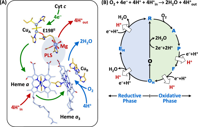
Mammalian CcO is a large integral membrane protein comprised of 13 subunits. It contains four redox active centers, CuA, heme a, and a heme a3/CuB binuclear center (BNC) (Fig. 1A). Molecular oxygen binds to the heme a3 iron in the BNC, where it is reduced to two water molecules by accepting four electrons from cytochrome c and four protons (the “substrate” protons) from the negative side (N-side) of the mitochondrial membrane. The energy derived from the oxygen reduction chemistry is used to drive the translocation of four protons (the “pumped” protons) from the N-side to the positive side (P-side) of the membrane1,2. Strong evidence suggests that the substrate protons are delivered to the BNC via the D and K-channel (see Supplementary Fig. 1), while the pumped protons are translocated via the D-channel3–5 or the H-channel6 through a putative proton loading site (PLS) located between the heme a3 propionates and a Mg2+ center7–10.
Fig. 1. Oxygen reduction reaction catalyzed by bCcO.
A Schematic illustration of the four redox active metal centers in bCcO and the electron and proton transfers associated with the O2 reduction reaction. The entry of O2 and four substrate protons into the heme a3/CuB binuclear center (BNC), as well as the release of the product water molecules out of it, are indicated by the blue arrows. The associated entry of four electrons into the BNC and the translocation of four pumped protons across the membrane are indicated by the green and red arrows, respectively. The putative proton loading site (PLS) between heme a3 and the Mg center is highlighted by the light blue background. B The overall O2 reduction reaction and the associated mechanism. The P intermediate is a general term for the PM and PR intermediates. The entry of the electrons and substrate protons into the BNC and the release of the product water molecules are indicated in each step of the reaction as described in the main text. The coupled proton translocation reactions are indicated by the white arrows. If the OH intermediate produced at the end of the oxidative phase is allowed to relax to the resting O state, its reduction to R does not support proton translocation.
The oxygen reduction reaction catalyzed by CcO has been well-characterized11–15. As illustrated in Fig. 1B, the reaction is initiated by O2 binding to the reduced enzyme, R, to generate the primary O2-complex (A). Upon accepting 2 electrons and 2 substrate protons into the BNC, A is converted to the oxidized OH state, via the P and F intermediates. This oxidative phase of the reaction is followed by the reductive phase, where OH is reduced back to R via the EH intermediate, by accepting two additional electrons and 2 substrate protons into the BNC. During the reaction cycle, each time an electron and a substrate proton enter the BNC, a pumped proton is translocated across the mitochondrial membrane, as indicated by the white arrows. Intriguingly, if the metastable OH state is allowed to relax to the resting O state, its reduction to R, unlike the OH →R transition, does not drive proton translocation16,17, despite the fact that O and OH are redox equivalents.
The structural properties of OH, distinguishing it from the O state, have been elusive. Time-resolved resonance Raman spectroscopic studies of bovine CcO (bCcO) revealed that the heme a3 iron in the BNC of the OH state is coordinated by a hydroxide ion12,18, as evidenced by its characteristic νFe-OH stretching mode at 450 cm−1. In contrast, a comparable νFe-OH band has never been identified in the O state. As such, it has been thought that the inability of the O state to drive proton translocation is at least a partial result of a unique BNC ligation state11,19,20. The three dimensional structure of OH has not been determined, while the crystal structures of O have been reported for various homologs of CcO21–23. It is clear that, in the O state, strong electron density associated with exogeneous ligand(s) is present between heme a3 and CuB in the BNC; its assignment, however, has been controversial. It was first assigned to a peroxide ion bridging the two metal centers21,24, but the best fitted O-O bond distance is much longer than that of a typical ferric peroxide species (~1.48 Å); in addition, it is unclear as to how the peroxides were formed and why they were stable under the equilibrium conditions22. As a result, alternative assignments have been considered. Based on theoretical perspectives, the BNC ligand has been proposed to be a dioxygen25 or a water molecule26. In contrast, other crystallographic studies of bCcO (PDB ID: 7TIE)27 and R. sphaeroides CcO (PDB ID: 2GSM)22 suggest that a water and a hydroxide are coordinated to the heme a3 and CuB, although the O-O distance between the two ligands (~1.9–2.0 Å) is too close for a typical H-bond.
Recently, it was recognized that macromolecular crystallographic structures obtained with intense synchrotron light sources at cryogenic temperatures often suffer from radiation damage problems, in particular for proteins containing redox sensitive metal centers28–30. It has been shown that, in the O structure of bCcO acquired with a typical synchrotron light source, all the four redox active metal centers were reduced, although its polypeptide scaffold remained in a native-like conformation27,31. Accordingly, approaches, such as serial femtosecond X-ray crystallography (SFX)32, have been developed and employed to overcome the radiation damage problems. With SFX, the diffraction patterns of randomly oriented microcrystals suspended in a solution jet are collected sequentially with an X-ray free electron laser (XFEL) before they are destroyed by the intense femtosecond laser pulses. As such, virtually radiation damage-free structures can be obtained at room temperature. Using SFX, Branden and coworkers obtained a radiation damage-free O structure of the ba3 oxidase from Thermus thermophilus, based on which it was concluded that the electron density in the BNC was best modeled by a ligand with a single oxygen atom, either a water or a hydroxide ion23. Likewise, we have used SFX to determine the radiation damage free O structure of bCcO33; however, we found that it required two oxygen atoms to account for the ligand electron density in the BNC, although the resolution (2.9 Å) was insufficient to clarify the identity of the ligands. On the other hand, Hirata et al. employed a different point-by-point scanning approach, using an XFEL as a light source, to circumvent radiation damage problems, based on which the BNC ligands of the O derivative of bCcO were assigned as two co-existing peroxide moieties34, implying possible heterogeneity of CcO samples35,36. More recently, Kolbe et al. used a cryo-EM technique to solve the structure of a bacterial CcO from P. denitrificans, which suggests that the BNC ligand is a peroxide with an O–O distance of 1.42 Å37. Despite these and other efforts, no consensus on the ligand identity has been reached to date.
Here, we sought to clarify the structural properties of the O derivatives of bCcO by using a combination of resonance Raman spectroscopy and SFX.
Results
Identification of the heme a3 ligand by resonance Raman spectroscopy
To determine the identity of the heme a3 iron ligand, we carried out resonance Raman spectroscopic studies in free soluton. The 413.1 nm output from a krypton ion laser was selected as the excitiation light source to selectively enhance the signals associated with heme a3. We reasoned that if the heme a3 iron ligand is a water or a hydroxide ion, it should be able to be exchanged with the solvent water molecules. Accordingly, we incubated the resting O derivative of bCcO in isotope-substituted H218O buffer for 12 h to ensure complete solvent exchange prior to the resonance Raman measurements. To prevent photoreduction, the laser power was kept low (~5 mW) and the spectral acquisition time was kept short (3 min); in addition, the spectra from 6 fresh samples were acquired and summed to improve the signal-to-noise ratio of the spectrum. As a comparison, the spectrum of O in naturally abundant H216O buffer was obtained in the same fashion.
As shown in Fig. 2, an oxygen isotope sensitive band was identified at 451 cm−1 in the H216O buffer, which shifted to 428 cm−1 in the H218O buffer. The isotopic shift of 23 cm−1 is consistent with the theoretical shift of a Fe-OH− stretching mode (νFe-OH) (24 cm−1), indicating that the heme a3 iron ligand of the resting O state is a hydroxide ion. It should be noted that, this solvent isotope sensitive mode was not detected in prior studies12, possibly due to the complications resulting from laser induced photodamage38. The νFe-OH mode is identical to that found in the OH state12,18, suggesting that the BNC ligands in O and OH are the same, contrary to the common belief that they are distinct11,19,20.
Fig. 2. Resonance Raman spectrum of the O state of bCcO in H216O (black) overlaid with that in H218O (red).
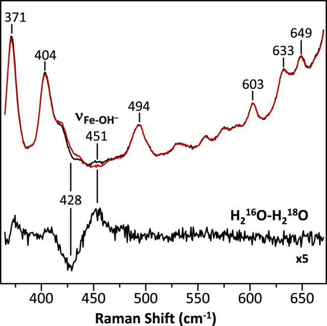
The H216O-H218O difference spectrum (expanded by 5-fold) is shown in black at the bottom. The oxygen sensitive mode in the H216O sample centered at 451 cm−1 that shifted to 428 cm−1 in the H218O sample is assigned to the νFe-OH mode.
It is noteworthy that this Fe-OH− stretching frequency (451 cm−1) is remarkably low as compared to those of other ferric heme species (~490–550 cm−1)39, indicating an unusually weak Fe-OH− coordination bond. Hydroxide is generally a strong field ligand for ferric heme iron, which is associated with a strong Fe-OH− bond and a low spin electronic configuration. However, previous resonance Raman12 and electron paramagnetic resonance (EPR)40 spectroscopic studies revealed that the heme a3 in the O state, like that in the OH state, has an unusual high spin configuration. We attribute the weak Fe-OH− bond and unique high spin configuration of the heme a3 to a strong H-bond between the hydroxide ligand and its surrounding environment, as that detected in a hemoglobin from M. tuberculosis, which exhibits a similar low Fe-OH− stretching frequency (454 cm−1) and a high spin electronic configuration due to a H-bond between the hydroxide ligand and a nearby tyrosine residue41. The presence of a strong H-bond to the hydroxide ligand in the BNC of bCcO is supported by the structural data discussed below. It is notable that previous computational studies19 suggested that the OH state has a single hydroxide ligand bridging heme a3 iron and CuB, thereby accounting for the high spin configuration of the heme a3. However, if that were the case, the vFe-OH mode of the OH state would be expected to be distinct from that of the O state due to the unique bonding interactions between the hydroxide ion and the two highly charged metal centers.
Determination of the structure of the O state by SFX
For the SFX measurements, microcrystals of bCcO were prepared in the resting O state using a previously reported protocol33. To ensure the homogeneity of the O state, the microcrystals were reduced under anaerobic conditions and then allowed to turn over by exposing to O2 and subsequently relax back to the O state by incubating overnight in a stabilizing solution. The suspension of the microcrystals was loaded into a gas-tight syringe and injected into the XFEL beam as a free solution jet with a single capillary MESH injector42,43. The XFEL beam, perpendicular to the solution jet, was directed into the tip of the Taylor cone formed at the output of the MESH injector. X-ray diffraction was collected for ~2 h, from which 84,736 indexable diffraction patterns were selected for structural analysis. The structure was solved and refined to a resolution of 2.38 Å (Supplementary Table 1). The structural markers for the oxidation states of the four redox centers confirm that the enzyme is in the fully oxidized O state (Supplementary Fig. 2).
Structural characterization of the BNC
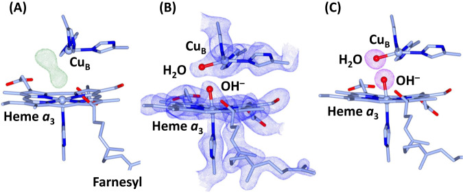
In the FO-FC electron density map associated with the SFX data (Fig. 3A), a large 2-lobe electron density is evident in the BNC, indicating the presence of ligands coordinated to heme a3 iron and CuB. As guided by the resonance Raman data (Fig. 2), we modeled the heme a3 ligand density with a OH− ion (Fig. 3B). In addition, we modeled the CuB ligand density with a water molecule, as the coexistence of two negatively charged hydroxide ions in the BNC is expected to be energetically unfavorable. The occupancy of the ligands is confirmed by the polder maps shown in Fig. 3C. The Fe-OH−, CuB-H2O, and Fe-CuB bond lengths, which were unrestrained during the refinement, are determined to be 1.90, 2.14, and 4.74 Å, respectively. The O–O distance between the two oxygen ligands is 2.53 Å, which is shorter than a typical H-bond, but is consistent with a strong low-barrier H-bond44,45. This strong H-bond between the two ligands plausibly weakens the ligand field strength of the hydroxide and destabilizes the Fe-OH− bond, thereby accounting for the high spin electronic configuration and the low Fe-OH− stretching frequency revealed by the spectroscopic studies. Taken together our data demonstrate that the heme a3 iron and CuB in the O state, like those in the OH state, are coordinated by a hydroxide ion and a water molecule, respectively.
Fig. 3. Electron density maps of the BNC in the O state of bCcO.
A The FO-FC electron density map (contoured at 7.0σ) showing clear 2-lobe electron density associated with the BNC ligands. B The 2FO-FC electron density map (contoured at 2.5σ) obtained with the electron density modeled with a hydroxide ion coordinated to the heme a3 iron and a water molecule coordinated to CuB. C The Polder map (contoured at 7.0σ) associated with the BNC ligands.
Protonation state of Y244
CuB in the BNC is coordinated by three histidine ligands, one of which (H240) forms a covalent bond with Y244 by a posttranslational modification (Fig. 4). It is well-established that, during the catalytic reaction, Y244 temporarily donates an electron and a proton to the heme a3 bound O2 to promote the O-O bond scission, by forming a tyrosyl radical46, and that its re-reduction to a tyrosinate and re-protonation back to its neutral protonated form are tightly coupled to the ensuing electron and proton transfer processes19,46,47.
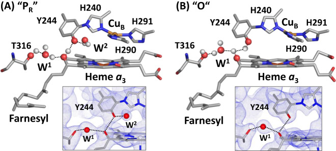
Fig. 4.
Protonation state of Y244 in the PR (A) and O (B) states. The post translationally modified Y244 forms a H-bonding network with the OH group of the farnesyl side chain of heme a3, a water molecule (W1) and T316. In the PR state, an additional water, W2, is recruited into the heme a3/CuB binuclear center (BNC) to stabilize the tyrosinate configuration of Y244. This water is absent in the O state reported here, as evident in the 2FO-FC electron density map (contoured at 1.0σ) shown in the lower inset, signifying that Y244 is in the neutral protonated state. For clarity the BNC ligands are not shown. The oxygen atom and hydrogen atoms are shown as red and white spheres.
Previously, with time-resolved SFX, we showed that, in a P intermediate of bCcO (PR), Y244 is in the tyrosinate form33. Next to the Y244 there is a water molecule within H-bonding distance from the oxygen atom of the tyrosinate (indicated as W2 in Fig. 4A), which is not present in the reduced R state (PDB IDs: 6NMF and 7THU) or the CO complex (PDB ID: 5W97, a structural analog of the A state), where Y244 is in the neutral protonated form. It suggests that, in the PR state, W2 is recruited into the BNC to stabilize the tyrosinate configuration of Y244, similar to the water rearrangement induced by tyrosine deprotonation found in a photosynthetic reaction center from B. viridis48. Intriguingly, our current work reveals that W2 is not present in the O state (Fig. 4B), signifying that Y244 is in the neutral protonated form, consistent with the conclusion drawn from computational studies carried out by Blomberg49. Although the structures of the F and OH intermediates have not been determined, infrared spectroscopic studies demonstrate that Y244 in both F and OH, like that in the PR intermediate, is deprotonated47, indicating that during the PR→F→OH transition, Y244 remains in the tyrosinate configuration. This scenario is in good agreement with recent structural data showing the presence of W2 in the mixed oxygen intermediates of bCcO20,50. Our current data further demonstrate that the OH→O transition is associated with the protonation of the tyrosinate (Y244).
Discussion
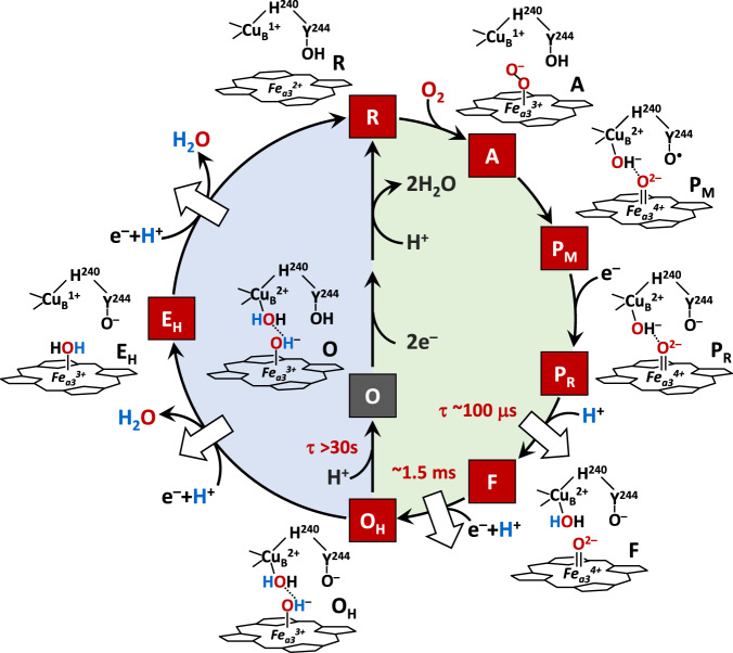
With the clarification of the O structure, we propose a complete reaction cycle of CcO as illustrated in Fig. 5. The reduced R state first binds O2 to form the primary oxy intermediate A, with a Fe3+-O2− electronic configuration51. By accepting an electron and a proton from Y244, the O-O bond in A is heterolytically cleaved, leading to the formation of the putative PM intermediate, where one oxygen remains on the heme a3 iron, in a ferryl (Fe4+=O2−) configuration, and the other oxygen is coordinated to CuB as a hydroxide, while Y244 is converted to a neutral tyrosyl radical. The entry of one electron into the BNC leads to the conversion of PM to PR, where the tyrosyl radical is reduced to a tyrosinate. The PM→PR transition is associated with the entry of a new water (W2, not shown in Fig. 5 for clarity), which stabilizes the tyrosinate configuration of Y24433. The subsequent entry of a substrate proton into the BNC further transforms PR to F, where the hydroxide ligand of CuB is protonated to a water, which strengthens the iron-oxygen bond as evidenced by the shift of the frequency of the Fe4+=O2− stretching mode from 785 to 804 cm−1 52. Finally, the entry of an additional electron and a substrate proton into the BNC converts F to OH, where the Fe4+=O2− moiety is reduced to the Fe3+-OH− species12,18,52. During this oxidative phase, one pumped proton is translocated during each of the PR→F and F→OH transitions (as indicated by the white arrows) and, throughout the PR→F→OH transformation, Y244 remains in the tyrosinate form.
Fig. 5. Hypothesized ligand transformation in the BNC during the CcO reaction cycle.
The oxidative and reductive phase of the reaction cycle are highlighted with green and blue backgrounds, respectively. The white arrows indicate the proton translocation reactions associated with the reaction cycle. The lifetimes associated with the PR→F, F→OH and OH→O transitions are indicated in red.
In the ensuing reductive phase, an additional electron and a substrate proton enter the BNC, leading to the conversion of OH to EH, where CuB is reduced from the cupric to cuprous state and the hydroxide ligand of the heme a3 is protonated to a water11,53. At the same time, the water ligand of CuB is released out of the BNC. The entry of another electron and a substrate proton into the BNC leads to the conversion of EH to R, where the heme a3 iron is reduced from the ferric to ferrous state and its water ligand is released out of the BNC; at the same time the tyrosinate is protonated back to its neutral form. It should be noted that in our proposed model the EH state has Y244 in the tyrosinate form and a water molecule coordinated to the heme a3 iron11,53, but an alternative configuration with Y244 in the protonated form and a hydroxide coordinated to the heme a3 iron, as suggested by infrared spectroscopic studies47, cannot be excluded. In any case, during this reductive phase, one additional pumped proton is translocated during each of the OH→EH and EH→R transitions. Under electron deficient conditions, the OH intermediate produced at the end of the oxidative phase can spontaneously and slowly relax to the resting O state16. Our current data revealed that during the OH→O transition, the tyrosinate (Y244) is protonated to the neutral form, leading to the dissociation of the W2 that stabilizes the tyrosinate, while the BNC ligands remain unchanged.
The results reported here have important implications on proton translocation. It has been shown that the entry of the first two substrate protons into the BNC, associated with the PR→F and F→OH transitions, is mediated by the D-channel and occurs rapidly (within ~100 μs and 1.5 ms, respectively)2. Similarly, the entry of the other two substrate protons into the BNC, associated with the OH→EH and EH→R transitions, also takes place quickly (on the submillisecond time scale), although it is mediated by a different channel, the K-channel54. In sharp contrast, the off-pathway OH→O transition is sluggish and does not reach completion until ~30 s16, suggesting that the tyrosinate (Y244) is likely protonated by an adventitious proton from its surrounding environment or some other part of the protein, rather than a substrate proton from the K-channel, despite the fact that Y244 sits at the end of the K-Channel (see Supplementary Fig. 1). If the proton were derived from the K-Channel, a much more efficient OH→O transition and a unique sidechain conformation of K319 (a critical component of the K-Channel that is predicted to undergo conformational changes during proton translocation55,56), which was not detected in the current structure, would be expected.
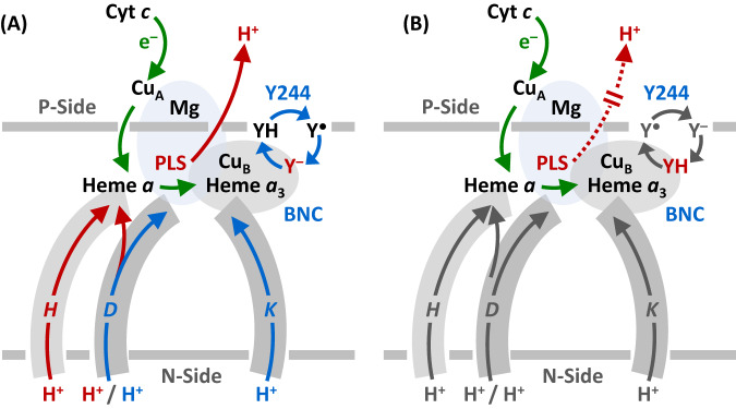
Based on the electroneutrality principle proposed by refs. 57,58, during the catalytic reaction, the entry of each electron into the BNC (see the green arrows in Fig. 6A) is charge-compensated by the entry of one substrate proton from the D or K-channel (blue arrows). The redox energy thereby derived is then used to drive the release of a pumped proton into the P-side of the membrane from the PLS, which is preloaded with the pumped proton(s) via the D or H-channel. As illustrated in Fig. 5, Y244 in all the intermediate states active in proton translocation (PR, F, OH, and EH) is in the deprotonated tyrosinate form, suggesting that the premature protonation of the tyrosinate (Y244) during the OH→O transition, without the input of any electron into the BNC, perturbs the charge balance in the BNC of the O state, thereby disabling the release of pumped protons out of the PLS upon reduction (Fig. 6B). This analysis of our structure is in good agreement with the conclusions drawn from recent computational studies carried out by Blomberg, showing that the protonation of Y244 associated with the OH→O transition lowers the reduction potential of the metal centers in the BNC, such that the proton translocation is no longer energetically favorable upon reduction49. These data demonstrate the importance of deprotonated forms of Y244 in modulating the energy landscape of the CcO reaction to promote the coupling of the oxygen reduction chemistry to proton translocation53.
Fig. 6. Functional role of Y244 during the CcO reaction cycle.
A During the active turnover, Y244 cycles between the neutral form (YH), tyrosyl radical form (Y●) and deprotonated tyrosinate form (Y–) as depicted by the blue cycle. In all the intermediate states active in proton translocation, Y244 is in the Y– form (in red), which ensures the tight coupling of electron transfer (green arrows) and substrate proton transfer (blue arrows), via the D and K-channel into the heme a3/CuB binuclear center (BNC), to drive the proton translocation (red arrows) via the D or H channel from the N to P-side of the membrane. B The protonation of the tyrosinate to the YH form (red) in the O state disables the proton translocation upon its reduction to R.
In summary, previous studies revealed that O and OH are redox equivalents, but only the latter, not the former, is capable of translocating protons upon reduction; with a combination of resonance Raman spectroscopy and SFX, we now clarify that the heme a3 iron and CuB in the BNC of the O state, like those in the OH state, are coordinated by a hydroxide ion and water, respectively; in addition, we show that the post-translationally modified Y244 is in the neutral protonated form, which distinguishes the O state from the OH state, where Y244 is in the deprotonated tyrosinate form. Our data underscore the pivotal role of the deprotonated form of Y244, a residue fully conserved in the CcO family of enzymes, in energizing the enzyme for proton translocation, as supported by reported computational studies53.
Methods
Protein sample preparation
Bovine CcO (bCcO) was isolated from bovine heart by slight modifications of a reported procedure59,60 as described below. All the procedures were done at 4 oC. Bovine hearts, obtained from cows freshly slaughtered in a local slaughterhouse, were immediately cooled in a cold sodium phosphate buffer (pH 7.4, 50 mM). The skin, fat tissues and vascular tubes of the bovine hearts were trimmed from the heart and cut into small cubes. 550 g meat was grounded in a grinder. The ground meat was mixed with 3250 ml cold sodium phosphate buffer (pH 7.4, 23 mM) and homogenized in a blender at 13,000 rpm for 10 min. The solution was centrifuged at 2000 × g for 20 min. The supernatant was filtered through cheesecloth and kept aside. The precipitate was collected and mixed with 1500 ml cold sodium phosphate buffer (pH 7.4, 20 mM) and homogenized again in a blender at 13,000 rpm for 10 min. It was centrifuged at 2000 × g for 20 min and filtered through cheesecloth. The two red-colored supernatants were then combined and acidified to pH 5.15 with 30% acetic acid and centrifuged at 2000 × g for 15 min. The yellow soil-colored precipitate was collected and mixed with 400 ml cold water. It was then homogenized in a blender at 3500 rpm for 2 min. The volume was adjusted to 1.5 L with cold water and centrifuged at 2000 × g for 15 min. The tea-colored precipitate was collected and dissolved in 50 ml sodium phosphate buffer (pH 7.4, 200 mM). It was homogenized in a blender at 7000 rpm for 3 min and then centrifuged at 1500 × g for 10 min to remove the bubbles in the solution. The solution was mixed with 94 ml sodium phosphate buffer (pH 7.4, 200 mM) and the pH was adjusted to 7.4 with small aliquots of 3 N NaOH, and then water was added to make a final volume of 288 ml. The sample was kept at 4 oC overnight.
On the second day, 25 ml sodium cholate (40% w/v) was added to the sample, which was then subjected to seven rounds of ammonium sulfate fractionation at 4 oC. In the 1st ammonium sulfate fractionation, 61.35 g ammonium sulfate was added to the sample to achieve 33% saturation. The sample was stirred at 4 oC for 30 min and then centrifuged at 25,000 × g for 20 min. The supernatant was collected, and additional ammonium sulfate was added (with the pH kept at ~7.3–7.4 by adding small aliquots of 3 N NaOH) to achieve 50% saturation to precipitate out bCcO. The sample was centrifuged at 25,000 × g for 30 min. The precipitate was collected and homogenized in 0.5% (w/v) sodium cholate in sodium phosphate buffer (pH 7.4, 100 mM) with a final volume of 90 ml. The solution was dialyzed against 3 L sodium phosphate buffer (pH 7.4, 40 mM) for 90 min. It was then centrifuged at 100,000 × g for 50 min. The precipitate was collected and homogenized in 2.0% (w/v) sodium cholate in sodium phosphate buffer (pH 7.4, 100 mM) with a final volume of 100 ml. The sample was then subjected to the 2nd ammonium sulfate fractionation. Ammonium sulfate was added to the sample to achieve 25% saturation, which was then incubated at 4 oC for 30 min. It was centrifuged at 35,000 × g for 10 min. The supernatant was collected, and additional ammonium sulfate was added (with the pH kept at ~7.3–7.4) to achieve 45% saturation to precipitate out bCcO. The solution was centrifuged at 35,000 × g for 10 min. The precipitate was collected and homogenized in 0.5% (w/v) sodium cholate in sodium phosphate buffer (pH 7.4, 100 mM) with a final volume of 100 ml. In the 3rd ammonium sulfate fractionation, ammonium sulfate was added to the sample to reach 25% saturation. The solution was incubated at 4 oC for 30 min, and then centrifuged at 35,000 × g for 10 min. The supernatant was collected, and additional ammonium sulfate was added (with the pH kept at ~7.3–7.4) to achieve 40% saturation to precipitate out bCcO. The sample was centrifuged at 35,000 × g for 10 min. The precipitate was collected and homogenized in 0.5% (w/v) sodium cholate in sodium phosphate buffer (pH 7.4, 100 mM) with a final volume of 65 ml. In the 4th ammonium sulfate fractionation, ammonium sulfate was added to the sample to reach 25% saturation. It was incubated at 4 oC for 30 min, and then centrifuged at 35,000 × g for 5 min. The supernatant was collected, and additional ammonium sulfate was added (with the pH kept at ~7.3–7.4) to achieve 35% saturation to precipitate out bCcO. It was centrifuged at 35,000 × g for 5 min. The precipitate was collected and homogenized in 0.34% (w/v) n-Decyl-β-D-maltoside in sodium phosphate buffer (pH 7.4, 100 mM) with a final volume of 100 ml. In the 5th ammonium sulfate fractionation, ammonium sulfate was added to the sample to reach 40% saturation. The solution was centrifuged at 35,000 × g for 5 min. The supernatant was collected, and additional ammonium sulfate was added (with the pH kept at ~7.3–7.4) to achieve 60% saturation to precipitate out bCcO. It was centrifuged at 35,000 × g for 12 min. The precipitate was collected and homogenized in 0.2% (w/v) n-Decyl-β-D-maltoside in sodium phosphate buffer (pH 7.4, 100 mM) with a final volume of 50 ml. The solution was stored in a cold room overnight.
On the third day, the sample volume was increased to 110 ml by adding 0.2% (w/v) n-Decyl-β-D-maltoside in sodium phosphate buffer (pH 7.4, 100 mM). The sample was subjected to the 6th ammonium sulfate fractionation. Ammonium sulfate was added to the sample to reach 50% saturation, which was then centrifuged at 35,000 × g for 5 min. The supernatant was collected, and additional ammonium sulfate was added (with the pH kept at ~7.3–7.4) to achieve 70% saturation to precipitate out bCcO. The sample was centrifuged at 35,000 × g for 15 min. The precipitate was collected and homogenized in 0.2% (w/v) n-Decyl-β-D-maltoside in sodium phosphate buffer (pH 7.4, 100 mM) with a final volume of 100 ml. In the last ammonium sulfate fractionation, ammonium sulfate was added to the sample to reach 55% saturation. The solution was centrifuged at 35,000 × g for 5 min. The supernatant was collected, and additional ammonium sulfate was added (with the pH kept at ~7.3–7.4) to achieve 70% saturation to precipitate out bCcO. The sample was centrifuged at 35,000 × g for 15 min. The precipitate was collected and dissolved in 5 ml sodium phosphate buffer (pH 7.4, 10 mM). It was dialyzed against 1 L fresh sodium phosphate buffer (pH 7.4, 10 mM) three times for 1 h, 2 h and 3 h each. After dialysis, the sample was centrifuged at 35,000 × g for 20 min. The supernatant was collected and concentrated to reach a protein concentration of ~80–90 mg /ml, using an Amicon Diaflo apparatus with an ultrafiltration membrane (Advantec) with a pore size of 200,000 Da.
On the fourth day, the concentrated sample was washed three times with 0.2% (w/v) n-Decyl-β-D-maltoside in sodium phosphate buffer (pH 7.4, 20 mM) using the Amicon Diaflo apparatus. It was followed by three additional washes with 0.2% (w/v) n-Decyl-β-D-maltoside in sodium phosphate buffer (pH 6.8, 40 mM). After the 2nd wash, the sample was further concentrated until micro-crystals were formed on top of the Amicon membrane. The micro-crystal slurry was collected and centrifuged at 35,000 × g for 10 min. The micro-crystal pellet was collected and dissolved in 0.2% (w/v) n-Decyl-β-D-maltoside in sodium phosphate buffer (pH 6.8, 40 mM) to generate the protein stock for the resonance Raman measurements and microcrystal preparation for the SFX studies. To determine the concentration of bCcO, the absorption spectra of the sample reduced by dithionite was acquired and the concentration was calculated based on the absorbance difference at 603 and 630 nm using an extinction coefficient, εred (603–630 nm), of 46.6 nM−1 cm−1. Typical yields of the purified enzyme are 150–330 mg.
It is well-known that oxidized bCcO can exist in two distinct forms, the so-called “fast” and “slow” forms35,36, depending on the purification and preparation methods. The optical absorption spectra of the bCcO samples used in this work displayed a Soret maximum at 423 nm in both the solution phase and crystalline form (see Supplementary Fig. 3), indicating that they are the fast form (i.e., the active form) of the enzyme35,36.
Resonance Raman measurements
To prepare the sample in H218O, a concentrated bCcO sample (in 40 mM pH 7.4 phosphate buffer containing 0.2% decylmaltoside) was diluted by tenfold with H218O containing the same buffer and then incubated overnight prior to the resonance Raman measurements. The final protein concentration was 30 µM. An equivalent sample in H216O was prepared in the same fashion as a comparison. Resonance Raman spectra were obtained by using 413.1 nm excitation from a Kr ion laser (Spectra Physics, Mountain View, CA). The laser beam was focused to a ~30 µm spot on a spinning sample cell. The scattered light, collected at right angle to the incident laser beam, was focused on the 100 µm wide entrance slit of a 1.25 m Spex spectrometer equipped with a 1200 grooves/mm grating (Horiba Jobin Yvon, Edison, NJ), where it was dispersed and then detected by a liquid-nitrogen cooled CCD detector (Princeton Instruments, Trenton, NJ). A holographic notch filter (Kaiser, Ann Arbor, MI) was used to remove the laser scattering. The Raman shift was calibrated by using indene (Sigma).
It has been shown that high laser power can induce photoreduction linked artifacts in the resonance Raman spectra of bCcO38, which prevented identification of the Fe-OH stretching mode in a prior study12. To avoid photo-induced artifacts, we kept the sample in a spinning quartz cell that rotates at 1000 rpm and kept the laser power low, ~5 mW at the output of the laser (prior to its passage through mirrors and a focusing lens to the sample). In addition, we limited the laser exposure time to 3 min for each sample. The final spectrum was obtained by averaging six spectra acquired from six individual fresh samples. Under these conditions none of the previously reported bands associated with photodamage derived from high laser power were found in the spectra. To confirm that there was no photoreduction, spectra of reduced bCcO were acquired under the same conditions. None of the reduced marker lines were detected in the spectra of the oxidized enzyme. To determine if there were any oxygen species containing oxygen atoms that were not exchangeable with H218O, such as a peroxide, we carried out the following experiment. We first placed the enzyme in H218O buffer to exchange all exchangeable oxygen to 18O. We then reduced the enzyme to the fully reduced state, to expel all oxygen ligands from the BNC, and exposed it to 18O2 to initiate the turnover. Subsequently, we allowed the enzyme to relax back to the fully oxidized state and then acquired a spectrum. As a reference, we carried out a comparable reaction with H216O and 16O2 and acquired another spectrum. Our data showed that no isotope differences were detected other than that assigned as the Fe-OH stretching mode. To ensure that no artifacts present in the buffer background, spectra of the H216O and H218O buffers alone were obtained. It was confirmed that no isotopic differences in the 400–500 cm−1 window were detected.
Microcrystal preparation
The microcrystals were prepared with a previously reported method33. The crystal growth was initiated by mixing the protein stock with the precipitant solution (0.2% decylmaltoside and 2.5% PEG4000 in 40 mM pH 6.8 phosphate buffer) and a seeding solution (prepared by crushing and sonicating large crystals in the mother solution). The microcrystals were allowed to grow at 4 oC for ~36 h before they were harvested and characterized by polarized optical microscopy. The microcrystals have a planar shape with approximate dimensions of ~20 × 20 × 4 μm. To obtain a homogeneous sample of the oxidized O crystals for the SFX measurements, the microcrystals were reduced anaerobically by a minimum amount of dithionite in a glove box and then thoroughly washed with the mother solution to remove excess dithionite and its oxidized products. The reduced microcrystals were then exposed to O2 to initiate the enzyme turnover. They were allowed to relax in a stabilizing solution (0.2% decylmaltoside and 6.25% PEG-4000 in 40 mM pH 6.8 sodium phosphate solution) at 4 oC for ~24 hours prior to the SFX measurements.
SFX data acquisition and structural analysis
The SFX measurements were conducted at the Macromolecular Femtosecond Crystallography (MFX) end station of the Linac Coherent Light Source (LCLS) at the SLAC National Accelerator Laboratory. A bCcO microcrystal slurry was loaded into a gas tight syringe and driven by a HPLC pump into a 100 µm diameter capillary of a Microfluidic Electrokinetic Sample Holder (MESH) injector42,43, which has recently been developed as a useful and reliable technique for delivering microcrystal slurries into the XFEL beam61–65. A high voltage (~+2500 V) was applied to the sample at the entrance of the capillary against a grounded waste collector. It was used to electro-focus the microcrystal jet down to a Taylor cone at the capillary output. The 10 keV/30 fs pulses from the X-ray free electron laser (XFEL) intersected the microcrystal slurry at the tip of the Taylor cone prior to its development into a thin jet, as the jet was too unstable for data collection, while the thick region of the cone gave too many multiple hits and a high background. The diameter of the XFEL beam was ~3 μm. The sample flow rate was set to ~3 µl/min and the sample temperature was kept at ~293 K. The XFEL wavelength was 1.24 Å. A series of diffraction patterns from randomly orientated microcrystals were collected with a Rayonix MX340-XFEL CCD detector at a 30 Hz rep rate.
The quality and hit rate of the SFX data were monitored in real time using OM66. The data were collected for ~2 h. Psocake67,68 was used to determine the initial diffraction geometry and find crystal hits. 84,736 patterns in the acquired dataset were indexed and merged with the CrystFEL program suite developed for SFX experiments69,70. The initial structure was solved with molecular replacement with Phaser-MR through the CCP4 program suite71 using a 1.9 Å resolution structure of bCcO (PDB ID: 7TIE) as the search model. Waters and BNC ligands were excluded from the search model. Further model building was performed using Coot72. Structure refinements were done using Refmac5 and PDB-Redo73. The final structure was refined to a resolution of 2.38 Å (see Supplementary Table 1) (PDB ID: 8GCQ). Similar structures were observed in the two monomers of the bCcO dimer. All the structural data presented here are based on the first monomer (subunit A-M), as it exhibited a better resolution owing to crystal packing.
Reporting summary
Further information on research design is available in the Nature Portfolio Reporting Summary linked to this article.
Supplementary information
Source data
Acknowledgements
The SFX experiments were carried out at the LCLS at the SLAC National Accelerator Laboratory. LCLS is an Office of Science User Facility operated for the US Department of Energy Office of Science by Stanford University. Use of the LCLS, SLAC National Accelerator Laboratory, is supported by the U.S. Department of Energy, Office of Science and Office of Basic Energy Sciences under Contract No. DE-AC02-76SF00515. The HERA system for in helium experiments at MFX was developed by Bruce Doak and funded by the Max-Planck Institute for Medical Research. This work was supported by National Science Foundation (NSF) CHE−1404929 (D.L.R. and S.-R.Y.) and National Institutes of Health (NIH) grants S10 OD023453, P41 GM139687, GM126297 (D.L.R. and S.-R.Y.) and GM115773 (S.-R.Y.).
Author contributions
S.R.Y., I.I., and D.L.R. designed experiments; I.I. isolated and crystallized bCcO and measured and interpreted the resonance Raman spectra; R.G.S. designed and operated the MESH injector; R.E.S. prepared the injector auxiliary equipment; F.P., S.L., B.H., F.R.M., and S.B. collected the SFX data; Z.S., A.P., C.W., and C.H.Y. processed the SFX data; S.R.Y., I.I., C.H.Y., and D.L.R. analyzed and interpreted the SFX data; S.R.Y. and D.L.R. drafted the manuscript and all authors contributed to the revisions.
Peer review
Peer review information
Nature Communications thanks the anonymous reviewer(s) for their contribution to the peer review of this work. A peer review file is available.
Data availability
Atomic coordinates and structure factors of the O state of bCcO generated in this study have been deposited in the Protein Data Bank (PDB) under accession code 8GCQ. PDB codes of previously published structures used in this study are 7TIE, 6NMF, 7THU, 5W97 and 2GSM. Source data are provided with this paper. Any additional requests for data should be directed to the corresponding authors. Source data are provided with this paper.
Competing interests
The authors declare no competing interests.
Footnotes
Publisher’s note Springer Nature remains neutral with regard to jurisdictional claims in published maps and institutional affiliations.
Contributor Information
Syun-Ru Yeh, Email: syun-ru.yeh@einsteinmed.edu.
Denis L. Rousseau, Email: denis.rousseau@einsteinmed.edu
Supplementary information
The online version contains supplementary material available at 10.1038/s41467-023-41533-x.
References
- 1.Brzezinski P. Redox-driven membrane-bound proton pumps. Trends Biochem. Sci. 2004;29:380–387. doi: 10.1016/j.tibs.2004.05.008. [DOI] [PubMed] [Google Scholar]
- 2.Wikstrom M, Krab K, Sharma V. Oxygen activation and energy conservation by cytochrome c oxidase. Chem. Rev. 2018;118:2469–2490. doi: 10.1021/acs.chemrev.7b00664. [DOI] [PMC free article] [PubMed] [Google Scholar]
- 3.Konstantinov AA, Siletsky S, Mitchell D, Kaulen A, Gennis RB. The roles of the two proton input channels in cytochrome c oxidase from Rhodobacter sphaeroides probed by the effects of site-directed mutations on time-resolved electrogenic intraprotein proton transfer. Proc. Natl Acad. Sci. USA. 1997;94:9085–9090. doi: 10.1073/pnas.94.17.9085. [DOI] [PMC free article] [PubMed] [Google Scholar]
- 4.Rich PR, Marechal A. Functions of the hydrophilic channels in protonmotive cytochrome c oxidase. J. R. Soc. Interface. 2013;10:20130183. doi: 10.1098/rsif.2013.0183. [DOI] [PMC free article] [PubMed] [Google Scholar]
- 5.Wikstrom M, Jasaitis A, Backgren C, Puustinen A, Verkhovsky MI. The role of the D- and K-pathways of proton transfer in the function of the haem-copper oxidases. Biochim. Biophys. Acta. 2000;1459:514–520. doi: 10.1016/S0005-2728(00)00191-2. [DOI] [PubMed] [Google Scholar]
- 6.Shimokata K, et al. The proton pumping pathway of bovine heart cytochrome c oxidase. Proc. Natl Acad. Sci. USA. 2007;104:4200–4205. doi: 10.1073/pnas.0611627104. [DOI] [PMC free article] [PubMed] [Google Scholar]
- 7.Kaila VR, Sharma V, Wikstrom M. The identity of the transient proton loading site of the proton-pumping mechanism of cytochrome c oxidase. Biochim. Biophys. Acta. 2011;1807:80–84. doi: 10.1016/j.bbabio.2010.08.014. [DOI] [PubMed] [Google Scholar]
- 8.Lu J, Gunner MR. Characterizing the proton loading site in cytochrome c oxidase. Proc. Natl Acad. Sci. USA. 2014;111:12414–12419. doi: 10.1073/pnas.1407187111. [DOI] [PMC free article] [PubMed] [Google Scholar]
- 9.Sharpe MA, Ferguson-Miller S. A chemically explicit model for the mechanism of proton pumping in heme-copper oxidases. J. Bioenerget. Biomembr. 2008;40:541–549. doi: 10.1007/s10863-008-9182-6. [DOI] [PMC free article] [PubMed] [Google Scholar]
- 10.Supekar S, Gamiz-Hernandez AP, Kaila VR. A protonated water cluster as a transient proton-loading site in cytochrome c oxidase. Angew. Chem. 2016;55:11940–11944. doi: 10.1002/anie.201603606. [DOI] [PubMed] [Google Scholar]
- 11.Belevich I, Verkhovsky MI. Molecular mechanism of proton translocation by cytochrome c oxidase. Antioxid. Redox Signal. 2008;10:1–29. doi: 10.1089/ars.2007.1705. [DOI] [PubMed] [Google Scholar]
- 12.Han S, Takahashi S, Rousseau DL. Time dependence of the catalytic intermediates in cytochrome c oxidase. J Biol. Chem. 2000;275:1910–1919. doi: 10.1074/jbc.275.3.1910. [DOI] [PubMed] [Google Scholar]
- 13.Kitagawa T, Ogura T. Time-resolved resonance Raman investigation of oxygen reduction mechanism of bovine cytochrome c oxidase. J. Bioenerget. Biomembr. 1998;30:71–79. doi: 10.1023/A:1020511612194. [DOI] [PubMed] [Google Scholar]
- 14.Wikstrom M, Gennis RB, Rich PR. Structures of the intermediates in the catalytic cycle of mitochondrial cytochrome c oxidase. Biochim. Biophys. Acta. Bioenerget. 2022;1864:148933. doi: 10.1016/j.bbabio.2022.148933. [DOI] [PubMed] [Google Scholar]
- 15.Varotsis C, Zhang Y, Appelman EH, Babcock GT. Resolution of the reaction sequence during the reduction of O2 by cytochrome oxidase. Proc. Natl Acad. Sci. USA. 1993;90:237–241. doi: 10.1073/pnas.90.1.237. [DOI] [PMC free article] [PubMed] [Google Scholar]
- 16.Bloch D, et al. The catalytic cycle of cytochrome c oxidase is not the sum of its two halves. Proc. Natl Acad Sci. USA. 2004;101:529–533. doi: 10.1073/pnas.0306036101. [DOI] [PMC free article] [PubMed] [Google Scholar]
- 17.Verkhovsky MI, Jasaitis A, Verkhovskaya ML, Morgan JE, Wikstrom M. Proton translocation by cytochrome c oxidase. Nature. 1999;400:480–483. doi: 10.1038/22813. [DOI] [PubMed] [Google Scholar]
- 18.Han S, Ching YC, Rousseau DL. Ferryl and hydroxy intermediates in the reaction of oxygen with reduced cytochrome c oxidase. Nature. 1990;348:89–90. doi: 10.1038/348089a0. [DOI] [PubMed] [Google Scholar]
- 19.Sharma V, Karlin KD, Wikstrom M. Computational study of the activated O(H) state in the catalytic mechanism of cytochrome c oxidase. Proc. Natl Acad. Sci. USA. 2013;110:16844–16849. doi: 10.1073/pnas.1220379110. [DOI] [PMC free article] [PubMed] [Google Scholar]
- 20.Shimada A, et al. Critical roles of the CuB site in efficient proton pumping as revealed by crystal structures of mammalian cytochrome c oxidase catalytic intermediates. J. Biol. Chem. 2021;297:100967. doi: 10.1016/j.jbc.2021.100967. [DOI] [PMC free article] [PubMed] [Google Scholar]
- 21.Koepke J, et al. High resolution crystal structure of Paracoccus denitrificans cytochrome c oxidase: new insights into the active site and the proton transfer pathways. Biochim. Biophys. Acta. 2009;1787:635–645. doi: 10.1016/j.bbabio.2009.04.003. [DOI] [PubMed] [Google Scholar]
- 22.Qin L, Hiser C, Mulichak A, Garavito RM, Ferguson-Miller S. Identification of conserved lipid/detergent-binding sites in a high-resolution structure of the membrane protein cytochrome c oxidase. Proc. Natl Acad. Sci. USA. 2006;103:16117–16122. doi: 10.1073/pnas.0606149103. [DOI] [PMC free article] [PubMed] [Google Scholar]
- 23.Andersson R, et al. Serial femtosecond crystallography structure of cytochrome c oxidase at room temperature. Sci. Rep. 2017;7:4518. doi: 10.1038/s41598-017-04817-z. [DOI] [PMC free article] [PubMed] [Google Scholar]
- 24.Aoyama H, et al. A peroxide bridge between Fe and Cu ions in the O2 reduction site of fully oxidized cytochrome c oxidase could suppress the proton pump. Proc. Natl Acad. Sci. USA. 2009;106:2165–2169. doi: 10.1073/pnas.0806391106. [DOI] [PMC free article] [PubMed] [Google Scholar]
- 25.Kaila VR, et al. A combined quantum chemical and crystallographic study on the oxidized binuclear center of cytochrome c oxidase. Biochim. Biophys. Acta. 2011;1807:769–778. doi: 10.1016/j.bbabio.2010.12.016. [DOI] [PubMed] [Google Scholar]
- 26.Noodleman L, et al. Coupled transport of electrons and protons in a bacterial cytochrome c oxidase-DFT calculated properties compared to structures and spectroscopies. Phys. Chem. Chem. Phys. 2020;22:26652–26668. doi: 10.1039/D0CP04848H. [DOI] [PMC free article] [PubMed] [Google Scholar]
- 27.Ishigami I, Russi S, Cohen A, Yeh SR, Rousseau DL. Temperature-dependent structural transition following X-ray-induced metal center reduction in oxidized cytochrome c oxidase. J.Biol. Chem. 2022;298:101799. doi: 10.1016/j.jbc.2022.101799. [DOI] [PMC free article] [PubMed] [Google Scholar]
- 28.Beitlich T, Kuhnel K, Schulze-Briese C, Shoeman RL, Schlichting I. Cryoradiolytic reduction of crystalline heme proteins: analysis by UV-Vis spectroscopy and X-ray crystallography. J. Synchrotron Radiat. 2007;14:11–23. doi: 10.1107/S0909049506049806. [DOI] [PubMed] [Google Scholar]
- 29.Pfanzagl V, et al. X-ray-induced photoreduction of heme metal centers rapidly induces active-site perturbations in a protein-independent manner. J. Biol. Chem. 2020;295:13488–13501. doi: 10.1074/jbc.RA120.014087. [DOI] [PMC free article] [PubMed] [Google Scholar]
- 30.Yi J, Orville AM, Skinner JM, Skinner MJ, Richter-Addo GB. Synchrotron X-ray-induced photoreduction of ferric myoglobin nitrite crystals gives the ferrous derivative with retention of the O-bonded nitrite ligand. Biochemistry. 2010;49:5969–5971. doi: 10.1021/bi100801g. [DOI] [PMC free article] [PubMed] [Google Scholar]
- 31.Liu J, Qin L, Ferguson-Miller S. Crystallographic and online spectral evidence for role of conformational change and conserved water in cytochrome oxidase proton pump. Proc. Natl Acad Sci. USA. 2011;108:1284–1289. doi: 10.1073/pnas.1012846108. [DOI] [PMC free article] [PubMed] [Google Scholar]
- 32.Boutet S, et al. High-resolution protein structure determination by serial femtosecond crystallography. Science. 2012;337:362–364. doi: 10.1126/science.1217737. [DOI] [PMC free article] [PubMed] [Google Scholar]
- 33.Ishigami I, et al. Snapshot of an oxygen intermediate in the catalytic reaction of cytochrome c oxidase. Proc. Nat Acad. Sci. USA. 2019;116:3572–3577. doi: 10.1073/pnas.1814526116. [DOI] [PMC free article] [PubMed] [Google Scholar]
- 34.Hirata K, et al. Determination of damage-free crystal structure of an X-ray-sensitive protein using an XFEL. Nat. Methods. 2014;11:734–736. doi: 10.1038/nmeth.2962. [DOI] [PubMed] [Google Scholar]
- 35.Baker GM, Noguchi M, Palmer G. The reaction of cytochrome oxidase with cyanide. Preparation of the rapidly reacting form and its conversion to the slowly reacting form. J. Biol. Chem. 1987;262:595–604. doi: 10.1016/S0021-9258(19)75825-6. [DOI] [PubMed] [Google Scholar]
- 36.Kruse F, et al. A resonance raman marker band characterizes the slow and fast form of cytochrome c oxidase. J. Am. Chem. Soc. 2021;143:2769–2776. doi: 10.1021/jacs.0c10767. [DOI] [PubMed] [Google Scholar]
- 37.Kolbe F, et al. Cryo-EM structures of intermediates suggest an alternative catalytic reaction cycle for cytochrome c oxidase. Nat. Commun. 2021;12:6903. doi: 10.1038/s41467-021-27174-y. [DOI] [PMC free article] [PubMed] [Google Scholar]
- 38.Han SW, Ching YC, Rousseau DL. Evidence for a hydroxide intermediate in cytochrome c oxidase. J. Biol. Chem. 1989;264:6604–6607. doi: 10.1016/S0021-9258(18)83469-X. [DOI] [PubMed] [Google Scholar]
- 39.Feis A, Marzocchi MP, Paoli M, Smulevich G. Spin state and axial ligand bonding in the hydroxide complexes of metmyoglobin, methemoglobin, and horseradish peroxidase at room and low temperatures. Biochemistry. 1994;33:4577–4583. doi: 10.1021/bi00181a019. [DOI] [PubMed] [Google Scholar]
- 40.Brudvig GW, Stevens TH, Chan SI. Reactions of nitric oxide with cytochrome c oxidase. Biochemistry. 1980;19:5275–5285. doi: 10.1021/bi00564a020. [DOI] [PubMed] [Google Scholar]
- 41.Yeh SR, Couture M, Ouellet Y, Guertin M, Rousseau DL. A cooperative oxygen binding hemoglobin from Mycobacterium tuberculosis. Stabilization of heme ligands by a distal tyrosine residue. J. Biol. Chem. 2000;275:1679–1684. doi: 10.1074/jbc.275.3.1679. [DOI] [PubMed] [Google Scholar]
- 42.Sierra RG, et al. Concentric-flow electrokinetic injector enables serial crystallography of ribosome and photosystem II. Nat. Methods. 2016;13:59–62. doi: 10.1038/nmeth.3667. [DOI] [PMC free article] [PubMed] [Google Scholar]
- 43.Sierra RG, et al. Nanoflow electrospinning serial femtosecond crystallography. Acta Crystallogr. Sect. D Biol. Crystallogr. 2012;68:1584–1587. doi: 10.1107/S0907444912038152. [DOI] [PMC free article] [PubMed] [Google Scholar]
- 44.Cleland, W. W. In: Advances in Physical Organic Chemistry Vol. 44 (ed John P. Richard) 1−17 (Academic Press, 2010).
- 45.Hosur MV, et al. Low-barrier hydrogen bonds in proteins. Crystallogr. Rev. 2013;19:3–50. doi: 10.1080/0889311X.2013.771633. [DOI] [Google Scholar]
- 46.Gorbikova EA, Belevich I, Wikstrom M, Verkhovsky MI. The proton donor for O-O bond scission by cytochrome c oxidase. Proc. Natl Acad. Sci. USA. 2008;105:10733–10737. doi: 10.1073/pnas.0802512105. [DOI] [PMC free article] [PubMed] [Google Scholar]
- 47.Gorbikova EA, Wikstrom M, Verkhovsky MI. The protonation state of the cross-linked tyrosine during the catalytic cycle of cytochrome c oxidase. J. Biol. Chem. 2008;283:34907–34912. doi: 10.1074/jbc.M803511200. [DOI] [PMC free article] [PubMed] [Google Scholar]
- 48.Ishikita H. Tyrosine deprotonation and associated hydrogen bond rearrangements in a photosynthetic reaction center. PloS One. 2011;6:e26808. doi: 10.1371/journal.pone.0026808. [DOI] [PMC free article] [PubMed] [Google Scholar]
- 49.Blomberg MR. Mechanism of oxygen reduction in cytochrome c oxidase and the role of the active site tyrosine. Biochemistry. 2016;55:489–500. doi: 10.1021/acs.biochem.5b01205. [DOI] [PubMed] [Google Scholar]
- 50.Shimada A, et al. X-ray structures of catalytic intermediates of cytochrome c oxidase provide insights into its O2 activation and unidirectional proton-pump mechanisms. J. Biol. Chem. 2020;295:5818–5833. doi: 10.1074/jbc.RA119.009596. [DOI] [PMC free article] [PubMed] [Google Scholar]
- 51.Han SW, Ching YC, Rousseau DL. Primary intermediate in the reaction of oxygen with fully reduced cytochrome c oxidase. Proc. Natl Acad Sci. USA. 1990;87:2491–2495. doi: 10.1073/pnas.87.7.2491. [DOI] [PMC free article] [PubMed] [Google Scholar]
- 52.Ogura T, et al. Time-resolved resonance raman evidence for tight coupling between electron transfer and proton pumping of cytochrome c oxidase upon the change from the FeV oxidation level to the FeIV oxidation level. J. Am. Chem. Soc. 1996;118:5443–5449. doi: 10.1021/ja951922i. [DOI] [Google Scholar]
- 53.Blomberg MRA. The redox-active tyrosine is essential for proton pumping in cytochrome c oxidase. Front. Chem. 2021;9:640155. doi: 10.3389/fchem.2021.640155. [DOI] [PMC free article] [PubMed] [Google Scholar]
- 54.Ruitenberg M, et al. Single-electron reduction of the oxidized state is coupled to proton uptake via the K pathway in Paracoccus denitrificans cytochrome c oxidase. Proc. Natl Acad Sci. USA. 2000;97:4632–4636. doi: 10.1073/pnas.080079097. [DOI] [PMC free article] [PubMed] [Google Scholar]
- 55.Sharma V, Wikstrom M. The role of the K-channel and the active-site tyrosine in the catalytic mechanism of cytochrome c oxidase. Biochim. Biophys. Acta. 2016;1857:1111–1115. doi: 10.1016/j.bbabio.2016.02.008. [DOI] [PubMed] [Google Scholar]
- 56.Woelke AL, Galstyan G, Knapp EW. Lysine 362 in cytochrome c oxidase regulates opening of the K-channel via changes in pKA and conformation. Biochim. Biophys. Acta. 2014;1837:1998–2003. doi: 10.1016/j.bbabio.2014.08.003. [DOI] [PubMed] [Google Scholar]
- 57.Mitchell R, Rich PR. Proton uptake by cytochrome c oxidase on reduction and on ligand binding. Biochim. Biophys. Acta. 1994;1186:19–26. doi: 10.1016/0005-2728(94)90130-9. [DOI] [PubMed] [Google Scholar]
- 58.Rich PR, Meunier B, Mitchell R, John Moody A. Coupling of charge and proton movement in cytochrome c oxidase. Biochim. Biophys. Acta (BBA) - Bioenergetics. 1996;1275:91–95. doi: 10.1016/0005-2728(96)00055-2. [DOI] [Google Scholar]
- 59.Yoshikawa S, Choc MG, O’Toole MC, Caughey WS. An infrared study of CO binding to heart cytochrome c oxidase and hemoglobin A. Implications re O2 reactions. J. Biol. Chem. 1977;252:5498–5508. doi: 10.1016/S0021-9258(19)63379-X. [DOI] [PubMed] [Google Scholar]
- 60.Mochizuki M, et al. Quantitative reevaluation of the redox active sites of crystalline bovine heart cytochrome c oxidase. J. Biol. Chem. 1999;274:33403–33411. doi: 10.1074/jbc.274.47.33403. [DOI] [PubMed] [Google Scholar]
- 61.Ayan E, et al. Cooperative allostery and structural dynamics of streptavidin at cryogenic- and ambient-temperature. Commun. Biol. 2022;5:73. doi: 10.1038/s42003-021-02903-7. [DOI] [PMC free article] [PubMed] [Google Scholar]
- 62.Ciftci H, et al. Structural insight into host plasma membrane association and assembly of HIV−1 matrix protein. Sci. Rep. 2021;11:15819. doi: 10.1038/s41598-021-95236-8. [DOI] [PMC free article] [PubMed] [Google Scholar]
- 63.Tetreau G, et al. Serial femtosecond crystallography on in vivo-grown crystals drives elucidation of mosquitocidal Cyt1Aa bioactivation cascade. Nat. Commun. 2020;11:1153. doi: 10.1038/s41467-020-14894-w. [DOI] [PMC free article] [PubMed] [Google Scholar]
- 64.Wolff AM, et al. Comparing serial X-ray crystallography and microcrystal electron diffraction (MicroED) as methods for routine structure determination from small macromolecular crystals. IUCrJ. 2020;7:306–323. doi: 10.1107/S205225252000072X. [DOI] [PMC free article] [PubMed] [Google Scholar]
- 65.Colletier JP, et al. De novo phasing with X-ray laser reveals mosquito larvicide BinAB structure. Nature. 2016;539:43–47. doi: 10.1038/nature19825. [DOI] [PMC free article] [PubMed] [Google Scholar]
- 66.Mariani V, et al. OnDA: online data analysis and feedback for serial X-ray imaging. J. Appl. Crystallogr. 2016;49:1073–1080. doi: 10.1107/S1600576716007469. [DOI] [PMC free article] [PubMed] [Google Scholar]
- 67.Damiani D, et al. Linac Coherent Light Source data analysis using psana. J. Appl. Cryst. 2016;49:672–679. doi: 10.1107/S1600576716004349. [DOI] [Google Scholar]
- 68.Thayer J, et al. Data systems for the Linac coherent light source. Adv. Struct. Chem. Imaging. 2017;3:3. doi: 10.1186/s40679-016-0037-7. [DOI] [PMC free article] [PubMed] [Google Scholar]
- 69.White TA, et al. CrystFEL: a software suite for snapshot serial crystallography. J. Appl. Crystallogr. 2012;45:335–341. doi: 10.1107/S0021889812002312. [DOI] [Google Scholar]
- 70.White TA, et al. Recent developments in CrystFEL. J. Appl. Crystallogr. 2016;49:680–689. doi: 10.1107/S1600576716004751. [DOI] [PMC free article] [PubMed] [Google Scholar]
- 71.Computational C. The CCP4 suite: programs for protein crystallography. Acta Crystallogr. Section D Biol. Crystallogr. 1994;50:760–763. doi: 10.1107/S0907444994003112. [DOI] [PubMed] [Google Scholar]
- 72.Emsley P, Cowtan K. Coot: model-building tools for molecular graphics. Acta Crystallogr. Section D Biol. Crystallogr. 2004;60:2126–2132. doi: 10.1107/S0907444904019158. [DOI] [PubMed] [Google Scholar]
- 73.Joosten RP, Long F, Murshudov GN, Perrakis A. The PDB_REDO server for macromolecular structure model optimization. IUCrJ. 2014;1:213–220. doi: 10.1107/S2052252514009324. [DOI] [PMC free article] [PubMed] [Google Scholar]
Associated Data
This section collects any data citations, data availability statements, or supplementary materials included in this article.
Supplementary Materials
Data Availability Statement
Atomic coordinates and structure factors of the O state of bCcO generated in this study have been deposited in the Protein Data Bank (PDB) under accession code 8GCQ. PDB codes of previously published structures used in this study are 7TIE, 6NMF, 7THU, 5W97 and 2GSM. Source data are provided with this paper. Any additional requests for data should be directed to the corresponding authors. Source data are provided with this paper.


