

Abstract
Butis genus is characterised by their small body size and morphological variability, allowing them to adapt to different habitats. This paper analyses the mitochondrial cytochrome C oxidase subunit I gene sequences and morphology of Butis for identification purposes and to understand genetic relationships. The morphological characteristics of Butis koilomatodon differed obviously from Butis humeralis and Butis butis. After classification based on morphology, the total deoxyribonucleic acid of fish samples was isolated, and the mitochondrial cytochrome C oxidase subunit I genes were successfully amplified using the polymerase chain reaction method. At approximately 617bp, the obtained mitochondrial cytochrome C oxidase subunit I gene sequences were highly similar to the reference sequences on Genbank (85.90–100%). The phylogenetic graphic was divided into five distinct groups, where B. koilomatodon was grouped in one group; and B. humeralis and B. butis were grouped together. The results suggest that B. humeralis was an entirely different species from B. butis, with a mean genetic divergence of up to 14%. However, further studies using a combination of other types of deoxyribonucleic acid barcoding together with morphological features should be undertaken to confirm these findings.
Keywords: Deoxyribonucleic acid barcoding, Goby, Mitochondrial cytochrome C oxidase subunit I gene, Morphology, Phylogeny
1. Introduction
Butis is a genus in the family Butidae (Gobiiformes) and is found in marine, brackish and freshwaters [1]. Currently, there are six identified species in the Butis genus: B. amboinnensis (Bleeker, 1853); B. butis (Hamilton, 1822); B. gymnopomus (Bleeker, 1853); B. humeralis (Valenciennes, 1837); B. koilomatodon (Bleeker, 1849) and B. melanostigma (Bleeker, 1849) [2]. In the Southwest of Vietnam or the Vietnamese Mekong Delta (VMD), three species have been recorded: B. humeralis, B. koilomatodon and B. butis [[3], [4], [5]]. Traditionally, morphological characteristics have been used to classify fish species, especially in adult individuals, yet this method is considered ineffective for larval or damaged specimens [6]. It is typically easier to identify B. koilomatodon since its morphological characteristics are different from the other two Butis species. Yet, it is difficult to distinguish between B. humeralis and B. butis due to their similar exterior traits such as blackish body colour [7], the slightly flattened head, the long and pointed snout, and a pair of serrated bones within the interorbital spaces [3]. Several articles related to the morphology and morphometrics of Butis species in the VMD have recently been published [[8], [9], [10]]. These studies have revealed that the species reaches different average body sizes over four different sampling sites. Hence, an unanswered question remains whether these morphological differences are related to heredity factors or just an organism's adaptation to different habitat conditions.
The mitochondrial cytochrome C oxidase subunit I (mtCOI) gene is a valuable and widely used tool for fish identification [[11], [12], [13]]. This technique, also termed deoxyribonucleic acid (DNA) barcoding [14], has played an important role in accurately classifying fish species from marine to freshwater ecosystems, as well as species over different geographic areas [ [12,[15], [16], [17], [18]]]. Bingpeng et al. [19] note the main advantages of DNA barcoding over traditional morphological methods. Firstly, it is difficult to distinguish some species with highly similar external morphological features based only on morphology. Here, DNA barcoding techniques can help accurately determine such species. Secondly, morphological differences can vary considerably during the different growth periods, yet DNA barcoding can identify individuals at any stage in their development. Thirdly, DNA barcoding techniques can detect mysterious species that are morphologically similar, but genetically distinct and inadvertently classified as the same species. Hebert et al. [14] note that the intraspecific diversity of the mtCOI gene in animals is much lower than that in interspecies, so using the mtCOI gene as a DNA barcode to identify members of the same species is highly efficient. Moreover, the sequence change in the mtCOI gene region evolves so that all members of the same species’ gene regions are in a cluster of very similar sequences. In contrast, the mtCOI sequences of other species, even within sister species, are outside that cluster [20]. Thus, the 650-bp fragment of the mitochondrial COI gene has been widely used for species-level identification in many invertebrates [ [21,22]] and vertebrates [ [23,24]]. In this present study, the sequence of the mtCOI gene combined with fish morphology is applied to discriminate three species in the Butis genus collected from the estuaries of the VMD. The paper aims to investigate whether sampling location differences affect fish genetics. An improved understanding of the genetic relationship of Butis genus is valuable in conserving genetic diversity and species management.
2. Materials & methods
2.1. Sampling and morphological identification
This study was conducted in four sampling sites of the VMD, namely in Duyen Hai, Tra Vinh (DHTV) (9°41′18.6 “N, 106°30′35.8 ″E), Tran De, Soc Trang (TDST) (9°29′26.8″N, 106°11′58.5″E), Hoa Binh, Bac Lieu (HBBL) (9°12′24.8″N, 105°42′54.9″E) and Dam Doi, Ca Mau (DDCM) (8°58′17.5″N, 105°22′51.8″E) (Fig. 1).
Fig. 1.

The sampling map modified from Fig. 1 of Dinh [25] (•: Sampling sites; 1: Duyen Hai, Tra Vinh; 2: Tran De, Soc Trang; 3: Hoa Binh, Bac Lieu; 4: Dam Doi, Ca Mau).
These sites are characterised by two distinct seasons, e.g., the dry season from January to May with no rain and the wet season from June to December with heavy rain [26]. The pH ranged 7.6–8.0, and the salinity varied from 12.3 to 23.5‰ [27]. Fish samples (715 B. butis, 871 B. humeralis and 876 B. koilomatodon) collected using trawl nets from March 2019 to March 2020 were used for morphological identification and classification. Accordingly, fish specimens were measured and observed for exterior taxonomic characteristics, for example, the body colour, the distribution of melanophores on the fish body, the amount of spine, and soft rays. All fish samples were anesthetised with Tricaine Methanesulfonate 10 g/L before being fixed in 5% formalin buffer and transported to the laboratory.
In terms of genetic analysis, twelve samples representing three goby species: B. humeralis, B. butis, and B. koilomatodon, at four sampling sites were collected by trawl (with a cod-end mesh size of 1.5 cm) in April 2020. These samples were preserved in 96% ethanol and kept on ice after anesthesia. At the laboratory, these samples were maintained in the refrigerator until the fish fins were used for DNA extraction.
At the laboratory, the weight of fish (W), total length (TL), eye diameter (ED), the distance between eyes (DE), body depth (BD), head length (HL), and morphometrics traits such as HL/TL, BD/TL, ED/HL, DE/HL, were measured and recorded.
2.2. DNA extraction and polymerase chain reaction (PCR) and sequencing
At the laboratory of the Department of Molecular Biotechnology, Institute of Food and Biotechnology, Can Tho University, fish fin samples were used to extract total DNA according to the method of Rogers & Bendich [28]. Twelve DNA samples corresponding to three species B. humeralis, B. butis, and B. koilomatodon at DHTV, TDST, HBBL, and DDCM, were used to amplify the mtCOI gene. A 650bp fragment of the mtCOI gene was amplified with primers Fish F1 and Fish R1, according to Ward et al. [12].
Each PCR reaction contained 3 μl DNA template (approximately 50 ng/μl); 20 μl My Taq mix buffer 1×; 1 μl of each primer (2.5 pmoles); and nuclease-free water at a total volume of 50 μl. The thermal cycle was set up with an initial denaturation for 5 min at 94 °C; 35 cycles of 1 min at 94 °C, 45 s at the annealing temperature of 54 °C, 90 s at 72 °C, and a final extension of 5 min at 72 °C; and kept at 10 °C for the remaining experimental time. DL2000 DNA marker (Phusa company, Vietnam) was used to refer to the size of the DNA product. Agarose gel 0.8% and 1.5% were used to check isolated DNA total and PCR products, respectively.
The PCR products were purified using the PCR Purification Kit (Jena Bioscience) according to the manufacturer's recommendations as follows: Mix Binding Buffer and PCR product in a ratio of 5:1; Transfer the entire solution to the Spin Column (activated with 100 μl Activation Buffer). Centrifuge 10,000 g for 30 s, discarding the solution through the column; Wash the column with 700 μl Washing Buffer, centrifuge 10,000 g for 30 s, and pour the solution through the column. Repeat Step 3 one more time; Centrifuge 10,000 g for 2 min to completely remove the Washing Buffer remaining on the column; Transfer the Spin Column to a new 1.5 ml tube, add 40 μl Elution Buffer to the center of the Spin Column, let stand 1 min; Centrifuge 10,000 g for 1 min; and Collect the solution through the column.
Determination of product concentration and purity was performed by Nanodrop (Denovix). Products with an A260/280 ratio in the range of 1.8–2.0 were considered to be of sufficient quality to conduct sequencing by the Sanger method. These products were then sequenced by Macrogen Ltd. Company, Korea, according to a method of Sanger et al. [29].
2.3. Sequence alignment and data analysis
The mtCOI gene sequences of the same species in different sampling sites were checked for quality and edited using Finch TV 1.4.0 software (Geospiza Inc. http://www.geospiza.com/finchtv). The nucleotide sequence was considered reliable when the Q value was greater than or equal to 20. The nucleotide composition was analyzed using Bioedit v.7.2 [30]. The mtCOI sequences of B. humeralis, B. butis, and B. koilomatodon were submitted to Genbank. GenBank numbers received were ON552550 and OK076879 to OK076889, matching 12 sequences. The BLAST tool of the National Center for Biotechnology Information (NCBI) was used to find regions of local similarity between sequences. The genetic distance was identified by “Compute pairwise distances” using the Kimura 2-parameters method after aligning the sequences in Mega v.7 software, and the genetic differentiation between Butis species was obtained by “DNA flow and genetic differentiation” in DNASP v.5. The genetic phylogeny amongst species in the Butis genus was determined by the “Maximum Likelihood method” with a bootstrapped value of 1000 times and performed by Mega v.7 software [31]. The “Maximum likelihood” is a commonly used method to construct the phylogenetic tree and is used by many molecular biologists [[32], [33], [34]]. Furthermore, phylogenetic analysis by “Maximum Likelihood” is a powerful tool to investigate the presence of codon-level positive selection through stochastic models of sequence evolution [35].
Morphometric and meristic parameters, including fish weight (W), total length (TL), body depth (BD), head length (HL), eye diameter (ED), the distance between eyes (DE), ED/HL, DE/HL, BD/TL, HL/TL in previous studies [[8], [9], [10]], were re-analyzed in the present study. The Shapiro-Wilk test examined the normal distribution of these parameters [36]. After that, the Kruskal-Wallis test was applied to analyse them if they were not normally distributed. On the contrary, one-way ANOVA with Turkey post hoc test was used to test the spatial variation of these meristic parameters.
3. Results
3.1. Morphological identification and classification
Morphological and meristic features of the three Butis species are shown in Table 1, Table 2, respectively. In general, numerous melanophores on the body and a pair of pointed bones at the inter-orbit were similar traits observed in all three species. B. koilomatodon, nevertheless, displayed its own unique characteristics that made it easy to distinguish from the two other congeners. In particular, it showed four to seven dark bands on the lateral body and a serrated snout. Identifying B. humeralis and B. butis was more challenging because their appearances were relatively identical. However, the height of the caudal peduncle in B. butis was greater than that of B. humeralis, the number of melanophores in B. humeralis was denser than in B. butis, and the starting points of the second dorsal and anal fins were equal in B. humeralis but unequal in B. butis.
Table 1.
The morphological characteristics of three species of Butis genus.
| Species | Morphological characteristics |
|---|---|
Butis koilomatodon [37]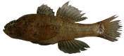
|
The body colour is pale beige to greyish beige. The head and body are relatively short. There are four to six dark-brown bands on the body and one dark band on the caudal peduncle. The snout is serrated. Melanophores were scattered on the body and dense at the pectoral, caudal fin bases, and pelvic fin. |
Butis humeralis [38]
|
The body is blackish, and the head is long, pointed, and relatively depressed. The caudal peduncle is relatively deep. Melanophores are covered throughout the fish body, notably in the first and second dorsal, pelvic and anal fins, and pectoral and caudal fin bases. |
Butis butis [39]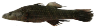
|
The colour of the body is blackish. The head is relatively depressed with the long and pointed snout. The caudal peduncle is slender. Melanophores are dense at the bases of pectoral and caudal fins, but scattered on opercula; first and second dorsal and anal fins. |
Table 2.
Variation of total length, weight and meristic parameters for the three Butis species (TL: total length (cm), W: weight (g); HL: head length (cm); BD: body depth (cm); ED: eye diameter (cm); DE: distance between eyes (cm); SE: Standard error).
| Species (number of fishes) | W±SE | TL±SE | ED±SE | DE±SE | BD±SE | HL±SE | HL/TL±SE | BD/TL±SE | ED/HL±SE | DE/HL±SE | |
|---|---|---|---|---|---|---|---|---|---|---|---|
| B. butis (715) | 11.35 ± 0.31 | 9.97 ± 0.07 | 0.43 ± 0.003 | 0.65 ± 0.007 | 1.47 ± 0.02 | 2.75 ± 0.02 | 0.15 ± 0.001a | 0.28 ± 0.001b | 0.16 ± 0.001b | 0.24 ± 0.002b | |
| Shapiro-Wilk test | W | 0.72 | 0.95 | 0.91 | 0.93 | 0.94 | 0.95 | 0.92 | 0.86 | 0.69 | 0.53 |
| p | <0.01 | <0.01 | <0.01 | <0.01 | <0.01 | <0.01 | <0.01 | <0.01 | <0.01 | <0.01 | |
| B. humeralis (871) | 14.66 ± 0.35 | 10.48 ± 0.07 | 0.44 ± 0.003 | 0.70 ± 0.006 | 1.61 ± 0.01 | 2.91 ± 0.02 | 0.15 ± 0.001b | 0.28 ± 0.001b | 0.15 ± 0.001a | 0.24 ± 0.001b | |
| Shapiro-Wilk test | W | 0.81 | 0.98 | 0.16 | 0.97 | 0.95 | 0.96 | 0.95 | 0.87 | 0.97 | 0.80 |
| p | <0.01 | <0.01 | <0.01 | <0.01 | <0.01 | <0.01 | <0.01 | <0.01 | <0.01 | <0.01 | |
| B. koilomatodon (876) | 5.46 ± 0.07 | 7.05 ± 0.03 | 0.36 ± 0.002 | 0.34 ± 0.003 | 1.37 ± 0.01 | 1.86 ± 0.01 | 0.20 ± 0.001c | 0.26 ± 0.001a | 0.20 ± 0.001c | 0.19 ± 0.002a | |
| Shapiro-Wilk test | W | 0.98 | 0.99 | 0.20 | 0.96 | 0.99 | 0.99 | 0.82 | 0.74 | 0.98 | 0.96 |
| p | <0.01 | <0.01 | <0.01 | <0.01 | <0.01 | <0.01 | <0.01 | <0.01 | <0.01 | <0.01 | |
| Kruskal-Wallis test | χ2 | 1169.90 | 329.13 | 781.41 | 608.93 | ||||||
| p | <0.01 | <0.01 | <0.01 | <0.01 | |||||||
Table 3 presents some of the measurement parameters of three study species, such as weight (W), total length (TL), eye diameter (ED), the distance between eyes (DE), head length (HL), body depth (BD), and their ratios. A total of 715 B. butis, 871 B. humeralis, and 876 B. bkoilomatodon were used in the morphological analysis. Regarding fish body size and weight, B. koilomatodon was found to be the smallest, and B. humeralis was the largest. This outcome was similar to the observation made by Tran et al. [3]. Butis humeralis and B. butis showed statistical similarity in BD/TL and DE/HL values and were greater than those in B. koilomatodon (Kruskal-Wallis Test, χ2BD/TL = 329.13, χ2DE/HL = 608.93, p < 0.01). Meanwhile, the three Butis species exhibited different HL/TL and ED/HL values (χ2HL/TL = 1169.90, χ2ED/HL = 781.41, p < 0.01). Overall, B. humeralis and B. butis were quite similar in both morphological features and meristic measurements, while B. koilomatodon showed marked differences from the other two species.
Table 3.
Nucleotide percentage (%) of mtCOI gene in Butis genus (DHTV: Duyen Hai, Tra Vinh, TDST: Tran De, Soc Trang, VHBL: Vinh Hau, Bac Lieu, DDCM: Dam Doi, Ca Mau).
| Species | Sampling sites | Number accession | %A | %C | %G | %T | %AT | %GC |
|---|---|---|---|---|---|---|---|---|
| Butis koilomatodon | DHTV | ON552550 | 25.62 | 28.44 | 17.81 | 28.13 | 53.75 | 46.25 |
| TDST | OK076879 | 25.45 | 29.01 | 17.18 | 28.36 | 53.81 | 46.19 | |
| HBBL | OK076880 | 25.61 | 28.85 | 17.02 | 28.53 | 54.14 | 45.87 | |
| DDCM | OK076881 | 25.45 | 29.01 | 17.18 | 28.36 | 53.81 | 46.19 | |
| Butis humeralis | DHTV | OK076882 | 23.50 | 29.66 | 18.48 | 28.36 | 51.86 | 48.14 |
| TDST | OK076883 | 23.50 | 29.66 | 18.48 | 28.36 | 51.86 | 48.14 | |
| HBBL | OK076884 | 24.64 | 26.58 | 18.15 | 30.63 | 55.27 | 44.73 | |
| DDCM | OK076885 | 24.15 | 26.90 | 18.15 | 30.79 | 54.94 | 45.05 | |
| Butis butis | DHTV | OK076886 | 23.66 | 29.66 | 18.31 | 28.36 | 52.02 | 47.97 |
| TDST | OK076887 | 24.80 | 26.74 | 17.99 | 30.47 | 55.27 | 44.73 | |
| HBBL | OK076888 | 24.15 | 26.90 | 18.31 | 30.63 | 54.78 | 45.21 | |
| DDCM | OK076889 | 23.99 | 27.07 | 18.31 | 30.63 | 54.62 | 45.38 |
3.2. Gene sequencing
The total DNA extracted from the fish samples all showed bands on agarose gel, indicating that the extraction process had obtained DNA (Fig. 2). The mtCOI gene was obtained from 12 fish specimens for a total of three Butis species. The length of the PCR product was 617bp (Fig. 3), and all nucleotides were unambiguous. The nucleotide composition of the mtCOI gene is shown in Table 3. In general, all B. koilomatodon samples showed the relatively same nucleotide percentage, %C content was the highest, followed by %T, %A, and the lowest was %G. A similar order was observed in the two specimens of B. humeralis from DHTV and TDST and one B. butis sample from DHTV. Different arrangements of nucleotide percentages were found in B. humeralis from HBBL and DDCM; and B. butis collected in TDST, HBBL, and DDCM. The highest %T, %C, %A, and the lowest %G; nevertheless, %AT of all sequences were consistently higher than %GC. These differences in nucleotide ratios may help understand the subgroups of the three groups in the phylogenetic tree of the genus Butis (Fig. 3).
Fig. 2.

Agarose gel electrophoresis of total DNA products (M: DL2000 DNA marker; lanes 1–4: B. humeralis; lanes 5–8: B. butis; lanes 9–12: B. koilomatodon).
Fig. 3.

Agarose gel electrophoresis of PCR products after amplification of mtCOI gene (M: DL2000 DNA marker, lanes 1 to 4 represent DNA amplification mtCOI gene of B. humeralis, lanes 5 to 8 represent DNA amplification mtCOI gene of B. butis, lanes 9 to 12 represent DNA amplification mtCOI gene of B. koilomatodon, lane (−): negative control).
After the intraspecific alignment by “Align by ClustalW” on Bioedit v.7.2, the results showed the variable nucleotide in B. koilomatodon was only 5/617 and conserved nucleotide was 612/617. A different result was seen for B. humeralis (DHTV - TDST) and B. butis (HBBL - DDCM) with homologous nucleotides 470/617 and 468/617, respectively. Notably, the alignment result between B. humeralis in HBBL and DDCM; and B. butis in DHTV and TDST showed significantly variable nucleotides as 147/617 and 149/617, respectively. These differences contributed to the different clustering of mtCOI genes of Butis species on the phylogenetic tree.
The obtained sequences were compared with homologous sequences from the Genbank to re-identify the species after using the morphological characteristics (Table 4). Four of B. koilmatodon specimens in this study were more than 98% homologous to B. koilmatodon from Taiwan and Malaysia. However, the comparison results of B. humeralis and B. butis (DHTV) were not shown as the same species. B. humeralis in HBBL and DDCM were the same B. butis from India with 99.68% and 86.08% percent identity, respectively. Meanwhile, B. butis-DHTV resembled B. humeralis in Bangladesh at 98.68%. Whereas, the B. butis collected in HBBL and DDCM and the B. butis in India were two different species because their identity match was only 85.90%.
Table 4.
The similarity of the mtCOI gene sequence of Butis species in the study with species on Gene Bank (DHTV: Duyen Hai, Tra Vinh, TDST: Tran De, Soc Trang, VHBL: Vinh Hau, Bac Lieu, DDCM: Dam Doi, Ca Mau).
| No. | Morphology method | DNA barcoding method |
|||||
|---|---|---|---|---|---|---|---|
| Species | Accession | Gene size (bp) | Query Cover (%) | Percent identity (%) | Sites | ||
| 1 | ON552550 B. koilomatodon - DHTV | B. koilomatodon | MW498548 | 655 | 99% | 99.53% | Malaysia |
| 2 | OK076879 B. koilomatodon - TDST | B. koilomatodon | MG574473 | 655 | 97% | 98.84% | Taiwan |
| 3 | OK076880 B. koilomatodon - HBBL | B. koilomatodon | MG574473 | 655 | 97% | 99.17% | Taiwan |
| 4 | OK076881 B. koilomatodon - DDCM | B. koilomatodon | MG574473 | 655 | 97% | 98.84% | Taiwan |
| 5 | OK076882 B. humeralis - DHTV | B. humeralis | MK572083 | 655 | 98% | 98.84% | Bangladesh |
| 6 | OK076883 B. humeralis - TDST | B. humeralis | MK572083 | 655 | 98% | 98.84% | Bangladesh |
| 7 | OK076884 B. humeralis - HBBL | B. butis | MH827972 | 630 | 99% | 99.68% | Bangladesh |
| 8 | OK076885 B. humeralis - DDCM | B. butis | MT765266 | 691 | 100% | 86.06% | India |
| 9 | OK07687986 B. butis - DHTV | B. humeralis | MK572083 | 655 | 98% | 98.68% | Bangladesh |
| 10 | OK076887 B. butis - TDST | B. butis | MH827972 | 630 | 99% | 100% | Bangladesh |
| 11 | OK076888 B. butis - HBBL | B. butis | MT765266 | 691 | 100% | 85.90% | India |
| 12 | OK076889 B. butis - DDCM | B. butis | MT765266 | 691 | 100% | 85.90% | India |
3.3. Genetic distance
Table 5 illustrates the genetic distances amongst the samples of Butis species. The mean values of intraspecific Kimura genetic distances of B. koilomatodon, B. humeralis, and B. butis were 0.00; 0.17; and 0.16, respectively. Whereas the interspecific Kimura genetic distances of B. koilomatodon - B. humeralis and B. koilomatodon - B. butis and B. humeralis - B. butis were 0.19; 0.20; and 0.14, respectively. This showed that identifying these B. koilomatodon specimens was unambiguous, and their mtCOI sequences were not vastly different. As such, they might belong to the same group in the genetic tree. The average interspecific genetic distance between B. humeralis and B. butis was 0.14, lower than their intraspecific distances (0.16–0.17).
Table 5.
Genetic distances among samples of Butis species (DHTV: Duyen Hai, Tra Vinh, TDST: Tran De, Soc Trang, HBBL: Hoa Binh, Bac Lieu, DDCM: Dam Doi, Ca Mau).
| Samples | (1) | (2) | (3) | (4) | (5) | (6) | (7) | (8) | (9) | (10) | (11) | (12) |
|---|---|---|---|---|---|---|---|---|---|---|---|---|
| (1) ON552550 B. koilomatodon - DHTV | _ | _ | _ | _ | _ | _ | _ | _ | _ | _ | _ | _ |
| (2) OK076879 B. koilomatodon - TDST | 0.01 | _ | _ | _ | _ | _ | _ | _ | _ | _ | _ | _ |
| (3) OK076880 B. koilomatodon - HBBL | 0.00 | 0.00 | _ | _ | _ | _ | _ | _ | _ | _ | _ | _ |
| (4) OK076881 B. koilomatodon - DDCM | 0.01 | 0.00 | 0.00 | _ | _ | _ | _ | _ | _ | _ | _ | _ |
| (5) OK076882 B. humeralis - DHTV | 0.20 | 0.19 | 0.19 | 0.19 | _ | _ | _ | _ | _ | _ | _ | _ |
| (6) OK076883 B. humeralis - TDST | 0.20 | 0.19 | 0.19 | 0.19 | 0.00 | _ | _ | _ | _ | _ | _ | _ |
| (7) OK076884 B. humeralis - HBBL | 0.18 | 0.17 | 0.18 | 0.17 | 0.22 | 0.22 | _ | _ | _ | _ | _ | _ |
| (8) OK076885 B. humeralis - DDCM | 0.21 | 0.21 | 0.21 | 0.21 | 0.21 | 0.21 | 0.15 | _ | _ | _ | _ | _ |
| (9) OK076886 B. butis - DHTV | 0.20 | 0.19 | 0.20 | 0.19 | 0.00 | 0.00 | 0.22 | 0.21 | _ | _ | _ | _ |
| (10) OK076887 B. butis - TDST | 0.17 | 0.17 | 0.17 | 0.17 | 0.22 | 0.22 | 0.00 | 0.16 | 0.22 | _ | _ | _ |
| (11) OK076888 B. butis - HBBL | 0.21 | 0.21 | 0.21 | 0.21 | 0.21 | 0.21 | 0.15 | 0.00 | 0.21 | 0.16 | _ | _ |
| (12) OK076889 B. butis - DDCM | 0.21 | 0.21 | 0.21 | 0.21 | 0.21 | 0.21 | 0.15 | 0.01 | 0.21 | 0.16 | 0.00 | _ |
3.4. Genetic differentiation
The results of analysing the genetic diversity of the 12 mtCOI gene sequences using DnaSP v.5 software (Table 6) obtained 10 haplotypes/12 individuals, haplotype diversity Hd = 0.97; nucleotide diversity π = 0.14. The number of segregating sites S = 174, and the average number of differences K = 83.68. The comparison results also showed that the genetic differentiation of B. humeralis (4 haplotypes, Hd = 0.83, π = 0.14; S = 147; K = 89.00) and B. butis (4 haplotypes, Hd = 1.00, π = 0.14; S = 149; K = 85.50) was higher than that of B. koilomatodon (3 haplotypes, Hd = 0.83, π = 0.005; S = 5; K = 2.83).
Table 6.
Genetic differentiation among three populations of Butis species.
| Populations | Sample size | Genetic differentiation |
||||
|---|---|---|---|---|---|---|
| Number of segregating sites (S) | Number of haplotypes (h) | Haplotype diversity (Hd) | Average number of differences (K) | Nucleotide diversity (π) | ||
| Butis koilomatodon | 4 | 5 | 3 | 0.83 | 2.83 | 0.005 |
| Butis humeralis | 4 | 147 | 4 | 0.83 | 89.00 | 0.14 |
| Butis Butis | 4 | 149 | 4 | 1.00 | 85.50 | 0.14 |
| Total | 12 | 174 | 10 | 0.97 | 83.68 | 0.14 |
3.5. Phylogenetic analysis
The phylogeny expressed the genetic relationship amongst Butis species is shown in Fig. 4. Three mtCOI sequences were used as in-group controls consisting of B. koilomatodon from Taiwan with an accession number of MG574473; B. humeralis from Bangladesh (MK572083); and B. butis from India (MT765266). All three control mtCOI gene sequences displayed significant similarities with the investigated mtCOI gene sequences (Table 4). The out-group controls were the mtCOI gene sequences of Glossogobius aureus-Philippines (KJ013044) and Periophthalmus chrysospilos-Bangladesh (MK572461). G. aureus belonged to the family Gobiidae, Glossogonius genus, while P. chrysospilos was of the family Oxudercidae, Periophthalmus genus. Two out-group control mtCOI gene sequences were chosen to ensure the genetic difference from the mtCOI gene of the Butis species.
Fig. 4.

The phylogenetic tree based on the COI gene was inferred by using the Maximum Likelihood method based on the Kimura 2-parameter model, with a bootstrap value of 1000 times on the nodes. Branch lengths correspond to the mean number of nucleotide substitutions per site. The scale bar indicates substitutions per site (DHTV: Duyen Hai, Tra Vinh, TDST: Tran De, Soc Trang, HBBL: Hoa Binh, Bac Lieu, DDCM: Dam Doi, Ca Mau).
The mtCOI phylogenetic graph in Fig. 4 was divided into five groups with reliable bootstrap values of 100%. The phylogram in Fig. 4 did not suggest that a different living environment had resulted in a change in the heredity of the mtCOI gene in Butis species. For example, in group I, B. koilomatodon obtained from TDST and DDCM were identical and did not vary much from HBBL and DHTV. Furthermore, often in groups II, II, and IV the samples of B. humeralis and B. butis were not grouped by sampling sites. Specifically, in group IV, B. humeralis collected from DHTV and TDST were found to be identical with a genetic distance of 0.00, or in group III, B. butis from HBBL and DDCM were seen to be very similar with a genetic distance of 0.00.
Group I displayed a similar pattern since all samples of B. koilomatodon and control were identical. Group V consisted of two out-group control, namely G. aureus from the Philippines and P. chrysospilos from Bangladesh. The fact that two out-group controls were separated into the group from the Butis species reinforces the reliability of the genetic analysis and the phylogenetic tree in the present study.
All three groups II, III, and IV of B. humeralis and B. butis samples were interspersed on the clades with a bootstrap value of 100%, and not separated into two separate groups for each species. Whilst in group II, B. humeralis from TDST and DHTV; B. butis-DHTV; and B. humeralis control from Bangladesh were grouped together because their genetic distance was 0.00. Group III consisted of B. butis control from India, B. humeralis-HBBL, and B. butis-TDST due to their low genetic distance of ∼0.00. B. humeralis from DDCM and B. butis from HBBL and DDCM were grouped into group IV with interspecific divergence of 0.00 and 0.01.
4. Discussion
The distinctive morphology of B. koilomatodon in this study was similar to that documented by Yokoo et al. [7] and Macieira et al. [40], who also reported scattered dark bands from the head to tail peduncle and a saw-toothed snout. Meanwhile, B. humeralis and B. butis shared many of the same features. However, they also expressed some different exterior features, such as differences in the height of the caudal peduncle. According to the research of Tran et al. [3], the first and second dorsal and anal fins were covered with numerous melanophores in B. humeralis, but scattered in B. butis [7]. Besides, the anal fins showed different starting positions [ [41,42]]. Compared with B. butis in this study, B. butis in India had many similarities such as an elongated and cylindrical body, pointed and depressed head, pointed snout, prominent lower jaw, the length of caudal peduncle was two times the height, the colour body was blackish with covered red spots on the head, body, and base of the fins. Like B. butis, but B. humeralis in India had distinct morphological features such as a head, body, and cheek covered with dark spots [43]. Meristic counts and measurements did not depend on the fish body size but were noted to be impacted by species origin and sexes [ [44,45]]. The present results showed B. koilomatodon was different from the two sister species regarding morphological characteristics and meristic parameters. Nevertheless, to determine if these differences were due to heredity or environmental adaptation, it was necessary to study the combination of morphology with the mtCOI gene.
Bingpeng et al. [19] noted that the mitochondrial mtCOI gene was conserved highly at the intraspecific level and showed slight variation at the interspecific level. The mtCOI gene has been used as DNA barcoding for the identification of marine fishes from Japan [46], freshwater fishes from India [47], fish from the Mediterranean [48], and from Australia [12]. In the present study, 12 mitochondrial mtCOI genes were amplified successfully with a size of 617bp after editing. After removing the interfering fragments at the beginning and the end, all sequences did not appear to stop codon or read frameshifting error.
Nucleotide composition was a fundamental feature of the genome that strongly influences gene function and regulation. The GC content was one of the main parameters used to describe nucleotide composition and is often related to genome size [49]. The fish genomes had a homogeneous AT/GC overall, containing only a narrow GC% [50]. In the present study, the base composition of the mtCOI gene revealed AT content was higher than GC content for all sequences; however, this difference was insignificant, only about 10%. Similar findings were also reported by Ward et al. [12], investigating Australian fish species; Hubert et al. [51], studying Canadian freshwater fishes; Lara et al. [52], focusing on Cuban freshwater fish; and Bingpeng et al. [19], studying Taiwanese fish.
Comparison results with homogenous sequences from the Genbank revealed B. humeralis-HBBL and B. humeralis-DDCM were similar to B. butis from India, whereas B. butis-DHTV was identifcal to B. humeralis from Bangladesh. Confusion may have been caused if the species identification was only based on morphological similarities between the two species. Several previous studies have also reported it was difficult to discriminate between B. humeralis and B. butis based solely on morphological methods [41]. Hence, the B. humeralis and B. butis specimens were reclassified by the molecular method using the mtCOI gene sequence.
The efficiency and reliability of using the mtCOI gene as DNA barcoding to identify fish species depended on both interspecific and intraspecific divergence. However, there was still a lack of a reliable standard threshold of genetic difference for identifying intraspecific and interspecific species [19]. In this study, the intraspecific genetic distance of B. humeralis and B. butis were 0.17 (17%) and 0.16 (16%) compared with 0.14 (14%) for species within genera. In the research of Nguyen & Duong [53], the conspecific genetic distances of B. butis and B. humeralis were merely 0.3% and 0.4%, which is much lower than the distance in this study. It highlights that B. humeralis and B. butis were two different species.
The genetic relationship of the three Butis species was seen in the phylogenetic tree. Although Lam & Dinh [8] reported the morphometric values such as the total length (TL) and weight (W) of B. koilomatodon in DDCM were higher than that in HBBL and TDST, the four B. koilomatodon samples along with the control B. koilmatodon sample originating from Taiwan were clustered into a monophyletic clade in the phylogram. This demonstrated that the efficiency of DNA barcoding in species identification was high. In the same way, several previous studies showed that the body sizes of B. humeralis in TDST were larger than in HBBL [9] and also that DDCM was the sampling site where TL and W values of B. butis were lower than HBBL [10]. However, in clusters III and IV, they were grouped. The differences in TL and W of Butis species were due to the adaptation of fish to different habitats, including food availability, and were not influenced by genetic factors. Here, the different habitat conditions did not affect the congeneric mtCOI gene.
The four B. koilomatodon samples and the in-group control were the same. The morphological traits of B. koilomatodon were sharply different from two congeners, such as the number of dark bands on the body and the serrated snout [ [7,40]]. Therefore, B. koilomatodon mtCOI genes were separated into their own groups that was reliable. Two out-group controls, G. aureus from the Philippines and P. chrysospilos from Bangladesh, clustered separately, helped increase the genetic analysis's reliability and the phylogenetic tree in these findings.
The bootstrap value is the percentage of replicate phylogenies that recovered a particular clade from the original phylogenetic system constructed using the original association. However, the accuracy of the bootstrap value can be significantly reduced as different genes may have different evolutionary histories due to ancestral polymorphism, recombination within a locus, etc. Therefore, bootstrap test results must be cautiously performed, especially when using a single DNA marker [54]. Butis humeralis and B. butis were interspersed on three clusters II, III and IV with a bootstrap value of 100%, rather than divided into two separate groups corresponding to the two species. The intraspecific genetic distances could differ from species to species, but are usually at a low level (∼1%), for example, 1.2% in goby species in India [11] or 0.39% in fish in Australia [12]. In the present study, kimura genetic distances between B. humeralis and B. butis were 0.14 (14%), and as such, they were seen as different species, although their similar morphological characteristics might confuse fish classification. Incorrect morphological identification could lead to misclassifications and alter the results of the genetic tree [19]. Tautz et al. [55] and Radulovici et al. [56] emphasised that morphological misidentifications, DNA contamination, and insufficient knowledge about the taxonomy could also contribute to ambiguous barcoding results. On the other hand, the reference library of DNA barcoding and species identification requires many samples at different stages, such as egg, larval and adult stages, due to the many changes in morphological characteristics over the development stages [19]. Therefore, misidentification is not feasible; for example, in this study, DNA barcoding could detect cases of morphological misidentification. The combination of morphological and molecular methods was necessary for more accurate taxonomy and species identification.
5. Conclusion
This study constructed the phylogeny of the mtCOI gene of Butis species collected from river mouths in the VMD with a high bootstrap of 100%. The phylogram was divided into five groups, where four B. koilomatodon specimens were separated into a single group, and B. humeralis and B. butis were interspersed in groups II, III, and IV. The meristic and morphology of B. koilomatodon differed from the two remaining congeners. Meanwhile, B. humeralis and B. butis shared similar exterior traits, apart from the high caudal peduncle and the pigments at the first and second dorsal and anal fins. The outcome suggests that B. humeralis and B. butis differed because their interspecific genetic distance was 14%.
Author contribution statement
Tran Thi Huyen Lam: Conceived and designed the experiments; Performed the experiments; Analyzed and interpreted the data; Contributed reagents, materials, analysis tools or data; Wrote the paper.
Quang Minh Dinh: Conceived and designed the experiments; Performed the experiments; Analyzed and interpreted the data; Contributed reagents, materials, analysis tools or data; Wrote the paper.
Van Thi Bich Truong: Contributed reagents, materials, analysis tools or data.
Ngon Trong Truong: Contributed reagents, materials, analysis tools or data.
Nam Sy Tran: Contributed reagents, materials, analysis tools or data.
Ton Huu Duc Nguyen: Performed the experiments; Analyzed and interpreted the data; Contributed reagents, materials, analysis tools or data.
Ethical statement
The use of fish in this research was approved by the Scientific Committee of the School of Education, Can Tho University (Animal welfare assessment BQ2019–02/KSP).
Data availability statement
Data included in article/supplementary material/referenced in article.
Funding statement
This research was funded by the Ministry of Education and Training of Vietnam under grant number B2019-TCT-02. Tran Thi Huyen Lam was funded by Vingroup JSC and supported by the Master, PhD Scholarship Programme of Vingroup Innovation Foundation (VINIF), Institute of Big Data, code VINIF.2021.TS.162.
Additional information
No additional information is available for this paper.
Declaration of competing interest
The authors declare that they have no known competing financial interests or personal relationships that could have appeared to influence the work reported in this paper.
Acknowledgements
We are grateful to local fishers for fish collection help and Nigel K. Downes for proofreading assistance.
Abbreviations
- VMD
Vietnamese Mekong Delta
- mtCOI
Mitochondrial cytochrome C oxidase subunit I
- DNA
Deoxyribonucleic acid
- DHTV
Duyen Hai, Tra Vinh
- TDST
Tran De, Soc Trang
- HBBL
Hoa Binh, Bac Lieu
- DDCM
Dam Doi, Ca Mau
- W
Weight of fish
- TL
Total length
- ED
Eye diameter
- DE
Distance between eyes
- BD
Body depth
- HL
Head length
- PCR
Polymerase chain reaction
- NCBI
National Center for Biotechnology Information
References
- 1.Thacker C. Phylogeny of Gobioidei and placement within acanthomorpha, with a new classification and investigation of diversification and character evolution. Copeia. 2009;2009(1):93–104. [Google Scholar]
- 2.Froese R., Pauly D. 2023. FishBase.www.fishbase.org Accessed: 2023. [Google Scholar]
- 3.Tran D.D., Shibukawa K., Nguyen T.P., Ha P.H., Tran X.L., Mai V.H., Utsugi K. Can Tho University Publisher; 2013. Fishes of Mekong Delta, Vietnam, Can Tho. [Google Scholar]
- 4.Tran D.D., Nguyen V.T., To H.T.M., Nguyen T.T., Dinh Q.M. Species composition and biodiversity index of gobiid assemblage in estuarine areas of the Mekong Delta, Vietnam. Egyptian Journal of Aquatic Biology and Fisheries. 2020;24(7):931–941. [Google Scholar]
- 5.Le K.N., Son N.H., Nguyen T.N.H., Le H.A., Tran V.D., Nguyen T.D., Tran D.D., Ha P.H., To T.M.H., Nguyen T.V., Vo T.T., Nguyen T.T., Dinh Q.M. Fish species composition in Hau river basin at Hau giang province. VNU Journal of Science: Natural Sciences and Technology. 2018;34(1):90–104. [Google Scholar]
- 6.Larmuseau M., Guelinckx J., Hellemans B., Van Houdt J., Volckaert F. Fast PCR‐RFLP method facilitates identification of Pomatoschistus species from the North Atlantic. J. Appl. Ichthyol. 2008;24(3):342–344. [Google Scholar]
- 7.Yokoo T., Kanou K., Moteki M., Kohno H., Tongnunui P., Kurokura H. Juvenile morphology and occurrence patterns of three Butis species (Gobioidei: eleotridae) in a mangrove estuary, southern Thailand. Ichthyol. Res. 2006;53(4):330–336. [Google Scholar]
- 8.Lam T.T.H., Dinh Q.M. Morphometric and meristic variability in Butis koilomatodon in estuarine and coastal areas of the Mekong Delta. Vietnam Agricultural Science Journal. 2020;3(4):806–816. [Google Scholar]
- 9.Dinh Q.M., Lam T.T.H., Nguyen T.H.D. Study on external morphology of Butis humeralis in the coastline regions of Mekong Delta. Science and Technology Journal of Agriculture & Rural Development. 2021;10:146–153. [Google Scholar]
- 10.Phan G.H., Dinh Q.M., Truong N.T., Nguyen T.H.D., Tran N.S. Morphometric and meristic variations of Butis butis along the coastline in the Mekong Delta, Vietnam. AACL Bioflux. 2021;14(4):2544–2553. [Google Scholar]
- 11.Viswambharan D., Pavan-Kumar A., Singh D.P., Jaiswar A.K., Chakraborty S.K., Nair J.R., Lakra W.S. DNA barcoding of gobiid fishes (Perciformes, Gobioidei) Mitochondrial DNA. 2015;26(1):15–19. doi: 10.3109/19401736.2013.834438. [DOI] [PubMed] [Google Scholar]
- 12.Ward R.D., Zemlak T.S., Innes B.H., Last P.R., Hebert P.D. DNA barcoding Australia’s fish species. Philosophical Transactions of the Royal Society B. 2005;360(1462):1847–1857. doi: 10.1098/rstb.2005.1716. [DOI] [PMC free article] [PubMed] [Google Scholar]
- 13.Jeon H.-B., Choi S.-H., Suk H.Y. Exploring the utility of partial cytochrome c oxidase subunit 1 for DNA barcoding of gobies. Animal Systematics, Evolution and Diversity. 2012;28(4):269–278. [Google Scholar]
- 14.Hebert P.D., Cywinska A., Ball S.L., Dewaard J.R. Biological identifications through DNA barcodes. Proceedings of the Royal Society B: Biological Sciences. 2003;270(1512):313–321. doi: 10.1098/rspb.2002.2218. [DOI] [PMC free article] [PubMed] [Google Scholar]
- 15.Zemlak T.S., Ward R.D., Connell A.D., Holmes B.H., Hebert P.D. DNA barcoding reveals overlooked marine fishes. Molecular Ecology Resources. 2009;9:237–242. doi: 10.1111/j.1755-0998.2009.02649.x. [DOI] [PubMed] [Google Scholar]
- 16.Knebelsberger T., Thiel R. Identification of gobies (Teleostei: perciformes: Gobiidae) from the north and baltic seas combining morphological analysis and DNA barcoding. Zoological Journal of the Linnean Society. 2014;172(4):831–845. [Google Scholar]
- 17.DNA barcoding application of mitochondrial COI gene to identify some fish of family Gobiidae in Vietnam. Linh N.M., Van Quan N., Van Chien P., Ly D.H., Van Nhan D., Len D.T., editors. Vietnam Journal of Marine Science and Technology. 2018;18(4):443–451. [Google Scholar]
- 18.Thacker C.E. Molecular phylogeny of the gobioid fishes (Teleostei: perciformes: gobioidei) Molecular Phylogenetics and Evolution. 2003;26(3):354–368. doi: 10.1016/s1055-7903(02)00361-5. [DOI] [PubMed] [Google Scholar]
- 19.Bingpeng X., Heshan L., Zhilan Z., Chunguang W., Yanguo W., Jianjun W. DNA barcoding for identification of fish species in the Taiwan Strait. PLoS One. 2018;13(6) doi: 10.1371/journal.pone.0198109. [DOI] [PMC free article] [PubMed] [Google Scholar]
- 20.Asgharian H., Sahafi H.H., Ardalan A.A., Shekarriz S., Elahi E. Cytochrome c oxidase subunit 1 barcode data of fish of the Nayband National Park in the Persian Gulf and analysis using meta‐data flag several cryptic species. Molecular Ecology Resources. 2011;11(3):461–472. doi: 10.1111/j.1755-0998.2011.02989.x. [DOI] [PubMed] [Google Scholar]
- 21.Costa F.O., DeWaard J.R., Boutillier J., Ratnasingham S., Dooh R.T., Hajibabaei M., Hebert P.D. Biological identifications through DNA barcodes: the case of the Crustacea. Canadian Journal of Fisheries and Aquatic Sciences. 2007;64(2):272–295. [Google Scholar]
- 22.Mikkelsen N.T., Schander C., Willassen E. Local scale DNA barcoding of bivalves (Mollusca): A case study. Zoologica Scripta. 2007;36(5):455–463. [Google Scholar]
- 23.Hebert P.D.N., Stoeckle M.Y., Zemlak T.S., Francis C.M. Identification of birds through DNA barcodes. PLoS Biology. 2004;2(10):e312. doi: 10.1371/journal.pbio.0020312. [DOI] [PMC free article] [PubMed] [Google Scholar]
- 24.Hajibabaei M., Singer G.A., Hickey D.A. Benchmarking DNA barcodes: an assessment using available primate sequences. Genome. 2006;49(7):851–854. doi: 10.1139/g06-025. [DOI] [PubMed] [Google Scholar]
- 25.Dinh Q.M. Aspects of reproductive biology of the red goby Trypauchen vagina (Gobiidae) from the Mekong Delta. Journal of Applied Ichthyology. 2018;34(1):103–110. [Google Scholar]
- 26.Le T., Nguyen M.T., Nguyen V.P., Nguyen D.C., Pham X.H., Nguyen T.S., Hoang V.C., Hoang P.L., Le H., Dao N.C. In: Le T., editor. vol. vol. I. Ha Noi, Vietnam Education Publishing House; 2006. Provinces and city in the Mekong Delta; pp. 49–94. (Geography of Provinces and Cities in Vietnam). [Google Scholar]
- 27.Dinh Q.M., Lam T.T.H., Nguyen T.H.D., Nguyen T.M., Nguyen T.T.K., Nguyen N.T. First reference on reproductive biology of Butis koilomatodon in Mekong Delta, Vietnam. BMC Zoology. 2021;6(1):1–14. doi: 10.1186/s40850-021-00072-y. [DOI] [PMC free article] [PubMed] [Google Scholar]
- 28.Rogers S.O., Bendich A.J. Plant Molecular Biology Manual. Springer; 1989. Extraction of DNA from plant tissues; pp. 73–83. [Google Scholar]
- 29.Sanger F., Nicklen S., Coulson A.R. DNA sequencing with chain-terminating inhibitors. Proceedings of the National Academy of Sciences of the United States of America. 1977;74(12):5463–5467. doi: 10.1073/pnas.74.12.5463. [DOI] [PMC free article] [PubMed] [Google Scholar]
- 30.Hall T. Nucleic Acids Symposium Series. 1999. BioEdit: a user-friendly biological sequence alignment editor and analysis program for Windows 95/98/NT; pp. 95–98. [Google Scholar]
- 31.Kumar S., Stecher G., Tamura K. MEGA7: molecular evolutionary genetics analysis version 7.0 for bigger datasets. Molecular Biology and Evolution. 2016;33(7):1870–1874. doi: 10.1093/molbev/msw054. [DOI] [PMC free article] [PubMed] [Google Scholar]
- 32.Agorreta A., Rueber L. A standardized reanalysis of molecular phylogenetic hypotheses of Gobioidei. Systematics and Biodiversity. 2012;10(3):375–390. [Google Scholar]
- 33.Agorreta A., San Mauro D., Schliewen U., Van Tassell J.L., Kovačić M., Zardoya R., Rüber L. Molecular phylogenetics of Gobioidei and phylogenetic placement of European gobies. Molecular Phylogenetics and Evolution. 2013;69(3):619–633. doi: 10.1016/j.ympev.2013.07.017. [DOI] [PubMed] [Google Scholar]
- 34.Çiftci Y., Eroğlu O., Firidin Ş. Mitochondrial cytochrome b sequence variation in three sturgeon species (A. stellatus pallas, 1771, A. gueldenstaedtii brandt, 1833, H. huso Linnaeus, 1758) from the black sea coasts of Turkey. Turkish Journal of Fisheries and Aquatic Sciences. 2013;13(2):291–303. [Google Scholar]
- 35.Merl D., Escalante A., Prado R. Report No AMS2005-03 (2005). Applied Mathematics & Statistics. University of California; United States.: 2005. Comparison of Bayesian, maximum likelihood and parsimony methods for detecting positive selection. [Google Scholar]
- 36.Drezner Z., Turel O., Zerom D. A modified Kolmogorov–Smirnov test for normality. Communications in Statistics - Simulation and Computation. 2010;39(4):693–704. [Google Scholar]
- 37.Bleeker P. vol. 22. Lange & Company; 1849. Bijdrage tot de kennis der Blennioïden en Gobioïden van den Soenda-Molukschen archipel, met beschrijving van 42 nieuwe species; pp. 1–40. (Verhandelingen van het Bataviaasch Genootschap van Kunsten en Wetenschappen). 6. [Google Scholar]
- 38.Valenciennes A. vol. 4. FG Levrault; Paris: 1837. (Cuvier GL & Valenciennes. A Histoire Naturelle des Poissons. Levrault). 12. [Google Scholar]
- 39.Hamilton F. vol. 1. Archibald Constable; 1822. (An Account of the Fishes Found in the River Ganges and its Branches). [Google Scholar]
- 40.Macieira R., Giarrizzo T., Gasparini J., Sazima I. Geographic expansion of the invasive mud sleeper Butis koilomatodon (Perciformes: eleotridae) in the western Atlantic Ocean. Journal of Fish Biology. 2012;81(1):308–313. doi: 10.1111/j.1095-8649.2012.03285.x. [DOI] [PubMed] [Google Scholar]
- 41.Vo T.T., Ha P.H. Species composition andabundance of goby fish in family Eleotridae in Hau river. Can Tho University Journal of Science, Part B: Agriculture, Fisheries and Biotechnology. 2013;28:168–176. [Google Scholar]
- 42.Truong T.K., Tran T.T.H. Can Tho University, Can Tho University book store; 1993. Identification of Freshwater Fish in Mekong Delta. [Google Scholar]
- 43.Koumans F.P. Manager of Publications; 1941. Gobioid Fishes of India. [Google Scholar]
- 44.Talwar P.K., Jhingran A.G. vol. 2. Balkema; Rotterdam, Netherlands: 1991. (Inland Fishes of India and Adjacent Countries). [Google Scholar]
- 45.Gonzalez-Martinez A., Lopez M., Molero H.M., Rodriguez J., González M., Barba C., García A. Morphometric and meristic characterization of native chame fish (Dormitator latifrons) in Ecuador using multivariate analysis. Animals. 2020;10(10):1805. doi: 10.3390/ani10101805. [DOI] [PMC free article] [PubMed] [Google Scholar]
- 46.Zhang J.-B., Hanner R. DNA barcoding is a useful tool for the identification of marine fishes from Japan. Biochemical Systematics and Ecology. 2011;39(1):31–42. [Google Scholar]
- 47.Chakraborty M., Ghosh S.K. An assessment of the DNA barcodes of Indian freshwater fishes. Gene. 2014;537(1):20–28. doi: 10.1016/j.gene.2013.12.047. [DOI] [PubMed] [Google Scholar]
- 48.Karahan A., Douek J., Paz G., Stern N., Kideys A.E., Shaish L., Goren M., Rinkevich B. Employing DNA barcoding as taxonomy and conservation tools for fish species censuses at the southeastern Mediterranean, a hot-spot area for biological invasion. Journal for Nature Conservation. 2017;36:1–9. [Google Scholar]
- 49.Li X.-Q., Du D. Variation, evolution, and correlation analysis of C+ G content and genome or chromosome size in different kingdoms and phyla. PLoS One. 2014;9(2) doi: 10.1371/journal.pone.0088339. [DOI] [PMC free article] [PubMed] [Google Scholar]
- 50.Symonová R., Suh A. Nucleotide composition of transposable elements likely contributes to AT/GC compositional homogeneity of teleost fish genomes. Mobile DNA. 2019;10(1):1–8. doi: 10.1186/s13100-019-0195-y. [DOI] [PMC free article] [PubMed] [Google Scholar]
- 51.Hubert N., Hanner R., Holm E., Mandrak N.E., Taylor E., Burridge M., Watkinson D., Dumont P., Curry A., Bentzen P. Identifying Canadian freshwater fishes through DNA barcodes. PLoS One. 2008;3(6) doi: 10.1371/journal.pone.0002490. [DOI] [PMC free article] [PubMed] [Google Scholar]
- 52.Lara A., Ponce de León J.L., Rodriguez R., Casane D., Cote G., Bernatchez L., García‐Machado E. DNA barcoding of Cuban freshwater fishes: evidence for cryptic species and taxonomic conflicts. Molecular Ecology Resources. 2010;10(3):421–430. doi: 10.1111/j.1755-0998.2009.02785.x. [DOI] [PubMed] [Google Scholar]
- 53.Nguyen T.P., Duong Y.T. Comparing morphological characteristics and DNA barcoding of two goby species Butis butis and Butis humeralis. Can Tho University Journal of Science. 2015;40(B):23–30. [Google Scholar]
- 54.Russo C.A.d.M., Selvatti A.P. Bootstrap and rogue identification tests for phylogenetic analyses. Molecular Biology and Evolution. 2018;35(9):2327–2333. doi: 10.1093/molbev/msy118. [DOI] [PubMed] [Google Scholar]
- 55.Tautz D., Arctander P., Minelli A., Thomas R.H., Vogler A.P. A plea for DNA taxonomy. Trends in Ecology & Evolution. 2003;18(2):70–74. [Google Scholar]
- 56.Radulovici A.E., Archambault P., Dufresne F. DNA barcodes for marine biodiversity: moving fast forward? Diversity. 2010;2(4):450–472. [Google Scholar]
Associated Data
This section collects any data citations, data availability statements, or supplementary materials included in this article.
Data Availability Statement
Data included in article/supplementary material/referenced in article.