Abstract
The cell type-specific expression of key transcription factors is central to development and disease. Brachyury/T/TBXT is a major transcription factor for gastrulation, tailbud patterning, and notochord formation; however, how its expression is controlled in the mammalian notochord has remained elusive. Here, we identify the complement of notochord-specific enhancers in the mammalian Brachyury/T/TBXT gene. Using transgenic assays in zebrafish, axolotl, and mouse, we discover three conserved Brachyury-controlling notochord enhancers, T3, C, and I, in human, mouse, and marsupial genomes. Acting as Brachyury-responsive, auto-regulatory shadow enhancers, in cis deletion of all three enhancers in mouse abolishes Brachyury/T/Tbxt expression selectively in the notochord, causing specific trunk and neural tube defects without gastrulation or tailbud defects. The three Brachyury-driving notochord enhancers are conserved beyond mammals in the brachyury/tbxtb loci of fishes, dating their origin to the last common ancestor of jawed vertebrates. Our data define the vertebrate enhancers for Brachyury/T/TBXTB notochord expression through an auto-regulatory mechanism that conveys robustness and adaptability as ancient basis for axis development.
Subject terms: Developmental biology, Molecular biology
This study identifies three conserved shadow enhancers (T3, C, I) regulating notochord expression of Brachyury across vertebrates using genomic data, transgenic assays, and enhancer knockouts, which are critical for vertebrate notochord development.
Introduction
The defining feature of the chordate body plan is the notochord, a principal structure formed by the axial or chorda mesoderm that provides stability and rigidity along the body axis1,2. As mammals form an ossified spine, their notochord progressively regresses and its remnants form the nucleus pulposus within the intervertebral discs3–7. Notochord precursors emerge from the initial organizer and form in a stereotypical rostral-to-caudal trajectory as gastrulation proceeds, manifesting among the earliest visible structures in chordate embryos1,8. The deeply conserved T-box transcription factor gene Brachyury (also called T or TBXT) is a key regulator of notochord formation. Originally identified as dominant mutation T that caused short tails in mice, Brachyury expression and function has been linked to notochord emergence across chordates9–15. In addition to its central role in notochord fate specification, the function of vertebrate Brachyury is required for proper primitive streak formation, tailbud specification, and subsequent neuromesodermal progenitor control16–18. However, how the expression of this central developmental transcription factor is selectively regulated to achieve its notochord activity in mammals remains unresolved.
The central contribution of the notochord and the tailbud to different morphological adaptions and locomotion strategies shows in the diversification of axial structures across vertebrates19. Gain and loss of gene copies and of their associated gene-regulatory elements are major drivers of evolutionary innovation, and the Brachyury gene family itself is a prime example of this process. Brachyury predates the origin of, and was present as, a single copy gene in the chordate ancestor20,21. Following two whole genome duplications in early vertebrates and the subsequent loss of one of four Brachyury paralogs, three gene paralogs were present in the jawed vertebrate ancestor: Tbxta, Tbxtb, and Tbx1921. Tbxta became subsequently lost within the tetrapod lineage, resulting in mammals and birds ultimately only retaining Tbxtb (commonly called Brachyury/T/TBXT in tetrapods including humans)22. In contrast, ray-finned fishes retained both tbxta/ntla and tbxtb/ntlb, the latter being the ortholog of the remaining human Brachyury/T/TBXT (de facto TBXTB) gene17.
Curiously, tbxta/ntla has become the predominant functional Brachyury/T/TBXT gene in zebrafish, as documented in classic mutants for ntla (no tail a) that fail to form a tail and notochord13,15. While no mutant for zebrafish tbxtb/ntlb has been reported to date, morpholino-based knockdown studies indicate that tbxtb function adds minimally to the dominant role of zebrafish tbxta17. This variable copy number of Brachyury genes across vertebrates came along with selection and divergence of regulatory elements controlling Brachyury gene expression during distinct developmental timepoints and cell types. Promoter-proximal regions in the Ciona Brachyury gene and in the zebrafish tbxta gene drive early organizer and notochord activity10,23. In contrast, the promoter-proximal region called Tstreak of Brachyury/T/Tbxtb in mouse, human, and Xenopus has previously been found to drive primitive streak expression in response to canonical Wnt/beta-catenin signaling, yet lacks any notochord-driving activity24–26. Further, recent work documented that deleting a large 37 kb-spanning region upstream of mouse Brachyury/T/Tbxtb leads to mutant phenotypes consistent with a selective loss of Brachyury notochord expression27. A small element termed TNE in the 37 kb interval was sufficient to drive specific notochord expression in mouse reporter assays, yet its deletion showed mild to no phenotypic consequences27. These pioneering data show that additional regulatory element(s) in addition to Tstreak and TNE contribute to Brachyury/Tbxtb expression specifically in the notochord. Uncovering the regulation of the vertebrate Brachyury notochord enhancer(s) will expand our understanding of the evolutionary history of this key developmental regulator and of the mechanisms leading to notochord formation. In particular, comparison to the Ciona Brachyury locus containing two upstream shadow enhancers with well-defined regulatory grammar28,29 may inform cis-regulatory adaptations at the onset of vertebrate emergence.
Uncovering the regulatory elements responsible for its notochord expression also promises to shed light onto the role of Brachyury in adult human spine health and in chordoma tumors, a rare sarcoma of the spine that is hypothesized to arise from notochord remnants30–32. Several familial chordomas harbor duplications or further complex amplifications of the Brachyury/T/TBXTB locus that possibly convey chordoma susceptibility to carriers33–35. These findings suggest that chordoma-associated Brachyury/T/TBXTB locus amplifications contain, or hijack the action of, cis-regulatory elements to possibly drive Brachyury/T/TBXTB expression in chordoma, potentially with Brachyury controlling its own expression as indicated by ChIP-seq findings36.
Here, we identify the complement of three auto-regulated shadow enhancers T3, C, and I in the Brachyury/T/Tbxtb locus that convey notochord activity. We combined (i) genomic data from human chordoma tumor cell lines, human embryonic stem cells, and mouse embryonic stem cells; (ii) non-coding element conservation across mammals (human, mouse, Monodelphis) and all vertebrates; (iii) transgenic reporter assays in zebrafish, mouse, axolotl, and Ciona; (iv) and enhancer knockouts in mice. In triple enhancer knockout mice, we document the selective absence of Brachyury protein in the notochord and subsequent neural tube and trunk defects as linked to notochord perturbations. Using comparative genomics, we uncover that the location and activity of the enhancers T3, C, and I is conserved within the Brachyury/tbxtb loci across jawed vertebrates. Our data uncover a deep conservation of shadow enhancers regulating Brachyury expression in the notochord, one of the most prominent developmental structures of the vertebrate body and involved in spine and neural tube defects.
Results
Defining a region for human Brachyury notochord expression
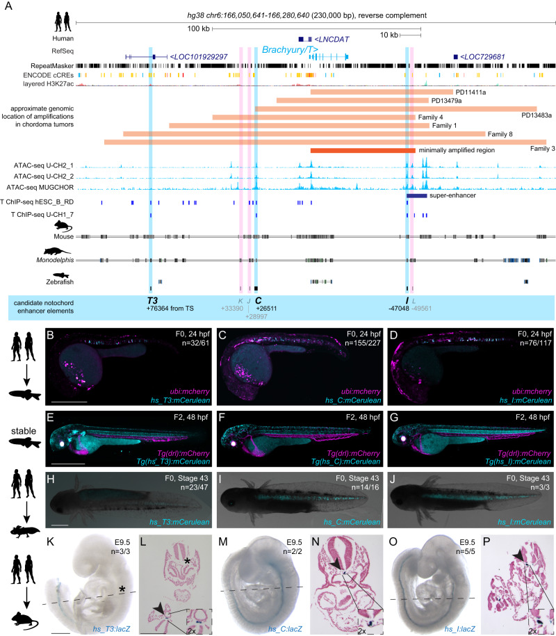
To identify enhancer elements with notochord activity in the human Brachyury/T/TBXTB locus, we analyzed the Brachyury/T/TBXTB locus to narrow down a minimally required genomic region around the Brachyury gene body. Familial and sporadic chordoma feature duplications and/or complex amplifications of Brachyury33–35,37, suggesting that essential cis-regulatory elements for notochord expression lie within the commonly amplified region. Available genomic patient data outlined a minimally amplified region of ~50 kb surrounding the human Brachyury gene body, with individual tumors extending their amplifications proximal or distal of this minimal region34,37 (Fig. 1A). Within this minimal interval and its vicinity, we uncovered several regions that have been charted as open chromatin in the chordoma cell lines U-CH2 and MUGCHOR using ATAC-seq36,38, indicating potential regulatory elements in accessible chromatin, including a super-enhancer region previously proposed to be active in chordoma38 (Fig. 1A). Further, mammalian Brachyury has been postulated to control its own notochord expression27,39. Using Brachyury/T ChIP-seq data from the human chordoma tumor cell line U-CH1 and human ES-derived mesoderm progenitor cells36,40, we found discrete Brachyury binding events within the minimal amplification interval and its vicinity (Fig. 1A). Genome alignment of human versus other mammalian species indicated candidate enhancer regions (conserved non-coding elements; CNEs) through non-coding sequence conservation in mouse and the more distant marsupial Monodelphis domestica41 (Fig. 1A).
Fig. 1. Human Brachyury enhancer elements T3, C, and I are active in different species.
A Human Brachyury/T/TBXTB locus with surrounding gene loci adapted from UCSC genome browser. Repeats marked in black using the RepeatMasker track; additional tracks include the ENCODE conserved cis-regulatory elements (cCREs) and layered H3K27ac signals. Further annotated are approximate amplifications (light orange) and the minimally amplified region (dark orange) in chordoma tumors. ATAC-sequencing (light blue peaks) and T ChIP-sequencing (dark blue lines) suggest enhancer elements (light pink highlight, not active; light blue highlight, active) that are conserved in mouse and the marsupial Monodelphis domestica. B–D Representative F0 zebrafish embryos injected with the human enhancer elements hs_T3 (B), hs_C (C), and hs_I (D) showing mosaic mCerulean reporter expression in the notochord at 24 hpf and expression of ubi:mCherry as injection control. N represents the number of animals expressing mCerulean in the notochord relative to the total number of animals expressing mCherry. Scale bar in (B): 0.5 mm, applies to B, C. E–G Representative images of stable transgenic F2 embryos at 48 hpf for each of the human enhancer elements hs_T3, hs_C, and hs_I crossed to Tg(drl:mCherry) that labels lateral plate mesoderm and later cardiovascular lineages. Transgenic F2 embryos recapitulate the F0 expression pattern in the notochord, with hs_T3 (E) additionally expressing mCerulean in the pharyngeal arches and fin, and hs_I (G) in the proximal kidney close to the anal pore. Enhancer element hs_C (F) stable transgenic lines have lower relative notochord reporter activity than hs_T3 and hs_I. Scale bar in (E): 0.5 mm, applies to E–G. H–J Representative F0 axolotl embryos at peri-hatching stages expressing mCerulean from the human enhancers hs_T3 (G), hs_C (H), hs_I (I). N represent the number of animals expressing mCerulean in the notochord relative to the total number of animals showing any mCerulean expression. Scale bar in (H): 1 mm, applies to H–J. K, M, and O Representative images of transgenic E9.5 mouse embryos expressing lacZ (encoding beta-galactosidase) under the human enhancers hs_T3 (K), hs_C (M), and hs_I (O) visualized with X-gal whole-mount staining. While hs_C and hs_I express beta-galactosidase in the entire notochord, beta-galactosidase expression from hs_T3 is restricted to the posterior notochord. Black asterisk marks absence of beta-galactosidase in the anterior notochord. N represent the number of animals expressing beta-galactosidase in the notochord relative to the total number of animals with tandem integrations at H11. Dotted lines represent the sectioning plane. Scale bar in (K): 0.5 mm, applies to (K, M, O). L, N, P Representative images of Fast Red-stained cross sections from embryos shown on the left, hs_T3 (L), hs_C (N), and hs_I (P). Black arrowheads point at notochord, and inserts show notochords at 2x higher magnification. Scale bar in (L): 0.25 mm, applies to L, N, P. The species silhouettes were adapted from the PhyloPic database (www.phylopic.org).
From our combined locus analysis, we identified the six initial candidates T3, K, J, C, I, and L as putative notochord enhancer elements in the vicinity of the human Brachyury gene (Fig. 1A, Supplementary Data 1; all Supplementary Data is included in the Supplementary Information file). While K and J represent conserved sequence to other mammalian genomes, candidates I and L notably lie in the annotated chordoma super-enhancer region38. From this combined analysis, we hypothesized that individual or combined elements among the six enhancer candidates could convey notochord activity to the human Brachyury gene.
Brachyury enhancers have autonomous notochord activity
Given the evolutionarily conserved notochord expression of vertebrate Brachyury genes, we hypothesized that the human enhancers may be correctly interpreted in a model vertebrate. We initially tested all six enhancer candidates in zebrafish that allows for highly efficient reporter gene activity screening in developing embryos. To test their activity within a broad evolutionary framework, we cloned the human enhancer element candidates T3, K, J, C, I, and L into reporter vectors coupled with the mouse betaE-globin minimal promoter to express the blue fluorophore mCerulean for enhancer testing in zebrafish embryos42. Upon co-injection into one cell-stage zebrafish embryos together with ubi:mCherry as injection control, the human hs_T3, hs_C, and hs_I elements resulted in mCerulean expression in the developing zebrafish notochord during early somitogenesis, followed by strong, selective notochord activity in injected embryos at 24 h post-fertilization (hpf) (n = 32/61, n = 155/227, n = 76/117; mCerulean-positive notochord/total mCherry-positive embryos) (Fig. 1B–D, Supplementary Data 2). Zebrafish embryos injected with hs_T3, hs_C, and hs_I reporters maintained notochord-specific mCerulean expression throughout our observations until 5 days post-fertilization (dpf). In contrast, we did not observe any specific mCerulean reporter expression at any timepoint with elements hs_K, hs_J, and hs_L (n = 0/68, n = 0/63, n = 0/254) (Supplementary Data 2). Notably, hs_C was still active when further trimming the sequence 5’ and 3’ (hs_Cshort, n = 55/103) (Supplementary Fig. 1A–C, Supplementary Data 2). Germline-transmitted, stable transgenic integrations for mCerulean reporters based on hs_T3, hs_C, and hs_I recapitulated the transient reporter results and consistently showed selective notochord expression, with minimal variability across independent transgenic insertions for each enhancer reporter (followed to at least F3 generation) (Fig. 1E–G). Together, these data indicate that the three enhancer elements hs_T3, hs_C, and hs_I within the human Brachyury/T/TBXTB locus convey notochord activity when tested in zebrafish.
Next, we tested the activity of hs_T3, hs_C, and hs_I in axolotl (Ambystoma mexicanum) as a representative amphibian species43,44. Upon microinjection, reporters based on hs_T3, hs_C, and hs_I enhancer elements showed consistent reporter expression in the notochord of axolotl embryos (n = 23/47, n = 14/16, n = 3/3) throughout tailbud stages (st. 30-41) and beyond (Fig. 1H–J, Supplementary Fig. 1D–M, Supplementary Data 2). Notably, 50% of hs_T3-positive F0 animals had additional expression in other mesodermal tissues such as trunk muscles. In contrast, 80% and 100% of positive hs_C and hs_I F0 animals, respectively, showed expression exclusively in the notochord. In addition, hs_C and hs_I reporter expression was distributed along the entire rostral-caudal axis in all observed embryos, while hs_T3 reporter expression was frequently restricted to more caudal portions of the notochord. Combined, these results indicate that the human enhancers hs_T3, hs_C, and hs_I also integrate regulatory input for driving notochord activity in amphibians.
We next tested if human enhancers hs_T3, hs_C, and hs_I also drive notochord-specific reporter activity in mouse embryos. For increased specificity and reproducibility, we used a site-directed transgenic integration strategy at the H11 locus (enSERT)45 to generate mouse embryos harboring enhancer-LacZ reporter transgenes. As observed in zebrafish and axolotl, hs_T3, hs_C, and hs_I elements exhibited specific and selective notochord expression in mouse embryos at E9.5 (n = 3/3, n = 2/2 and n = 5/5) (Fig. 1K, M, O, Supplementary Data 2). Of note, hs_T3 reporter activity appeared predominantly confined to the posterior notochord compared to hs_C or hs_I, which showed reporter activity in the entire mouse notochord. Histological analysis of Nuclear Fast Red-stained transversal sections from transgenic mouse embryos further confirmed reproducible, notochord-specific activity for human notochord enhancer elements hs_T3, hs_C, and hs_I (Fig. 1L, N, P).
Taken together, we identified three enhancer candidates in the human Brachyury/T/TBXTB locus, that all display notochord enhancer activity as transgenic reporters when tested in teleost fish, amphibian, and rodent embryos, suggesting pan-bony vertebrate activity and function.
Dependence of human Brachyury enhancers on T-box motifs
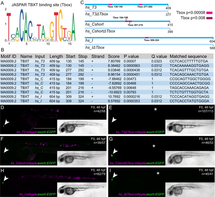
Published ChIP-seq data indicated Brachyury binding at hs_T3, hs_C, and hs_I (Fig. 1A), suggesting that notochord expression of the Brachyury/T/Tbxtb gene might be auto-regulated by Brachyury itself27,39. We investigated if the three human notochord enhancer elements contained a TBXT binding motif (short T-box, Fig. 2A) using FIMO46. We found that enhancer element hs_T3 contained two low p-value T-box motifs, enhancer element hs_I contained one low p-value T-box motif, whereas enhancer element hs_C contained two possibly degenerate T-box motifs that we only identified when significantly increasing the p-value (Fig. 2B), with two additional T-box motifs with even higher p-values that we did not further pursue in this work (Supplementary Fig. 2A, B). We then generated the reporter constructs hs_T3ΔTbox:mApple, hs_CshortΔTbox:mApple, and hs_IΔTbox:mApple in which we deleted the respective T-box motifs in the enhancer elements, as well as constructs containing the wildtype enhancer elements in an identical backbone (Fig. 2C). The reporter constructs further harbored the transgenesis marker exorh:EGFP (expression in the pineal gland, Fig. 2D–I) for precise quantification of reporter activity42. After injection into zebrafish embryos and in line with the enhancer element activity at 24 hpf (Fig. 1B–D), we observed continued and reproducible notochord expression at 48 hpf with all three wildtype enhancer element reporters hs_T3:mApple, hs_C:mApple, and hs_I:mApple (n = 42/58, n = 39/57 and n = 62/79; mCerulean-positive notochord/total EGFP pineal gland-positive embryos) (Fig. 2D, F, H, Supplementary Data 2). However, we observed a complete loss of specific notochord reporter activity in zebrafish embryos injected with the deletion constructs hs_T3ΔTbox:mApple, hs_CshortΔTbox:mApple, and hs_IΔTbox:mApple (n = 6/113, n = 7/53, n = 1/41), with positive embryos containing few labeled notochord cells (Fig. 2E, G, I, Supplementary Data 2). In contrast, individual deletion of the high p-value T-box motifs in enhancer element hs_C did not result in significant loss of reporter activity (n = 28/50, n = 15/63, Supplementary Fig. 2C, D).
Fig. 2. Identified TBXT binding sites in the enhancer elements are essential for reporter activity.
A Sequence of the human TBXT binding site (T-box) using JASPAR. B FIMO output with location of the T-box, statistical significance, and matched sequence within the enhancer elements. P-values were calculated by FIMO which computes a log-likelihood ratio score for each position in the sequence, then converts this score to a P-value, and then applies false discovery rate analysis to estimate a Q-value for each position. C Schematic depiction of the wildtype human enhancer elements with the TBXT binding site/T-box (pink, red, purple boxes) and the enhancer elements without the respective T-box sites (ΔTbox). The human enhancer elements are depicted in the reverse complement direction. Tbox130-145, Tbox277-292, Tbox309-324: p < 0.00008, Tbox184-199: p < 0.005, Tbox201-216: p < 0.008. D–I Injection of the wildtype enhancer elements hs_T3 (D), hs_Cshort (F), and hs_I (H) as reporter constructs results in mApple fluorophore expression in the notochord at 48 hpf, whereas injection of hs_T3ΔTbox (E), hs_CshortΔTbox (G), and hs_IΔTbox (I) show complete loss of notochord expression (asterisks in E, G, I). Arrowheads (D–I) mark EGFP expression in the pineal gland from the transgenesis marker exorh:EGFP. Scale bar in (D): 0.5 mm, applies to D–I.
Together, we conclude that the T-box motifs in the notochord enhancers hs_T3, hs_C, and hs_I are critical to the activity of these regulatory elements in our reporter assays. These data support the model in which Brachyury/T/TBXTB auto-regulates its own expression in the notochord through a defined motif in its notochord-regulatory elements27,39.
Brachyury notochord enhancers are conserved across mammals
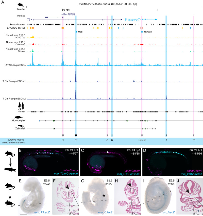
We next sought to determine if other mammalian genomes harbor orthologous T3, C, and I enhancer regions in their Brachyury/T/Tbxtb loci. Here, we focused on the orthologous T3, C, and I enhancer candidate regions from mouse (Fig. 3A). As in the human Brachyury/T/TBXTB locus, we found open chromatin and Brachyury protein binding events at the mouse orthologs of the putative enhancer elements T3, C, and I, as well as the well-characterized murine Brachyury/T/Tbxtb promoter Tstreak (Fig. 3A).
Fig. 3. Mouse Brachyury enhancer elements are active in different species.
A Mouse Brachyury/T/TBXTB locus adapted from the UCSC genome browser. Repeats marked in black using the RepeatMasker track; additional tracks include the ENCODE cCREs, H3K27ac (yellow), H3K4me (red) and DNase (green) signals. ATAC-sequencing (light blue peaks) and T ChIP-sequencing (dark blue lines) indicate enhancer elements (light pink highlight, not active; light blue highlight, active) that are conserved in human and Monodelphis. B–D Representative F0 zebrafish embryos injected with the mouse enhancer elements mm_T3 (B), mm_C (C), and mm_I (D). mm_T3 and mm_I show mosaic mCerulean reporter expression in the notochord at 24 hpf and mosaic ubi:mCherry expression as injection control. Mouse enhancer element mm_C is not active in the zebrafish notochord (asterisk in C). N represent the number of animals expressing mCerulean in the notochord relative to the total number of animals expressing mCherry. Scale bar in (B): 0.5 mm, applies to (B–D). E, G, I Representative images of transgenic E9.5 mouse embryos expressing lacZ (encoding beta-galactosidase) under the mouse enhancer elements mm_T3 (E), mm_C (G) and mm_I (I) visualized with X-gal whole mount staining. While mm_T3 and mm_I express beta-galactosidase in the entire notochord, beta-galactosidase expression from mouse mm_C is absent (asterisk in G). N represent the number of animals expressing beta-galactosidase in the notochord relative to the total number of animals with tandem integrations at H11. Dotted lines represent the sectioning plane. Scale bar in (E): 0.5 mm, applies to E, G, I. F, H, J Representative images of Fast Red-stained cross sections from embryos shown on the left, mm_T3 (F), mm_C (H), and mm_I (J). Black arrowheads point at notochord, and inserts show notochords at 2x higher magnification. Scale bar in F: 0.25 mm, applies to F, H, J. The species silhouettes were adapted from the PhyloPic database (www.phylopic.org).
When transiently tested in zebrafish, both mouse enhancer mm_T3 and mm_I showed reporter activity emerging arbitrarily throughout the gastrulating embryo at around 6 hpf (50% epiboly, shield stage) (Supplementary Fig. 3A–D), before expression became restricted to the developing notochord (n = 46/67, n = 61/66) at 24 hpf (Fig. 3B, D, Supplementary Data 2). Of note, our mouse enhancer mm_T3 contains the previously identified element TNE, which has been established to act as autonomous notochord enhancer when tested in mouse embryos and gastruloid cultures27. In contrast, mouse enhancer mm_C failed to drive any reporter expression in the zebrafish notochord (n = 0/88) (Fig. 3C, Supplementary Data 2). Imaging transgenic zebrafish carrying mouse mm_I as stable reporter documented robust notochord expression, again with little variability across independent transgenic insertions (Supplementary Fig. 3E). In contrast, the murine Brachyury/T/Tbxtb promoter region Tstreak24–26 showed transient, variable reporter expression in the zebrafish shield at around 6 hpf, with no reporter activity upon somitogenesis (n = 79/102) (Supplementary Data 2). We further tested the mouse ortholog of enhancer candidate mm_J, as well as the two lesser conserved elements mm_T1 and mm_T5, none of which showed reporter activity in zebrafish embryos up to 5 dpf (n = 0/98, n = 0/98, n = 0/79) (Supplementary Data 2).
Tested with site-directed reporter transgenesis at H11, mm_T3 and mm_I conveyed specific notochord activity in mouse embryos at E9.5 (n = 2/2, n = 4/4) (Fig. 3E, G, Supplementary Data 2). In contrast, and consistent with our observations in zebrafish reporter assays, mm_C did not show any detectable reporter activity in the notochord in mouse embryos at E9.5 (n = 0/2) (Fig. 3F, Supplementary Data 2).
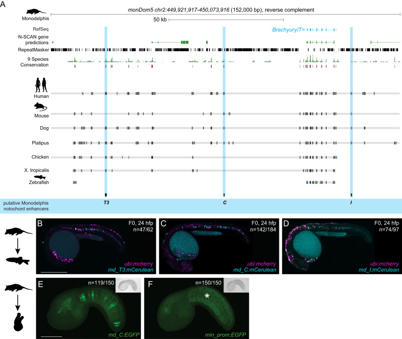
While humans and mice diverged ~90 million years ago, marsupials split from eutherians (placental mammals) ~160 million years ago41,47–50. The opossum Monodelphis domestica is a representative marsupial species and provides a more distant comparative species to human and mouse (Supplementary Fig. 4A). Detailed sequence alignments documented dispersed conserved regions along the entire sequences for all three enhancer candidates in Monodelphis (Fig. 4A). When injected into zebrafish embryos as mCerulean reporters, the Monodelphis-derived md_T3, md_C, and md_I enhancer element candidates all conveyed specific notochord activity at 24 hpf (n = 47/62, n = 142/184, n = 74/97) (Fig. 4B–D, Supplementary Data 2). Similar to the mouse elements, md_T3 transiently started reporter expression at around 6 hpf (Supplementary Fig. 4B, C), whereas md_C and md_I started to be active at early somitogenesis, similar to the human ones. In addition to the notochord activity, md_C reporter-injected zebrafish embryos showed transient reporter expression in the heart whereas md_I reporter-injected embryos showed transient expression in the brain and spinal cord neurons (Fig. 4C, D).
Fig. 4. Monodelphis Brachyury enhancer elements are active in different species.
A Monodelphis Brachyury/T/TBXTB locus adapted from the UCSC genome browser. Repeats are marked in black using the RepeatMasker track. Further annotated are tracks containing N-SCAN gene predictions and 9 Species Conservation. The light blue highlighted boxes mark the Monodelphis enhancer elements T3, C and I and their conservation in other species. B–D Representative F0 zebrafish embryos injected with the Monodelphis enhancer elements md_T3 (B), md_C (C), and md_I (D) showing mosaic mCerulean reporter expression in the zebrafish notochord at 24 hpf. ubi:mCherry was used as injection control. N represent the number of animals expressing mCerulean in the notochord relative to the total number of animals expressing mCherry. Scale bar in (B): 0.5 mm, applies to (B, C). E, F Representative images of Ciona embryos electroporated with Monodelphis enhancer element md_C (E), and minimal forkhead promoter (fkh) only as control (F). Monodelphis enhancer element md_C expresses EGFP throughout the entire Ciona notochord, compared to minimal fkh promoter only which does not express EGFP at all (asterisk in F). N represent the number of animals expressing EGFP in the notochord relative to the total number of animals. Inserts on the top right represent bright field images of respective embryos. Scale bar in (E): 0.05 mm, applies to E, F. The species silhouettes were adapted from the PhyloPic database (www.phylopic.org).
Given the mammalian sequence conservation and differential responses in reporter assays, we next tested the notochord enhancer element candidates in the tunicate Ciona intestinalis as non-vertebrate outgroup. As a chordate, Ciona forms a bona fide notochord51. Testing T3, C, and I of human, mouse, and Monodelphis by reporter gene assays in Ciona, we found that only Monodelphis-derived md_C showed specific and robust reporter activity in the notochord (n = 119/150) compared to all other eight elements (n = 0/150) and minimal promoter only control (n = 0/150) (Fig. 4E, F, Supplementary Data 2).
Taken together, and extending previous work on the mouse TNE element27, our data indicate that three distant elements in the mammalian Brachyury/T/Tbxtb locus with differential activity converge on providing notochord-specific activity in reporter assays across chordates.
Enhancer deletions cause selective loss of Brachyury in mice
While especially enhancer element C seems to have diverged in activity (or is sensitive to the specific trans environment it was tested in), all three elements T3, C, and I remain conserved and detectable at the sequence level throughout the mammalian clade. In mice, homozygous Brachyury/T/Tbxtb mutations in the gene body cause post-implantation defects leading to embryonic lethality between E9.5 and E10.552–54. Previous work established that deletion of mouse enhancer TNE does not cause a fully penetrant loss of Brachyury/T/Tbxtb expression in the developing notochord, indicating the presence of additional shadow elements interacting with, or compensating for, TNE27. To functionally test if the three enhancer elements are involved in Brachyury/T/Tbxtb expression in the mouse notochord, we generated a series of knockout alleles targeting the three mouse enhancer elements T3, C, and I (Fig. 5A).
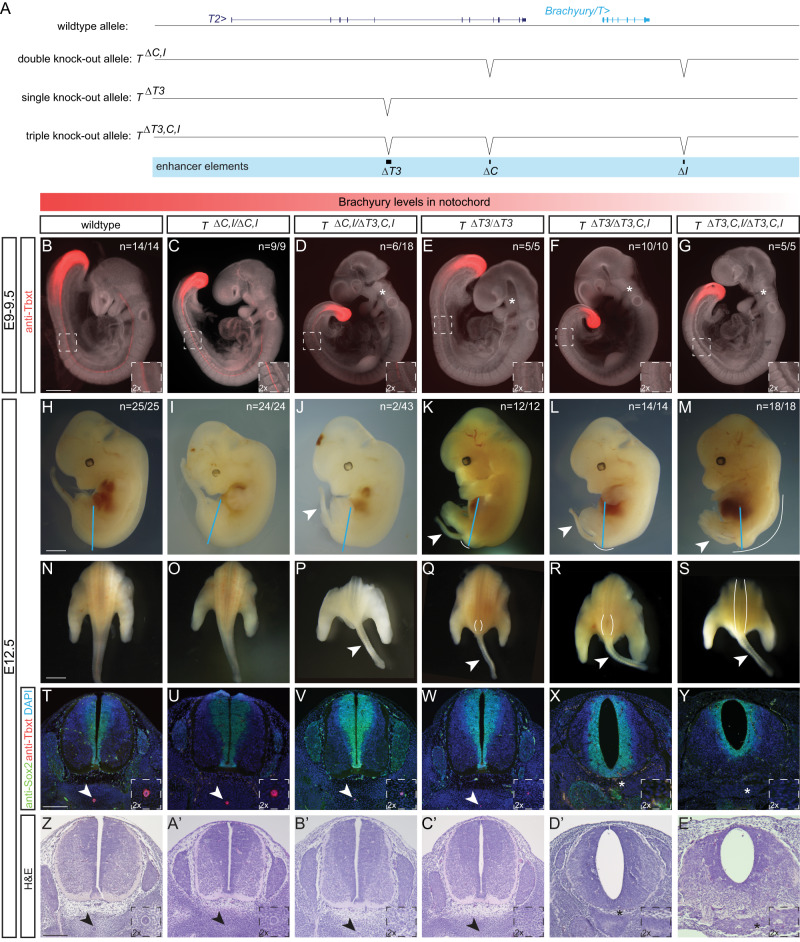
Fig. 5. Deletion of the three enhancer elements T3, C and I results in selective loss of Brachyury protein expression in the notochord at E9.5 and posterior defects at E12.5.
A Overview of wildtype mouse Brachyury/T/TBXTB locus adapted from the UCSC genome browser and deletion alleles generated with CRISPR-Cas9 genome editing. Exact coordinates and sequences of target sites, deletions, and genotyping primer sequences can be found in Supplementary Data 5. B–G Brachyury/T antibody staining (red) of E9.5 embryos. White dashed square in panels represents location of right bottom inserts with 2x magnification. Brachyury/T protein expression in the notochord is dose-dependent on the three enhancer elements. Asterisks in (D–G) mark absent notochord in rostral portion of the embryo. Scale bar in (B): 1 mm, applies to panels (B–G). H–M Overall morphology of E12.5 embryos with different genotypes. Blue lines indicate the location of immunofluorescence and H&E sections. Spina bifida and tail defects are dose-dependent. Arrowheads mark rudimentary tails. White lines mark spina bifida. Scale bar in H: 1 mm, applies to (H–M). N–S Dorsal view of embryos (sectioned at blue line in H–M). White lines mark areas of spina bifida. Arrowheads mark rudimentary tails compared to tails in wildtype control and double knock-out allele. Scale bar in (N): 2.5 mm, applies to panels (N–S). T–Y Immunofluorescence of mouse transverse sections. Anti-Sox2 labels the neural plate, anti-Tbxt the notochord, and DAPI marks nuclei. Sox2 expression is comparable amongst all genotypes, even in the genotypes with spina bifida, while there is loss of Brachyury/T staining in the notochord with increased loss of the enhancers. Arrowheads point to notochord. Asterisks mark absent notochord. Scale bar in (T): 0.2 mm, applies to panels (T–Y). Z–E' H&E staining of transverse sections confirm the dose-dependent loss of the notochord and spina bifida. Arrowheads point to notochord. Asterisks mark absent notochord. Scale bar in (Z): 0.2 mm, applies to (Z–E').
We employed CRISPR-Cas9 genome editing using target sites flanking the enhancers and established heterozygous and homozygous mice carrying individual and combined enhancer deletions (Fig. 5A, Supplementary Fig. 5A). Compared to E9.5 wildtype control embryos (Fig. 5B) (n = 14/14), neither homozygous deletion of mouse C (TΔC/ΔC) (n = 7/7) or I (TΔI/ΔI) (n = 7/7) alone, nor heterozygous (T+/ΔC,I) (n = 12/12), heterozygous (T+/ΔT3) (n = 7/7) (Supplementary Fig. 5B–F) or homozygous deletion of both C and I (TΔC,I/ΔC,I) (n = 9/9) (Fig. 5C) altered Brachyury/T/Tbxtb expression in the notochord as determined by Brachyury/T antibody staining.
In contrast, we observed reduced Brachyury/T/Tbxtb expression in the notochord of E9.5 embryos in a dose-dependent manner when we combined ΔT3 with ΔC,I deletions. E9.5 embryos heterozygous for the triple knockout chromosome carrying ΔT3,C,I (T+/ΔT3,C,I) in cis appeared normal (n = 14/14) (Supplementary Fig. 5F). In contrast, in trans-heterozygous E9.5 embryos carrying ΔC,I and ΔT3,C,I alleles (TΔC,I/ΔT3,C,I), we documented reduced Brachyury/T/Tbxtb protein in the caudal portion of the notochord in all embryos (n = 18/18) with individual embryos also displaying reduced or lost Brachyury/T/Tbxtb protein in the trunk and rostral portion (n = 6/18) (Fig. 5D). Similarly, in E9.5 embryos homozygous for ΔT3 (TΔT3/ΔT3) (n = 5/5) (Fig. 5E), we observed reduced Brachyury/T/Tbxtb protein levels, as previously reported for homozygous TNE embryos27. Brachyury/T/Tbxtb protein levels were even further reduced or lost in the entire notochord of trans-heterozygous for ΔT3 and ΔT3,C,I alleles (TΔT3/ΔT3,C,I) (n = 10/10) (Fig. 5F). These data are consistent with, and expand upon, previous observations that the severity of Brachyury/T/Tbxtb phenotypes correlate with gene dosage54. Importantly, the TΔT3/ΔT3,C,I genotype with severely reduced Brachyury/T/Tbxtb protein levels is consistent with the loss of Brachyury/T/Tbxtb protein in the notochord in mice trans-heterozygous for the TNE deletion and a large, locus-spanning Brachyury/T/Tbxtb deletion that includes elements C and I27, revealing the actual relevant enhancer regions (Figs. 1, 3, and 4) and motifs (Fig. 2). Finally, E9.5 homozygous triple knockout ΔT3,C,I embryos (TΔT3,C,I/ΔT3,C,I) showed a complete absence of Brachyury/T/Tbxtb protein in the entire notochord region (n = 5/5) yet all embryos retained Brachyury/T/Tbxtb protein in the tailbud (n = 5/5) (Fig. 5G). Taken together, our data establish the notochord-specific Brachyury/T/Tbxtb loss-of-function mutant in mice by means of deleting three conserved enhancer elements in cis.
Next, we examined phenotypic defects resulting from perturbed Brachyury/T/Tbxtb expression using various allele combinations involving ΔC,I and ΔT3,C,I. Consistent with the phenotypes at E9.5 (Fig. 5B–G), we observed a gradual increase of phenotype severity with deletion of the three different enhancer elements at E12.5 (Fig. 5H–E’). Wildtype control (n = 25/25) (Fig. 5H, N), homozygous TΔC,I/ΔC,I embryos (n = 24/24) (Fig. 5I, O), heterozygous T+/ΔC,I (n = 5/5), heterozygous T+/ΔT3 (n = 23/23) and T+/ΔT3,C,I embryos (n = 23/23) (Supplementary Fig. 5G–I) appeared grossly normal. In contrast, we observed rudimentary tails with additional enhancer deletions. Rudimentary tails appeared in trans-heterozygous TΔC,I/ΔT3,C,I embryos in 4.7 % (n = 2/43) (Fig. 5J, P) and were fully penetrant in homozygous TΔT3/ΔT3 (n = 12/12) (Fig. 5K, Q) similar to homozygous TNE embryos27, and trans-heterozygous TΔT3/ΔT3,C,I embryos (n = 14/14) (Fig. 5L, R), as well as in triple homozygous TΔT3,C,I/ΔT3,C,I embryos (n = 18/18) (Fig. 5M, S). In addition, homozygous TΔT3/ΔT3 embryos (n = 11/12) (Fig. 5Q) seemed to display defects in neural tube closure very close to the tail, comparable to spina bifida; upon sectioning however, we identified this region to be very small and not a fully developed spina bifida phenotype (Fig. 5Q). In comparison, trans-heterozygous TΔT3/ΔT3,C,I embryos displayed caudal spina bifida with 100% penetrance (n = 14/14) (Fig. 5R). Finally, triple-homozygous TΔT3,C,I/ΔT3,C,I embryos lacking all three enhancers displayed spina bifida along 3/4 of the spine (n = 18/18) (Fig. 5S), reminiscent of previous observations using Brachyury/T/Tbxtb-targeting RNAi in mouse embryos55,56. These results provide compelling phenotypic evidence of the impact of cumulative enhancer deletions on Brachyury/T/Tbxtb expression in the notochord.
We further validated these phenotypes with immunohistochemistry and histology. We visualized Brachyury/T/Tbxtb protein in transversal sections of E12.5 embryos together with the neural plate marker Sox2: compared to wildtype (Fig. 5T), heterozygous T+/ΔC,I, T+/ΔT3, T+/ΔT3,C,I (Supplementary Fig. 5J–L) as well as homozygous TΔC,I/ΔC,I (Fig. 5U) embryos that were all grossly normal, we found decreased Brachyury protein in the notochord of TΔC,I/ΔT3,C,I (Fig. 5V) and TΔT3/ΔT3 (Fig. 5W) embryos. Strikingly, we observed a complete absence of Brachyury protein in TΔT3/ΔT3,C,I embryos (Fig. 5X) and TΔT3,C,I/ΔT3,C,I (Fig. 5Y) embryos. In contrast, Sox2 expression was comparable in all embryos (Fig. 5T–Y, Supplementary Fig. 5J–L), even in TΔT3,C,I/ΔT3,C,I embryos that clearly displayed spina bifida along the entire spine (Fig. 5Y). Compared to wildtype embryos (Fig. 5Z), additional histology assessed by H&E staining confirmed wildtype-looking notochords in T+/ΔC,I, T+/ΔT3, T+/ΔT3,C,I, and homozygous TΔC,I/ΔC,I embryos (Supplementary Fig. 5M–O, Fig. 5A’), smaller (in diameter) notochords in TΔC,I/ΔT3,C,I (Fig. 5B’) and TΔT3/ΔT3 (Fig. 5C’) embryos, and absent notochords in TΔT3/ΔT3,C,I and TΔT3,C,I/ΔT3,C,I embryos (Fig. 5D’-E’).
We found that the two most severe enhancer mutants are not viable as adults since we did not recover homozygous triple TΔT3,C,I/ΔT3,C,I (n = 0/59) or trans-heterozygote TΔT3/ΔT3,C,I (n = 0/31) animals at term (Supplementary Fig. 5P), indicating lethality prior to or shortly after birth. In contrast, homozygous TΔT3/ΔT3 animals were born, but died within 14 days after birth, with one exception where we identified one homozygous TΔT3/ΔT3 (n = 1/34) animal without a tail that survived until adulthood (Supplementary Fig. 5P). In contrast, TΔC,I/ΔT3,C,I (n = 46) trans-heterozygotes and homozygous TΔC,I/ΔC,I (n = 100) animals survived to adulthood (Supplementary Fig. 5P). Notably, a variable percentage of TΔC,I/ΔC,I, TΔC,I/ΔT3,C,I, and T+/ΔT3 animals presented with kinked tails (Supplementary Fig. 5Q), with two TΔC,I/ΔT3,C,I animals displaying a small tail (Supplementary Fig. 5R), reminiscent of hypomorphic Brachyury/T/Tbxtb mutants and in vivo Brachyury/T/Tbxtb knockdown by siRNA9,27,55,56. Taken together, our data are consistent with the correlation of Brachyury/T/Tbxtb-mutant phenotypes and gene dosage controlled by enhancer activity, as revealed by increasing phenotype severity with an increasing number of combined enhancer deletions in Brachyury/T/Tbxt.
In summary, our data establishes that the combined activity of the enhancers T3, C, and I in the mouse Brachyury/T/Tbxtb locus are necessary to convey notochord expression of Brachyury/T/Tbxtb. Upon combined loss of these enhancers, the notochord is lost.
T3, C and I are conserved among jawed vertebrates
The evolutionary trajectory of chordate Brachyury control in the notochord remains unresolved. The notochord-regulatory elements driving Brachyury expression in Ciona are promoter-proximal8,10,31. Zebrafish tbxta/ntla harbors a −2.1 kb upstream notochord element containing the two smaller elements E1 and E223. In contrast, zebrafish tbxtb descended from the same ancestral Brachyury gene as the single mammalian Tbxtb gene. Further, while zebrafish tbxtb remains expressed in the notochord21,57, its regulatory elements have not been reported. Using direct sequence comparisons of mammalian T3, C, and I to the zebrafish genome, we did not find any sequences of significant sequence similarity (Fig. 1A).
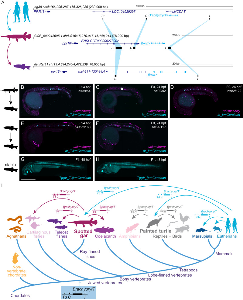
Identifying non-coding sequence conservation across vertebrate lineages, whether from human or other tetrapods to the fast-evolving teleost fishes like zebrafish, remains notoriously challenging. Species with slow rates of molecular evolution can help as “genomic bridges” to provide sequence connectivity across distant vertebrate groups58,59. The spotted gar (Lepisosteus oculatus) is a slowly evolving ray-finned fish that has diverged from zebrafish and other teleosts before a teleost-specific whole-genome duplication, providing a bridge species for genomic comparisons between tetrapods and teleosts58. Using BLAST searches, we found sequence similarity between human T3, C, and I and regions of the spotted gar tbxtb locus with equivalent positions relative to the gar tbxtb gene body compared to mammals (Fig. 6A). Next, we used these spotted gar T3, C, and I regions as BLAST queries to bridge to the genomes of zebrafish and other fish lineages (Supplementary Data 4). This approach uncovered candidate regions for T3 and I, but not C, within the zebrafish tbxtb locus (Fig. 6A).
Fig. 6. Bridge species establish the presence of Tbxtb enhancers across jawed vertebrates.
A Location of the enhancer elements in the human (top), gar (middle), and zebrafish (bottom) Brachyury/T/Tbxtb loci, adapted from the UCSC browser as established through the “gar bridge”. B–D Representative F0 zebrafish embryos injected with the gar enhancer elements Io_T3 (B), Io_C (C), and Io_I (D). T3 and I show mosaic mCerulean reporter expression in the notochord at 24 hpf compared to gar element C with is not active in the zebrafish notochord (asterisk). N represent the number of animals expressing mCerulean in the notochord relative to the total number of animals expressing mosaic ubi:mCherry as injection control. Scale bar in (B): 0.5 mm, applies to (B–F). E, F Representative F0 zebrafish embryos injected with the conserved zebrafish enhancer elements dr_T3 (E) and dr_I (F). T3 and I show mosaic mCerulean reporter expression in the notochord at 24 hpf. N represent the number of animals expressing mCerulean in the notochord relative to the total number of animals expressing mosaic ubi:mCherry as injection control. G, H Representative images of stable F1 embryos at 2 dpf of zebrafish enhancer elements T3 and I recapitulate the F0 expression pattern in the notochord, with dr_T3 (E) additionally expressing mCerulean in the brain, heart, and fin, and dr_I (G) in the proximal kidney close to the anal pore, pharyngeal arches, heart, fin, and spinal cord neurons. Scale bar in (G): 0.5 mm, applies to (G, H). I Phylogenetic representation of species investigated using the bridging approach with spotted gar and painted turtle as anchor species within ray-finned fish and tetrapod lineages. Arrows indicate informative phylogenetic comparisons to uncover conservation of enhancer elements T3, I, and C. The species silhouettes were adapted from the PhyloPic database (www.phylopic.org).
Analogous to our tests with mammalian enhancer candidates, we cloned reporter transgenes coupled with the betaE-globin:mCerulean cassette using the T3, C, and I enhancer elements from the spotted gar tbxtb locus. Upon injection into zebrafish embryos, both spotted gar lo_T3 and lo_I displayed specific and reproducible notochord reporter activity (n = 39/54, n = 82/122) (Fig. 6B, D, Supplementary Data 2). In contrast, and akin to the mouse mm_C enhancer element, spotted gar element lo_C did not convey any notochord reporter activity in zebrafish embryos (n = 0/92) (Fig. 6C, Supplementary Data 2). The zebrafish-derived dr_T3 and dr_I also showed selective notochord activity when tested in zebrafish transgenic reporter assays (n = 122/160, n = 81/117) (Fig. 6E, F, Supplementary Data 2). Further confirming our results, we found robust reporter activity in the notochord of stable transgenic zebrafish lines based on dr_T3 and dr_I (Fig. 6G, H). All fish enhancer elements started to express the mCerulean reporter during early somitogenesis, similar to the human elements.
Using the three gar elements as queries, in addition to clupeocephalan teleosts (e.g. zebrafish), we found T3 and I also in the other two major teleost lineages elopomorphs (e.g. eel) and osteoglossomorphs (e.g. arowana). However, we did not detect any equivalent sequence for C in any teleosts, indicating that this element has been lost or diverged beyond recognition in the teleost lineage (Fig. 6I). However, we detected orthologs of all three elements, including C, at expected locations around the tbxtb genes in additional non-teleost ray-finned fishes (e.g. bowfin, sturgeon, reedfish) as well as in the more basally diverging cartilaginous fishes (e.g. sharks, skate) (Supplementary Data 4); in contrast, we only detected T3 and I in the lobe-finned coelacanth (Fig. 6I). To explore the presence of the three enhancer elements among tetrapods, we used the painted turtle, characterized by a slow genomic evolutionary rate60,61, as an additional bridge species within tetrapods. We found all three elements in the turtle Brachyury/T/Tbxtb locus and through use of the painted turtle as reference also in other reptiles and birds, as well as in amphibians (e.g. axolotl) (Fig. 6I, Supplementary Data 4), but did not detect any of the three elements in the jawless cyclostome (e.g. lamprey, hagfish) genomes. Furthermore, we found that the human T-box motifs, which we identified using FIMO (Fig. 2) in our enhancers, are conserved across tetrapods and fishes as distantly related as ghost shark based on sequence alignments (Supplementary Fig. 6A–C) as well as multi-species FIMO analyses (Supplementary Data 7). Cross-species sequence conservation centers at the T-box motifs (Supplementary Fig. 6A–C) which supports both their functional importance as well as their evolutionary ancestry since at least the last common ancestor of jawed vertebrates.
Taken together, our observations provide strong evidence that notochord enhancers T3, I, and C are deeply conserved cis-regulatory elements of the Brachyury/T/Tbxtb gene that were already present in the last common ancestor of jawed vertebrates over 430 million years ago.
Discussion
How the Brachyury/T/Tbxtb gene is controlled during notochord development is fundamental to our understanding of how basic concepts of body plan formation remain conserved or have diverged across species. Shadow enhancers, seemingly redundant transcriptional cis-regulatory elements that regulate the same gene and drive overlapping expression patterns, are a pervasive feature of developmental gene regulation62. The concept of enhancer redundancy through one or more shadow enhancers acting on the same gene in addition to a primary enhancer has been established for numerous loci62–67. Shadow enhancers are thought to provide robustness to gene expression and buffer against genetic and environmental variations62,65, a hypothesis validated in mammals66,67.
Here, we discovered a deeply conserved set of three notochord-specific shadow enhancers within the human TBXT locus as ancient cis-regulatory elements. While we cannot draw conclusions about reporter initiation or early reporter expression patterns, cross-species enhancer testing reveals that the cis-regulatory grammar of the three human enhancers T3, C, and I, is correctly interpreted in vertebrates including mice, salamanders, and zebrafish, but not in the invertebrate chordate Ciona. The three notochord enhancers described here are not the only non-coding conserved elements across mammalian Brachyury/T/Tbxtb loci (Figs. 1A, 3A, and 4A). Even though our zebrafish reporter assays did not reveal any notochord activity in three out of the six tested human enhancer elements (K, J, and L), we cannot rule out synergistic or interdependent notochord activity conveyed by additional elements. Further, our reporter assays indicate that not all three Brachyury/T/Tbxtb notochord enhancers T3, C, and I have equal potency. Enhancer element C shows variable activity and remains unrecognized in teleost fishes and Coelacanth. Compared to human C with reproducible notochord activity in all tested models (Fig. 1C, F, I, M) and Monodelphis C that is active in zebrafish and uniquely in Ciona (Fig. 4C, E), mouse C showed no discernible activity in any assay including in mouse embryos (Fig. 3C, G) despite significant sequence conservation. We speculate that while mouse C is not active in isolation, it may contribute together with T3 and I to Brachyury activity in the notochord. This model is consistent with the impact of TNE deletions when combined with larger deletions that include TNE and C in mouse trans-heterozygotes27 (Fig. 5). The potential auto-regulation of Brachyury/T/Tbxtb by its protein product via in part conserved T-box motifs in enhancers T3 and I might contribute to the enhancer redundancy and divergent activity of element C when tested in isolation (Fig. 2). Our data propose that enhancer C is an auxiliary element to T3 and might contribute to duration, expression levels, or other features that differ among Brachyury/T/Tbxtb notochord expression across vertebrates. Our combined data proposes a model in which notochord expression of vertebrate Brachyury/T/Tbxtb is cumulatively or cooperatively driven by enhancers T3, C, and I. In this model, sequence variants of T3, C, and I that modulate their individual potency became selected for modulating Brachyury/T levels to species-specific requirements.
The conservation of gene order (micro-synteny) between species can be indicative of the presence of cis-regulatory elements, which are crucial for controlling expression of the physically linked genes68. The finding of functionally relevant distant enhancers 5’ and 3’ of the Brachyury/T/Tbxtb gene body is further supported by the conserved gene linkage Sftd2-(Prr18)-Brachyury/T/Tbxtb-Pde10a across the entire jawed vertebrate phylogeny. In agreement with a distinct gene linkage surrounding Brachyury/T/Tbxtb in agnathans (Fig. 6I), we were unable to identify any of the three distant enhancers in two species representing this clade. Likewise, a distinct gene linkage associates with Tbxta, the second Tbxtb paralog in fish, which apparently lacks any of the three notochord enhancers described here. tbxta/ntla expression is instead controlled by two mesoderm/notochord enhancers located close to the gene promoter (Harvey et al., 2010), a possible example of evolutionary novelty following ancestral gene duplication. In contrast, the functionally less impactful zebrafish tbxtb/ntlb gene retained the regulation of the Tbxtb gene from the jawed vertebrate ancestor (Fig. 6). We did not find any evidence for sequence conservation of the Tbxtb T3, I, or C regions within vertebrate Tbxta loci or any other genomic regions. Future detailed studies across vertebrate Tbxt paralogs are needed to evaluate whether or not the three Tbxtb regulatory elements identified here were already part of the single Tbxt gene in a vertebrate ancestor. Notably, zebrafish mutants of tbxta/ntla have been widely studied as model for Brachyury function in notochord formation13,15,69, while the seemingly less impactful tbxtb has retained ancestral regulation. Why zebrafish, and possibly other fish lineages, use tbxta as their main functional Brachyury paralog, and how the regulatory balance between T3, C, and I plays out across individual vertebrate lineages, warrants future efforts.
We found that Brachyury/T/Tbxtb notochord enhancers T3 and I, and possibly further supported by enhancer C, represent a shadow enhancer combination that contributes to the robust Brachyury/T/Tbxt expression in mammals. In mice, neither deletion of enhancer T3/TNE27, nor deletion of enhancer C, I, or C and I, resulted in a discernable notochord phenotype (Fig. 5). Nonetheless, by combining deletions of all three notochord enhancer elements, we showed a dose response for Brachyury/T expression in the notochord. In particular, in embryos where ΔT3 is combined with a chromosome harboring ΔT3,C,I as trans-heterozygotes (TΔT3/ΔT3,C,I) or in triple homozygous knock-out embryos (TΔT3,C,I/ΔT3,C,I), we observed loss of Brachyury/T protein in the notochord as well as notochord-specific phenotypes, such as spina bifida (Fig. 5). The neural tube closure defects are similar to phenotypes observed in Brachyury/T/Tbxtb knockdown embryos55,56 or hypomorphic Brachyury/T/Tbxtb mutants9. These results assign an essential, combinatorial role to the enhancer elements T3/TNE, C and I in regulating Brachyury/T/Tbxtb in the notochord. Notably, previous work70,71 has described the T2 mutant caused by a large viral integration 5’ of the mouse Brachyury/Tbxt locus that (i) is recessive lethal with phenotypes reminiscent of Brachyury loss, and (ii) does complement loss-of-function alleles for Brachyury. T2 has been hypothesized to encode a short protein off a long mRNA70,71. The described genomic position of the viral integration in T2 places it in the vicinity and upstream of enhancer element C. We note that various vertebrate Brachyury/tbxtb loci feature annotated long non-coding RNAs upstream of the main gene body that are reminiscent of enhancer RNAs (Figs. 3A and 6A). We therefore hypothesize that the T2 mutation is caused by a disruption of the gene-regulatory landscape of the mouse Brachyury/Tbxt gene by the viral integration, changing the interaction of distant enhancer elements with the promoter. Inspection of the chromatin landscape of the Brachyury/Tbxt locus, also in T2 mutants, could shed light on the architecture of the locus during notochord development.
The significance of Brachyury/T/Tbxtb regulation in the notochord translates to chordoma tumors that feature expression of this T-box transcription factor as key diagnostic readout32,72,73. Both sporadic and familial chordoma are hypothesized to derive from notochord remnants in the spine that do not convert to nucleus pulposus tissue32,74,75. Native Brachyury-expressing cells in the nucleus pulposus decrease in number with age along with a concomitant increase in cartilage-like cells4,76–78. What role these long-lasting Brachyury-positive cells play in the adult spine, if they progressively differentiate into cartilage, and how Brachyury gene activity is sustained, remains unknown. Detection of Brachyury protein is a diagnostic marker for chordoma32, yet the functional contribution of its re-activated or persistent expression in the tumor is not known56,79–81. Our analysis of reported familial and sporadic chordoma amplifications indicate that amplifications invariantly retain the notochord enhancer I together with the gene body including the promoter34,37. Enhancer I lies within a super-enhancer region identified in chordoma cell lines38, further implicating its transcriptional engagement in chordoma. Amplifications occurring in tandem with the original locus propose a scenario where the retained enhancer I could synergize with C and T3 from the original locus on the newly amplified gene copies, potentially resulting in increased Brachyury/T/TBXTB expression (Fig. 1A). Beyond chordoma, changes in enhancer sequence or relative distance to the Brachyury/T/TBXTB gene body could also impact spine formation and health by altering the robustness of Brachyury expression in the notochord and subsequent nucleus pulposus.
Tremendous progress with in vitro differentiation regimens have resulted in stem cell-derived models for body segmentation and different organ structures82–85. However, notochord formation has only been reported in more complex systems that recapitulate major hallmarks of embryo patterning86–88. Reporters based on our isolated enhancers could potentially provide potent readouts to screen for differentiation regimens that result in notochord fates. Together, our uncovered set of shadow enhancers in Brachyury/T/TBXTB advance our concepts of how this key contributor to notochord formation is regulated and de-regulated in development and disease.
Methods
Ethical regulations
All research within this manuscript complies with all relevant ethical regulations that are described and named individually in each paragraph.
Brachyury locus annotations
The UCSC genome browser was used to identify and visualize enhancer elements in the human, mouse, and Monodelphis Brachyury locus. *.bed files were generated with the approximate genomic location of human Brachyury amplifications in chordoma tumors from different patients34,37. Previously published ATAC-sequencing data of U-CH2 cells and MUGCHOR cells38, as well as Brachyury/T ChIP sequencing data of human embryonic stem cells (hESCs)40 and U-CH1 cells36 were added. Further, the repeat masker track, ENCODE cCREs, layered H3K27ac, and the conservation track for mouse and Monodelphis were added. Ultimately, using this strategy, the human enhancer element candidates T3, K, J, C, I, and L were identified. For detailed information, see Supplementary Data 1 and 3.
The same strategy was applied to find the corresponding mouse enhancer elements. Published ATAC-seq data of mouse ESCs89 and Brachyury/T-positive fluorescence-activated cell sorted cells from the caudal ends of wild-type mouse embryos (TS12/8 dpc and TS13/8.5 dpc)90, as well as Brachyury/T ChIP sequencing data of mouse ESCs39,90 were used. Again, the repeat masker track, the ENCODE Candidate Cis-Regulatory Elements (cCREs, combined from all cell types) track, tracks containing H3K27ac, H3K4me, DNase signals from E11.5 neural tube as it likely contains notochord tissue as well due to extraction91, and the Vertebrate Multiz Alignment & Conservation track to check for conservation in human, Monodelphis, and zebrafish, were added. This approach identified the mouse enhancer element candidates T1, T2, T3, J, C2/next to C, C, Tstreak, I, T4, T5, and T6, of which T1, T3, J, C, Tstreak, I, and T5 were pursued and tested (Supplementary Data 3 and 5).
To find the corresponding Monodelphis elements, the repeat masker and 9-Way Multiz Alignment & Conservation track were included to identify T3, C, and I (Supplementary Data 3 and 5).
Cloning of the enhancer element reporter plasmids
Each Brachyury enhancer element candidate was amplified from either human, mouse, Monodelphis, spotted gar, or zebrafish genomic DNA using the Expand Hi-Fidelity PCR System (11732641001, Roche). Exact coordinates are listed in Supplementary Data 3. Each enhancer candidate was TOPO-cloned into the pENTR5′-TOPO plasmid (K59120, Invitrogen) according to the manufacturer’s instructions (half-volume reactions). Subsequent Multisite Gateway cloning were performed using LR Clonase II Plus (12538120, Invitrogen) according to the manufacturer’s instructions (half-volume reactions) and recommended reaction calculations92. 5’ entry plasmids containing the different enhancer elements were assembled into reporter expression plasmids together with the middle entry plasmid (pME) containing the mouse betaE-globin minimal promoter expressing mCerulean (pSN001) as well as mApple (pCK068), the 3’plasmid #302 (p3E_SV40polyA), and the destination plasmid pDESTTol2A2 containing crybb1:mKate2 (pCB59) and pDESTexorh:EGFP containing EGFP expression in the pineal gland (pCK017) as transgenesis markers42. Assembled vectors were verified using restriction digest and Sanger sequencing using standard sequencing primers for Multisite Gateway assemblies42,92.
Zebrafish husbandry, transgenic reporter assays and stable transgenic lines
Zebrafish animal care and procedures were carried out in accordance with the IACUC of the University of Colorado Anschutz Medical Campus (protocol # 00979), Aurora, Colorado. Adult AB and TU wildtype zebrafish were obtained from the Zebrafish International Resource Center (ZIRC) and maintained as per standard husbandry procedures93.
To test the transient activity of the putative enhancer elements, 25 ng/µL Tol2 mRNA, 12.5 ng/µL reporter expression plasmid DNA, and 12.5 ng/µL ubi:mCherry plasmid94 as injection control were co-injected into one-cell stage wild-type zebrafish embryos44. At 24 hpf, embryos were anesthetized with 0.016% Tricaine-S (MS-222, Pentair Aquatic Ecosystems Inc.) in E3 embryo medium and embedded in E3 with 1% low-melting-point agarose (A9045, Sigma Aldrich).
To generate stable transgenic lines, 25 ng/µL Tol2 mRNA were co-injected with 25 ng/µL reporter expression plasmid DNA95,96. Multiple F0 founders were screened for specific mCerulean and mKate2 expression, raised to adulthood, and screened for germline transmission. Resulting F1 single-insertion transgenic strains were established and verified through screening for a 50% germline transmission rate outcrosses in the subsequent generations as per our previously outlined procedures96. Tg(drl:mCherry) was used as a marker for lateral plate mesoderm derivatives41.
For imaging, embryos were mounted laterally on glass bottom culture dishes (627861, Greiner Bio-One) and confocal images were acquired with a Zeiss LSM880 using a ×10/0.8 air-objective lens. Fluorescence channels were acquired sequentially with maximum speed in bidirectional mode in 3 µM slices. The range of detection for each channel was adapted to avoid any crosstalk between the channels. Images of acquired Z-stacks were reconstructed with ImageJ/Fiji as a maximum intensity projections.
Axolotl husbandry, transgenic reporter assays and immunostaining
Procedures for care and manipulation of all animals used in this study were performed in compliance with the laws and regulations of the State of Saxony, Germany. Axolotl husbandry and experiments (non-free feeding stages) were performed at the Center for Regenerative Therapies Dresden (CRTD), Dresden, Germany. Adult axolotls (Ambystoma mexicanum) were obtained from the axolotl facility at the Technische Universität Dresden (TUD)/CRTD Center for Regenerative Therapies Dresden. Animals were maintained in individual aquaria at ~18–20 °C97. Axolotls of the white (d/d) strain were used in all experiments.
Transgenic axolotl embryos were generated using Tol2 transposase following standard protocols98. For live imaging, the embryos were anaesthetized by bathing in 0.01% benzocaine and imaged on an Olympus SZX16 fluorescence stereomicroscope. Embryos were staged as described previously99.
For immunostaining, axolotl embryos were fixed in MEMFA at 4 °C overnight, washed in PBS, embedded in 2% low-melting temperature agarose, and sectioned by vibratome into 200 μm-thick sections. Fibronectin was detected using mouse anti-Fibronectin (ab6328, Abcam; dilution 1:400) and donkey anti-mouse Alexa Fluor™ 568 (A-10037, Invitrogen; dilution 1:600). After staining, sections were mounted with Mowiol (81381, Millipore Sigma). Confocal images were acquired on a Zeiss LSM780-FCS inverted microscope.
Transgenic mouse reporter assays
Research was conducted at the E.O. Lawrence Berkeley National Laboratory (LBNL) and performed under U.S. Department of Energy Contract DE-AC02-05CH11231, University of California (UC). Transgenic mouse assays were performed in Mus musculus FVB mice (obtained from The Jackson Laboratory), animal protocol number 290003; reviewed and approved by the Animal Welfare and Research Committee at Lawrence Berkeley National Laboratory.
For comprehensive analysis of species-specific T3, C and I, enSERT enhancer analysis was used, allowing for site-directed insertion of transgenic constructs at the H11 safe-harbor locus100,101. EnSERT is based on co-injection of Cas9 protein and H11-targeted sgRNA in the pronucleus of FVB single cell-stage mouse embryos (E0.5) with the targeting vector encoding a candidate enhancer element upstream of the Shh-promoter-LacZ reporter cassette45. Enhancer elements were PCR-amplified from human, mouse and Monodelphis genomic DNA and cloned into the respective LacZ expression vector102. Embryos were excluded from further analysis if they did not contain a reporter transgene in tandem. CD-1 females (The Jackson Laboratory) served as pseudo-pregnant recipients for embryo transfer to produce transgenic embryos which were collected at E9.5 and stained with X-gal using standard methods102.
Histological analysis of Nuclear Fast Red-stained sections from transgenic mouse embryos
After LacZ staining, E9.5 transgenic mouse embryos were dehydrated in serial alcohols (1 × 70%, 1 × 80%, 1 × 90%, 2 × 96%, 2 × 100% ethanol, followed by 1 × 100% isopropanol for 20 min each) and cleared twice for 30 min with Histo-Clear II (HS-202, National Diagnostics) for paraffin wax embedding. 10 µm-thick transverse sections were obtained with a Leica Biosystems RM2245 Semi-Automated Rotary Microtome. Sections were de-waxed, rehydrated, and stained with Nuclear Fast Red (R5463200, Ricca Chemical) for 2 min. After staining, sections were dehydrated and mounted with Omnimount (HS-110, National Diagnostics). Images were obtained using a Leica M205 FA stereo microscope.
Ciona reporter assays
Ciona experiments were performed at UCSD as described previously29,103. Adult Ciona intestinalis type A aka Ciona robusta (obtained from M-Rep) were maintained under constant illumination in seawater (obtained from Reliant Aquariums) at 18 °C. Briefly, human, mouse and Monodelphis enhancer elements T3, C and I were subcloned into appropriate plasmids suited for expression in Ciona, upstream of a basal Ciona Forkhead promoter driving GFP28,104. Ciona embryos were electroporated with 70 μg of each plasmid as previously described105 and reporter expression was counted blind in 50 embryos per biological repeat. All constructs were electroporated in three biological replicates. Images were taken of representative embryos with an Olympus FV3000 microscope using a 40X objective.
Deletion of mouse enhancer elements T3, C, and I
All mouse experimental procedures and animal care were approved by the Animal Care Committee of the Institute of Molecular Genetics (IMG), Czech Academy of Sciences, Prague, Czech Republic, and covered under protocol permission number 357/2021. Experiments were performed in compliance with the European Communities Council Directive of November 24, 1986 (86/609/EEC), as well as national and institutional guidelines.
For this study, inbred C57BL/6 N mice (The Jackson Laboratory) were used. Mice carrying deletions of enhancer elements T3, C, and I were generated using CRISPR-Cas9 technology. The cRNAs (purchased from Integrated DNA technologies, IDT) were designed to target the 5’ and 3’ ends of the mouse enhancer elements T3, C and I to delete the genomic regions in between. For genomic location and sequence of the selected target sites, as well as genomic coordinates of the deleted enhancer element sequences, see Supplementary Data 5.
A ribonucleoprotein (RNP) complex of crRNA/TRACR (1072532, IDT) and SpCas9 protein (1081058, IDT) was electroporated into fertilized zygotes isolated from C57BL/6 N mice. Zygote electroporation and transfer into pseudo-pregnant foster females was performed as previously described106. Founder animals from multiple embryo transfers were genotyped from tail biopsies using PCR and Sanger sequencing and the positive animals were backcrossed to C57BL/6 N mice.
Independent knockout lines for enhancer element C (ΔC) and I (ΔI) were generated. Heterozygous ΔC and ΔI (T+/ΔC and T+/ΔI) and homozygous ΔC and ΔI (TΔC/ΔC and TΔI/ΔI) embryos were investigated for potential overall phenotypes, but appeared phenotypically normal. Pups were born normally and grew up into fertile adults.
To generate a double knockout ΔC,I strain, homozygous TΔC/ΔC mice were used for electroporation of CRISPR-Cas9 RNP complexes deleting enhancer element I. Pups homozygous for ΔC,I (TΔC,I/ΔC,I) were born phenotypically normal and developed into fertile adults; however, around 20% of the animals had a kinked tail (Supplementary Fig. 5M, N).
To generate a triple knockout ΔT3,C,I mouse strain, heterozygous ΔC,I (T+/ΔC,I) mice were used for electroporation of CRISPR-Cas9 RNP complexes deleting enhancer element T3 (ΔT3). Heterozygous T+/ΔT3,C,I or trans-heterozygous TΔT3/ΔC,I embryos were phenotypically normal and grew up into fertile adults. To establish a single knockout line for enhancer element T3 (ΔT3), TΔT3/ΔC,I animals were outcrossed to establish T+/ΔT3.
TΔC,I/ΔT3,C,I animals were generated by mating ΔC,I (TΔC,I/ΔC,I) and ΔT3,C,I (T+/ΔT3,C,I) strains and TΔT3/ΔT3,C,I by mating ΔT3 (T+/ΔT3) and ΔT3,C,I (T+/ΔT3,C,I) strains, respectively. Finally, homozygous TΔT3,C,I/ΔT3,C,I animals were generated by mating trans-heterozygous ΔC,I/ΔT3,C,I (TΔC,I/ΔT3,C,I) animals.
Around 60% of TΔC,I/ΔT3,C,I pups were born with a tail defect and adult animals displayed a kinked tail, with around 2% of the TΔC,I/ΔT3,C,I pups displaying a small tail. In contrast, adult trans-heterozygous TΔT3/ΔT3,C,I and homozygous TΔT3,C,I/ΔT3,C,I animals were never recovered likely due to lethality at around birth or during early postnatal life.
The deletion breakpoints in the individual enhancer alleles were determined by Sanger sequencing. Mice were genotyped using PCR with dedicated primer sets (Supplementary Data 5). Mouse embryos of the given stage were harvested from timed pregnant mice. The day of plug was counted as embryonic day 0.5 (E0.5).
E9.5 whole mount immunostaining and imaging
E9.5 mouse embryos were collected and whole mount immunostaining was done as previously described107. Brachyury/T/Tbxt expression in E9.5 embryos was visualized using rabbit anti-Brachyury (ab209665, Abcam; dilution 1:2000) and donkey anti-rabbit Alexa Fluor™ 594 (A-21207, Invitrogen, dilution 1:500). Images were obtained using a Zeiss AxioZoom V16 macroscope with Apotome with a Zeiss Axiocam 512 mono camera. A qualitative analysis of all investigated embryos can be found in Supplementary Data 6.
E12.5 embryo preparation, immunostaining and imaging
E12.5 mouse embryos were collected and fixed overnight in 4% paraformaldehyde. Whole embryo images were acquired using a Olympus SZX9 stereo microscope with a Olympus DP72 camera. Afterwards, embryos were embedded in paraffin, and 9 µm-thick transverse sections were obtained using a Microtome Leica RM2255. Sections were deparaffinized, rehydrated, and stained with hematoxylin & eosin (H-3502, Vectorlabs) for histology, or rabbit anti-Brachyury (ab209665, Abcam; dilution 1:2000) and donkey anti-rabbit Alexa Fluor™ 594 (A- 21207, Invitrogen, dilution 1:500), or goat anti-Sox2 Y-17 (sc-17320, Santa Cruz; dilution 1:400) and donkey anti-goat Alexa Fluor™ 488 (A-11055, Invitrogen, dilution 1:500) together with DAPI (10236276001, Roche Diagnostics) according to the manufacturer’s instructions. After staining, sections were mounted with Mowiol (81381, Millipore Sigma). Images of sections were obtained using a Leica DM6000 widefield fluorescence microscope with a Leica DFC 9000 camera.
Gar and turtle bridge alignment
To establish genomic connectivity across distant vertebrate lineages, a bridging approach that leverages species with slowly evolving genomic sequences, such as spotted gar within ray-finned fishes58 and painted turtle within tetrapods60, was used. Using human T3, C, and I as queries, BLASTN searches at ensembl.org108 (search sensitivity: distant homologies) against the bridge species genomes were performed. Candidate BLAST hit regions were manually inspected for their location in relation to the Tbxtb gene locus for further consideration. Core regions based on the initial BLAST hits in both bridge species were expanded in both directions up to the next annotated repeat element. Once the three elements were established in the bridge species, their sequences were used for as queries for BLASTN searches with genomes representative species across all major vertebrate lineages as targets (see Supplementary Data 4 for species list, genome assemblies, and enhancer element coordinates). Further BLASTN chaining through additional species was performed as needed (e.g., human->gar->goldfish->zebrafish for T3 and I). All BLAST hits were manually inspected for proximity to the Tbxtb gene. Multi-species alignments of the three elements were generated with MAFFT version 1.5.0109.
Identifying T-box motifs
The presence of T-box motifs in the individual species was established with FIMO version 5.5.446 at https://meme-suite.org/meme/tools/fimo using as input sequence the human TBXT motif TBXT_MA0009.2.meme obtained from JASPAR 2022110 at https://jaspar.genereg.net/.
Statistics and Reproducibility
The authors declare that key measures of statistics and reproducibility are built into the work throughout. For the zebrafish, axolotl, mouse, and Ciona reporter assays, as well as the mouse knockout studies, sufficient embryos were analyzed to achieve statistical significance based on previous experience in transgenic reporter assays and mouse knockout studies. Experimental sample sizes were chosen by common standards in the field and in accordance with solid phenotype designation42,44,105,107. For the mouse reporter assays, sample sizes were selected empirically for >3000 total putative enhancers (VISTA Enhancer Browser, https://enhancer.lbl.gov/)111.
All transgenic reporter assays, as well as the knockout experiments, were treated with identical experimental conditions across species and performed at least twice or more times in the majority of instances. All attempts at replication were successful.
No data were excluded in the zebrafish, axolotl, mouse or Ciona reporter assays, as well as the mouse knockout studies.
Data analyses of the transgenic reporter quantification was based on injections into zebrafish, axolotl, and mouse embryos/electroporation into Ciona embryos, and knockout quantification was based on defined genotypes of mouse embryos from crosses. No other randomizations were applicable.
Data collection for transgenic and knockout analyses was unblinded as it required reporter activity and phenotype assessment as well as genotyping analysis to confirm transgenic or mutant versus wildtype.
Zebrafish and axolotl embryos were not selected by gender as sex determination happens later in development. Ciona are hermaphroditic, therefore there is only one possible sex for individuals. Mouse embryos of both sexes were used in transgenic and knockout analyses and no differences in gender were observed in those experiments.
Reporting summary
Further information on research design is available in the Nature Portfolio Reporting Summary linked to this article.
Supplementary information
Description of Additional Supplementary Files
Acknowledgements
We thank Christine Archer, Molly Waters, Nikki Tsuji, Ainsley Gilbard, and Olivia Gomez (CU Anschutz), as well as Vesna Barros, Lukas Obernosterer, and Yorgos Panayotu (UZH) for excellent zebrafish husbandry support; Jitka Lachova (IMG) for outstanding expert assistance with mouse embryo preparation and strain maintenance; Beate Gruhl and Anja Wagner (TU Dresden) for excellent axolotl husbandry support; Dr. Alexa Sadier and Dr. Karen Sears (UCLA) for generously providing us with Monodelphis genomic DNA; Dr. Jelena Kresoja and Elena Cabello for bioinformatics input; and all members of our individual laboratories for critical input and discussion on experiments, concepts, and the manuscript. The species silhouettes were adapted from the PhyloPic database (www.phylopic.org). This work has been supported by an UZH URPP “Translational Cancer Research” seed grant and NIH/NIDDK grant 1R01DK129350-01A1 to A.B.; the Children’s Hospital Colorado Foundation, National Science Foundation Grant 2203311, and Swiss National Science Foundation (SNSF) Sinergia grant CRSII5_180345 to C.M.; a project grant from the Swiss Cancer League, the SwissBridge Award 2016 from the SwissBridge Foundation, Additional Ventures SVRF2021-1048003 grant, and the University of Colorado Anschutz Medical Campus to C.M. and A.B.; NIH/NIGMS 1T32GM141742-01, 3T32GM121742-02S1, and NIH/NHLBI F31HL167580 to H.R.M.; Czech Science Foundation grant 23-07056 S to Z.K. and Czech Center for Phenogenomics infrastructure support LM2018126, OP VaVpI CZ.1.05/2.1.00/19.0395, and CZ.1.05/1.1.00 /02.0109 to IMG; NIH grant 5R01DE024745-09 to L.S.; NSF EDGE Award #2029216 to I.B.; SNSF Eccellenza professorship PCEFP3_186993 to M.O.; an Alexander von Humboldt postdoctoral Fellowship to A.C.; Deutsche Forschungsgemeinschaft (DFG) Grants 22137416, 450807335 and 497658823, as well as TUD and CRTD funds to M.Y.; NIH grant DP2HG010013 to E.K.F and F.L.; NIH grants R01HG003988, R01DE028599, and R01HL162304 to A.V. and B.J.M.
Author contributions
C.L.K., H.R.M., S.B., C.M. and A.B. zebrafish experiment design and performance, UCSC browser data analysis, manuscript writing. D.K., A.C. and M.Y. axolotl experiment design and performance, data analysis, manuscript writing. B.J.M., V.R., A.V. and M.O. mouse enhancer testing experiment design and performance, data analysis, manuscript writing. V.H.A. and L.S. design and performance of experiments to assess enhancer activity on mouse embryonic sections, histology, data analysis, manuscript writing. F.L. and E.F. Ciona experiment design and performance, data analysis, manuscript writing. J.S. and Z.K. mouse CRIPSR-Cas9 knockout experiment design and performance, data analysis, manuscript writing. O.E.F. and I.B. bridge species research design, data analysis, manuscript writing.
Peer review
Peer review information
Nature Communications thanks Pedro Rocha, Billie Swalla and the other anonymous reviewer(s) for their contribution to the peer review of this work. A peer review file is available.
Data availability
The authors declare that all the data supporting the findings of this study are available within the paper and its supplementary information files. The genome tracks using published data are deposited in a publicly accessible repository (UCSC browser). The hg38 UCSC browser session can be found here: https://genome.ucsc.edu/cgi-bin/hgTracks?db=hg38&lastVirtModeType=default&lastVirtModeExtraState=&virtModeType=default&virtMode=0&nonVirtPosition=&position=chr6%3A166055376%2D166285375&. The hg38 UCSC browser session can be found here: https://genome.ucsc.edu/cgi-bin/hgTracks?db=hg38&lastVirtModeType=default&lastVirtModeExtraState=&virtModeType=default&virtMode=0&nonVirtPosition=&position=chr6%3A166055376%2D166285375&hgsid=1668196600_TyrXKpANjNuIeK9hJyKBqwmyA2yAhgsid=1668196600_TyrXKpANjNuIeK9hJyKBqwmyA2yA. The hg19 UCSC browser session can be found here: https://genome.ucsc.edu/cgi-bin/hgTracks?db=hg19&lastVirtModeType=default&lastVirtModeExtraState=&virtModeType=default&virtMode=0&nonVirtPosition=&position=chr6%3A166464129%2D166694128&hgsid=1668176188_UwkZBA1qkTeo3E3sOlYoMYl3FJC3. The mouse (mm10) UCSC browser session can be found here: https://genome.ucsc.edu/cgi-bin/hgTracks?db=mm10&lastVirtModeType=default&lastVirtModeExtraState=&virtModeType=default&virtMode=0&nonVirtPosition=&position=chr17%3A8368806%2D8468805&hgsid=1670749280_ioGL9AfZ5ZfCwVzWxcAwM4s0PHxk. The Monodelphis (monDom5) UCSC browser session can be found here: https://genome.ucsc.edu/cgi-bin/hgTracks?db=monDom5&lastVirtModeType=default&lastVirtModeExtraState=&virtModeType=default&virtMode=0&nonVirtPosition=&position=chr2%3A449921917%2D450073916&hgsid=1668178122_QQzeb4abeiOPvFBIo1AeXQ56AAQr. The spotted gar (GCF_000242695.1) UCSC browser session can be found here: https://genome.ucsc.edu/cgi-bin/hgTracks?db=hub_2243239_GCF_000242695.1&lastVirtModeType=default&lastVirtModeExtraState=&virtModeType=default&virtMode=0&nonVirtPosition=&position=chrLG16%3A15070915%2D15148914&hgsid=1668181420_WCqDJoX4D50Wvt0W5P7oYAFrAjcN. The zebrafish (danRer11) UCSC browser session can be found here: https://genome.ucsc.edu/cgi-bin/hgTracks?db=danRer11&lastVirtModeType=default&lastVirtModeExtraState=&virtModeType=default&virtMode=0&nonVirtPosition=&position=chr13%3A4394240%2D4472239&hgsid=1668178552_e2IT5zOlZFk3BhQoKpd0yek6naG5. Plasmids, stable transgenic zebrafish lines, and mouse knockout lines are available from the corresponding authors upon reasonable request.
Competing interests
The authors declare no competing interests.
Footnotes
Publisher’s note Springer Nature remains neutral with regard to jurisdictional claims in published maps and institutional affiliations.
These authors contributed equally: Cassie L. Kemmler, Jana Smolikova, Hannah R. Moran.
Contributor Information
Zbynek Kozmik, Email: zbynek.kozmik@img.cas.cz.
Alexa Burger, Email: alexa.burger@cuanschutz.edu.
Supplementary information
The online version contains supplementary material available at 10.1038/s41467-023-42151-3.
References
- 1.Stemple DL. Structure and function of the notochord: an essential organ for chordate development. Development. 2005;132:2503–2512. doi: 10.1242/dev.01812. [DOI] [PubMed] [Google Scholar]
- 2.Corallo D, Trapani V, Bonaldo P. The notochord: structure and functions. Cell Mol. Life Sci. 2015;72:2989–3008. doi: 10.1007/s00018-015-1897-z. [DOI] [PMC free article] [PubMed] [Google Scholar]
- 3.Wang, F. et al. The embryonic and evolutionary boundaries between notochord and cartilage: a new look at nucleus pulposus-specific markers. Osteoarthr. Cartil.10.1016/j.joca.2018.05.022 (2018). [DOI] [PubMed]
- 4.Risbud MV, Schaer TP, Shapiro IM. Toward an understanding of the role of notochordal cells in the adult intervertebral disc: from discord to accord. Dev. Dyn. 2010;239:2141–2148. doi: 10.1002/dvdy.22350. [DOI] [PMC free article] [PubMed] [Google Scholar]
- 5.Bagnat, M. & Gray, R. S. Development of a straight vertebrate body axis. Development.10.1242/dev.175794 (2020). [DOI] [PMC free article] [PubMed]
- 6.Stosiek P, Kasper M, Karsten U. Expression of cytokeratin and vimentin in nucleus pulposus cells. Differentiation. 1988;39:78–81. doi: 10.1111/j.1432-0436.1988.tb00083.x. [DOI] [PubMed] [Google Scholar]
- 7.Peck SH, et al. Whole transcriptome analysis of notochord-derived cells during embryonic formation of the nucleus pulposus. Sci. Rep. 2017;7:1–14. doi: 10.1038/s41598-017-10692-5. [DOI] [PMC free article] [PubMed] [Google Scholar]
- 8.Satoh, N., Tagawa, K. & Takahashi, H. How was the notochord born? Evol. Dev.14, 56–75 (2012). [DOI] [PubMed]
- 9.Dobrovolskaia-Zavadskaia N. Sur la mortification spontanee de la chez la souris nouveau-nee et sur l’existence d’un caractere (facteur) hereditaire, non-viable. Crit. Rev. Soc. Biol. 1927;97:114–116. [Google Scholar]
- 10.Corbo JC, Levine M, Zeller RW. Characterization of a notochord-specific enhancer from the Brachyury promoter region of the ascidian, ciona intestinalis. Development. 1997;124:589–602. doi: 10.1242/dev.124.3.589. [DOI] [PubMed] [Google Scholar]
- 11.Herrmann BG. The mouse Brachyury (T) gene. Semin. Dev. Biol. 1995;6:385–394. [Google Scholar]
- 12.Holland PW, Koschorz B, Holland LZ, Herrmann BG. Conservation of Brachyury (T) genes in amphioxus and vertebrates: developmental and evolutionary implications. Development. 1995;121:4283–4291. doi: 10.1242/dev.121.12.4283. [DOI] [PubMed] [Google Scholar]
- 13.Schulte-Merker S, van Eeden FJ, Halpern ME, Kimmel CB, Nüsslein-Volhard C. No tail (ntl) is the zebrafish homologue of the mouse T (Brachyury) gene. Development. 1994;120:1009–1015. doi: 10.1242/dev.120.4.1009. [DOI] [PubMed] [Google Scholar]
- 14.Smith JC, Price BMJJ, Green JBAA, Weigel D, Herrmann BG. Expression of a xenopus homolog of Brachyury (T) is an immediate-early response to mesoderm induction. Cell. 1991;67:79–87. doi: 10.1016/0092-8674(91)90573-h. [DOI] [PubMed] [Google Scholar]
- 15.Halpern ME, Ho RK, Walker C, Kimmel CB. Induction of muscle pioneers and floor plate is distinguished by the zebrafish no tail mutation. Cell. 1993;75:99–111. [PubMed] [Google Scholar]
- 16.Rivera-Pérez JA, Magnuson T. Primitive streak formation in mice is preceded by localized activation of Brachyury and Wnt3. Dev. Biol. 2005;288:363–371. doi: 10.1016/j.ydbio.2005.09.012. [DOI] [PubMed] [Google Scholar]
- 17.Martin BL, Kimelman D. Regulation of canonical wnt signaling by Brachyury is essential for posterior mesoderm formation. Dev. Cell. 2008;15:121–133. doi: 10.1016/j.devcel.2008.04.013. [DOI] [PMC free article] [PubMed] [Google Scholar]
- 18.Henrique D, Abranches E, Verrier L, Storey KG. Neuromesodermal progenitors and the making of the spinal cord. Development. 2015;142:2864–2875. doi: 10.1242/dev.119768. [DOI] [PMC free article] [PubMed] [Google Scholar]
- 19.Schwaner MJ, Hsieh ST, Swalla BJ, McGowan CP. An introduction to an evolutionary tail: evodevo, structure, and function of post-anal appendages. Integr. Comp. Biol. 2021;61:352–357. doi: 10.1093/icb/icab134. [DOI] [PubMed] [Google Scholar]
- 20.Sebé-Pedrós A, et al. Early eèolution of the T-box transcription factor family. Proc. Natl Acad. Sci. USA. 2013;110:16050–16055. doi: 10.1073/pnas.1309748110. [DOI] [PMC free article] [PubMed] [Google Scholar]
- 21.Inoue J, Yasuoka Y, Takahashi H, Satoh N. The chordate ancestor possessed a single copy of the Brachyury gene for notochord acquisition. Zool. Lett. 2017;3:1–7. doi: 10.1186/s40851-017-0064-9. [DOI] [PMC free article] [PubMed] [Google Scholar]
- 22.Amemiya CT, et al. The African coelacanth genome provides insights into tetrapod evolution. Nature. 2013;496:311–316. doi: 10.1038/nature12027. [DOI] [PMC free article] [PubMed] [Google Scholar]
- 23.Harvey SAA, et al. no tail integrates two modes of mesoderm induction. Development. 2010;137:1127–1135. doi: 10.1242/dev.046318. [DOI] [PMC free article] [PubMed] [Google Scholar]
- 24.Clements D, Taylor HC, Herrmann BG, Stott D. Distinct regulatory control of the Brachyury gene in axial and non-axial mesoderm suggests separation of mesoderm lineages early in mouse gastrulation. Mech. Dev. 1996;56:139–149. doi: 10.1016/0925-4773(96)00520-5. [DOI] [PubMed] [Google Scholar]
- 25.Latinkić BV, et al. The Xenopus Brachyury promoter is activated by FGF and low concentrations of activin and suppressed by high concentrations of activin and by paired-type homeodomain proteins. Genes. Dev. 1997;11:3265–3276. doi: 10.1101/gad.11.23.3265. [DOI] [PMC free article] [PubMed] [Google Scholar]
- 26.Arnold SJ, et al. Brachyury is a target gene of the Wnt/β-catenin signaling pathway. Mech. Dev. 2000;91:249–258. doi: 10.1016/s0925-4773(99)00309-3. [DOI] [PubMed] [Google Scholar]
- 27.Schifferl, D. et al. A 37 kb region upstream of Brachyury comprising a notochord enhancer is essential for notochord and tail development. Development10.1242/DEV.200059 (2021). [DOI] [PMC free article] [PubMed]
- 28.Farley EK, Olson KM, Zhang W, Rokhsar DS, Levine MS. Syntax compensates for poor binding sites to encode tissue specificity of developmental enhancers. Proc. Natl Acad. Sci. USA. 2016;113:6508–6513. doi: 10.1073/pnas.1605085113. [DOI] [PMC free article] [PubMed] [Google Scholar]
- 29.Song BP, et al. Diverse logics and grammar encode notochord enhancers. Cell Rep. 2023;42:112052. doi: 10.1016/j.celrep.2023.112052. [DOI] [PMC free article] [PubMed] [Google Scholar]
- 30.Yakkioui, Y., van Overbeeke, J. J., Santegoeds, R., van Engeland, M. & Temel, Y. The origin of chordoma. Biochim. Biophys. Acta. 10.1016/j.bbcan.2014.07.012 (2014). [DOI] [PubMed]
- 31.Nibu Y, Jose-Edwards DS, Di Gregorio A, José-Edwards DS, Di Gregorio A. From notochord formation to hereditary chordoma: The many roles of brachyury. BioMed. Res. Int. 2013;2013:826435. doi: 10.1155/2013/826435. [DOI] [PMC free article] [PubMed] [Google Scholar]
- 32.Vujovic S, et al. Brachyury, a crucial regulator of notochordal development, is a novel biomarker for chordomas. J. Pathol. 2006;209:157–165. doi: 10.1002/path.1969. [DOI] [PubMed] [Google Scholar]
- 33.Bhadra AK, Casey AT. Familial chordoma. A report of two cases. J. Bone Jt. Surg. Br. 2006;88:634–636. doi: 10.1302/0301-620X.88B5.17299. [DOI] [PubMed] [Google Scholar]
- 34.Yang XR, et al. T (brachyury) gene duplication confers major susceptibility to familial chordoma. Nat. Genet. 2009;41:1176–1178. doi: 10.1038/ng.454. [DOI] [PMC free article] [PubMed] [Google Scholar]
- 35.Hsu W, et al. Generation of chordoma cell line JHC7 and the identification of Brachyury as a novel molecular target. J. Neurosurg. 2011;115:760–769. doi: 10.3171/2011.5.JNS11185. [DOI] [PMC free article] [PubMed] [Google Scholar]
- 36.Nelson AC, et al. An integrated functional genomics approach identifies the regulatory network directed by brachyury (T) in chordoma. J. Pathol. 2012;228:274–285. doi: 10.1002/path.4082. [DOI] [PMC free article] [PubMed] [Google Scholar]
- 37.Tarpey PS, et al. The driver landscape of sporadic chordoma. Nat. Commun. 2017;8:1–6. doi: 10.1038/s41467-017-01026-0. [DOI] [PMC free article] [PubMed] [Google Scholar]
- 38.Sharifnia, T. et al. Small-molecule targeting of brachyury transcription factor addiction in chordoma. Nat. Med. 10.1038/s41591-018-0312-3 (2019). [DOI] [PMC free article] [PubMed]
- 39.Beisaw A, et al. BRACHYURY directs histone acetylation to target loci during mesoderm development. EMBO Rep. 2018;19:118. doi: 10.15252/embr.201744201. [DOI] [PMC free article] [PubMed] [Google Scholar]
- 40.Faial T, et al. Brachyury and SMAD signalling collaboratively orchestrate distinct mesoderm and endoderm gene regulatory networks in differentiating human embryonic stem cells. Development. 2015;142:2121. doi: 10.1242/dev.117838. [DOI] [PMC free article] [PubMed] [Google Scholar]
- 41.Mikkelsen TS, et al. Genome of the marsupial monodelphis domestica reveals innovation in non-coding sequences. Nature. 2007;447:167–177. doi: 10.1038/nature05805. [DOI] [PubMed] [Google Scholar]
- 42.Kemmler CL, et al. Next-generation plasmids for transgenesis in zebrafish and beyond. Development. 2023;150:dev201531. doi: 10.1242/dev.201531. [DOI] [PMC free article] [PubMed] [Google Scholar]
- 43.Nowoshilow S, et al. The axolotl genome and the evolution of key tissue formation regulators. Nature. 2018;554:50–55. doi: 10.1038/nature25458. [DOI] [PubMed] [Google Scholar]
- 44.Prummel KD, et al. A conserved regulatory program initiates lateral plate mesoderm emergence across chordates. Nat. Commun. 2019;10:3857. doi: 10.1038/s41467-019-11561-7. [DOI] [PMC free article] [PubMed] [Google Scholar]
- 45.Kvon EZ, et al. Comprehensive in vivo interrogation reveals phenotypic impact of human enhancer variants. Cell. 2020;180:1262. doi: 10.1016/j.cell.2020.02.031. [DOI] [PMC free article] [PubMed] [Google Scholar]
- 46.Grant CE, Bailey TL, Noble WS. FIMO: scanning for occurrences of a given motif. Bioinform. 2011;27:1017. doi: 10.1093/bioinformatics/btr064. [DOI] [PMC free article] [PubMed] [Google Scholar]
- 47.Pippucci T, et al. Mutations in the 5’ UTR of ANKRD26, the ankirin repeat domain 26 gene, cause an autosomal-dominant form of inherited thrombocytopenia, THC2. Am. J. Hum. Genet. 2011;88:115–120. doi: 10.1016/j.ajhg.2010.12.006. [DOI] [PMC free article] [PubMed] [Google Scholar]
- 48.Nei M, Xu P, Glazko G. Estimation of divergence times from multiprotein sequences for a few mammalian species and several distantly related organisms. Proc. Natl Acad. Sci. USA. 2001;98:2497–2502. doi: 10.1073/pnas.051611498. [DOI] [PMC free article] [PubMed] [Google Scholar]
- 49.Goodstadt L, Heger A, Webber C, Ponting CP. An analysis of the gene complement of a marsupial, monodelphis domestica: evolution of lineage-specific genes and giant chromosomes. Genome Res. 2007;17:969–981. doi: 10.1101/gr.6093907. [DOI] [PMC free article] [PubMed] [Google Scholar]
- 50.Kumar S, Stecher G, Suleski M, Blair Hedges S. Time tree: a resource for timelines, timetrees, and divergence times. Mol. Biol. Evol. 2017;34:1812–1819. doi: 10.1093/molbev/msx116. [DOI] [PubMed] [Google Scholar]
- 51.Passamaneck YJ, et al. Direct activation of a notochord cis-regulatory module by Brachyury and FoxA in the ascidian ciona intestinalis. Development. 2009;136:3679–3689. doi: 10.1242/dev.038141. [DOI] [PMC free article] [PubMed] [Google Scholar]
- 52.Gluecksohn-Schoenheimer S. The development of two tailless mutants in house mouse. Genetics. 1938;23:573–584. doi: 10.1093/genetics/23.6.573. [DOI] [PMC free article] [PubMed] [Google Scholar]
- 53.Gluecksohn-Schoenheimer S. The development of normal and homozygous brachy (T/T) mouse embryos in the extraembryonic coelom of the chick. Proc. Natl Acad. Sci. 1944;30:134–140. doi: 10.1073/pnas.30.6.134. [DOI] [PMC free article] [PubMed] [Google Scholar]
- 54.Yanagisawa KO. Does the T gene determine the anteroposterior axis of a mouse embryo? Jpn. J. Genet. 1990;65:287–297. doi: 10.1266/jjg.65.287. [DOI] [PubMed] [Google Scholar]
- 55.Pennimpede T, et al. In vivo knockdown of Brachyury results in skeletal defects and urorectal malformations resembling caudal regression syndrome. Dev. Biol. 2012;372:55–67. doi: 10.1016/j.ydbio.2012.09.003. [DOI] [PubMed] [Google Scholar]
- 56.Zhu, J., Kwan, K. M. & Mackem, S. Putative oncogene Brachyury (T) is essential to specify cell fate but dispensable for notochord progenitor proliferation and EMT. Proc. Natl Acad. Sci. USA10.1073/pnas.1601252113 (2016). [DOI] [PMC free article] [PubMed]
- 57.Martin BL, Kimelman D. Brachyury establishes the embryonic mesodermal progenitor niche. Genes. Dev. 2010;24:2778–2783. doi: 10.1101/gad.1962910. [DOI] [PMC free article] [PubMed] [Google Scholar]
- 58.Braasch I, et al. The spotted gar genome illuminates vertebrate evolution and facilitates human-teleost comparisons. Nat. Genet. 2016;48:427–437. doi: 10.1038/ng.3526. [DOI] [PMC free article] [PubMed] [Google Scholar]
- 59.Thompson, A. W. et al. The bowfin genome illuminates the developmental evolution of ray-finned fishes. Nat. Genet.10.1038/s41588-021-00914-y (2021). [DOI] [PMC free article] [PubMed]
- 60.Bradley Shaffer H, et al. The western painted turtle genome, a model for the evolution of extreme physiological adaptations in a slowly evolving lineage. Genome Biol. 2013;14:R28. doi: 10.1186/gb-2013-14-3-r28. [DOI] [PMC free article] [PubMed] [Google Scholar]
- 61.Takezaki N. Global rate variation in bony vertebrates. Genome Biol. Evol. 2018;10:1803–1815. doi: 10.1093/gbe/evy125. [DOI] [PMC free article] [PubMed] [Google Scholar]
- 62.Kvon, E. Z. et al. Enhancer redundancy in development and disease. Nat. Rev. Genet. 22, 324–336 (2021). [DOI] [PMC free article] [PubMed]
- 63.Letelier J, et al. A conserved Shh cis-regulatory module highlights a common developmental origin of unpaired and paired fins. Nat. Genet. 2018;50:504–509. doi: 10.1038/s41588-018-0080-5. [DOI] [PMC free article] [PubMed] [Google Scholar]
- 64.Cannavò, E. et al. Shadow enhancers are pervasive features of developmental regulatory networks. Curr. Biol. 10.1016/j.cub.2015.11.034 (2015). [DOI] [PMC free article] [PubMed]
- 65.Hong JW, Hendrix DA, Levine MS. Shadow enhancers as a source of evolutionary novelty. Science. 2008;321:1314. doi: 10.1126/science.1160631. [DOI] [PMC free article] [PubMed] [Google Scholar]
- 66.Antosova B, et al. The gene regulatory network of lens induction is wired through meis-dependent shadow enhancers of Pax6. PLoS Genet. 2016;12:e1006441. doi: 10.1371/journal.pgen.1006441. [DOI] [PMC free article] [PubMed] [Google Scholar]
- 67.Osterwalder M, et al. Enhancer redundancy provides phenotypic robustness in mammalian development. Nature. 2018;554:239–243. doi: 10.1038/nature25461. [DOI] [PMC free article] [PubMed] [Google Scholar]
- 68.Irimia M, et al. Extensive conservation of ancient microsynteny across metazoans due to cis-regulatory constraints. Genome Res. 2012;22:2356–2367. doi: 10.1101/gr.139725.112. [DOI] [PMC free article] [PubMed] [Google Scholar]
- 69.Amacher, S. L., Draper, B. W., Summers, B. R. & Kimmel, C. B. The zebrafish T-box genes no tail and spadetail are required for development of trunk and tail mesoderm and medial floor plate. Development10.1242/DEV.129.14.3311 (2002). [DOI] [PubMed]
- 70.Rennebeck GM, et al. Is there a Brachyury the second? analysis of a transgenic mutation involved in notochord maintenance in mice. Dev. Biol. 1995;172:206–217. doi: 10.1006/dbio.1995.0016. [DOI] [PubMed] [Google Scholar]
- 71.Rennebeck G, Lader E, Fujimoto A, Lei EP, Artzt K. Mouse Brachyury the second (T2) is a gene next to classical T and a candidate gene for tct. Genetics. 1998;150:1125–1131. doi: 10.1093/genetics/150.3.1125. [DOI] [PMC free article] [PubMed] [Google Scholar]
- 72.Takei H, Powell SZ. Novel immunohistochemical markers in the diagnosis of nonglial tumors of nervous system. Adv. Anat. Pathol. 2010;17:150–153. doi: 10.1097/PAP.0b013e3181cfb7ae. [DOI] [PubMed] [Google Scholar]
- 73.Sangoi AR, et al. Specificity of brachyury in the distinction of chordoma from clear cell renal cell carcinoma and germ cell tumors: a study of 305 cases. Mod. Pathol. 2011;24:425–429. doi: 10.1038/modpathol.2010.196. [DOI] [PubMed] [Google Scholar]
- 74.Heaton JM, Turner DR. Reflections on notochordal differentiation arising from a study of chordomas. Histopathol. 1985;9:543–550. doi: 10.1111/j.1365-2559.1985.tb02835.x. [DOI] [PubMed] [Google Scholar]
- 75.Choi KS, Cohn MJ, Harfe BD. Identification of nucleus pulposus precursor cells and notochordal remnants in the mouse: implications for disk degeneration and chordoma formation. Dev. Dyn. 2008;237:3953–3958. doi: 10.1002/dvdy.21805. [DOI] [PMC free article] [PubMed] [Google Scholar]
- 76.Tang X, Jing L, Chen J. Changes in the molecular phenotype of nucleus pulposus cells with intervertebral disc aging. PLoS One. 2012;7:52020. doi: 10.1371/journal.pone.0052020. [DOI] [PMC free article] [PubMed] [Google Scholar]
- 77.Richardson SM, et al. Notochordal and nucleus pulposus marker expression is maintained by sub-populations of adult human nucleus pulposus cells through aging and degeneration. Sci. Rep. 2017;7:1–11. doi: 10.1038/s41598-017-01567-w. [DOI] [PMC free article] [PubMed] [Google Scholar]
- 78.Nakamichi R, Asahara H. The transcription factors regulating intervertebral disc development. JOR Spine. 2020;3:e1081. doi: 10.1002/jsp2.1081. [DOI] [PMC free article] [PubMed] [Google Scholar]
- 79.Hu Y, Mintz A, Shah SRR, Quinones-Hinojosa A, Hsu W. The FGFR/MEK/ERK/brachyury pathway is critical for chordoma cell growth and survival. Carcinogene. 2014;35:1491–1499. doi: 10.1093/carcin/bgu014. [DOI] [PMC free article] [PubMed] [Google Scholar]
- 80.D’Agati, G. et al. Active receptor tyrosine kinases, but not Brachyury, are sufficient to trigger chordoma in zebrafish. Dis. Model Mech. 12, 7 (2019). [DOI] [PMC free article] [PubMed]
- 81.Presneau N, et al. Role of the transcription factor T (brachyury) in the pathogenesis of sporadic chordoma: a genetic and functional-based study. J. Pathol. 2011;223:327–335. doi: 10.1002/path.2816. [DOI] [PubMed] [Google Scholar]
- 82.Brink, S. C. van den et al. Single-cell and spatial transcriptomics reveal somitogenesis in gastruloids. Nature10.1038/s41586-020-2024-3 (2020). [DOI] [PubMed]
- 83.Lópezlópez-Anguita N, et al. Hypoxia induces an early primitive streak signature, enhancing spontaneous elongation and lineage representation in gastruloids. Development. 2022;149:dev200679. doi: 10.1242/dev.200679. [DOI] [PMC free article] [PubMed] [Google Scholar]
- 84.Veenvliet JV, et al. Mouse embryonic stem cells self-organize into trunk-like structures with neural tube and somites. Science. 2020;370:eaba4937. doi: 10.1126/science.aba4937. [DOI] [PubMed] [Google Scholar]
- 85.Moris, N. et al. An in vitro model of early anteroposterior organization during human development. Nature10.1038/s41586-020-2383-9 (2020). [DOI] [PubMed]
- 86.Xu PF, et al. Construction of a mammalian embryo model from stem cells organized by a morphogen signalling centre. Nat. Commun. 2021;12:3277. doi: 10.1038/s41467-021-23653-4. [DOI] [PMC free article] [PubMed] [Google Scholar]
- 87.Xu P-F, Houssin N, Ferri-Lagneau KF, Thisse B, Thisse C. Construction of a vertebrate embryo from two opposing morphogen gradients. Science. 2014;344:87–89. doi: 10.1126/science.1248252. [DOI] [PubMed] [Google Scholar]
- 88.Rito, T., Libby, A. R. G., Demuth, M. & Briscoe, J. Notochord and axial progenitor generation by timely BMP and NODAL inhibition during vertebrate trunk formation. bioRxiv. 10.1101/2023.02.27.530267 (2023).
- 89.Tosic J, et al. Eomes and Brachyury control pluripotency exit and germ-layer segregation by changing the chromatin state. Nat. Cell Biol. 2019;21:1518–1531. doi: 10.1038/s41556-019-0423-1. [DOI] [PubMed] [Google Scholar]
- 90.Koch, F. et al. Antagonistic activities of Sox2 and brachyury control the fate choice of neuro-mesodermal progenitors. Dev. Cell10.1016/j.devcel.2017.07.021 (2017). [DOI] [PubMed]
- 91.Abascal F, et al. Expanded encyclopaedias of DNA elements in the human and mouse genomes. Nature. 2020;583:699–710. doi: 10.1038/s41586-020-2493-4. [DOI] [PMC free article] [PubMed] [Google Scholar]
- 92.Mosimann, C. Multisite gateway calculations: excel spreadsheet. protocols.io Preprint at 10.17504/protocols.io.b4xdqxi6 (2022).
- 93.Westerfield, M. The Zebrafish Book: A Guide for the Laboratory Use of Zebrafish (Oregon University Press, 2007).
- 94.Mosimann C, et al. Ubiquitous transgene expression and cre-based recombination driven by the ubiquitin promoter in zebrafish. Development. 2011;138:169–177. doi: 10.1242/dev.059345. [DOI] [PMC free article] [PubMed] [Google Scholar]
- 95.Kwan KM, et al. The Tol2kit: a multisite gateway-based construction kit for Tol2 transposon transgenesis constructs. Dev. Dyn. 2007;236:3088–3099. doi: 10.1002/dvdy.21343. [DOI] [PubMed] [Google Scholar]
- 96.Felker A, Mosimann C. Contemporary zebrafish transgenesis with Tol2 and application for Cre/lox recombination experiments. Methods Cell Biol. 2016;135:219–244. doi: 10.1016/bs.mcb.2016.01.009. [DOI] [PubMed] [Google Scholar]
- 97.Yun MH, Gates PB, Brockes JP. Regulation of p53 is critical for vertebrate limb regeneration. Proc. Natl Acad. Sci. USA. 2013;110:17392–17397. doi: 10.1073/pnas.1310519110. [DOI] [PMC free article] [PubMed] [Google Scholar]
- 98.Khattak S, et al. Optimized axolotl (Ambystoma mexicanum) husbandry, breeding, metamorphosis, transgenesis and tamoxifen-mediated recombination. Nat. Protoc. 2014;9:529–540. doi: 10.1038/nprot.2014.040. [DOI] [PubMed] [Google Scholar]
- 99.Armstrong, J. B. & Malacinski, G. M. Developmental Biology of the Axolotl (Oxford University Press, 1989).
- 100.Hippenmeyer S, et al. Genetic mosaic dissection of lis1 and Ndel1 in neuronal migration. Neuron. 2010;68:695. doi: 10.1016/j.neuron.2010.09.027. [DOI] [PMC free article] [PubMed] [Google Scholar]
- 101.Tasic B, et al. Site-specific integrase-mediated transgenesis in mice via pronuclear injection. Proc. Natl Acad. Sci. USA. 2011;108:7902–7907. doi: 10.1073/pnas.1019507108. [DOI] [PMC free article] [PubMed] [Google Scholar]
- 102.Osterwalder M, et al. Characterization of mammalian in vivo enhancers using mouse transgenesis and CRISPR genome editing. Methods Mol. Biol. 2022;2403:147–186. doi: 10.1007/978-1-0716-1847-9_11. [DOI] [PubMed] [Google Scholar]
- 103.Farley EK, et al. Suboptimization of developmental enhancers. Science. 2015;350:325–328. doi: 10.1126/science.aac6948. [DOI] [PMC free article] [PubMed] [Google Scholar]
- 104.Harafuji N, Keys DN, Levine M. Genome-wide identification of tissue-specific enhancers in the ciona tadpole. Proc. Natl Acad. Sci. USA. 2002;99:6802–6805. doi: 10.1073/pnas.052024999. [DOI] [PMC free article] [PubMed] [Google Scholar]
- 105.Christiaen L, Wagner E, Shi W, Levine M. Electroporation of transgenic DNAs in the sea squirt Ciona. Cold Spring Harb. Protoc. 2009;2009:pdb.prot5345. doi: 10.1101/pdb.prot5345. [DOI] [PubMed] [Google Scholar]
- 106.Jenickova I, et al. Efficient allele conversion in mouse zygotes and primary cells based on electroporation of Cre protein. Methods. 2021;191:87–94. doi: 10.1016/j.ymeth.2020.07.005. [DOI] [PubMed] [Google Scholar]
- 107.Mašek J, Machoň O, Kořínek V, Taketo MM, Kozmik Z. Tcf7l1 protects the anterior neural fold from adopting the neural crest fate. Development. 2016;143:2206–2216. doi: 10.1242/dev.132357. [DOI] [PubMed] [Google Scholar]
- 108.Martin FJ, et al. Ensembl 2023. Nucleic Acids Res. 2023;51:D933–D941. doi: 10.1093/nar/gkac958. [DOI] [PMC free article] [PubMed] [Google Scholar]
- 109.Katoh K, Asimenos G, Toh H. Multiple alignment of DNA sequences with MAFFT. Methods Mol. Biol. 2009;537:39–64. doi: 10.1007/978-1-59745-251-9_3. [DOI] [PubMed] [Google Scholar]
- 110.Castro-Mondragon JA, et al. JASPAR 2022: the 9th release of the open-access database of transcription factor binding profiles. Nucleic Acids Res. 2022;50:D165–D173. doi: 10.1093/nar/gkab1113. [DOI] [PMC free article] [PubMed] [Google Scholar]
- 111.Visel A, Minovitsky S, Dubchak I, Pennacchio LA. VISTA enhancer browser–a database of tissue-specific human enhancers. Nucleic Acids Res. 2007;35:D88–92. doi: 10.1093/nar/gkl822. [DOI] [PMC free article] [PubMed] [Google Scholar]
Associated Data
This section collects any data citations, data availability statements, or supplementary materials included in this article.
Supplementary Materials
Description of Additional Supplementary Files
Data Availability Statement
The authors declare that all the data supporting the findings of this study are available within the paper and its supplementary information files. The genome tracks using published data are deposited in a publicly accessible repository (UCSC browser). The hg38 UCSC browser session can be found here: https://genome.ucsc.edu/cgi-bin/hgTracks?db=hg38&lastVirtModeType=default&lastVirtModeExtraState=&virtModeType=default&virtMode=0&nonVirtPosition=&position=chr6%3A166055376%2D166285375&. The hg38 UCSC browser session can be found here: https://genome.ucsc.edu/cgi-bin/hgTracks?db=hg38&lastVirtModeType=default&lastVirtModeExtraState=&virtModeType=default&virtMode=0&nonVirtPosition=&position=chr6%3A166055376%2D166285375&hgsid=1668196600_TyrXKpANjNuIeK9hJyKBqwmyA2yAhgsid=1668196600_TyrXKpANjNuIeK9hJyKBqwmyA2yA. The hg19 UCSC browser session can be found here: https://genome.ucsc.edu/cgi-bin/hgTracks?db=hg19&lastVirtModeType=default&lastVirtModeExtraState=&virtModeType=default&virtMode=0&nonVirtPosition=&position=chr6%3A166464129%2D166694128&hgsid=1668176188_UwkZBA1qkTeo3E3sOlYoMYl3FJC3. The mouse (mm10) UCSC browser session can be found here: https://genome.ucsc.edu/cgi-bin/hgTracks?db=mm10&lastVirtModeType=default&lastVirtModeExtraState=&virtModeType=default&virtMode=0&nonVirtPosition=&position=chr17%3A8368806%2D8468805&hgsid=1670749280_ioGL9AfZ5ZfCwVzWxcAwM4s0PHxk. The Monodelphis (monDom5) UCSC browser session can be found here: https://genome.ucsc.edu/cgi-bin/hgTracks?db=monDom5&lastVirtModeType=default&lastVirtModeExtraState=&virtModeType=default&virtMode=0&nonVirtPosition=&position=chr2%3A449921917%2D450073916&hgsid=1668178122_QQzeb4abeiOPvFBIo1AeXQ56AAQr. The spotted gar (GCF_000242695.1) UCSC browser session can be found here: https://genome.ucsc.edu/cgi-bin/hgTracks?db=hub_2243239_GCF_000242695.1&lastVirtModeType=default&lastVirtModeExtraState=&virtModeType=default&virtMode=0&nonVirtPosition=&position=chrLG16%3A15070915%2D15148914&hgsid=1668181420_WCqDJoX4D50Wvt0W5P7oYAFrAjcN. The zebrafish (danRer11) UCSC browser session can be found here: https://genome.ucsc.edu/cgi-bin/hgTracks?db=danRer11&lastVirtModeType=default&lastVirtModeExtraState=&virtModeType=default&virtMode=0&nonVirtPosition=&position=chr13%3A4394240%2D4472239&hgsid=1668178552_e2IT5zOlZFk3BhQoKpd0yek6naG5. Plasmids, stable transgenic zebrafish lines, and mouse knockout lines are available from the corresponding authors upon reasonable request.


