

Abstract
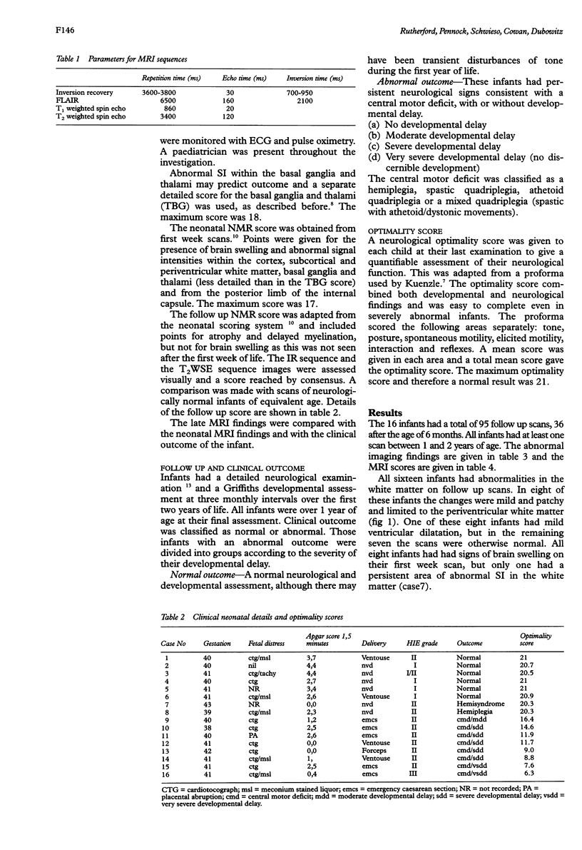
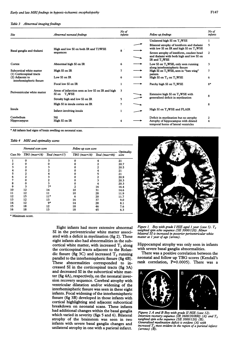
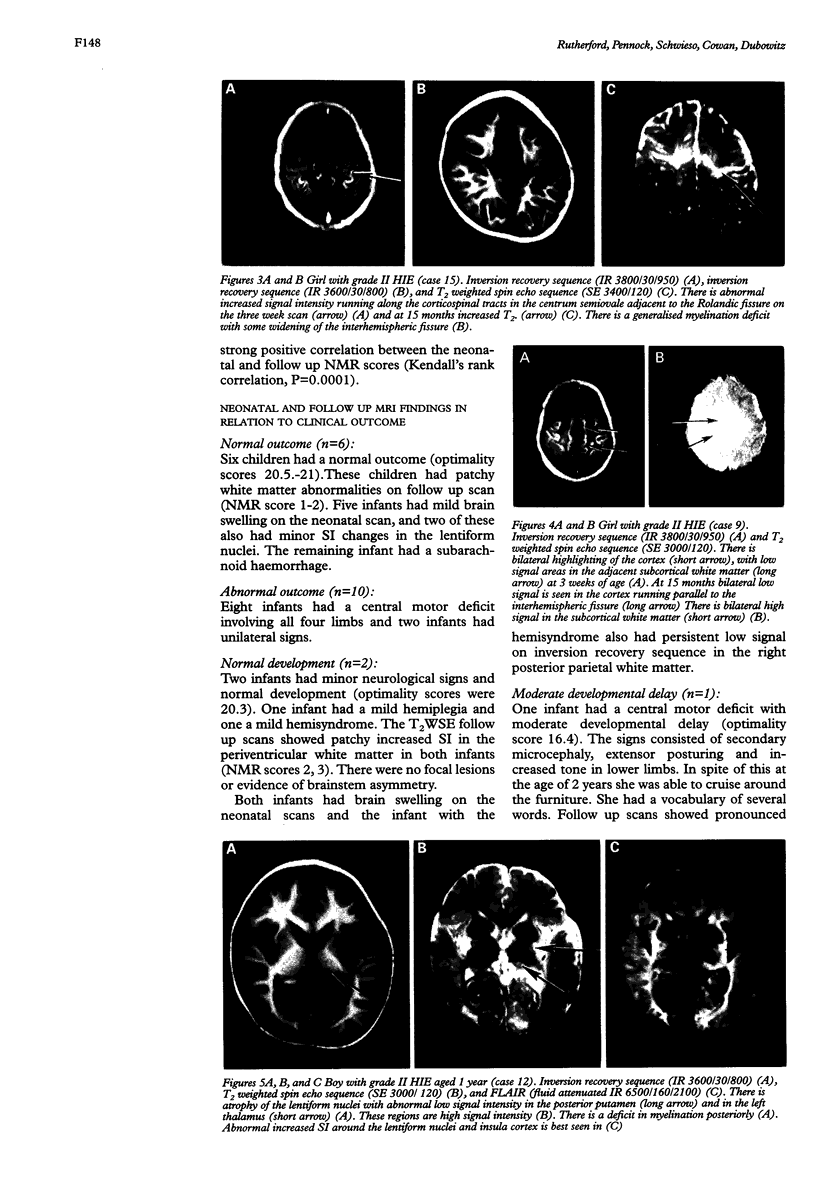
Sixteen infants with hypoxic-ischaemic encephalopathy (HIE) were studied using serial magnetic resonance imaging (MRI) up to the age of 2 years. The infants had regular neurological and developmental assessments. An nuclear magnetic resonance (NMR) score was devised to quantify the early and late MRI findings and a neurological optimality score was used to quantify abnormal neurological signs at the time of the final examination. The follow up MRI score was compared with the neonatal MRI score and the outcome of the child. There was a strong positive correlation between the neonatal and follow up MRI scores and between MRI scores and optimality score. All infants with a normal outcome had patchy white matter abnormalities. All infants with an abnormal outcome had extensive white matter abnormalities. The outcome was most severe in those infants with additional basal ganglia atrophy with or without cyst formation. Infants with mild HIE who are developmentally normal at the age of 2 years do not have normal MRI scans and may be at risk of minor neurological problems by school age. Bilateral basal ganglia abnormalities are associated with severe developmental delay, but infants with mainly white matter and cortical abnormalities have less severe problems despite extensive tissue loss.
Full text
PDF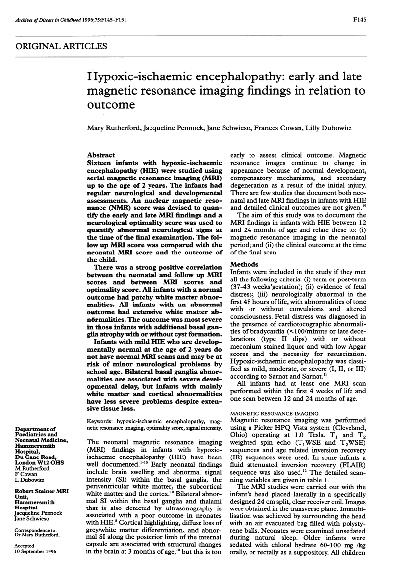
Images in this article


Selected References
These references are in PubMed. This may not be the complete list of references from this article.
- Baenziger O., Martin E., Steinlin M., Good M., Largo R., Burger R., Fanconi S., Duc G., Buchli R., Rumpel H. Early pattern recognition in severe perinatal asphyxia: a prospective MRI study. Neuroradiology. 1993;35(6):437–442. doi: 10.1007/BF00602824. [DOI] [PubMed] [Google Scholar]
- Ball W. S., Jr Magnetic resonance imaging of the infant brain. Semin Ultrasound CT MR. 1991 Oct;12(5):379–409. [PubMed] [Google Scholar]
- Barkovich A. J. MR and CT evaluation of profound neonatal and infantile asphyxia. AJNR Am J Neuroradiol. 1992 May-Jun;13(3):959–975. [PMC free article] [PubMed] [Google Scholar]
- Barkovich A. J., Truwit C. L. Brain damage from perinatal asphyxia: correlation of MR findings with gestational age. AJNR Am J Neuroradiol. 1990 Nov-Dec;11(6):1087–1096. [PMC free article] [PubMed] [Google Scholar]
- Byrne P., Welch R., Johnson M. A., Darrah J., Piper M. Serial magnetic resonance imaging in neonatal hypoxic-ischemic encephalopathy. J Pediatr. 1990 Nov;117(5):694–700. doi: 10.1016/s0022-3476(05)83323-2. [DOI] [PubMed] [Google Scholar]
- Dambska M., Dydyk L., Szretter T., Wozniewicz J., Myers R. E. Topography of lesions in newborn and infant brains following cardiac arrest and resuscitation. Damage to brain and hemispheres. Biol Neonate. 1976;29(3-4):194–206. doi: 10.1159/000240864. [DOI] [PubMed] [Google Scholar]
- Eken P., Jansen G. H., Groenendaal F., Rademaker K. J., de Vries L. S. Intracranial lesions in the fullterm infant with hypoxic ischaemic encephalopathy: ultrasound and autopsy correlation. Neuropediatrics. 1994 Dec;25(6):301–307. doi: 10.1055/s-2008-1073044. [DOI] [PubMed] [Google Scholar]
- Hajnal J. V., De Coene B., Lewis P. D., Baudouin C. J., Cowan F. M., Pennock J. M., Young I. R., Bydder G. M. High signal regions in normal white matter shown by heavily T2-weighted CSF nulled IR sequences. J Comput Assist Tomogr. 1992 Jul-Aug;16(4):506–513. doi: 10.1097/00004728-199207000-00002. [DOI] [PubMed] [Google Scholar]
- Hajnal J. V., Saeed N., Oatridge A., Williams E. J., Young I. R., Bydder G. M. Detection of subtle brain changes using subvoxel registration and subtraction of serial MR images. J Comput Assist Tomogr. 1995 Sep-Oct;19(5):677–691. doi: 10.1097/00004728-199509000-00001. [DOI] [PubMed] [Google Scholar]
- Keeney S. E., Adcock E. W., McArdle C. B. Prospective observations of 100 high-risk neonates by high-field (1.5 Tesla) magnetic resonance imaging of the central nervous system. II. Lesions associated with hypoxic-ischemic encephalopathy. Pediatrics. 1991 Apr;87(4):431–438. [PubMed] [Google Scholar]
- Kuenzle C., Baenziger O., Martin E., Thun-Hohenstein L., Steinlin M., Good M., Fanconi S., Boltshauser E., Largo R. H. Prognostic value of early MR imaging in term infants with severe perinatal asphyxia. Neuropediatrics. 1994 Aug;25(4):191–200. doi: 10.1055/s-2008-1073021. [DOI] [PubMed] [Google Scholar]
- Martin E., Barkovich A. J. Magnetic resonance imaging in perinatal asphyxia. Arch Dis Child Fetal Neonatal Ed. 1995 Jan;72(1):F62–F70. doi: 10.1136/fn.72.1.f62. [DOI] [PMC free article] [PubMed] [Google Scholar]
- McArdle C. B., Richardson C. J., Hayden C. K., Nicholas D. A., Amparo E. G. Abnormalities of the neonatal brain: MR imaging. Part II. Hypoxic-ischemic brain injury. Radiology. 1987 May;163(2):395–403. doi: 10.1148/radiology.163.2.3550882. [DOI] [PubMed] [Google Scholar]
- Myers R. E. Two patterns of perinatal brain damage and their conditions of occurrence. Am J Obstet Gynecol. 1972 Jan 15;112(2):246–276. doi: 10.1016/0002-9378(72)90124-x. [DOI] [PubMed] [Google Scholar]
- Pasternak J. F., Predey T. A., Mikhael M. A. Neonatal asphyxia: vulnerability of basal ganglia, thalamus, and brainstem. Pediatr Neurol. 1991 Mar-Apr;7(2):147–149. doi: 10.1016/0887-8994(91)90014-c. [DOI] [PubMed] [Google Scholar]
- Roland E. H., Hill A., Norman M. G., Flodmark O., MacNab A. J. Selective brainstem injury in an asphyxiated newborn. Ann Neurol. 1988 Jan;23(1):89–92. doi: 10.1002/ana.410230115. [DOI] [PubMed] [Google Scholar]
- Rutherford M. A., Pennock J. M., Dubowitz L. M. Cranial ultrasound and magnetic resonance imaging in hypoxic-ischaemic encephalopathy: a comparison with outcome. Dev Med Child Neurol. 1994 Sep;36(9):813–825. doi: 10.1111/j.1469-8749.1994.tb08191.x. [DOI] [PubMed] [Google Scholar]
- Rutherford M. A., Pennock J. M., Murdoch-Eaton D. M., Cowan F. M., Dubowitz L. M. Athetoid cerebral palsy with cysts in the putamen after hypoxic-ischaemic encephalopathy. Arch Dis Child. 1992 Jul;67(7 Spec No):846–850. doi: 10.1136/adc.67.7_spec_no.846. [DOI] [PMC free article] [PubMed] [Google Scholar]
- Rutherford M. A., Pennock J. M., Schwieso J. E., Cowan F. M., Dubowitz L. M. Hypoxic ischaemic encephalopathy: early magnetic resonance imaging findings and their evolution. Neuropediatrics. 1995 Aug;26(4):183–191. doi: 10.1055/s-2007-979751. [DOI] [PubMed] [Google Scholar]
- Sarnat H. B., Sarnat M. S. Neonatal encephalopathy following fetal distress. A clinical and electroencephalographic study. Arch Neurol. 1976 Oct;33(10):696–705. doi: 10.1001/archneur.1976.00500100030012. [DOI] [PubMed] [Google Scholar]
- Truwit C. L., Barkovich A. J., Koch T. K., Ferriero D. M. Cerebral palsy: MR findings in 40 patients. AJNR Am J Neuroradiol. 1992 Jan-Feb;13(1):67–78. [PMC free article] [PubMed] [Google Scholar]
- Yokochi K., Aiba K., Kodama M., Fujimoto S. Magnetic resonance imaging in athetotic cerebral palsied children. Acta Paediatr Scand. 1991 Aug-Sep;80(8-9):818–823. doi: 10.1111/j.1651-2227.1991.tb11955.x. [DOI] [PubMed] [Google Scholar]
- van der Knaap M. S., Smit L. S., Nauta J. J., Lafeber H. N., Valk J. Cortical laminar abnormalities--occurrence and clinical significance. Neuropediatrics. 1993 Jun;24(3):143–148. doi: 10.1055/s-2008-1071532. [DOI] [PubMed] [Google Scholar]