Abstract
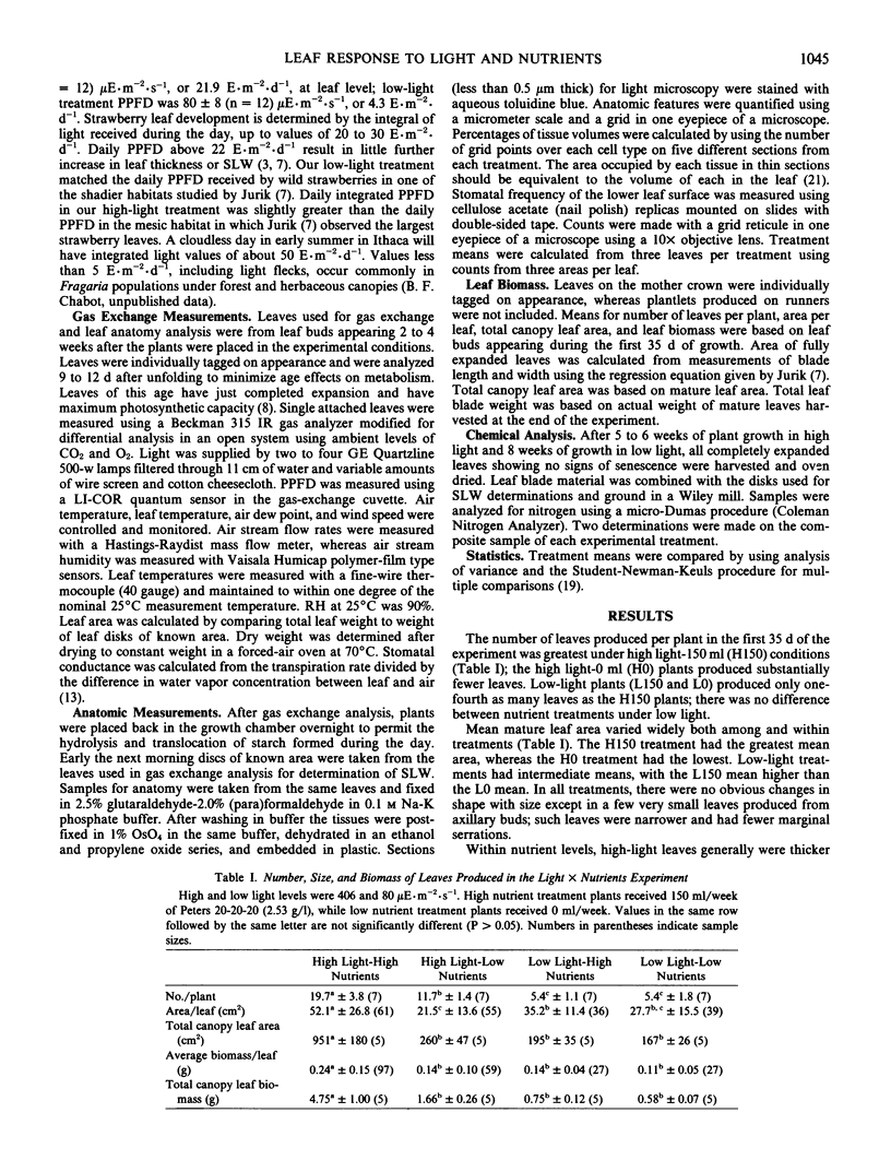
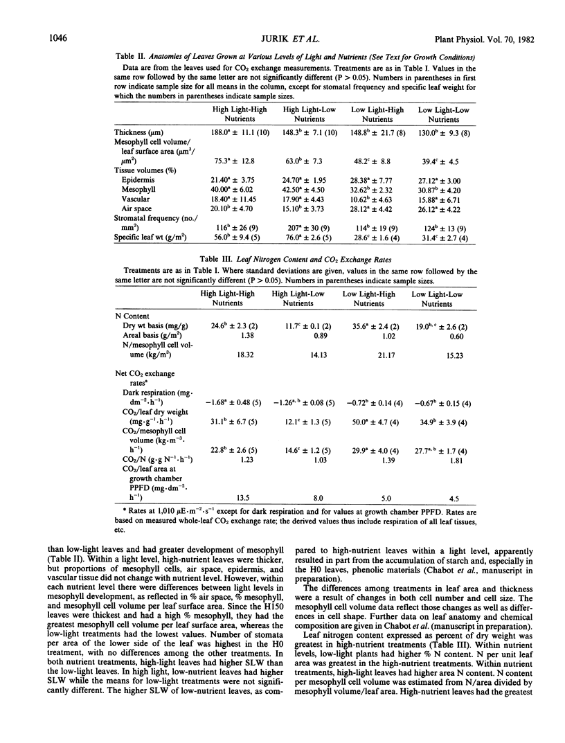
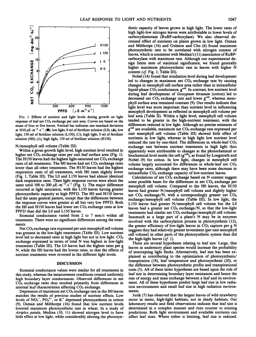
Plants of a single genotype of wild strawberry, Fragaria virginiana Duchesne, were grown with or without fertilizer in high (406 microeinsteins per square meter per second) and low (80 microeinsteins per square meter per second) light. High-light leaves were thicker than low-light leaves and had greater development of the mesophyll. Within a light level, high-nutrient leaves were thicker, but the proportions of leaf tissues did not change with nutrient level. Maximum net CO2 exchange rate and leaf size were greatest in high-light, high-nutrient leaves and lowest in high-light, low-nutrient leaves. Changes in mesophyll cell volume largely accounted for differences in CO2 exchange rate in low-light leaves, but not in high-light leaves.
Leaf size in these experiments was apparently determined by nutrient and carbon supply. This may explain the observation that the largest leaves produced by wild strawberries in the field occur in high-light, mesic habitats, rather than in shady habitats.
Full text
PDF
Selected References
These references are in PubMed. This may not be the complete list of references from this article.
- Jurik T. W., Chabot J. F., Chabot B. F. Ontogeny of Photosynthetic Performance in Fragaria virginiana under Changing Light Regimes. Plant Physiol. 1979 Mar;63(3):542–547. doi: 10.1104/pp.63.3.542. [DOI] [PMC free article] [PubMed] [Google Scholar]
- Longstreth D. J., Nobel P. S. Nutrient Influences on Leaf Photosynthesis: EFFECTS OF NITROGEN, PHOSPHORUS, AND POTASSIUM FOR GOSSYPIUM HIRSUTUM L. Plant Physiol. 1980 Mar;65(3):541–543. doi: 10.1104/pp.65.3.541. [DOI] [PMC free article] [PubMed] [Google Scholar]
- Nobel P. S., Zaragoza L. J., Smith W. K. Relation between Mesophyll Surface Area, Photosynthetic Rate, and Illumination Level during Development for Leaves of Plectranthus parviflorus Henckel. Plant Physiol. 1975 Jun;55(6):1067–1070. doi: 10.1104/pp.55.6.1067. [DOI] [PMC free article] [PubMed] [Google Scholar]
- Weibel E. R. Stereological principles for morphometry in electron microscopic cytology. Int Rev Cytol. 1969;26:235–302. doi: 10.1016/s0074-7696(08)61637-x. [DOI] [PubMed] [Google Scholar]


