Abstract
This study was conducted to investigate the antagonistic potential of endophytic and rhizospheric bacterial isolates obtained from Citrullus colocynthis in suppressing Fusarium solani and Pythium aphanidermatum and promoting the growth of cucumber. Molecular identification of bacterial strains associated with C. colocynthis confirmed that these strains belong to the Achromobacter, Pantoea, Pseudomonas, Rhizobium, Sphingobacterium, Bacillus, Sinorhizobium, Staphylococcus, Cupriavidus, and Exiguobacterium genera. A dual culture assay showed that nine of the bacterial strains exhibited antifungal activity, four of which were effective against both pathogens. Strains B27 (Pantoea dispersa) and B28 (Exiguobacterium indicum) caused the highest percentage of inhibition towards F. solani (48.5% and 48.1%, respectively). P. aphanidermatum growth was impeded by the B21 (Bacillus cereus, 44.7%) and B28 (Exiguobacterium indicum, 51.1%) strains. Scanning electron microscopy showed that the strains caused abnormality in phytopathogens’ mycelia. All of the selected bacterial strains showed good IAA production (>500 ppm). A paper towel experiment demonstrated that these strains improved the seed germination, root/shoot growth, and vigor index of cucumber seedlings. Our findings suggest that the bacterial strains from C. colocynthis are suppressive to F. solani and P. aphanidermatum and can promote cucumber growth. This appears to be the first study to report the efficacy of these bacterial strains from C. colocynthis against F. solani and P. aphanidermatum.
Keywords: antagonistic bacteria, biological control, damping-off, Fusarium solani, plant growth promotion, Pythium aphanidermatum
1. Introduction
Cucumber is an important crop in Oman [1]. Soilborne plant diseases represent a challenge to the cultivation and production of vegetable crops in Oman. In cucumber, damping-off is a serious problem in the USA, Canada, China, the Middle East, and other parts of the world [2,3,4,5,6,7,8]. In Oman, damping-off and decline diseases have been reported to occur in 77% of greenhouses and farms in the Al-Batinah regions and cause up to 100% losses in cucumbers and melons [9,10,11]. Soilborne diseases are also important in other vegetable crops including tomatoes, radish, and beans [12,13,14].
Soilborne diseases of cucumber are caused by a number of fungal and oomycete pathogens. Pythium species are among the most common soilborne pathogens affecting cucumber, with P. aphanidermatum being the most widespread soilborne pathogen of cucumber in Oman [11,15,16]. Fusarium solani and Rhizoctonia solani are also important soilborne pathogens of cucumber [11,17].
Management strategies of damping-off can be divided into four major categories, namely, development of resistant cultivars, chemical treatments, cultural practices, and biological control. Most soilborne pathogens, especially Pythium, are non-host-specific and resistant cultivars are not known yet. Chemical control is used extensively for the management of Pythium and other soilborne pathogens. However, systemic fungicides carry greater environmental risk [18,19] and are subject to the development of resistance [15].
Biological control (or biocontrol) indicates the use of microbial antagonists to suppress plant diseases. The biocontrol of P. aphanidermatum has been achieved using Pseudomonas sp., Trichoderma spp. [20,21,22], Bacillus cereus [23], and B. subtilis [24,25] isolated from various sources. Biocontrol agents are divided into two groups: endophytes, that live inside the plant; and rhizospheric microorganisms, that live attached to or beside plant roots [26,27].
A number of studies have been conducted in Oman on the biocontrol of soilborne pathogens. Pseudomonas aeruginosa showed antagonistic activities against Pythium and Fusarium [22,28]. The tomato rhizosphere bacteria, Bacillus cereus and Exiguobacterium indicum, and the endophytic fungus Aspergillus terreus isolated from the native plants Rhazya stricta and Tephrosia apollinea [29], were found to be effective against P. aphanidermatum. A novel fungus, Cladosporium omanense, and an endophytic bacterium, Enterobacter cloaca, showed antagonistic activity against cucumber damping-off [30,31]. Several other biocontrol agents were reported in Oman during the last 3 years, isolated from desert plants, medicinal plants, cultivated crops, and marine environments [32,33,34,35,36].
Citrullus colocynthis is a wild plant species belonging to the Citrullus genus [37]. It is a medicinal plant with several benefits [38,39]. Wild plants are known to have more resistance to diseases and tolerance to environmental stresses including drought, heat, and salinity compared to cultivated crops [40,41]. Although several studies addressed the medicinal values of C. colocynthis, no reports exist on the antagonistic activities of bacterial strains from this crop against P. aphanidermatum or F. solani. This study was, therefore, designed to investigate the antagonistic potential of endophytic and rhizospheric bacterial strains obtained from Citrullus colocynthis in suppressing Pythium and Fusarium, and in promoting cucumber growth. Knowledge in this field will help in proposing integrated management strategies for Pythium damping-off diseases.
2. Materials and Methods
2.1. Sampling
Healthy plant samples of Citrullus colocynthis, at the mature fruit stage, were randomly collected from Wadi Al Alaq (Al-Dakhiliya region) and Wadi Bani Khalid (Al-Sharqiya region), Oman. The two samples were collected with sterile tools and transferred to the laboratory in sterile plastic bags. The rhizospheric soil was collected from the roots by brushing the soil into sterile Petri plates. The roots, stems, leaves, and fruits were separated and the collected samples were stored in sterile containers at 4 °C. Bacterial isolation was performed within 48 h of sampling.
2.2. Bacteria Isolation from Citrullus colocynthis Rhizosphere and Endosphere
The isolation of cultivable bacteria was carried out as described by Ferjani et al. [42]. One gram of rhizospheric soil from each sample was added to 9 mL of sterile physiological saline solution (9 g/L NaCl), then the tube for each sample was agitated for 15 min at 200 rpm. After that, suspensions were diluted using physiological water in 10-fold series and 0.1 mL of each dilution was spread on nutrient agar (NA) culture medium (Oxoid Ltd., Basingstoke, UK).
For isolation of endophytic bacteria, the surfaces of collected samples (root, stem, leaf, and fruit) were sterilized with sodium hypochlorite (2% NaClO, 1 min), before being rinsed three times in sterile distilled water (SDW). In the next step, samples were dried out on sterile filter paper. The dried samples were either cut into small pieces with a sterile razor blade and cultured on NA medium, or they were crushed using a stomacher (Atkinson, NH, USA). The samples (10 g/sample) were placed in a sterile stomacher bag containing sterile physiological saline solution (190 mL) and homogenized for two minutes. Further, the suspensions were serially diluted and plated onto NA medium [43].
All plates were kept in an incubator for 3–7 days at 28 ± 2 °C. Different colonies were selected and purification was carried out on NA medium via sub-culturing. The bacterial strains were sorted based on phenotypic characteristics and Gram staining. Moreover, pure strains were maintained in nutrient broth (NB) (Sigma Aldrich, St. Louis, MO, USA) containing 25% glycerol at −80 °C for further study.
2.3. DNA Extraction, PCR Amplification, and Sequence Analysis of the Bacterial Strains
Genomic DNA of the bacterial strains was extracted from these strains using the foodproof® StarPrep Two Kit (Windsor, CA, USA). The qualitative and quantitative analysis of the extracted DNA was conducted using a NanoDropTM 2000 spectrophotometer (Thermo Fisher Scientific, MA, USA). A polymerase chain reaction (PCR) was used on the extracted DNA of each bacterial strain to amplify the 16s rDNA coding region. Two universal bacterial primers, 27F (5′-AGAGTTTGATCMTGGCTCAG-3′) and 1492R (5′-TACGGYTACCTTGTTACGACTT-3′), were used for the amplification, following the described thermocycler conditions given by dos Santos, et al. [44]. The PCR reactions consisted of genomic DNA, primers, and PuRe Taq Ready-To-Go™ PCR beads (Cytiva, Marlborough, MA, USA). The PCR products were purified and sequenced by Macrogen Inc. (Seoul, Republic of Korea). The obtained sequences from this study were compared with reference sequences of closely related species in the National Center for Biotechnology Information (NCBI) database. GenBank accession numbers were allocated for the 16S ribosomal DNA sequences of the strains.
2.4. Screening for Antagonistic Activity
2.4.1. In Vitro Antifungal Assays
All rhizospheric and endophytic bacterial strains were inspected for their antifungal activity against the cucumber fungal pathogens Fusarium solani and Pythium aphanidermatum through a dual culture assay, as described by Anith, et al. [45]. The used fungal pathogens, F. solani and P. aphanidermatum, were part of a collection of plant pathogens maintained in the plant pathology laboratory at the Department of Plant Sciences (Sultan Qaboos University, Muscat, Oman). They were cultured on potato dextrose agar (PDA) culture medium (Sigma Aldrich, MO, USA) and then a mycelial disc (5 mm diameter) was excised from the growing edge of a fungal pathogen colony (5 days old) using a sterile cork borer. The mycelial disc was placed in the middle of PDA medium inoculated with a 2-day-old bacterial strain. PDA plates inoculated with the fungal pathogen alone were considered as the control. After that, all cultures were maintained at 28 ± 2 °C for 3–7 days and the fungal growth diameter was measured from the edge of the fungal disc up to the active growing edges of the fungus compared with the control. The inhibitory impact of the bacterial strains was estimated as the inhibition percentage [45].
2.4.2. Scanning Electron Microscope (SEM)
PDA plates from the dual culture assays which showed antifungal activity of the bacterial strains were chosen for electron microscopy study. The hyphal morphology of fungal pathogens (F. solani and P. aphanidermatum) on the edge of bacterial colonies was observed under SEM (JEOL JSM-5600, Tokyo, Japan). The hyphal samples of fungal pathogens were cut out and fixed in glutaraldehyde (2.5% C5H8O2, 4 °C, 2 h) and then washed in phosphate buffer saline (4 times). Afterwards, the samples were desiccated in a graded ethanol series (25%, 75%, 95%, and 100%) for 10 min each. Finally, they were air dried, coated with gold, and scanned using an SEM to record abnormalities in the fungal hyphae [46].
2.5. Determination of Growth-Promoting Potential
2.5.1. Indole Acetic Acid (IAA) Quantification
The antagonistic bacterial strains were evaluated for their in vitro putative growth-promoting attribute, IAA production. The method, as adopted earlier [47], was applied to determine the IAA quantity. Strains were cultured for 72 h on NB medium supplied with 1 mM of L-tryptophan at 28 ± 2 °C. Then, the bacterial cultures were centrifuged and the supernatant was analyzed directly using high-performance liquid chromatography (HPLC) (SIL-30A, Shimadzu, Tokyo, Japan). The chromatograms from the HPLC were produced by injecting 20 µL of each filtered sample into the C18 HPLC column (5 µm particle size, 250 mm column length, 4.6 mm column internal diameter; Hewlett Packard Enterprise, San Jose, CA, USA). Methanol and water were used as the mobile phase at the specific ratio (80:20 v/v]), and the pH was adjusted to 3.8 using sulfuric acid. The mobile phase flow rate was 1 mL/min and the spectra scanning of the compound was performed using a photodiode array detector (Prominence SPD-M20A, Shimadzu, Tokyo, Japan) at 278 nm.
2.5.2. Seed Germination Test via Paper Towel Method
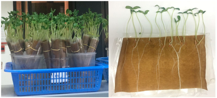
Nine (B1, B2, B20, B21, B23, B24, B27, B28, and B29) of the thirty bacterial strains with antagonistic activity and IAA-producing ability were evaluated for growth promotion of cucumber seedlings using the paper towel method in accordance with the International Seed Testing Association [48]. Cucumber seeds were surface sterilized with sodium hypochlorite (1% NaClO, 5 min) and rinsed three times with autoclaved distilled water. The bacterial strains were grown in NB medium with shaking (200 rpm) at 28 °C for 48 h. Then, the bacterial suspension was prepared and adjusted to 106–107 CFU/mL spectrophotometrically at 600 nm wavelength (Thermo Fisher Scientific, Waltham, MA, USA). The sterilized seeds were mixed with a bacterial suspension in a 50 mL flask at room temperature (140 rpm, 3 h). Sterile sieves were used to separate the seeds from the bacterial suspension. After bacterization, 25 seeds were placed in each germination paper and the paper was rolled and placed in a beaker with a suitable amount of sterile distilled water and maintained in a growth chamber at 28 °C for 10 days (Figure 1). Seeds treated only with water served as the control. Eventually, germinated seeds, and root and shoot lengths were measured to calculate the vigor index using the following formula: Vigor index = (mean shoot length + mean root length) × %Germination [49].
Figure 1.
Paper towel seed germination.
2.6. Statistical Analysis
All the experiments were conducted in a completely randomized design with three replications in each treatment and repeated three times. All data were subjected to one-way analysis of variance (ANOVA) and means were compared by the least significant difference test (p < 0.05) using the R software (version 4.0.3). The data are presented as means ± standard deviation (SD) and displayed in graphical form.
3. Results
3.1. Isolation of Endophytic and Rhizospheric Bacteria
Thirty bacterial strains were isolated from the C. colocynthis plants. Eleven strains came from the rhizosphere (B1–B11), while nineteen were endophytes from the roots (B12–B18), stems (B19–B22), leaves (B23–B26), and fruits (B27–B30). Some strains were found in only one location, while other strains were found in both locations (Wadi Al Alaq and Wadi Bani Khalid) (Table 1).
Table 1.
Identification of rhizospheric and endophytic bacteria associated with Citrullus colocynthis.
| Bacterial Code | Source of Isolation | Identified Species | Max. Identity (%) | GenBank Accession No. | Place of Collection |
|---|---|---|---|---|---|
| B1 | Soil | Achromobacter xylosoxidans | 99.79% | JQ724537 | Wadi Al Alaq |
| B2 | Soil | Pantoea dispersa | 100% | MN725743 | Wadi Al Alaq |
| B3 | Soil | Pseudomonas lini | 99.75% | MT102313 | Wadi Al Alaq |
| B4 | Soil | Pseudomonas plecoglossicida | 99.71% | MF574326 | Wadi Al Alaq |
| B5 | Soil | Pseudomonas aeruginosa | 100% | JQ659890 | Wadi Al Alaq |
| B6 | Soil | Pseudomonas fluorescens | 100% | HM439968 | Wadi Al Alaq |
| B7 | Soil | Rhizobium pusense | 100% | MK734334 | Wadi Al Alaq |
| B8 | Soil | Sphingobacterium spiritivorum | 100% | KR349259 | Wadi Bani Khalid |
| B9 | Soil | Pseudomonas plecoglossicida | 100% | MF574326 | Wadi Bani Khalid |
| B10 | Soil | Achromobacter xylosoxidans | 99.82% | MK537386 | Wadi Bani Khalid |
| B11 | Soil | Pseudomonas putida | 100% | KU672371 | Wadi Bani Khalid |
| B12 | Root | Bacillus aryabhattai | 100% | MT078622 | Wadi Al Alaq |
| B13 | Root | Sinorhizobium meliloti | 99.85% | MT197365 | Wadi Al Alaq |
| B14 | Root | Bacillus anthracis | 98.89% | KF601916 | Wadi Al Alaq |
| B15 | Root | Bacillus anthracis | 99.66% | KF601916 | Wadi Bani Khalid |
| B16 | Root | Unknown | Wadi Bani Khalid | ||
| B17 | Root | Achromobacter mucicolens | 99.48% | MT534143 | Wadi Bani Khalid |
| B18 | Root | Achromobacter xylosoxidans | 98.79% | MN889379 | Wadi Bani Khalid |
| B19 | Stem | Achromobacter xylosoxidans | 99.86% | MK332530 | Wadi Al Alaq |
| B20 | Stem | Unknown | Wadi Al Alaq | ||
| B21 | Stem | Bacillus cereus | 100% | MK648340 | Wadi Bani Khalid |
| B22 | Stem | Pseudomonas plecoglossicida | 99.86% | MF574326 | Wadi Bani Khalid |
| B23 | Leaf | Pseudomonas stutzeri | 97.34% | KY606628 | Wadi Al Alaq |
| B24 | Leaf | Staphylococcus gallinarum | 99.93% | MH542297 | Wadi Al Alaq |
| B25 | Leaf | Cupriavidus gilardii | 99.50% | AY860225 | Wadi Bani Khalid |
| B26 | Leaf | Achromobacter xylosoxidans | 100% | MK537386 | Wadi Bani Khalid |
| B27 | Fruit | Pantoea dispersa | 100% | MN725743 | Wadi Al Alaq |
| B28 | Fruit | Exiguobacterium indicum | 99.86% | KT986092 | Wadi Al Alaq |
| B29 | Fruit | Unknown | Wadi Bani Khalid | ||
| B30 | Fruit | Achromobacter xylosoxidans | 98% | HQ288926 | Wadi Bani Khalid |
3.2. Molecular Identification
The obtained nucleotide sequences were deposited in GenBank under defined accession numbers (Table 1). The identity indices of the bacterial strains with their relevant strain on the NCBI database are presented in Table 1. There was up to nearly 100% resemblance detected amongst the strains and sequences in the database (except B16, B20, and B29). Identification of the bacterial strains showed that they belonged to different genera and species. These strains belonged to 10 bacterial genera, namely, Achromobacter, Pantoea, Pseudomonas, Rhizobium, Sphingobacterium, Bacillus, Sinorhizobium, Staphylococcus, Cupriavidus, and Exiguobacterium (Table 1). Pseudomonas, Achromobacter, and Bacillus were the most abundant genera. The most abundant species were Achromobacter xylosoxidans and Pseudomonas plecoglossicida. The bacterial species Achromobacter xylosoxidans, Pseudomonas plecoglossicida, and Pantoea dispersa were detected in the rhizosphere as well as the endosphere of Citrullus colocynthis (Table 1).
3.3. Antagonistic Activity against F. solani and P. aphanidermatum
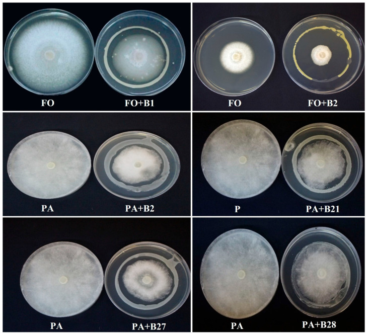
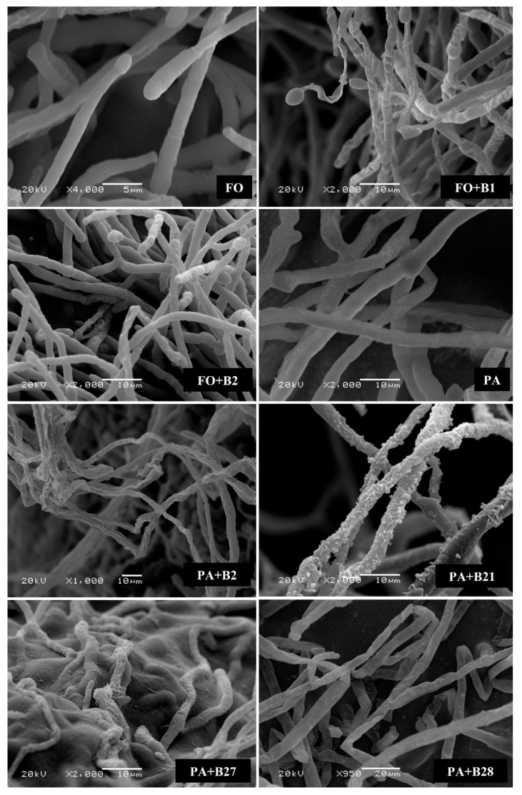
The bacterial strains were examined for their ability to retard the growth of F. solani and P. aphanidermatum in vitro using the dual culture technique (Figure 2). Out of the 30 bacterial strains, only 9 rhizospheric and endophytic bacterial strains caused inhibition against F. solani growth, while 4 strains inhibited P. aphanidermatum growth. Among these strains, only four of the strains demonstrated an inhibitory effect toward the growth of both fungal pathogens. SEM observation of F. solani and P. aphanidermatum hyphae at the inhibition zone showed negative effects of the selected bacterial strains on the morphology of the pathogens. The bacterial strains caused shrinking and deformation of their hyphae, indicative of a loss in turgidity and cellular content (Figure 3).
Figure 2.
Dual culture assay showing the antifungal activity of selected bacterial strains against Fusarium solani (FO) and Pythium aphanidermatum (PA). (B) refers to the different bacterial strains.
Figure 3.
Antagonistic activity of selected bacterial strains on Fusarium solani (FO) and Pythium aphanidermatum (PA) mycelial morphology depicted using a scanning electron microscope (SEM). Normal patterns of hyphae presented in the control (FS and PA). Distorted mycelial structure, wrinkled or shrunken patterns presented in FO + B1, FO + B2, PA + B2, PA + B21, PA + B27, and PA + B28.
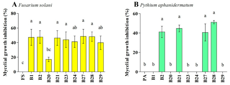
The nine antagonistic strains belonged to Achromobacter xylosoxidans, Pantoea dispersa, Bacillus cereus, Pseudomonas stutzeri, Staphylococcus gallinarum, and Exiguobacterium indicum. These strains hindered the growth of F. solani and P. aphanidermatum with different percentages of inhibition. The inhibition percentage of F. solani and P. aphanidermatum growth by the active strains was in the range of 16.9–48.5% and 40.5–51.2%, respectively. Strains B27 (48.5%) and B28 (48.1%) caused the highest percentage of inhibition toward F. solani, while strains B21 (44.7%) and B28 (51.1%) caused the highest percentage of inhibition toward P. aphanidermatum (Figure 4). Strains B2, B21, B27, and B28 were inhibitory toward both pathogens, and the inhibition percentage caused by these strains ranged from 41.1 to 51.2%.
Figure 4.
Effect of bacterial antagonists on the inhibition of Fusarium solani (FO) (A) and Pythium aphanidermatum (PA) (B) mycelial growths. Values show the means ± standard error (n = 3), and significant differences at p < 0.05 are indicated by different lowercase letters above the columns.
3.4. Growth-Promoting Assays
3.4.1. Determination of IAA Production
The HPLC method was applied to determine the IAA generated by the selected bacterial strains. Production of IAA (ppm) was detected in all of the selected bacterial strains, comprising B1 (749.1), B2 (660.3), B20 (714.4), B21 (512.9), B23 (699.6), B24 (706.7), B27 (568.7), B28 (581.2), and B29 (732.9). Strain B1 produced the highest amount of IAA, followed by B29. The lowest amount of IAA was produced by the B21 strain.
3.4.2. Effect of Bacterial Strains on Plant Growth Parameters
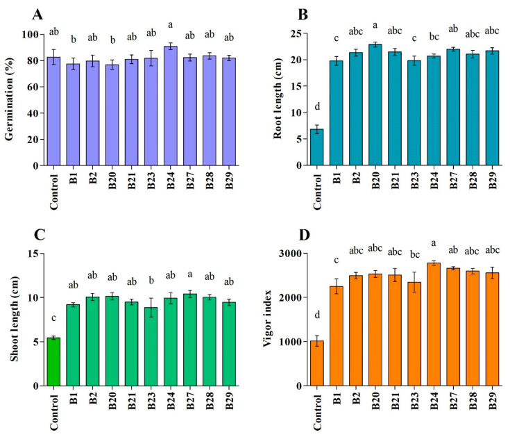
All of the tested strains promoted the growth of cucumber, as evidenced by the enhanced seedling vigor compared to the control. The seeds’ germination percentage varied among all strains, ranging from 76.7 to 90.7%. The bacterial strains B24 and B20 were the most active in promoting germination (90.7%) and root growth (22.8%), respectively. In addition, B27 and B24 resulted in the highest shoot length (10.4%) and seedling vigor (63.4%), respectively (Figure 5).
Figure 5.
Growth-promoting impact of selected bacterial strains on cucumber plants. Values show the means ± standard error (n = 3), and significant differences at p < 0.05 are indicated by different lowercase letters above the columns. The subfigures indicate % germination (A), root length (B), shoot length (C), and vigor index (D).
4. Discussion
Pythium aphanidermatum and Fusarium solani are major soilborne pathogens of vegetable crops, especially cucumber, in Oman and different parts of the world, causing up to 100% mortality [5,11,16,50,51,52,53]. Although different methods are used for the control of Pythium and Fusarium, biological control using antagonistic microorganisms has received more attention during recent years. Pseudomonas fluorescens [54,55,56], P. aeruginosa [22,28,57], P. resinovorans [32], Lysobacter sp. [58], Bacillus cereus [23,59], B. subtilis [25,60,61,62], and Serratia marcescens [8,34] have shown antagonistic potential against several soilborne pathogens.
In this study the endophytic bacterial strain Exiguobacterium indicum (B28) inhibited F. solani and Pythium aphanidermatum growth by up to 48.1% and 51.1%, respectively, in a dual culture assay. Exiguobacterium species have been used industrially in many applications, such as enzyme production and bioremediation. There are some reports that have indicated the ability of Exiguobacterium sp. in promoting plant growth [23,63]. It has been reported that E. acetylicum inhibited the growth of Rhizoctonia solani, Sclerotium rolfsii, Pythium, and Fusarium solani [64]. In a previous study, Al-Hussini, et al. [65] reported that E. indicum strain D1/8 inhibited the growth of P. aphanidermatum in vitro and reduced the incidence of damping-off of tomato by 13%. The strain of E. indicum (B28) isolated in this study was very effective in inhibiting F. solani and P. aphanidermatum growth.
The bacterial strain Achromobacter xylosoxidans (B1) inhibited F. solani by 47.1%. A. xylosoxidans has been reported as a biocontrol agent against F. solani associated with melon wilt [66]. A. xylosoxidans has also been found to increase yields and alleviate drought stress in some crops [67,68]. Another study showed that A. xylosoxidans can be used in the control of root-knot nematodes and improve the growth of eggplants [69].
Pantoea dispersa (B2) showed high inhibitory activity (~47%) against F. solani and P. aphanidermatum. It has been reported as a biocontrol agent of sweet potato black rot [70]. P. dispersa also proved efficacious in managing Fusarium wilt of pigeon pea [71]. In addition, P. dispersa has also been found to stimulate the growth of different crops [72,73,74]. However, no previous reports are available on the use of P. dispersa against F. solani or P. aphanidermatum.
The mycelial growth inhibition of F. solani and P. aphanidermatum in the dual culture assay plate in this study might be due to diffusible antimicrobial compounds released by the antagonistic bacteria into the agar medium [29]. The differences in the antagonistic potential among the bacterial strains against F. solani and P. aphanidermatum might be related to the type of antimicrobial compounds produced by them and the sensitivity of the pathogens.
The bacterial strains caused various morphological abnormalities to F. solani and P. aphanidermatum hyphae, which could be related to the secretion of antifungal metabolites that damage the mycelial cell wall [29,75,76,77]. Hydrolytic enzymes such as cellulase, chitinase, glucanase, and protease are examples of antifungal metabolites that can cause deformations in fungal mycelium [78,79,80,81,82]. Trichoderma [78,79,83], Aspergillus [29], and Talaromyces [13,84] have been reported to produce cellulase enzymes as one of their biocontrol mechanisms. It is evident from this study that the antagonistic bacterial strains induced shrinkage and deformation of the hyphae of the test pathogens. These findings are similar to those reported by Al-Daghari, et al. [36], who recorded shrinkage of the hyphae of Monosporascus cannonballus due to the antagonistic effects of Pseudomonas spp. The shrinkage of hyphae indicates cell membrane damage and leakage of cytoplasmic contents. Alterations in the intracellular osmotic pressure often result in hyphal distortion.
The bacterial strains improved the growth of cucumber seedlings. The bacterial strain Achromobacter xylosoxidans (B1) resulted in the improvement of cucumber growth. Several endophytes and rhizosphere bacteria have been characterized as growth-promoting bacteria since they stimulate root development, improve water and mineral uptake, and produce IAA. These include Bradyrhizobium, Alcaligenes, Azoarcus, Acetobacter, Pseudomonas, Enterobacter, and Xanthomonas [85,86,87,88,89,90,91,92,93]. Achromobacter xylosoxidans was reported as a growth-promoting bacterium in rice plants [67,94]. The growth-promoting activity of this bacterium could be explained by the high production of IAA, high nitrogenase activity, and P-solubilization. In our study, the high IAA production by this bacterium could be the reason for its high efficacy in promoting cucumber growth. Our study also showed that the antagonistic activity and the growth-promoting activity of the bacteria were different, which may indicate that there is no relationship between the two parameters.
5. Conclusions
This study revealed that bacterial strains isolated from C. colocynthis have the potential, as biocontrol agents, to prevent damage caused by F. solani, P. aphanidermatum, and other phytopathogens to vegetable crops, and enhance plant growth. The strains Pantoea dispersa and Exiguobacterium indicum showed the most antifungal activity against F. solani. The mycelial growth of P. aphanidermatum was suppressed significantly by Bacillus cereus and Exiguobacterium indicum. This is the first study to report the efficacy of these bacterial strains from C. colocynthis against F. solani and P. aphanidermatum. Future studies will be required to examine the efficiency of these strains under field conditions, and to prepare their stable bioformulations.
Acknowledgments
We would like also to thank Issa Al-Mahmooli and Zainab Al-Balushi for their helpful advice and guidance in the laboratory.
Author Contributions
Conceptualization, A.M.A.-S. and R.V.; methodology, B.K.A.-S.; validation, E.A.K.; formal analysis, B.K.A.-S., S.H. and E.A.K.; resources, A.M.A.-S.; writing—original draft preparation, B.K.A.-S., E.A.K. and A.M.A.-S.; writing—review and editing, R.V., S.H.; supervision, A.M.A.-S. and R.V.; project administration, A.M.A.-S.; funding acquisition, A.M.A.-S. All authors have read and agreed to the published version of the manuscript.
Data Availability Statement
Sequences were deposited in GenBank under the accession numbers listed in Table 1.
Conflicts of Interest
The authors declare no conflict of interest.
Funding Statement
This research was funded by Sultan Qaboos University, CR/AGR/CROP/23/01.
Footnotes
Disclaimer/Publisher’s Note: The statements, opinions and data contained in all publications are solely those of the individual author(s) and contributor(s) and not of MDPI and/or the editor(s). MDPI and/or the editor(s) disclaim responsibility for any injury to people or property resulting from any ideas, methods, instructions or products referred to in the content.
References
- 1.FAO FAOSTAT. [(accessed on 1 September 2023)]. Available online: http://www.fao.org/faostat/en/#data/QC/visualize.
- 2.Hashemi L., Golparvar A.R., Nasr Esfahani M., Golabadi M. Correlation between cucumber genotype and resistance to damping-off disease caused by Phytophthora melonis. Biotechnol. Biotechnol. Equip. 2019;33:1494–1504. doi: 10.1080/13102818.2019.1675535. [DOI] [Google Scholar]
- 3.Jaiswal A.K., Graber E.R., Elad Y., Frenkel O. Biochar as a management tool for soilborne diseases affecting early stage nursery seedling production. Crop Prot. 2019;120:34–42. doi: 10.1016/j.cropro.2019.02.014. [DOI] [Google Scholar]
- 4.Abbasi P.A., Renderos W., Fillmore S. Soil incorporation of buckwheat as a pre-plant amendment provides control of Rhizoctonia damping-off and root rot of radish and Pythium damping-off and root rot of cucumber. Can. J. Plant Pathol. 2019;41:24–34. doi: 10.1080/07060661.2018.1559224. [DOI] [Google Scholar]
- 5.Owen W.G., Jackson B.E., Whipker B.E., Fonteno W.C., Benson M.D. Assessing the severity of damping-off caused by Pythium ultimum and Rhizoctonia solani in peat-based greenhouse substrates amended with pine wood chip aggregates. Acta Hortic. 2019;1266:27–33. doi: 10.17660/ActaHortic.2019.1266.5. [DOI] [Google Scholar]
- 6.You X., Kimura N., Okura T., Murakami S., Okano R., Shimogami Y., Matsumura A., Tokumoto H., Ogata Y., Tojo M. Suppressive effects of vermicomposted-bamboo powder on cucumber damping-off. Jpn. Agric. Res. Q. 2019;53:13–19. doi: 10.6090/jarq.53.13. [DOI] [Google Scholar]
- 7.Chen W.L., Chien C.S. Improving productive efficiency of cucumber under greenhouse cultivation by grafting and bee pollination. Acta Hortic. 2019;1257:187–194. doi: 10.17660/ActaHortic.2019.1257.27. [DOI] [Google Scholar]
- 8.Roberts D.P., McKenna L.F., Buyer J.S. Consistency of control of damping-off of cucumber is improved by combining ethanol extract of Serratia marcescens with other biologically based technologies. Crop Prot. 2017;96:59–67. doi: 10.1016/j.cropro.2017.01.007. [DOI] [Google Scholar]
- 9.Al-Mawaali Q.S., Al-Sadi A.M., Al-Said F.A., Deadman M.L. Etiology, development and reaction of muskmelon to vine decline under arid conditions of Oman. Phytopathol. Mediterr. 2013;52:457–465. doi: 10.14601/Phytopathol_Mediterr-11673. [DOI] [Google Scholar]
- 10.Al-Mawaali Q.S., Al-Sadi A.M., Khan A.J., Al-Hasani H.D., Deadman M.L. Response of cucurbit rootstocks to Pythium aphanidermatum. Crop Prot. 2012;42:64–68. doi: 10.1016/j.cropro.2012.07.017. [DOI] [Google Scholar]
- 11.Al-Sadi A.M., Al-Said F.A., Al-Kiyumi K.S., Al-Mahrouqi R.S., Al-Mahmooli I.H., Deadman M.L. Etiology and characterization of cucumber vine decline in Oman. Crop Prot. 2011;30:192–197. doi: 10.1016/j.cropro.2010.10.013. [DOI] [Google Scholar]
- 12.Al-Azizi A., Al-Sadi A.M., Dietz H., Al-Said F.A., Deadman M.L. Influence of carbon/nitrogen ratio on Pythium aphanidermatum and on Pythium-induced damping-off of radish. J. Plant Pathol. 2013;95:181–185. [Google Scholar]
- 13.Halo B.A., Al-Yahyai R.A., Maharachchikumbura S.S.N., Al-Sadi A.M. Talaromyces variabilis interferes with Pythium aphanidermatum growth and suppresses Pythium-induced damping-off of cucumbers and tomatoes. Sci. Rep. 2019;9:11255. doi: 10.1038/s41598-019-47736-x. [DOI] [PMC free article] [PubMed] [Google Scholar]
- 14.Al-Jaradi A., Al-Mahmooli I., Janke R., Maharachchikumbura S., Al-Saady N., Al-Sadi A.M. Isolation and identification of pathogenic fungi and oomycetes associated with beans and cowpea root diseases in Oman. PeerJ. 2018;2018:e6064. doi: 10.7717/peerj.6064. [DOI] [PMC free article] [PubMed] [Google Scholar]
- 15.Al-Balushi Z.M., Agrama H., Al-Mahmooli I.H., Maharachchikumbura S.S.N., Al-Sadi A.M. Development of resistance to hymexazol among Pythium species in cucumber greenhouses in Oman. Plant Dis. 2018;102:202–208. doi: 10.1094/PDIS-11-16-1680-RE. [DOI] [PubMed] [Google Scholar]
- 16.Al-Sadi A.M., Al-Ghaithi A.G., Al-Balushi Z.M., Al-Jabri A.H. Analysis of diversity in Pythium aphanidermatum populations from a single greenhouse reveals phenotypic and genotypic changes over 2006 to 2011. Plant Dis. 2012;96:852–858. doi: 10.1094/PDIS-07-11-0624. [DOI] [PubMed] [Google Scholar]
- 17.Al-Sadi A.M., Al-Masoodi R.S., Al-Ismaili M., Al-Mahmooli I.H. Population structure and development of resistance to hymexazol among Fusarium solani populations from date palm, citrus and cucumber. J. Phytopathol. 2015;163:947–955. doi: 10.1111/jph.12397. [DOI] [Google Scholar]
- 18.Zhang Q., Hua X.D., Shi H.Y., Liu J.S., Tian M.M., Wang M.H. Enantioselective bioactivity, acute toxicity and dissipation in vegetables of the chiral triazole fungicide flutriafol. J. Hazard. Mater. 2015;284:65–72. doi: 10.1016/j.jhazmat.2014.10.033. [DOI] [PubMed] [Google Scholar]
- 19.Kitchen J.L., Van Den Bosch F., Paveley N.D., Helps J., Van Den Berg F. The evolution of fungicide resistance resulting from combinations of foliar-acting systemic seed treatments and foliar-applied fungicides: A modeling analysis. PLoS ONE. 2016;11:e0161887. doi: 10.1371/journal.pone.0161887. [DOI] [PMC free article] [PubMed] [Google Scholar]
- 20.Meena R.P., Kalariya K.A., Saran P.L., Roy S. Efficacy of fungicides and biocontrol agents against Pythium aphanidermatum causes damping off disease in ashwagandha (Withania somnifera L. dunal) Med. Plants. 2019;11:404–409. doi: 10.5958/0975-6892.2019.00052.2. [DOI] [Google Scholar]
- 21.Charoenrak P., Chamswarng C., Intanoo W., Keawprasert N. The effects of vermicompost mixed with Trichoderma asperellum on the growth and pythium root rot of lettuces. Int. J. GEOMATE. 2019;17:215–221. doi: 10.21660/2019.61.4728. [DOI] [Google Scholar]
- 22.Al-Ghafri H.M., Velazhahan R., Shahid M.S., Al-Sadi A.M. Antagonistic activity of Pseudomonas aeruginosa from compost against Pythium aphanidermatum and Fusarium solani. Biocontrol. Sci. Technol. 2020;30:642–658. doi: 10.1080/09583157.2020.1750562. [DOI] [Google Scholar]
- 23.Qulsum M.U., Islam M.M., Chowdhury M.E.K., Hossain S.M.M., Hasan M.M. Management of bacterial wilt (Ralstonia solanacearum) of brinjal using Bacillus cereus, Trichoderma harzianum and Calotropis gigantea consortia in Bangladesh. Egyp. J. Biol. Pest Cont. 2023;33:74. doi: 10.1186/s41938-023-00720-0. [DOI] [Google Scholar]
- 24.Ramzan N., Noreen N., Shahzad S. Inhibition of in vitro growth of soil-borne pathogens by compost-inhabiting indigenous bacteria and fungi. Pak. J. Bot. 2014;46:1093–1099. [Google Scholar]
- 25.Ramzan N., Noreen N., Perveen Z., Shahzad S. Effect of seed pelleting with biocontrol agents on growth and colonisation of roots of mungbean by root-infecting fungi. J. Sci. Food Agric. 2016;96:3694–3700. doi: 10.1002/jsfa.7553. [DOI] [PubMed] [Google Scholar]
- 26.Segaran G., Sathiavelu M. Fungal endophytes: A potent biocontrol agent and a bioactive metabolites reservoir. Biocatal. Agric. Biotechnol. 2019;21:101284. doi: 10.1016/j.bcab.2019.101284. [DOI] [Google Scholar]
- 27.De Silva N.I., Brooks S., Lumyong S., Hyde K.D. Use of endophytes as biocontrol agents. Fungal Biol. Rev. 2019;33:133–148. doi: 10.1016/j.fbr.2018.10.001. [DOI] [Google Scholar]
- 28.Al-Hinai A.H., Al-Sadi A.M., Al-Bahry S.N., Mothershaw A.S., Al-Said F.A., Al-Harthi S.A., Deadman M.L. Isolation and characterization of Pseudomonas aeruginosa with antagonistic activity against Pythium aphanidermatum. J. Plant Pathol. 2010;92:653–660. [Google Scholar]
- 29.Halo B.A., Al-Yahyai R.A., Al-Sadi A.M. Aspergillus terreus inhibits growth and induces morphological abnormalities in Pythium aphanidermatum and suppresses Pythium-induced damping-off of cucumber. Front. Microbiol. 2018;9:95. doi: 10.3389/fmicb.2018.00095. [DOI] [PMC free article] [PubMed] [Google Scholar]
- 30.Halo B.A., Al-Yahyai R.A., Al-Sadi A.M. Biological control of Pythium aphanidermatum-induced cucumber and radish damping-off by an endophytic fungus, Cladosporium omanense isolate 31R. Biocontrol Sci. Technol. 2021;31:235–251. doi: 10.1080/09583157.2020.1844148. [DOI] [Google Scholar]
- 31.Kazerooni E.A., Al-Shibli H., Nasehi A., Al-Sadi A.M. Endophytic Enterobacter cloacae exhibits antagonistic activity against Pythium Damping-off of cucumber. Ciência Rural. 2020;50:e20191035. doi: 10.1590/0103-8478cr20191035. [DOI] [Google Scholar]
- 32.Al-Daghari D.S.S., Al-Abri S.A., Al-Mahmooli I.H., Al-Sadi A.M., Velazhahan R. Efficacy of native antagonistic rhizobacteria in the biological control of Pythium aphanidermatum-induced damping-off of cucumber in Oman. J. Plant Pathol. 2019;102:305–310. doi: 10.1007/s42161-019-00438-9. [DOI] [Google Scholar]
- 33.Al-Shibli H., Dobretsov S., Al-Nabhani A., Maharachchikumbura S.S.N., Rethinasamy V., Al-Sadi A.M. Aspergillus terreus obtained from mangrove exhibits antagonistic activities against Pythium aphanidermatum-induced damping-off of cucumber. PeerJ. 2019;2019:7884. doi: 10.7717/peerj.7884. [DOI] [PMC free article] [PubMed] [Google Scholar]
- 34.Gaya Karunasinghe T., Hashil Al-Mahmooli I., Al-Sadi A.M., Velazhahan R. The effect of salt-tolerant antagonistic bacteria from tomato rhizosphere on plant growth promotion and damping-off disease suppression under salt-stress conditions. Acta Agric. Scand. Sect. B Soil Plant Sci. 2020;70:69–75. doi: 10.1080/09064710.2019.1668956. [DOI] [Google Scholar]
- 35.Al-Badri B.A.S., Al-Maawali S.S., Al-Balushi Z.M., Al-Mahmooli I.H., Al-Sadi A.M., Velazhahan R. Cyanide degradation and antagonistic potential of endophytic Bacillus subtilis strain BEB1 from Bougainvillea spectabilis Willd. All Life. 2020;13:92–98. doi: 10.1080/26895293.2020.1728393. [DOI] [Google Scholar]
- 36.Al-Daghari D.S.S., Al-Sadi A.M., Janke R., Al-Mahmooli I.H., Velazhahan R. Potential of indigenous antagonistic rhizobacteria in the biological control of Monosporascus root rot and vine decline disease of muskmelon. Acta Agric. Scand. Sect. B-Soil Plant Sci. 2020;70:371–380. doi: 10.1080/09064710.2020.1748703. [DOI] [Google Scholar]
- 37.Dhiman K., Gupta A., Sharma D.K., Gill N.S., Goyal A. A review on the medicinally important plants of the family Cucurbitaceae. Asian J. Clin. Nutr. 2012;4:16–26. doi: 10.3923/ajcn.2012.16.26. [DOI] [Google Scholar]
- 38.Yatoo M.I., Saxena A., Gopalakrishnan A., Alagawany M., Dhama K. Promising antidiabetic drugs, medicinal plants and herbs: An update. Int. J. Pharmacol. 2017;13:732–745. doi: 10.3923/ijp.2017.732.745. [DOI] [Google Scholar]
- 39.Hussain A.I., Rathore H.A., Sattar M.Z.A., Chatha S.A.S., Sarker S.D., Gilani A.H. Citrullus colocynthis (L.) Schrad (bitter apple fruit): A review of its phytochemistry, pharmacology, traditional uses and nutritional potential. J. Ethnopharmacol. 2014;155:54–66. doi: 10.1016/j.jep.2014.06.011. [DOI] [PubMed] [Google Scholar]
- 40.Luu V.T., Weinhold A., Ullah C., Dressel S., Schoettner M., Gase K., Gaquerel E., Xu S., Baldwin I.T. O-Acyl sugars protect a wild Tobacco from both native fungal pathogens and a specialist herbivore. Plant Physiol. 2017;174:370–386. doi: 10.1104/pp.16.01904. [DOI] [PMC free article] [PubMed] [Google Scholar]
- 41.Lebeda A., Křístková E., Kitner M., Mieslerová B., Jemelková M., Pink D.A.C. Wild Lactuca species, their genetic diversity, resistance to diseases and pests, and exploitation in lettuce breeding. Eur. J. Plant Pathol. 2014;138:597–640. doi: 10.1007/s10658-013-0254-z. [DOI] [Google Scholar]
- 42.Ferjani R., Marasco R., Rolli E., Cherif H., Cherif A., Gtari M., Boudabous A., Daffonchio D., Ouzari H.-I. The Date Palm Tree Rhizosphere Is a Niche for Plant Growth Promoting Bacteria in the Oasis Ecosystem. BioMed Res. Int. 2015;2015:153851. doi: 10.1155/2015/153851. [DOI] [PMC free article] [PubMed] [Google Scholar]
- 43.Abid L., Smiri M., Federici E., Lievens B., Manai M., Yan Y., Sadfi-Zouaoui N. Diversity of rhizospheric and endophytic bacteria isolated from dried fruit of Ficus carica. Saudi J. Biol. Sci. 2022;29:103398. doi: 10.1016/j.sjbs.2022.103398. [DOI] [PMC free article] [PubMed] [Google Scholar]
- 44.dos Santos H.R.M., Argolo C.S., Argôlo-Filho R.C., Loguercio L.L. A 16S rDNA PCR-based theoretical to actual delta approach on culturable mock communities revealed severe losses of diversity information. BMC Microbiol. 2019;19:74. doi: 10.1186/s12866-019-1446-2. [DOI] [PMC free article] [PubMed] [Google Scholar]
- 45.Anith K.N., Nysanth N.S., Natarajan C. Novel and rapid agar plate methods for in vitro assessment of bacterial biocontrol isolates’ antagonism against multiple fungal phytopathogens. Lett. Appl. Microbiol. 2021;73:229–236. doi: 10.1111/lam.13495. [DOI] [PubMed] [Google Scholar]
- 46.Vinayarani G., Prakash H. Growth promoting rhizospheric and endophytic bacteria from Curcuma longa L. as biocontrol agents against rhizome rot and leaf blight diseases. Plant Pathol. J. 2018;34:218. doi: 10.5423/PPJ.OA.11.2017.0225. [DOI] [PMC free article] [PubMed] [Google Scholar]
- 47.KC B.M., Gauchan D.P., Khanal S.N., Lamichhane J. Quantification of indole-3-acetic acid from Bambusa tulda Roxb. seedlings using high performance liquid chromatography. Afr. J. Biotechnol. 2020;19:781–788. doi: 10.5897/AJB2020.17238. [DOI] [Google Scholar]
- 48.International Seed Testing Association (ISTA) International Rules of Seed Testing. Seed Sci. Technol. 2005;15:1–9. [Google Scholar]
- 49.Lee D.G., Lee J.M., Choi C.G., Lee H., Moon J.C., Chung N. Effect of plant growth-promoting rhizobacterial treatment on growth and physiological characteristics of Triticum aestivum L. under salt stress. Appl. Biol. Chem. 2021;64:89. doi: 10.1186/s13765-021-00663-w. [DOI] [Google Scholar]
- 50.Philosoph A.M., Dombrovsky A., Elad Y., Koren A., Frenkel O. Insight into late wilting disease of cucumber demonstrates the complexity of the phenomenon in fluctuating environments. Plant Dis. 2019;103:2877–2883. doi: 10.1094/PDIS-12-18-2141-RE. [DOI] [PubMed] [Google Scholar]
- 51.Philosoph A.M., Dombrovsky A., Elad Y., Jaiswal A.K., Koren A., Lachman O., Frenkel O. Combined infection with Cucumber green mottle mosaic virus and Pythium species causes extensive collapse in cucumber plants. Plant Dis. 2018;102:753–759. doi: 10.1094/PDIS-07-17-1124-RE. [DOI] [PubMed] [Google Scholar]
- 52.Klochkova T.A., Jung S., Kim G.H. Host range and salinity tolerance of Pythium porphyrae may indicate its terrestrial origin. J. Appl. Phycol. 2017;29:371–379. doi: 10.1007/s10811-016-0947-8. [DOI] [Google Scholar]
- 53.Morris K.A., Langston D.B., Dutta B., Davis R.F., Timper P., Noe J.P., Dickson D.W. Evidence for a disease complex between Pythium aphanidermatum and root-knot nematodes in cucumber. Plant Health Prog. 2016;17:200–201. doi: 10.1094/PHP-BR-16-0036. [DOI] [Google Scholar]
- 54.Abdalmoohsin R.G., Alhumairy Y.N., Abood N.T., Lahuf A.A. Bioefficacy of Pseudomonas fluorescens and Saccharomyces cerevisiae against Pythium aphanidermatum under laboratory and greenhouse aquaculture conditions in cucumber plants, Cucumis sativus. Biopestic. Int. 2019;15:15–22. [Google Scholar]
- 55.Saberi-Riseh R., Fathi F. Biocontrol of Fusarium oxysporum in cucumber by some antagonist bacteria under drought stress. J. Crop Prot. 2018;7:375–385. [Google Scholar]
- 56.Prabhukarthikeyan S.R., Keerthana U., Raguchander T. Antibiotic-producing Pseudomonas fluorescens mediates rhizome rot disease resistance and promotes plant growth in turmeric plants. Microbiol Res. 2018;210:65–73. doi: 10.1016/j.micres.2018.03.009. [DOI] [PubMed] [Google Scholar]
- 57.Abo-Zaid G.A., Wagih E.E., Matar S.M., Ashmawy N.A., Hafez E.E. Scaling-up production of pyocyanin from Pseudomonas aeruginosa JY21 as biocontrol agent against certain plant pathogenic fungi. Int. J. ChemTech Res. 2015;8:213–224. [Google Scholar]
- 58.Islam M.T., Hashidoko Y., Deora A., Ito T., Tahara S. Suppression of damping-off disease in host plants by the rhizoplane bacterium Lysobacter sp. strain SB-K88 is linked to plant colonization and antibiosis against soilborne peronosporomycetes. Appl. Environ. Microbiol. 2005;71:3786–3796. doi: 10.1128/AEM.71.7.3786-3796.2005. [DOI] [PMC free article] [PubMed] [Google Scholar]
- 59.Janahiraman V., Anandham R., Kwon S.W., Sundaram S., Pandi V.K., Krishnamoorthy R., Kim K., Samaddar S., Sa T. Control of wilt and rot pathogens of tomato by antagonistic pink pigmented facultative methylotrophic Delftia lacustris and Bacillus spp. Front. Plant Sci. 2016;7:1626. doi: 10.3389/fpls.2016.01626. [DOI] [PMC free article] [PubMed] [Google Scholar]
- 60.Kipngeno P., Losenge T., Maina N., Kahangi E., Juma P. Efficacy of Bacillus subtilis and Trichoderma asperellum against Pythium aphanidermatum in tomatoes. Biol. Control. 2015;90:92–95. doi: 10.1016/j.biocontrol.2015.05.017. [DOI] [Google Scholar]
- 61.Khabbaz S.E., Zhang L., Cáceres L.A., Sumarah M., Wang A., Abbasi P.A. Characterisation of antagonistic Bacillus and Pseudomonas strains for biocontrol potential and suppression of damping-off and root rot diseases. Ann. App. Biol. 2015;166:456–471. doi: 10.1111/aab.12196. [DOI] [Google Scholar]
- 62.Khabbaz S.E., Abbasi P.A. Isolation, characterization, and formulation of antagonistic bacteria for the management of seedlings damping-off and root rot disease of cucumber. Can. J. Microbiol. 2014;60:25–33. doi: 10.1139/cjm-2013-0675. [DOI] [PubMed] [Google Scholar]
- 63.Ali A., Shahzad R., Khan A.L., Halo B.A., Al-Yahyai R., Al-Harrasi A., Al-Rawahi A., Lee I.J. Endophytic bacterial diversity of Avicennia marina helps to confer resistance against salinity stress in Solanum lycopersicum. J. Plant Interact. 2017;12:312–322. doi: 10.1080/17429145.2017.1362051. [DOI] [Google Scholar]
- 64.Selvakumar G., Joshi P., Nazim S., Mishra P.K., Kundu S., Gupta H.S. Exiguobacterium acetylicum strain 1P (MTCC 8707) a novel bacterial antagonist from the North Western Indian Himalayas. World J. Microbiol. Biotechnol. 2009;25:131–137. doi: 10.1007/s11274-008-9874-4. [DOI] [Google Scholar]
- 65.Al-Hussini H.S., Al-Rawahi A.Y., Al-Marhoon A.A., Al-Abri S.A., Al-Mahmooli I.H., Al-Sadi A.M., Velazhahan R. Biological control of damping-off of tomato caused by Pythium aphanidermatum by using native antagonistic rhizobacteria isolated from Omani soil. J. Plant Pathol. 2019;101:315–322. doi: 10.1007/s42161-018-0184-x. [DOI] [Google Scholar]
- 66.Dhaouadi S., Rouissi W., Mougou-Hamdane A., Nasraoui B. Evaluation of biocontrol potential of Achromobacter xylosoxidans against Fusarium wilt of melon. Eur. J. Plant Pathol. 2019;154:179–188. doi: 10.1007/s10658-018-01646-2. [DOI] [Google Scholar]
- 67.Wang K., Li Y., Wu Y., Qiu Z., Ding Z., Wang X., Chen W., Wang R., Fu F., Rensing C., et al. Improved grain yield and lowered arsenic accumulation in rice plants by inoculation with arsenite-oxidizing Achromobacter xylosoxidans GD03. Ecotoxicol. Environ. Saf. 2020;206:111229. doi: 10.1016/j.ecoenv.2020.111229. [DOI] [PubMed] [Google Scholar]
- 68.Danish S., Zafar-ul-Hye M., Fahad S., Saud S., Brtnicky M., Hammerschmiedt T., Datta R. Drought stress alleviation by ACC deaminase producing Achromobacter xylosoxidans and Enterobacter cloacae, with and without timber waste biochar in maize. Sustainabilty. 2020;12:6286. doi: 10.3390/su12156286. [DOI] [Google Scholar]
- 69.Ramadan W.A., Soliman G.M. Effect of different applications of bio-agent Achromobacter xylosoxidans against Meloidogyne incognita and gene expression in infected eggplant. Jordan J. Biol. Sci. 2020;13:363–370. [Google Scholar]
- 70.Jiang L., Jeong J.C., Lee J.S., Park J.M., Yang J.W., Lee M.H., Choi S.H., Kim C.Y., Kim D.H., Kim S.W., et al. Potential of Pantoea dispersa as an effective biocontrol agent for black rot in sweet potato. Sci. Rep. 2019;9:16354. doi: 10.1038/s41598-019-52804-3. [DOI] [PMC free article] [PubMed] [Google Scholar]
- 71.Maisuria V.B., Gohel V., Mehta A.N., Patel R.R., Chhatpar H.S. Biological control of Fusarium wilt of pigeonpea by Pantoea dispersa, a field assessment. Ann. Microbiol. 2008;58:411–419. doi: 10.1007/BF03175536. [DOI] [Google Scholar]
- 72.Li Y., Guo P.Y., Sun J.G. Isolation, identification, phylogeny and growth promoting characteristics of endophytic diazotrophs from tuber and root crops. Sci. Agric. Sin. 2017;50:104–122. doi: 10.3864/j.issn.0578-1752.2017.01.010. [DOI] [Google Scholar]
- 73.Shahzad R., Waqas M., Khan A.L., Al-Hosni K., Kang S.M., Seo C.W., Lee I.J. Indoleacetic acid production and plant growth promoting potential of bacterial endophytes isolated from rice (Oryza sativa L.) seeds. Acta Biol. Hung. 2017;68:175–186. doi: 10.1556/018.68.2017.2.5. [DOI] [PubMed] [Google Scholar]
- 74.Selvakumar G., Kundu S., Joshi P., Nazim S., Gupta A.D., Mishra P.K., Gupta H.S. Characterization of a cold-tolerant plant growth-promoting bacterium Pantoea dispersa 1A isolated from a sub-alpine soil in the North Western Indian Himalayas. World J. Microbiol. Biotechnol. 2008;24:955–960. doi: 10.1007/s11274-007-9558-5. [DOI] [Google Scholar]
- 75.Moustafa S.M.N. Influence of Pythium oligandrum on mycelia and production of zoo-and oo-spores of two phytopathogenic Pythium spp. J. Pure Appl. Microbiol. 2019;13:233–240. doi: 10.22207/JPAM.13.1.24. [DOI] [Google Scholar]
- 76.Culebro-Ricaldi J.M., Ruíz-Valdiviezo V.M., Rodríguez-Mendiola M.A., Ávila-Miranda M.E., Gutiérrez-Miceli F.A., Cruz-Rodríguez R.I., Dendooven L., Montes-Molina J.A. Antifungal properties of Beauveria bassiana strains against Fusarium oxysporum f. Sp. Lycopersici race 3 in tomato crop. J. Environ. Biol. 2017;38:821–827. doi: 10.22438/jeb/38/5/MRN-412. [DOI] [Google Scholar]
- 77.Kunova A., Bonaldi M., Saracchi M., Pizzatti C., Chen X., Cortesi P. Selection of Streptomyces against soil borne fungal pathogens by a standardized dual culture assay and evaluation of their effects on seed germination and plant growth. BMC Microbiol. 2016;16:272. doi: 10.1186/s12866-016-0886-1. [DOI] [PMC free article] [PubMed] [Google Scholar]
- 78.Li J., Wu Y., Chen K., Wang Y., Hu J., Wei Y., Yang H. Trichoderma cyanodichotomus sp. nov., a new soil-inhabiting species with a potential for biological control. Can. J. Microbiol. 2018;64:1020–1029. doi: 10.1139/cjm-2018-0224. [DOI] [PubMed] [Google Scholar]
- 79.Wu Q., Sun R., Ni M., Yu J., Li Y., Yu C., Dou K., Ren J., Chen J. Identification of a novel fungus, Trichoderma asperellum GDFS1009, and comprehensive evaluation of its biocontrol efficacy. PLoS ONE. 2017;12:e0179957. doi: 10.1371/journal.pone.0179957. [DOI] [PMC free article] [PubMed] [Google Scholar]
- 80.Yin J., Yuan L. Phytophthora disease control and growth promotion of pepper by Pythium oligandrum. Acta Hortic. Sin. 2017;44:2327–2337. doi: 10.16420/j.issn.0513-353x.2017-0273. [DOI] [Google Scholar]
- 81.Wang S., Liang Y., Shen T., Yang H., Shen B. Biological characteristics of Streptomyces albospinus CT205 and its biocontrol potential against cucumber Fusarium wilt. Biocontrol Sci. Technol. 2016;26:951–963. doi: 10.1080/09583157.2016.1172203. [DOI] [Google Scholar]
- 82.Saravanakumar K., Yu C., Dou K., Wang M., Li Y., Chen J. Synergistic effect of Trichoderma-derived antifungal metabolites and cell wall degrading enzymes on enhanced biocontrol of Fusarium oxysporum f. sp. cucumerinum. Biol. Control. 2016;94:37–46. doi: 10.1016/j.biocontrol.2015.12.001. [DOI] [Google Scholar]
- 83.Mayo S., Gutiérrez S., Cardoza R.E., Hermosa R., Monte E., Casquero P.A. Trichoderma species as biocontrol agents in legumes. In: Jimenez-Lopez J.C., Clemente A., editors. Legumes for Global Food Security. Nova Science Publishers, Inc.; Hauppauge, NY, USA: 2017. pp. 73–100. [Google Scholar]
- 84.Inglis G.D., Kawchuk L.M. Comparative degradation of oomycete, ascomycete, and basidiomycete cell walls by mycoparasitic and biocontrol fungi. Can. J. Microbiol. 2002;48:60–70. doi: 10.1139/w01-130. [DOI] [PubMed] [Google Scholar]
- 85.Gu Y., Wang J., Xia Z., Wei H.L. Characterization of a versatile plant growth-promoting rhizobacterium Pseudomonas mediterranea strain s58. Microorg. 2020;8:334. doi: 10.3390/microorganisms8030334. [DOI] [PMC free article] [PubMed] [Google Scholar]
- 86.Ullah H., Yasmin H., Mumtaz S., Jabeen Z., Naz R., Nosheen A., Hassan M.N. Multitrait Pseudomonas spp. isolated from monocropped wheat (Triticum aestivum) suppress Fusarium root and crown rot. Phytopathology. 2020;110:582–592. doi: 10.1094/PHYTO-10-19-0383-R. [DOI] [PubMed] [Google Scholar]
- 87.Bhattacharyya C., Banerjee S., Acharya U., Mitra A., Mallick I., Haldar A., Haldar S., Ghosh A., Ghosh A. Evaluation of plant growth promotion properties and induction of antioxidative defense mechanism by tea rhizobacteria of Darjeeling, India. Sci. Rep. 2020;10:15536. doi: 10.1038/s41598-020-72439-z. [DOI] [PMC free article] [PubMed] [Google Scholar]
- 88.Rincón-Molina C.I., Martínez-Romero E., Ruiz-Valdiviezo V.M., Velázquez E., Ruiz-Lau N., Rogel-Hernández M.A., Villalobos-Maldonado J.J., Rincón-Rosales R. Plant growth-promoting potential of bacteria associated to pioneer plants from an active volcanic site of Chiapas (Mexico) Appl. Soil Ecol. 2020;146:103390. doi: 10.1016/j.apsoil.2019.103390. [DOI] [Google Scholar]
- 89.Aeron A., Maheshwari D.K., Meena V.S. Endophytic bacteria promote growth of the medicinal legume Clitoria ternatea L. by chemotactic activity. Arch. Microbiol. 2020;202:1049–1058. doi: 10.1007/s00203-020-01815-0. [DOI] [PubMed] [Google Scholar]
- 90.Singh T.B., Sahai V., Ali A., Prasad M., Yadav A., Shrivastav P., Goyal D., Dantu P.K. Screening and evaluation of PGPR strains having multiple PGP traits from hilly terrain. J. Appl. Biol. Biotechnol. 2020;8:38–44. doi: 10.7324/JABB.2020.80406. [DOI] [Google Scholar]
- 91.Minaeva O.M., Akimova E.E., Tereshchenko N.N., Kravets A.V., Zyubanova T.I., Apenysheva M.V. Pseudomonads associated with soil lumbricides as promising agents in root rod biocontrol for spring grain crops. Sel’skokhozyaistvennaya Biol. 2019;54:91–100. doi: 10.15389/agrobiology.2019.1.91eng. [DOI] [Google Scholar]
- 92.Liu X., Li Q., Li Y., Guan G., Chen S. Paenibacillus strains with nitrogen fixation and multiple beneficial properties for promoting plant growth. PeerJ. 2019;2019:e7445. doi: 10.7717/peerj.7445. [DOI] [PMC free article] [PubMed] [Google Scholar]
- 93.Cui W., He P., Munir S., He P., Li X., Li Y., Wu J., Wu Y., Yang L., He P., et al. Efficacy of plant growth promoting bacteria Bacillus amyloliquefaciens B9601-Y2 for biocontrol of southern corn leaf blight. Biol. Control. 2019;139:104080. doi: 10.1016/j.biocontrol.2019.104080. [DOI] [Google Scholar]
- 94.Jha P., Kumar A. Characterization of novel plant growth promoting endophytic bacterium Achromobacter xylosoxidans from wheat plant. Microb. Ecol. 2009;58:179–188. doi: 10.1007/s00248-009-9485-0. [DOI] [PubMed] [Google Scholar]
Associated Data
This section collects any data citations, data availability statements, or supplementary materials included in this article.
Data Availability Statement
Sequences were deposited in GenBank under the accession numbers listed in Table 1.


