Abstract
OBJECTIVE
Proton pump inhibitors (PPIs) are commonly used to manage children with upper gastrointestinal symptoms and without a formal diagnosis. We investigated the effect of PPIs on esophageal mucosal transcriptome and active microbiota in children with normal esophagi. Furthermore, we examined whether the differences in host esophageal mucosal gene expression were driven by an underlying esophageal epithelial cell type composition.
METHODS
Using metatranscriptomics, the host transcriptional and active microbial profiles were captured from 17 esophageal biopsy samples (PPI naïve [PPI−], n = 7; PPI exposed [PPI+], n = 10) collected from children without any endoscopic and histologic abnormalities in their esophagus (normal esophagus). Deconvolution computational analysis was performed with xCell to assess if the observed epithelial gene expression changes were related to the cell type composition in the esophageal samples.
RESULTS
The median (IQR) age of our cohort was 14 years (12–16) with female (63%) preponderance. Both groups were similar in terms of their demographics and clinical features. Compared with PPI−, the PPI+ had upregulation of 27 genes including the MUC genes. The cell type composition was similar between the PPI− and PPI+ groups. Prevotella sp and Streptococcus sp were abundant in PPI+ group.
CONCLUSIONS
In children with normal esophagus, PPI exposure can be associated with upregulation of esophageal mucosal homeostasis and epithelial cell function genes in a cell-type independent manner, and an altered esophageal microbiome. Additional studies are warranted to validate our findings and to investigate the causal effect of PPIs on the normal esophageal epithelium and microbial communities.
Keywords: deconvolution analysis, esophagus, metatranscriptomics, microbiome, pediatrics, proton pump inhibitors
Introduction
Proton pump inhibitors (PPIs) are frequently used in children with symptomatic gastroesophageal reflux disease (GERD) and eosinophilic esophagitis (EoE).1 The effect of PPIs on esophageal mucosal transcription and microbiome in diseases such as GERD and EoE are being studied.2,3 PPIs can be used to empirically manage upper gastrointestinal (GI) symptoms in children with normal esophageal anatomy and histology (normal esophagus).4,5 However, the effect of PPIs on the esophageal mucosal transcription and local microbiome in normal pediatric esophagus remains to be understood.
We investigated the association between PPI exposure and the esophageal mucosal gene expression and microbiota in children with a normal esophagus, using metatranscriptomic approaches optimized for low-bacterial-load human samples. Additionally, we investigated whether differences in the host mucosal gene expression were driven by composition of the esophageal cell types. We hypothesized that PPI exposure alters the normal pediatric esophageal mucosal transcriptional profile in a cell-type−dependent manner and affects the local microbiome.
Materials and Methods
The distal esophageal mucosal biopsy samples collected by using conventional biopsy forceps and corresponding metadata collected as part of previously published studies were used for this research.6,7 In brief, children 6 to 18 years of age with upper GI symptoms and undergoing an esophagogastroduodenoscopy (EGD) at Vanderbilt Children’s Hospital were enrolled in the original studies. In addition to 4 to 6 esophageal mucosal biopsy samples (from proximal and distal esophagus combined) collected for clinical care, distal (≤5 cm from the lower esophageal sphincter) esophageal biopsy samples were collected for research. The demographic (age at EGD, sex, ethnicity) and clinical (presenting symptoms, allergic comorbidities, PPI exposure [name, dose, duration], and the number of esophageal biopsies) information were gathered from electronic medical records. Those with known GERD; EoE; prior esophageal injury or surgery; celiac disease; inflammatory bowel disease; exposure to antibiotics, topical steroids, or systemic steroids within the past 30 days; and/or on concurrent histamine-2 receptor blockers (i.e., famotidine) were excluded to minimize confounding. Children with endoscopically normal-appearing esophagus and with no histologic alterations in their esophageal biopsy samples were considered as having a normal esophagus.
Total RNA was extracted as previously described.8 After rRNA depletion, Illumina sequencing libraries were made, and the libraries were sequenced on an Illumina NovaSeq6000 platform (San Diego, California, USA) see Supplemental Material for details). A threshold of log2 fold change >1 and adjusted p < 0.05 was used to identify significantly upregulated or downregulated genes. Following quality control and initial data-processing steps, the sequence reads mapped to bacteria were used to profile the esophageal microbiome. The reads mapped to human transcripts were used to analyze the esophageal mucosal transcriptome. The microbial reads were subjected to marker gene−based operational taxonomic unit (mOTU) analysis to obtain the relative abundance for each mOTU.9 The statistical significance of the differences in proportions (or relative abundance) of bacteria of interest was tested by using a Kruskal-Wallis test.10 The Phyloseq R package version 1.30.0 was used to analyze microbial richness and alpha diversity metrics.11 Shannon and Chao1 diversity indices were calculated from the mOTU counts in the samples to assess the alpha diversity of the microbial communities they represent. Wilcoxon rank sum test was used to test for significant differences in microbial richness or alpha diversity. To assess whether observed gene expression changes could be related to the cell type composition in the biopsy samples, deconvolution analysis was performed by using xCell.12,13 Briefly, the raw gene counts were converted to log counts per million with a prior count of 3 using limma (version 3.50.3). Raw enrichment scores were then calculated and normalized. Subsequently, a beta distribution was used to exclude cell types not in the gene count mixture to improve accuracy, using an arbitrary threshold of cell types present in at least 3 samples. Finally, spillover was calculated with the default alpha of 0.5, and Z-scores of enrichment score were graphed by using Complex Heatmap (version 2.10.0).14 A Wilcoxon signed rank test with correction for multiple testing according to Benjamin-Hochberg was executed to assess the statistical relation between the cell type scores and treatment status.
Results
The median (IQR) age of our cohort was 14 years (12–16) with female (63%) predominance. Seven (37%) were PPI naïve (PPI−) and 10 (63%) were on a PPI (PPI+) at the time of EGD. Both groups were similar in terms of their demographics and clinical features. In the PPI+ group, most (80%) were on omeprazole for a median (minimum–maximum) duration of 53 days (3–224) and dose of 0.6 mg/kg/day (0.4–0.9) (Supplemental Table S1).
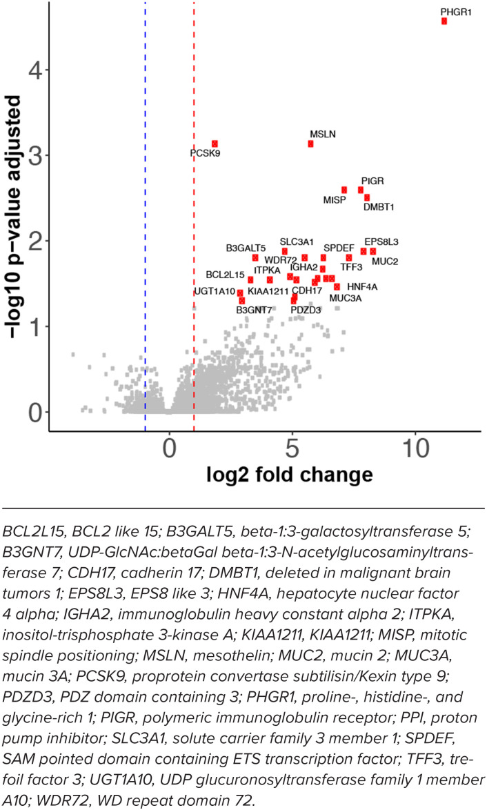
As compared with the PPI− group, 27 genes were significantly upregulated in the PPI+ group. Proline-, histidine-, and glycine-rich protein 1 (PHGR1), mucin glycoprotein genes (MUC2, MUC3A), deleted in malignant brain tumors 1 (DMBT1), and polymeric immunoglobulin receptor (PIGR) were among the most highly upregulated genes. Proprotein convertase subtilisin/kexin type 9 (PCSK9), which is a regulator of low-density lipoprotein cholesterol clearance, was slightly but not statistically significantly upregulated in the patients receiving PPI. No genes were significantly downregulated (Figure 1 and Supplemental Table S2). The deconvolution analysis revealed that cell type composition was not significantly different between the PPI− and PPI+ groups, indicating that the observed gene expression changes could not be readily explained by a difference in proportion of cell types between the 2 groups of patients (Supplemental Figure).
Figure 1.
Volcano plot showing differentially expressed host genes between children on PPI and children not on PPI. We used a threshold of log2 fold change >1 and adjusted p < 0.05 to call the genes that are upregulated or downregulated. The upregulated genes that satisfy the threshold are shown in red dots.
BCL2L15, BCL2 like 15; B3GALT5, beta-1:3-galactosyltransferase 5; B3GNT7, UDP-GlcNAc:betaGal beta-1:3-N-acetylglucosaminyltransferase 7; CDH17, cadherin 17; DMBT1, deleted in malignant brain tumors 1; EPS8L3, EPS8 like 3; HNF4A, hepatocyte nuclear factor 4 alpha; IGHA2, immunoglobulin heavy constant alpha 2; ITPKA, inositol-trisphosphate 3-kinase A; KIAA1211, KIAA1211; MISP, mitotic spindle positioning; MSLN, mesothelin; MUC2, mucin 2; MUC3A, mucin 3A; PCSK9, proprotein convertase subtilisin/Kexin type 9; PDZD3, PDZ domain containing 3; PHGR1, proline-, histidine-, and glycine-rich 1; PIGR, polymeric immunoglobulin receptor; PPI, proton pump inhibitor; SLC3A1, solute carrier family 3 member 1; SPDEF, SAM pointed domain containing ETS transcription factor; TFF3, trefoil factor 3; UGT1A10, UDP glucuronosyltransferase family 1 member A10; WDR72, WD repeat domain 72.
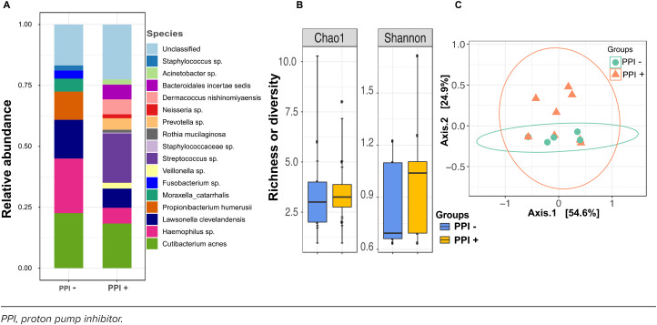
The esophageal microbiome of both PPI+ and PPI− groups was dominated by Cutibacterium acnes and members of Streptococcus and Haemophilus species. Interestingly, the mean abundance of Streptococcus sp was significantly higher in the PPI+ group than the PPI− group (20% vs 0%; p = 0.053, Kruskal-Wallis). At the species level, there were no significant differences between PPI+ and PPI− for the alpha (Shannon and Chao1 indices) and beta (Bray-Curtis dissimilarities) diversity as well as for the overall community composition (Figure 2).
Figure 2.
(A) A color-coded bar plot shows the average relative abundance of all microbial taxa that could be identified in the esophagus in PPI− and PPI+ groups. (B) Richness and alpha diversity of the esophagus microbiome. Alpha diversity and richness (measured by Shannon and S.Chao1 index) are compared between the PPI− and PPI+ groups. Differences in alpha diversity between the groups were not significant. (C) A principal coordinate analysis plot of Bray-Curtis dissimilarities (beta diversity) over the first two-axis is shown. Dots represent individual data points, and the color represents PPI+ and PPI− groups. Overall, the microbiome community composition was not significantly dissimilar among the PPI+ and PPI− groups.
PPI, proton pump inhibitor.
Discussion
Using metatranscriptomics and a gene-signature–based method to enumerate cell subsets from transcriptomes, we found that in normal pediatric esophagus PPI exposure can be associated with esophageal mucosal changes in a cell-type–independent manner and with altered local microbiome. This study provides original insights into the association between PPI use and the esophageal mucosal molecular mechanisms and local microbiome. Taken together our results hold promise to enhance our understanding of the complex community behavior of the microbiome in esophagus and can also inform the use of PPIs in pediatric practice.
Previously, PPIs have been shown to curtail transcriptomic processes involved in cellular proliferation and interleukin (IL)-13–induced responses in esophageal cell culture experiments.15 In GERD and EoE, omeprazole has been shown to inhibit the expression of eotaxin-3 (an eosinophil chemoattractant, also known as C-C motif chemokine ligand or CCL26) in the esophageal epithelium.16 Our results indicate that in endoscopically and histologically normal pediatric esophagus, PPIs induce genes involved in O-linked glycosylation of mucins (MUC2, MUC3A) and B3GNT7. Transcription of mucin genes is important in attenuating effects of noxious stimuli in the GI tract.17 A recent study using human-derived enteroids demonstrated that an increased expression of B3GNT7 alone is sufficient to promote the augmented display of Lex-decorated carbohydrate glycan structures primarily on O-glycosylated intestinal epithelial glycoproteins.18 Taken together, these genes play an important role in enhancing protective mucus secretion and epithelial cell differentiation.19,20 Genes involved in vesicular transport and metabolic processes (PHGR1),21 regulation of beta-cell development (HNF4A), and cell-cell junction organization (CDH17) are also upregulated in children exposed to PPIs.
In a setting of altered transcriptional landscape, studying the contribution of individual cell types to the gene expression patterns is pivotal because cells may behave differently depending on microenvironment.22 We used deconvolution of bulk RNA-sequencing data and assessed the proportion of 64 immune and stromal cell types and found no differences between PPI+ and PPI− subjects. These results suggest that the differences in esophageal epithelial gene expression upon exposure to PPI is unlikely to be driven by changes in the proportion of specific cell types. To investigate this further, a more focused approach, such as single cell–RNA sequencing, might be required.
With regard to microbiome, PPI exposure has been shown to decrease Comamondadaceae and increase Clostridiaceae and Micrococcaceae in esophageal biopsy samples obtained from healthy adults.23,24 We found that PPI exposure was associated with increased abundance of Streptococcus sp without any notable difference in the richness, diversity, or overall community composition when compared with those who were PPI naïve. While the significance of these differences remains to be investigated, we postulate that these differences may be related to the pediatric age and the type, duration, and dose of PPI.25
The small sample size, wide range for PPI exposure, and cross-sectional design are major limitations of our study and limit the generalizability of our results and our ability to make any causal inferences. Next, we did not have dietary modification data, which could confound our findings. Despite these limitations our study has multiple strengths. Given how often PPIs are used to manage upper GI symptoms in children,26 understanding their effect on the esophagus is an important scientific pursuit. By including children, we were able to eliminate several confounders that can be relatively more common in adults (e.g., multiple medications). To the best of our knowledge, this is the first study to apply metatranscriptomics optimized for low-bacterial-load human samples and a validated gene-signature–based method to enumerate cell subsets from transcriptomes to interrogate the contribution of individual cell types to the gene expression patterns.
Adequately designed studies in the future can allow validation of our findings and investigate the causal effect of PPIs on the normal esophageal epithelium and microbial communities, and examine if these vary by PPI type, duration of exposure, or dose.
Supplementary Material
Acknowledgments.
Authors acknowledge Regina Tyree for her assistance with this project. Preliminary results were presented at the American College of Gastroenterology Meeting in Charlotte, NC, on October 21–26. Funding: SVR, GH, and SRD are supported by R21AI168832. YAC is supported by the US Department of Veterans Affairs Office of Medical Research (IK2BX004648). SRD is supported by funds from the Centers for Disease Control and Prevention (75D3012110094), the National Institute of Allergy and Infectious Diseases (R21AI142321, R21AI154016, and R21AI149262), and the Vanderbilt Technologies for Advanced Genomics Core (grant support from the National Institutes of Health under award numbers UL1RR024975, P30CA68485, P30EY08126, and G20RR030956). JJ is supported by The Academy Ter Meulen Grant from the Royal Netherlands Academy of Arts and Sciences and the Cultural Foundation Grant from Prince Bernhard Cultural Foundation. JAG is supported by National Institute of Diabetes and Digestive and Kidney Diseases (R03DK123489). GH is supported by the American College of Gastroenterology Junior Faculty Development Award, 2021 American College of Gastroenterology Clinical Research Award. The contents are solely the responsibility of authors and do not necessarily represent official views of the funding agencies.
ABBREVIATIONS
- DBMT1
deleted in malignant brain tumors 1;
- EGD
esophagogastroduodenoscopy;
- EoE
eosinophilic esophagitis;
- GERD
gastroesophageal reflux disease;
- GI
gastrointestinal;
- mOTU
marker gene–based operational taxonomic unit;
- MUC
mucin glycoprotein genes;
- PCSK9
proprotein convertase subtilisin/kexin type 9;
- PHGR1
proline-, histidine-, and glycine-rich protein 1;
- PIGR
polymeric immunoglobulin receptor;
- PPI
proton pump inhibitor
Footnotes
Disclosures. SRD is one of the advisory board members of Premas Biotech, and consultant to Jansen and Jansen. GH serves as a consultant to Allakos, Bristol Myer Squibb, Regeneron, and Sanofi. He has received speaker fees from Bristol Myer Squibb. All other authors declare that the research was conducted in the absence of any commercial or financial relationships that could be construed as a potential conflict of interest.
Ethical Approval and Informed Consent. The authors assert that all procedures contributing to this work comply with the ethical standards of the relevant national guidelines on human experimentation and have been approved by the Institutional Review Board at Vanderbilt University. All patients and/or parents/caregiver(s) provided written informed consent and/or assent (as applicable) at enrollment.
Supplemental Material. DOI: 10.5863/1551-6776-28.6.504.S1F
DOI: 10.5863/1551-6776-28.6.504.S1T
DOI: 10.5863/1551-6776-28.6.504.S2T
References
- 1.Katz PO, Dunbar KB, Schnoll-Sussman FH et al. ACG Clinical Guideline for the Diagnosis and Management of Gastroesophageal Reflux Disease. Am J Gastroenterol . 2022;117(1):27–56. doi: 10.14309/ajg.0000000000001538. [DOI] [PMC free article] [PubMed] [Google Scholar]
- 2.Shoda T, Matsuda A, Nomura I et al. Eosinophilic esophagitis versus proton pump inhibitor–responsive esophageal eosinophilia: transcriptome analysis. J Allergy Clin Immunol . 2017;139(6):2010–2013. doi: 10.1016/j.jaci.2016.11.028. [DOI] [PubMed] [Google Scholar]
- 3.Park CH, Lee SK. Exploring esophageal microbiomes in esophageal diseases: a systematic review. J Neurogastroenterol Motil . 2020;26(2):171–179. doi: 10.5056/jnm19240. [DOI] [PMC free article] [PubMed] [Google Scholar]
- 4.Prager JD. Empirical proton pump inhibitor therapy in children. JAMA Otolaryngol Head Neck Surg . 2018;144(12):1124–1125. doi: 10.1001/jamaoto.2018.1952. [DOI] [PubMed] [Google Scholar]
- 5.Poddar U. Gastroesophageal reflux disease (GERD) in children. Paediatr Int Child Health . 2019;39(1):7–12. doi: 10.1080/20469047.2018.1489649. [DOI] [PubMed] [Google Scholar]
- 6.Hiremath G, Locke A, Thomas G et al. Novel insights into tissue-specific biochemical alterations in pediatric eosinophilic esophagitis using Raman spectroscopy. Clin Transl Gastroenterol . 2020;11(7):1–10. doi: 10.14309/ctg.0000000000000195. [DOI] [PMC free article] [PubMed] [Google Scholar]
- 7.Hiremath G, Shilts MH, Boone HH et al. The salivary microbiome is altered in children with eosinophilic esophagitis and correlates with disease activity. Clin Transl Gastroenterol . 2019;10(6):e1–e9. doi: 10.14309/ctg.0000000000000039. [DOI] [PMC free article] [PubMed] [Google Scholar]
- 8.Rajagopala SV, Bakhoum NG, Pakala SB et al. Metatranscriptomics to characterize respiratory virome, microbiome, and host response directly from clinical samples. Cell Rep Methods . 2021;1(6):1–13. doi: 10.1016/j.crmeth.2021.100091. [DOI] [PMC free article] [PubMed] [Google Scholar]
- 9.Milanese A, Mende DR, Paoli L et al. Microbial abundance, activity and population genomic profiling with mOTUs2. Nat Commun . 2019;10(1):1–11. doi: 10.1038/s41467-019-08844-4. [DOI] [PMC free article] [PubMed] [Google Scholar]
- 10.Kruskal WH, Wallis WA. Use of ranks in one-criterion variance analysis. J Am Stat Assoc . 1952;47(260):583–621. [Google Scholar]
- 11.McMurdie PJ, Holmes S. Phyloseq: an R package for reproducible interactive analysis and graphics of microbiome census data. PLoS One . 2013;8(4):e61217. doi: 10.1371/journal.pone.0061217. [DOI] [PMC free article] [PubMed] [Google Scholar]
- 12.Aran D, Hu Z, Butte AJ. xCell: digitally portraying the tissue cellular heterogeneity landscape. Genome Biol . 2017;18(1):220–234. doi: 10.1186/s13059-017-1349-1. [DOI] [PMC free article] [PubMed] [Google Scholar]
- 13.Aran D. Cell-type enrichment analysis of bulk transcriptomes using xcell. In: Methods in Molecular Biology. 2020;2120:263–276. doi: 10.1007/978-1-0716-0327-7_19. [DOI] [PubMed] [Google Scholar]
- 14.Gu Z, Hübschmann D. Make interactive complex heatmaps in R. Bioinformatics . 2022;38(5):1460–1462. doi: 10.1093/bioinformatics/btab806. [DOI] [PMC free article] [PubMed] [Google Scholar]
- 15.Rochman M, Xie YM, Mack L et al. Broad transcriptional response of the human esophageal epithelium to proton pump inhibitors. J Allergy Clin Immunol . 2021;147(5):1924–1935. doi: 10.1016/j.jaci.2020.09.039. [DOI] [PMC free article] [PubMed] [Google Scholar]
- 16.Cheng E, Zhang X, Huo X et al. Omeprazole blocks eotaxin-3 expression by oesophageal squamous cells from patients with eosinophilic oesophagitis and GORD. Gut . 2013;62(6):824–832. doi: 10.1136/gutjnl-2012-302250. [DOI] [PMC free article] [PubMed] [Google Scholar]
- 17.Corfield AP. Mucins in the gastrointestinal tract in health and disease. Front Biosci . 2001;6(1):D1321–D1357. doi: 10.2741/corfield. [DOI] [PubMed] [Google Scholar]
- 18.Carroll DJ, Burns MWN, Mottram L et al. Interleukin-22 regulates B3GNT7 expression to induce fucosylation of glycoproteins in intestinal epithelial cells. J Biol Chem . 2022;298(2):101463. doi: 10.1016/j.jbc.2021.101463. [DOI] [PMC free article] [PubMed] [Google Scholar]
- 19.Sarosiek I, Olyaee M, Majewski M et al. Significant increase of esophageal mucin secretion in patients with reflux esophagitis after healing with rabeprazole: its esophagoprotective potential. Dig Dis Sci . 2009;54(10):2137–2142. doi: 10.1007/s10620-008-0589-z. [DOI] [PubMed] [Google Scholar]
- 20.Guillem P, Billeret V, Buisine MP et al. Mucin gene expression and cell differentiation in human normal, premalignant and malignant esophagus. Int J Cancer . 2000;88(6):856–861. doi: 10.1002/1097-0215(20001215)88:6<856::aid-ijc3>3.0.co;2-d. [DOI] [PubMed] [Google Scholar]
- 21.Oltedal S, Skaland I, Maple-Grødem J et al. Expression profiling and intracellular localization studies of the novel Proline-, Histidine-, and Glycine-rich protein 1 suggest an essential role in gastro-intestinal epithelium and a potential clinical application in colorectal cancer diagnostics. BMC Gastroenterol . 2018;18(1):26–41. doi: 10.1186/s12876-018-0752-8. [DOI] [PMC free article] [PubMed] [Google Scholar]
- 22.Jin H, Liu Z. A benchmark for RNA-seq deconvolution analysis under dynamic testing environments. Genome Biol . 2021;22(1):102–125. doi: 10.1186/s13059-021-02290-6. [DOI] [PMC free article] [PubMed] [Google Scholar]
- 23.Amir I, Konikoff FM, Oppenheim M et al. Gastric microbiota is altered in oesophagitis and Barrett’s oesophagus and further modified by proton pump inhibitors. Environ Microbiol . 2014;16(9):2905–2914. doi: 10.1111/1462-2920.12285. [DOI] [PubMed] [Google Scholar]
- 24.Tasnim S, Miller AL, Jupiter DC et al. Effects of proton pump inhibitor use on the esophageal microbial community. BMC Gastroenterol . 2020;20(1):312–321. doi: 10.1186/s12876-020-01460-3. [DOI] [PMC free article] [PubMed] [Google Scholar]
- 25.Deshpande NP, Riordan SM, Castaño-Rodríguez N et al. Signatures within the esophageal microbiome are associated with host genetics, age, and disease. Microbiome . 2018;6(1):227–241. doi: 10.1186/s40168-018-0611-4. [DOI] [PMC free article] [PubMed] [Google Scholar]
- 26.Arnoux A, Bailhache M, Tetard C et al. Proton pump inhibitors are still overprescribed for hospitalized children. Arch Pediatr . 2022;29(4):258–262. doi: 10.1016/j.arcped.2022.02.004. [DOI] [PubMed] [Google Scholar]
Associated Data
This section collects any data citations, data availability statements, or supplementary materials included in this article.


