Abstract
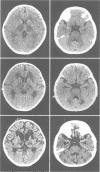
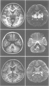
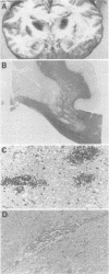
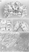
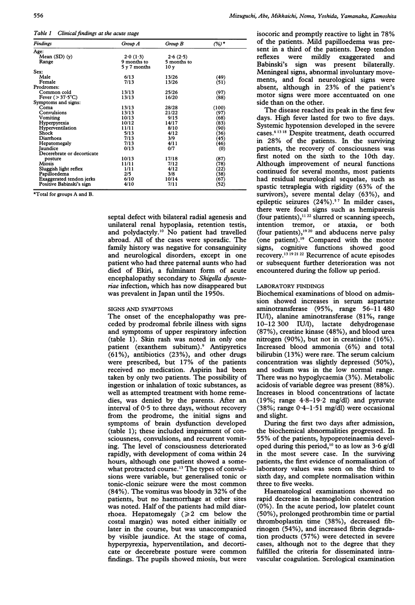
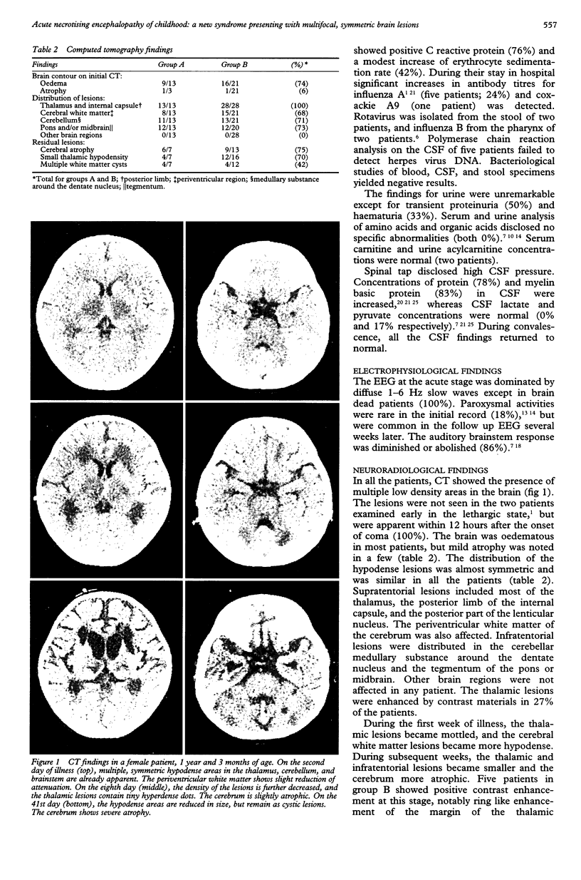
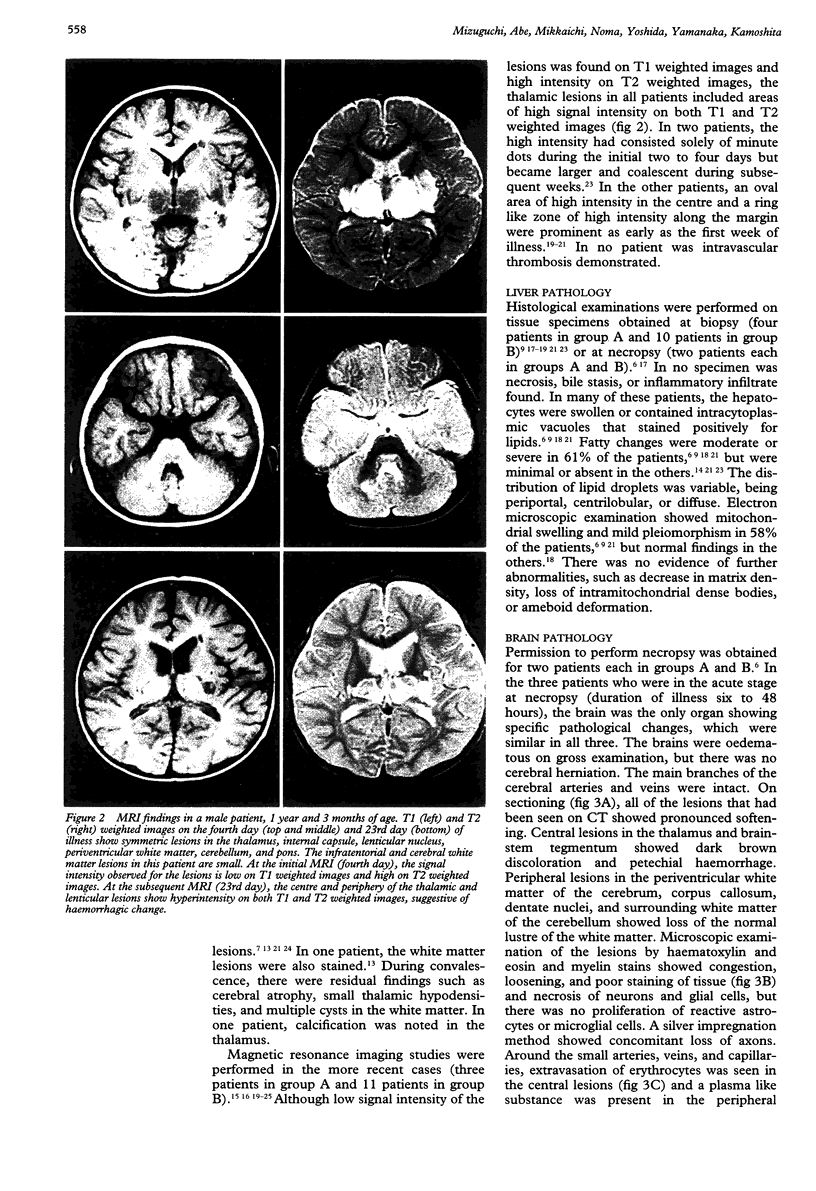
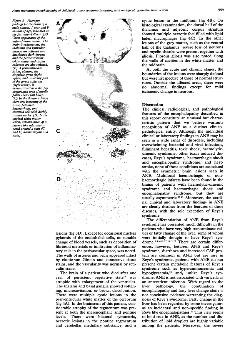
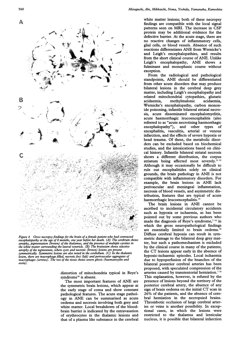
The clinicopathological features of a previously unrecognised type of acute encephalopathy prevalent among Japanese children is described by reviewing the records of 13 consecutive patients treated and 28 previously reported cases. The hallmark of this encephalopathy, proposed to be a novel entity termed acute necrotising encephalopathy of childhood, is multiple, necrotic brain lesions showing a symmetric distribution. The encephalopathy was noted in previously healthy children after respiratory tract infections, with presenting symptoms of coma, convulsions, vomiting, hyperpyrexia, and hepatomegaly. Laboratory examinations disclosed liver dysfunction, uraemia, and hypoproteinaemia. The histological appearance of the liver was variable and non-specific. Cerebrospinal fluid contained an increased amount of protein. Computed tomography and MRI showed the presence of symmetrically distributed brain lesions of the thalamus, cerebral white matter, brainstem, and cerebellum. Necropsy examination confirmed extensive fresh necrosis of these regions with evidence of local breakdown of the blood-brain barrier. Based on the characteristic combination of clinical and pathological findings, acute necrotising encephalopathy of childhood can be distinguished from previously known encephalopathies, including Reye's syndrome.
Full text
PDF
Images in this article
Selected References
These references are in PubMed. This may not be the complete list of references from this article.
- ADAMS R. D., KUBIK C. S. The morbid anatomy of the demyelinative disease. Am J Med. 1952 May;12(5):510–546. doi: 10.1016/0002-9343(52)90234-9. [DOI] [PubMed] [Google Scholar]
- Aoki N. Acute toxic encephalopathy with symmetrical low density areas in the thalami and the cerebellum. Childs Nerv Syst. 1985;1(1):62–65. doi: 10.1007/BF00706733. [DOI] [PubMed] [Google Scholar]
- Aoki N., Kaneshi K., Mizuguchi M., Kurihara E. [Computerized tomography in acute toxic encephalopathy--report of 3 cases with symmetrical low density areas in the thalamus and the cerebellum]. No To Hattatsu. 1983 Jul;15(4):345–349. [PubMed] [Google Scholar]
- Barkovich A. J., Kjos B. O., Jackson D. E., Jr, Norman D. Normal maturation of the neonatal and infant brain: MR imaging at 1.5 T. Radiology. 1988 Jan;166(1 Pt 1):173–180. doi: 10.1148/radiology.166.1.3336675. [DOI] [PubMed] [Google Scholar]
- Corey L., Rubin R. J., Bregman D., Gregg M. B. Diagnostic criteria for influenza B-associated Reye's syndrome: clinical vs. pathologic criteria. Pediatrics. 1977 Nov;60(5):702–708. [PubMed] [Google Scholar]
- Cullity G. J., Kakulas B. A. Encephalopathy and fatty degeneration of the viscera: an evaluation. Brain. 1970;93(1):77–88. doi: 10.1093/brain/93.1.77. [DOI] [PubMed] [Google Scholar]
- Evans H., Bourgeois C. H., Comer D. S., Keschamras N. Brain lesions in Reye's syndrome. Arch Pathol. 1970 Dec;90(6):543–546. [PubMed] [Google Scholar]
- Goutières F., Aicardi J. Acute neurological dysfunction associated with destructive lesions of the basal ganglia in children. Ann Neurol. 1982 Oct;12(4):328–332. doi: 10.1002/ana.410120403. [DOI] [PubMed] [Google Scholar]
- Hino T., Sai H., Morikawa Y., Mizuta R., Okuno T. [A case of clinical Reye's syndrome presenting characteristic CT changes]. No To Hattatsu. 1984;16(3):210–217. [PubMed] [Google Scholar]
- Iai M., Tanabe Y., Goto M. [A case of acute encephalopathy with symmetrical low density areas in the thalami on CT; serial CT and MRI findings]. No To Hattatsu. 1992 Jul;24(4):370–374. [PubMed] [Google Scholar]
- Mizuguchi M., Tomonaga M., Fukusato T., Asano M. Acute necrotizing encephalopathy with widespread edematous lesions of symmetrical distribution. Acta Neuropathol. 1989;78(1):108–111. doi: 10.1007/BF00687411. [DOI] [PubMed] [Google Scholar]
- Nagai T., Yagishita A., Tsuchiya Y., Asamura S., Kurokawa H., Matsuo N. Symmetrical thalamic lesions on CT in influenza A virus infection presenting with or without Reye syndrome. Brain Dev. 1993 Jan-Feb;15(1):67–73. doi: 10.1016/0387-7604(93)90009-w. [DOI] [PubMed] [Google Scholar]
- Partin J. C., Schubert W. K., Partin J. S. Mitochondrial ultrastructure in Reye's syndrome (encephalopathy and fatty degeneration of the viscera). N Engl J Med. 1971 Dec 9;285(24):1339–1343. doi: 10.1056/NEJM197112092852402. [DOI] [PubMed] [Google Scholar]
- Sunaga Y., Fujinaga T., Tamura H. [A study on computed tomography of three cases of Reye syndromes]. No To Hattatsu. 1991 Jan;23(1):100–102. [PubMed] [Google Scholar]
- Takano T., Matsui E., Yamano T., Shimada M., Okumura K. [A case of clinical Reye syndrome with symmetrical abnormal signal areas in the pons and thalami by MRI]. No To Hattatsu. 1994 Jan;26(1):63–67. [PubMed] [Google Scholar]
- Tanaka J. [Developmental changes in electrically elicited blink reflex in infancy and childhood]. No To Hattatsu. 1989 May;21(3):271–277. [PubMed] [Google Scholar]
- Tateno A., Sakai K., Sakai S., Koya N., Aoki T. Computed tomography of bilateral thalamic hypodensity in acute encephalopathy. J Comput Assist Tomogr. 1988 Jul-Aug;12(4):637–639. doi: 10.1097/00004728-198807000-00020. [DOI] [PubMed] [Google Scholar]
- Vles J. S., de Vries L. S., Wilms G., de Roo M., Casaer P. J. Computed cranial tomography, magnetic resonance imaging and single photon emission computed tomography in hemorrhagic shock and encephalopathy syndrome: a report of three cases. Neuropediatrics. 1992 Feb;23(1):24–27. doi: 10.1055/s-2008-1071306. [DOI] [PubMed] [Google Scholar]


