Abstract
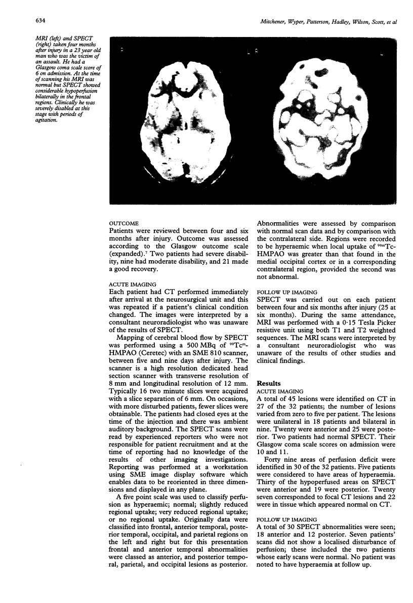
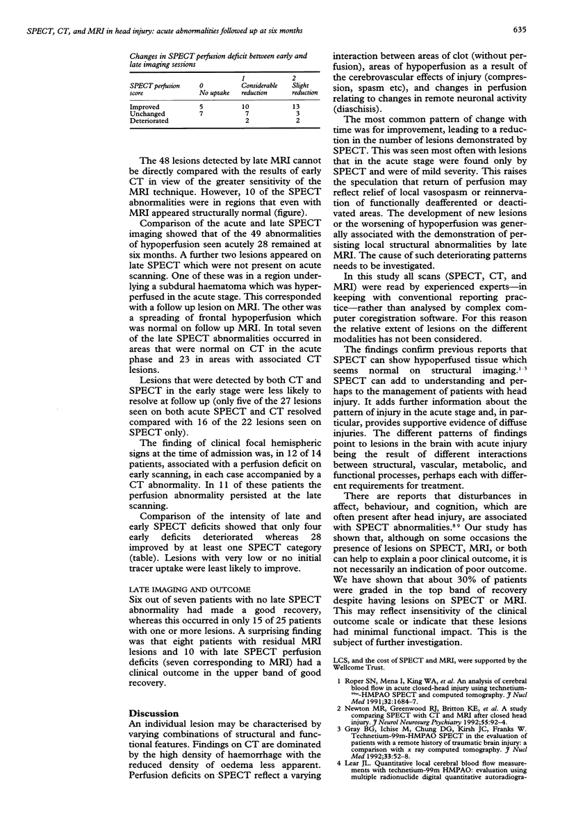
Neuroimaging with single photon emission computed tomography (SPECT) using the cerebral blood flow tracer 99Tc(m)-HMPAO has been used to study acute functional alterations after head injury and residual abnormalities at six month follow up in 32 patients. Comparison has been made with anatomical abnormalities defined acutely with CT and on follow up with MRI. SPECT showed slightly more abnormalities than CT in the acute phase (49 regions of abnormally low tracer uptake on SPECT and 45 lesions on CT). Twenty two of the acute SPECT abnormalities were in normal regions on CT. At follow up MRI showed more abnormalities than SPECT (30 on SPECT and 48 on MRI). Ten of the SPECT deficits were in regions with normal MRI. Comparison of the intensity of late and early SPECT deficits showed that only four early deficits deteriorated whereas 28 improved. Only five of 27 lesions seen on both acute SPECT and CT resolved compared with 16 of 22 lesions seen on SPECT but not on CT. Regions of abnormally high tracer uptake were detected in the acute stage in five of the patients. No high focal uptake was evident on follow up. Ten patients with a residual SPECT abnormality and eight with residual MRI lesions were graded clinically in the upper band of good recovery.
Full text
PDF


Images in this article
Selected References
These references are in PubMed. This may not be the complete list of references from this article.
- Bullock R., Patterson J., Park C. Evaluation of 99mTc-hexamethylpropyleneamine oxime cerebral blood flow mapping after acute focal ischemia in rats. Stroke. 1991 Oct;22(10):1284–1290. doi: 10.1161/01.str.22.10.1284. [DOI] [PubMed] [Google Scholar]
- Goldenberg G., Oder W., Spatt J., Podreka I. Cerebral correlates of disturbed executive function and memory in survivors of severe closed head injury: a SPECT study. J Neurol Neurosurg Psychiatry. 1992 May;55(5):362–368. doi: 10.1136/jnnp.55.5.362. [DOI] [PMC free article] [PubMed] [Google Scholar]
- Jennett B., Snoek J., Bond M. R., Brooks N. Disability after severe head injury: observations on the use of the Glasgow Outcome Scale. J Neurol Neurosurg Psychiatry. 1981 Apr;44(4):285–293. doi: 10.1136/jnnp.44.4.285. [DOI] [PMC free article] [PubMed] [Google Scholar]
- Newton M. R., Greenwood R. J., Britton K. E., Charlesworth M., Nimmon C. C., Carroll M. J., Dolke G. A study comparing SPECT with CT and MRI after closed head injury. J Neurol Neurosurg Psychiatry. 1992 Feb;55(2):92–94. doi: 10.1136/jnnp.55.2.92. [DOI] [PMC free article] [PubMed] [Google Scholar]
- Oder W., Goldenberg G., Spatt J., Podreka I., Binder H., Deecke L. Behavioural and psychosocial sequelae of severe closed head injury and regional cerebral blood flow: a SPECT study. J Neurol Neurosurg Psychiatry. 1992 Jun;55(6):475–480. doi: 10.1136/jnnp.55.6.475. [DOI] [PMC free article] [PubMed] [Google Scholar]
- Roper S. N., Mena I., King W. A., Schweitzer J., Garrett K., Mehringer C. M., McBride D. An analysis of cerebral blood flow in acute closed-head injury using technetium-99m-HMPAO SPECT and computed tomography. J Nucl Med. 1991 Sep;32(9):1684–1687. [PubMed] [Google Scholar]
- Teasdale G., Jennett B. Assessment of coma and impaired consciousness. A practical scale. Lancet. 1974 Jul 13;2(7872):81–84. doi: 10.1016/s0140-6736(74)91639-0. [DOI] [PubMed] [Google Scholar]


