Persoonia : Molecular Phylogeny and Evolution of Fungi logoLink to Persoonia : Molecular Phylogeny and Evolution of Fungi
. 2022 Jul 12;48:261–371. doi: 10.3767/persoonia.2023.48.08

Fungal Planet description sheets: 1383–1435

PW Crous 1,2, J Boers 3, D Holdom 4, ER Osieck 5, TV Steinrucken 6, YP Tan 7, JS Vitelli 4, RG Shivas 8, M Barrett 9, A-G Boxshall 10, J Broadbridge 11, E Larsson 12, T Lebel 13, U Pinruan 14, S Sommai 14, P Alvarado 15, G Bonito 16, CA Decock 17, S De la Peña-Lastra 18, G Delgado 19, J Houbraken 1, JG Maciá-Vicente 20,21, HA Raja 22, A Rigueiro-Rodríguez 18, A Rodríguez 23, MJ Wingfield 2, SJ Adams 24, A Akulov 25, T AL-Hidmi 8, V Antonín 26, S Arauzo 27, F Arenas 23, F Armada 28, J Aylward 2, J-M Bellanger 29, A Berraf-Tebbal 30, A Bidaud 31, F Boccardo 32, J Cabero 33, F Calledda 34, G Corriol 35, JL Crane 36, JDW Dearnaley 8, B Dima 37, F Dovana 38, A Eichmeier 30, F Esteve-Raventós 39, M Fine 40, L Ganzert 41, D García 42, D Torres-Garcia 42, J Gené 42, A Gutiérrez 23, P Iglesias 27, Ł Istel 1, P Jangsantear 43, GM Jansen 44, M Jeppson 12, NC Karun 45, A Karich 46, P Khamsuntorn 14, K Kokkonen 47, M Kolařík 48, A Kubátová 49, R Labuda 50, AC Lagashetti 51, N Lifshitz 52, C Linde 53, M Loizides 54, JJ Luangsa-ard 14, P Lueangjaroenkit 55, S Mahadevakumar 56, AE Mahamedi 57, DW Malloch 58, S Marincowitz 2, A Mateos 59, P-A Moreau 60, AN Miller 36, A Molia 61, A Morte 23, A Navarro-Ródenas 23, J Nebesářová 62, E Nigrone 63, BR Nuthan 64, NH Oberlies 22, AL Pepori 63, T Rämä 41, D Rapley 4, K Reschke 65, BM Robicheau 24,66, F Roets 67, J Roux 68, M Saavedra 69, B Sakolrak 43, A Santini 63, H Ševčíková 26, PN Singh 51, SK Singh 51, S Somrithipol 14, M Spetik 30, KR Sridhar 45, M Starink-Willemse 1, VA Taylor 24,70, AL van Iperen 1, J Vauras 71, AK Walker 24, BD Wingfield 2, O Yarden 52, AW Cooke 72, AG Manners 72, KG Pegg 72, JZ Groenewald 1
PMCID: PMC10792288  PMID: 38234686

Abstract

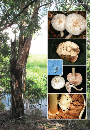
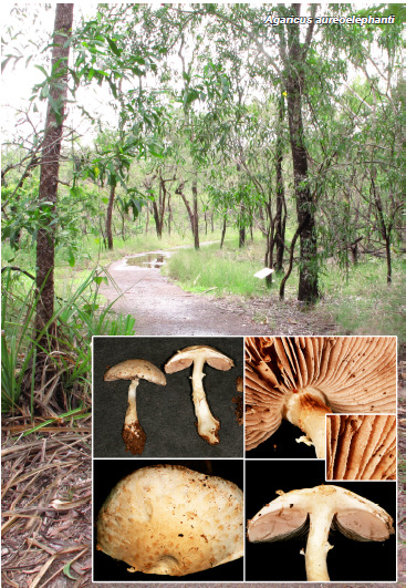
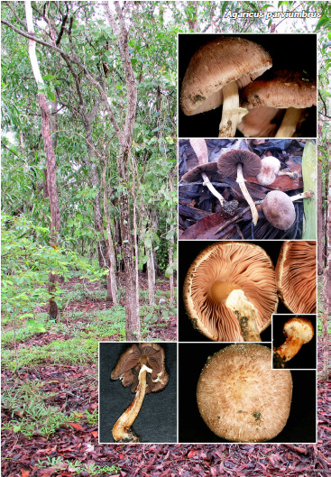
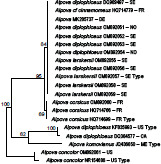
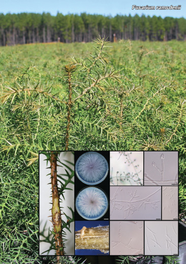
Novel species of fungi described in this study include those from various countries as follows: Australia, Agaricus albofoetidus, Agaricus aureoelephanti and Agaricus parviumbrus on soil, Fusarium ramsdenii from stem cankers of Araucaria cunninghamii, Keissleriella sporoboli from stem of Sporobolus natalensis, Leptosphaerulina queenslandica and Pestalotiopsis chiaroscuro from leaves of Sporobolus natalensis, Serendipita petricolae as endophyte from roots of Eriochilus petricola, Stagonospora tauntonensis from stem of Sporobolus natalensis, Teratosphaeria carnegiei from leaves of Eucalyptus grandis × E. camaldulensis and Wongia ficherai from roots of Eragrostis curvula. Canada, Lulworthia fundyensis from intertidal wood and Newbrunswickomyces abietophilus (incl. Newbrunswickomyces gen. nov.) on buds of Abies balsamea. Czech Republic, Geosmithia funiculosa from a bark beetle gallery on Ulmus minor and Neoherpotrichiella juglandicola (incl. Neoherpotrichiella gen. nov.) from wood of Juglans regia. France, Aspergillus rouenensis and Neoacrodontium gallica (incl. Neoacrodontium gen. nov.) from bore dust of Xestobium rufovillosum feeding on Quercus wood, Endoradiciella communis (incl. Endoradiciella gen. nov.) endophytic in roots of Microthlaspi perfoliatum and Entoloma simulans on soil. India, Amanita konajensis on soil and Keithomyces indicus from soil. Israel, Microascus rothbergiorum from Stylophora pistillata. Italy, Calonarius ligusticus on soil. Netherlands, Appendopyricularia juncicola (incl. Appendopyricularia gen. nov.), Eriospora juncicola and Tetraploa juncicola on dead culms of Juncus effusus, Gonatophragmium physciae on Physcia caesia and Paracosmospora physciae (incl. Paracosmospora gen. nov.) on Physcia tenella, Myrmecridium phragmitigenum on dead culm of Phragmites australis, Neochalara lolae on stems of Pteridium aquilinum, Niesslia nieuwwulvenica on dead culm of undetermined Poaceae, Nothodevriesia narthecii (incl. Nothodevriesia gen. nov.) on dead leaves of Narthecium ossifragum and Parastenospora pini (incl. Parastenospora gen. nov.) on dead twigs of Pinus sylvestris. Norway, Verticillium bjoernoeyanum from sand grains attached to a piece of driftwood on a sandy beach. Portugal, Collybiopsis cimrmanii on the base of living Quercus ilex and amongst dead leaves of Laurus and herbs. South Africa, Paraproliferophorum hyphaenes (incl. Paraproliferophorum gen. nov.) on living leaves of Hyphaene sp. and Saccothecium widdringtoniae on twigs of Widdringtonia wallichii. Spain, Cortinarius dryosalor on soil, Cyphellophora endoradicis endophytic in roots of Microthlaspi perfoliatum, Geoglossum lauri­silvae on soil, Leptographium gemmatum from fluvial sediments, Physalacria auricularioides from a dead twig of Castanea sativa, Terfezia bertae and Tuber davidlopezii in soil. Sweden, Alpova larskersii, Inocybe alpestris and Inocybe boreogodeyi on soil. Thailand, Russula banwatchanensis, Russula purpureoviridis and Russula lilacina on soil. Ukraine, Nectriella adonidis on overwintered stems of Adonis vernalis. USA, Microcyclus jacquiniae from living leaves of Jacquinia keyensis and Penicillium neoherquei from a minute mushroom sporocarp. Morphological and culture characteristics are supported by DNA barcodes.

Citation: Crous PW, Boers J, Holdom D, et al. 2022. Fungal Planet description sheets: 1383–1435. Persoonia 48: 261–371. https://doi.org/10.3767/persoonia.2022.48.08.

Keywords: ITS nrDNA barcodes, LSU, new taxa, systematics

Acknowledgments

Teresa Lebel and colleagues acknowledge the Royal Botanic Gardens Victoria and the Friends of the Royal Botanic Gardens Victoria for funding and support for Northern Territory field and laboratory work; Northern Territory Herbarium staff for space and laboratory access and help with collection curation; State Herbarium of South Australia for support of the undergraduate intern J. Broadbridge. This research was also supported through funding from Australian Biological Resources Study grant (4-EHOJ161) to the State Herbarium of South Australia. Ellen Larsson acknowledges the Swedish Taxonomy Initiative, SLU, Artdatabanken, Uppsala (dha.2019.4.3-13) and the curators of herbaria O, S, UME and UPS. Fernando Esteve-Raventós acknowledges the FEDER/Ministerio de Ciencia, Innovación y Universidades – Agencia Estatal de Investigación, Spain (Project CGL2017-86540-P). Jordi Vila (Institut d’Estudis Catalans) is thanked for sending us material and a photograph of Inocybe alpestris. The studies of Hana Ševčíková and Vladimír Antonín was facilitated via institutional support of the long-term conceptual development of research institutions for the Moravian Museum provided by the Czech Ministry of Culture (ref. MK000094862). François Armada and colleagues acknowledge Clotilde & Didier Borgarino and Patrice Tanchaud for collections, and Francisco Donaire (JA-CUSSTA), as fungarium curator. Gregorio Delgado is grateful to William Colbert and Kamash Pillai (Eurofins EMLab P&K) for provision of laboratory facilities. Jose G. Maciá-Vicente acknowledges support from the German Research Foundation under grant MA7171/1-1, and from the Landes-Offensive zur Entwicklung Wissenschaftlich-ökonomischer Exzellenz (LOEWE) of the state of Hesse within the framework of the Cluster for Integrative Fungal Research (IPF). Kandikere R. Sridhar and colleagues are thankful to Prof. Sajeewa S.N. Maharachchikumbura, University of Electronic Science and Technology of China, P.R. China for facilitating phylogenetic tree construction and interpretation. Kai Reschke and colleagues thank Machiel E. Noordeloos for extensive support with their Entoloma research. The work of Bálint Dima was supported by the ELTE Thematic Excellence Programme 2020 (TKP2020-IKA-05), financed by the National Research, Development and Innovation Office of Hungary. Antonio Mateos and colleagues thank Amalio Gutiérrez for his collaboration and cooperation in the field collections in Extremadura. Ajay C. Lagashetti and colleagues thank the Director, MACS-Agharkar Research Institute, Pune, for providing necessary facilities, the CSIR, New Delhi for a Senior Research Fellowship (SRF) and S.P. Pune University for granting permission to register for a PhD degree. The study of Daniel Torres-Garcia and colleagues was partially supported by the Spanish Ministerio de Economía, Industria y Competitividad (grant CGL2017-88094-P). Lukasz Istel and co-authors thank Jan Dijksterhuis for the examination of Microascus rothbergiorum using Scanning Electron Micro­scopy. Oded Yarden and Nofar Lifshitz were funded by the Israel Science Foundation. The research of Milan Spetik and co-authors was supported by project No. IGA-ZF/2021-ST2003. Saúl De la Peña-Lastra and colleagues thank the Atlantic Islands National Maritime-Terrestrial Park authorities and guards, especially Víctor Rivas Iglesias, and also Cristóbal Burgos Morillo for his generous help in processing the images. Tracey Steinrucken and colleagues were supported by AgriFutures Australia (Rural Industries Research and Development Corporation), through funding from the Australian Government Department of Agriculture, Water and the Environment, as part of its Rural Research and Development for Profit program (PRJ-010527). Janneke Aylward and colleagues are grateful for funding received from the Department of Science and Innovation (DSI)-National Research Foundation (NRF) Centre of Excellence in Plant Health Biotechnology (CPHB), South Africa and the DST-NRF SARChI chair in Fungal Genomics. Julio Cabero acknowledges Berta Martín from the regional administration of Zamora (Spain) for supporting this work, as well as Hipólito Hernandez, environmental agent in the area of Sanabria, for sharing his knowledge about local flora and ecology. Asunción Morte is grateful to Fundación Séneca- Agencia de Ciencia y Tecnología de la Región de Murcia (20866/PI/18) for financial support. Umpawa Pinruan and colleagues were financially supported by the Platform Technology Management Section, National Center for Genetic Engineering and Biotechnology (BIOTEC), Project Grant No. P19-50231. Konstanze Bensch (Westerdijk Fungal Biodiversity Institute, Utrecht) is thanked for correcting the spelling of various Latin epithets.

REFERENCES

  1. Abdelrazek S, Choudhari S, Thimmapuram J, et al. 2020. Changes in the core endophytic mycobiome of carrot taproots in response to crop management and genotype. Scientific Reports 10: 13685. [DOI] [PMC free article] [PubMed] [Google Scholar]
  2. Agerer R. 1980. Contribution to neotropical cyphellaceous fungi. 11. Deigloria gen. nov. (Physalacriaceae). Mycotaxon 12: 185–200. [Google Scholar]
  3. Al Subeh ZY, Raja HA, Burdette JE, et al. 2021. Three diketomorpholines from a Penicillium sp. (strain G1071). Phytochemistry 189: 112830. [DOI] [PMC free article] [PubMed] [Google Scholar]
  4. Alfieri SA, Jr, Samuels GJ. 1979. Nectriella pironii and its kutilakesa-like anamorph, a parasite of ornamental shrubs. Mycologia 71: 1178–1185. [Google Scholar]
  5. Andjic V, Pegg GS, Carnegie AJ, et al. 2010. Teratosphaeria pseudoeucalypti, new cryptic species responsible for leaf blight of Eucalyptus in subtropical and tropical Australia. Plant Pathology 59: 900–912. [Google Scholar]
  6. Antonín V, Noordeloos M. 2010. A monograph of marasmioid and collybioid fungi in Europe. IHW Verlag. [Google Scholar]
  7. Arauzo S, Iglesias P. 2014. La familia Geoglossaceae ss. str. en la península Ibérica y la Macaronesia. Errotari 11: 166–259. [Google Scholar]
  8. Arya CP, Manoj Kumar A, Pradeep CK, et al. 2022. Agaricus brunneodiscus, a new species of Agaricus sect. Rarolentes from India. Phytotaxa 533: 181–193. [Google Scholar]
  9. Arzanlou M, Groenewald JZ, Gams W, et al. 2007. Phylogenetic and morphotaxonomic revision of Ramichloridium and allied genera. Studies in Mycology 58: 57–93. [DOI] [PMC free article] [PubMed] [Google Scholar]
  10. Aylward J, Havenga M, Dreyer LL, et al. 2021. Genetic diversity of Terato­sphaeria pseudoeucalypti in Eucalyptus plantations in Australia and Uruguay. Australasian Plant Pathology 50: 639–649. [Google Scholar]
  11. Azevedo E, Barata M, Marques MI, et al. 2017. Lulworthia atlantica: a new species supported by molecular phylogeny and morphological analysis. Mycologia 109: 287–295. [DOI] [PubMed] [Google Scholar]
  12. Azuddin NF, Mohd MH, Rosely NFN, et al. 2021. Molecular phylogeny of endo­phytic fungi from Rattan (Calamus castaneus Griff.) spines and their antagonistic activities against plant pathogenic fungi. Journal of Fungi 7: 301. [DOI] [PMC free article] [PubMed] [Google Scholar]
  13. Bandini D, Oertel B, Schüssler C, et al. 2020. Noch mehr Risspilze: Fünfzehn neue und zwei wenig bekannte Arten der Gattung Inocybe. Mycologia Bavarica 20: 13–101. [Google Scholar]
  14. Bao D-F, Hyde KD, McKenzie EHC, et al. 2021. Biodiversity of lignicolous freshwater hyphomycetes from China and Thailand and description of sixteen species. Journal of Fungi 7: 669. [DOI] [PMC free article] [PubMed] [Google Scholar]
  15. Barr ME. 1972. Preliminary studies on the Dothideales in temperate North America. Contributions from the University of Michigan Herbarium 9: 523–638. [Google Scholar]
  16. Bashir H, Chen J, Jabeen S, et al. 2021. An overview of Agaricus section Hondenses and Agaricus section Xanthodermatei with description of eight new species from Pakistan. Scientific Reports 11: 12905. [DOI] [PMC free article] [PubMed] [Google Scholar]
  17. Bell J, Yokoya K, Kendon JP, et al. 2020. Diversity of root-associated culturable fungi of Cephalanthera rubra (Orchidaceae) in relation to soil characteristics. PeerJ 8: e8695. [DOI] [PMC free article] [PubMed] [Google Scholar]
  18. Bellanger J-M, Moreau P-A, Corriol G, et al. 2015. Plunging hands into the mushroom jar: a phylogenetic framework for Lyophyllaceae (Agaricales, Basidiomycota). Genetica 143: 169–194. [DOI] [PubMed] [Google Scholar]
  19. Berger F, Braun U, Heuchert B. 2015. Gonatophragmium lichenophilum sp. nov. – a new lichenicolous hyphomycete from Austria. Mycobiota 5: 7–13. [Google Scholar]
  20. Berthier J. 1985. Les Physalacriaceae du Globe. Biblioteca Mycologica 98: 1–128. [Google Scholar]
  21. Bidaud A. 2011. Cortinaires rares ou nouveaux de la région Rhône-Alpes (France). Journal des J.E.C. 13: 4–24. [Google Scholar]
  22. Bonito G, Hameed K, Ventura R, et al. 2016. Isolating a functionally relevant guild of fungi from the root microbiome of Populus. Fungal Ecology 22: 35–42. [Google Scholar]
  23. Brandrud TE, Lindström H, Marklund H, et al. 1992. Cortinarius: Flora Photo­graphica. Vol. II (Swedish version). Cortinarius HB, Sweden. [Google Scholar]
  24. Braun U, Crous PW, Nakashima C. 2015. Cercosporoid fungi (Mycosphaerellaceae) 4. Species on dicots (Acanthaceae to Amaranthaceae). IMA Fungus 6: 373–469. [DOI] [PMC free article] [PubMed] [Google Scholar]
  25. Braun U, Nakashima C, Crous PW. 2013. Cercosporoid fungi (Mycosphaerellaceae) 1. Species on other fungi, Pteridophyta and Gymnospermae. IMA Fungus 4: 265–345. [DOI] [PMC free article] [PubMed] [Google Scholar]
  26. Burgess TI, Wingfield MJ. 2017. Pathogens on the move: A 100-year global experiment with planted eucalypts. Bioscience 67: 14–25. [Google Scholar]
  27. Cannon P, Carmarán C, Romero A. 1995. Studies on biotrophic fungi from Argentina: Microcyclus porlieriae, with a key to South American species of Microcyclus. Mycological Research 99: 353–356. [Google Scholar]
  28. Ceruti A, Fontana A, Nosenzo C. 2003. Le specie europee del genere Tuber: una revisione storica. Vol. 37. Turin, Italy, Museo Regionale di Scienze Naturali. [Google Scholar]
  29. Chang R, Cao W, Wang Y, et al. 2022. Melanodevriesia, a new genus of endolichenic oleaginous black yeast recovered from the Inner Mongolia Region of China. Fungal Systematics and Evolution 9: 1–9. [DOI] [PMC free article] [PubMed] [Google Scholar]
  30. Chen J, Callac P, Parra LA, et al. 2017. Study in Agaricus subgenus Minores and allied clades reveals a new American subgenus and contrasting phylo­genetic patterns in Europe and Greater Mekong Subregion. Persoonia 38: 170–196. [DOI] [PMC free article] [PubMed] [Google Scholar]
  31. Chen J, Parra L, Kesel A, et al. 2016. Inter- and intra-specific diversity in Agaricus endoxanthus and allied species reveals a new taxon, A. punjabensis. Phytotaxa 252: 1–16. [Google Scholar]
  32. Cochard H, Réaudin D. 2019. Physalacria stilboidea (Cooke) Sacc., espèce exotique nouvelle pour la France. Bulletin Mycologique et Botanique Dauphiné-Savoie 235: 11–16. [Google Scholar]
  33. Cooke MC. 1889. New Australian fungi. Grevillea 18: 1–8. [Google Scholar]
  34. Corner EJH. 1950. A monograph of Clavaria and allied genera. Annals of Botany Memoirs 1: 1–740. [Google Scholar]
  35. Cripps CL, Larsson E, Vauras J. 2020. Nodulose-spored Inocybe species from the Rocky Mountain alpine zone: molecularly linked to European type specimens. Mycologia 112: 133–153. [DOI] [PubMed] [Google Scholar]
  36. Crous PW, Cowan DA, Maggs-Colling G, et al. 2020a. Fungal Planet des­cription sheets: 1112–1181. Persoonia 45: 251–409. [DOI] [PMC free article] [PubMed] [Google Scholar]
  37. Crous PW, Cowan DA, Maggs-Colling G, et al. 2021a. Fungal Planet des­cription sheets: 1182–1283. Persoonia 46: 313–528. [DOI] [PMC free article] [PubMed] [Google Scholar]
  38. Crous PW, Groenewald JZ. 2011. Why everlastings don’t last. Persoonia 26: 70–84. [DOI] [PMC free article] [PubMed] [Google Scholar]
  39. Crous PW, Hernández-Restrepo M, Schumacher RK, et al. 2021b. New and interesting fungi 4. Fungal Systematics and Evolution 7: 255–343. [DOI] [PMC free article] [PubMed] [Google Scholar]
  40. Crous PW, Osieck ER, Jurjević Ž, et al. 2021c. Fungal Planet description sheets: 1284–1382. Persoonia 47: 178–374. [DOI] [PubMed] [Google Scholar]
  41. Crous PW, Schumacher RK, Akulov A, et al. 2019. New and interesting fungi. 2. Fungal Systematics and Evolution 3: 57–134. [DOI] [PMC free article] [PubMed] [Google Scholar]
  42. Crous PW, Shivas RG, Quaedvlieg W, et al. 2014. Fungal Planet description sheets: 214–280. Persoonia 32: 184–306. [DOI] [PMC free article] [PubMed] [Google Scholar]
  43. Crous PW, Summerell BA, Shivas RG, et al. 2011. Fungal Planet description sheets: 92–106. Persoonia 27: 130–162. [DOI] [PMC free article] [PubMed] [Google Scholar]
  44. Crous PW, Wingfield MJ, Burgess TI, et al. 2018. Fungal Planet description sheets: 716–784. Persoonia 40: 239–392. [DOI] [PMC free article] [PubMed] [Google Scholar]
  45. Crous PW, Wingfield MJ, Chooi Y-H, et al. 2020b. Fungal Planet description sheets: 1042–1111. Persoonia 44: 301–459. [DOI] [PMC free article] [PubMed] [Google Scholar]
  46. Crous PW, Wingfield MJ, Richardson DM, et al. 2016. Fungal Planet description sheets: 400–468. Persoonia 36: 316–458. [DOI] [PMC free article] [PubMed] [Google Scholar]
  47. Crous PW, Wingfield MJ, Schumacher RK, et al. 2020c. New and interesting fungi 3. Fungal Systematics and Evolution 6: 157–231. [DOI] [PMC free article] [PubMed] [Google Scholar]
  48. Da Hora Júnior BT, De Macedo DM, Barreto RW, et al. 2014. Erasing the past: a new identity for the Damoclean pathogen causing South American leaf blight of rubber. PLoS ONE 9: e104750. [DOI] [PMC free article] [PubMed] [Google Scholar]
  49. De Beer ZW, Wingfield MJ. 2013. Emerging lineages in the Ophiostomatales. In: Seifert KA, De Beer ZW, Wingfield MJ. (eds), The Ophiostomatoid fungi: expanding frontiers: 21–46. CBS-KNAW Fungal Biodiversity Centre, Utrecht, The Netherlands. [Google Scholar]
  50. De Hoog GS. 1972. The genera Beauveria, Isaria, Tritirachium and Acrodontium gen. nov. Studies in Mycology 1: 1–41. [Google Scholar]
  51. Dereeper A, Guignon V, Blanc G, et al. 2008. Phylogeny.fr: robust phylogenetic analysis for the non-specialist. Nucleic Acids Research 1: 36 (Web Server issue): W465–W469. [DOI] [PMC free article] [PubMed] [Google Scholar]
  52. Desjardin DE. 1987. New and noteworthy marasmioid fungi from California. Mycologia 79: 123–134. [Google Scholar]
  53. Desjardin DE, Perry BA. 2017. The gymnopoid fungi (Basidiomycota, Agari-cales) from the Republic of São Tomé and Príncipe, West Africa. Mycosphere 8: 1317–1391. [Google Scholar]
  54. Devadatha B, Calabon MS, Abeywickrama PD, et al. 2020. Molecular data reveals a new holomorphic marine fungus, Halobyssothecium estuariae, and the asexual morph of Keissleriella phragmiticola. Mycology 11:167–183. [DOI] [PMC free article] [PubMed] [Google Scholar]
  55. Dutta AK, Wilson AW, Antonín V, et al. 2015. Taxonomic and phylogenetic study on gymnopoid fungi from Eastern India I. Mycological Progress 14: 79. [Google Scholar]
  56. Eberhart J, Trappe J, Piña Páez C, et al. 2020. Tuber luomae, a new spiny-spored truffle species from the Pacific Northwest, USA. Fungal Systematics and Evolution 6: 299–304. [DOI] [PMC free article] [PubMed] [Google Scholar]
  57. Ellis MB. 1949. Tetraploa. Transactions British Mycological Society 32: 246–251. [Google Scholar]
  58. Esteve-Raventós F, Bandini D, Oertel B, et al. 2018. Advances in the knowledge of the Inocybe mixtilis group (Inocybaceae, Agaricomycetes), through molecular and morphological studies. Persoonia 41: 213–236. [DOI] [PMC free article] [PubMed] [Google Scholar]
  59. Fritsche Y, Lopes ME, Selosse M-A, et al. 2021. Serendipita restingae sp. nov. (Sebacinales); an orchid mycorrhizal agaricomycete with wide host range. Mycorrhiza 30: 1–15. [DOI] [PubMed] [Google Scholar]
  60. Gams W, Philippi S. 1992. A study of Cyathicula strobilina and its Chalara anamorph in vitro. Persoonia 14: 547–552. [Google Scholar]
  61. Gams W, Stielow B, Gräfenhan T. et al. 2019. The ascomycete genus Niesslia and associated monocillium-like anamorphs. Mycological Progress 18: 5–76. [Google Scholar]
  62. Garnica S, Schön ME, Abarenkov K, et al. 2016. Determining threshold values for barcoding fungi: lessons from Cortinarius (Basidiomycota), a highly diverse and widespread ectomycorrhizal genus. FEMS Microbiology Ecology 92: fiw045. [DOI] [PubMed] [Google Scholar]
  63. Geiser DM, Ivery MLL, Hakiza G, et al. 2005. Gibberella xylarioides (anamorph: Fusarium xylarioides), a causative agent of coffee wilt disease in Africa, is a previously unrecognized member of the G. fujikuroi species complex. Mycologia 97: 191–201. [DOI] [PubMed] [Google Scholar]
  64. Gerlach W. 1977. Fusarium robustum spec. nov., der Erreger einer Stamm­fäule an Araucaria angustifolia (Bertol.) O. Kuntze in Argentinien? Journal of Phytopathology 88: 29–37. [Google Scholar]
  65. Gerlach W, Nirenberg H. 1982. The genus Fusarium – a pictorial atlas. Mitteilungen aus der Biologischen Bundesanstalt für Land- und Forstwirtschaft Berlin-Dahlem 209: 1–406. [Google Scholar]
  66. Glynou K, Ali T, Buch A-K, et al. 2016. The local environment determines the assembly of root endophytic fungi at a continental scale. Environmental Microbiology 18: 2418–2434. [DOI] [PubMed] [Google Scholar]
  67. Greif MD, Gibas CFC, Currah RS. 2006. Leptographium piriforme sp. nov., from a taxonomically diverse collection of arthropods collected in an aspen-dominated forest in western Canada. Mycologia 98: 771–780. [DOI] [PubMed] [Google Scholar]
  68. Gui Y, Zhu GS, Callac P, et al. 2015. Agaricus section Arvenses: three new species in highland subtropical Southwest China. Fungal Biology 119: 79–94. [DOI] [PubMed] [Google Scholar]
  69. Guindon S, Dufayard JF, Lefort V, et al. 2010. New algorithms and methods to estimate maximum likelihood phylogenies: Assessing the performance of PhyML 3.0. Systematic Biology 59: 307–321. [DOI] [PubMed] [Google Scholar]
  70. Havenga M, Wingfield BD, Wingfield MJ, et al. 2020. Diagnostic markers for Teratosphaeria destructans and closely related species. Forest Pathology 50: e12645. [Google Scholar]
  71. Hayward J, Tourtellot SG, Horton TR. 2014. A revision of the Alpova diplophloeus complex in North America. Mycologia 106: 846–855. [DOI] [PubMed] [Google Scholar]
  72. Hernández-Restrepo M, Giraldo A, Van Doorn R, et al. 2020. The genera of fungi - G6: Arthrographis, Kramasamuha, Melnikomyces, Thysanorea, and Verruconis. Fungal Systematics and Evolution 6: 1–24. [DOI] [PMC free article] [PubMed] [Google Scholar]
  73. Herrera CS, Rossman AY, Samuels GJ, et al. 2013. Pseudocosmospora, a new genus to accommodate Cosmospora vilior and related species. Mycologia 105: 1287–1305. [DOI] [PubMed] [Google Scholar]
  74. Hoang DT, Chernomor O, Von Haeseler A, et al. 2018. UFBoot2: Improving the ultrafast bootstrap approximation. Molecular Biology and Evolution 35: 518–522. [DOI] [PMC free article] [PubMed] [Google Scholar]
  75. Horak E, Desjardin DE. 1994. Reduced marasmioid and mycenoid Agarics from Australasia. Australian Systematic Botany 7: 153–170. [Google Scholar]
  76. Hou LW, Groenewald JZ, Pfenning LH, et al. 2020. The phoma-like dilemma. Studies in Mycology 96: 309–396. [DOI] [PMC free article] [PubMed] [Google Scholar]
  77. Houbraken J, Kocsubé S, Visagie CM, et al. 2020. Classification of Aspergillus, Penicillium, Talaromyces and related genera (Eurotiales): An overview of families, genera, subgenera, sections, series and species. Studies in Mycology 95: 5–169. [DOI] [PMC free article] [PubMed] [Google Scholar]
  78. Hudson O, Buchholz M, Doyle V, et al. 2019. Multilocus phylogeny of Acrospermaceae: New epibiotic species and placement of Gonatophragmium, Pseudovirgaria, and Phaeodactylium anamorphs. Mycologia 111: 1041–1055. [DOI] [PubMed] [Google Scholar]
  79. Huelsenbeck JP, Ronquist F. 2001. MrBayes: Bayesian inference of phylogenetic trees. Bioinformatics 17: 754–755. [DOI] [PubMed] [Google Scholar]
  80. Hyde KD, Hongsanan S, Jeewon R, et al. 2016. Fungal diversity notes 367–490: taxonomic and phylogenetic contributions to fungal taxa. Fungal Diversity 80: 1–270. [Google Scholar]
  81. Inderbitzin P, Bostock RM, Davis RM. et al. 2011. Phylogenetics and taxon­omy of the fungal vascular wilt pathogen Verticillium, with the descriptions of five new species. PLoS ONE 6: e28341. [DOI] [PMC free article] [PubMed] [Google Scholar]
  82. Jacob M, Bhat DJ. 2000. Two new endophytic fungi from India. Cryptogamie, Mycologie 21: 81–88. [Google Scholar]
  83. Jacobsson S, Larsson E. 2018. Inocybe (Fr.) Fr. In: Knudsen H, Vesterholt J. (eds), Funga Nordica. Agaricoid, boletoid, cyphelloid and gasteroid genera. Second Edition: 981–1021. Nordsvamp, Copenhagen. [Google Scholar]
  84. Jankowiak R, Kolařík M. 2010. Leptographium piriforme – first record for Europe and of potential pathogenicity. Biologia 65: 754–757. [Google Scholar]
  85. Jayawardena RS, Hyde KD, McKenzie EH, et al. 2019. One stop shop III: taxonomic update with molecular phylogeny for important phytopathogenic genera: 51–75 (2019). Fungal Diversity 98: 77–160. [Google Scholar]
  86. Jones EBG, Pang K-L, Abdel-Wahab MA, et al. 2019. An online resource for marine fungi. Fungal Diversity 96: 347–433. [Google Scholar]
  87. Jones EBG, Suetrong S, Sakayaroj J, et al. 2015. Classification of marine Ascomycota, Basidiomycota, Blastocladiomycota and Chytridiomycota. Fungal Diversity 73: 1–72. [Google Scholar]
  88. Kalyaanamoorthy S, Minh BQ, Wong TKF, et al. 2017. ModelFinder: Fast model selection for accurate phylogenetic estimates. Nature Methods 14: 587–589. [DOI] [PMC free article] [PubMed] [Google Scholar]
  89. Kamara AM, El-Lakany MH, Badran OA, et al. 1981. Seed pathology of Araucaria spp. 1. A survey of seed-borne fungi associated with four Araucaria spp. Australian Forest Research 11: 269–274. [Google Scholar]
  90. Karunarathna A, Działak P, Jayawardena RS, et al. 2021. A novel addition to the Pezizellaceae (Rhytismatales, Ascomycota). Phytotaxa 480: 251–261. [Google Scholar]
  91. Katoh K, Standley DM. 2013. MAFFT Multiple Sequence Alignment Software version 7 : improvements in performance and usability. Molecular Biology and Evolution 30: 772–780. [DOI] [PMC free article] [PubMed] [Google Scholar]
  92. Kearse M, Moir R, Wilson A, et al. 2012. Geneious Basic: an integrated and extendable desktop software platform for the organization and analysis of sequence data. Bioinformatics 28: 1647–1649. [DOI] [PMC free article] [PubMed] [Google Scholar]
  93. Kerrigan RW. 2016. Agaricus of North America. Memoirs of The New York Botanical Garden Volume 114. NYBG Press. [Google Scholar]
  94. Kers LE. 1981. Några anmärkningsvärda fynd av hypogeiska svampar i Sverige. Svensk Botanisk Tidskrift 75: 129–140. [Google Scholar]
  95. Kers LE. 1983. Några svenska fynd av hypogeiska svampar. Svensk Botanisk Tidskrift 77: 259–268. [Google Scholar]
  96. Kers LE. 1986. Några norska fynd av hypogeer. Agarica 7: 30–48. [Google Scholar]
  97. Khemmuk W, Geering ADW, Shivas RG. 2016. Wongia gen. nov. (Papulo­saceae, Sordariomycetes), a new generic name for two root-infecting fungi from Australia. IMA Fungus 7: 247–252. [DOI] [PMC free article] [PubMed] [Google Scholar]
  98. Klaubauf S, Tharreau D, Fournier E, et al. 2014. Resolving the polyphyletic nature of Pyricularia (Pyriculariaceae). Studies in Mycology 79: 85–120. [DOI] [PMC free article] [PubMed] [Google Scholar]
  99. Knapp DG, Imrefi I, Boldpurev E, et al. 2019. Root-colonizing endophytic fungi of the dominant grass Stipa krylovii from a Mongolian Steppe grassland. Frontiers in Microbiology 10: 2565. [DOI] [PMC free article] [PubMed] [Google Scholar]
  100. Kolařík M, Hulcr J, Tisserat N, et al. 2017. Geosmithia associated with bark beetles and woodborers in the western USA: taxonomic diversity and vector specificity. Mycologia 109: 185–199. [DOI] [PubMed] [Google Scholar]
  101. Kolařík M, Jankowiak R. 2013. Vector affinity and diversity of Geosmithia fungi living on subcortical insects inhabiting Pinaceae species in Central and Northeastern Europe. Microbial Ecology 66: 682–700. [DOI] [PubMed] [Google Scholar]
  102. Kolařík M, Kostovčík M, Pažoutová S. 2007. Host range and diversity of the genus Geosmithia (Ascomycota: Hypocreales) living in association with bark beetles in the Mediterranean area. Mycological Research 111: 1298–1310. [DOI] [PubMed] [Google Scholar]
  103. Kolařík M, Kubátová A, Pažoutová S. 2004. Morphological and molecular characterisation of Geosmithia putterillii, G. pallida comb. nov. and G. flava sp. nov., associated with subcorticolous insects. Mycological Research 108: 1053–1069. [PubMed] [Google Scholar]
  104. Kornerup A, Wanscher JH. 1978. Methuen handbook of colour. 3rd ed. Eyre Methuen, London. [Google Scholar]
  105. Kornerup A, Wanscher JH. 1981. Taschenlexikon der Farben. Muster-Schmidt Verlag, Göttingen. [Google Scholar]
  106. Kovăcs G, Calonge FD, Martín MP. 2011. The diversity of Terfezia desert truffles: new species and a highly variable species complex with intra­sporocarpic nrDNA ITS heterogeneity. Mycologia 103: 841–853. [DOI] [PubMed] [Google Scholar]
  107. Kozlov AM, Darriba D, Flouri T, et al. 2019. RAxML-NG: a fast, scalable and user-friendly tool for maximum likelihood phylogenetic inference. Bioinformatics 35: 4453–4455. [DOI] [PMC free article] [PubMed] [Google Scholar]
  108. Kumar S, Stecher G, Li M, et al. 2018. MEGA X: Molecular Evolutionary Genetics Analysis across computing platforms. Molecular Biology and Evolution 35: 1547–1549. [DOI] [PMC free article] [PubMed] [Google Scholar]
  109. Kumar S, Stecher G, Tamura K. 2016. MEGA7: Molecular Evolutionary Genetics Analysis Version 7.0 for Bigger Datasets. Molecular Biology and Evolution 33: 1870–1874. [DOI] [PMC free article] [PubMed] [Google Scholar]
  110. Kuyper TW. 1986. A revision of the genus Inocybe in Europe. I. Subgenus Inosperma and the smooth-spored species of subgenus Inocybe. Persoonia Supplement 3(1): 1–247. [Google Scholar]
  111. Laessøe T, Spooner BM. 1993. New British records. 103. Physalacria crypto­meriae Berthier & Rogerson. 104. Physalacria stilboidea (Cooke) Sacc. Mycologist 7: 162–163. [Google Scholar]
  112. Letunic I, Bork P. 2019. Interactive Tree Of Life (iTOL) v4: recent updates and new developments. Nucleic Acids Research 47: W256–W259. [DOI] [PMC free article] [PubMed] [Google Scholar]
  113. Li T, Deng WQ, Li TH, et al. 2018. Endophytic fungal communities associated with leaves, stems and roots of four medicinal plants in South China. Studies in Fungi 3: 126–140. [Google Scholar]
  114. Li X, Li W, Chu L, et al. 2016. Diversity and heavy metal tolerance of endophytic fungi from Dysphania ambrosioides, a hyperaccumulator from Pb–Zn contaminated soils. Journal of Plant Interactions 11: 186–192. [Google Scholar]
  115. Liu H, Li T, Ding Y, et al. 2017. Dark septate endophytes colonizing the roots of ‘non-mycorrhizal’ plants in a mine tailing pond and in a relatively undisturbed environment, Southwest China. Journal of Plant Interactions 12: 264–271. [Google Scholar]
  116. Liu JK, Hyde KD, Gareth EBG, et al. 2015. Fungal diversity notes 1–110: taxonomic and phylogenetic contributions to fungal species. Fungal Diversity 72: 1–197. [Google Scholar]
  117. Loizides M, Kyriakou T, Tziakouris A. 2011. Edible & toxic Fungi of Cyprus. Published by the authors. [Google Scholar]
  118. Madrid H, Hernández M, Gené J, et al. 2016. New and interesting chaetothyrialean fungi from Spain. Mycological Progress 15: 1179–1201. [Google Scholar]
  119. Maharachchikumbura SSN, Guo LD, Chukeatirote E, et al. 2011. Pestalotiopsis-morphology, phylogeny, biochemistry and diversity. Fungal Diversity 50: 167–187. [Google Scholar]
  120. Maharachchikumbura SSN, Hyde KD, Groenewald JZ, et al. 2014. Pestalotiopsis revisited. Studies in Mycology 79: 121–186. [DOI] [PMC free article] [PubMed] [Google Scholar]
  121. McAlpine D. 1902. Fungus diseases of stone-fruit trees in Australia and their treatment. Melbourne, Victorian Department of Agriculture. [Google Scholar]
  122. Mello A, Vizzini A, Longato S, et al. 2000. Tuber borchii versus Tuber maculatum: neotype studies and DNA analyses. Mycologia 92: 326–331. [Google Scholar]
  123. Miller MA, Pfeiffer W, Schwartz T. 2010. Creating the CIPRES Science Gateway for inference of large phylogenetic trees. In: Proceedings of the Gateway Computing Environments Workshop (GCE), 14 Nov. 2010, New Orleans: 1–8. [Google Scholar]
  124. Minh BQ, Schmidt HA, Chernomor O, et al. 2020. IQ-TREE 2: New models and efficient methods for phylogenetic inference in the genomic era. Molecular Biology and Evolution 37: 1530–1534. [DOI] [PMC free article] [PubMed] [Google Scholar]
  125. Mitkowski NA, Browning M. 2004. Leptosphaerulina australis associated with intensively managed stands of Poa annua and Agrostis palustris. Canadian Journal of Plant Pathology 26: 193–198. [Google Scholar]
  126. Mongkolsamrit S, Khonsanit A, Thanakitpipattana D, et al. 2020. Revisiting Metarhizium and the description of new species from Thailand. Studies in Mycology 95: 171–251. [DOI] [PMC free article] [PubMed] [Google Scholar]
  127. Monkai J, Liu J-K, Boonmee S, et al. 2013. Planistromellaceae (Botryosphae­riales). Cryptogamie, Mycologie 34: 45–77. [Google Scholar]
  128. Moreau PA, Rochet J, Richard F, et al. 2011. Taxonomy of Alnus-associated hypogeous species of Alpova and Melanogaster (Basidiomycota, Paxillaceae) in Europe. Cryptogamie, Mycologie 32: 33–62. [Google Scholar]
  129. Münzmay T, Saar G, Schmidt-Stohn G, et al. 2009. Cortinarius laberiae Münzmay, B. Oertel & Saar nov. spec. und zwei weitere, wenig bekannte Arten aus der Gattung Cortinarius, Untergattung Phlegmacium in Europa. Journées Européenes du Cortinaire 11: 32–40. [Google Scholar]
  130. Nag Raj TR, Kendrick WB. 1975. A monograph of Chalara and allied genera. Wilfred Laurier University Press, Waterloo, Ontario, Canada. [Google Scholar]
  131. Nguyen L-T, Schmidt HA, Von Haeseler A, et al. 2015. IQ-TREE: A fast and effective stochastic algorithm for estimating maximum likelihood phylo­genies. Molecular Biology and Evolution 32: 268–274. [DOI] [PMC free article] [PubMed] [Google Scholar]
  132. Noordeloos ME. 1980. Entoloma subgenus Nolanea in the Netherlands and adjacent regions with a reconnaissance of its remaining taxa in Europe. Persoonia 10: 427–534. [Google Scholar]
  133. Noordeloos ME, Hausknecht A. 1998. Rezente Rötlingsfunde aus Österreich und Italien. Österreichische Zeitschrift für Pilzkunde 7: 227–261. [Google Scholar]
  134. Noordeloos ME, Vila J, Jordal JB, et al. 2022. Contributions to the revision of the genus Entoloma (Basidiomycota, Agaricales) in Europe: six new species from subgenus Cyanula and typification of E. incarnatofuscescens. Fungal Systematics and Evolution 9: 87–97. [DOI] [PMC free article] [PubMed] [Google Scholar]
  135. Oktalira FT, May TW, Dearnaley JDW, et al. 2021. Seven new Serendipita species associated with Australian terrestrial orchids. Mycologia 113: 968–987. [DOI] [PubMed] [Google Scholar]
  136. Pegler DN. 1977. A preliminary Agaric flora of East Africa. Kew Bulletin Additional Series 6: 1–615. [Google Scholar]
  137. Pepori AL, Kolařík M, Bettini PP, et al. 2015. Morphological and molecular characterisation of Geosmithia species on European elms. Fungal Biology 119: 1063–1074. [DOI] [PubMed] [Google Scholar]
  138. Petersen RH, Hughes KW. 2021. Collybiopsis and its type species, Co. ramealis. Mycotaxon 136: 263–349. [Google Scholar]
  139. Phookamsak R, Hyde KD, Jeewon R, et al. 2019. Fungal diversity notes 929–1035: taxonomic and phylogenetic contributions on genera and species of fungi. Fungal Diversity 95: 1–273. [Google Scholar]
  140. Pitt JI. 1980. The genus Penicillium and its teleomorphic states Eupenicillium and Talaromyces. London, Academic Press. [Google Scholar]
  141. Quaedvlieg W, Binder M, Groenewald JZ, et al. 2014. Introducing the Consolidated Species Concept to resolve species in the Teratosphaeriaceae. Persoonia 33: 1–40. [DOI] [PMC free article] [PubMed] [Google Scholar]
  142. Quin J, Yang ZL. 2016. Three new species of Physalacria from China, with a key to the Asian taxa. Mycologia 108: 215–226. [DOI] [PubMed] [Google Scholar]
  143. Radic T, Likar M, Hancevic K, et al. 2021. Root-associated community com­position and co-occurrence patterns of fungi in wild grapevine. Fungal Ecology 50: 101034. [Google Scholar]
  144. Ramirez C. 1982. Manual and atlas of the Penicillia. Amsterdam-New York-Oxford, Elsevier Biomedical Press. [Google Scholar]
  145. Raper KB, Thom C. 1949. A manual of the Penicillia. Baltimore, Waverly Press, INC for The Williams & Wilkins Company. [Google Scholar]
  146. Redhead SA. 1979. Physalacria subpeltata sp. nov. from Hawaii. Mycotaxon 10: 46–48. [Google Scholar]
  147. Ronquist F, Huelsenbeck JP. 2003. MrBayes 3: Bayesian phylogenetic inference under mixed models. Bioinformatics 19: 1572–1574. [DOI] [PubMed] [Google Scholar]
  148. Ronquist F, Teslenko M, Van der Mark P, et al. 2012. MrBayes 3.2: efficient Bayesian phylogenetic inference and model choice across a large model space. Systematic Biology 61: 539–542. [DOI] [PMC free article] [PubMed] [Google Scholar]
  149. Rossman AY, Samuels GJ, Rogerson CT, et al. 1999. Genera of Bionectriaceae, Hypocreaceae and Nectriaceae (Hypocreales, Ascomycetes). Studies in Mycology 42: 1–248. [Google Scholar]
  150. Roux C. 1986. Leptosphaerulina chartarum sp.nov., the teleomorph of Pitho-myces chartarum. Transactions of the British Mycological Society 86: 319–323. [Google Scholar]
  151. Sandoval-Denis S, Gené J, Sutton DA, et al. 2016. Redefining Microascus, Scopulariopsis and allied genera. Studies in Mycology 36: 1–36. [DOI] [PMC free article] [PubMed] [Google Scholar]
  152. Santana-Ortiz B, Chen J, Parra L, et al. 2021. The genus Agaricus in the Caribbean II. Refined phylogeny of Agaricus subg. Spissicaules with des­cription of two new sections and eight new species. Mycological Progress 20: 381–411. [Google Scholar]
  153. Seifert KA, Nickerson NL, Corlett M, et al. 2004. Devriesia, a new hyphomycete genus to accommodate heat-resistant, cladosporium-like fungi. Canadian Journal of Botany 82: 914–926. [Google Scholar]
  154. Shemesh H, Boaz BE, Millar CI, et al. 2020. Symbiotic interactions above treeline of long-lived pines: Mycorrhizal advantage of limber pine (Pinus flexilis) over Great Basin bristlecone pine (Pinus longaeva) at the seedling stage. Journal of Ecology 108: 908–916. [Google Scholar]
  155. Singer R. 1969. Mycoflora australis. Beihefte zur Nova Hedwigia 29: 1–405. [Google Scholar]
  156. Singer R. 1973. The genera Marasmiellus, Crepidotus, and Simocybe in the Neotropics. Beihefte zur Nova Hedwigia 44: 1–517. [Google Scholar]
  157. Singer R. 1976. Marasmieae (Basidiomycetes-Tricholomataceae). Flora Neo-tropica Monograph 17: 1–347. [Google Scholar]
  158. Singer R. 1986. The Agaricales in modern taxonomy. 4th edn. Koeltz Scien­tific Books, Kijnigstein, Germany. [Google Scholar]
  159. Singer R, Digilio APL. 1951. Pródromo de la Flora Agaricina Argentina. Lilloa 25: 5–461. [Google Scholar]
  160. Soop K, Dima B, Cooper JA, et al. 2019. A phylogenetic approach to a global supraspecific taxonomy of Cortinarius (Agaricales) with an emphasis on the southern mycota. Persoonia 42: 261–290. [DOI] [PMC free article] [PubMed] [Google Scholar]
  161. Stamatakis A. 2014. RAxML version 8: A toll for phylogenetic analysis and post-analysis of large phylogenenies. Bioinformatics 30: 1312–1313. [DOI] [PMC free article] [PubMed] [Google Scholar]
  162. Stamatakis A, Hoover P, Rougemont J. 2008. A rapid bootstrap algorithm for the RAxML web servers. Systematic Biology 57: 758–771. [DOI] [PubMed] [Google Scholar]
  163. Stangl J. 1989. Die Gattung Inocybe in Bayern. Hoppea 46: 5–388. [Google Scholar]
  164. Strzałka B, Jankowiak R, Bilański P, et al. 2020. Two new species of Ophio­stomatales (Sordariomycetes) associated with the bark beetle Dryocoetes alni from Poland. MycoKeys 68: 23. [DOI] [PMC free article] [PubMed] [Google Scholar]
  165. Strzałka B, Kolařík M, Jankowiak R. 2021. Geosmithia associated with hardwood-infesting bark and ambrosia beetles, with the description of three new species from Poland. Antonie van Leeuwenhoek 114: 169–194. [DOI] [PubMed] [Google Scholar]
  166. Sutton BC. 1980. The Coelomycetes. Fungi Imperfecti with Pycnidia, Acervuli and Stromata. CMI, Kew, UK. [Google Scholar]
  167. Swofford DL. 2003. PAUP* 4.0b10. Phylogenetic Analysis Using Parsimony (*and other methods). Version 4. Sinauer Associates, Sunderland, MA, USA. [Google Scholar]
  168. Takahashi H. 2000. Two new species of Marasmiellus from eastern Honshu, Japan. Mycoscience 41: 467–472. [Google Scholar]
  169. Tamura K, Stecher G, Kumar S. 2021. MEGA 11: Molecular Evolutionary Genetics Analysis Version 11. Molecular Biology and Evolution 38: 3022–3027. [DOI] [PMC free article] [PubMed] [Google Scholar]
  170. Tamura K, Stecher G, Peterson D, et al. 2013. MEGA6: Molecular Evolutionary Genetics Analysis version 6.0. Molecular Biology and Evolution 30: 2725–2729. [DOI] [PMC free article] [PubMed] [Google Scholar]
  171. Tanaka K, Hirayama K, Yonezawa H, et al. 2009. Molecular taxonomy of bambusicolous fungi: Tetraplosphaeriaceae, a new pleosporalean family with tetraploa-like anamorphs. Studies in Mycology 64: 175–209. [DOI] [PMC free article] [PubMed] [Google Scholar]
  172. Tennakoon DS, Thambugala KM, De Silva ED, et al. 2019. Leaf litter sapro­bic Didymellaceae (Dothideomycetes): Leptosphaerulina longiflori sp., nov. and Didymella sinensis, a new record from Roystonea regia. Asian Journal of Mycology 2: 87–100. [Google Scholar]
  173. Thambugala KM, Wanasinghe DN, Phillips AJL, et al. 2017. Mycosphere notes 1–50: Grass (Poaceae) inhabiting Dothideomycetes. Mycosphere 8: 697–796. [Google Scholar]
  174. Thongklang N, Nawaz R, Khalid AN, et al. 2014. Morphological and molecular characterization of three Agaricus species from tropical Asia (Pakistan, Thailand) reveals a new group in section Xanthodermatei. Mycologia 106: 1220–1232. [DOI] [PubMed] [Google Scholar]
  175. Tulasne LR, Tulasne C. 1851. Fungi Hypogaei. In: Klincksieck F. (ed.), Histoire et Monographie des Champignons Hypogés. Paris, France. [Google Scholar]
  176. Vauras J, Larsson E. 2016. Inocybe caprimulgi and I. lacunarum, two new nodulose-spored species from Fennoscandia. Karstenia 55: 1–18. [Google Scholar]
  177. Videira S, Groenewald JZ, Nakashima C, et al. 2017. Mycosphaerellaceae – chaos or clarity? Studies in Mycology 87: 257–421. [DOI] [PMC free article] [PubMed] [Google Scholar]
  178. Videira SIR, Groenewald JZ, Braun U, et al. 2016. All that glitters is not Ramularia. Studies in Mycology 83: 49–163. [DOI] [PMC free article] [PubMed] [Google Scholar]
  179. Vila J, Carbó J, Caballero F, et al. 2013. A first approach to the study of the genus Entoloma subgenus Nolanea sensu lato using molecular and morphological data. Fungi non Delineati 66: 3–62. [Google Scholar]
  180. Visagie CM, Goodwell M, Nkwe DO. 2021. Aspergillus diversity from the Gcwihaba Cave in Botswana and description of one new species. Fungal Systematics and Evolution 8: 81–89. [DOI] [PMC free article] [PubMed] [Google Scholar]
  181. Walker J, Sutton BC, Pascoe IG. 1992. Phaeoseptoria eucalypti and similar fungi on Eucalyptus, with description of Kirramyces gen. nov. (Coelomycetes). Mycological Research 96: 911–924. [Google Scholar]
  182. Walz A, De Hoog GS. 1987. A new species of Cyphellophora. Antonie van Leeuwenhoek 53: 143–146. [DOI] [PubMed] [Google Scholar]
  183. Wanasinghe DN, Phukhamsakda C, Hyde KD, et al. 2018. Fungal diversity notes 709–839: taxonomic and phylogenetic contributions to fungal taxa with an emphasis on fungi on Rosaceae. Fungal Diversity 89: 1–236. [Google Scholar]
  184. Wang XC, Chen K, Zeng ZQ, et al. 2017. Phylogeny and morphological analyses of Penicillium section Sclerotiora (Fungi) lead to the discovery of five new species. Scientific Reports 7: 8233. [DOI] [PMC free article] [PubMed] [Google Scholar]
  185. Warcup JH, Talbot PHB. 1967. Perfect states of Rhizoctonias associated with orchids. New Phytologist 66: 631–641. [Google Scholar]
  186. Weiss M, Waller F, Zuccaro A, et al. 2016. Sebacinales – one thousand and one interactions with land plants. New Phytologist 211: 20–40. [DOI] [PubMed] [Google Scholar]
  187. Wijayawardene NN, Hyde KD, Wanasinghe DN, et al. 2016. Taxonomy and phylogeny of dematiaceous coelomycetes. Fungal Diversity 77: 1–316. [Google Scholar]
  188. Wijayawardene NN, Phillips AJL, Tibpromma S, et al. 2021. Looking for the undiscovered asexual taxa; case studies from lesser studied life modes and habitats. Mycosphere 12: 1290–1333. [Google Scholar]
  189. Wong PTW, Dong C, Stirling AM, et al. 2012. Two new Magnaporthe species pathogenic to warm-season turfgrassses in Australia. Australasian Plant Pathology 41: 321–329. [Google Scholar]
  190. Woudenberg JHC, Meijer M, Houbraken J, et al. 2017. Scopulariopsis and scopulariopsis-like species from indoor environments. Studies in Mycology 88: 1–35. [DOI] [PMC free article] [PubMed] [Google Scholar]
  191. Yaguchi T, Miyadoh S, Udagawa S. 1993. Chromocleista, a new cleistothecial genus with a Geosmithia anamorph. Transactions of the Mycological Society of Japan 34: 101–108. [Google Scholar]
  192. Yen LTH, Dung NL, Van Hop D, et al. 2012. Condylospora vietnamensis, a new Ingoldian hyphomycete isolated from fallen leaves in Vietnam. Mycoscience 53: 326–329. [Google Scholar]
  193. Yin M, Wingfield MJ, Zhou X, et al. 2019. Taxonomy and phylogeny of the Leptographium olivaceum complex (Ophiostomatales, Ascomycota), including descriptions of six new species from China and Europe. MycoKeys 60: 93. [DOI] [PMC free article] [PubMed] [Google Scholar]
  194. Yokoya K, Postel S, Fang R, et al. 2017. Endophytic fungal diversity of Fragaria vesca, a crop wild relative of strawberry, along environmental gradients within a small geographical area. PeerJ 5: e2860. [DOI] [PMC free article] [PubMed] [Google Scholar]
  195. Zhang D, Gao F, Jakovlić I, et al. 2020. PhyloSuite: An integrated and scalable desktop platform for streamlined molecular sequence data management and evolutionary phylogenetics studies. Molecular Ecology Resources 20: 348–355. [DOI] [PubMed] [Google Scholar]
  196. Zhang X, Li Y, Si H, et al. 2022. Geosmithia species associated with bark beetles from China, with the description of eleven new species. Frontiers in Microbiology 124. 10.3389/fmicb.2022.820402. [DOI] [PMC free article] [PubMed] [Google Scholar]
  197. Zhang Z-F, Zhou S-Y, Eurwilaichitr L, et al. 2021. Culturable mycobiota from Karst caves in China II, with descriptions of 33 new species. Fungal Diversity 106: 29–136. [Google Scholar]
  198. Zhou J-L, Su S-Y, Su H, et al. 2016. A description of eleven new species of Agaricus sections Xanthodermatei and Hondenses collected from Tibet and the surrounding areas. Phytotaxa 257: 99–121. [Google Scholar]
Persoonia. 2022 Jul 12;48:264–265.

Fungal Planet 1383 – 12 July 2022

Appendopyricularia Crous & Osieck, gen. nov.

Pedro W Crous 1, Johannes Z Groenewald 2, Eduard R Osieck 2

Etymology. Name refers to a morphological similarity to Pyricularia, but with apical conidial appendices.

Classification — Barbatosphaeriaceae, Sordariomycetes incertae sedis, Sordariomycetes.

Mycelium consisting of hyaline, smooth, branched, septate hyphae. Conidiophores dimorphic, solitary or in fascicles of 2–3. Microconidiophores subcylindrical, hyaline, smooth, geniculate-sinuous. Macroconidiophores subcylindrical, straight to curved to geniculate-sinuous, brown, thick-walled, smooth, base swollen or not so, 1–2-septate. Conidiogenous cells integrated, terminal, subcylindrical, at times slightly clavate; subdenticulate, denticles cylindrical, with one to several per conidiogenous cell. Conidia solitary, hyaline, smooth, guttulate, fusoid to fusoid-ellipsoid, 0–2-septate, hilum truncate, with flexuous central apical appendage.

Type species. Appendopyricularia juncicola Crous & Osieck

MycoBank MB 844242.

graphic file with name per-2023-48-8-g001.jpg

Appendopyricularia juncicola

Persoonia. 2022 Jul 12;48:265.

Fungal Planet 1383 – 12 July 2022

Appendopyricularia juncicola Crous & Osieck, sp. nov.


Etymology. Name refers to the host genus Juncus from which it was isolated.

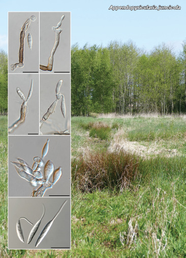
Mycelium consisting of hyaline, smooth, branched, septate, 1.5–2 µm diam hyphae. Conidiophores dimorphic, solitary or in fascicles of 2–3. Microconidiophores subcylindrical, hyaline, smooth, geniculate-sinuous, 7–30 × 3–4 µm. Macroconidio­phores subcylindrical, straight to curved to geniculate-sinuous, brown, thick-walled, smooth, base swollen or not so, 4–7 µm diam, 1–2-septate, 40–80 × 4–5 µm. Conidiogenous cells integrated, terminal, subcylindrical, at times slightly clavate, (7–)25–40 × 3–5 µm; subdenticulate, denticles cylindrical, 1–2 × 1 µm, with one to several per conidiogenous cell. Conidia (13–)15–20(–22) × 3(–4) µm, solitary, hyaline, smooth, guttulate, fusoid to fusoid-ellipsoid, 0–2-septate, hilum truncate, 1–1.5 µm diam with flexuous central apical appendage, 4–15 µm long.

Culture characteristics — Colonies erumpent, spreading, with sparse to moderate aerial mycelium and feathery margins, reaching 6 mm diam in 2 wk. On MEA surface smoke grey, reverse honey; on PDA surface smoke grey, reverse pale mouse grey; on OA surface pale mouse grey.

Typus. Netherlands, Utrecht Province, Nieuw Wulven, near Houten, 1.5 m a.s.l., N52°02'41" E05°09'49", on dead culm of Juncus effusus (Juncaceae), 25 Feb. 2021, E.R. Osieck, HPC 3600 = WI-26/#4224 (holotype CBS H-24981, cultures ex-type CPC 41278 = CBS 149232, CPC 41279, ITS, LSU, actA and tef1 (first part) sequences GenBank ON603767.1, ON603787.1, ON605619.1, ON605627.1 and ON605635.1, MycoBank MB 844243).

Additional material examined. Netherlands, Utrecht Province, Nieuw Wulven, near Houten, 1.5 m a.s.l., N52°02'45" E05°10'34", on dead culm of J. effusus, 24 June 2021, E.R. Osieck, HPC 3659 = WI-41/#4274, culture CPC 42171 = CBS 149233, ITS, LSU and tef1 (first part) sequences GenBank ON603768.1, ON603788.1, ON605628.1 and ON605636.1.

Notes — Appendopyricularia resembles genera in the Pyri­cularia complex (Klaubauf et al. 2014), in that it has solitary, pigmented conidiophores that terminate in denticulate conidiogenous cells. It is distinct in that the conidia are hyaline, fusoid to fusoid-ellipsoid, and have a characteristic flexuous central apical appendage, with conidia arranged in an apical circle, curved upwards, bowl-like, when viewed under a dissecting microscope.

Based on a megablast search of NCBIs GenBank nucleotide database, the closest hits using the ITS sequence of CPC 41278 had highest similarity to Phialemoniopsis curvata (strain EXF-8700, GenBank KP035004.1; Identities = 389/462 (84 %), 29 gaps (6 %)), Phialemoniopsis limonesiae (strain CBS 146752, GenBank NR_172355.1; Identities = 388/461 (84 %), 27 gaps (5 %)) and Thyridium curvatum (strain UTHSC R-3448, Gen­Bank HE599292.1; Identities = 387/462 (84 %), 29 gaps (6 %)). The ITS sequences of CPC 41278 and 42171 differ by one substitution and two gaps caused by two separate single nucleotide repeat strings (484/487 nt (99 %), including two gaps). Closest hits using the LSU sequence of CPC 41278 are Barbatosphaeria varioseptata (strain CBS 137797, GenBank NG_058674.1; Identities = 776/816 (95 %), five gaps (0 %)), Paradiplococcium singulare (strain CBS 126091, GenBank NG_066271.1; Identities = 775/815 (95 %), three gaps (0 %)) and Fluminicola striata (strain MFLUCC 18-0990, GenBank MW287770.1; Identities = 767/808 (95 %), five gaps (0 %)). The LSU sequences of CPC 41278 and 42171 differ by one gap caused by an extra C in a single nucleotide repeat string (659/660 nt (99 %), including one gap). Closest hits using the actA sequence of CPC 41278 did not reveal any significant hits, only a very distant association with Xylariales. Closest hits using the tef1 (first part) sequence of CPC 41278 did not reveal any significant hits, only a very distant association with Sorda­riales and Hypocreales. The tef1 sequences of CPC 41278 and 42171 differ by one gap caused by an extra C in a single nucleotide repeat string (447/448 nt (99 %), including one gap). Closest hits using the tub2 sequence of CPC 41278 did not reveal any significant hits, only a very distant association with Glomerellales and Xylariales. The tub2 sequences of CPC 41278 and 42171 are identical (678/678 nt).

Colour illustrations. Juncus effusus growing at Nieuw Wulven, near Houten in Utrecht. Conidiophores giving rise to conidia on SNA; conidiogenous cell with conidia; conidia with apical appendage. Scale bars = 10 µm.

Supplementary material

FP1383-1

Phylogenetic ITS tree.

FP1383-2

Phylogenetic LSU tree.

Persoonia. 2022 Jul 12;48:266–267.

Fungal Planet 1384 – 12 July 2022

Myrmecridium phragmitigenum Crous & Osieck, sp. nov.

Pedro W Crous 1, Johannes Z Groenewald 1, Eduard R Osieck 2

Etymology. Name refers to the host genus Phragmitis from which it was isolated.

Classification — Myrmecridiaceae, Myrmecridiales, Sorda­riomycetidae, Sordariomycetes.

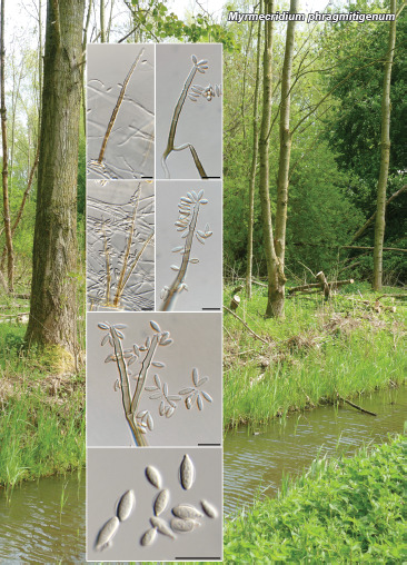
On SNA. Mycelium consisting of hyaline, smooth, branched, septate, 2–3 µm diam hyphae. Conidiophores unbranched, erect, straight, flexuous, medium brown, at times with nodulose swellings, thick-walled, 3–8-septate, up to 140 µm tall, 3–4 µm diam, with T-shaped basal cell, lacking rhizoids. Conidiogenous cells terminal, integrated, subcylindrical, 20–45 µm long, pale brown, forming a rachis with pimple-shaped denticles less than 1 µm long, 0.5 µm diam, slightly thickened. Conidia solitary, aseptate, pale brown, thin- and smooth-walled, guttulate with wing-like gelatinous sheath in middle, fusoid, (6–)7–8(–10) × 2.5(–3) µm; hilum unthickened but slightly darkened, 0.5 µm diam.

Culture characteristics — Colonies flat, spreading, with sparse to moderate aerial mycelium and smooth, even margin, reaching 45 mm diam after 2 wk at 25 °C. On MEA surface hazel, reverse hazel to isabelline; on PDA surface and reverse buff; on OA surface hazel.

Typus. Netherlands, Utrecht Province, Nieuw Wulven, near Houten, 1.5 m a.s.l., N52°03'03" E05°09'46", on dead culm of Phragmites australis (Poaceae), 25 Feb. 2021, E.R. Osieck, HPC 3613 = WI-32/#4221 (holotype CBS H-24955, cultures ex-type CPC 41394 = CBS 148945, ITS and LSU sequences GenBank ON603769.1 and ON603789.1, MycoBank MB 844244).

Notes — Myrmecridium phragmitigenum is closely related to M. phragmiticola known from Phragmites australis in Ukraine (conidia ellipsoid to fusoid, (7–)8–9 × (2.5–)3 μm, conidio­phores 2–4-septate, up to 70 μm tall, 3–3.5 μm diam; basal cell 4–6 μm diam; Crous et al. 2020b) and M. sambuci, known from Sambucus nigra in the Netherlands (conidia ellipsoid to fusoid, (7–)8–9(–10) × (2.5–)3(–3.5) μm, conidiophores (1–)3–7-septate, up to 170 μm tall, 2.5–3 μm diam; Crous et al. 2021c). It can be distinguished from M. phragmiticola by its longer, multiseptate conidiophores, but is best distinguished from M. sambuci based on DNA data, as the two species are morphologically similar, but phylogenetically distinct. A further species occurring on Phragmites, M. phragmites has similar ellipsoid to obovoid or fusoid conidia, (6.5–)7–8(–9) × (2.5–)3(–3.5) μm, but shorter conidiophores, 1–4-septate, up to 100 μm tall (Crous et al. 2011).

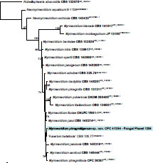
Based on a megablast search of NCBIs GenBank nucleotide database, the closest hits using the ITS sequence had highest similarity to ‘Neocosmospora striata / Fusarium bataticola’ (strain CBS 105.77, GenBank LR583781.1; Identities = 528/535 (99 %), one gap (0 %)), Myrmecridium schulzeri (strain CBS 188.96, GenBank EU041772.1; Identities = 508/518 (98 %), three gaps (0 %)) and Myrmecridium sambuci (strain CBS 148444, GenBank NR_175209.1; Identities = 523/534 (98 %), two gaps (0 %)). Closest hits using the LSU sequence are Myrmecridium phragmiticola (strain CPC 36367, GenBank NG_074444.1; Identities = 776/779 (99 %), no gaps), Myrmecridium schulzeri (strain CBS 188.96, GenBank EU041829.1; Identities = 775/779 (99 %), no gaps) and Myrmecridium spartii (strain CPC 24953, GenBank KR611902.1; Identities = 773/779 (99 %), no gaps).

graphic file with name per-2023-48-8-i001.jpg

Consensus phylogram (50 % majority rule) obtained from the maximum likelihood analysis with IQ-TREE v. 2.1.3 (Minh et al. 2020) of the Myrmecridium phragmitigenum ITS nucleotide alignment. Bootstrap support values (> 69 % are shown; only values > 94 % are significant) from 5 000 ultrafast (Hoang et al. 2018) bootstrap replicates are shown at the nodes. Culture collection numbers and GenBank accession numbers (superscript) are indicated for all species. The tree was rooted to Rubellisphaeria abscondita (culture CBS 132078; GenBank NR_154424.1) and the novelty described here is highlighted with a coloured block and bold font. Sequences from material with a type status are indicated in bold font. Alignment statistics: 21 strains including the outgroup; 565 characters including alignment gaps analysed: 250 distinct patterns, 135 parsimony-informative, 70 singleton sites, 360 constant sites. The best-fit model identified for the entire alignment in IQ-TREE using the TESTNEW option was TIM2e+R2. The alignment and tree were deposited at figshare.com (10.6084/m9.figshare.19745380).

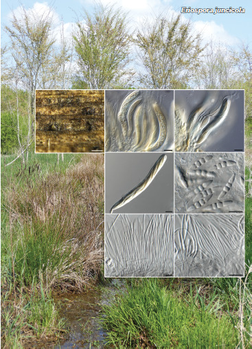
Colour illustrations. Phragmites australis growing along ditch at Nieuw Wulven, near Houten in Utrecht. Conidiophores and conidiogenous cells giving rise to conidia on SNA; conidia with mucoid sheath. Scale bars = 10 µm.

graphic file with name per-2023-48-8-g002.jpg

Myrmecridium phragmitigenum

Persoonia. 2022 Jul 12;48:268–269.

Fungal Planet 1385 – 12 July 2022

Paraproliferophorum Crous, gen. nov.

Pedro W Crous 1, Johannes Z Groenewald 1, Wingfield Michael J 2, Roux Jolanda 3

Etymology. Name refers to the fact that it is related to the genus Proli­ferophorum.

Classification — Diaporthomycetidae incertae sedis, Diaporthomycetidae, Sordariomycetes.

Mycelium consisting of pale brown, smooth, branched, septate hyphae. Conidiophores solitary, erect, subcylindrical, olivaceous brown to medium brown, unbranched or branched above, finely verruculose, 1–2-septate. Conidiogenous cells terminal and intercalary, olivaceous brown, smooth to finely verruculose, subcylindrical with slight apical taper, with aggregated cluster of apical denticles, loci slightly darkened. Conidia olivaceous brown, smooth, guttulate, fusoid, 0(–1)-septate, tapering to truncate ends, darkened, thickened; with age apical locus elongating sympodially to become ramoconidia, giving rise to 1–2 secondary conidia, fusoid, apex subobtuse, base truncate, hilum darkened, thickened, pale brown, guttulate, smooth.

Type species. Paraproliferophorum hyphaenes Crous

MycoBank MB 844245.

graphic file with name per-2023-48-8-g003.jpg

Paraproliferophorum hyphaenes

Persoonia. 2022 Jul 12;48:269.

Fungal Planet 1385 – 12 July 2022

Paraproliferophorum hyphaenes Crous, sp. nov.


Etymology. Name refers to the host genus Hyphaene from which it was isolated.

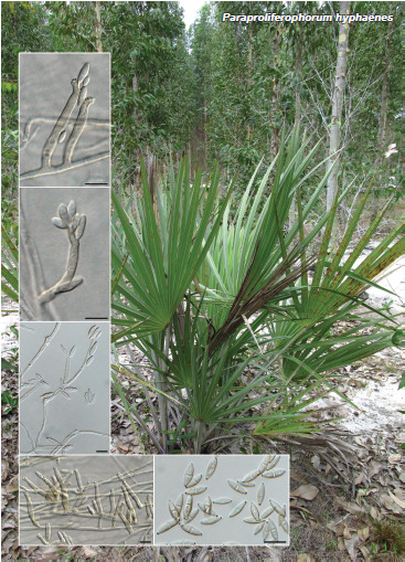
Mycelium consisting of pale brown, smooth, branched, septate, 1.5–2 µm diam hyphae. Conidiophores solitary, erect, subcylindrical, olivaceous brown to medium brown, unbranched or branched above, finely verruculose, 1–2-septate, 20–60 × 3–4 µm. Conidiogenous cells terminal and intercalary, olivaceous brown, smooth to finely verruculose, subcylindrical with slight apical taper, 15–45 × 2.5–3 µm, with aggregated cluster of apical denticles, 0.5 × 0.5–1 µm, slightly darkened. Conidia olivaceous brown, smooth, guttulate, fusoid, 0(–1)-septate, tapering to truncate ends, darkened, thickened, 0.5–1 µm diam, (8–)10–12(–15) × (2.5–)3(–4) µm; with age apical locus elongating sympodially to become ramoconidia, giving rise to 1–2 secondary conidia, fusoid, apex subobtuse, base truncate, hilum darkened, thickened, 0.5 µm diam, pale brown, guttulate, smooth, 4–6 × 2–2.5 µm.

Culture characteristics — Colonies erumpent, spreading, surface folded, with sparse aerial mycelium and smooth, even margin, reaching 8 mm diam after 2 wk at 25 °C. On MEA, PDA and OA surface and reverse isabelline.

Typus. SOUTH AFRICA, near Mozambique, on living leaves of Hyphaene sp. (Arecaceae), 19 Oct. 2017, M.J. Wingfield & J. Roux, HPC 3501 (holotype CBS H-24949, culture ex-type CPC 40103 = CBS 148939, ITS, LSU, rpb1 and tef1 (second part) sequences GenBank ON603770.1, ON603790.1, ON605643.1 and ON605632.1, MycoBank MB 844246).

Notes — Paraproliferophorum is phylogenetically related to Proliferophorum (Phookamsak et al. 2019), but distinct from the latter genus in that the conidiophores do not proliferate percurrently, have terminal and intercalary clusters of subdenticulate conidiogenous loci, and form ramoconidia that give rise to secondary conidia with slightly thickened hila. Morphologically Paraproliferophorum resembles Semipseudocercospora and Veronaeopsis, although the former genus has longer, more flexuous conidiophores with terminal conidiogenous cells, and Veronaeopsis tends to form a rachis of conidiogenous loci (Arzanlou et al. 2007, Braun et al. 2015).

Based on a blastn search of NCBIs GenBank nucleotide data­base, the closest hits using the ITS sequence had highest similarity to Proliferophorum thailandicum (strain MFLUCC 17-0293, GenBank MK028344.1; Identities = 352/422 (83 %), 18 gaps (4 %)), Plagiosphaera immersa (strain D270, GenBank MN727889.1; Identities = 178/209 (85 %), two gaps (0 %)) and Pseudopyricularia higginsii (strain 09/2007/1470, GenBank KM484875.1; Identities = 183/216 (85 %), eight gaps (3 %)). Closest hits using the LSU sequence are Proliferophorum thai­landicum (strain MFLUCC 17-0293, GenBank MK028343.1; Identities = 802/848 (95 %), ten gaps (1 %)), Ophiostoma saponiodorum (strain CBS 128125, GenBank MH877992.1; Identities = 741/845 (88 %), 19 gaps (2 %)) and Ophiostoma pallidulum (strain VPRI 43846, GenBank MW046139.1; Identities = 741/845 (88 %), 19 gaps (2 %)). No significant hits were obtained when the rpb1 sequence was used in blastn and megablast searches. Closest hits using the tef1 (second part) sequence had distant similarity to Phaeoacremonium sphinctrophorum (strain MFLUCC 11-0629, GenBank KU940202.1; Identities = 481/514 (94 %), no gaps), Phaeoacremonium minimum (strain AFTOL-ID 924, GenBank DQ471083.2; Identities = 476/514 (93 %), no gaps) and Codinaea amazonensis (strain MUCL 41171, GenBank OL653996.1; Identities = 472/512 (92 %), no gaps).

Colour illustrations. Hyphaene sp. in South Africa near Mozambique. Conidiophores and conidiogenous cells giving rise to conidia on SNA; conidia. Scale bars = 10 µm.

Supplementary material

FP1385

Phylogenetic tree.

per-2023-48-8-SF1385.jpg (32.8KB, jpg)
Persoonia. 2022 Jul 12;48:270–271.

Fungal Planet 1386 – 12 July 2022

Niesslia nieuwwulvenica Crous & Osieck, sp. nov.

Pedro W Crous 1, Johannes Z Groenewald 1, Eduard R Osieck 2

Etymology. Name refers to Nieuw Wulven where it was collected.

Classification — Niessliaceae, Hypocreales, Hypocreomycetidae, Sordariomycetes.

Mycelium consisting of smooth, septate, hyaline, branched, 1.5–2 µm diam hyphae. Conidiophores reduced to conidiogenous cells, erect, flexuous, solitary or aggregated, subcylindrical, hyaline, smooth, thick-walled, 65–100 × 2–2.5 µm, swollen in middle part with wavy apical region, terminating in a phialidic apex with non-flared collarette up to 1 µm long, 1.5–2 µm diam. Stipes intermingled among conidiophores, straight to flexuous, shorter than conidiophores, 20–40 µm long, thick-walled, terminating in sphaeropedunculate vesicle, 5–6 µm diam. Conidia solitary, aseptate, hyaline, smooth, subcylindrical, straight, apex subobtuse, base truncate, (8–)10–12(–13) × 2.5(–3) µm.

Culture characteristics — Colonies flat, spreading, with moderate aerial mycelium and smooth, even margin, reaching 30 mm diam after 2 wk at 25 °C. On MEA surface dirty white, reverse luteous; on PDA surface and reverse dirty white; on OA surface pale luteous.

Typus. Netherlands, Utrecht Province, Nieuw Wulven, near Houten, 1.5 m a.s.l., N52°02'48" E05°10'33", on dead culm of undetermined Poaceae, 19 Mar. 2021, E.R. Osieck, HPC 3623 = WI-38/#4238 (holotype CBS H-24953, cultures ex-type CPC 41374 = CBS 148943, ITS, LSU, actA, tef1 (first part) and tub2 sequences GenBank ON603771.1, ON603791.1, ON605620.1, ON605629.1 and ON605637.1, MycoBank MB 844247).

Notes — Using the key of Gams et al. (2019), N. nieuwwulvenica is identified as N. curvisetosa, which has much smaller dimorphic conidia, with globose conidia (3.5–)4.0–4.5 μm diam and ellipsoid conidia 3.5–6 × 1.5–2.0 μm. Phylogenetically, N. nieuwwulvenica is related to N. aemula (conidia cylindrical, 4.5–6.5 × 1.2–2.0 μm) and N. indica (conidia guttiliform to clavate, 3-5–4.7 × 1.8–2.2(–3) μm and N. neoexosporioides (conidia subcylindrical, (7–)8–10(–14) × 2(–2.5) μm (Gams et al. 2019, Crous et al. 2021b), which differ based on their conidial dimensions.

Based on a megablast search of NCBIs GenBank nucleotide database, the closest hits using the ITS sequence had highest similarity to Niesslia aemula (strain CBS 556.75, GenBank MG827004.1; Identities = 535/564 (95 %), 15 gaps (2 %)), Niesslia indica (strain CBS 313.61, GenBank MH858063.1; Identities = 516/545 (95 %), 17 gaps (3 %)) and Niesslia neo­exosporioides (strain CBS 146810, GenBank NR_173014.1; Identities = 533/563 (95 %), 14 gaps (2 %)). Closest hits using the LSU sequence are Niesslia exosporioides (strain CBS 515.72 l, GenBank MH872254.1; Identities = 788/798 (99 %), no gaps), Niesslia neoexosporioides (strain CBS 146810, Gen-Bank NG_076714.1; Identities = 791/802 (99 %), one gap (0 %)) and Niesslia aemula (strain CBS 556.75, GenBank MG826805.1; Identities = 791/804 (98 %), no gaps). Closest hits using the actA sequence had highest similarity to Niesslia neoexosporioides (strain CPC 38177, GenBank MW890027.1; Identities = 519/574 (90 %), 14 gaps (2 %)) and the protein-coding part of the sequences of Coccinonectria pachysandricola (strain CBS 501.63, GenBank KM231167.1; Identities = 398/418 (95 %), no gaps) and Gliocephalotrichum bulbilium (strain CBS 242.62, GenBank KM231118.1; Identities = 398/419 (95 %), no gaps). Closest hits using the tef1 (first part) sequence had highest similarity to Niesslia neoexosporioides (strain CPC 38177, GenBank MW890097.1; Identities = 284/348 (82 %), 27 gaps (7 %)) and the protein-coding part of the sequences of Alfaria ossiformis (strain CBS 324.54, GenBank KU846009.1; Identities = 202/234 (86 %), 9 gaps (3 %)) and Trichoderma applanatum (strain 7781, GenBank KJ634757.1; Identities = 144/150 (96 %), no gaps). No significant hits were obtained when the tub2 sequence was used in blastn and megablast searches. However, a blast2 comparison against Niesslia beta-tubulin sequences in GenBank revealed highest similarity to Niesslia ilicifolia (strain CBS 459.74, GenBank MG896293.1 (unverified sequence); Identities = 244/296 (82 %), 12 gaps (4 %)), Niesslia exilis (strain CBS 389.70A, GenBank MG896278.1 (unverified sequence); Identities = 235/293 (80 %), eight gaps (2 %)) and Monocillium constrictum (strain CBS 407.70A, GenBank MG896289.1 (unverified sequence); Identities = 239/304 (79 %), 15 gaps (4 %)).

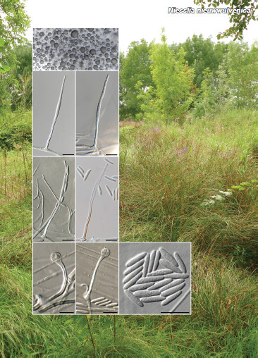
Colour illustrations. Poaceae with Juncus effusus clumps along ditches in open woodland, in Nieuw Wulven, near Houten in Utrecht. Conidiophores with conidia on SNA; sphaeropedunculate vesicles; conidia. Scale bars = 10 µm.

graphic file with name per-2023-48-8-g004.jpg

Niesslia nieuwwulvenica

Supplementary material

FP1386

Phylogenetic tree.

per-2023-48-8-SF1386.jpg (46.9KB, jpg)
Persoonia. 2022 Jul 12;48:272–273.

Fungal Planet 1387 & 1388 – 12 July 2022

Paracosmospora Crous & Boers, gen. nov.

Pedro W Crous 1, Johannes Z Groenewald 1, Boers Jannes 2

Etymology. Name refers to its phylogenetic relationship with the genus Cosmospora.

Classification — Hypocreales incertae sedis, Hypocreales, Hypocreomycetidae, Sordariomycetes.

Mycelium consisting of hyaline, smooth, septate, branched hyphae. Conidiophores solitary, but mostly aggregated in clusters, forming sporodochia, subcylindrical, smooth, hyaline, erect to wavy, branched or not, 1–2-septate. Conidiogenous cells terminal or intercalary, subcylindrical, hyaline, smooth, apex phialidic, with periclinal thickening. Conidia solitary, hyaline, smooth, guttulate, subcylindrical, medianly 1-septate, straight, apex obtuse to somewhat flattened, base tapering to truncate scar.

Type species. Paracosmospora physciae Crous & Boers

MycoBank MB 844248.

graphic file with name per-2023-48-8-g005.jpg

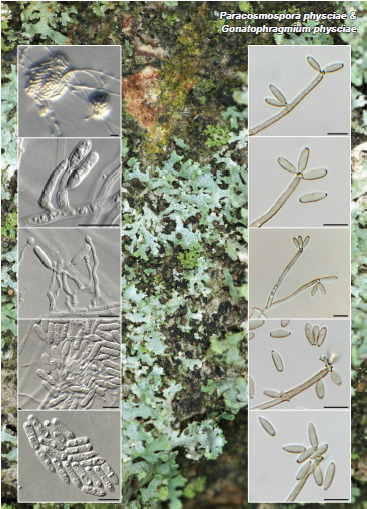
Paracosmospora physciae & Gonatophragmium physciae

Persoonia. 2022 Jul 12;48:273.

Fungal Planet 1387 & 1388 – 12 July 2022

Paracosmospora physciae Crous & Boers, sp. nov.


Etymology. Name refers to the host genus Physcia from which it was isolated.

Mycelium consisting of hyaline, smooth, septate, branched, 2.5–3 µm diam hyphae. Conidiophores solitary, but mostly aggregated in clusters, forming sporodochia, subcylindrical, smooth, hyaline, erect to wavy, branched or not, 1–2-septate, 20–50 × 2.5–3 µm. Conidiogenous cells terminal or intercalary, subcylindrical, hyaline, smooth, 15–25 µm long, apex phialidic, with periclinal thickening. Conidia solitary, hyaline, smooth, guttulate, subcylindrical, medianly 1-septate, straight, apex obtuse to somewhat flattened, base tapering to truncate scar, 1–1.5 µm diam, (11–)13–15(–18) × 4(–4.5) µm.

Culture characteristics — Colonies flat, spreading, with folded surface and moderate aerial mycelium with smooth, lobate margin, reaching 25 mm diam after 2 wk at 25 °C. On MEA surface saffron, reverse sienna; on PDA surface saffron, reverse orange; on OA surface saffron.

Typus. Netherlands, Drenthe Province, Dwingeloo, 52.8358892, 6.3644129, on Physcia tenella (Physciaceae), 10 Mar. 2021, J. Boers, HPC 3602 (holotype CBS H-24952 culture ex-type CPC 41288 = CBS 148942, ITS, LSU and tef1 (first part) sequences GenBank ON603772.1, ON603792.1 and ON605630.1, MycoBank MB 844249).

Notes — Paracosmospora resembles genera in the Cosmo­spora complex and is allied to Pseudocosmospora. It is mor­phologically distinct, however, in that Pseudocosmospora has acremonium-like to verticillium-like asexual morphs with aseptate, ellipsoidal, ovoid, or reniform conidia (Herrera et al. 2013).

(Notes continues on Supplementary material page FP1387 & 1388)

Persoonia. 2022 Jul 12;48:273.

Fungal Planet 1387 & 1388 – 12 July 2022

Gonatophragmium physciae Crous & Boers, sp. nov.


Etymology. Name refers to the host genus Physcia from which it was isolated.

Classification — Acrospermaceae, Acrospermales, Dothi­deomycetes.

Mycelium consisting of hyaline, smooth, branched, septate, 1.5–2 µm diam hyphae. Conidiophores solitary, erect, unbranched, subcylindrical, pale brown, smooth, 1–3-septate, 30–60 × 2.5–3 µm. Conidiogenous cells terminal, subcylindrical, smooth, pale brown, 10–30 × 2–2.5 µm, with terminal and intercalary loci, consisting of swollen areas with denticulate loci, 0.5–1 µm, darkened, not thickened. Conidia pale brown, smooth, (7–)8–9(–10) × (2.5–)3 µm, solitary, subcylindrical to narrowly ellipsoid, aseptate, apex obtuse, base obconically truncate, darkened, 0.5–1 µm diam.

Culture characteristics — Colonies erumpent, spreading, with folded surface and moderate aerial mycelium and smooth, even margin, reaching 35 mm diam after 2 wk at 25 °C. On MEA surface sienna to isabelline and diffuse scarlet pigment, reverse bay; on PDA surface fulvous, reverse sienna; on OA surface umber.

Typus. Netherlands, Drenthe Province, Dwingeloo graveyard, 52.831376, 6.365404, on Physcia caesia (Physciaceae), 28 Mar. 2021, J. Boers, HPC 3614 (holotype CBS H-24967, culture ex-type CPC 41464 = CBS 149048, ITS and LSU sequences GenBank ON603773.1 and ON603793.1, MycoBank MB 844250).

Additional material examined. Netherlands, Drenthe Province, Dwingeloo graveyard, 52.831376, 6.365404, on Physcia tenella (Physciaceae), 28 Mar. 2021, J. Boers, HPC 3614, CPC 41466, 41467, ITS and LSU sequences of CPC 41466 GenBank ON603774.1 and ON603794.1.

Notes — Hudson et al. (2019) suggested that although the asexual morph of Acrospermum is not known, close relatives include Gonatophragmium, Pseudovirgaria and Radulidium. Furthermore, Crous et al. (2021b) also added Pseudoacrospermum to the complex. If these taxa are congeneric, then the older name Acrospermum (1790) would have precedence. However, the type species, A. compressum (on dry stems of Heracleum sphondylium, Germany), is not yet known from culture or DNA. It thus remains to be determined whether Acrospermum is monophyletic. Based on its morphology, we have tentatively described the present taxon in Gonatophragmium. Gonatophragmium physciae should also be compared to G. lichenophilum (on Xanthoria parietina in Austria), although the latter has longer, septate conidia ((7–)9–15(–17) × (2.5–)3–4(–4.5) μm, (0–)1(–2)-septate) (see key in Berger et al. 2015).

Colour illustrations. Physcia tenella at Dwingeloo graveyard. Paracosmospora physciae (left column). Sporodochia on SNA; conidiophores and conidiogenous cells giving rise to conidia; conidia. Gonatophragmium physciae (right column). Conidiophores and conidiogenous cells giving rise to conidia. Scale bars = 10 µm.

(Notes continues on Supplementary material page FP1387 & 1388)

Supplementary material

FP1387-1

Paracosmospora physciae phylogenetic ITS tree.

FP1387-2

Paracosmospora physciae phylogenetic LSU tree.

FP1388

Gonatophragmium physciae phylogenetic ITS tree.

Persoonia. 2022 Jul 12;48:274–275.

Fungal Planet 1389 – 12 July 2022

Neoacrodontium Crous & Decock, gen. nov.

Pedro W Crous 1, Johannes Z Groenewald 1, Cony A Decock 2

Etymology. Name refers to the morphologically similar genus Acrodontium.

Classification — Amplistromataceae, Amplistromatales, Sor-dariomycetidae, Sordariomycetes.

Mycelium consisting of subhyaline to pale olivaceous, smooth, thin-walled, septate, branched, hyphae. Conidiophores erect, arising from hyphae, brown at base, becoming subhyaline toward apex, branched subverticillately, septate. Conidiogenous cells integrated, terminal and intercalary, basal part flask-shaped, with long flexuous, elongated neck, proliferating sympodially to form a long rachis; denticles pimple-like or elongated. Conidia solitary, subhyaline, subglobose, base with unthickened truncate scar.

Type species. Neoacrodontium gallicum Crous & Decock

MycoBank MB 844251.

graphic file with name per-2023-48-8-g006.jpg

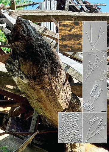
Neoacrodontium gallicum

Persoonia. 2022 Jul 12;48:275.

Fungal Planet 1389 – 12 July 2022

Neoacrodontium gallicum Crous & Decock, sp. nov.


Etymology. Name refers to France where it was collected.

Mycelium consisting of subhyaline to pale olivaceous, smooth, thin-walled, septate, branched, 1.5–2 µm diam hyphae. Coni­dio­phores erect, arising from hyphae, brown at base, becoming subhyaline toward apex, branched subverticillately, 1–3-septate. Conidiogenous cells integrated, terminal and intercalary, basal part flask-shaped, with long flexuous, elongated neck, proliferating sympodially to form a long rachis, 20–60 × 2–3 µm; denticles pimple-like, 0.5 µm diam, or elongated, up to 2 µm long, 0.5 µm diam. Conidia 2–3 µm diam, solitary, subhyaline, subglobose, base with truncate scar, 0.5 µm diam.

Culture characteristics — Colonies flat, spreading, with moderate aerial mycelium and smooth, even margin, reaching 10 mm diam after 2 wk at 25 °C. On MEA surface and reverse ochreous; on PDA surface and reverse saffron; on OA surface ochreous.

Typus. France, Ile de France, Paris, 7ième arrondissement, in house, from bore dust of Xestobium rufovillosum feeding on Quercus wood (death-watch beetle), Sept. 2020, C. Decock (holotype CBS H-24951 culture ex-type CPC 41118 = CBS 148941 = MUCL 58099, ITS and LSU sequences GenBank ON603775.1 and ON603795.1, MycoBank MB 844252).

Notes — Neoacrodontium (Amplistromataceae) is phylogenetically distinct from morphologically similar genera such as Acrodontium (Teratosphaeriaceae; Videira et al. 2016) and Xenoacrodontium (Xenoacrodontiaceae; Crous et al. 2021c). Neoacrodontium gallicum clusters with CBS 349.55 (unknown country, keratinous substrate, F. Blank, 3 Jan. 1955), which was identified as Acrodontium hydnicola (De Hoog 1972), and belongs to the same genus.

Neoacrodontium hydnicola (Peck) Crous & Decock, comb. nov. — MycoBank MB 844253

Basionym. Virgaria hydnicola Peck, Ann. Rep. N.Y. State Mus. 42: 128. 1889.

Synonyms. Tritirachium hydnicola (Peck) Hughes, Canad. J. Bot. 31: 604. 1953.

Acrodontium hydnicola (Peck) de Hoog, Stud. Mycol. 1: 31. 1972.

Based on a megablast search of NCBIs GenBank nucleotide database, the closest hits using the ITS sequence had highest similarity to ‘Acrodontiumhydnicola (strain CBS 349.55, GenBank MH857507.1; Identities = 515/602 (86 %), 44 gaps (7 %)), ‘Acrodontiumsimplex (strain CBS 127.53, GenBank MH857128.1; Identities = 378/462 (82 %), 41 gaps (8 %)) and Acidothrix acidophila (strain MH566, GenBank FJ430780.3; Identities = 341/420 (81 %), 28 gaps (6 %)). Closest hits using the LSU sequence are ‘Acrodontiumhydnicola (strain CBS 349.55, GenBank MH869047.1; Identities = 806/833 (97 %), two gaps (0 %)), Wallrothiella congregata (strain SMH1760, GenBank FJ532375.1; Identities = 753/839 (90 %), 16 gaps (1 %)) and Amplistroma erinaceum (strain CBS 134881, GenBank MH877572.1; Identities = 655/733 (89 %), 17 gaps (2 %)).

Colour illustrations. Quercus wood with beetle damage. Insect galleries in wood; conidiophores and conidiogenous cells giving rise to conidia; conidia. Scale bars = 10 µm.

Supplementary material

FP1389-1

Phylogenetic ITS tree.

FP1389-2

Phylogenetic LSU tree.

Persoonia. 2022 Jul 12;48:276–277.

Fungal Planet 1390 – 12 July 2022

Parastenospora Crous, gen. nov.

Pedro W Crous 1, Arien L van Iperen 1

Etymology. Name refers to Stenospora, which is morphologically similar to this genus.

Classification — Incertae sedis, Pleosporales, Pleosporomycetidae, Dothideomycetes.

Mycelium consisting of hyaline, smooth, branched, septate hyphae. Conidiophores terminal on hyphae, or intercalary, erect, solitary, subcylindrical, straight to flexuous, hyaline, smooth, septate, branched below or unbranched. Conidiogenous cells subcylindrical, hyaline, smooth, terminal or intercalary, holoblastic with one to several denticulate loci, unthickened, not darkened, frequently with rosette of denticulate loci. Conidia solitary, hyaline, smooth, guttulate, obclavate, apex subobtuse, base obconically truncate, straight to curved, septate; hilum unthickened, not darkened.

Type species. Parastenospora pini Crous

MycoBank MB 844254.

graphic file with name per-2023-48-8-g007.jpg

Parastenospora pini

Persoonia. 2022 Jul 12;48:277.

Fungal Planet 1390 – 12 July 2022

Parastenospora pini Crous, sp. nov.


Etymology. Name refers to the host genus Pinus from which it was isolated.

Mycelium consisting of hyaline, smooth, branched, septate, 1.5–2 µm diam hyphae. Conidiophores terminal on hyphae, or intercalary, erect, solitary, subcylindrical, straight to flexuous, hyaline, smooth, 0–3-septate, branched below or unbranched, 10–30 × 2.5–3 µm. Conidiogenous cells 3–10 × 2–5 µm, subcylindrical, hyaline, smooth, terminal or intercalary, holoblastic with one to several denticulate loci, 1–3 × 1.5–2 µm, unthickened, not darkened, frequently with rosette of denticulate loci. Conidia solitary, hyaline, smooth, guttulate, obclavate, apex subobtuse, base obconically truncate, straight to curved, (1–)3(–6)-septate, (25–)45–70(–85) × (3–)4(–5) µm; hilum unthickened, not darkened, 1.5–2 µm.

Culture characteristics — Colonies erumpent, spreading, with moderate aerial mycelium and smooth, even margin, reaching 5 mm diam after 2 wk at 25 °C. On MEA surface and reverse ochreous; on PDA surface and reverse dirty white; on OA surface dirty white.

Typus. Netherlands, Utrecht Province, Soest, De Zoom, on dead twigs of Pinus sylvestris (Pinaceae), 1 Nov. 2020, A.L. van Iperen, HPC 3492 (holotype CBS H-24950, culture ex-type CPC 40385 = CBS 148940, ITS, LSU, rpb2 and tub2 sequences GenBank ON603776.1, ON603796.1, ON605622.1 and ON605638.1, MycoBank MB 844255).

Notes — Parastenospora is reminiscent of Condylospora and Stenospora. However, Stenospora is mycoparasitic, and has slightly thickened scars and hila (Braun et al. 2013), while Condylospora is aquatic, having unique L- or N-shaped conidia (Yen et al. 2012). The latter genera are thus morphologically and phylogenetically distinct from Parastenospora.

Based on a megablast search of NCBIs GenBank nucleotide database, the closest hits using the ITS sequence had distant similarity over mainly the 5.8S nrRNA gene to Neocamarospo­rium solicola (strain IBRC M 30257, GenBank KX817217.1; Identities = 347/393 (88 %), ten gaps (2 %)), Didymocyrtis cladoniicola (strain 19E070, GenBank MZ206178.1; Identities = 373/431 (87 %), 11 gaps (2 %)),and Ascochyta manawaorae (strain 1213, GenBank MZ400577.1; Identities = 373/432 (86 %), 11 gaps (2 %)). Closest hits using the LSU sequence are Pseudopyrenochaeta lycopersici (strain CBS 306.65, GenBank NG_057799.1; Identities = 847/860 (98 %), no gaps), Subplenodomus galicola (voucher MFLU 15-1368, GenBank NG_070410.1; Identities = 843/863 (98 %), no gaps) and Subplenodomus urticae (voucher MFLU 17-1694, GenBank NG_073768.1; Identities = 843/863 (98 %), no gaps). Distant hits were obtained using the rpb2 sequence, with highest similarity to Podonectria kuwanaspis (strain SICAUCC 21-0007, GenBank MW462123.1; Identities = 617/735 (84 %), no gaps), Neocucurbitaria aetnensis (strain C261, GenBank MF795811.1; Identities = 609/736 (83 %), no gaps) and Neocucurbitaria cinereae (strain KU9, GenBank MF795813.1; Identities = 607/736 (82 %), no gaps). Distant hits were obtained using the tub2 sequence, with highest similarity to Parafenestella pseudo­salicis (strain C301, GenBank MK357620.1; Identities = 253/291 (87 %), one gap (0 %)), Parafenestella vindobonensis (strain C302, GenBank MK357632.1; Identities = 258/300 (86 %), 11 gaps (3 %)) and Neocucurbitaria keratinophila (strain CNM-CM 6401, GenBank LT992261.1; Identities = 255/295 (86 %), one gap (0 %)).

Colour illustrations. Pinus sylvestris in De Zoom, Soest. Conidiogenous cells giving rise to conidia; conidia. Scale bars = 10 µm.

Supplementary material

FP1390

Phylogenetic tree.

Persoonia. 2022 Jul 12;48:278–279.

Fungal Planet 1391 – 12 July 2022

Eriospora juncicola Crous & Osieck, sp. nov.

Pedro W Crous 1, Johannes Z Groenewald 1, Eduard R Osieck 2

Etymology. Name refers to the host genus Juncus from which it was isolated.

Classification — Stictidaceae, Ostropales, Ostropomyce­tidae, OSLEUM clade, Lecanoromycetes.

Ascomata pseudothecial, immersed in host tissue, globose, 250–300 µm diam, with central ostiole; wall of 2–3 layers of brown textura angularis. Pseudoparaphyses trabeculae-like, hyaline, smooth, branched, septate, anastomosing, 1.5–2 µm diam. Asci hyaline, smooth, fusoid-ellipsoid, apex obtuse with stipitate base, ocular chamber well defined, bitunicate with 2–3 seriate ascospores, 100–130 × 15–17 µm. Ascospores hyaline, smooth, guttulate, fusoid ellipsoid, initially medianly septate, becoming 3-septate, constricted at septa, prominently swollen in cell above median septum, encased in 5–7 µm diam thick mucoid sheath, (22–)24–26(–30) × 5(–6) µm (in Melzer). Conidiomata pycnidial, immersed in agar, separate, globose, unilocular with thick wall and central ostiole. Conidiophores reduced to conidiogenous cells that proliferate sympodially at apex, subcylindrical, hyaline, smooth, 5–10 × 1.5–2 µm. Conidia frequently remaining attached to conidiogenous cells, hyaline, smooth, curved, multi-septate, cylindrical with obtuse ends, 30–100 × 1.5–2 µm.

Culture characteristics — Colonies erumpent, spreading, sur-face folded, with moderate aerial mycelium and smooth, even margin, reaching 20 mm diam after 2 wk at 25 °C. On MEA surface ochreous, reverse sienna; on PDA surface and reverse pale luteous; on OA surface ochreous.

Typus. Netherlands, Utrecht Province, Nieuw Wulven, near Houten, N52°02'48" E05°10'33", on dead culm of Juncus effusus (Juncaceae), 19 Mar. 2021, E.R. Osieck, HPC 3620 = WI-35/#4233 (holotype CBS H-24958, culture ex-type CPC 41498 = CBS 148948, ITS and LSU sequences GenBank ON603777.1 and ON603797.1, MycoBank MB 844256).

Notes — Eriospora is a genus of saprobic coelomycetes for which there was no known sexual morph (Sutton 1980). The type species, E. leucostoma, was treated by Crous et al. (2020c), with cultures from Typha sp. (the type host genus), and Juncus effusus. Eriospora juncicola has a sexual morph, which distinguishes it from the asexual E. leucostoma. The two species have morphologically similar asexual morphs, but are phylogenetically distinct.

Based on a megablast search of NCBIs GenBank nucleotide database, the closest hits using the ITS sequence had highest similarity to Eriospora leucostoma (strain CPC 35594, GenBank MT223795.1; Identities = 589/651 (90 %), 15 gaps (2 %)), Neofitzroyomyces nerii (strain CBS 145088, GenBank NR_161144.1; Identities = 580/656 (88 %), 38 gaps (5 %)) and Fitzroyomyces cyperacearum (strain MFLU 19-2725, GenBank MW293953.1; Identities = 391/441 (89 %), 14 gaps (3 %)). Clo­sest hits using the LSU sequence are Eriospora leuco­stoma (strain CPC 35594, GenBank MT223890.1; Identities = 778/780 (99 %), no gaps), Neofitzroyomyces nerii (strain CBS 145088, GenBank NG_068278.1; Identities = 778/786 (99 %), no gaps) and Phacidiella alsophilae (strain CPC 37041, GenBank MT373344.1; Identities = 728/769 (95 %), five gaps (0 %)).

Colour illustrations. Juncus effusus in Nieuw Wulven, near Houten, Utrecht. Ascomata immersed in host tissue; asci and pseudoparaphyses; ascus; ascospores; conidiogenous cells giving rise to branched conidia. Scale bars: ascomata = 300 µm, all others = 10 µm.

graphic file with name per-2023-48-8-g008.jpg

Eriospora juncicola

Supplementary material

FP1391

Phylogenetic tree.

per-2023-48-8-SF1391.jpg (55.8KB, jpg)
Persoonia. 2022 Jul 12;48:280–281.

Fungal Planet 1392 – 12 July 2022

Nectriella adonidis Crous & Akulov, sp. nov.

Pedro W Crous 1, Johannes Z Groenewald 1, Akulov Alex 2

Etymology. Name refers to the host genus Adonis from which it was isolated.

Classification — Bionectriaceae, Hypocreales, Hypocreomycetidae, Sordariomycetes.

Mycelium consisting of hyaline, septate, smooth, branched, 1.5–2 µm diam hyphae. Conidiophores erect, solitary, unbranched or subverticillately branched with terminal and intercalary conidiogenous cells. Conidiogenous cells acremonium-like, monophialidic, somewhat thick-walled, cylindrical, flexuous, with slight apical taper, 35–80 × 2.5–3 µm. Conidia hyaline, smooth, aseptate, thin-walled, guttulate, fusoid-ellipsoid, ends obtuse, straight, hilum bluntly rounded to truncate, 1.5 µm diam, (7–)8–10(–11) × (2–)2.5(–3) µm. Sexual morph unknown.

Culture characteristics — Colonies flat, spreading, with moderate aerial mycelium and smooth, lobate margin, reaching 50 mm diam after 2 wk at 25 °C. On MEA, PDA and OA surface pale luteous, reverse luteous.

Typus. Ukraine, Kharkiv region, Dvorichna district, Krasne Pershe village, National Park Dvorichanskyi, on overwintered stems of Adonis vernalis (Ranunculaceae), 11 Apr. 2021, A. Akulov, ex CWU (MYC) AS 8121, HPC 3630 (holotype CBS H-24960, culture ex-type CPC 41516 = CBS 148950, ITS and LSU sequences GenBank ON603778.1 and ON603798.1, MycoBank MB 844257).

Notes — Nectriella (based on N. fuckellii) is characterised by white to pale yellow or pale brown ascomatal perithecia (frequently immersed) and ellipsoid to fusoid, 1-septate, hyaline, smooth to faintly spinulose to striate ascospores. Where known from culture, species produce kutilakesa-like sporodochia, and acremonium-like asexual morphs (Alfieri & Samuels 1979, Rossman et al. 1999). However, most Nectriella spp. occur on dead wood and herbaceous substrates, and are not considered plant pathogenic (Rossman et al. 1999). Based on the few cultures that are available, Nectriella is paraphyletic, and the only plant pathogen, N. pironii, together with N. adonidis probably represents a distinct genus.

Nectriella adonidis is closely related to N. pironii, which produces kutilakesa-like sporodochia on PDA, and an acremonium-like asexual morph on corn meal agar. Nectriella adonidis was isolated from orange sporodochia, but sporulated only on SNA, producing an acremonium-like morph, and no sporodochia, making it difficult to compare to other known species. This is the first Nectriella species described from Adonis vernalis, which occurs in the dry meadows and steppes of Eurasia.

Based on a megablast search of NCBIs GenBank nucleotide database, the closest hits using the ITS sequence had highest similarity to ‘Ijuhya sp.’ (strain daef39, GenBank MH550509.1; Identities = 555/555 (100 %), no gaps), Nectriella pironii (strain CBS 264.80, GenBank MH861261.1; Identities = 548/555 (99 %), no gaps) and Parvothecium terrestre (strain CBS 198.89, GenBank NR_145081.1; Identities = 516/557 (93 %), 12 gaps (2 %)). Closest hits using the LSU sequence are ‘Sarcopodium circinatum’ (strain CBS 376.81, GenBank HQ232167.1; Identities = 788/788 (100 %), no gaps), Nectriella pironii (strain CBS 264.80, GenBank MH873030.1; Identities = 780/788 (99 %), one gap (0 %)) and Pseudoachroiostachys krabiense (strain MFLUCC 16-0325, GenBank NG_068839.1; Identities = 774/788 (98 %), three gaps (0 %)).

graphic file with name per-2023-48-8-i002.jpg

Consensus phylogram (50 % majority rule) obtained from the maximum likelihood analysis with IQ-TREE v. 2.1.3 (Minh et al. 2020) of the Nectriella adonidis ITS nucleotide alignment. Bootstrap support values (> 69 % are shown; only values > 94 % are significant) from 5 000 ultrafast (Hoang et al. 2018) bootstrap replicates are shown at the nodes. Culture collection numbers and GenBank accession numbers (superscript) are indicated for all species. The tree was rooted to Fusarium tonkinense (culture CBS 115.40; GenBank NR_170733.1) and the novelty described here is highlighted with a coloured block and bold font. Sequences from material with a type status are indicated in bold font. Alignment statistics: 22 strains including the outgroup; 644 characters including alignment gaps analysed: 228 distinct patterns, 88 parsimony-informative, 113 singleton sites, 443 constant sites. The best-fit model identified for the entire alignment in IQ-TREE using the TESTNEW option was TNe+R2. The alignment and tree were deposited at figshare.com (10.6084/m9.figshare.19745380).

Colour illustrations. National Park Dvorichanskyi, Krasne Pershe village, Kharkiv region, Ukraine. Conidiophores and conidiogenous cells giving rise to conidia; conidia. Scale bars = 10 µm.

graphic file with name per-2023-48-8-g009.jpg

Nectriella adonidis

Persoonia. 2022 Jul 12;48:282–283.

Fungal Planet 1393 – 12 July 2022

Nothodevriesia Crous & Boers, gen. nov.

Pedro W Crous 1, Johannes Z Groenewald 1, Boers Jannes 2

Etymology. Phylogenetically related but distinct from Devriesia.

Classification — Teratosphaeriaceae, Mycosphaerellales, Dothideomycetidae, Dothideomycetes.

Saprobic on leaf litter. Ascomata pseudothecial, amphigenous, immersed to erumpent, superficial, globose, ostiole inconspicuous; ascomata linked by immersed brown hyphal network; hyphae medium brown, smooth, constricted at septa; ascomatal wall consisting of 2–3 layers of medium brown textura angularis. Asci aparaphysate, bitunicate, subsessile, obovoid to broadly ellipsoid, 8-spored. Ascospores multiseriate, overlapping, hyaline to pale brown (verruculose upon discharge), guttulate, thin-walled, straight, obovoid, ends obtuse, widest in middle of apical cell, medianly 1-septate, constricted at septum, tapering towards both ends, but more prominently towards lower end.

Type species. Nothodevriesia narthecii Crous & Boers

MycoBank MB 844258.

graphic file with name per-2023-48-8-g010.jpg

Nothodevriesia narthecii

Persoonia. 2022 Jul 12;48:283.

Fungal Planet 1393 – 12 July 2022

Nothodevriesia narthecii Crous & Boers, sp. nov.


Etymology. Name refers to the host genus Narthecium from which it was isolated.

Leaf spots absent, associated with leaf litter. Ascomata pseudo­thecial, amphigenous, immersed to erumpent, superficial, globose, 30–45 µm diam, ostiole inconspicuous; ascomata linked by immersed brown hyphal network; hyphae medium brown, smooth, 4–5 µm diam, constricted at septa; ascomatal wall consisting of 2–3 layers of medium brown textura angularis. Asci aparaphysate, bitunicate, subsessile, obovoid to broadly ellipsoid, 8-spored, 15–20 × 9–10 µm. Ascospores multiseriate, overlapping, hyaline to pale brown (verruculose upon discharge), guttulate, thin-walled, straight, obovoid, ends obtuse, widest in middle of apical cell, medianly 1-septate, constricted at septum, tapering towards both ends, but more prominently towards lower end, (7–)8–9(–10) × (2.5–)3 µm. Germinating from one end, with germ tube at angle to the long axis.

Culture characteristics — Colonies erumpent, spreading, surface folded with moderate aerial mycelium and lobate, even margin, reaching 4 mm diam after 2 wk at 25 °C. On MEA, PDA and OA surface and reverse iron-grey.

Typus. Netherlands, Drenthe Province, Dwingelderveld National Park, 52.829188, 6.432495, on dead leaves of Narthecium ossifragum (Narthecia­ceae), 13 July 2021, J. Boers, HPC 3655 (holotype CBS H-24985, cultures ex-type CPC 42166 = CBS 149066, ITS, LSU, actA, rpb2, tef1 (first part) and tub2 sequences GenBank ON603779.1, ON603799.1, ON605621.1, ON605623.1, ON605631.1 and ON605639.1, MycoBank MB 844259).

Notes — Nothodevriesia narthecii represents a sexual morph in the Teratosphaeriaceae occurring on dead leaves of Narthecium ossifragum. Although related, it is morphologically and phylogenetically distinct from Devriesia, which includes hypho­mycetous species that are soil-borne, as well as saprobic taxa found in dead plant material (Seifert et al. 2004, Crous & Groenewald 2011, Chang et al. 2022).

Based on a megablast search of NCBIs GenBank nucleotide database, the closest hits using the ITS sequence had highest similarity to Phaeothecoidea melaleuca (strain CPC 17223, GenBank HQ599594.1; Identities = 444/501 (89 %), 11 gaps (2 %)), Teratosphaeria jonkershoekensis (strain CBS 122897, GenBank EU707864.1; Identities = 443/503 (88 %), 14 gaps (2 %)) and Neocatenulostroma microsporum (strain HFJN1, GenBank MH349092.1; Identities = 433/485 (89 %), 13 gaps (2 %)). Closest hits using the LSU sequence are Devriesia thermodurans (strain CBS 115878, GenBank NG_059078.1; Identities = 802/833 (96 %), no gaps), Teratosphaeria hortaea (strain CBS 124156, GenBank MH874881.1; Identities = 798/834 (96 %), five gaps (0 %)) and Readeriella considenianae (strain CMW37676, GenBank JQ732948.1; Identities = 798/834 (96 %), five gaps (0 %)). Closest hits using the actA sequence had highest similarity to Neocatenulostroma germanicum (strain CAT104, GenBank KU612288.1; Identities = 444/508 (87 %), 12 gaps (2 %)), Teratosphaeria dunnii (strain CBS 145548, GenBank MK876463.1; Identities = 372/404 (92 %), no gaps) and Teratosphaeria gracilis (strain CBS 145090, GenBank MK047523.1; Identities = 442/507 (87 %), nine gaps (1 %)). Closest hits using the rpb2 sequence had highest similarity to Teratosphaeria corymbiicola (strain CBS 146047, GenBank MN556802.1; Identities = 472/623 (76 %), ten gaps (1 %)), Teratosphaeria eucalypti (strain CPC 12552, GenBank KX348102.1; Identities = 509/685 (74 %), 10 gaps (1 %)) and Teratosphaeria biformis (strain CBS 124578, GenBank KX348100.1; Identities = 471/624 (75 %), 12 gaps (1 %)). No significant hits were obtained when the tef1 (first part) and tub2 sequences were used in blastn and megablast searches.

Colour illustrations. Narthecium ossifragum in Dwingelderveld National Park. Ascomata in host tissue; broken ascoma with asci; brown ascospores on host tissue; germinating ascospores on MEA. Scale bars: intact ascomata = 30 and 45 µm, respectively, all others = 10 µm.

Supplementary material

FP1393-1

Nothodevriesia narthecii phylogenetic ITS tree.

FP1393-2

Nothodevriesia narthecii phylogenetic LSU tree.

Persoonia. 2022 Jul 12;48:284–285.

Fungal Planet 1394 – 12 July 2022

Tetraploa juncicola Crous & Osieck, sp. nov.

Pedro W Crous 1, Johannes Z Groenewald 1, Eduard R Osieck 2

Etymology. Name refers to the host genus Juncus from which it was isolated.

Classification — Tetraplosphaeriaceae, Pleosporales, Pleo­sporomycetidae, Dothideomycetes.

Mycelium consisting of medium brown, verruculose, septate, branched, 2–3 µm diam hyphae. Conidiogenous cells monoblastic, intercalary on hyphae, doliiform to ellipsoid, 3–6 × 3–5 µm. Conidia solitary, dry, composed on basal conidial body and 1–4 brown, septate apical appendages; conidia body (17–)20–30(–35) × (15–)18–20(–23) µm, medium brown, gut-tulate, verruculose, narrowly ellipsoid to subcylindrical, composed of brown, verruculose, four vertical columns of cells, 3–5 distoseptate. Appendages 30–100 µm long, medium brown, 3–8-septate, thick-walled, guttulate, smooth-walled.

Culture characteristics — Colonies erumpent, spreading, with moderate aerial mycelium and smooth, even margin, reaching 50 mm diam after 2 wk at 25 °C. On MEA, PDA and OA surface and reverse olivaceous grey.

Typus. Netherlands, Flevoland Province, Horsterwold, N52°18'52" E05°28'10", on dead culm of Juncus inflexus (Juncaceae), 31 Mar. 2021, E.R. Osieck, HPC 3631 = WI-39/#4240 (holotype CBS H-24965, culture ex-type CPC 41580 = CBS 149046, ITS, LSU and rpb2 sequences GenBank ON603780.1, ON603800.1 and ON605624.1, MycoBank MB 844260).

Notes — Tetraploa (= Tetraplosphaeria), based on T. aris­tata, is characterised by micronematous conidiophores, mono-blastic conidiogenous cells and tetraploate conidia (4-euseptate, short-cylindrical, brown, vertical columns that are verrucose at the base, with 4-setose, divergent, short or long septate appendages at the apex; Tanaka et al. 2009). Tetraploa aristata, long believed to be a single species with high ecological variation, is considered by Tanaka (loc. cit.) as species complex. Approximately 20 species are known (Bao et al. 2021), of which some have been linked to a massarina-like sexual morph (Crous et al. 2021a). Tetraploa juncicola is similar to T. aquatica and T. thailandica, having monoblastic conidiogenous cells and four vertical columns, with up to four apical appendages. It is distinct in that vertical columns of T. aquatica are 2–3-septate, while conidia of T. juncicola and T. thailandica are 3–5-septate, but T. thailandica has longer appendages, being 73–136 µm long, 5–10-distoseptate (Bao et al. 2021). It is also rather similar to T. aristata which has a slightly larger conidial body with 3–6 columns and somewhat shorter appendages (Ellis 1949). Tetraploa species are saprobic mostly on monocotyledons (more than half of the species) and/or in freshwater habitats. Tetraploa juncicola is the 3rd Tetraploa species recorded from Europe.

Based on a megablast search of NCBIs GenBank nucleotide database, the closest hits using the ITS sequence had highest similarity to Tetraploa yunnanensis (strain MFLUCC 19-0319, GenBank NR_171886.1; Identities = 522/544 (96 %), six gaps (1 %)), Tetraploa sasicola (strain 5-1, GenBank KX440178.1; Identities = 513/535 (96 %), four gaps (0 %)) and Tetraploa endophytica (strain P6086, GenBank KT270279.1; Identities = 489/515 (95 %), nine gaps (1 %)). Closest hits using the LSU sequence are Tetraploa endophytica (strain CBS 147114, GenBank MW659165.1; Identities = 678/683 (99 %), one gap (0 %)), Tetraploa sasicola (voucher HHUF 27566, GenBank NG_042329.1; Identities = 758/766 (99 %), no gaps) and Tetraploa yunnanensis (strain MFLUCC 18-0652, GenBank MN913697.1; Identities = 767/776 (99 %), no gaps). Closest hits using the rpb2 sequence had highest similarity to Tetraploa yunnanensis (strain MFLUCC 19-0319, GenBank MT878451.1; Identities = 677/737 (92 %), no gaps), Melanomma pulvis-pyrius (strain KH 77, GenBank LC203400.1; Identities = 542/740 (73 %), 12 gaps (1 %)) and Ernakulamia krabiensis (strain MFLUCC 18-0237, GenBank MK434872.1; Identities = 535/734 (73 %), seven gaps (0 %)).

Colour illustrations. Juncus inflexus in woodland border, Horsterwold, Flevoland Province. Conidiogenous cells giving rise to conidia; conidia with apical appendages. Scale bars = 10 µm.

graphic file with name per-2023-48-8-g011.jpg

Tetraploa juncicola

Supplementary material

FP1394

Phylogenetic tree.

per-2023-48-8-SF1394.jpg (45.4KB, jpg)
Persoonia. 2022 Jul 12;48:286–287.

Fungal Planet 1395 – 12 July 2022

Neochalara lolae Crous, sp. nov.

Pedro W Crous 1, Johannes Z Groenewald 1

Etymology. Named after Lola, a Labradoodle member of the Crous family, who indicated where to collect this specimen in her favourite forest.

Classification — Pezizellaceae, Rhytismatales, Leotiomycetes.

Mycelium consisting of hyaline, smooth, branched, 2–2.5 µm diam hyphae. Conidiophores solitary, erect, medium brown, smooth, 1–2-septate, with gradual transition between venter and neck; conidiophores 50–70 µm tall; venter 20–30 × 4–6 µm, without any sign of percurrent rejuvenation; neck cylindrical, 10–25 × 3–4 µm. Conidia occurring in long flexuous, unbranched chains, hyaline, smooth, thin-walled, guttulate, ends bluntly rounded, (9–)12–16(–28) × (2.5–)3 µm, 1–3(–4)-septate.

Culture characteristics — Colonies erumpent, with sparse aerial mycelium and lobate, even margin, reaching 6 mm diam after 2 wk at 25 °C. On MEA surface and reverse ochreous; on PDA surface dirty white, reverse ochreous; on OA surface ochreous.

Typus. Netherlands, Utrecht Province, Bilthoven, on stems of Pteridium aquilinum (Dennstaedtiaceae), 24 May 2021, P.W. Crous, K.L. Crous & L. Crous, HPC 3644 (holotype CBS H-24986, culture ex-type CPC 41931 = CBS 149065, ITS, LSU, rpb1 and tub2 sequences GenBank ON603781.1, ON603801.1, ON605644.1 and ON605640.1, MycoBank MB 844261).

Notes — Neochalara lolae is common on the fronds of Pteri-dium aquilinum in the Netherlands, and forms extremely long conidial chains. It must be compared with C. pteridina, that occurs on this host in Europe. It differs from the latter that has single or short conidial chains, conidiophores with percurrent rejuvenation, a wider venter (5–9.2 µm), longer collarettes (12–44(–50) × 3–4.5 µm), and mostly 3-septate conidia, (8–)12(–18) × (2–)2.5(–3) µm (Nag Raj & Kendrick 1975). Neochalara lolae is similar to N. spiraeae, which also has septate conidia with truncate ends (on Spiraea japonica, Netherlands), but is distinct in that the latter species has smaller, 1-septate conidia ((11–)12–14(–16) × (3–)4(–4.5) μm, av. 13 × 4 µm; Crous et al. 2021c).

Based on a megablast search of NCBIs GenBank nucleotide database, the closest hits using the ITS sequence had highest similarity to Neochalara spiraeae (strain CBS 148332, GenBank NR_175217.1; Identities = 457/489 (93 %), 16 gaps (3 %)), Lambertella advenula (strain FC-1007, GenBank AB705236.1; Identities = 336/380 (88 %), 25 gaps (6 %)) and Orbiliopsis callistea (voucher PDD 97932, GenBank HQ533049.1; Identities = 403/461 (87 %), nine gaps (1 %)). Closest hits using the LSU sequence are Neochalara spiraeae (strain CPC 39565, GenBank OK663754.1; Identities = 806/817 (99 %), no gaps), Calycina citrina (voucher G.M. 2014-12-14-4, GenBank KY462815.1; Identities = 785/817 (96 %), no gaps) and Bisporella citrina (strain M253, GenBank EU940087.1; Identities = 785/817 (96 %), no gaps). No significant hits were obtained when the rpb1 and tub2 sequences were used in blastn and megablast searches.

graphic file with name per-2023-48-8-i003.jpg

Consensus phylogram (50 % majority rule) obtained from the maximum likelihood analysis with IQ-TREE v. 2.1.3 (Minh et al. 2020) of the Neochalara lolae ITS nucleotide alignment. Bootstrap support values (> 69 % are shown; only values > 94 % are significant) from 5 000 ultrafast (Hoang et al. 2018) bootstrap replicates are shown at the nodes. Culture collection numbers and GenBank accession numbers (superscript) are indicated for all species. The tree was rooted to Rhytisma acerinum (voucher K(M) 234484; GenBank MZ159658.1) and the novelty described here is highlighted with a coloured block and bold font. Sequences from material with a type status are indicated in bold font. Alignment statistics: 24 strains including the outgroup; 579 characters including alignment gaps analysed: 299 distinct patterns, 164 parsimony-informative, 83 singleton sites, 332 constant sites. The best-fit model identified for the entire alignment in IQ-TREE using the TESTNEW option was TIM2e+I+G4. The alignment and tree were deposited at figshare.com (10.6084/m9.figshare.19745380).

Colour illustrations. Lola inspecting stems of Pteridium aquilinum in Bilt­hoven. Conidiophores with conidiogenous cells giving rise to conidia that form in chains; conidia. Scale bars = 10 µm.

graphic file with name per-2023-48-8-g012.jpg

Neochalara lolae

Persoonia. 2022 Jul 12;48:288–289.

Fungal Planet 1396 – 12 July 2022

Aspergillus rouenensis Crous & Decock, sp. nov.

Pedro W Crous 1, Cony A Decock 2

Etymology. Name refers to Rouen, capital of the northern French region of Normandy, where this species was collected.

Classification — Aspergillaceae, Eurotiales, Eurotiomycetidae, Eurotiomycetes.

Mycelium consisting of hyaline, smooth, branched, septate, 2–2.5 µm diam hyphae. Conidiophores reduced to conidio­genous cells arising laterally from hyphae, erect, hyaline, smooth, subcylindrical to narrowly ampulliform, phialidic, at times with inconspicuous percurrent proliferation, 4–20 × 2.5–3 µm. Conidia occurring in short chains (–15), ovoid to ellipsoid to ovoid, apex obtuse, base truncate, thick-walled, aseptate, guttulate, hyaline, smooth, becoming somewhat verruculose and on MEA greenish (hyaline on SNA), (3.5–)4–5 × (3.5–)4–5 µm.

Culture characteristics — Colonies erumpent, spreading, sur-face folded with moderate aerial mycelium and smooth, lobate margin, reaching 6 mm diam after 2 wk at 25 °C. On MEA sur­face olivaceous buff, reverse olivaceous; on PDA surface and reverse olivaceous buff; on OA surface olivaceous buff.

Typus. France, Normandy, Rouen, in house, bore dust of Xestobium rufovillosum feeding on Quercus wood (death-watch beetle), Feb. 2021, C. Decock ٣٧٦a (holotype CBS H-24987, culture ex-type CPC 41585 = CBS 149067 = MUCL 58110, ITS, LSU, BenA , CaM and rpb2 sequences GenBank ON603782.1, ON603802.1, ON605641.1, ON653193.1 and ON653194.1, MycoBank MB 844262).

Additional material examined. France, Normandie, Rouen, in house, bore dust of Xestobium rufovillosum feeding on Quercus wood (death-watch beetle), Feb. 2021, C. Decock ٣٧٦a, culture CBS 149068 = CPC 41586 = MUCL 58109, ITS, LSU and tub2 sequences GenBank ON603783.1, ON603803.1 and ON605642.1.

Notes — Aspergillus is subdivided in six subgenera with 27 sections (Houbraken et al. 2020, Visagie et al. 2021). Aspergillus rouenensis is phylogenetically distinct and is sister to a clade containing A. baarnensis, A. loretoensis, A. salinarum and A. salisburgensis. Houbraken et al. (2020) accepted 17 species in Aspergillus subgenus Polypaecilum (currently 20 species are accepted), and introduced six series in the subgenus: Canini, Kalimarum, Noonimiarum, Polypaecilum, Salinarum and Whitfieldiorum. Aspergillus rouenensis belongs to series Salinarum and shares the production of simple, solitary, monophialidic conidiogenous cells borne laterally or terminally on hyphae. No chlamydospores were observed in A. rouenensis, while these structures were observed in A. salinarum and A. salisburgensis cultures. Aspergillus baarnensis, A. loretensis and A. salinarum are strict halophiles (unable to grow in medium without NaCl) in contrast to A. rouenensis that is able to grow on media without NaCl. Aspergillus loretensis also differs by production of smaller conidia (2.5–4 µm vs (3.5–)4–5 µm).

Based on a megablast search of NCBIs GenBank nucleotide data­base, the closest hits using the ITS sequence of CPC 41585 had highest similarity to Aspergillus baarnensis (strain CBS 348.68, GenBank MH859155.1; Identities = 551/598 (92 %), nine gaps (1 %)), Aspergillus salinarum (strain CBS 138583, GenBank NR_157473.1; Identities = 548/596 (92 %), nine gaps (1 %)) and Aspergillus kalimae (strain CBS 143506, GenBank NR_156332.1; Identities = 546/594 (92 %), 16 gaps (2 %)). The ITS sequences of CPC 41585 and 41586 are identical (575/575 nt). Closest hits using the LSU sequence of CPC 41585 are Aspergillus caninus (strain CBS 128032, GenBank NG_064243.1; Identities = 815/831 (98 %), no gaps), Aspergillus limoniformis (strain FZ4148-2, GenBank MK328972.1; Identities = 813/829 (98 %), one gap (0 %)) and Aspergillus baarnensis (strain CBS 129.65, GenBank MH870150.1; Identities = 816/836 (98 %), five gaps (0 %)). The LSU sequences of CPC 41585 and 41586 are identical (831/831 nt). Closest hits using the BenA sequence of CPC 41585 had highest similarity to Aspergillus salisburgensis (strain CBS 142047, GenBank MN969414.1; Identities = 238/271 (88 %), no gaps), Aspergillus baarnensis (strain DAOMC 251735, GenBank KY980551.1; Identities = 236/271 (87 %), six gaps (2 %)) and Aspergillus phialosimplex (strain LC12658, GenBank MK336099.1; Identities = 218/256 (85 %), two gaps (0 %)). The tub2 sequences of CPC 41585 and 41586 are identical (381/381 nt). Closest hits using the CaM sequence had highest similarity to Aspergillus salinarum (strain CBS 138583, GenBank KY980583.1; Identities = 433/508 (85 %), 15 gaps (2 %)), Aspergillus baarnensis (strain CBS 380.74, GenBank KY980585.1; Identities = 432/513 (84 %), 21 gaps (4 %)) and Aspergillus telluris (as Aspergillus sp.; strain CGMCC3.19701, GenBank MN635264.1; Identities = 432/514 (84 %), 24 gaps (4 %)). Closest hits using the rpb2 sequence had highest similarity to Aspergillus salisburgensis (strain CBS 142047, GenBank MN969191.1; Identities = 589/632 (93 %), no gaps), Aspergillus baarnensis (strain CBS 380.74, GenBank JN121509.1; Identities = 597/643 (93 %), no gaps) and Aspergillus loretoensis (strain CM-CNRG 624, GenBank MK312162.1; Identities = 657/710 (93 %), no gaps).

Colour illustrations. Roof of house with beetle damage in Rouen, Normandy. Conidiogenous cells on SNA giving rise to conidia; conidial chains. Scale bars = 10 µm.

graphic file with name per-2023-48-8-g013.jpg

Aspergillus rouenensis

Supplementary material

FP1396-1

Phylogenetic BenA tree.

FP1396-2

Phylogenetic rpb2 tree.

Persoonia. 2022 Jul 12;48:290–291.

Fungal Planet 1397 – 12 July 2022

Newbrunswickomyces Crous & Malloch, gen. nov.

Pedro W Crous 1, Johannes Z Groenewald 1, David W Malloch 2

Etymology. Name refers to New Brunswick, Canada, where this species was collected.

Classification — Pezizales incertae sedis, Pezizales, Pezizo­mycetes.

Mycelium consisting of hyaline to brown, smooth to verruculose, branched, septate hyphae. Sporodochia developing on agar surface with mucoid conidial mass. Conidiophores aggregated in sporodochia, erect, branched, septate. Conidiogenous cells terminal and intercalary, subcylindrical, hyaline, smooth, phiali­dic with inconspicuous percurrent proliferation at apex. Conidia solitary, hyaline, subcylindrical to narrowly ellipsoid, apex obtuse, tapering to truncate hilum, septate.

Type species. Newbrunswickomyces abietophilus Crous & Malloch

MycoBank MB 844263.

graphic file with name per-2023-48-8-g014.jpg

Newbrunswickomyces abietophilus

Persoonia. 2022 Jul 12;48:291.

Fungal Planet 1397 – 12 July 2022

Newbrunswickomyces abietophilus Crous & Malloch, sp. nov.


Etymology. Name refers to the host genus Abies from which it was isolated.

Mycelium consisting of hyaline to brown, smooth to verruculose, branched, septate, 2–3 µm diam hyphae. Sporodochia developing on agar surface with mucoid conidial mass. Conidiophores aggregated in sporodochia, erect, branched, 1–5-septate, up to 80 µm tall. Conidiogenous cells terminal and intercalary, subcylindrical, hyaline, smooth, phialidic with inconspicuous percurrent proliferation at apex, 5–15 × 3–4 µm. Conidia solitary, hyaline, subcylindrical to narrowly ellipsoid, apex obtuse, tapering to truncate hilum, 2.5–3 µm diam, 3-septate, (14–)16–18(–21) × (4–)5 µm.

Culture characteristics — Colonies erumpent, spreading, with sparse aerial mycelium and lobate, even margin, reaching 10 mm diam after 2 wk at 25 °C. On MEA, PDA and OA surface and reverse greenish black.

Typus. Canada, New Brunswick, Charlotte Co., 1.5 km SW of Little Lepreau, 45.135614° -66.492269°, on buds of Abies balsamea (Pinaceae), 4 May 2021, D.W. Malloch, HPC 3633 (holotype CBS H-24961, culture ex-type CPC 41918 = CBS 149042, ITS, LSU, rpb1 and SSU sequences GenBank ON603784.1, ON603804.1, ON605645.1 and ON603974.1, MycoBank MB 844264).

Notes — Newbrunswickomyces abietophilus is phylogene­tically closely related to Phialea strobilina (CBS 643.85), which has Chalara strobilina as asexual morph (Gams & Philippi 1992), and is thus morphologically quite distinct. Although future research might resolve Newbrunswickomyces as asexual morph for one of the known sexual genera in Pezizales, we have been unable to resolve this question and have therefore chosen to name it in a new genus.

Based on a megablast search of NCBIs GenBank nucleotide database, the closest hits using the ITS sequence had highest similarity to Phialea strobilina (strain CBS 643.85, GenBank EF596821.1; Identities = 513/551 (93 %), nine gaps (1 %)), Scleropezicula alnicola (strain CBS 200.46, GenBank AF141168.1; Identities = 512/551 (93 %), 10 gaps (1 %)) and Mollisina un-cinata (voucher KUS-F52307, GenBank JN033404.1; Identities = 481/518 (93 %), 11 gaps (2 %)). Closest hits using the LSU sequence are Phialea strobilina (strain CBS 643.85, GenBank EF596821.1; Identities = 799/810 (99 %), no gaps), Rhytisma acerinum (voucher J. Platt (DUKE), GenBank AF356696.1; Identities = 775/787 (98 %), two gaps (0 %)) and Calycellina fagina (voucher SBRH925, GenBank OM218631.1; Identities = 795/810 (98 %), no gaps). No significant hits were obtained when the rpb1 sequence was used in blastn and megablast searches. Closest hits using the SSU sequence are Rhytisma acerinum (voucher J. Platt (DUKE), GenBank AF356695.1; Identities = 865/878 (99 %), no gaps), Dicephalo­spora huangshanica (voucher MFLU 18-1828, GenBank MK585051.1; Identities = 864/878 (98 %), no gaps) and Dice­phalospora rufocornea (voucher MFLU 18-1827, GenBank MK585050.1; Identities = 863/878 (98 %), no gaps).

Colour illustrations. Abies balsamea in Charlotte Co., New Brunswick. Conidiophores and conidiogenous cells on SNA giving rise to septate conidia. Scale bars = 10 µm.

Supplementary material

FP1397

Phylogenetic tree.

per-2023-48-8-SF1397.jpg (66.7KB, jpg)
Persoonia. 2022 Jul 12;48:292–293.

Fungal Planet 1398 – 12 July 2022

Saccothecium widdringtoniae Crous, sp. nov.

Pedro W Crous 1, Johannes Z Groenewald 1, Roets Francois 2, Michael J Wingfield 3

Etymology. Name refers to the host genus Widdringtonia from which it was isolated.

Classification — Saccotheciaceae, Dothideales, Dothideomycetidae, Dothideomycetes.

Sporodochia solitary, superficial, hyaline, becoming pale brown, up to 250 µm diam, giving rise to mucoid conidial mass. Conidio­phores reduced to conidiogenous cells which are solitary loci on hyphae forming sporodochia, intercalary, subdenticulate, pale brown, smooth. Conidia solitary, 0–1-septate, hyaline to brown, smooth, fusoid-ellipsoid, straight to slightly curved, (10–)12–14(–18) × (5–)6–7(–8) µm.

Culture characteristics — Colonies flat, spreading, with sparse aerial mycelium and lobate, smooth margin, reaching 30 mm diam after 2 wk at 25 °C. On MEA, PDA and OA surface brown vinaceous, reverse grey olivaceous.

Typus. SOUTH AFRICA, Northern Cape Province, Cederberg, on twigs of Widdringtonia wallichii (Cupressaceae), 3 Sept. 2018, F. Roets & M.J. Wingfield, HPC 3770 (holotype CBS H-24973, culture ex-type CPC 42458 = CBS 149071, ITS, LSU, rpb2 and tef1 (second part) sequences GenBank ON603785.1, ON603805.1, ON605625.1 and ON605633.1, MycoBank MB 844265).

Notes — Saccothecium widdringtoniae was isolated from sporodochia occurring on twigs of Widdringtonia wallichii in the Cederberg, South Africa. Only the aureobasidium-like morph (Barr 1972) of Saccothecium was found, complicating its generic placement. However, given its phylogenetic similarity to other taxa in the genus, Saccothecium is appropriate.

Based on a megablast search of NCBIs GenBank nucleotide database, the closest hits using the ITS sequence had highest similarity to Pringsheimia smilacis (strain CBS 873.71, GenBank AJ244257.1; Identities = 520/525 (99 %), four gaps (0 %; all due to variability in number of nucleotide repeats)), Saccothecium rubi (strain MFLUCC 14-1171, GenBank NR_148096.1; Identities = 519/531 (98 %), five gaps (0 %)) and Saccothecium sepincola (strain CBS 748.71, GenBank MH860330.1; Identities = 512/527 (97 %), six gaps (1 %)). Closest hits using the LSU sequence are Saccothecium rubi (strain MFLUCC 14-1171, GenBank NG_059644.1; Identities = 756/765 (99 %), no gaps), Cryptocline arctostaphyli (strain 19GCAS004, Gen­Bank MW077311.1; Identities = 755/765 (99 %), no gaps) and Pseudosydowia indooroopillyensis (strain BRIP 28248, GenBank MW443081.1; Identities = 753/765 (98 %), two gaps (0 %)). The LSU sequence of Pringsheimia smilacis (strain CBS 873.71, GenBank FJ150970.1) is only 94 % similar (Identities = 521/557 (94 %), four gaps (0 %)). No significant hits were obtained when the rpb2 and tef1 (second part) sequences were used in blastn and megablast searches.

graphic file with name per-2023-48-8-i004.jpg

Consensus phylogram (50 % majority rule) obtained from the maximum likelihood analysis with IQ-TREE v. 2.1.3 (Minh et al. 2020) of the Saccothecium widdringtoniae ITS nucleotide alignment. Bootstrap support values (> 69 % are shown; only values > 94 % are significant) from 5 000 ultrafast (Hoang et al. 2018) bootstrap replicates are shown at the nodes. Culture collection numbers and GenBank accession numbers (superscript) are indicated for all species. The tree was rooted to Dothidea sambuci (culture CBS 197.58; GenBank MH857756.1) and the novelty described here is highlighted with a coloured block and bold font. Sequences from material with a type status are indicated in bold font. Alignment statistics: 12 strains including the outgroup; 615 characters including alignment gaps analysed: 117 distinct patterns, 40 parsimony-informative, 57 singleton sites, 518 constant sites. The best-fit model identified for the entire alignment in IQ-TREE using the TESTNEW option was TIM2e+G4. The alignment and tree were deposited at figshare.com (10.6084/m9.figshare.19745380).

Colour illustrations. Widdringtonia wallichii in Cederberg, Northern Cape Province. Sporodochia on pine needle agar; conidiophores and conidiogenous cells giving rise to conidia; conidia. Scale bars: sporodochia = 250 µm, all others = 10 µm.

graphic file with name per-2023-48-8-g015.jpg

Saccothecium widdringtoniae

Persoonia. 2022 Jul 12;48:294–295.

Fungal Planet 1399 – 12 July 2022

Verticillium bjoernoeyanum Crous & Rämä, sp. nov.

Teppo Rämä 1, Lars Ganzert 1, Pedro W Crous 2, Johannes Z Groenewald 2

Etymology. Name refers to Bear Island (Bjørnøya in Norwegian), an island in the Barents Sea where the fungus was collected.

Classification — Plectosphaerellaceae, Glomerellales, Hypo-creomycetidae, Sordariomycetes.

Mycelium consisting of hyaline, smooth, branched, septate, 2–3 µm diam hyphae. Microsclerotia not seen. Conidiophores erect, subcylindrical, branched or not, hyaline, smooth, arising from superficial hyphae, 1–2-septate. Conidiogenous cells phialidic, solitary, or verticillate to subverticillate on hyphae, subulate, with slight apical taper, (8–)20–30(–40) × 2.5–3.5 µm, apex 1.5 µm diam, with non-flared collarette, 0.5–1.5 µm long. Conidia hyaline, smooth, 0–2-septate, subcylindrical with obtuse ends, dimorphic, smaller 0–1-septate conidia (7–)10–15(–17) × 2–3 µm, whereas longer conidia 1–2-septate, 18–25(–32) × 3 µm; conidia accumulating in crystalline mucoid mass, with both conidial types developing on the same phialide. On SNA developing chains or small clusters of chlamydospores, hyaline, smooth, ellipsoid, 5–11 µm diam.

Culture characteristics — Colonies flat, spreading, with sparse aerial mycelium and smooth, even margin, reaching 5 mm diam after 2 wk at 25 °C MEA and PDA, 35 mm diam on OA. On MEA surface and reverse ochreous; on PDA and OA surface and reverse dirty white.

Typus. Norway, Svalbard, Bjørnøya, Kvalrossfjæra, isolated from sand grains attached to a piece of driftwood that was found on a sandy beach, N74°30'10" E18°58'11", 21 Aug. 2020, T. Rämä, TRä3203I, (Governor of Svalbard´s collection permission RiS-ID 11551) (holotype CBS H-25000, culture ex-type 149167 = CPC 43267, ITS, LSU, rpb1, rpb2, SSU and tef1 (second part) sequences GenBank ON603786.1, ON603806.1, ON605646.1, ON605626.1, ON603975.1 and ON605634.1, MycoBank MB 844266).

Notes — There are currently three Verticillium species that are considered as marine fungi. These include V. dahliae, V. cellulosae and V. terrestre, according to the marinefungi.org website (Jones et al. 2019). These species are encountered both in the terrestrial and the marine environments, whereas V. bjoernoeyanum described in this study has thus far only been found in the marine environment. Verticillium bjoernoeyanum was isolated on an autoclaved agar medium consisting of freeze-dried diatom (Porosira glacialis) pellets (2 g/L), and Sigma Sea salts (40 g/L). The isolation was done by streaking sand grains attached to a piece of driftwood (2 cm diam, 17 cm long) over the agar surface. The fungus was subcultured on corn meal agar and malt extract agar supplemented with Sigma Sea salts. The isolation site was a sandy beach of Kvalrossfjæra that is located on the Northern shore of Bear Island, alongside the weather station. The ITS sequence and sequences from three protein-coding genes showed that V. bjoernoeyanum falls in the genus but is phylogenetically distinct from other marine and terrestrial species of Verticillium. The morphological observations and culture characteristics provided by Inderbitzin et al. (2011) support this conclusion.

Based on a megablast search of NCBIs GenBank nucleotide database, the closest hits using the ITS sequence had highest similarity to Verticillium tricorpus (strain CBS 237.75, GenBank MH860910.1; Identities = 523/543 (96 %), eight gaps (1 %)), V. albo-atrum (strain CBS 127169, GenBank MH864456.1; Identities = 521/546 (95 %), nine gaps (1 %)) and V. longisporum (strain CBS 128316, GenBank MH864843.1; Identities = 520/546 (95 %), nine gaps (1 %)). Closest hits using the LSU sequence are Verticillium zaregamsianum (strain CBS 130342, GenBank NG_069489.1; Identities = 812/818 (99 %), two gaps (0 %)), V. klebahnii (strain CBS 130344, GenBank NG_069486.1; Identities = 812/818 (99 %), two gaps (0 %)) and V. isaacii (strain CBS 130343, GenBank NG_069485.1; Identities = 812/818 (99 %), two gaps (0 %)). Closest hits using the rpb1 sequence had highest similarity to Verticillium isaacii (strain NO-1, GenBank AB548758.1; Identities = 525/591 (89 %), three gaps (0 %)), V. tricorpus (strain CE98Vt1, GenBank AB545917.1; Identities = 523/591 (88 %), three gaps (0 %)) and V. zaregamsianum (strain Shichi6, GenBank AB545914.1; Identities = 522/591 (88 %), three gaps (0 %)). Closest hits using the rpb2 sequence had highest similarity to Verticillium albo-atrum (strain CBS 130340, GenBank LR026233.1; Identities = 385/440 (88 %), no gaps), V. zare­gamsianum (strain V204, GenBank KJ443183.1; Identities = 385/441 (87 %), two gaps (0 %)) and V. tricorpus (strain CBS 803.97, GenBank LR026285.1; Identities = 378/437 (86 %), no gaps). Closest hits using the SSU sequence are Verticillium zare­gamsianum (strain V202, GenBank KJ443093.1; Identities = 1020/1021 (99 %), no gaps), V. nonalfalfae (strain CBS 382.66, GenBank CP069146.1; Identities = 1018/1021 (99 %), no gaps) and V. dahliae (strain ATCC 16535, GenBank AY489705.1; Identities = 1018/1021 (99 %), no gaps). Closest hits using the tef1 (second part) sequence had highest similarity to Verticillium nonalfalfae (strain CBS 321.91, GenBank LR026579.1; Identities = 356/374 (95 %), no gaps), V. alfalfae (strain CBS 127169, GenBank LR026546.1; Identities = 356/374 (95 %), no gaps) and V. albo-atrum (strain CBS 102464, GenBank LR026541.1; Identities = 355/375 (95 %), two gaps (0 %)).

Colour illustrations. Kvalrossfjæra on the northern shore of Bear Island, the type location of V. bjoernoeyanum. Sporulating colony on SNA; conidiophores and conidiogenous cells giving rise to conidia; conidia. Scale bars = 10 µm.

graphic file with name per-2023-48-8-g016.jpg

Verticillium bjoernoeyanum

Supplementary material

FP1399

Phylogenetic tree.

per-2023-48-8-SF1399.jpg (28.8KB, jpg)
Persoonia. 2022 Jul 12;48:296–297.

Fungal Planet 1400 – 12 July 2022

Agaricus albofoetidus Boxshall, Broadbridge & T. Lebel, sp. nov.

Boxshall Amelia-Grace 1, Jasmin Broadbridge 2, Teresa Lebel 3, Matthew Barrett 4

Etymology. Named for the large white basidiomata with strong persistent phenol odour.

Classification — Agaricaceae, Agaricales, Agaricomycetes.

Pileus 25–65(–75) mm diam, apex and general shape square when young, becoming hemispherical with flattened apex (square) with age; appearing shiny and smooth with very fine fibrils and scales scattered over surface, white or barely tinged cream-coloured, margin of pileus can appear slightly roughened with veil remnants; context staining bright yellow immediately. Lamellae free, crowded, thin, pale pink becoming dull brown; lamellulae in four series. Stipe (25–)38–70(–90) × 5–13 mm, central, cylindrical, tapering slightly to apex, with a very slightly bulbous base, surface silky, appearing smooth above and below the annulus, white handling slightly reddish brown; context staining bright yellow immediately. Annulus sub-apical to supra-median, very broad, becoming pendent with maturity, comprising universal and partial veils as a ‘double annulus’; universal veil thick, pale to light brown, friable ridge breaking into large fragments; partial veil thin and elastic, lower surface floccose, white becoming beige-light brown towards margins with age, sometimes producing pale yellowish brown droplets at margins, connected to stipe by robust, arachnoid fibres that may break upon drying causing annulus to become moveable. Odour strong, persistently of phenol when fresh and dried. Basidiospores 5–6.5 × (2.8–)3–3.5(–4.0) μm, (5.53 ± 0.34 × 3.35 ± 0.28, Q = 1.37–2.01, n = 36), ellipsoid to elongate ellipsoid, smooth, brown when mature, hyaline when immature, thick-walled 0.5–0.8 μm, germ pore absent; 2-spored 6.4–7 × (3.6–)4–4.9(–5.1) μm, (6.64 ± 0.2 × 4.39 ± 0.48, Q = 1.28–1.77, n = 8), thick-walled. Basidia 15–20 × (4.5–)5–8 μm, clavate, hyaline, mostly 4-spored, some 2-spored; sterigmata inconspicuous. Cheilocystidia absent. Pleurocystidia absent. Hymenophoral trama composed of interwoven, septate, hyaline hyphae, 3–4 μm wide; subhymenium 17–20 μm wide, composed of 3–4 parenchymatous cells, 6.4–8 × 4.5–7.0 μm. Pileipellis a cutis composed of hyaline, smooth, unpigmented, septate hyphae, 3–5 μm wide, cylindrical, smooth, sometimes constricted at the septa; terminal elements cylindrical and apex obtuse. Pileus context of interwoven hyphae, slightly inflated, 6–8 μm wide. Clamp connections not observed.

Habit, Habitat & Distribution — Sporulating singly or in groups, sometimes caespitose at base. Sporulating in deep leaf litter along roadside embankments in open Eucalyptus and Acacia woodland. Known from two locations in the Northern Territory and in south-eastern Queensland.

Typus. Australia, Northern Territory, Fogg Dam, 1st parking bay, 24 Jan. 2014, G.M. Bonito, M.D. Barrett, T. Lebel & C.N. Barrett, GB538 (holotype MEL2382883, ITS-LSU sequence GenBank KP012754, MycoBank MB 843302).

Additional material examined. Australia, Northern Territory, Fogg Dam site2, parking area at end of dam, 24 Jan. 2014, G.M. Bonito, M.D. Barrett, T. Lebel & C.N. Barrett, GB545, MEL2382890 (ITS-LSU sequence GenBank KP012761); Litchfield National Park, site 3 on roadside, 25 Jan. 2014, G.M. Bonito, M.D. Barrett, T. Lebel & C.N. Barrett, GB561, MEL2382906 (ITS-LSU sequence GenBank KP012775); Queensland, Lamington National Park, Caves track, Binna Burra, 1 Mar. 2003, N.A. Fechner & A.M. Young, LNP893, BRI-AQ794575 (ITS and LSU sequences GenBank OM955546 and OM955549); Queensland, 12 Mar. 2016, P. Leonard, 1120316, BRI-AQ (ITS and LSU sequences GenBank OM955545 and OM955548).

Notes — According to phylogenetic analyses, A. albofoetidus and A. atrodiscus appear as an unbranched, strongly supported lineage arising near the common ancestor of the clade named Xan III by Thongklang et al. (2014), which is one of the three major clades constituting the section Xanthodermatei. Many of the species in this broader clade, A. atrodiscus, A. daliensis, A. endoxanthus, A. rotalis, A. punjabensis and A. xanthosarcus, have dark pigmented pilei, a strong chemical odour and robust veils (Thongklang et al. 2014, Liu et al. 2015, Chen et al. 2016, Zhou et al. 2016, Bashir et al. 2021). Agaricus albofoetidus is unique in this clade in having a white pileus and lacking any darkly pigmented fibrils. Morphologically, A. albofoetidus and A. atrodiscus are easily distinguished. In A. albofoetidus the context stains bright yellow immediately (no staining observed in A. atrodiscus), the sporocarps are smaller (cap up to 75 mm diam vs 90–130 mm), cheilocystidia are absent, and the spores are longer (A. atrodiscus 4.7–5.9 × 3–3.6 μm). Agaricus atrodiscus is currently found amongst bamboo in subtropical vegetation while A. albofoetidus is found in subtropical open eucalypt and Acacia woodland. Differences in the ITS gene region are small (less than 5 bp) between Australian collections from Queensland and the Northern Territory, and A. albofoetidus and A. atrodiscus from Thailand. Further sampling from both northern Australia and Thailand, and analysing other genes such as tef1-α or rpb2 (which have been found useful in distinguishing other closely related pairs of Agaricus), will potentially aid in further discriminating A. albofoetidus and A. atrodiscus.

Another species in the same lineage, Agaricus daliensis from China, varies in the degree of dark fibrils on the pileus from dots with dark apical disc to almost completely covered in dark scales, the stipe surface changing to reddish brown when touched, and spores 4.3–5.1 × 2.7–3.2 μm (Zhou et al. 2016).

Colour illustrations. Subtropical rainforest, Fogg Dam, Northern Territory, Australia, holotype site. Basidiomata showing pale pileus with some scattered fibrils; double annulus, with partial veil still attached; (inset) showing two sizes of basidiospores; basidiomes showing pale pileus and immature lamellae; closeup of double annulus, composed of universal and partial veils. Scale bar = 10 µm. (Photo credit C.N. Barrett, M. Barrett & J. Broadbridge (spores)).

graphic file with name per-2023-48-8-g017.jpg

Agaricus albofoetidus

Supplementary material

FP1400

Phylogenetic tree.

per-2023-48-8-SF1400.jpg (38.2KB, jpg)
Persoonia. 2022 Jul 12;48:298–299.

Fungal Planet 1401 – 12 July 2022

Agaricus aureoelephanti Broadbridge, Boxshall & T. Lebel, sp. nov.

Jasmin Broadbridge 2, Boxshall Amelia-Grace 1, Teresa Lebel 3, Matthew Barrett 4, Gregory Bonito 5

Etymology. In reference to the overall colour and texture of the sporocarps. L = golden (aureo) elephant (elephanti).

Classification — Agaricaceae, Agaricales, Agaricomycetes.

Pileus 18–48 mm diam, convex, pale cream to tawny white, ageing to brownish orange cream or sometimes very pale brown in centre, smooth, dry, fibrils forming indistinct, broad, flat scales 2–4 mm across, that show as a tessellated pattern on drying (pellis becoming slightly leathery and shiny on drying); context staining slowly but distinctly yellowish/brownish. Lamellae dull, pale pink, becoming dark chocolate brown when mature, densely crowded, 2–3 series of short lamellulae; margins appearing slightly eroded. Stipe 30–65 mm long × 4.5–8.5 mm diam, 4 mm diam at apex widening to 8–13 mm in clavate to elongate-bulbous base, central, cylindrical, smooth, white to pale cream, but bruising yellowish orange upon handling; outer parts of solid context flesh and lower 3/4 of stipe staining slowly but distinctly yellowish brown. Annulus comprising partial veil, fragile and easily lost or malformed, milky white to buff at margin, matte, margin uneven, becoming thin towards stipe attachment. Odour unpleasant, very strong mushroom to ‘hot-asphalt’ scent or of almonds (MEL2382663). Basidiospores 6.20–7.40 × 4.50–5.30 μm, (6.86 ± 0.36 × 4.80 ± 0.25, Q = 1.31–1.61, n = 15), broadly ellipsoid, smooth, brown when mature, hyaline when immature, thick-walled ± 0.5 μm, germ pore absent. Basidia 22–27 × 8–9.5 μm, clavate, hyaline, 4-spored; sterigmata inconspicuous. Cheilocystidia absent. Pleurocystidia absent. Hymenophoral trama composed of interwoven, septate hyaline hyphae 4–11 μm wide; subhyme­nium 17–20 μm wide, composed of 2–4 parenchymatous cells 4–8 × 4–7 μm. Pileipellis a narrow cutis, 30–40 μm wide, composed of hyaline, smooth, unpigmented, septate hyphae, 4–6 μm wide, cylindrical, smooth, terminal elements cylindrical with an obtuse apex, repent to upright. Pileus context a broad layer 200–400 μm wide, of interwoven hyaline hyphae 3–5 μm wide. Clamp connections not observed.

Habit, Habitat & Distribution — On soil in fairly dense Eu­calyptus miniata woodland with understory shrub layer of Erythrophleum chlorostachys and other shrubs, with deep litter layer; long unburnt or subtropical mixed forest of Eucalyptus, Acacia, Callitris with open understory of herbs and grasses.

Typus. Australia, Northern Territory, Palmerston, Howard Springs Reserve, Site 1 approximately 500 m from entrance, 23 Jan. 2014, G.M. Bonito, M.D. Barrett, T. Lebel & C.N. Barrett, GB513 (holotype MEL2382860, ITS-LSU sequence GenBank KP012734, MycoBank MB 843303).

Additional material examined. Australia, Northern Territory, Darwin, Palmer­ston, NE corner of Buscall Terrace and Chung-wa Terrace, M.D. Barrett, G.M. Bonito, T. Lebel & C.N. Barrett, MD F71/14, MEL2382663 (ITS-LSU sequence GenBank KP013026).

Notes — Agaricus aureoelephanti is the first recorded member of subgenus Spissicaules section Rarolentes in Australasia. Kerrigan (2016) and Santana-Ortiz et al. (2021) state that members of subgenus Spissicaules section Rarolentes occur in tropical to subtropical habitats, have pilei that are pale toned and convex with fibrils ranging from cream to brown, pink-toned gills that darken to chocolate brown with maturity, an unchanging or locally sordid stipe context, an odour of solvent, rubber or sometimes almonds or anise.

Phylogenetically it is placed in a poorly supported clade with an unnamed species from Queensland (tle2660), A. brunneodiscus (India), A. leucolepidotus (Thailand), A. albosquamosus (Thailand) and more distantly A. butyreburneus (USA) and A. furfuripes (Martinique). Agaricus aureoelephanti resembles A. leucolepidotus and A. albosquamosus, but has a smaller pileus and sordid brown staining reaction which is lacking in Thai material. The spore size is more similar to A. leucolepidotus than A. albosquamosus. Agaricus brunneodiscus from India has much larger basidiomata, an anise odour, narrower basidiospores, and abundant cheilocystidia (Arya et al. 2022).

Colour illustrations. Eucalyptus woodland, Palmerston, Northern Territory, Australia. Basidiomata of Agaricus aureoelephanti; lamellae and (inset) closeup of serrated lamellae margins; showing stipe context with some yellow-orange staining in cross section; golden-scaled pileus, more densely pigmented at centre. (Photo credit C.N. Barrett & M. Barrett).

graphic file with name per-2023-48-8-g018.jpg

Agaricus aureoelephanti

Supplementary material

FP1401

Phylogenetic tree.

per-2023-48-8-SF1401.jpg (39.4KB, jpg)
Persoonia. 2022 Jul 12;48:300–301.

Fungal Planet 1402 – 12 July 2022

Agaricus parviumbrus Broadbridge, Boxshall & T. Lebel, sp. nov.

Jasmin Broadbridge 1, Boxshall Amelia-Grace 2, Teresa Lebel 3, Matthew Barrett 4, Gregory Bonito 5

Etymology. In reference to the overall shape of the pileus, darkening with maturity, and the small shadow cast by this species; L. parvi (small), umbra (a little shadow).

Classification — Agaricaceae, Agaricales, Agaricomycetes.

Pileus 12–35 mm diam, convex becoming planoconvex and slightly umbonate, dry, minutely fibrillose, with purple to brownish fibrils scattered over pileus surface, becoming dark dull brown overall, margin faintly striate when young; no context staining. Lamellae free, crowded, lamellulae present in series of > 3, pale pinkish brown, becoming pinkish brown to dark brown. Stipe 3–6 mm wide × 16–65 mm long, central, cylindrical, tapering slightly at apex and slightly bulbous at base, white, finely fibrillose, silky, bruising orange with handling; context immediately stains yellow to orange-brown, becoming brown. Universal veil cortina-like, forming a thick web enclosing the entire pileus, creamy to golden, bruising strong orange upon handling, remnants of universal veil present on base to lower third of stipe as broken strands resembling roughly broken tufts of fibrils, remnants may also be visible on pileus margin or lower partial veil surface in young specimens. Partial veil initially erect becoming pendent, white, fragile, upper surface finely striate, lower surface finely floccose, attached to upper third of stipe with white, arachnoid fibres. Odour almond-scented. Basidio­spores 5.4–6.3(–6.7) × 3.5–4.7 μm (5.96 ± 0.35 × 3.93 ± 0.33 μm, Q= 1.19–1.81, n = 23) ellipsoid, thick walled, 0.2–0.5 μm, brown, smooth, germ pore absent. Basidia 15–22(–26.5) × 6.5–8(–9.0) μm, mostly 4-spored, clavate, hyaline to broadly clavate; sterigmata conspicuous. Cheilocystidia absent. Pleurocystidia absent. Hymenophoral trama composed of irregularly inflated interwoven hyphae, 5–12.5 μm wide, septate and indented around septa; subhymenium 14–20 μm wide, composed of 3–5 parenchymatous cells, 4–10.5 × 4–10.5 μm, with scattered much larger cells, 12–20 × 10–17 μm. Pileipellis a cutis 30–60 μm wide, composed of hyaline and golden brown (KOH) to brown (water) pigmented septate hyphae, ٤–٨ μm diam, terminal elements cylindrical with an obtuse apex, repent to upright. Pileus context 200–400 μm wide, composed of interwoven hyaline hyphae 4–7 μm diam and inflated elements 10–18 μm diam more common towards the cutis, with hyphae 4–8 μm diam becoming more consistent and interwoven towards the hymenophoral trama. Clamp connections not observed.

Habit, Habitat & Distribution — Solitary or in small groups, in scattered leaf litter and bare soil in open eucalypt woodland and subtropical mixed forest of Acacia, Callitris and Eucalyptus with open understory of herbs and grasses.

Typus. Australia, Northern Territory, Howard Springs Reserve Site 1 approximately 500 m from entrance, 23 Jan. 2014, G.M. Bonito, M.D. Barrett, T. Lebel & C.N. Barrett, GB511 (holotype MEL2382858, ITS-LSU sequence GenBank KP012732, MycoBank MB 843301).

Additional material examined: Australia, Northern Territory, Palmerston, Territory Wildlife Park, walking track to Goose Lagoon, 21 Jan. 2014, G.M. Bonito, M.D. Barrett, T. Lebel & C.N. Barrett, GB487, MEL2382834 (ITS-LSU sequence GenBank KP012709).

Notes — Agaricus parviumbrus is in section Minores but placement is currently unresolved, with poor branch support in deeper nodes, utilising only the nuclear ITS gene region. While the smallish basidiome size and almond smell are also typical of the closely related sects. Minoriopsis and Kerrigania, our analyses support placement in the very diverse sect. Minores (Gui et al. 2015, Chen et al. 2017). The lack of yellow staining and orange bruising are uncommon characters of species in this section (Kerrigan 2016).

Colour illustrations. Eucalypt and subtropical mixed woodland with open understory of herbs and grasses, Territory Wildlife Park, Palmerston, Northern Territory, Australia, paratype site. Basidiomes of Agaricus parviumbrus, showing colour and texture of pileus, including faintly striate margin in younger specimens; basidiomes of A. parviumbrus in deep leaf litter; lamellae and partial veil; (inset) orange bruising of button, and partial and universal veils visible; pileus texture; and yellow to orange-brown bruising of stipe context in older specimen. (Photo credit C.N. Barrett & M. Barrett).

graphic file with name per-2023-48-8-g019.jpg

Agaricus parviumbrus

Supplementary material

FP1402

Phylogenetic tree.

per-2023-48-8-SF1402.jpg (36.3KB, jpg)
Persoonia. 2022 Jul 12;48:302–303.

Fungal Planet 1403 – 12 July 2022

Alpova larskersii Jeppson & E. Larss., sp. nov.

Mikael Jeppson 1, Ellen Larsson 1, Anne Molia 2

Etymology. The name refers to the Swedish botanist Lars Erik Kers (1931–2017) who made important contributions to the knowledge of hypogeous fungi in Sweden.

Classification — Paxillaceae, Boletales, Agaricomycetidae, Agaricomycetes.

Basidiomata hypogeous to emergent, subglobose, irregularly lobed, 10–25 mm diam. Peridial surface smooth to finely felted, when young whitish ochraceous, with age yellow brown to reddish brown, when dry dark brown to almost black. Fresh peri-dium turning reddish brown on handling. No rhizomorphs covering the peridial surface although inconspicuous rhizomorphs may sometimes be present in the basal area. Peri­dium yellowish brown in section 0.4–1.0 mm thick fresh, dry 0.2–0.3 mm, turning slightly reddish brown on exposure. Gleba composed of gelatinised, olivaceous brown to blackish brown chambers divided by distinct narrow, whitish to yellowish brown walls. Peridium with an exterior peridiopellis 20–50 µm thick, of brownish to yellowish, smooth to slightly encrusted hyphae, 2–8 µm diam with slightly thickened walls and occasional clamps. Interior part of peridium (subpellis) 180–300 µm thick, composed of more or less hyaline, cylindrical to isodiametric cells forming a pseudoparenchymatic structure with occasional larger follicle-like features and bundles of compacted hyphae. Clamps present but difficult to discern. Walls of the glebal chambers composed of compacted hyaline to yellowish hyphae and pseudoparenchymatic cells. Basidia club-shaped, 8–12 × 4–5 µm, with a short tapering, elongate base, in mature specimens largely collapsed, with up to eight basidiospores developing on thin and short sterigmata. Basidiospores narrowly ellipsoid to cylindrical or allantoid, in KOH hyaline-pale yellowish grey, smooth, 5.0–5.3–6.5 × 2.0–2.5–3.0 µm, Q = 2.1– 2.5, sometimes biguttulate.

Ecology & Distribution — Associated with Alnus alnobetula, A. glutinosa and A. incana in alder fens and along lake shores and streams lined with alders. In Northern Europe with A. glutinosa and A. incana in hemiboreal and boreal areas of Norway and Sweden. It is also recorded under A. alnobetula in the French Alps (Moreau et al. 2011; HQ714779) and ITS sequence data from environmental samples from Germany (Schwarzwald; GenBank MK285737) and Slovenia (UNITE UDB009830), suggest a wider distribution also comprising montane habitats in Central Europe.

Typus. Sweden, Västergötland, Amnehärad s:n, Kvarntorpet, c. 9.6 km SSW of Amnehärad church, c. 4 km S of Otterbäcken, c. 600 m NNO of Kvarntorpet, on the south bank of the rivulet Kvarntorpsbäcken, under Alnus glutinosa, Corylus avellana, Picea abies and Populus tremula, N58.910305° E14.021087°, 24 July 2009, R. Carlsson, 2009-07-24:3 (holotype S-F413318, ITS-LSU sequence GenBank OM892057, MycoBank MB 843356).

Additional materials examined. Alpova corsicus: France, Corsica, Mol­tifao, Campo Longo, Valdo, under Alnus glutinosa along stream, 7 Nov. 2019, M. Jeppson, E. Larsson, P.A. Moreau et al., MJ10823 (GB-0207628). Alpova larskersii: Norway, Møre og Romsdal, Nesset, Eikesdalen, along path at Vike, under Alnus incana, Corylus and Betula, 16 Sept. 2011, J.-O. Aarnes (O-21018); Oppland, Lunner, S. Oppdalen, mold-rich soil, roadside verge, 28 July 1979, T.E. Brandrud 52-79 (O-F151994); Oslo, SW Sognsvann, along stream with old Picea and scattered Alnus and Betula, 17 Sept. 1985, J. Nitare & M. Jeppson (O-F88449); Viken, Larvik, at Gjønnesvannet, south of Kvelde, 2018, J.T. Jensen (GB-0207627). – Sweden, Ångermanland, Nordingrå, 1 km N Röksta, slope towards Ullåkersfjärden, under Alnus incana and Picea abies, 21 July 1989, R. Carlsson, 447 (S-F410823); Dalarna, Leksand, Åsledens fäbod, Åsledsberget, under Alnus incana, 21 Sept. 1987, L.E. Kers, 6681 (S-F410837); Jämtland, Ragunda, below Vättaberget, near Skaltjärn, under Alnus incana, 6 Aug. 1986. L.E. Kers & R. Carlsson (S-F410832); Småland, Hörreda, Kulla, N57.616439° E14.843744°, under Alnus glutinosa, L.E. Kers & R. Carlsson (S-F413316); Värmland, Nordmark, 1 km N of Motjärnshyttan, under Alnus incana, 14 Sept. 1988, L.E. Kers & R. Carlsson, 8812 (S-F410846); Västergötland, Amnehärad, 4.3 km SSV of Otterbäcken, just S of Kvarntorpsbäcken, 16 Aug. 1980, J. Nitare (UPS-F153582); ibid., UME33851; Hova, 1.5 km N of Mälltorp, 27 June 1981, C. Eriksson (UPS-F153581).

Notes — Alpova larskersii is closely related to A. corsicus, a species that appears to be endemic to Corsica (France) (Moreau et al. 2011). In contrast to A. corsicus, A. larskersii has a wide European distribution particularly in montane and boreal regions. In morphology it differs from A. corsicus by slightly larger spores. Based on molecular data it differs by a single insertion/deletion event in ITS1 and three substitutions in ITS2. Alpova larskersii was first reported and illustrated with line drawings from Scandinavia by Kers (1981, 1983, 1986) under the name A. diplophloeus. Hayward et al. (2014) provided an extensive description of a Swedish sample (as Alpova sp.) collected by L.E. Kers and also provided ITS data. They further concluded that a previously published French record (as A. cf. cinnamomeus; Moreau et al. 2011) was conspecific. In macromorphology A. larskersii is reminiscent of Melanogaster luteus with which it shares its habitat under Alnus spp. The latter can be readily recognised under the microscope by its yellowish brown, thick-walled, wider spores with a truncate base and a conspicuous sterigmal remnant.

graphic file with name per-2023-48-8-i005.jpg

Phylogram obtained using PAUP v. 4.0a (Swofford 2003) based on ITS data showing the position of A. larskersii as a sister species to A. corsicus. Heuristic searches with 1 000 random-addition sequence replicates and tree bisection-reconnection (TBR) branch swapping were performed. Relative robustness of clades was assessed by the bootstrap method using 1 000 heuristic search replicates with 100 random taxon addition sequence replicates and TBR branch swapping. Bootstrap support values are indicated on branches, the holotype of A. larskersii is indicated and marked in bold.

Colour illustrations. Alpova larskersii habitat from the type locality in Västergötland, Sweden. Basidiomata (fresh); basidiospores (holotype). Scale bars = 10 mm (basidiomata), 10 µm (basidiospores).

graphic file with name per-2023-48-8-g020.jpg

Alpova larskersii

Persoonia. 2022 Jul 12;48:304–305.

Fungal Planet 1404 – 12 July 2022

Amanita konajensis K.R. Sridhar, Mahadevak., B.R. Nuthan & N.C. Karun, sp. nov.

Kandikere R Sridhar 1, Shivannegowda Mahadevakumar 2, Bettadapura R Nuthan 3

Etymology. Name refers to the place/region, Konaje, Mangalore, Karnataka, India where this species was collected.

Classification — Amanitaceae, Agaricales, Agaricomycetidae, Agaricomycetes.

The immature sporocarp is oval to dumbbell-shaped, (0.5–)0.6–1.9(–2.1) × (0.4–)1.2–1.6(–2.0) cm (n = 11); mature sporocarp (5.4–)5.5–10.6(–11.3) cm (n = 28); pileus pale grey to greyish brown, hemispherical to convex at maturity, smooth, viscid, non-striated, (1.2–)1.5–6.1(–6.3) cm; lamellae white, free, narrow to inflated, crowded, regular, short gills of 3–4 lengths. Stipes white to smokey white with age, fibrillose, cylindrical stuffed, equal or slightly tapering towards the apex, (3.5–)3.7–8.0(–8.4) × (0.4–)0.5–1.2(–1.4) cm (n = 28); Annulus white, membranous, superior, skirt-like, flaring, persistent, striate on the upper surface, smooth to the silky inner surface, emerging out from the apex region, (0.6–)0.7–1.8(–1.9) cm (n = 28); volva white, saccate, membranous, lobed, smooth, spongy to puffy, (1.4–)1.6–2.9(–3.3) × (1.3–)1.4–2.4(–2.6) cm (n = 28); basidia hyaline, long, cylindrical, thin-walled, club-shaped with 4 sterigmata with each basidiospore, (15.6–)18.2–28.6(–31.2) × (5.2–)7.8–11.7(–13) µm (n = 25); basidiospores hyaline, smooth, thin-walled sub-spherical to broadly ellipsoidal, (7.8–)9.1–11.7(–13) × (5.2–)6.5–9.1(–10.4) µm (n = 50); cheilocystidia are hyaline, short broadly clavate. Taste edible in immature stage and not edible in mature state.

Habit & Habitat — Ectomycorrhizal on hosts Acacia auriculiformis, Acacia mangium and Anacardium occidentale. Basidio­mata growing solitary on moist soil near the forest of Konaje.

Typus. India, Karnataka, Mangalore on scrub jungles of Konaje on the basins of Acacia auriculiformis, Acacia mangium and Anacardium occidentale 15 June 2013, K.R. Sridhar (holotype UOM2021-10; ITS sequence GenBank MW354955, MycoBank MB 840212).

Notes — Based on a megablast search of NCBIs GenBank nucleotide database, the closest hits using the ITS sequence of UOM2021-10 had highest similarity to Amanita marmorata (voucher RET 685-9, GenBank MG252696; Identities = 587/611 (96 %), six gaps (0 %)), Amanita eucalypti (voucher PERTH 8809828, GenBank KU057396; Identities = 579/610 (95 %), five gaps (0 %)) and the type of Amanita gardneri (voucher PERTH 08776121, GenBank NR_169902; Identities = 569/615 (93 %), ten gaps (1 %)). Morphologically, A. konajensis has similar characters to A. marmorata except for the pileus, stipe, and annulus characteristics. The pileus in A. marmorata is pale grey to dried dull brown, broadly convex, with no signs of a universal veil. However, in A. konajensis it is pale grey to greyish brown, hemi-spherical to convex at maturity, smooth, viscid and non-striate. The stipe in A. marmorata is white, equal or enlarging to form a clavate base, slightly raised with loose fibrils, and in A. konajensis, it is white to smoky white, with age fibrillose, cylindrical, stuffed, and equal or slightly tapering towards the apex. The annulus in A. marmorata is white, skirt-like, superior, and often almost at the lamellae. In contrast, in A. konajensis it is white, membranous, superior, skirt-like, flaring, persistent, striating on the upper surface, smooth to the silky inner surface, and emerging from the apex region.

Colour illustrations. Amanita konajensis (holotype specimen, Collection UOM2021-10) on soil in Konaje, Karnataka, India. Immature basidioma; mature basidioma with pileus; basidium with sterigmata with immature basidio­spores. Scale bars: 20 mm – specimen in situ (left panel); 20 µm – basidium and basidiospores (right panel).

graphic file with name per-2023-48-8-g021.jpg

Amanita konajensis

Supplementary material

FP1404

Phylogenetic tree.

per-2023-48-8-SF1404.jpg (34.5KB, jpg)
Persoonia. 2022 Jul 12;48:306–307.

Fungal Planet 1405 – 12 July 2022

Calonarius ligusticus Calledda, Boccardo & Dovana, sp. nov.

Francesco Dovana 1, Fabrizio Boccardo 2, Federico Calledda 3

Etymology. The epithet ‘ligusticus’ reflects the name Liguria, a region in Italy where the holotype was collected.

Classification — Cortinariaceae, Agaricales, Agaricomycetes.

Basidiomata small-sized, phlegmacioid. Pileus 25–45 mm diam, hemispherical, then convex to plano-convex, when old depressed, slightly viscid to dry, radially fibrillose, centre with a few whitish to pale brown veil remnants, margin involute for a long time then inflexed; cream, ochraceous to ochraceous brown with some faint brown hues and with pinkish to violaceous tinges. Lamellae moderately distant, emarginate to adnate with decurrent tooth, edge even to slightly crenulated, violet at the beginning, darkening to rusty brown with age. Stipe 20–40 × 6–12 mm, cylindrical, with a relatively broad marginate bulb up to 20 mm diam long, violet, later becoming brownish, solid, covered with fibrils of the partial veil; bulbipellis whitish with a lilac tinge, that becomes brownish when old; whitish to lilac universal veil on the bulb margin; mycelial strands white. Cortina fairly sparse, whitish, with age heavily covered with rust-brown spore powder. Context whitish to ochraceous in the pileus, with blue tinge in the stipe, brown in the bulb of older specimens. Odour and taste not distinctive. Macrochemical reaction 30 % KOH on pileus orange-brown, in context pale orange-brown, brown on bulb edge surface. Basidiospores (10.5–)10.7–11.7–12.6(–14.5) × (5.0–)5.9–6.4–6.9(–7.5) µm Q = (1.49–)1.61–1.83–2.05(–2.36) citriform, amygdaliform to subamygdaliform, strongly and coarsely, net-like verrucose, suprahilar plage indistinct, apiculus smooth. Basidia 27–32 × 8–10 μm, clavate, four-spored, thin-walled and hyaline in KOH, sterigmata up to 3.5 μm long. Lamella edge fertile, presence of cystidioid cylindrical elements. Cheilocystidia and pleurocystidia not observed. Pileipellis as an ixocutis, hyphae hyaline, yellow to brown, cylindrical to slightly moniliform with subcapitate terminal elements 4–6 μm wide. Pigments cytoplasmic and parietal. Clamp connections frequent at all septa.

Habitat & Distribution — In deciduous forest with Quercus ilex on calcareous soil. Found, so far as we know, only in North Italy in Liguria Region.

Typus. Italy, Zoagli, Località Le Grazie, in a dense forest of Quercus ilex, near the path, on calcareous soil, 30 Nov. 2019, F. Calledda, M. Carbone & E. Pini (holotype GDOR5237, ITS and LSU sequences GenBank OM980183 and OM980184, MycoBank MB 843353).

Notes — Spore dimensions are expressed as (a)b–c–d(e), where (a) = minimum value, b = (average – standard deviation), c = average, d = (average + standard deviation) and (e) = maxi-mum value. Calonarius ligusticus is characterised by a small basidioma, cream to ochraceous brown fibrillose pileus with pinkish to light violaceous tinges and with some brown hues, lamellae moderately distant and distinctly violaceous, violaceous stipe with broad marginate bulb covered by whitish to lilac universal veil, mycelial strands white and orange alkaline reaction on the pileus surface and in the context. Microscopically, Calonarius ligusticus shows high variability in spore shape (citri­form, amygdaliform to subamygdaliform, Q = 1.49–2.36) and size (10.5–14.5 × 5.0–7.5 µm) and a pileipellis as an ixocutis, with brown subcapitate terminal elements. In the nrITS phylogenetic analysis, Calonarius ligusticus is in a well-supported /Calochroi clade (maximim-likelihood bootstrap, MLB = 98 %), and it is a sister species of Calonarius laberiae (MLB = 100 %), with which it shares 97 % bp. Based on a megablast search of NCBIs GenBank nucleotide database, the best hit using the LSU sequence is Calonarius sodagnitus (voucher AFTOL-ID 811, GenBank; Identities = 909/915 (99 %), no gaps). Morphologically, C. laberiae differs mainly from C. ligus­ticus by yellow to yellow-ochraceous pileus, greyish white lamellae, white to ochraceous stipe, smaller basidiospores (9.3–12.2 × 5.9–7.3 µm) and different habitat under Abies and Picea (Münzmay et al. 2009). Calonarius sodagnitus is easily distinguished from C. ligusticus by its violaceous pileus with dark spots, intense alkaline reactions on bulbipellis and pileus and smaller basidiospores (8.5–10 × 5–6 um) (Brandrud et al. 1992).

Colour illustrations. Zoagli, Italy, Quercus ilex forest. Calonarius ligusticus basidiomata in habitat; basidiospores and pileipellis. Scale bars = 10 μm.

graphic file with name per-2023-48-8-g022.jpg

Calonarius ligusticus

Supplementary material

FP1405

Phylogenetic tree.

per-2023-48-8-SF1405.jpg (51.6KB, jpg)
Persoonia. 2022 Jul 12;48:308–309.

Fungal Planet 1406 – 12 July 2022

Collybiopsis cimrmanii Ševčíková & P.-A. Moreau, sp. nov.

Hana Ševčíková 1, Vladimír Antonín 1, Pierre-Arthur Moreau 2

Etymology. cimrmanii in honour of ‘The greatest Czech Jára Cimrman’, a great playwright, poet, musician, teacher, traveller, philosopher, inventor, scientist, criminologist and athlete.

Classification — Omphalotaceae, Agaricales, Agaricomycetes.

Pileus 7‒12 mm broad, hemispherical to convex, then applan­ate, obtuse or with or without small umbo at centre, slightly involute then straight at margin, sulcate or non-striate, indistinctly undulate or crenulate at the margin, very finely tomentose, whitish, with very pale ochre to brownish tinge at the centre, not striate, not hygrophanous. Lamellae moderately close, L = c. 20, l = 3–4, ± emarginated and broadly adnate, anastomosed when old, white, with concolourous edge. Stipe 10‒25 × 0.5‒1.5 mm, cylindrical or slightly attenuated towards base, minutely pubescent, even finely floccose when young, white at apex, pale ochre then pale brown at lower half, chestnut brown at the base. Smell none. Basidiospores 7.0‒9.0 × (2.25–)2.5‒3.0(–3.5) μm, av. 7.8 × 2.7 μm, E = 2.3‒3.2, Q = 2.9, narrowly fusoid, almost lacrimoid, cylindrical-ellipsoid, thin-walled, separate, rarely in tetrads in preparations. Basidia 15.5‒20 × 5.5‒6.0 μm, 4-spored, clavate or subfusoid. Cheilocystidia 25‒60(–75) × 16‒30 μm, in the form of broom cells of both the Siccus- and Rotalis-type, broadly clavate to vesiculose or (sub)fusoid, rarely clavate, thin-walled, mostly diverticulate, rarely smooth or rostrate, hyaline, colourless. Pleurocystidia absent. Pileipellis a cutis of diverticulate, 3.0‒10 μm wide hyphae and solitary broom cells of the Rotalis-type. Stipitipellis hyphae 3.0–9.5 μm broad, cylindrical. Caulocystidia 30‒60 × 5.0‒22 μm, variable in shape, clavate, subulate, (sub)cylindrical, submoniliform, irregular, smooth or diverticulate, thin-walled. Clamp connections present.

Habit, Habitat & Distribution — In groups on the base of living Quercus ilex and amongst dead leaves Laurus and herbs. So far known only from Madeira, Portugal.

Typus. Portugal, Madeira, a way from Ribeiro Frio to Portela, Levada do Furado, on the base of living Quercus ilex (Fagaceae), 520‒870 m a.s.l., 23 Sept. 2015, leg. H. Ševčíková, (holotype BRNM 828679; GenBank sequences ITS, LSU and EF1-a MW924062, OM333232 and OM675755, MycoBank MB 839325).

Additional material examined. Portugal, Ribeiro Frio, trail PR11, Vereda dos Balcoes, amongst the dead leaves Laurus novocanariensis and herbs along the trail, 21 July 2019, leg. P.-A. Moreau, BRNM 828680, GenBank sequences ITS, LSU and EF1-a MW924061, OM333231 and OM675754.

Notes — The morphologically similar species C. ramealis has larger spores on average, 7.5‒11(‒12.5) × 2.5‒4.5(‒5.5) µm; yellow upper parts of cheilocystidia in KOH; shorter stipe, often a darker stipe base and a pileus (Antonín & Noordeloos 2010). Our unpublished results show a wider variability in C. ramealis, with some basidiomata being paler, and their stipes may be longer than the 20 mm mentioned by Antonín & Noordeloos (2010). Collybiopsis ramealis may grow on rotting leaves or decomposed plants. Marasmiellus subramealis differs by its narrowly adnexed to subfree lamellae, wider spores, cheilocystidia only up to 55 × 21 µm, slightly thicker hyphae of upper stipe portion and longer caulocystidia (Singer & Digilio 1951, Singer 1973). African Gymnopus ugandensis (= Marasmiellus ugandensis) differs by having a brown stipe, shorter basidio­spores, 5‒7.5 × 2.5‒3.5 μm, narrower cheilocystidia and different caulocystidia (Pegler 1977, Desjardin & Perry 2017). Marasmiellus antarcticus differs by its glabrous pileus, smaller cheilocystidia; and caulocystidia of different size and shape (Singer 1969). Marasmiellus atrostipitatus differs by a radially sulcate-striate pileus, broader spores, 7–9 × 3.5–4.5 µm, mostly clavate cheilocystidia; and clavate to subcylindrical, smooth or with a few irregular, knob-like diverticula (Takahashi 2000). Old basidiomata smell alliaceous. A phylogenetically closely related species, C. foliiphila, differs by a thinner stipe (< 1 mm), greyish yellow to buff-brown at the base; smaller cheilocystidia up to 31(‒37) × 12(‒18) μm and the absence of the pileipellis broom cells; it grows on leaves of dicotyledonous plants in India (Dutta et al. 2015); C. filamentipes has a pinkish buff coloured pileus, more distant lamellae (L = 12‒13), adnexed to subdecurrent, lamellar edge with diverticulate hyphae and narrower cheilocystidia with long projections, presence of pleurocystidia, different caulocystidia, and grows apparently associated with Poaceae (Petersen & Hughes 2021); C. furtiva differs by a darker pileus, lamellae adnexed to decurrent by tooth and sometimes almost pale olive buff or buff when fresh, a shorter (4‒11 × 0.3‒1.2 mm) stipe sometimes abruptly pinched at base, different cheilocystidia and well-developed pleurocystidia (Petersen & Hughes 2021); C. californica has a pinkish buff to cinnamon pileus, buff coloured lamellae, slightly narrower, thick-walled cheilocystidia which are sometimes pale yellow and smaller, never diverticulate caulocystidia, 18‒30 × 3.6‒9 μm (Desjardin 1987).

Based on a megablast search of NCBIs GenBank nucleotide database, the closest hits using the ITS sequence had highest similarity to Uncultured Gymnopus (FONf09, GenBank HM488468, Identities = 528/545 (97 %), seven gaps (1 %)) and Collybiopsis filamentipes type (TENN F-065861, GenBank NR_174048; Identities = 619/644 (96 %), 12 gaps (1 %)). Closest hits using the EF1-a sequence are Chaetocalathus cf. columellifer (MCA2538, GenBank AY916688, Identities = 350/394 (89 %), 13 gaps (3 %)) and Marasmius sp. (MCA1708, GenBank AY916722, Identities = 341/385 (89 %), seven gaps (1 %)). Closest hits using the LSU sequence are Uncultured soil fungus (NCD_LSU_otu2179, GenBank KF567149, Identities = 611/619 (99 %), no gaps) and Collybiopsis filamentipes (TFB13962, GenBank MN897832, Identities = 609/619 (98 %), no gaps).

Colour illustrations. Type locality, Madeira island, Levada do Furado, laurisilva. Drawings: Collybiopsis cimrmanii holotype BRNM 828679 from top to bottom: cheilocystidia, basidiospores, basidia, pileipellis elements, caulocystidia. Photo: Collybiopsis cimrmanii basidiomata BRNM 828680. Scale bars = 10 μm (micro characters).

graphic file with name per-2023-48-8-g023.jpg

Collybiopsis cimrmanii

Supplementary material

FP1406

Phylogenetic tree.

per-2023-48-8-SF1406.jpg (29.5KB, jpg)
Persoonia. 2022 Jul 12;48:310–311.

Fungal Planet 1407 – 12 July 2022

Cortinarius dryosalor Armada, Bidaud, Bellanger & Loizides, sp. nov.

François Armada 1, André Bidaud 2, Jean-Michel Bellanger 3, Michael Loizides 4

Synonym. Cortinarius largodelibutus var. caducifolius Bidaud, Journal des J.E.C. 13: 5. 2011.

Etymology. From the Greek word δρῦς (= oak) and salor, referring to the species’ putative association with oaks and close resemblance to C. salor.

Classification — Cortinariaceae, Agaricales, Agaricomycetes.

Pileus 20–105 mm diam, subglobose at first, then campanulate to convex or plano-convex, broadly umbonate, with an inrolled, sometimes dented or contracted margin; coating viscous, smooth or finely felted-fibrillated under the magnifying glass, dull to bright sky-blue, lilac, lavender or mauve when fresh, paler ochre-yellow, livid ivory to ochraceous in the centre, not reacting to 5 % KOH but strongly gold yellow with Tl4. Lamellae up to 9 mm wide, fairly thick, adnate to emarginate, grey-blue to mauve when immature, becoming rusty brown to grey brown at full maturity; edges paler, smooth to crenate. Stipe 33–110(–220) × 6–25(–31) mm, cylindrical to clavate, often enlarged towards the base and then attenuated to rooting, sometimes bent or somewhat twisted, surface smooth to fibrillose or somewhat speckled and bluish lilac at the apex, pure white with occasional faint bluish hues elsewhere, often staining ochre-brown, with a fleeting cortina forming a fibrillose orange-rust annular zone at the apex. Context thick and firm in young basidiomata, whitish with bluish tinges in the upper stipe, yellowish at the stipe base, odour weak but complex, fruity, herbaceous, honey-like or raddish-like; taste mild; reacting to Tl4 (yellow), null or grey yellowish in 5 % KOH, null or grey-blue in AgNO3, null or golden yellow in phenolaniline, null in gaïacol, formol, FMP and FeSO4. Basidiospores 7–9.5(–11) × 6–7.5(–8.5) µm, Me = 9 × 7.1 μm; Q = 1.13–1.42; Qm = 1.26, ovoid to subglobose, thick-walled, fairly densely ornamented by unconnected spiny warts, tapering to an obligue hilar appendage. Lamellar edge fertile. Basidia 30–52 × 8–12 µm, tetrasporic, cylindro-clavate, thick-walled, with a basal clamp. Hymenophoral trama composed of tightly packed, septate, 2–12 µm wide hyphae. Marginal cells abundant, 3–13 µm wide, thick-walled, cylindro-clavate or ± cylindrical, frequently bifurcate, with a basal clamp. Pileipellis a cutis, composed of a thick layer of long (2.5–)3–6 µm wide hyphae overlying an undifferentiated or weakly differentiated layer of slightly wider parallel hyphae < 15(–19) µm with yellowish parietal pigmentation, encrusted or finely zebra-like in Congo red, appearing more subtly encrusted in KOH, rarely smooth. Clamp connections abundant throughout.

Habit, Habitat & Distribution — In small groups near Quercus, on calcareous or basic soil. So far known from southern Spain, Cyprus and Mediterranean or relatively thermophilous localities in France.

Typus. Spain, Granada, Huétor de Santillán, Arroyo Palacios, under Quercus ilex, Q. faginea, and scattered Pinus halepensis and P. pinaster on calcareous soil, 1300 m a.s.l., 24 Nov. 2018, F. Armada & M.-J. Díaz de Haro (holotype in Herbarium of Granada: JA-CUSSTA 9619; isotype in herb. pers. F. Armada: FA 4700, ITS and LSU sequences GenBank OM964838 and ON032996, MycoBank MB 844316).

Additional material examined. See Supplementary material page.

Notes — Two clades are currently candidates for the widely applied binomial Cortinarius salor, referred to as ‘C. salor’ and ‘C. salor II’ in the reference phylogeny by Garnica et al. (2016). The species described here constitutes a third well-supported lineage distinct from both these clades (Supplementary material), distant from them by 9 SNPs + 3 indels, and 4 SNPs + 5 indels, respectively (data not shown). Cortinarius dryosalor has been previously described at an infraspecific rank as C. largodelibutus var. caducifolius by Bidaud (2011). We refrain from recombining this variety at species level because the epithet is unfortunately grammatically incorrect and would literally translate as ‘Cortinarius with deciduous leaves’ also not accurately describing the ecology of this species which is frequently found with evergreen oaks. Therefore the new name C. dryosalor is proposed for this lineage.

Morphologically, these three species are all very similar, but C. dryosalor usually displays somewhat paler sky-blue to grey-blue tinges on the pileus whereas ‘C. salor’ and ‘C. salor II’ display generally more vibrant violet colours. The new species can additionally be distinguished by its ecology and biogeography. Available data so far indicate that C. dryosalor is present in the distribution area of Q. ilex, Q. calliprinos s.lat. and Q. alnifolia in southern Europe, but also in thermophilous woodlands populated by Q. pubescens or Q. cerris in more continental localities. Conversely, sequenced collections of ‘C. salor’ and ‘C. salor II’ originate from colder/moister localities dominated by conifers and Fagus sylvatica in lower latitudes or, e.g., Tilia cordata in northern European ecoregions. It is thus unlikely that C. dryosalor co-occurs with either of the two C. salor candidates and reports of C. salor from the Mediterranean or the Atlantic coast (e.g., https://www.mycocharentes.fr/pdf1/1104.pdf), most likely correspond to the new species described here. A collection from Cyprus (ML902151CS) previously reported as ‘C. salor’ in Loizides et al. (2011) also represents C. dryosalor, as probably do Mediterranean collections identified as C. largodelibutus.

Colour illustrations. Holotype collection area at Huétor de Santillán, Spain; basidiomata in situ, holotype coll. JA-CUSSTA 9619 (top left, scale bar = 10 cm); coll. DB041143 (top right, scale bar = 5 cm); marginal cells in congo red (bottom left, coll. JA-CUSSTA 9619, scale bar = 20 μm); basidiospores in congo red (bottom right, coll. DB041143, scale bar = 10 μm).

graphic file with name per-2023-48-8-g024.jpg

Cortinarius dryosalor

Supplementary material

FP1407

Phylogeny of Cortinarius sect. Delibuti.

per-2023-48-8-SF1407.jpg (43.8KB, jpg)
Persoonia. 2022 Jul 12;48:312–313.

Fungal Planet 1408 – 12 July 2022

Cyphellophora endoradicis G. Delgado & Maciá-Vicente, sp. nov.

Gregorio Delgado 1, Jose G Maciá-Vicente 2

Etymology. Epithet refers to the isolation source, inside the root tissues of the host plant.

Classification — Cyphellophoraceae, Chaetothyriales, Euro­tiomycetes.

Root endophyte isolated on culture media from surface-sterilised roots of living plants. Mycelium composed of hyaline, subhyaline to pale brown or pale olivaceous, branched, septate, smooth, thin-walled hyphae, 1–2(–3) µm wide, at first pale olivaceous brown in mass turning brown to dark brown with age, single or often aggregated in tightly packed, brown hyphal cords up to 56 µm wide, rarely forming hyphal coils.

Culture characteristics — Colonies on potato dextrose agar (PDA) moderately slow growing, reaching 10–15 mm diam after 2 wk at 25 °C, velvety, pale grey to grey, circular, convex and slightly raised up to 2 mm, margin entire, reverse blackish grey; culture P1380 smaller, 10–11 mm diam, umbonate, with darker grey centre often developing a concentric ring. On malt extract agar (MEA) reaching 10–14 mm diam, grey, circular, pulvinate and raised up to 5 mm, margin entire or slightly undulose, dark grey to black, reverse black. Cultures sterile.

Typus. Spain, Palencia, Grijera, endophytic in roots of Microthlaspi perfoliatum (Brassicaceae), N42°48'36.0" W٤°١٥'٠٠.٠", ١055 m a.s.l., isolated from surface-sterilised, asymptomatic roots of a wild plant, 16 May 2013, coll. K. Glynou & J.G. Maciá-Vicente, isol. K. Glynou, culture P1577 (holotype and culture ex-type permanently preserved in a metabolically inactive state CBS 148862, ITS, LSU, tub2 and tef1 sequences GenBank KT268871, OM527235, OM574614 and OM574612, MycoBank MB 843143) (= culture DSM 111321).

Additional materials examined. France, Auvergne-Rhône-Alpes, Drôme, near Ruisseau d’Establet, endophytic in roots of M. perfoliatum, N44°29'31.7" E٥°٢٦'٣٥.٧", ٧٤٢ m a.s.l., isolated from surface-sterilised, asymptomatic roots of a wild plant, 8 May 2013, coll. A.-K. Buch & X. Xia, isol. K. Glynou, culture P2009 = CBS 146853, ITS, LSU, tub2 and tef1 sequences GenBank KT269272, OM527236, OM574615 and OM574613; Nouvelle-Aquitaine, Vienne, Celle-Lévescault, ibid., N46°24'29.9" E٠°١٢'٥٤.٥", ١١٥ m a.s.l., ibid., 5 May 2013, coll. & isol. K. Glynou & J.G. Maciá-Vicente, culture P2429, ITS sequence GenBank KT269660. – Germany, Baden-Württemberg, Bad Mergentheim, endophytic in roots of Microthlaspi erraticum (Brassicaceae), N49°27'09.7" E٩°٤٩'٠٩.٦", ٢٧٣ m a.s.l., ibid., coll. & isol. K. Glynou & J.G. Maciá-Vicente, 6 June 2013, culture P2927, ITS sequence GenBank KT270124. – Spain, Granada, Puebla de Don Fadrique, Puerto del Pinar, ibid., N38°02'40.9" W٢°٢٨'٥٤.٨", ١636 m a.s.l., ibid., 2 May 2013, coll. & isol. J.G. Maciá-Vicente, culture P1380 = CBS 148861, ITS, LSU and tef1 sequences GenBank KT268675, OM527234, and OM574611.

Notes — Among the 35 described species of Cyphellophora (http://www.indexfungorum.org; search date 01.31.2022), only C. guyanensis, C. indica and C. vermispora have been isolated as plant endophytes (Walz & De Hoog 1987, Jacob & Bhat 2000, Azuddin et al. 2021). However, there are several reports of Cyphellophora taxa determined only to genus level that occur as endophytes in different plant hosts (Liu et al. 2017, Li et al. 2018, Abdelrazek et al. 2020), suggesting that the endophytic lifestyle is probably widespread among its members and further novelties still await formal description. Cyphellophora endoradicis was isolated during an extensive sampling for root endophytic fungi across Europe (Glynou et al. 2016). It was recovered as a sterile endophyte and none of the five isolates studied sporulated in any of the culture media used, even after an extended incubation period of over 3 mo. Phylogenetically, they clustered in a fully supported monophyletic group (100 % BS, 1 BPP) with several other root endophytic Cyphellophora strains, represented in GenBank mainly by their ITS sequences. They are Cyphellophora sp. Cyph2 (GenBank MN450628), isolated from roots of Cephalanthera rubra (Orchidaceae) in France (Bell et al. 2020), and Cyphellophora sp. TU18 (GenBank MN537681) and MD68 (GenBank MN537651), both root endophyte isolates obtained from the grass Stipa krylovii (Poaceae) in Mongolia (Knapp et al. 2019). Phylogenetic analyses suggest that they may be conspecific with C. endoradicis and the latter two isolates expand its distribution from Europe to north-central Asia. Moreover, strains of C. endoradicis were phylogenetically close but distinct from C. chlamydospora, another sterile species isolated from soil in Spain and characterised by the production of abundant chlamydospores in culture (Madrid et al. 2016).

Based on a megablast search of NCBIs GenBank nucleotide database, the closest hits using the ITS sequence of Cyphello­phora endoradicis (CBS 148862) are Cyphellophora sp. (isolate Cyph2, GenBank MN450628.1; Identities = 577/580 (99 %), no gaps), Cyphellophora sp. (isolate TU18, GenBank MN537681.1; Identities = 479/483 (99 %), no gaps) and Cyphellophora sp. (isolate MD68, GenBank MN537651.1; Identities = 474/484 (98 %), three gaps (0 %)). Closest hits using the LSU sequence are Cyphellophora europaea (strain CBS 101466, GenBank KC455259.1; Identities = 913/920 (99 %), no gaps), Cyphellophora europaea (strain CBS 129.96, GenBank NG_067264.1; Identities = 975/983 (99 %), no gaps) and Cyphellophora olivacea (strain CBS 123.74, GenBank NG_067280.1; Identities = 970/983 (99 %), no gaps). The closest hits using the tub2 sequence are Cyphellophora phyllostachydis (strain HLHNZWYZZ08, GenBank KP122929.1; Identities = 321/369 (87 %), three gaps (0 %)), Cyphellophora europaea (strain CBS 129.96, GenBank JQ766364.1; Identities = 312/364 (86 %), three gaps (0 %)) and Cyphellophora europaea (strain CBS 656.82, GenBank JQ766367.1; Identities = 311/364 (85 %), three gaps (0 %)). Closest hits using the tef1 sequence had highest similarity to Cyphellophora europaea (strain CBS 101466, GenBank XM_008714006.1; Identities = 758/812 (93 %), four gaps (0 %)), Phialophora attae (strain CBS 131958, GenBank XM_018148973.1; Identities = 730/798 (91 %), no gaps) and Cladophialophora immunda (strain CBS 83496, GenBank XM_016388514.1; Identities = 705/792 (89 %), two gaps (0 %)).

Colour illustrations. Grassland near Grijera (Palencia, Spain), with the stand of Microthlaspi perfoliatum from where the type specimen was isolated. Colonies on PDA and MEA (after 2 wk at 25 °C) on surface view; mycelium with hyphae forming hyphal coil and cord. Scale bars = 10 µm.

graphic file with name per-2023-48-8-g025.jpg

Cyphellophora endoradicis

Supplementary material

FP1408

Phylogenetic tree.

per-2023-48-8-SF1408.jpg (41.2KB, jpg)
Persoonia. 2022 Jul 12;48:314–315.

Fungal Planet 1409 – 12 July 2022

Endoradiciella G. Delgado & Maciá-Vicente, gen. nov.

Gregorio Delgado 1, Jose G Maciá-Vicente 2

Etymology. Name refers to the fungus lifestyle as a root endophyte.

Classification — Porodiplodiaceae, Helotiales, Leotiomycetes.

Root-colonising endophyte living on a variety of plant species. Mycelium sterile and isolated on culture media from surface-sterilised roots of plant hosts. Hyphae often aggregated in tightly packed hyphal cords, with intercalary or terminal, thin-walled chlamydospore-like cells and formation of crystal bundles in large and complex structures. The genus differs from its closest phylogenetic relative, Chalara clidemiae (CBS 141319, ex-type culture), by unique fixed alleles in the ITS and LSU loci based on alignments of each separate locus deposited in figshare (10.6084/m9.figshare.19310963). ITS positions: 13 (G), 60 (C), 72 (T), 82 (T), 84 (G), 87 (A), 94 (A), 114 (T), 117 (T), 128 (G), 151 (deletion), 152 (A), 155 (C), 399 (G), 405 (C), 467 (C), 475 (T), 486 (G), 503 (C), 504 (T); LSU positions: 36 (A), 37 (G), 38 (C), 114 (C), 115-116 (T), 119 (insertion), 329 (T), 384 (C), 395 (C), 413 (T), 433 (T), 434 (C), 435 (G), 445 (G).

Type species. Endoradiciella communis G. Delgado & Maciá-Vicente

MycoBank MB 843305.

graphic file with name per-2023-48-8-g026.jpg

Endoradiciella communis

Persoonia. 2022 Jul 12;48:315.

Fungal Planet 1409 – 12 July 2022

Endoradiciella communis G. Delgado & Maciá-Vicente, sp. nov.


Etymology. Epithet refers to the apparent widespread distribution of the fungus.

Mycelium composed of branched, septate, smooth, hyaline or subhyaline 1–2 µm wide hyphae, single or often aggregated in tightly packed hyphal cords, hyaline when young, reddish brown to blackish brown with age, up to 75 µm wide, rarely forming hyphal coils and often with swollen, intercalary or terminal, thin-walled, uni- or bicellular chlamydospore-like cells, 4–7 µm wide.

Culture characteristics — Colonies on potato dextrose agar (PDA) moderately fast growing, reaching 27–30 mm diam after 2 wk at 25 °C, velvety, whitish to pale cream, circular, flat, sometimes slightly raised in the centre and whitish cream or pale orange to orange, margin diffuse, reverse whitish cream. On malt extract agar (MEA) reaching 18–22 mm diam, whitish cream, floccose and funiculose in the centre, with hyphal cords visible under the dissecting microscope and brown in colour, velvety and flattening toward the edge, margin diffuse, reverse whitish yellow, brown in the centre; formation of greenish yellow bundles of needle-shaped or thin rectangular-bladed crystals, often in large and complex structures, was observed after 2 mo of incubation. Cultures sterile.

Typus. France, Côte-d’Or, Auxonne, endophytic in roots of Microthlaspi perfoliatum (Brassicaceae), N47°10'43.1" E٥°٢٧'٢١.٠", ٢١١ m a.s.l., isolated from surface-sterilised, asymptomatic roots of a wild plant, 4 May 2013, coll. A.-K. Buch & X. Xia, isol. K. Glynou, culture P2333 (holotype and culture ex-type permanently preserved in a metabolically inactive state CBS 148863, ITS and LSU sequences GenBank KT269568 and OM527237, MycoBank MB 843306).

Additional material examined. France, Vienne, Celle-Lévescault, endophytic in roots of M. perfoliatum, N46°24'29.9" E٠°١٢'٥٤.٥", 115 m a.s.l., isolated from surface-sterilised, asymptomatic roots of a wild plant, 5 May 2013, coll. K. Glynou & J.G. Maciá-Vicente, isol. K. Glynou, culture P2433, ITS GenBank KT269664. – Germany, Baden-Württemberg, Bad Mergentheim, endophytic in roots of Microthlaspi erraticum (Brassicaceae), N49°27'09.7" E٩°٤٩'٠٩.٦", 273 m a.s.l., isolated from surface-sterilised, asymptomatic roots of a wild plant, 6 June 2013, coll. K. Glynou & J.G. Maciá-Vicente, isol. K. Glynou, culture P2928, ITS sequence GenBank KT270125.

Notes — The recently introduced helotialean family Porodiplodiaceae currently includes coelomycetous and chalara-like asexual morphs inhabiting living leaves or saprobic on dead plant material belonging to different hosts (Crous et al. 2016, 2018, 2019, Wijayawardene et al. 2016, 2021). However, none of its members has been reported so far as a plant endophyte. The novel genus Endoradiciella is introduced to accommodate a sterile root endophyte which strains did not sporulate in any culture medium used even after an extended incubation period of over 2 mo. Similar to previously described taxa (Crous et al. 2021a), Endoradiciella communis was isolated during an extensive sampling for root endophytic fungi across Europe (Glynou et al. 2016). Blast searches of the fungus ITS sequences reveal a considerable number of isolates available in GenBank annotated as Helotiaceae sp. or Helotiales sp. that are identical or almost identical to Endoradiciella. They share a similar root endophytic lifestyle whereas phylogenetic analyses suggest they might be conspecific with E. communis. Most of them have been isolated from several locations all over Europe, including roots of Vitis vinifera subsp. sylvestris (Vitaceae) in Bosnia and Herzegovina and Croatia (Radic et al. 2021), Cephalanthera rubra (Orchidaceae) in France (Bell et al. 2020) and Fragaria vesca (Rosaceae) in the UK (Yokoya et al. 2017). Moreover, the distribution of Endoradiciella is apparently not limited to Europe but also includes western North America and China. Available ITS sequences also suggest several conspecific isolates recovered from roots of Pinus flexilis (Pinaceae) seedlings in the US state of California (Shemesh et al. 2020) and Populus spp. (Salicaceae) in Oregon, Washington, and the Canadian province of British Columbia (Bonito et al. 2016). A further isolate named Helotiales sp. FT2G58 (GenBank KT291427.1) was obtained as an endophyte of Dysphania ambrosioides (Amaranthaceae), a hyperaccumulator of heavy metals in contaminated soils of China (Li et al. 2016). Phylogenetically, these isolates of disparate geographical origins clustered with our strains in a strongly supported monophyletic group (100 % BS but lacking significant BPP) and they all formed a well-distinct lineage within the family Porodiplodiaceae. The presence of needle-shaped crystals in bundles on the hyphae of E. communis could be tentatively attributed to calcium oxalate but their true nature remains pending of future chemical analysis.

Colour illustrations. Plants of Microthlaspi erraticum near Bad Mergent­heim, Germany. Colonies on PDA and MEA (after 2 wk at 25 °C) on surface view; hyphal cords, hyphae with swollen chlamydospore-like cells, bundles of needle-shaped crystals. Scale bars (from left to right) = 10 µm, 5 µm, 10 µm.

(Notes continued on Supplementary page)

Supplementary material

FP1409

Phylogenetic tree.

per-2023-48-8-SF1409.jpg (42.8KB, jpg)
Persoonia. 2022 Jul 12;48:316–317.

Fungal Planet 1410 – 12 July 2022

Entoloma simulans Reschke, Karich, Corriol, G.M. Jansen & Dima, sp. nov.

Kai Reschke 1, Alexander Karich 2, Gilles Corriol 3, Gerrit M Jansen 4, Bálint Dima 5

Etymology. simulans (L.) = pretending, i.e., referring to the fact that this species pretends to belong to subgenus Nolanea.

Classification — Entolomataceae, Agaricales, Agaricomycetes.

Basidiocarps nolaneoid (mycenoid). Pileus 3–22 mm, conico-convex to convex, finally flattened, with or without small papilla, sordid yellow-brown to grey-brown, mostly not or only slightly translucently striate at the margin, predominantly with somewhat fibrillose, silvery-greyish pruina, especially in the centre, sometimes smooth, finely felted, particularly at centre, becoming more distinctly translucently striate up to centre in fragile or soaked specimens, thin-fleshed. Lamellae L = 20–30, l = 1–3(–5), moderately distant, narrowly adnate, sometimes with small, decurrent tooth, ventricose, brownish pink to dark greyish with concolourous edge. Stipe 15–70 × 0.8–2 mm, filiform to cylindrical, sometimes almost polished, but often whitish fibrillose, concolorous with pileus, sometimes darker towards base. Basal mycelium white, somewhat cottony, without conspicuous rhizomorphs. Odour and taste predominantly weak, sometimes farinaceous. Basidiospores 8.0–11.0 × 6.0–8.5 µm, av. 9.0–9.6 × 6.6–7.1 µm, Q = 1.15–1.60, Qav 1.3–1.4, hetero­diametrical, 5–6-angled with pronounced and sharp angles. Basidia 25–45 × 9–13 µm, clavate, mainly 4-spored, few 2-spored, clamped. Lamella edge heterogenous, with scattered cheilocystidia slightly exceeding the hymenium. Cheilocystidia 20–50 × 3.5–9.0 µm, subcylindrical, somewhat moniliform to subclavate, often subcapitate, clamped. Hymenophoral trama regular, made up of slightly inflated hyphae, 4.5–20 µm wide, with pale brown, incrusting pigment. Pileipellis a cutis of cylindrical hyphae, 4–12 µm wide, with transitions to a loose trichoderm, particularly at centre, with rather frequent, variable terminal cells often emerging from below the suprapellis. Pigment incrusting and faintly intracellular. Stipitipellis a cutis with somewhat loose suprapellis of often relatively wide hyphae, 4.5–12 µm, and a dense subcutis of narrow hyphae, 2–4 µm wide, with pale brown, minutely incrusting pigment; scattered terminal cells of the suprapellis cylindric-clavate to irregular, often (sub-)capitate, or rather moniliform. Clamp connections abundant in all parts.

Habitat & Distribution — In poorly managed, nutrient-poor grasslands and parks, and in heath-like vegetations on poor soil. Known from Estonia, France, Denmark, Germany, Latvia, Norway, and The Netherlands.

Typus. France, Hautes-Pyrénées, Gavarnie ski station, 1850 m a.s.l., in a grazed subalpine nutrient poor basophilous grassland with other Entoloma species, 10 Sept. 2003, G. Corriol, GC03091006 (holotype in BBF; ITS-LSU sequence GenBank ON006551, MycoBank MB 843348).

Additional materials examined. Denmark, Biowide 089 Eskebjerg Vester-lyng, 23 Sept. 2015, T. Læssøe, DMS-718511, ITS sequence GenBank ON006557; Biowide 079 Melby Hede, 18 Sept. 2020, T. Læssøe, DMS-716711, ITS sequence GenBank ON006556. – Germany, Lückendorf, Parkwiese, 19 Sept. 2019, A. Karich, IHI-19Ent01, ITS sequence GenBank ON006552; 2 Sept. 2021, A. Karich, IHI-20Ent06, ITS sequence GenBank ON006554; Herrnhut, Gottesacker, 23 Sept. 2020, A. Karich, IHI-20Ent05, ITS sequence GenBank ON006553. – Netherlands, Utrecht Province, Soesterberg, former military airport, 30 Sept. 2019, M.E. Noordeloos, J. van Dongen & JP. Keizer, L-0607879, ITS sequence GenBank ON006558. – Norway, Telemark, Pors­grunn, Heistad, Lundebukta, 11 Oct. 2013, A. Molia & T. Læssøe, O-F-21950, ITS sequence GenBank ON006555.

Notes — This study is part of a large-scale molecular phylo­genetic and morphological revision of the genus Entoloma in Europe (Noordeloos et al. 2022). Entoloma simulans belongs to the subgenus Leptonia, where it occupies an isolated position. It is distinctive by its rather small basidiocarps with mycenoid habit and fibrillose-pruinose pileus surface, a heterogenous lamella edge with scattered, rather undifferentiated cheilocystidia, rather simple, heterodiametrical spores with sharp angles, and abundant clamp connections. It has some affinity to E. sanvitalense, which has, however, a translucently striate pileus about halfway to the centre, a blackish stipe, and a fertile lamella edge (Noordeloos & Hausknecht 1998, Vila et al. 2013). Specimens of E. simulans with rather smooth pileus and stipe may resemble E. clandestinum. This species has, however, differently shaped spores with 6–8, relatively blunt angles, a fertile lamella edge, and a regular cutis without conspicuous trichodermal parts (Noordeloos 1980). Molecularly it is quite distant, belonging to another subgenus (Nolanea).

Colour illustrations. Subalpine site with grassland patches near Garvarnie ski station, holotype location. Basidiocarps of GC03091006, holotype, and IHI-20Ent06; drawings (from holotype) of cheilocystidia and basidiospores; drawing (from holotype) of the pileipellis with incrusting pigment indicated on hyphae at the right; microscopical pictures of basidiospores (from holotype), hymenium with cheilocystidia among immature basidia (from holotype), and pileipellis (from L-0607897). Scale bars = 10 mm (basidiocarps), 20 µm (drawing of pileipellis with incrusting pigment), 10 µm (all others).

graphic file with name per-2023-48-8-g027.jpg

Entoloma simulans

Supplementary material

FP1410

Phylogenetic tree.

per-2023-48-8-SF1410.jpg (43.2KB, jpg)
Persoonia. 2022 Jul 12;48:318–319.

Fungal Planet 1411 – 12 July 2022

Geoglossum laurisilvae A. Mateos, S. De la Peña-Lastra, Arauzo & P. Iglesias, sp. nov.

Antonio Mateos 1, Saúl De la Peña-Lastra 2, Antonio Rigueiro-Rodríguez 2, Sabino Arauzo 3, Plácido Iglesias 4

Etymology. The specific epithet refers to the ecosystem where it was found, evergreen laurel forest, commonly called ‘laurisilva’.

Classification — Geoglossaceae, Geoglossales, Geoglosso­mycetes.

Apothecia gracile *14–48 mm high, dry, cylindrical or subcapitate but usually clavate, dull grey blackish, dark brown or blackish. Ascogenous portion *5–17 × 1.5–4 mm, cylindrical to clavate, frequently flattened (spatulate), sometimes longitudinally furrowed, (1/4) 1/3 or 40 % of the total apothecium, felted in appearance and with grey-blackish colour in general; ascogenous portion and sterile portion they are not clearly distinguishable, at times lower on one side or the other, smoothly shaped. Sterile portion *9–37 × 1.1–2.2 mm, terete, sometimes compressed lobed with longitudinal furrow, generally recurved, occasionally serpentiform, narrowed towards the base, which ends somewhat widened; upper part finely fibrillose and with small scales, more abundant near fertile part; lower part more cylindrical, with surface more uniform and smooth, sometimes granulate; colour is greyish brown especially in dry weather and blackish towards the base, darker in wet weather, the hairs shorter and finer than fertile part along the whole length. Flesh somewhat fistulous at ascogenous portion, with a slight pleasant odour, whitish when cut, but soon turning greyish and brownish on the underside of the stipe, blackish at the apex of fertile part. Asci *(160–)175.5–182.7–190(–205) × (14.4–)14.7–15.6–16(–19) µm; Q = (10–)10.8–11.8–12.67(–12.7), unitunicate, consistently 8-spored, clavate, cylindrical or fusiform, with rounded apex, narrowed below, pore I+ euamylloid, with pretreatment in KOH and later in Lugol’s solution (IKI 2), apical ring strongly amyloid (bb) deep blue-greenish, with pleurorhynchous base provided with croziers. Paraphyses slightly protruding above the asci, fragile, filiform and hyaline towards the base, 2.5–3.5 mm wide, constricted at the septa, last elements with parietal or encrusting grey-fuliginous pigment, distinctly and variously enlarged, broadly clavate, capitate, pyriform or obovoid, sometimes with appendage or somewhat curved or flexuous, hook-like or circinate, usually 180–270(–360) degrees, *(14–)17–37 × (5–)5.5–8(–11) µm. Ascospores *(65–)71.6–79.3–86(–90) × (4.8–)5–5.8–6.47(–6.5) µm; Q = (10.8–)11.2–13.8–17(–17.4); N = 20; Vm = 1416 µm3; cylindrical-clavate, sub-fusiform, somewhat curved, acute basal end; initially hyaline and aseptate, finally dark brownish full-brown and with well-marked 7-septate, rarely with 1- or 3-septate; *pluriguttulate (LBs) and sometimes with larger guttules forming a more or less complete row. Medullary excipulum banal composed of rather compact textura porrecta-prismatica, elements *(20–)22.2–29.1–36.1(–50) × (8.2–)8.4–11.7–14.3(–15) µm, shallower hyphae with abundant encrusting pigment. Subhymenial trama with globose elements up to 15 × 10 µm. Ectal excipulum composed of cauline hairs *(35–)50–65(–90) µm long, consisting of chains of 3–5 elements, moniliform, light brown parietal pigment. Basal element clavate, all other elements are elliptic, ovoid or subglobose *(10–)13–18(–20) × 6–9 µm.

Habitat & Distribution — Gregarious, in more or less nume­rous groups in laurel forest areas, among the leaf litter of Prunus lusitanica subsp. lusitanica. Known from two different and distant laurel forest locations, one continental (Cáceres, Spain) and one island (Madeira, Portugal).

Typus. Spain, Cáceres, Alía, Lorera de la Trucha, N39°32'50.57" W5°14'55.08", 640 m a.s.l., gregarious growth on a slope near a watercourse, under Prunus lusitanica subsp. lusitanica (Rosaceae) in acidic soils, 20 Feb. 2021, A. Mateos, S. De la Peña & A. Gutiérrez (holotype AMI-SPL642, ITS and LSU sequences GenBank OM691497 and OM691457, MycoBank MB 843160).

Additional materials examined. Portugal, Madeira, Ribeiro Frío, N32°44'08.0" W16°53'11.0", 888 m a.s.l., in wet area under laurel forest (Laurus novocanariensis), 23 Nov. 2018, P. Iglesias, J. Fernández, R. Ibarretxe & R. Martínez (ERRO2018112301, ITS sequence GenBank OM691496). – Spain, Cáceres, Alía, Lorera de la Trucha, N39°32'50.57" W5°14'55.08", 640 m a.s.l., gregarious growth on a slope near a watercourse, under Prunus lusitanica subsp. lusitanica in acidic soils, 20 Feb. 2021, A. Mateos, S. De la Peña & A. Gutiérrez (AMI-SPL641).

Notes — Geoglossum laurisilvae is morphologically charac­terised by gracile and slender apothecia, brown stipe with scaly decoration, paraphyses with variously shaped terminal elements and ascospores with seven pluriguttulate septa. Closely related species are: G. subumbratile nom. prov. ‘sp. SA-2015c’ (Arauzo & Iglesias 2014) with more robust and black hymenium and stipe, partially similar but less claviform and more curved paraphyses, the ectal excipulum has generally claviform cells; G. pseudoumbratile nom. prov. ‘sp. SA-2015a’ (Arauzo & Iglesias 2014), has much larger apothecia, with wider and blacker clavula and stipe, the paraphyses without hooks and larger spores; G. umbratile with larger ascomata, no scaly stipe, with monotonous, only slightly claviform paraphyses and somewhat larger spores; although G. gesteranii is also close, it differs mainly by the paraphyses with very thickened and curved apex, and the spores with non-acute base and much smaller spores. Geoglossum brunneipes and G. scabripes, despite their morphological and phylogenetic affinity, differ well in having the pore of the asci hemiamyloid, paraphyses never circinate and smaller spores. The most closely phylogenetically related species appearing in the same clade in our analyses are G. subbarlae nom. prov. ‘sp. SA-2015b’ (ITS 95.2 % match), G. scabripes (ITS 94.1 % match) and G. brunneipes (ITS 91.4 % match). Geoglossum scabripes and G. subbarlae differ in having a more strongly ornamented stipe, hemiamyloid ascal pore and frequent pseudoparaphyses; G. brunneipes differs in having a smooth stipe, hemiamyloid ascal pore and shorter ascospores (Arauzo & Iglesias 2014).

Colour illustrations. Spain, Alía, Lorera de la Trucha, laurissilva of Prunus lusitanica subsp. lusitanica, where the holotype of Geoglossum laurisilvae was collected. Right column: apothecia in upper photo correspond with the holotype; middle photo corresponds with: detail of *ascus apex (IKI 2), detail base ascus with crozier (two pictures) and mature asci (three pictures) in *H2O; the bottom photo is *medullary excipulum (left) and *ectal excipulum (right) (H2O). Left column: middle photo ascospores (left RC, right *H2O); the bottom photo is paraphyses (RC). = dead, * = living. Scale bars = 25 μm (ascospores), 10 μm (all others).

graphic file with name per-2023-48-8-g028.jpg

Geoglossum laurisilvae

Supplementary material

FP1411

Phylogenetic tree.

per-2023-48-8-SF1411.jpg (47.1KB, jpg)
Persoonia. 2022 Jul 12;48:320–321.

Fungal Planet 1412 – 12 July 2022

Geosmithia funiculosa Pepori, M. Kolařík, Nigrone & Santini, sp. nov.

Alessia Lucia Pepori 1, Kolařík Miroslav 2

Etymology. Named after the funiculose mycelium.

Classification — Bionectriaceae, Hypocreales, Sordariomycetes.

On MEA, 25 °C, 7 d: Conidiophores penicillium-like, arising from surface or aerial mycelium, verrucose, septate; base with our without a peg foot; stipe erect (20–)39–93(–162) × (1.1–)2.2–3.7(–5.0) µm; penicillus symmetrical, often with short branch appressed to each other, 1–5 × branched, (17–)23–40(–120) × (14–)19–37(–80) µm, primary branches (5–)6.5–11.5(–20) × 2.1–3.0 µm, 2–4 per cluster; secondary branches (5–)6.5–9.3(–17) × 1.7–2.6 µm, 2–3 per cluster, metulae 3–12 × (2–)2.5–3.7 µm, 2–3(–4) per cluster; phialides cylindrical without distinct neck, 5–7(–10) × 1.5–2.1 µm, 2–3 per cluster. Conidial chains not persistent, not forming crust. Conidia cylindrical, (1.6–)3.1–4.2(–5) × (0.9–)1.4–2.1(–2.5) µm. Substrate conidia absent. Sexual morph unknown.

Culture characteristics — On malt extract agar (MEA), margin narrow, entire, substrate mycelium hyaline; aerial mycelium hyaline; surface and texture plane, with velutinose, floccose and funicolose (typically in the centre) areas; sporulation weak to moderate, yellowish brown (5E 8–5, 5D 6–5), light brown (6D 8–5); reverse dull yellow (3B3); soluble pigment absent. On Czapek Yeast Autolysate Agar (CYA), similar to MEA, but sometimes radially furrowed, zonate, aerial mycelium more evolved, sporulation weak, reverse browning in the inner part (old part of mycelium). On Czapek Dox Agar (CZD), similar to MEA, substrate mycelium less shiny, sporulation weak, reverse pale grey. Yeast-like colonies absent. Colony diam, 14 d (mm): MEA at 25 °C: 39–48; CYA at 25 °C: 45–62; CZD at 25 °C: 40–74; MEA at 37 °C: no growth. Colour codes based on Kornerup & Wanscher (1981).

Distribution & Habitat — Italy, Czech Republic (Pepori et al. 2015, this study), Bulgaria, Hungary, Poland (Kolařík & Jankowiak 2013, Strzałka et al. 2021). Seems to be absent in other regions, such as Mediterranean basin (Kolařík et al. 2007), China (Zhang et al. 2022) and the USA (Kolařík et al. 2017) where Geosmithia was studied. Associated with broad spectrum of bark beetle species feeding on host plants from families Fagaceae, Oleaceae, Pinaceae, Rosaceae, Tiliaceae and Ulmaceae. See the distribution and host list of Geosmithia sp. 5 in Kolařík & Jankowiak (2013), Pepori et al. (2015) and Strzałka et al. (2021).

Typus. CZECH REPUBLIC, Velký Osek, Libický Luh, from a bark beetle gallery on Ulmus minor (Ulmaceae), 11 Oct. 2009, A. Pepori & M. Kolařík (holotype CNR48, dried culture on MEA, Fungal Collection Institute for Sustainable Plant Protection (CNR), Florence, Italy; ITS, tub2, tef1 sequences GenBank KR229897, KP990579, KR135506, ex-type culture CBS 149063, MycoBank MB 843066). Geosmithia sp. 5 in Pepori et al. (2015).

Additional materials examined. BULGARIA, Rodopy Mts, Bačkovo, from Ernoporus tiliae on Tilia sp., 16 Sept. 2005, M. Kolařík, tub2, rpb2, tef1 sequences GenBank HG799812, HG799903, HG799849. – CZECH REPUBLIC, Libický Luh, Velký Osek, from a bark beetle gallery on Ulmus minor, 11 Oct. 2009, A.L. Pepori & M. Kolařík, CNR31, CNR 33, ITS, tub2, tef1 sequences GenBank for CNR31: KR229887, KP990567, KF484889, for CNR33: KR229889, KP990569, KR135495; from a bark beetle gallery on U. laevis, 11 Oct. 2009, A.L. Pepori & M. Kolařík, CNR 30, ITS, tub2, tef1 sequences GenBank KR229886, KP990566, KR135493; Scolytus intricatus on Quercus petraea, 1998, A. Kubátová, CCF3341 (= AK 108/97) ITS, tub2, rpb2, tef1 sequences GenBank AJ578487, HG799801, HG799891, HG799837; Sušice, Maršovice, from a bark beetle gallery on U. glabra, 17 Oct. 2009, A.L. Pepori & M. Kolařík, CNR49, tef1 sequences GenBank KR135507; Libochovice, Hoštěnice, from a bark beetle gallery on U. glabra, 17 Oct. 2009, A.L. Pepori & M. Kolařík, CNR63, ITS, tub2, tef1 sequences GenBank KR229906, KP990588, KR135517. – Italy, Florence, from a bark beetle gallery on U. minor, 3 Nov. 2010, A.L. Pepori, CNR142.

Notes — Geosmithia funiculosa was previously classified as a broad species G. pallida, where it was distinguished as an own group G. pallida RAPD-type V ‘funiculosa’ by RAPD fingerprinting (Kolařík et al. 2004). Later, based on DNA sequence data, the group was treated as Geosmithia sp. 5, without a formal description (Kolařík & Jankowiak 2013, Pepori et al. 2015, Strzałka et al. 2021). Diagnostic features of G. funiculosa include beige shades of sporulation and funiculose texture of colonies, short conidiophore stipes and branches, and small cylindrical conidia. Geosmithia funiculosa grows faster on CZD than on the other substrates and does not grow at 37 °C. It is morphologically similar to other species from the G. pallida species complex, namely G. pumila and G. pulverea (Zhang et al. 2022). Some slight phenotypic differences are observed: G. pumila has a surface texture mostly velutinous and reverse less pigmented than G. funiculosa, especially on CZD. Geosmithia pulverea differs by its strictly velutinous colonies on MEA and smaller conidia. All G. funiculosa strains exhibited almost identical morphology, differing in colony growth rate only.

Colour illustrations. Ulmus minor plain forest. Geosmithia funiculosa colonies on (top to bottom) CYA, CZD, MEA; conidiophores and small cylindrical conidia. Scale bars = 10 µm.

graphic file with name per-2023-48-8-g029.jpg

Geosmithia funiculosa

Supplementary material

FP1412-1

Maximum likelihood tree.

FP1412-2

One of the three equally most parsimonious trees.

Persoonia. 2022 Jul 12;48:322–323.

Fungal Planet 1413 – 12 July 2022

Inocybe alpestris E. Larss. & Esteve-Rav., sp. nov.

Ellen Larsson 1, Fernando Esteve-Raventós 2

Etymology. Refers to the ecology, growing in the alpine zone.

Classification — Inocybaceae, Agaricales, Agaricomycetes.

Pileus 10–35 mm diam, as young campanulate, conical to con-vex, umbonate or with an obtuse to broad umbo, later plano-­convex to plane with or without broad umbo, as young with slightly incurved margin later plane to decurved, sometimes with undulate margin, dry, rather uniformly coloured yellowish brown to ochraceous brown, at centre smooth to matted fibrillose, radially fibrillose with depressed scale and rimose towards the margin, cortina absent, velipellis thin fugacious white visible in young basidiomata, soon disappearing. Lamellae moderately crowded L 46–60, interspersed with lamellulae, adnexed to almost free, first white with a greyish tone, later pale ochraceous brown, edge concolourous to pale. Stipe 15–45 × 3–8 mm, more or less equal with a distinctly marginate basal bulb, sometimes with a fibrillose false volva as a result of the remains of the velipellis, with age often a bit curved at the base, pruinose for the entire length, as young white and later with pale straw to a yellowish brown tone, longitudinally striate, solid. Context in pileus pale yellowish brown, in stipe whitish to pale buff, base white. Smell weakly spermatic to indistinct, taste indistinct. Basidiospores (9.2–)10.2–10.5–11.1(–12.7) × (5.5–)6.5–6.9–7.1(–8.5) µm, n = 95, Q = 1.34–1.51–1.80, variable, angular-nodulose, with about 7–10 prominent rounded obtuse nodules and a small apiculus, pale ochraceous brown. Basidia 30–34–40 × 10–11–13 µm, n = 25, clavate, 4-spored, hyaline, sterigmata 6.0–7.5 µm. Pleurocystidia 60–90 × ١6–26 µm, n = 35, lageniform to utriform to fusiform, with short pedicel, pedicellate to truncate-variable or with rounded base, thick-walled (1.5–)2–4(–5) µm, thicker towards the apex, with abundant crystals, hyaline to slightly yellow in ammonia solution. Cheilocystidia similar to pleurocystidia but shorter, 45–75 × 12–24 µm, n = 30, thick-walled, mixed with clavate paracystidia (1.5–)2–4(–5) µm, hyaline. Caulocysti­dia numerous over the entire length, similar to pleurocystidia, longer at stipe apex, abundant, with crystals, less so further down 50–100 × 10–20 µm, n = 30, fusiform to more cylindrical, with abundant crystals, cauloparacystidia clavate to pyriform abundant. Pileipellis a cutis of cylindrical to inflated thin-walled, 7–15 µm wide hyphae. Clamp connections present.

Ecology & Distribution — Occurs in the alpine zone growing associated with Dryas octopetala, Salix reticulata and S. retusa, also in the subalpine zone associated with S. lapponum. Based on the three studied collections it seems to be favoured by more nutrient rich soils and neutral to calcareous ground. It is known from Sweden and Spain, and from an ITS sequence generated from a soil sample in China originating from a northern temperate forest. The Chinese ITS sequence differs in a few base pairs from the ITS of the European collections.

Typus. Sweden, Härjedalen, Tännäs, Andersborgsvägen, subalpine area on sandy soil, associated with Salix lapponum (Salicaceae), 15 Aug. 2006, E. Larsson, EL85-06 (holotype GB-0207621, isotype AH, ITS-LSU sequence GenBank FN550892, MycoBank MB 843360).

Additional materials examined. Spain, Catalonia, Girona, Vall de Núria, Ras de l´Ortigar, 2250 m a.s.l., alpine meadows with Dryas octopetala and Salix retusa on calcareous soil, 23 July 1999, J. Vila 990723-8, AH26730 (ITS sequence GenBank OM891092). – Sweden, Lule lappmark, Jokkmokk, Padjelanta, Oarjep Slampet-Jåhkkå, in mosaic rich alpine vegetation on calcareous ground associated with Salix reticulata and Dryas octopetala, 18 Aug. 2016, E. Larsson, EL244A-16, GB-0207622 (ITS-LSU sequence GenBank OM891091).

Notes — Inocybe alpestris is characterised by the uniformly coloured, warm brown, smooth, radially fibrose pileus, entire white pruinose stipe with a distinct to volvate marginate bulb, hardly darkening context and prominent cystidia. In alpine habitats there are similar species that can cause confusion (Cripps et al. 2020); I. phaeocystidiosa (I. salicis-herbaceae) differs by having somewhat more robust basidiomata, yellowish brown appressed scaly to rimose pileus, stipe and context that becomes brownish, less distinct bulbous stipe base and broader spores. Inocybe occulta and I. alpinomarginata share with I. alpestris the greyish tone in young lamellae and not so darkening context; I. occulta belongs to the I. mixtilis group (Esteve-Raventós et al. 2018) characterised by more yellowish brown radially fibrose pileus, rounded stipe base and different shorter cystidia; otherwise I. alpinomarginata is similar to I. phaeocystidiosa but clearly differs by the less marginate stipe base, hardly darkening flesh and in ITS sequence data. In micro-morphology they all have distinct nodulose spores but the average number of prominent nodules vary, in I. phaeocystidiosa (10–14), I. occulta (8–13), I. alpestris (7–10) and in I. alpinomarginata (6–8). The pleurocystidia of I. phaeocystidiosa are distinctly yellow-brown in NH4OH, while they are pale to only pale yellowish in I. occulta, I. alpinomarginata and I. alpetris. Another nodulose species in the alpine zone is I. substellata, that differs by having a grey-brown colour of the pileus, often abundant white velipellis and larger spores (Vauras & Larsson 2016).

Colour illustrations. Inocybe alpestris habitat in the alpine zone, Padje­lanta, Oarjep Slampet-Jåhkkå. In situ basidiomata of the holotype (GB-0207621); photos of basidiospores, cheilo- and caulocystidia. Scale bars = 20 µm (cheilo- and caulocystidia), 10 µm (spores).

graphic file with name per-2023-48-8-g030.jpg

Inocybe alpestris

Supplementary material

FP1413

Phylogenetic tree.

per-2023-48-8-SF1413.jpg (41.6KB, jpg)
Persoonia. 2022 Jul 12;48:324–325.

Fungal Planet 1414 – 12 July 2022

Inocybe boreogodeyi Vauras, Kokkonen & E. Larss., sp. nov.

Ellen Larsson 1, Jukka Vauras 2, Kokkonen Katri 3

Etymology. Refers to the boreal distribution, and being closely related to Inocybe godeyi.

Classification — Inocybaceae, Agaricales, Agaricomycetes.

Pileus 10–33 mm diam, when young hemispherical or obtusely conical, later convex to plano-convex, sometimes with broad umbo, margin at first inflexed to deflexed, later often reflexed; surface smooth, slightly viscid to dry, innately fibrillose, sometimes cracking into small squares, centre at times with whitish velipellis, often abundantly in high altitudes; colour at first pale yellow-orange, reddish cream, brownish yellow to yellow-brown, later darkening and soon reddening to brownish red to red-brown, umbo often paler, yellowish or greyish; no cortina observed. Lamellae moderately crowded, up to 8 mm broad, narrowly adnate to emarginate, at first pale grey, then grey, later pale brown or grey-brown, reddening first at edges or as few red spots, later sometimes totally red; edge fimbriate, pale, concolourous, or red. Stipe 15–42 × 2.5–7 (bulb –8) mm, equal, often bulbous but not marginately so; when young white, soon partly pale yellow-brown, red-brown, brownish red to red, rarely turning red when touched or damaged; white-pruinose all over or visibly pruinose on the upper part, longitudinally striate. Context in pileus pale grey, brownish to watery pale red, in stipe whitish, pale red, or brownish. Smell slightly spermatic when cut. Basidiospores (8.9–)9.4–10.4–11.3(–11.7) × (5.4–)5.7–6.2–6.6(–6.8) µm, Q = (1.4–)1.5–1.67–1.9(–2.0) (n = 105/6), smooth, subamygdaloid, some with bulgy dorsal side, without or with suprahilar depression, apex obtuse, sub­acute to subpapillate, yellow-brown with reddish tinge. Basidia (24–)26–33–40(–44) × 9–11–13 µm (n = 40/3), subclavate to clavate, mainly 4-spored. Pleurocystidia (42–)50–60–74(–84) × (15–)16–21–26(–28) µm (n = 130/5), Qaw = 2.9, mainly subfusiform, broadly subfusiform to subclavate, with or without pedicel, walls up to 2 µm, yellowish in 10 % NH4OH, apex crystalliferous. Cheilocystidia similar to pleurocystidia, or shorter and broadly clavate, rarely yellow-brown inside, intermixed with abundant subclavate to pyriform thin-walled paracystidia. Caulocystidia present down to the base, similar to cheilocysti­dia but in general shorter, abundant, (25–)32–46–62(–64) × 11–16–20(–22) µm (n = 40/1), apex mostly crystalliferous. Clamps abundant.

Ecology & Distribution — The species is found both in the alpine zone and in boreal lowlands, likely associated with Salix spp. on calcareous soils. In the Scandinavian Mountains found growing with dwarf Salix and Bistorta viviparia, and in Finland on a boreal lake shore close to mixed forest as well as from a margin of fen. Additional ITS data from soil and environmental samples suggest a broader distribution and host preferences with occurrences in a wooded meadow in Estonia and associated with Dryas integrifolia in Alaska, Fairbanks.

Typus. Sweden, Lule lappmark, Jokkmokk, Padjelanta NP, Vielggisbákte, alpine fjeld area, S slope, under limestone cliff with Salix herbacea, S. reticulata (Salicaceae) and Bistorta vivipara (Polygonaceae), 760 m a.s.l., 12 Aug. 2016, J. Vauras, 31472F (holotype TUR-A 204256, isotype GB-0207623; ITS-LSU sequence GenBank OM859009, MycoBank MB 843268).

Additional materials examined. Finland, Pohjois-Savo, Kuopio, Säyneinen, Huosiaisniemi Nature Reserve, lake shore, near Salix myrsinifolia, Alnus incana, Betula, Picea abies and Populus tremula, 96 m a.s.l., 20 Sept. 2013, K. Kokkonen, 463/13, TUR-A 209628 (ITS sequence GenBank OM859015); ibid., 16 Sept. 2014, K. Kokkonen, 129/14, TUR-A 209629; ibid., 2. Sept. 2021, J. Vauras, 33447, TUR-A 209525 (ITS sequence GenBank OM859016); Oulun Pohjanmaa, Oulu, Kiiminki, Keskikylä, Vehmaansuo, margin of fen, 45 m a.s.l., 24 Aug. 1969, M. Ohenoja, OULU. – Sweden, Lule lappmark, Jokkmokk, Padjelanta NP, Vielggisbákte, alpine fjeld area, S slope, under limestone cliff with Salix herbacea, S. reticulata and Bistorta vivipara, 760 m a.s.l., 12 Aug. 2016, J. Vauras, 31473, TUR-A 204298, GB-0207626 (ITS-LSU sequence GenBank OM859010); ibid., J. Vauras, 31485F, TUR-A 204321, GB-0207624; Padjelanta NP, N side of Slahpejávrre, S slope, on sandy, calcareous soil with Salix herbacea and Bistorta vivipara, 790 m a.s.l., 14 Aug. 2016, J. Vauras, 31528, TUR-A 204263, GB-0207625 (ITS-LSU sequence GenBank OM859011).

Notes — Inocybe boreogodeyi is characterised by the red-dening basidiomata, entirely pruinose stipe, smooth and rather dark variable spores. It is a medium-sized species with a northern alpine and boreal distribution, and is clearly rare. In the phylogeny it comes out as a sister species to I. godeyi, that also must be regarded as rare but has a wide distribution in temperate and Mediterranean Europe in deciduous woods and parks associated with Fagus, Tilia, Corylus and Quercus on calcareous soils (Kuyper 1986, Jacobsson & Larsson 2018). The species are morphologically very similar but I. boreogodeyi has on average slightly longer spores and pleurocystidia. The average size of spores of I. godeyi in our measurements was 10.1 × 6.2 µm (n = 100/5) and of pleurocystidia 55 × 20 µm (n = 64/5). Further, the spores of I. boreogodeyi are darker in 10 % NH4OH. Both species have rather variable spores, as depicted by Stangl (1989) of I. godeyi. Inocybe roseifolia described from Florida, USA, is a species that belongs to the same species group but does not seem to occur in Europe. Also I. amelandica, described from dunes of the Netherlands is genetically closely related but does not have reddening basidio­-mata (Bandini et al. 2020).

Colour illustrations. Inocybe boreogodeyi habitat in the alpine zone with Salix herbacea, S. reticulata and Bistorta vivipara, Padjelanta NP, Lule lappmark, Sweden. In situ basidiomata of the holotype (TUR-A 204256); hymenial cheilocystidia; basidiospores. Drawing of pleurocystidia (left); basidiospores; caulocystidia (right). Scale bars = 10 µm.

graphic file with name per-2023-48-8-g031.jpg

Inocybe boreogodeyi

Supplementary material

FP1414-1

Phylogenetic tree.

FP1414-2

Photo of Inocybe boreogodeyi from the southern boreal zone (Kokkonen 129/14).

Persoonia. 2022 Jul 12;48:326–327.

Fungal Planet 1415 – 12 July 2022

Keithomyces indicus Lagashetti, S.K. Singh & P.N. Singh, sp. nov.

Lagashet Ajay C 1, Sin Sanjay K 1, Sin Paras N 1

Etymology. Name refers to India, the country from where this fungus was isolated.

Classification — Clavicipitaceae, Hypocreales, Hypocreomycetidae, Sordariomycetes.

Mycelium in simple or in parallel hyphal bundles, branched, sep­tate, pigmented, hyaline, thin- and smooth-walled, 0.7–4.9 μm wide. Conidiophores mononematous unbranched arising laterally from superficial hyphae, smooth-walled, hyaline, simple to verticillate, 11.25–112.5 × 0.85–3.15 μm. Phialides solitary, sometimes produced directly from superficial hyphae or in groups of 2–5, produced terminally from conidiophores, acerose with elongated neck, straight to slightly tapered towards neck, base narrow, smooth-walled, hyaline, 7–85.5 × 1.3–3.5 μm. Conidia globose to subglobose to rarely fusoid, catenate, echinulate, hyaline, 1.7–3.9 × 1.6–3.2 μm. Stroma and chlamydospores absent.

Culture characteristics — Colonies on potato dextrose agar (PDA) 16–18 mm diam after 7 d at 25 °C, circular, floccose, umbonate, colony from below white (1A1; Methuen handbook of colour) to pinkish white (9A2), reverse orange yellow (4B8) to pastel yellow (3A4), exudate lacking, margin regular, entire, smooth producing soluble yellow pigment diffused in media.

Typus. India, Maharashtra, Pune, Bhamburda, Vetal Hill, saprophyte from soil, 1 Oct. 2020, A.C. Lagashetti & S.K. Singh (holotype NFCCI 5106, preserved as metabolically inactive culture, culture ex-type SF-8; ITS, LSU, SSU, tef1 and rpb2 sequences GenBank OL584171, OL584177, OL584174, OM032806 and OM032809, MycoBank MB 842441).

Notes — Initially, members of the genus Keithomyces were placed in the genus Metarrhizium due to morphological similarity. A recent study on Clavicipitaceae especially Metarhizium using six genomic loci (ITS, SSU, LSU, tef, rpb1, rpb2), showed that the genus Keithomyces presently comprises of three species which includes K. aciculare, K. carneus and K. neogunnii (Mongkolsamrit et al. 2020). Among them, K. aciculare and K. carneus were isolated from soil in temperate regions, whereas K. neogunnii was isolated from Lepidoptera larvae. Morpho-logically, K. indicus is distinct from other species of Keithomyces. Conidiophores of K. indicus are significantly longer as compared to conidiophores in K. aciculare and K. neogunnii, while it is shorter in K. carneus. Similarly, the phialides of K. indicus are longer as compared to all other species of Keithomyces. The conidia are echinulate or spinulose in K. indicus, similar to those in K. carneus, K. neogunnii and K. aciculare.

Based on a megablast search of NCBIs GenBank nucleotide database, the closest hits using the ITS sequence had highest similarity to Keithomyces carneus (strain WCPX-FS04, GenBank KR296911.1; Identities = 521/521 (100 %), no gaps), Keithomyces carneus (strain GZDXIFR-XJ73-2, GenBank DQ836183.1; Identities = 521/525 (99 %), three gaps (0 %)) and Metarhizium flavoviride (strain SC01B03, GenBank MW113262.1; Identities = 523/524 (99 %), one gap (0 %)). Closest hits using the LSU sequence are Keithomyces carneus (strain CBS 126563, GenBank MT078856.1; Identities = 889/916 (97 %), no gaps), Metarhizium viride (strain VS10220, GenBank MH014982.1; Identities = 888/916 (97 %), two gaps (0 %)), Metarhizium gra­nulomatis (culture collection MUT:5385, GenBank KU314964.1; Identities = 877/902 (97 %), one gap (0 %)), and Drechmeria gunnii (GenBank HM119590.1; Identities = 889/916 (97 %), no gaps). Closest hits using the SSU sequence are Metarhizium aciculare (strain JCM 33284, GenBank NG_067656.1; Identities = 1059/1064 (99 %), no gaps), Metarhizium granulomatis (strain UAMH 11028, GenBank NG_064956.1; Identities = 1059/1064 (99 %), no gaps), Keithomyces carneus (strain JCM 6870, GenBank AB103379.1; Identities = 1059/1064 (99 %), no gaps) and Metarhizium viride (strain CBS 348.65, GenBank NG_062608.1; Identities = 1059/1064 (99 %), no gaps). Closest hits using the tef sequence are Metarhizium robertsii (strain TMGJ01, GenBank LT220799.1; Identities = 922/959 (96 %), no gaps), Metarhizium guizhouense (culture-collection CBS 258.90, GenBank EU248862.1; Identities = 922/959 (96 %), no gaps), Metarhizium novozealandicum (voucher ARSEF 4661, GenBank KJ398811.1; Identities = 921/959 (96 %), no gaps) and Metacordyceps taii (strain YHMG0928, GenBank KC244318.1; Identities = 919/956 (96 %), no gaps). Closest hits using the rpb2 sequence are Cordyceps gunnii (strain HMIGD 20873, GenBank EF495088.1; Identities = 917/1028 (89 %), no gaps), Rotiferophthora angustispora (strain RCEF4111, GenBank KP324768.1; Identities = 883/1004 (88 %), no gaps), and Metarhizium viride (voucher ARSEF 2456, GenBank KJ398717.1; Identities = 885/1025 (86 %), no gaps).

Colour illustrations. Vetal Hill, Bhamburda, Pune, Maharashtra, India. Conidiophore bearing conidia; phialide directly emerging from superficial hyphae bearing conidium; rough-walled, globose to subglobose, echinulate conidia; conidia at higher magnification. Scale bars = 2 μm.

graphic file with name per-2023-48-8-g032.jpg

Keithomyces indicus

Supplementary material

FP1415-1

Phylogenetic tree.

FP1415-2

Table showing taxa used for phylogenetic analysis and their corresponding GenBank accession numbers.

Persoonia. 2022 Jul 12;48:328–329.

Fungal Planet 1416 – 12 July 2022

Leptographium gemmatum Torres-Garcia, Dania García & Gené, sp. nov.

Garcia Daniel Torres 1, García Dania 1, Gené Josepa 1

Etymology. Name refers to the ability to produce yeast-like cells in culture.

Classification — Ophiostomataceae, Ophiostomatales, Sor­dariomycetes.

On malt extract agar (MEA) at 25 °C. Mycelium mostly immers­ed, composed of branched, septate, septa often irregularly distributed along hyaline, subhyaline to brown, smooth, thin- to thick-walled hyphae, 9–11.5 µm wide. Conidiophores micro­nematous, semi-macronematous or macronematous occurring simultaneously on vegetative hyphae; micronemateous conidiophores consisting in intercalary conidiogenous cells with a lateral peg, up to 4 × 2 µm, arising directly from vegetative hyphae; semi-macronematous unbranched to slightly branched, with hyaline, smooth and cylindrical stipes, bearing terminal or lateral conidiogenous cells, up to 50 × 2–3 µm; macronematous conidiophores branched, with stipes short, brown, smooth, cylindrical to slightly swollen, 0–1-septate, 10–25 × 4.5–7 µm, 1–4 stages of branching, each branch bearing terminally a group of 3–6 appressed conidiogenous cells, 40–110 µm long. Conidio­genous cells annellidic, hyaline, smooth, cylindrical, tapering at the apex, 10–20.5 × 0.5–1.5 µm. Conidia slimy, aseptate, hyaline, smooth- and thin-walled, narrowly oblong, some slightly curved; conidia from micronematous conidiophores 15–22 × 1.5–3 µm; conidia from semi- to macronematous conidiophores 3–8.5(–13) × 0.5–1.5(–2) µm. Pear-shaped cells not observed. Yeast-like cells present on MEA and potato dextrose agar (PDA) after 1 wk, 0(–1)-septate, hyaline, smooth- and thick-walled, cylindrical, somewhat curved, 13–15.5 × 2.5–3 µm, with terminal or subterminal conidiogenous loci. Sexual morph not observed.

Culture characteristics at 25 °C in ١ wk — Colonies on PDA reaching 82–85 mm diam, with granular surface and irregular cottony masses at the centre, olive brown (4E5) (Kornerup & Wanscher 1978), margin diffuse; reverse colourless to olive brown (4E5); abundant sporulation. On MEA it showed the same growth rate as on PDA, but colonies were flat, with superficial mycelium radially distributed, cream coloured (4A2) at centre, brown (5E5) towards periphery, margin fimbriate; reverse colourless to brown (5E5); sporulation moderate.

Cardinal temperature for growth — Minimum 5 °C, optimum 25 °C, maximum 30 °C.

Typus. Spain, Aragón, Huesca, Remáscaro stream, fluvial sediments, Sept. 2018, D. Torres-Garcia (holotype CBS H-24942, culture ex-type CBS 149024 = FMR 17558; ITS, LSU and tub2 sequences GenBank LR989050, LR990372 and LR989052, MycoBank MB 842301).

Notes — Based on a megablast search of NCBIs GenBank database, the LSU sequence of L. gemmatum showed a simi-larity of 98 % (532/542) with sequences of L. piriforme (CMW 52066, GenBank MH74031; KFL10618DA, GenBank MN901002), 97.45 % (529/542) with L. crassivaginatum (CMW 134, GenBank MN901003) and 97 % (528/542) with those of L. alni (CBS 144901-T, GenBank MN900997; CBS 144902, GenBank MN900994; CBS 144900, GenBank MN901000); the similarity with ITS sequences ranged from 94.4 % (559/587–590/625) with L. piriforme (UAMH 10680-T, GenBank DQ885241, KFL10618DA, GenBank MN901002; CMW 52066, GenBank MN901001) to 91 % (465/511) with those of L. alni (CBS 144901-T, GenBank MN900997; CBS 144902, GenBank MN900994; CBS 144900, GenBank MN9010009) and L. crassi­vaginatum (CMW 134, GenBank MN901003). Whereas the similarity using tub2 sequences was 99 % (374/375) with L. piriforme (CMW 52066, GenBank MH740984; KFL10618DA, GenBank MN901011) and 83 % or lower regarding sequences of the other Leptographium species mentioned above. Our phylogenetic reconstruction with the three gene markers places L. gemmatum, together with L. alni, L. crassivaginatum and L. piri-forme, in a fully-supported clade distant from other Lepto­graphium clades representing different species complexes described in the genus (De Beer & Wingfield 2013, Yin et al. 2019, Strzałka et al. 2020), with L. piriforme as its closest relative.

Relevant morphological features that distinguish L. gemmatum from the other species in the clade are the production of large yeast-like cells and the lack of pear-shaped cells. In addition, L. gemmatum differs from L. alni and L. crassivaginata (Greif et al. 2006, Strzałka et al. 2020) by the absence of a sexual morph and the presence of a micronematous conidial morph, and from L. piriforme (Greif et al. 2006) by showing macronematous conidiophores with shorter stipes (up to 25 µm vs up to 45.6 µm), longer conidiogenous cells (up to 20.5 µm vs up to 15.4 µm) and usually straight and longer conidia (3–13 µm vs curved and 2.4–4.6 µm long). In addition, L. gemmatum has an optimal temperature for growth at 25 °C, while L. piriforme grows optimally at 30–35 °C (Greif et al. 2006, Jankowiak & Kolařík 2010).

Colour illustrations. Cerler, Aragón, Spain. Colony on PDA after 1 wk at 25 °C; conidiophores and conidia after 7 d at 25 °C. Scale bars = 25 µm (habitat on MEA), 10 µm (microscopic structures on MEA).

graphic file with name per-2023-48-8-g033.jpg

Leptographium gemmatum

Supplementary material

FP1416

Phylogenetic tree.

per-2023-48-8-SF1416.jpg (79.9KB, jpg)
Persoonia. 2022 Jul 12;48:330–331.

Fungal Planet 1417 – 12 July 2022

Lulworthia fundyensis V.A. Taylor, S.J. Adams, B.M. Robicheau & A.K. Walker, sp. nov.

Taylor Victoria A 1, Robicheau Brent M 2, Walker Allison K 3, Adams Sarah J 3

Etymology. Named after the region where it was collected, the Bay of Fundy, Nova Scotia, Canada.

Classification — Lulworthiaceae, Lulworthiales, Sordario­mycetes.

Ascomata perithecial, known only from culture. Immature perithecia were observed after 2 mo on 50 % seawater malt extract agar (MEA) plates at 4 °C and were dark brown to grey and globose, 39–41 mm in width (based on small sample size, n = 6). Asci and ascospores were not observed after 3 yr incubation at several temperatures and on several media types. After 10 mo, no perithecia were observed on wood block inoculations. Chlamydospores were observed after 3 mo on 50 % seawater cornmeal agar (CMA) at 21 °C; cultures grown at 4 °C either lacked or had few chlamydospores. Chlamydospores formed chains and were hyaline and globose to subglobose with thick walls, measuring (7.18–)9.09–14.30(–18.55) µm in length and (6.61–)8.01–12.42(–15.18) µm in width (av. = 11.23 × 10.27 µm, S.D. 1.98, 1.71, n1 = 100, n2 = 100).

Culture characteristics — Colonies reaching 25 mm diam after 2 wk on 50 % seawater corn meal agar (CMA; Sigma) at 21 °C. Cultures were dark to pale brown/grey, velvety, appressed, with low dense white aerial mycelium. Diffusion of a dark brown pigment was observed in the media.

Typus. Canada, Nova Scotia, Cumberland County, Edgett’s Beach, N45°27'23.7" W64°51'10.7", dried perithecia from culture obtained from direct plating of recently exposed buried intertidal wood onto 50 % seawater CMA, 19 June 2017, S.J. Adams, E. Adams & S. Winters (holotype DAOMC 251940, ex-type culture ACAD21000F; ITS, LSU and SSU nrDNA sequences GenBank MH465123, MH458750 and MH465136, MycoBank MB 836622).

Notes — Wood samples were collected from Edgett’s Beach Sandbar, Nova Scotia, Canada after a spring storm. The storm redirected a tidal stream, exposing fragments of buried marine wood which had been buried in beach sediment for over 100 years.

Lulworthia presently includes approximately 13 species of obli-gate marine fungi (Jones et al. 2015, Azevedo et al. 2017) occurring on marine substrates such as submerged wood. A three-gene phylogeny (LSU, SSU, ITS rDNA concatenated gene ML tree) shows support for Lulworthia fundyensis as a sister species to Lulworthia atlantica. Lulworthia fundyensis reached 25 mm diam on 50 % seawater CMA after 14 d; L. atlantica reached 50 mm diam on 50 % seawater CMA after 15 d (Azevedo et al. 2017). Lulworthia fundyensis was pale brown to grey, with low dense white aerial mycelium and a dark brown pigment diffused in the medium. In contrast, under the same culture conditions, L. atlantica was green to pale brown, producing sparse aerial mycelia and no diffused pigments (Azevedo et al. 2017). Chlamydospores of L. fundyensis were almost twice as large as those reported for L. atlantica (Azevedo et al. 2017). For a key to other lignicolous species of Lulworthia, see Azevedo et al. (2017).

graphic file with name per-2023-48-8-i006.jpg

Maximum likelihood tree built from concatenated 28S/18S/ITS sequences. Bootstrap support values > 70 % (from 1 000 replicates) are shown mid-branch. Tree was built in MEGA v. 7 (Kumar et al. 2016) using a TrN+G model; nucleotide sites with missing data/gaps were excluded from the analysis. Alignment was trimmed to start and end positions without a gap. Tree was visualised in iTOL v. 5.6.2 (Letunic & Bork 2019). TreeBASE Study Accession URL: http://purl.org/phylo/treebase/phylows/study/TB2:S29426.

Colour illustrations. Intertidal wood at Bay of Fundy, Nova Scotia, Canada. Clockwise from top left: immature ascomata (MEA at 10 °C), scale bar = 100 µm; chlamydospores forming chains (after 3 mo on CMA at 21 °C), scale bar = 50 µm; culture macromorphology (after 4 mo on CMA at 21 °C). All plates contained 50 % artificial seawater.

graphic file with name per-2023-48-8-g034.jpg

Lulworthia fundyensis

Persoonia. 2022 Jul 12;48:332–333.

Fungal Planet 1418 – 12 July 2022

Microascus rothbergiorum Houbraken, Istel & Yarden, sp. nov.

Istel Lukasz 1, Houbraken Jos 1, Lifshitz Nofar 2, Yarden Oded 2, Fine Maoz 3

Etymology. Name refers to the Rothberg family, in recognition of continuous support of studies on marine environments.

Classification — Microascaceae, Microascales, Sordario­mycetes.

Vegetative hyphae septate, subhyaline, smooth- and thin-walled. Conidiogenous cells annellidic, solitary, somewhat lageniform, (6–)7.5–11.5(–13.5) μm long, 2.5–4.5(–6) µm broad at the widest part, tapering to a distinct neck, 1–2.5 μm wide. Conidia subglobose to broadly ellipsoidal, with a small, distinct apical base, highly reticulate, 4–5.5 × (٣–)٣.٥–٤.٥ μm, dull green to greenish grey en masse, arranged in short chains. Sexual morph not observed.

Culture characteristics (14 d) — Malt extract agar (MEA), 15 °C: Colonies 4 mm diam, elevated in centre, flat at the margins; mycelium white; sporulation absent; reverse pastel yellow. MEA, 25 °C: Colonies 21–24 mm diam, slightly elevated; margin irregular; texture velvety to lanose; mycelium white; sporulation sparse, present in darker spots of the colony, under aerial hyphae; reverse pale yellow to yellowish white, with greenish grey spots. MEA, 37 °C: no growth. Dichloran 18 % Glycerol agar (DG18), 25 °C: Colonies 22–28 mm, umbonate; margin regular, immersed; mycelium white to greyish; sporulation absent; reverse yellowish grey to greenish grey. Oatmeal agar (OA), 25 °C: Colonies 23–25 mm, flat, dull yellow to pale yellow with floccose spots in olive; texture velvety, floccose in centre; margins white, slightly irregular; sporulation sparse to moderate; mycelium white; reverse greenish white.

Typus. Israel, Eilat, N29.5° E34.9°, from Stylophora pistillata (Pocillo­poridae), Jan. 2019, N. Lifshitz (holotype CBS H-24913, culture ex-type CBS 148579 = DTO 454-F5; ITS, LSU, tub2 and tef sequences GenBank OM509733, OM509736, OM470474 and OM470475, MycoBank MB 842812).

Notes — Microascus rothbergiorum is phylogenetically distinct and related to M. murinus, M. restrictus, M. sparsimycelialis and M. verrucosus. This species can be easily distinguished from those species by its conidial ornamentation, as it is the only species that produces reticulate conidia. In contrast, the conidia of M. verrucosus are distinctively rough and M. murinus, M. restrictus and M. sparsimycelialis produce smooth to finely ornamented conidia (Sandoval-Denis et al. 2016, Woudenberg et al. 2017, Zhang et al. 2021).

A megablast search of the NCBIs nucleotide database using the ITS sequence revealed similarity with M. restrictus (GenBank KX923928; Identities = 444/459 (97 %), 3 gaps), M. verrucosus (GenBank KX923950; Identities = 450/470 (96 %), 5 gaps) and M. murinus (GenBank KX923908; Identities 411/431 (95 %), 7 gaps).

graphic file with name per-2023-48-8-i007.jpg

Maximum likelihood tree of M. rothbergiorum and phylogenetically closely related species based on 2 264 aligned nucleotides (combined ITS, LSU, tef and tub2 sequences). GenBank accession numbers used in the analysis can be found in Woudenberg et al. (2017) and Zhang et al. (2021). Analysis performed using RAxML v. 8.2.12 (Stamatakis 2014). Bootstrap support from 1 000 re-samplings; only bootstrap support values above 70 % are presented at the nodes and branches of > 95 % are thickened. Microascus pseudo­longirostris was used as outgroup. The scale bar indicates the expected number of substitutions per site.

Colour illustrations. Stylophora pistllata (photo credit Guilhem Banc-Prandi). Colonies on OA (14 d, 25 °C); conidia; annellides with conidia. Scale bars = 10 µm (light microscopical pictures), 1 µm (SEM picture).

graphic file with name per-2023-48-8-g035.jpg

Microascus rothbergiorum

Persoonia. 2022 Jul 12;48:334–335.

Fungal Planet 1419 – 12 July 2022

Microcyclus jacquiniae Raja & J.L. Crane, sp. nov.

Crane J Leland 1, Miller Andrew N 1, Raja Huzefa A 2

Etymology. Named after the host plant, Jacquinia keyensis from which the fungus is described.

Classification — Mycosphaerellaceae, Mycosphaerellales, Dothideomycetes.

Ascostromata 320–480 µm wide, pulvinate, irregularly shaped, developing from the adaxial surface of the leaf by lifting the upper epidermis, superficial, multilocular, composed of pseudoparenchymatous cells; textura angularis to textura prismatica, 10–20 µm wide, thick-walled, reddish brown, ostiole papillate, 28–30 µm wide, with periphyses. Asci 82–124 × 8–12 µm (av. = 96 × 10 μm), 8-spored, thick-walled, bitunicate, fissitunicate, cylindrical to clavate, with a pedicel, 10–17 µm long, with bulbous base. Ascospores 17–20 × 2–4 µm (av. = 18 × 3 µm), biseriate partially overlapping, 1-septate, fusiform, upper cell wider than lower cell, slightly constricted at the septum, smooth-walled granular, hyaline, with two bipolar pad-like appendages apparent only in fresh material in water, staining in aqueous nigrosin. Asexual morph not present.

Habitat & Distribution — Biotrophic on leaves, known only from Florida.

Typus. USA, Florida, Monroe County, Long Key State Recreation Area, East of Layton, Salt Pan Area, on living leaves of Jacquinia keyensis (Primulaceae), N24.8145, W80.8196, 2 Jan. 1988, J.L. Crane & J.D. Schoknecht, F263 (holotype ILLS00151164, isotypes BPI, NY; ITS-LSU sequence GenBank ON006513, MycoBank MB 843362).

Additional materials examined. USA, Florida, Monroe County, Long Key State Recreation Area, East of Layton, Salt Pan Area, on living leaves of Jacquinia keyensis, N24.8145, W80.8196, 1 Jan. 1989, J.L. Crane & J.D. Scho­knecht, 89-482, ILLS00151165; ibid., 30 Dec. 1991, J.L. Crane & J.D. Scho­knecht, F265. ILLS00151166; ibid., 2 Jan. 1992, J.L. Crane & J.D. Schoknecht, 92-567, ILLS00151167; ibid., 1 Jan. 1995, J.L. Crane & J.D. Schoknecht, 95-875, ILLS00151168; ibid., 26 Jan. 1996, J.L. Crane & J.D. Schoknecht, 96-29a, ILLS00151169; ibid., 18 May 1998, J.L. Crane & Payam M. Fallah, 98-249, ILLS00151170; ibid., 28 Dec. 2001, J.L. Crane, 01-393, ILLS00151171; ibid., 15 Mar. 2012, ILLS00151172; ibid., 10 Feb. 2015, J.L. Crane & J.D. Schoknecht, F267, ILLS00151173.

Notes — The new species from Florida, M. jacquiniae, agrees with the generic circumscription of Microcyclus, which is characterised as biotrophic and necrotrophic on leaves and stems of tropical and subtropical plants with irregularly-shaped multilocular ascostromata, composed of thick-walled, textura angularis cells, 8-spored, thick-walled, bitunicate, cylindrical to clavate asci, with an ocular chamber, periphysate ostiole, and 1-septate ascospores (Cannon et al. 1995, Monkai et al. 2013).

The Florida material agrees in all respects with the protologue of M. angolensis, the type species of the genus, but differs in being an epiphyllous biotrophic parasite on living leaves of Jacquinia keyensis, whereas M. angolensis was described from living leaves of Millettiae thonningii. The ascospores of M. jacquiniae show bipolar pad-like appendages, apparent only in fresh material in water stained with nigrosin. In contrast, the pad-like appendages are absent in the ascospores of M. angolensis. The genus appears to be polyphyletic and this is substantiated by the numerous asexual morphs associated with Microcyclus (Cannon et al. 1995, Da Hora Júnior et al. 2014). However, no asexual morph was found associated with M. jacquiniae. The host plant, J. keyensis, is native to Florida and the Greater Antilles and is found on the Keys and the coastal Everglades from areas subjected to extremes of salt spray, periods of salt-water inundation (Salt Pan Areas) and dryness.

Based on Maximum likelihood analysis using partial LSU, M. jacquiniae shows phylogenetic affinities to the Mycosphae­rellaceae, Mycosphaerellales (Monkai et al. 2013, Videira et al. 2017). We believe this study is the second report of molecular data from Microcyclus and might help shed light on the phylogenetic relationships of this poorly studied genus, which includes 36 species (Index Fungorum; Monkai et al. 2013). Recently, Da Hora Júnior et al. (2014) transferred the pleomorphic rubber pathogen, Microcylus ulei, to Pseudocercospora ulei based on a multi-gene phylogeny as it clustered with the Pseudocercospora s.str. clade within the Mycosphaerellaceae.

Colour illustrations. Background photo of Jacquinia keyensis (picture credit R.A. Hattaway). Microcyclus jacquiniae on living leaves of Jacquinia keyensis (picture credit J. Karakehian); ascomata on host; longitudinal section through ascomal wall; asci; ascospores. Scale bars = 50 µm and 200 µm (ascomata), 20 µm (all others).

graphic file with name per-2023-48-8-g036.jpg

Microcyclus jacquiniae

Supplementary material

FP1419

Phylogenetic tree.

per-2023-48-8-SF1419.jpg (42.4KB, jpg)
Persoonia. 2022 Jul 12;48:336–337.

Fungal Planet 1420 – 12 July 2022

Neoherpotrichiella Spetik, Eichmeier, Mahamedi & Berraf-Tebbal, gen. nov.

Spetik Milan 1, Eichmeier Ales 1, Berraf-Tebbal Akila 1, Mahamedi Alla Eddine 2

Etymology. Refers to the Herpotrichiellaceae, where the genus is accommodated.

Classification — Herpotrichiellaceae, Chaetothyriales, Chaetothyriomycetes.

Mycelium consisting of hyaline, septate hyphae. Conidiophores smooth, septate, arising from vegetative hyphae, bearing intercalary and/or terminal phialides. Conidiogenous cells solitary or in clusters of 2–3, phialidic, ampulliform to broadly ellipsoid, intercalary and/or terminal on conidiophores or reduced to subcylindrical conidiogenous loci on vegetative hyphae, producing conidia in slimy masses. Conidia dimorphic, hyaline, unicellular, aseptate, ellipsoid and subcylindrical.

Type genus. Neoherpotrichiella juglandicola Spetik, Eichmeier, Mahamedi & Berraf-Tebbal

MycoBank MB 843906.

graphic file with name per-2023-48-8-g037.jpg

Neoherpotrichiella juglandicola

Persoonia. 2022 Jul 12;48:337.

Fungal Planet 1420 – 12 July 2022

Neoherpotrichiella juglandicola Spetik, Eichmeier, Mahamedi & Berraf-Tebbal, sp. nov.


Etymology. Name refers to the host genus, Juglans.

Sexual morph: not observed. Asexual morph: Mycelium consisting of hyaline, septate, 1.9–2.2 μm diam hyphae. Conidio­phores 17–36 μm long, brown, smooth, 1–4-septate, arising from vegetative hyphae, bearing intercalary and/or terminal phialides. Conidiogenous cells 2.7–7.5 × 1.9–2.7 μm, solitary or in clusters of 2–3, brown, smooth, phialidic, ampulliform to broadly ellipsoid, intercalary and terminal on conidiophores or intercalary as subcylindrical loci on vegetative hyphae, producing conidia in slimy masses. Chlamydospores forming in culture after 4 wk, intercalary or terminal, smooth, brown, thick-walled. Conidia hyaline, smooth, aseptate, dimorphic: ellipsoidal conidia (1.9–)2.5–2.8(–3.4) × (1.7–)2.1–2.3(–2.9) μm, (av. ± S.D. 2.7 ± 0.4 × 2.2 ± 0.3 μm, L/W ration = 1.2); subcylindrical conidia (3.5–)4.4–5(–6.2) × (1.5–)1.8–2(–3.3) μm (av. ± S.D. 4.7 ± 0.8 × 1.9 ± 0.3 μm, L/W ration = 5.6).

Culture characteristics — Colonies on potato dextrose agar (PDA) circular, raised, entire margin, velvety with grey aerial mycelium, reverse greyish black, slow growing, 9.3 mm diam after 2 wk at 25 °C. On malt extract agar (MEA) circular, umbonate, entire margin, velvety with grey aerial mycelium, reverse greyish black, slow growing, 9 mm diam after 2 wk at 25 °C. Colonies on oatmeal agar (OA) circular, raised, entire margin, velvety, olivaceous grey with grey aerial mycelium, slimy in the centre, reverse greyish black, slow growing, 8.7 mm diam after 2 wk at 25 °C. On corn meal agar (centre) circular, raised, entire margin, velvety with grey aerial mycelium, reverse greyish black, slow growing, 8.3 mm diam after 2 wk at 25 °C. On yeast mannitol agar (YMA) circular, raised with entire margin, velvety, olivaceous grey in centre, grey in outer ring, reverse greyish black, slow growing, 10 mm diam after 2 wk at 25 °C.

Typus. CZECH REPUBLIC, Boleradice, isolated from wood of Juglans regia (Juglandaceae), 2018, M. Spetik (holotype CBS H-24969, ex-type culture CBS 147585 = MEND-F-٠٥٤٨, ITS, LSU, tub2 and tef1-α sequences GenBank ON110815, ON111439, ON181438 and ON314831, MycoBank MB 843907).

Notes — Neoherpotrichiella represents a new genus accom­modated in the Herpotrichiellaceae, being morphologically similar to Exophiala and the synasexual morph of Thysanorea (Hernández-Restrepo et al. 2020). However, it is phylogenetically distinct from both genera.

Based on a megablast search of NCBIs nucleotide database, the closest hits using the ITS sequence had the highest similarity to Veronaea japonica (strain D28, GenBank KX302057; Identities = 535/579 (93 %), 19/579 gaps (3 %)), Exophiala brunnea (CBS 587.66, GenBank MH858890; Identities = 530/577 (92 %), 17/577 gaps (2 %)) Veronaea aquatica (strain JAUCC2549, GenBank MW046892; Identities = 531/580 (92 %), 20/580 gaps (3 %)); the closest hits using the LSU sequence had the highest similarity to Annellophorella ellisii (strain CBS 738.70, GenBank MH871721; Identities = 817/833 (98 %), 2/833 gaps (0.2 %)), Exophiala sp. (strain LY-2021a, GenBank MZ779229; Identities = 817/833 (98 %), 2/833 gaps (0.2 %)) and Rhinocladiella coryli (strain CPC 26654, Gen-Bank NG_059697; Identities = 815/831 (98 %), 4/831 gaps (0.5 %)); the closest hits using the tub2 sequence had the highest similarity to Veronaea botryose (strain DI15-135, GenBank MN477327; Identities = 343/430 (80 %), 23/430 gaps (5 %)); Exophiala cancerae (strain CBS ١١٧٤٩١, GenBank JN112446; Identities = 269/332 (81 %), 11/332 gaps (3 %)) and Exophiala psychrophila (strain CBS ١٩١.٨٧, GenBank JN112497; Identities = 272/334 (81 %), 13/334 gaps (3 %)); the closest hits using the tef1-α sequence had the highest similarity to Exophiala embothrii (strain CBS ١٤٦٥٥٨, GenBank MW055980; Identities = 101/124 (81 %), 10/124 gaps (8 %)), Exophiala sp. (strain CBS ١٢٢٢٦٨, GenBank JN128794; Identities = 101/124 (81 %), 10/124 gaps (8 %)), Exophiala salmonis (strain CBS ١٢٠٢٧٤, GenBank JN128802; Identities = 99/122 (81 %), 8/122 gaps (6 %)).

Colour illustrations. Walnut tree in Lednice, Czech Republic. Mycelium with conidial masses; conidiogenous cells; conidiophores bearing conidiogenous cells; conidia. Scale bars = 10 µm.

Supplementary material

FP1420

Phylogenetic tree.

per-2023-48-8-SF1420.jpg (45.7KB, jpg)
Persoonia. 2022 Jul 12;48:338–339.

Fungal Planet 1421 – 12 July 2022

Penicillium neoherquei Labuda, Kubátová, J. Nebesářová, Oberlies & Raja, sp. nov.

Labuda Roman 1, Kubátová Alena 2, Nebesářová Jana 3, Raja Huzefa, A 4, Oberlies Nicholas H 4

Etymology. Latin, neoherquei refers to its resemblance with P. herquei.

Classification — Aspergillaceae, Eurotiales, Eurotiomycetes.

Conidiophores strictly biverticillate, born from surface or aerial hyphae; stipes (180–)250–480(–520) × (2–)3–3.5(–4) µm, hyaline, smooth-walled when young, coarsely rough to tuberculate (visible in water mounts) in older parts, non-vesicular to slightly swollen tip; metulae appressed, 5–6(–7) per stipe, (7–)8–12(–15) × (3–)3.5–4(–4.5) µm, terminally slightly swollen; phialides flask-shaped (ampulliform), in verticils of 5–7, (6.5–)8–10(–10.5) × (2.5–)3–3.5(–4) µm, with short necks; conidia ellipsoidal to slightly apiculate, (3–)3.5–4(–4.5) × 2.2–2.5(–3) µm (av. = 3.7 ± 0.3 × 2.5 ± 0.2 µm, n = 50), coarsely roughened and often striate (micro-tuberculate to lobate-reticulate under Scanning Electron Microscope; SEM), produced in short to long, tangled chains. Sclerotia not observed.

Culture characteristics — (in darkness, 25 °C, ٧ d): Colonies on Czapek agar (CZ) slow growing, 10–12 mm diam, plane, centrally slightly umbonate, texture velutinous to granulose, margins narrow; aerial mycelium inconspicuous, white at the margins, yellowish at the centre; sporulation good, dark dull green; exudate absent; soluble pigment produced, amber yellow; reverse orange to dark yellow brown. Colonies on Czapek yeast extract agar (CYA) slow growing, 18–22 mm diam, centrally slightly umbonate to crateriform, radially and slightly concentrically wrinkled, texture velutinous to floccose centrally, margins narrow and slightly lobate; aerial mycelium white to yellowish; sporulation strong, dark dull green; exudate absent or as very minute yellowish droplets; soluble pigment produced, amber yellow becoming green after prolonged incubation (after 12 d); reverse ochre to dark olive green with vivid orange areas especially at the centres and at the margins turning dark green after 14 d. Colonies on malt extract agar (MEA) moderately growing, 25–30 mm diam, plane, texture velutinous to slightly floccose centrally, margins filiform to fimbriate; aerial mycelium usually inconspicuous centrally and at the margins (sometimes also abundant yellow mycelium giving colonies a yellow character), white to yellowish; sporulation poor to good, dull dark green to glaucous blue green; exudate absent to very minute yellowish droplets; soluble pigment produced, amber yellow becoming green after prolonged incubation (after 9 d); reverse in orange yellow colours with green areas at subcentral and central parts. Colonies on yeast extract agar (YES) moderately growing, 27–30 mm diam, centrally strongly umbonate and crateriform, radially and concentrically deep furcate, margins narrow and slightly lobate, texture velutinous to deeply floccose; aerial mycelium white to yellowish; sporulation good, dark green to glaucous blue green; exudate absent; soluble pigment produced, amber yellow; reverse yellow brown with olive green at the centres. Colonies on creatine sucrose agar (CREA), poor and profusely growing, 8–10 mm diam, no acid production. Aerial mycelium of the colonies on CYA during the storage in fridge (7–10 °C) turned its colour to vivid malachite green. Growth (in mm, after 7 d) at 32 °C (CYA, MEA = 0, no spore germination), 31 °C (CYA = 5–8, MEA = 3–8), 30 °C (CYA = ٨–11, MEA = 13–15), 20 °C (CYA = 11–14, MEA = 19–21), 15 °C (CYA = 7–8, MEA = 11–12), 12 °C (CYA = 5–7, MEA = 10–11), 10 °C (CYA = 3–4, MEA = 4–7), 8 °C (CYA = 0, MEA = 1–2), 5 °C (CYA = 0, no spore germination, MEA = 0, spore germination). Spectrum of the extrolites was recently published (Al Subeh et al. 2021).

Typus. USA, Connecticut, Hebron, N41°41.26776 W72°26.52564, from a minute mushroom sporocarp, which was growing out of submerged wood collected in Jan. 2019 from a freshwater stream, isol. H. Raja (holotype PRM 956035 (dried culture), culture ex-type G1071 = CCF 6604 = CBS 148692; ITS, BenA, CaM and RPB2 sequences GenBank MW341222, OL840853 & OL840854, OL840855 & OL840856 and MW349119 & MW349120, MycoBank MB 842267).

Notes — See Supplementary material page.

Colour illustrations. A stream in the USA, Dec. 2021. Penicillium neoherquei G1071. Colonies on CYA, MEA, YES, CREA and CZ, rows, from top to bottom: obverses after 7 d, and reverse after 7 d at 25 °C; right bottom: unusually coloured colonies (malachite green) on CYA following 25 °C incubation and after storage in a fridge for 3 wk; conidiophores with conidia in long chains (in situ, on MEA, after 7 d); crystals formed on MEA; conidio­phores with conidia (MEA, after 7 d); conidia (on MEA, after 7 d); conidia in air bubbles (on MEA, after 7 d); scanning electron microscopy of conidia (on MEA, after 7 d). Scale bars = 50 µm (b), 20 µm (c–e), 10 µm (f), 5 µm (g–h), 1 µm (i–j), 0.5 µm (k).

graphic file with name per-2023-48-8-g038.jpg

Penicillium neoherquei

Supplementary material

FP1421-1

Table - Sequence identities and DNA gap frequencies among Penicillium neoherquei sp. nov. and close related species in series Herqueorum (section Sclerotiorum) *.

FP1421-2

Table - Comparison of the key phenotypic characteristics of Penicillium species (series Herqueorum, section Sclerotiorum).

FP1421-3

SEM of conidia of ex-type culture of P. herquei CCF 2769 (= MUCL 29213) (on MEA, after 7 d). Scale bar = 1 µm.

FP1421-4

Phylogenetic tree.

FP1421-5

Cartoon version of phylogeny.

Persoonia. 2022 Jul 12;48:340–341.

Fungal Planet 1422 – 12 July 2022

Physalacria auricularioides S. De la Peña-Lastra, A. Mateos, M. Saavedra & P. Alvarado, sp. nov.

Peña-Lastra Saúl De la 1, Antonio Mateos 2, Saavedra Mónica 3, Alvarado Pablo 4, Rigueiro-Rodríguez Antonio 5

Etymology. The epithet refers to the shape of the apothecium, similar to that of Auricularia spp.

Classification — Physalacriaceae, Agaricales, Agaricomycetes.

Basidiomata gregarious, isolated or in groups of 3–5 specimens, (1–)2.4–3.0 × 2.4–2.7(–3) mm, discoid-peltate, cupulate, lenti-cular when at the primordial state, usually regular in shape, but often laterally compressed or lobed; interior of the hymenial receptacle auriculiform, veined, concave, but sometimes flat-convex or with raised, wavy centre; hymenium surface smooth, gelatinous, pale beige, brownish orange or yellowish when mature or after drying; margin thick, rounded, 0.3 mm wide; outer surface irregular, covered with a fine pruina and transparent granules up to the edge. With a stipe or rudimentary pseudo-stipe 0.6 × 0.4 mm, somewhat broadened at the top and base (0.6 mm), tomentose, pale or concolourous with the outer surface, blackening at the base, sometimes sessile. Flesh hard and leathery. Basidia cylindrical or slightly clavate, fusiform when immature and fusoid when young (like basidioles), with two or four sterigmata up to 5 μm long, 26.6–35.2–42.0 × 6.7–9.2–12.5 µm. Basidiospores hyaline, smooth, thin-walled and inamyloid, ellipsoid to broadly ellipsoid or nearly subglobose, sub­ovoid, subcitriform, lacrymoid, elongated pip-shaped or broadly navicular, (7–)8.4–10.6–12.5(–14) × (4.2–)5.1–5.9–6.8(–7.3) µm; Q = (1.3–)1.4–1.8–2.1(–2.6); N = 95; Ve = 199 µm3, generally with medium or small oily droplets. Gloeocystidia hymenial protruded, fusiform to fusiform-acuminate or sometimes subglobose, with apical resinous droplets, (25.2–)31.2–38.7–49.0(–55.5) × (5.7–)7.1–10.5–14.5(–17.6) µm, with rounded capitate or uncapitate apices. Subhymenial hyphae 2.3–4.5 μm diam, sparsely interwoven, cylindrical, hyaline, thin-walled. Outer surface of the dome (sub-hymenium) sterile, with abundant cystidia, claviform, fusiform or capitate with rounded apex, thick-walled, with encrusting pigment, cyanophilic, covered with resinaceous exudates at the apex, and with yellowish pigment, (26.6–)31.2–36.1–42.9(–44) × (6.8–)7.0–8.2–9.8(–10.1) µm. Cortical hyphae 3–7 μm diam, cylindrical, protruding, smooth, hyaline, thick-walled. Stipe with subfusiform or subcylindrical thick-walled caulocystidia, 22.8–42.6 × 4.6–9.5 µm, with resinaceous exudates attached at apex. Clamp connections present in all tissues.

Distribution — Currently known only from the type location in north-western Spain.

Phylogeny — The analysis of 28S rDNA suggests that the specimens analysed belong to the genus Physalacria (FP1422 Suppl. Mat.). The ITS and 28S rDNA sequences produced do not match any other in public nucleotide databases. The ITS rDNA is only 91 % similar to multiple sequences of Cylindrobasidium and a few Physalacria, but with a very low coverage (< 45 %); LSU is 97.26 % close to Ph. corticola (GenBank DQ284913) and other species of Physalacria; finally, LSU is 99.13 % similar to Cylindrobasidium laeve (GenBank AF518576), and only 98.79 % close to some other sequences of Physalacria and Mycotribulus. A low coverage (< 40 %) can be seen also when the ITS rDNA sequences of P. lacrymispora (GenBank NR_154322, KT201648) are BLASTed. Both clades are by now interpreted as deviant lineages of genus Physalacria.

Typus. Spain, Galicia, Pontevedra, Parque Nacional de las Islas Atlánticas de Galicia, Illa de Cortegada, N42°36'56.27" W8°47'6.30", 20 m a.s.l., several apothecia found together on a dead twig of Castanea sativa (Fagaceae), 28 July 2021, S. De la Peña-Lastra (holotype AMI-SPL676, ITS and LSU sequences GenBank OM964475 and OM964480, MycoBank MB 843221).

Additional materials examined. See Supplementary material page.

Notes — Physalacria auricularioides is characterised by its discoid-peltate and cupulate basidiomata with an auriculiform hymenium surface and negative geotropism. Only Ph. subpeltata resembles it in some way, but it is not cupulate (only discoid to lenticular), its stipe is slenderer (up to 1 mm long × 0.125 mm wide), has smaller spores (10–12.5 × 4.5–5 µm), lacks hymenial cystidia, and the cystidia of the sterile tissues are somewhat different (Redhead 1979, Berthier 1985). These features led to this species being classified in section Pileolina by Singer (1976: 31), but he later (Singer 1986) transferred this section to genus Deigloria, which has sessile-cupulate basidiomata (Agerer 1980). Finally, Horak & Desjardín (1994) accommodated it in the new genus Anastrophella. This is not the case of Ph. auricularioides, which can have a rudimentary stipe or be almost sessile, presents fusiform or claviform, capitate or not capitate, hymenial and subhymenial oleocystidia (Corner 1950, cystidia with resinous droplets sensu Berthier 1985) with cyanophilic walls, and inamyloid spores, which are typical characters of the genus Physalacria (1882). Physalacria currently includes more than 40 known species (Quin & Yang 2016), almost all of which are saprophytic and have been found in the Southern Hemisphere and the tropics. The species found in the Northern Hemisphere are thought to have been introduced there together with their host plants (Cochard & Réaudin 2019). Furthermore, Ph. stilboidea (host species Griselinia littoralis) and Ph. cryptomeriae (host species Cryptomeria japonica; Laessøe & Spooner 1993), are the only species of Physalacria known to occur in Europe. Therefore, Ph. auricularioides is the first species to be described in Europe.

Colour illustrations. Spain, Pontevedra, Parque Nacional de las Islas Atlánticas de Galicia, Illa de Cortegada, laurel forest with specimens of Castanea sativa, place where the holotype of Physalacria auricularioides was collected. Right column: basidiomata in upper photo correspond with the holotype AMI-SPL688 (left) and MSS937 (right); the bottom photo corresponds with: in upper basidiospores (H2O, MLZ); in middle, caulocystidia (left) (RC), cortical hyphae (centre) (RC), hymenial gloecystidia (right and the bottom) (BL70, RC, H2O). Left column: basidiomata section in upper photo, middle and the bottom photo are outer cystidia (H2O, RC). Scale bar = 1 mm (section), 10 μm (all others). RC = Congo Red, MLZ= Melzer, BL70 = Blue Lactofenol heated to 70 °C.

graphic file with name per-2023-48-8-g039.jpg

Physalacria auricularioides

Supplementary material

FP1422

Phylogenetic tree.

per-2023-48-8-SF1422.jpg (54.9KB, jpg)
Persoonia. 2022 Jul 12;48:342–343.

Fungal Planet 1423 – 12 July 2022

Russula banwatchanensis Sommai, Pinruan, Somrith. & Luangsa-ard, sp. nov.

Sommai Sujinda 1, Pinruan Umpawa 1, Somrithipol Sayanh 1, Luangsa-ard Janet Jennifer 1

Etymology. Refers to the location where the fungus was collected, Banwatchan watershed forest, Kanlayaniwattana district, Chiang Mai province, Thailand.

Classification — Russulaceae, Russulales, Agaricomycetes.

Pileus medium-sized, 3.2–5.5 cm diam, pulvinate when young, plano-convex when mature, centre depressed with age; margin even, sometimes cracking; surface smooth, viscid when moist, greyish reddish purple (N77C, colour chart of RHS 2015) when young, greyish purplish red (N77D) when mature, unchanging when bruised. Lamellae free close, narrow, average, crowded, often forking, with scattered lamellulae, equal, white (N155D). Stipe 1.0–1.2 × 2.5–3.0 cm, central, equal sometimes tapering, longitudinally, smooth, dry, striate, solid, rigid, chalky, white (N155D), unchanging when bruised. Context 1.0–3.0 mm thick, firm, white (NN155D), unchanging when cut or bruised. Odour indistinct. Taste unrecorded. Spore print not obtained. Basidio­spores (30/3/1) 5.7–7.5(–8.5) × ٤.2–6.9 µm (Q = 0.83–1.61, Qm = 1.30 ± 0.19), globose to subglobose; ornamentation amyloid; minute warts, not exceeding 1.0–2.0 µm in height; suprahilar plage indistinct; hilar appendix distinct, not amyloid; hyaline in 10 ٪ KOH. Basidia (20.4–)21.7–35.3 × 4.0–8.2(–8.8) µm, ٤-spored with some 2-spored basidia present but rarely, clavate; sterigmata 4.0–6.0 µm in length, thick-walled. Lamellar trama mainly composed of hyphae, 5.0–6.0 µm width, without sphaerocysts. Hymenial cystidia numerous, c. 2133/mm2, 24.0–43.٠(–65.0) × ٢.5–8.0 µm abundant, clavate to subfusiform, thin-walled, apex round and some mucronate; contents completely heteromorphous (granular or crystalline), reacting weakly greyish in sulfovanillin; abundant near the lamellae edges, 47.5–65.0 × 7.5–8.8 µm, similar to those on the sides. Lamellae edges fertile; marginal cells 12.5–23.8 × 3.8–7.5 µm, usually broadly clavate and shorter than basidia. Pileipellis metachromatic in Cresyl Blue, not sharply delimited from underlying context, 100–120 µm, deep, not gelatinised, a trichodermium, composed of hyphae 2.0–6.0 µm wide, thin-walled, septate. Acid-resistant incrustations absent. Hyphal terminations near the pileus margin occasionally branched, flexuous, thin-walled, septate, terminal cells 12.5–27.5 × 2.5–5.0 µm, conical, subulate, lageniform or cylindrical, thin-walled, branched. Terminal cells of hyphae near the pileus centre cylindrical, 12.5–20.0 × 2.5–5.0 µm. Pileus trama interwoven with sphaerocysts. Pileocystidia near the pileus margin very abundant, always 1-celled, cylindrical, often very long and originating in the context, 42.5–127.5 × 2.5–5.0 µm, thin-walled, with dispersed granulations or locally heteromorphous-banded, non-reaction in sulfovanillin. Pileocystidia near the pileus centre similar. Stipitipellis a cutis, composed of hyphae 2.0–5.0 µm wide, thin-walled, septate. Caulocystidia and clamp connections absent.

Habitat & Distribution — Currently known only from the type locality, in association with Dipterocarpus spp. (Dipterocarpa­ceae).

Typus. Thailand, Chiang Mai, Kanlayaniwattana district, on soil under Dipterocarpus (Dipterocarpaceae) trees, 6 Nov. 2019, S. Sommai (holotype BBH 49228; ITS, LSU and rpb2 sequences GenBank MT940813, MT940823 and MT965687, MycoBank MB 841176).

Additional material examined. Thailand, Chiang Mai, Kanlayaniwattana district, on soil under Dipterocarpus trees, 6 Nov. 2019, S. Khamsuntorn, BBH 49230; ITS, LSU and rpb2 sequences GenBank MT940814, MT940824 and MT965688.

Notes — Based on a megablast search of NCBIs GenBank nucleotide database, the ITS sequence of the type collection has the closest GenBank BLAST match (99.5 ٪) with a sequence identified as unidentified Russula from Laos (GenBank MT252583) and similar sequences identified as R. variata (97.8 ٪). The nrLSU sequence of the type collection has the closest GenBank BLAST match (98.2 ٪) with a sequence identified as Russula lotus from China (GenBank MG214695).

The phylogenetic analyses showed R. banwatchanensis formed a highly supported sister group to the other species of subg. Heterophyllidia subsection Cyanoxanthinae. Russula banwatchanensis morphologically resembles other species in Cyanoxanthinae in the size of its pileus, colour of lamellae, the shape and size of basidia and basidiospores. Macromorphologically, apart from the difference in cap colour, the lamellae of R. banwatchanensis often fork from the centre of pileus to the margin as seen in R. lotus, R. phloginea and R. subpallidirosea, but forked lamellae are rare in R. dinghuensis and are absent in R. lilacina and R. purpureoviridis. Micromorphologically, the warts of basidiospores of R. banwatchanensis are similar to other species in Cyanoxanthinae. As for the structure of pleuro­cystidia, those of R. banwatchanensis are abundant and the same size as those of R. lilacina and R. purpureoviridis, while those of R. dinghuensis and R. subpallidirosea are abundant but larger in size, and those of R. lotus and R. phloginea are larger in size and not abundant. The cheilocystidia of R. banwatchanensis are similar to but smaller than those of R. dinghuensis, R. lotus, R. phloginea and R. subpallidirosea. Pileocystidia of R. banwatchanensis are absent, like R. lotus, R. lilacina and R. purpureoviridis while they are present in R. dinghuensis, R. phloginea and R. subpallidirosea. The caulocystidia of R. ban-watchanensis are absent, similar to R. lotus while it is present in other species in Cyanoxanthinae.

Colour illustrations. The dipterocarp forest in Banwatchan watershed, Kanlayaniwattana district, Chiang Mai province, where the holotype was collected. Left top: Basidiomata growing on the soil BBH 49228. Right top and bottom: Scanning electron photograph of spores from BBH 49228. Left bottom: Line drawings all from holotype BBH 49228; basidia (left top), marginal cells and basidiospores (centre), hymenial cystidia near the lamellae edges (right top), pileocystidia near the pileus centre (left bottom), pileocystidia near the pileus margin (centre), hypha terminations near the pileus centre (right centre) and hypha terminations near the pileus margin (right bottom). Centre: hymenial cystidia near the lamellae sides. Scale bars = 10 mm (basidiomata), 10 µm (all other microscopic structures), 5 µm (spores).

graphic file with name per-2023-48-8-g040.jpg

Russula banwatchanensis

Supplementary material

FP1423–1425-1

Table. Sequence data of Russula spp. used in this study.

FP1423–1425-2

Phylogenetic ITS tree.

FP1423–1425-3

Phylogenetic LSU tree.

FP1423–1425-4

Phylogenetic 3-gene tree.

Persoonia. 2022 Jul 12;48:344–345.

Fungal Planet 1424 – 12 July 2022

Russula purpureoviridis Khamsuntorn, Lueangjaroenkit, Sommai & Pinruan, sp. nov.

Khamsuntorn Phongsawat 1, Sommai Sujinda 1, Pinruan Umpawa 1, Lueangjaroenkit Piyangkun 2

Etymology. Refers to the purplish green colour of pileus.

Classification — Russulaceae, Russulales, Agaricomycetes.

Pileus medium to large-sized, 1.8–7.0 cm diam, convex, expanding to applanate when mature, centre infundibuliform with age; margin even, arched when old, sometime cracking, surface smooth, viscid when moist, greyish purple (N187B, colour chart of RHS 2015) when young, purplish green when mature, unchanging when bruised. Lamellae decurrent, narrow, crowded, equal, average, white (NN155C–D), unchanging when bruised. Stipe 1.0–1.8 × 2.0–5.5 cm, central, tapering, longitudinally, smooth, dry, striate, firm, solid, white (NN155D), unchanging when bruised. Context 1.0–3.0 mm thick, white (NN155C–D), without colour changing when bruised. Odour indistinct. Taste unrecorded. Spore print not obtained. Basidiospores (31/2/1) 6.1–9.3 × 6–8 µm (Q = 0.89–1.59, Qm = 1.20 ± 0.17), globose to subglobose or broadly ellipsoid; ornamentation amyloid; minute warts to bluntly conical to subcylindrical, not exceeding 1.0–2.0 µm in height; suprahilar plage indistinct; hilar appendix distinct, 1.0–1.5 µm in height, not amyloid; hyaline in 10 ٪ KOH. Basidia 30.9–43.8(–47.6) × ٥.0–9.0 µm, 4-spored with some 2-spored basidia present, narrowly clavate to clavate; sterigmata 3.0–6.0 µm in length. Lamellar trama mainly composed of nested sphaerocysts, 15.0–25.0 µm diam, and filamentous hyphae 3.0–6.5 µm thick, hyaline. Hymenial cystidia dispersed to moderately numerous, c. 800/mm2, (34.0–)36.3–49.7 × 4.3–7.0(–8.2) µm abundant, clavate to subfusiform, with oil-content, thin-walled, round or mucronate apex, non-reaction in sulfovanillin; abundant near the lamellae edges, 45.0–77.5 × 7.5–10.0 µm, similar to those on the sides. Lamellae edges fertile; marginal cells 10.0–32.5 × 7.5–12.5 µm, mainly clavate and shorter than basidia. Pileipellis metachromatic in Cresyl Blue, not sharply delimited from underlying context, 25.٠–62.5 µm, deep, not gelatinised, a trichodermium, composed of hyphae 2.٠–5.0 µm wide, thin-walled, septate. Acid-resistant incrustations absent. Hyphal terminations near the pileus margin scarcely branched, occasionally flexuous, thin-walled, septate, terminal cells 12.5–20.0 × 3.8–5.0 µm, occasionally cylindrical, rarely clavate, fusiform, some irregularly inflated near the base. Terminal cells of hyphae near the pileus centre, often clavate and longer 22.5–32.5 × 3.8–7.0 µm. Pileus trama interwoven with sphaerocysts. Pileocystidia near the pileus margin mainly 1-celled, clavate-pedicelate, often with strongly constricted and attenuated basal part, thin-walled, 52.5–87.5 × 5.0–8.3 µm, non-reaction in sulfovanillin. Pileocystidia near the pileus centre similar. Stipitipellis a cutis, composed of hyphae 2–4.5 µm in width, thin-walled, septate. Caulocystidia 38.5–42.5 × 5–6.5(–7.2) µm, cylindrical, not abundant. Clamp connections absent.

Habitat & Distribution — Currently known only from the type locality, in association with Pinus merkusii (Pinaceae).

Typus. Thailand, Chiang Mai, Kanlayaniwattana district, on soil under Pinus merkusii trees (Pinaceae), 7 Nov. 2019, S. Khamsuntorn (holotype BBH 49226; ITS, LSU and rpb2 sequences GenBank MT940807, MT940817 and MT965684, MycoBank MB 841177).

Notes — Based on a megablast search of NCBIs GenBank nucleotide database, the ITS sequence of the type collection has the closest GenBank BLAST match (94.5 ٪) with a sequence identified as unidentified Russula from Malaysia (GenBank GQ268649) and similar sequences identified as R. cyanoxantha (93.7 ٪) and R. variata (93.5 ٪).

The nrLSU sequence of the type collection has the closest GenBank BLAST match (97 ٪) with a sequence identified as Russula lotus from China (GenBank MG214695).

Russula purpureoviridis is proposed here as new to science based on morphological features and the phylogenetic analyses of DNA sequence data of ITS and LSU rDNA, and RPB2 gene, that consistently confirm that these two new taxa are placed into Russula subg. Heterophyllidia subsection Cyanoxanthinae. The phylogenetic analyses showed R. purpureoviridis and R. lilacina formed a highly supported sister group to the other species in subsection Cyanoxanthinae. The morphology of R. purpureoviridis is similar to R. lilacina in the size of basidiospores and the absence of cheilocystidia, pileocystidia and clamp connections. However, the morphological differences between the two species are obvious. The pileus colour of R. purpureoviridis is purplish green and unchanged with 3 ٪ KOH while those of R. lilacina is lilac/purple and turn to pale yellow with 3 ٪ KOH. Lamellae of R. purpureoviridis unchanged when bruised while they turn to pale purplish pink when bruised in R. lilacina. Moreover, R. purpureoviridis has larger basidia and smaller lamellar trama than those of R. lilacina. The pleurocystidia are abundant in R. purpureoviridis but rarely found in R. lilacina. The caulocystidia are slightly larger and abundant in R. lilacina but smaller and not abundant in R. purpureoviridis.

Colour illustrations. The dipterocarp forest in Banwatchan watershed, Kanlayaniwattana district, Chiang Mai province, where the holotype was collected. Left: basidiomata growing on the soil BBH 49226. Centre top; scanning electron photograph of spores from BBH 49226. Centre bottom; hymenial cystidia near the lamellae sides. Line drawings all from holotype BBH 49226. Left: basidia (left top), marginal cells and basidiospores (centre), hymenial cystidia near the lamellae edges (right top), pileocystidia near the pileus centre (left bottom), pileocystidia near the pileus margin (centre), hypha terminations near the pileus centre (right centre), and hypha terminations near the pileus margin (right bottom). Scale bars = 10 mm (basidiomata), 10 µm (all other microscopic structures), 5 µm (spores).

graphic file with name per-2023-48-8-g041.jpg

Russula purpureoviridis

Supplementary material

FP1423–1425-1

Table. Sequence data of Russula spp. used in this study.

FP1423–1425-2

Phylogenetic ITS tree.

FP1423–1425-3

Phylogenetic LSU tree.

FP1423–1425-4

Phylogenetic 3-gene tree.

Persoonia. 2022 Jul 12;48:346–347.

Fungal Planet 1425 – 12 July 2022

Russula lilacina Sakolrak, Jangsantear, Sommai & Pinruan, sp. nov.

Sakolrak Baramee 1, Jangsantear Panrada 1, Sommai Sujinda 2, Pinruan Umpawa 2

Etymology. Refers to the lilac colour of the pileus.

Classification — Russulaceae, Russulales, Agaricomycetes.

Pileus medium to large-sized, 0.4–8.0 cm diam, hemispheric when young, becoming plano-convex to applanate when mature, often slightly depressed at centre or to funnel-shaped (infundibuliform), surface smooth, viscid when moist, light brownish grey (201A, colour chart of RHS 2015) when young, greyish lilac (N77C) when mature with yellowish white (155D) at the centre of pileus, unchanging when bruised, turning light yellow with 3 ٪ KOH. Lamellae decurrent, narrow, crowded, even, equal, pale purplish pink (62D) when bruised. Stipe 0.7–1.3 × 2.0–4.8 cm, central, equal, longitudinally, smooth, dry, striate, firm, solid, unchanging when bruised, turning light yellow to yellow with 3 ٪ KOH, white (NN155D). Context 1–4 mm, thick, white (NN155D), spongy to firm changing when cut pale purplish pink (62D). Odour indistinct. Taste unrecorded. Spore print not obtained. Basidiospores (30/3/1) 6.2–9.7 × 5.8–8.8 µm (Q = 0.9–1.61, Qm = 1.15 ± 0.26), globose to subglobose or broadly ellipsoid; ornamentation amyloid; minute warts, not exceeding 1.0–2.0 µm in height; suprahilar plage indistinct; hilar appendix distinct, 1–1.5 µm in height, not amyloid; hyaline in 10 ٪ KOH. Basidia (20.4–)22.0–35.3(–39.5) × ٥.6–9.4(–10.6) µm, ٤-spored with some 2-spored basidia present, narrowly clavate to clavate; sterigmata 4.٠–6.0 µm in length. Lamellar trama mainly composed of nested sphaerocysts, 8.0–35.0 µm diam, and filamentous hyphae 3.0–5.0 µm thick, hyaline. Hymenial cystidia numerous, c. 1600/mm2, 32.2–46.9 × 4.1–9(–10) µm, rarely, clavate to subfusiform, with oil-content, thin-walled, round apex, weakly greyish in sulfovanillin; abundant near the lamellae edges, 35.0–57.5 × 10.0–10.5 µm, similar to those on the sides. Lamellae edges fertile; marginal cells 17.5–22.5 × 5.0–10.0 µm, mainly clavate and shorter than basidia. Pileipellis metachromatic in Cresyl Blue, not sharply delimited from underlying context, 30.0–40.0 µm, deep, not gelatinised, a trichodermium, composed of hyphae 2.0–7.0 µm wide, thin-walled, septate. Acid-resistant incrustations absent. Hyphal terminations near the pileus margin occasionally branched, flexuous, thin-walled, septate, terminal cells 17.5–35.0 × 3.8–5.0 µm, mainly lageniform, pyriform or clavate, apical obtuse and often with glutinous coating not colouring in any reagent. Terminal cells of hyphae near the pileus centre, often cylindrical and smaller, 10.0–17.5 × 2.5–4.5 µm. Pileus trama interwoven with sphaerocysts. Pileocystidia near the pileus margin relatively numerous, 1-celled, cylindrical, rarely lanceolate, often very long and originating in the context, thin-walled, 62.5–137.5 × 3.8–7.5 µm, with dispersed granulations or locally heteromorphous-banded, weakly greyish in sulfovanillin. Pileocystidia near the pileus centre similar and shorter. Stipitipellis a cutis, composed of hyphae 2.٠–5.0 µm wide, thin-walled, septate. Caulocystidia 35.5–45.8 × 8.5–10.3(–12.5) µm, cylindrical to digitate, abundant. Clamp connections absent.

Habitat & Distribution — Currently known only from the type locality, in association with Pinus merkusii (Pinaceae).

Typus. Thailand, Chiang Mai, Kanlayaniwattana district, on soil under Pinus merkusii trees (Pinaceae), 7 Nov. 2019, S. Sommai (holotype BBH 49227; ITS, LSU and rpb2 sequences GenBank, MT940809, MT940819 and MT965685, MycoBank MB 841178).

Additional material examined. Thailand, Chiang Mai, Kanlayaniwattana district, on soil under P. merkusii trees, 7 Nov. 2019, S. Sommai, BBH 49229; ITS, LSU and rpb2 sequences GenBank MT940810, MT940820 and MT965686.

Notes — See notes under Russula purpureoviridis (FP1424).

Colour illustrations. The pine forest, Kanlayaniwattana district, Chiang Mai province, where the holotype was collected. Left top and bottom: basidiomata growing on the soil BBH 49227. Right top: scanning electron photograph of spores from BBH 49227 and hymenial cystidia near the lamellae sides. Right bottom; line drawings all from holotype BBH 49227; basidia (left top), marginal cells and basidiospores (centre), hymenial cystidia near the lamellae edges (right top), pileocystidia near the pileus centre (left bottom), pileocystidia near the pileus margin (centre), hypha terminations near the pileus centre (right centre) and hypha terminations near the pileus margin (right bottom). Scale bars = 10 mm (basidiomata), 10 µm (all other microscopic structures), 5 µm (spores).

graphic file with name per-2023-48-8-g042.jpg

Russula lilacina

Supplementary material

FP1423–1425-1

Table. Sequence data of Russula spp. used in this study.

FP1423–1425-2

Phylogenetic ITS tree.

FP1423–1425-3

Phylogenetic LSU tree.

FP1423–1425-4

Phylogenetic 3-gene tree.

Persoonia. 2022 Jul 12;48:348–349.

Fungal Planet 1426 – 12 July 2022

Serendipita petricolae Dearnaley, Hidmi & C.C. Linde, sp. nov.

Dearnaley John DW 1, AL-Hidmi Tareq 1, Linde Celeste 2

Etymology. Name refers to the rocky location from where it was collected.

Classification — Serendipitaceae, Sebacinales, Agaricomycetes.

Sporophore produced by the soil on agar method (Warcup & Talbot 1967), cream-coloured, resupinate hyphae growing on the surface of soil clods. Probasidia ovate 8–10 × 6–7 µm diam, without sub-basidial cells. Metabasidia, basidia, sterigmata and basidiospores not seen.

Culture characteristics — Colonies on potato dextrose agar (PDA) up to 6 cm diam after 3 wk growth at 22 °C, cream, appressed, without aerial mycelium, margins irregular and translucent, surface wrinkled in the central part, reverse cream. Hyphae hyaline, thin-walled, lacking clamps, 1–3 µm in width. Monilioid cells globose, 4–7 µm diam, in short chains of 3–4 cells.

Typus. Australia, Queensland, Mt Mee, D’Aguilar National Park, open Eucalyptus woodland, S27°06'29" E151°42'21", 527 m a.s.l., isolated as an endophyte from roots of Eriochilus petricola (Orchidaceae), 3 Apr. 2018, J.D.W. Dearnaley, JDEADA2.4 (holotype BRIP 71159, preserved as metabolically inactive culture, culture ex-type BRIP 71159; ITS and LSU sequences GenBank OL679100 and OM327581, MycoBank MB 842605).

Notes — Serendipita spp. are cultivable agaricomycetous fungi found as mycobionts in a variety of plant hosts worldwide including grasses, ericoids, liverworts and orchids (Weiss et al. 2016). Recent descriptions include S. australiana, S. communis, S. occidentalis, S. rarihospitum, S. restingae, S. secunda, S. talbotii, S. warcupii and S. whamiae (Fritsche et al. 2021, Crous et al. 2020a, Oktalira et al. 2021). Serendipita petricolae is a new species of Serendipitaceae with some similarities to the recently described S. whamiae from the Australian terrestrial orchid Eriochilus cucullatus, including globose monilioid cells in chains and a growth rate on PDA of 3 mm/d. It differs from S. whamiae in that it has ovate probasidia without sub-basidial cells. Compared to other sequenced Serendipita taxa, S. petricolae is identical to the mycobiont isolated from the epiphytic Australian orchid, Bulbophyllum globuliforme (GenBank MT423705). It is distinct on BLAST matches from another Serendipita isolate (JDEADA2.1) from Eriochilus petricola (ITS; 82 % identity over 649 bp; GenBank MT423704) and S. restingae (ITS; 89 % identity over 640 bp; GenBank MN595219; Fritsche et al. 2021).

Colour illustrations. Eriochilus petricola in Eucalyptus woodland at D’Aguilar National Park. Serendipita petricolae (descending order) colony on PDA; monilioid cells; probasidia. Scale bars = 1 cm (host plant and colony), 10 μm (monilioid cells and probasidia).

graphic file with name per-2023-48-8-i008.jpg

Maximum Likelihood tree obtained by analysis of ITS DNA from S. petricolae (red, bold) and related Sebacinales species in GenBank. Phylogenetic analysis was conducted in MEGA v. 11 (Tamura et al. 2021) using ClustalW for alignment (422 bp in the final dataset) and Tamura-Nei parameters, Gamma distribution and 1 000 bootstrap re-samplings to build the tree. Helvellosebacina sp. (KF000449) and Sebacina incrustans (JQ665545) were used as outgroups. Scale bar = 0.10.

graphic file with name per-2023-48-8-g043.jpg

Serendipita petricolae

Persoonia. 2022 Jul 12;48:350–351.

Fungal Planet 1427 – 12 July 2022

Keissleriella sporoboli Steinrucken, Vitelli, Holdom, Y.P. Tan & R.G. Shivas, sp. nov.

Steinrucken Tracey V 1, Vitelli Joseph S 2, Holdom David 2, Tan Yu Pei 3, Shivas Roger G 4

Etymology. Named after Sporobolus, the grass on which the fungus was found.

Classification — Lentitheciaceae, Pleosporales, Dothideomycetes.

Conidia cylindrical, (19–)23–26(–28) × (5.5–)6–7(–7.5) µm, hyaline to subhyaline, rounded at apex, slightly truncate at base, 3-septate, straight, slightly constricted at septa, smooth-walled, cells with conspicuous guttules. Conidiomata, conidiophores, conidiogenous cells and sexual morph not seen.

Culture characteristics — Colonies on 1/2 potato dextrose agar (PDA) after 10 d in the dark at 23 °C reaching 22 mm diam, circular, olivaceous grey with a narrow pale grey margin, adpressed with scant aerial mycelium; reverse cream to pale yellow, with conidiomata apparent as small dark patches.

Typus. Australia, Queensland, Taunton, Williams Way, S24°26'48.04" E151°47'41.10", from stem of Sporobolus natalensis (Poaceae), 13 Nov. 2015, J.S. Vitelli (holotype BRIP 63688, preserved as metabolically inactive culture, culture ex-type BRIP 63688; ITS and LSU sequences GenBank MW682816 and MW682815, MycoBank MB 842477).

Notes — Phylogenetic analysis of the ITS region placed K. sporoboli in a well-supported monophyletic clade with sister K. phragmiticola, a saprobe of Phragmites sp. (Poaceae), first described from Dorset, UK (Wanasinghe et al. 2018). The asexual morph was described from isolates of woody P. communis in Cardiff Bay, Wales (Devadatha et al. 2020).

A BLASTn search of NCBIs GenBank shows that the closest hits using the ITS sequence were K. yonaguniensis (GenBank NR_155212; identities 477/527 (91 %); 11 gaps (2 %)), K. caraganae (GenBank NR_164447; identities 475/525 (90 %); 11 gaps (2 %)) and K. bambusicola (GenBank NR_165917; identities 474/530 (89 %); 15 gaps (2 %)). A BLASTn search of NCBIs GenBank shows that the closest hits using the LSU sequence were K. yonaguniensis (GenBank NG_059402; identities 823/828 (99 %); no gaps), K. caraganae (GenBank MK359439; identities 821/828 (99 %); no gaps) and Murilentithecium rosae (Gen-Bank MG829030; identities 821/828 (99 %); no gaps).

graphic file with name per-2023-48-8-i009.jpg

Phylogenetic tree of selected Keissleriella species based on maximum likelihood analysis of a combined multilocus alignment (ITS and LSU). Analyses were performed on the Geneious Prime ® 2021.0.3 platform (Biomatters Ltd.) using RAxML v. 8.2.11 (Stamatakis 2014) and MrBayes v. 3.2.6 (Huelsenbeck & Ronquist 2001), both based on the GTR substitution model with gamma-distribution rate variation. Branch lengths are proportional to distance. RAxML bootstrap (bs) values greater than 70 % and Bayesian posterior probabilities (pp) greater than 0.8 are given at the nodes (bs/pp). Massarina cisti was used as outgroup. Novel taxon is indicated in bold. Type specimens indicated with T.

Colour illustrations. Invasive Sporobolus spp. infestation of a paddock in Taunton, Queensland. Colony on 1/2 PDA at 1 wk; conidia. Scale bars = 1 cm (colony), 10 µm (conidia).

graphic file with name per-2023-48-8-g044.jpg

Keissleriella sporoboli

Persoonia. 2022 Jul 12;48:352–353.

Fungal Planet 1428 – 12 July 2022

Leptosphaerulina queenslandica Steinrucken, Vitelli, Holdom, Y.P. Tan & R.G. Shivas, sp. nov.

Steinrucken Tracey V 1, Vitelli Joseph S 2, Holdom David 2, Tan Yu Pei 3, Shivas Roger G 4

Etymology. Named after Queensland, where this fungus was collected.

Classification — Didymellaceae, Pleosporales, Dothideomycetes.

Ascomata 75–200 µm diam, pseudothecial, abundant and clustered on potato dextrose agar (PDA), solitary and scattered on straw pieces on Sach’s agar, immersed to erumpent, uniloculate, brown to dark brown, subglobose to cylindrical, pseudo­parenchymatous, ostiolate; peridium composed of several layers of brown to dark cells of textura angularis. Asci 80–100 × 55–75 µm, 8-spored, bitunicate, broadly obovoid, short pedicellate, apically rounded. Ascospores 29–34 × 10–14 µm, ellipsoid to obovoid, hyaline, muriform, with 5 transverse septa, and 1–2 longitudinal septa, smooth-walled, surrounded by a mucilaginous sheath.

Culture characteristics — Colonies on PDA reaching 60 mm diam after 10 d at 24 °C, velvety, cream to grey, with irregular dark scattered patches, darker at the centre; reverse dark brown to black, margin pale grey.

Typus. Australia, Queensland, Taunton, Tablelands Road, S24°27'0.47" E151°48'2.80", from leaves of Sporobolus natalensis (Poaceae), 8 Feb. 2017, J.S. Vitelli (holotype BRIP 65632, preserved as metabolically inactive culture, culture ex-type BRIP 65632; ITS, LSU and rpb2 sequences GenBank MW481667, MW481664 and MW626889, MycoBank MB 842478).

Notes — Leptosphaerulina queenslandica is morphologically similar to other species of Leptosphaerulina in having 8-spored, bitunicate asci and hyaline, muriform ascospores (Tennakoon et al. 2019). The size of the ascospores of L. queenslandica overlaps with several species, including the type of the genus, L. australis (McAlpine 1902), which was isolated from leaves of Prunus armeniaca in Queensland. There are 63 Leptosphaerulina epithets in MycoBank, but only16 have molecular data in GenBank (October 2020). Species of Leptosphaerulina include plant pathogens (e.g., L. australis; Mitkowski & Browning 2004) and saprobes (e.g., L. chartarum; Roux 1986). Multilocus (ITS, LSU and rpb2) phylogenetic analysis placed L. queenslandica in a monophyletic clade with L. australis (the generic type). Leptosphaerulina queenslandica is sister to L. chartarum, which was isolated from Galenia sp. (Aizoaceae) (Hou et al. 2020).

A BLASTn search of NCBIs GenBank shows that the closest hits using the ITS sequence were L. argentinensis (GenBank MH862490T), L. australis (GenBank MH857766T) and L. trifolii (GenBank MH857454T), all 100 % identical (522/522 nucleo-tides, no gaps). A BLASTn search of NCBIs GenBank shows that the closest hits using the LSU sequence were L. longiflori (GenBank NG_070082T), Epicoccum pruni (GenBank NG_069437T) and Nothophoma anigozanthi (GenBank NG_069045T) all 99 % identical (998/1 008 nucleotides, two gaps (0 %)). A BLASTn search of NCBIs GenBank shows that the closest hits using the rpb2 sequence were L. saccharicola (GenBank KF670714T; Identities 815/885 (92 %), no gaps), Saccothecium sepincola (GenBank GU371745T; Identities 815/885 (92 %), no gaps) and L. americana (GenBank MT649487; Identities 808/885 (91 %), no gaps).

graphic file with name per-2023-48-8-i010.jpg

Phylogenetic tree of selected Leptosphaerulina species based on maximum likelihood analysis of a combined multilocus alignment (ITS, LSU and rpb2). Analyses were performed on the Geneious Prime ® 2021.0.3 platform (Biomatters Ltd.) using RAxML v. 8.2.11 (Stamatakis 2014) and MrBayes v. 3.2.6 (Huelsenbeck & Ronquist 2001), both based on the GTR substitution model with gamma-distribution rate variation. Branch lengths are proportional to distance. RAxML bootstrap (bs) values greater than 70 % and Bayesian posterior probabilities (pp) greater than 0.8 are given at the nodes (bs/pp). Pleiochaeta setosa and Epicoccum proteae were used as outgroups. Novel taxon is indicated in bold. Type specimens indicated with T.

Colour illustrations. Sporobolus natalensis infestation in Taunton, Queens­land, Leptosphaerulina queenslandica. Colony on PDA at 10 d (upper and reverse); pseudothecia forming on Sabourad’s dextrose agar; pseudothecium; ascus and ascospores. Scale bars = 1 cm (colony), 100 μm (pseudothecia on agar), 10 μm (pseudothecium, ascus and ascospores).

graphic file with name per-2023-48-8-g045.jpg

Leptosphaerulina queenslandica

Persoonia. 2022 Jul 12;48:354–355.

Fungal Planet 1429 – 12 July 2022

Pestalotiopsis chiaroscuro Rapley, Steinrucken, Vitelli, Holdom & Y.P. Tan, sp. nov.

Rapley Drew 1, Vitelli Joseph S 1, Holdom David 1, Steinrucken Tracey V 2, Tan Yu Pei 3

Etymology. From chiaroscuro, which refers to the effect of contrasted light and dark. This is a reference to different colonies of the fungus that varied in colour from white to dark grey when grown on culture plates.

Classification — Pestalotiopsidaceae, Xylariales, Sordario­mycetes.

Conidiomata acervular on 1/2 PDA, globose or clavate, scattered or aggregated, immersed or semi-immersed, dark brown to black, up to 500 μm diam; exuding dark brown to black conidial masses. Conidiophores reduced to conidiogenous cells. Conidiogenous cells discrete, cylindrical, hyaline, smooth, 5–10 × 1–2 μm. Conidia fusoid, cylindrical, straight to slightly curved, 4-septate, 16–20 × 4–7 μm, basal cell conic, hyaline, smooth and thin-walled, 2–5 μm long; three median cells doliiform, 12–18 μm long, smooth, concolorous, septa darker than the rest of the cell; apical cell 2–4.5 μm long, hyaline, conic, thin-walled, smooth; with three tubular apical appendages, unbranched, filiform, 10–18 μm; basal appendage tubular, centric, 2–8 μm long. Sexual morph not seen.

Culture characteristics — Colonies on potato dextrose agar (PDA) after 7 d reach 8 cm diam, adpressed with scant aerial mycelium, margin entire, with radiating patches of dark brown to black acervuli in the centre becoming lighter towards the margin.

Typus. Australia, Queensland, Murgon, S26°08'38.5" E151°49'58.7", on leaf of Sporobolus natalensis (Poaceae), 31 May 2021, J. Mitchell (holotype BRIP 72970a, preserved as metabolically inactive culture, culture ex-type BRIP 72970a; ITS, LSU, tub2 and tef1a sequences GenBank OK422510, OK422511, OK423752 and OK423753, MycoBank MB 842479).

Notes — Pestalotiopsis chiaroscuro was isolated from leaves and stems of giant rat’s tail grass (Sporobolus natalensis), an introduced weedy pasture species in Australia. Pestalotiopsis chiaroscuro is a plant pathogen under investigation by Biosecurity Queensland for use as a potential biological control agent for this exotic grass in Queensland. Pestalotiopsis is a large genus of endophytes, saprobes or pathogens across diverse hosts and environments (Maharachchikumbura et al. 2011, 2014).

A multilocus phylogenetic analysis of the ITS, LSU, tub2 and tef1a loci placed P. chiaroscuro in a well-supported clade with P. malayana, P. adusta, P. papuana, P. humus, P. diploclisiae and P. colombiensis. A blastn search of NCBIs GenBank nucleo­tide database found the closest hits of ITS sequence were P. papuana (GenBank KU715152; Identities 539/539 (100 %), no gaps) and P. microspora (GenBank MT597837; identities 537/539 (99 %), no gaps). A BLASTn search of NCBIs GenBank nucleotide database found the closest hits of LSU sequence were P. thailandica (GenBank NG_070088; Identities 846/846 (100 %), no gaps), P. rhizophorae (GenBank NG_070087; Identities 846/846 (100 %), no gaps) and P. papuana (GenBank NG_069219; Identities 846/846 (100 %), no gaps). A BLASTn search of NCBIs GenBank nucleotide database found the closest hits of tub2 sequence were P. diploclisia (GenBank KM199417; Identities 771/777 (99 %), no gaps), P. humus (GenBank KM199418; Identities 766/772 (99 %), no gaps) and P. malayana (GenBank KM199411; Identities 767/774 (99 %), no gaps). A BLASTn search of NCBIs GenBank nucleotide database found the closest hits of tef1a sequence were P. humicola (GenBank MH554563; Identities 465/469 (99 %), no gaps), P. aggestorum (GenBank KY464151; Identities 465/469 (99 %), no gaps) and P. humus (GenBank KM199484; Identities 465/469 (99 %), no gaps).

graphic file with name per-2023-48-8-i011.jpg

Phylogenetic tree of Pestalotiopsis species based on maximum likelihood analysis of a combined multilocus alignment (ITS, LSU, tub2 and tef1a). Analyses were completed on the Geneious Prime ® 2022.0.1 platform (Biomatters Ltd.) using RAxML v. 8.2. (Stamatakis 2014) and MrBayes v. 3.2.6 (Huelsenbeck & Ronquist 2001), both based on the GTR substitution model with gamma-distribution rate variation. Branch lengths are proportional to distance. RAxML bootstrap (bs) values greater than 70 % and Bayesian posterior probabilities (pp) greater than 0.8 are given at the branches (bs/pp). Neopestalotiopsis steyaertii was used as the outgroup. The novel taxon is indicated in bold. Ex-type strains are marked with T.

Colour illustrations. Kondalilla Falls, Kondalilla National Park, Queensland (photo credit T..V. Steinrucken). Colony on PDA; acervulus; conidia. Scale bars = 1 cm (colony) and 10 μm (acervulus and conidia).

graphic file with name per-2023-48-8-g046.jpg

Pestalotiopsis chiaroscuro

Persoonia. 2022 Jul 12;48:356–357.

Fungal Planet 1430 – 12 July 2022

Stagonospora tauntonensis Steinrucken, Vitelli, Holdom, Y.P. Tan & R.G. Shivas, sp. nov.

Steinrucken Tracey V 1, Vitelli Joseph S 2, Holdom David 2, Tan Yu Pei 3, Shivas Roger G 4

Etymology. Named after Taunton, the rural locality where the fungus was collected.

Classification — Phaeosphaeriaceae, Pleosporales, Dothi­deomycetes.

Conidiomata abundant on potato carrot agar (PCA) after 3 wk at 25 °C, superficial and immersed, aggregated and often in irregular lines radiating from clusters, globose, 150–280 µm diam, dark brown, glabrous, with central ostiole. Conidiophores reduced to conidiogenous cells, hyaline, smooth, ampulliform. Conidia hyaline, smooth, guttulate, cylindrical to obclavate with rounded ends, (1–)2(–3)-septate, straight or curved, slightly constricted at septa, 20–25 × 5–7 µm.

Culture characteristics — Colonies on PCA after 2 wk at 25 °C cover the surface of 5.5 cm diam plates, aerial mycelium sparse, rosy vinaceous, with darker patches corresponding to aggregations of conidiomata; reverse pale peach with aggregations of conidiomata visible as dark patches.

Typus. Australia, Queensland, Taunton, Williams Way, S24°26'58.0" E151°47'05.6", on stem of Sporobolus natalensis (Poaceae), 30 Nov. 2017, J.S. Vitelli (holotype BRIP 70573, preserved as a metabolically inactive culture, culture ex-type BRIP 70573; ITS, LSU and tef1a sequences GenBank MW481668, MW481662 and OM219059, MycoBank MB 842475).

Additional materials examined. Australia, Queensland, Gladstone Region, on leaf of Sporobolus natalensis, 25 July 2017, J.S. Vitelli, BRIP 70684a (ITS, LSU and tef1a sequences GenBank OM220030, OM220036 and OM219058); Mareeba, Mareeba Dimbulah Road, on leaf of Sporobolus natalensis, 1 Nov. 2017, J.S. Vitelli, BRIP 70562 (ITS, LSU and tef1a sequences GenBank OM220029, OM220035 and OM219057); Mutchilba, Mareeba Dimbulah Road, on roots of Sporobolus natalensis, 30 Nov. 2017, J.S. Vitelli, BRIP 70560 (ITS, LSU and tef1a sequences GenBank OM220028, OM220034 and OM219056); Delaney’s Creek, Newman Road, on leaf of Sporobolus natalensis, Nov. 2017, D.G. Holdom, BRIP 66494 (ITS, LSU and tef1a sequences GenBank OM220027, OM220033 and OM219055).

Notes — Stagonospora tauntonensis is only known from Spo-robolus natalensis and has been collected from multiple locations in eastern Queensland as part of a survey of the invasive rat’s tail grasses (S. natalensis and S. pyramidalis). It is currently under assessment as a potential biological control agent for these agricultural weeds.

Phylogenetic analysis of the ITS gene placed S. tauntonensis in a monophyletic clade with sister S. trichophoricola from Trichophorum cespitosum in the Netherlands (Crous et al. 2014) and closest relatives S. forlicesenensis from Phragmites australis in Italy (Hyde et al. 2016) and S. multiseptata (Tham­bugala et al. 2017) and S. imperaticola from grasses. A BLASTn search of NCBIs GenBank shows that the closest hits using the ITS sequence were S. trichophoricola (GenBank MT228943; Identities 517/527 (98 %), two gaps (0 %)), S. bicolor (GenBank MT446144; Identities 516/527 (98 %), one gap (0 %)) and S. pseudoperfecta (GenBank MK442625; Identities 513/523 (98 %), two gaps (0 %)). A BLASTn search of NCBIs GenBank shows that the closest hits using the LSU sequence were S. tainanensis (GenBank AB807580; Identities 1000/1008 (99 %), two gaps (0 %)), S. paspali (GenBank EU754172; Identities 999/1008 (99 %), two gaps (0 %)) and S. pseudo­perfecta (GenBank NG_059399; Identities 998/1008 (99 %), two gaps (0 %)). A BLASTn search of NCBIs GenBank shows that the closest hits using the tef1a sequence were S. tainanensis (GenBank AB808556; Identities 537/551 (97 %), no gaps), S. pseudoperfecta (GenBank AB808553; Identities 536/551 (97 %), no gaps) and S. perfecta (GenBank AB808555l; Identities 529/551 (96 %), no gaps).

graphic file with name per-2023-48-8-i012.jpg

Phylogenetic tree of selected Stagonospora species based on maximum likelihood analysis of the ITS, LSU and tef1a gene. Analyses were performed on the Geneious Prime ® 2021.0.3 platform (Biomatters Ltd.) using RAxML v. 8.2.11 (Stamatakis 2014) and MrBayes v. 3.2.6 (Huelsenbeck & Ronquist 2001) both based on the GTR substitution model with gamma-distribution rate variation. Branch lengths are proportional to distance. RAxML bootstrap (bs) values greater than 70 % and Bayesian posterior probabilities (pp) greater than 0.8 are given at the nodes (bs/pp). Massarina cisti was used as outgroup. Novel taxon is indicated in bold. Ex-type strains indicated with T.

Colour illustrations. Pasture in central Queensland. Stagonospora tauntonensis (BRIP 70562) on PDA after 3 wk at 25 °C. Colony upper surface (left) and lower surface (right); conidiomata; conidia. Scale bars = 1 cm (colony), 100 µm (conidiomata), 10 µm (conidia).

graphic file with name per-2023-48-8-g047.jpg

Stagonospora tauntonensis

Persoonia. 2022 Jul 12;48:358–359.

Fungal Planet 1431 – 12 July 2022

Wongia ficherai Steinrucken, Vitelli, Holdom, Y.P. Tan & R.G. Shivas, sp. nov.

Steinrucken Tracey V 1, Vitelli Joseph S 2, Holdom David 2, Tan Yu Pei 3, Shivas Roger G 4

Etymology. In honour of Giovanni (Gio) Fichera, our friend and colleague, in the year of his retirement after three and a half decades of providing research and technical support, and friendship to the Tropical Weed Biological Control teams at the Commonwealth Scientific and Industrial Research Organisation, Brisbane.

Classification — Papulosaceae, Xenospadicoidales, Sorda­riomycetes.

Conidiophores subcylindrical, 70–120 × 3–5 µm, unbranched, solitary, septate, erect, straight or slightly flexuous, pale brown, smooth-walled. Conidiogenous cells subcylindrical, 18–30 × 2–4 µm, narrowed towards the apex, polyblastic, conspicuously denticulate, terminal, subhyaline to pale brown. Conidia 8–16 × 3–4 µm, solitary cylindrical to fusiform, rounded at the ends, 1–2-septate, pale brown, slightly constricted at the septa, smooth-walled.

Culture characteristics — Colonies on potato dextrose agar (PDA) reaching 60 mm diam after 10 d at 24 °C, velvety, umber, reverse fuscous black.

Typus. Australia, Queensland, Taunton, Williams Way, S24°26'58.0" E151°47'05.6", from roots of Eragrostis curvula (Poaceae), 18 May 2018, J.S. Vitelli (holotype BRIP 69019, preserved as metabolically inactive culture, culture ex-type BRIP 69019; ITS, LSU and rpb2 sequences GenBank OM230139, OM230140 and OM162025, MycoBank MB842476).

Notes — Wongia ficherai was discovered during a survey in Australia for pathogenic fungi on leaves and stems of the introduced grasses, Sporobolus natalensis, S. pyramidalis and Eragrostis curvula, which have become weedy in pastures. Wongia ficherai is currently under investigation as a potential biological control agent for these exotic grasses in Queensland. Wongia contains four other species, two (W. garrettii and W. griffinii) that are associated with disease symptoms in some grasses (Wong et al. 2012, Khemmuk et al. 2016), and two (W. aquatica and W. fusiformis) that have been isolated as saprobes on decayed wood in freshwater habitats (Bao et al. 2021).

A multilocus phylogenetic analysis of the ITS, LSU and rpb2 loci placed W. ficherai in a well-supported monophyletic clade with ex-types sequences of the four known species of Wongia (including the generic type W. garrettii). A blastn search of NCBIs GenBank nucleotide database shows that the closest hits using the ITS sequence were W. fusiformis MFLU 21-0029 (GenBank MZ412515; Identities 476/525 (91 %), 20 gaps (3 %)), W. fusiformis MFLU 21-0028T (GenBank MZ412517; Identities 450/495 (91 %), 18 gaps (3 %)) and W. garrettii DAR 79637 T (GenBank KU850474; Identities 446/493 (90 %), 16 gaps (3 %)). A blastn search of NCBIs GenBank nucleotide database shows that the closest hits using the LSU sequence were Wongia fusiformis DLUCC 1767 (GenBank MZ420761; Identities 982/994 (99 %), two gaps (0 %)), W. fusiformis MFLU 21-0029 (GenBank MZ412527; Identities 710/719 (99 %), no gaps) and W. fusiformis MFLU 21-0028T (GenBank MZ412529; Identities 698/707 (99 %), no gaps). A blastn search of NCBIs GenBank nucleotide database shows that the closest hits using the rpb2 sequence were W. aquatica MFLU 18-1596T (GenBank MN124536; Identities 677/752 (90 %), no gaps), Ceratosphaeria abietis CBS 125235T (GenBank JX066698; Identities 569/715 (80 %), 14 gaps (1 %)) and Ceratostomella cuspidata ICMP 17629 (GenBank KT991651; Identities 541/693 (78 %), 18 gaps (2 %)).

graphic file with name per-2023-48-8-i013.jpg

Phylogenetic tree of Wongia species based on maximum likelihood analysis of a combined multilocus alignment (ITS, LSU and rpb2). Analyses were performed on the Geneious Prime ® 2021.0.3 platform (Biomatters Ltd.) using RAxML v. 8.2. (Stamatakis 2014) and MrBayes v. 3.2.6 (Huelsenbeck & Ronquist 2001), both based on the GTR substitution model with gamma-distribution rate variation. Branch lengths are proportional to distance. RAxML bootstrap (bs) values greater than 70 % and Bayesian posterior probabilities (pp) greater than 0.8 are given at the branches (bs/pp). Ophiostoma floccosum CBS 799.73 was used as the outgroup. The novel taxon is indicated in bold. Ex-type strains are marked with T.

Colour illustrations. Queensland Hinterland. Colony on PDA after 4 wk; conidiophore and conidia. Scale bars = 1 cm (colony), 10 µm (all others).

graphic file with name per-2023-48-8-g048.jpg

Wongia ficherai

Persoonia. 2022 Jul 12;48:360–361.

Fungal Planet 1432 – 12 July 2022

Teratosphaeria carnegiei Aylward, Marinc. & M.J. Wingf., sp. nov.

Aylward Janneke 1, Marincowitz Seonju 1, Wingfield Brenda D 1, Wingfield Michael J 1

Etymology. Named for Dr Angus J. Carnegie who collected a population of Teratosphaeria isolates amongst which this species was found, and in recognition of his outstanding contributions to the study of Eucalyptus diseases.

Classification — Teratosphaeriaceae, Mycosphaerellales, Dothideomycetes.

Conidiomata on oatmeal agar (OA), stromatic, pycnidioid, super-ficial, embedded in white fluffy mycelia, exudating conidial mass at apex. Conidiophores borne along hymenial layer, often reduced to conidiogenous cells, mostly simple, rarely branched. Conidiogenous cells enteroblastic, discrete, hyaline to sub-hyaline, smooth to verruculous, ampulliform to sub-cylindrical, 3.5–12 × 2–5 µm. Conidia hyaline when young, becoming sub-hyaline when mature, greenish brown in mass, cylindrical to filiform, tapering toward apex, with blunt base, sometimes with marginal frills, curved in various ways, smooth, becoming verruculose when old, 0–1-septate, septation inconspicuous, mostly sub-median, (30–)36.5–44.5(–49) × (2–)3(–3.5) µm.

Culture characteristics — Colonies on 2 % malt extract agar (MEA) after 48 d in the dark at 25 °C producing slimy brown conidial mass, growing circular with lobate edges, raised, densely compact causing cracks on media, covered with short aerial hyphae, olivaceous grey with sporadic and intermixed patches of iron-grey and rosy buff on the surface and greenish black on reverse. Optimum growth temperature at 25 °C reaching 17.6 mm diam in 48 d, followed by at 20 °C (13.1 mm), 15 °C (9.5 mm), 10 °C (6.2 mm), 30 °C (5.7 mm) and no growth at 5 °C and 35 °C.

Typus. Australia, New South Wales, Mallanganee, Sandilands plantation, on leaf of Eucalyptus grandis × E. camaldulensis (Myrtaceae), 18 Apr. 2018, A.J. Carnegie (holotype PREM 63266, culture ex-type culture CMW 52484 = PPRI 29907; ITS, EF1-α and Btub sequences GenBank MZ285070, MZ318165 and MZ318163, MycoBank MB 841347).

Additional material examined. Australia, New South Wales, Mallanganee, on leaf of Eucalyptus grandis × E. camaldulensis, 2018, A.J. Carnegie, PREM 63267, culture CMW 52470 = PPRI 29908; ITS, EF1-α and Btub sequences GenBank MZ285069, MZ318164 and MZ318162.

Notes — Teratosphaeria species are primarily leaf-infecting fungi, many of which are also important pathogens of Eucalyptus (Quaedvlieg et al. 2014, Burgess & Wingfield 2017). Teratosphaeria carnegiei is closely related to the aggressive Eucalyptus pathogens T. eucalypti and T. pseudoeucalypti. It was first detected as a variant of T. eucalypti in New South Wales (NSW) that comprised distinct ‘KE’ haplotypes (Andjic et al. 2010) and was later isolated as part of a T. pseudoeucalypti population genetics study (Aylward et al. 2021), also in NSW. Molecular phylogenies and microsatellite genotypes (based on Havenga et al. 2020) clearly distinguish the NSW variants as a distinct and cryptic species.

Three Teratosphaeria species are morphologically similar with only small differences in conidial dimensions and the number of septa (Cooke 1889, Walker et al. 1992, Andjic et al. 2010). Conidial dimensions for the three species have been reported as follows: T. eucalypti 25–48 × 2–3 µm with 0–2 septa (Walker et al. 1992), T. pseudoeucalypti 26–58 × 2–3.5 (av. 35 × 2.2) µm in vivo, 27–43 × 1.5–3 (av. 35 × 2.5) µm in vitro with 0–3 septa (Andjic et al. 2010) and T. carnegiei 30–49 × 2–3.5 (av. 40 × 3) µm with ٠–١ septa in vitro (this study).

graphic file with name per-2023-48-8-i014.jpg

Maximum likelihood phylogeny constructed from the combined Btub and ITS sequences of Teratosphaeria eucalypti and T. pseudoeucalypti ‘KE’ haplotypes (Andjic et al. 2010) and ex-type (*) isolates. Support values for the three species clades represent bootstrap support for the maximum likelihood analysis in RAxML-NG v. 1.0.2 (Kozlov et al. 2019) and parsimony analysis in PAUP v. 4.0a (Swofford 2003) and the Bayesian posterior probabilities determined with MrBayes v. 3.2.7a (Ronquist et al. 2012), respectively.

Colour illustrations. Eucalyptus tree in the native range. Colony on oatmeal agar showing conidial mass exudated from conidiomata; conidiogenous cells (arrows); conidia (×٤٠٠); conidia (×١000). Scale bars = 1 mm (colony), 50 µm (conidia ×400), 10 µm (all others).

graphic file with name per-2023-48-8-g049.jpg

Teratosphaeria carnegiei

Persoonia. 2022 Jul 12;48:362–363.

Fungal Planet 1433 – 12 July 2022

Terfezia bertae Cabero, P. Alvarado & Ant. Rodr., sp. nov.

Cabero Julio 1, Alvarado Pablo 2, Rodríguez Antonio 3

Etymology. Named in honour of Berta Martín, a technician of the regional administration of Zamora (Spain), and supporter of the study of hypogeous fungi in this region.

Classification — Terfeziaceae, Pezizales, Ascomycota.

Ascomata globose, smooth, with small or subtle depressions, ocherish cream in colour when young, then reddish brown with darker areas, blackening when mature; 2–3 cm diam, showing a small tuft of mycelium in the base; odour slightly acidic. Gleba stuffed, whitish, with small dark cream irregular pockets containing fertile tissue. Trama formed by densely packed sterile irregular hyphae, septate, bifurcate, (1.23–)1.31–2.13(–2.36) µm diam. Peridium 300–400 µm thick, completely pseudoparenchymatic, formed by slightly angular cells measuring 45–60 µm, yellowish to reddish cells near the surface, which become globose and smaller, about 20–35 µm, in the inner layers. Asci subglobose to ovoid, 65–82 × 50–55 µm, sessile, inamyloid and indehiscent, developing 7–8 ascospores. Asco­spores spherical, (13.92–)13.93–14.57–15.19(–15.20) µm, ornamented with conical spikes, 2.26–2.48 µm high.

Distribution — Currently known only from the type locality in Zamora (central Spain). Occurring in mixed forests with Quercus pyrenaica and Castanea sativa, apparently without Cistaceae plants. Sporocarps found 2–3 cm deep in acidic soils (pH 5.5 approx.). In summer (July–Aug.).

Phylogeny — Terfezia bertae is probably closely related to T. dunensis, and also to T. honrubiae, T. cistophila, T. lusitanica and T. leptoderma. The two samples analysed form a signi­ficantly monophyletic clade (PP 1.00, BP 100) without any genetic difference between them. The ITS rDNA sequences of T. bertae are only 95–96 % similar to the closest species of Terfezia; 28S rDNA is < 99 % similar to homologous sequences of T. morenoi, T. honrubiae and T. leptoderma.

Typus. Spain, Castilla y León, Zamora, Ilanes de Sanabria, soil of a mixed forest with Quercus pyrenaica, Castanea sativa and Pteridium aquilinum, 31 Aug. 2014, J. Cabero, JC140831BT (holotype AH:51463; ITS, LSU and rpb2 sequences GenBank ON009054, ON009056 and ON012516, MycoBank MB 843364).

Additional material examined. Spain, Castilla y León, Zamora, Quintana de Sanabria, soil of a mixed forest with Quercus pyrenaica, Castanea sativa and Pteridium aquilinum, 13 Aug. 2018, J. Cabero (JC130818BT, ITS sequence GenBank ON009055).

Notes — Terfezia bertae differs from all other species of Terfezia because of its ecology, being linked with broadleaved trees (instead of Cistaceae or Pinaceae) in acidic soils, sporulating in summer (instead of spring), and the presence of irregular fuzzy dark fertile pockets in the gleba. Terfezia alsheikhii (Kovăcs et al. 2011) and T. olbiensis (Tulasne & Tulasne 1851) can also occur in broadleaved forests, but they sporulate much earlier, in winter or spring, and the former has reticulate spores.

graphic file with name per-2023-48-8-i015.jpg

Phylogenetic tree. A 50 % majority rule ITS rDNA-28S rDNA consensus phylogram of the genus Terfezia (with Tirmania nivea as outgroup) obtained using MrBayes v. 3.2.6 (Ronquist et al. 2012) from 1 275 sampled trees. Nodes were annotated if they were supported by ≥ 0.95 Bayesian posterior probability (left) or ≥ 70 % maximum likelihood bootstrap proportions (right). Sequences newly generated in this study are in bold.

Colour illustrations. Spain, Castilla y León, Zamora, Ilanes de Sanabria, mixed forest with Quercus pyrenaica and Castanea sativa where the holotype was found. Ascoma; peridium; tramal hyphae; ascospores. Scale bars = 40 μm (micro plates).

graphic file with name per-2023-48-8-g050.jpg

Terfezia bertae

Persoonia. 2022 Jul 12;48:364–365.

Fungal Planet 1434 – 12 July 2022

Tuber davidlopezii Ant. Rodr., Morte & Muñ.-Moh., sp. nov.

Rodríguez Antonio 1

Etymology. Named after David López Carreño, a prestigious Spanish mycological chef, for his valuable contribution to the dissemination of truffles in gastronomy.

Classification — Tuberaceae, Pezizales, Pezizomycetes.

Ascomata hypogeous, 0.5–1 cm in size, subglobose or irregular in form and gibbous, solid, firm, whitish at first, pale brown at maturity, smooth. Peridium 200–300 μm thick, two-layered: the outermost pseudoparenchymatous, 50–100 μm thick, composed of subglobose or subangular cells, 5–25 μm diam, hyaline to yellowish, thick-walled; the inner layer 100–200 μm thick, composed of hyaline, thin-walled, interwoven hyphae up to 8 μm broad at the septum, gradually intermixing into gleba. Gleba whitish when immature, becoming dark brown at maturity, marbled with thin, white veins. Odour slight and not distinctive. Asci inamyloid, 70–100 × 50–80 μm, the walls up to 2.5 μm thick, ellipsoid to subglobose, sessile or short-stalked, 1–4(–5)-spored. Ascospores globose to subglobose, 24–42 × 23–40 μm (av. Q = 1.02), excluding ornamentation, the walls 2–4 µm thick, at first hyaline, becoming yellowish brown at maturity; reticulum with 4–6(–8) alveolar meshes along the spore length, polygonal (5–6 sides), the alveolar walls up to 6 μm high.

Ecology & Distribution — Tuber davidlopezii grows in acidic soils of Spain dehesas forming mycorrhiza with Quercus ilex subsp. ballota in spring. Currently known only from Cáceres and Sevilla, Spain.

Typus. Spain, Cáceres, Talayuela, in acidic soil, under Quercus ilex subsp. ballota (Fagaceae), 13 Apr. 2006, A. Rodriguez (holotype MUB Fung-1001; ITS and LSU sequences GenBank MW340905 and MW547121, MycoBank MB 838176).

Additional material examined. Spain, Cáceres, Talayuela, in acidic soil, under Quercus ilex subsp. ballota, 5 May 2006, A. Rodriguez, MUB Fung-1002, ITS sequence GenBank MW340906.

Notes — Recently published DNA phylogenetic studies on Tuber have shown that the genus is more diverse than previously suspected (Eberhart et al. 2020), containing many still undescribed taxa. Among these, T. davidlopezii is a whitish truffle that clusters in the maculatum clade, and is characterised by its white-cream smooth peridium, brown gleba marbled with thin, white veins and globose, reticulate-alveolate spores. Tuber davidlopezii differs from all other species of the maculatum clade by its globose spores and unique DNA sequence data. Tuber rapaedorum has larger, ellipsoid spores (Ceruti et al. 2003). Tuber maculatum has a prosenchymatous peridium and ellipsoid spores (Mello et al. 2000).

graphic file with name per-2023-48-8-i016.jpg

Phylogenetic tree. Maximum likelihood phylogeny based on the ITS nuclear rDNA sequences showing placement of Tuber davidlopezii among other taxa in the maculatum clade. Type specimens are indicated in bold. Newly generated sequences are in blue. The new species is marked in blue square. Support values on branches indicate Maximum likelihood (ML) bootstrap values (BS) ≥ 70 % and Bayesian posterior probability values (PP) > 95 % (BS/PP). Tuber californicum and T. borchii were used as the outgroup. The scale bar indicates the expected number of changes per site. The ML analysis was done in IQ-TREE v. 1.6.12 (Nguyen et al. 2015) using the non-parametric bootstrap (1 000 replicates) and the best-fit model (TN+F+G4), according to BIC, determined with ModelFinder (Kalyaanamoorthy et al. 2017). Bayesian (PP) inference was done in MrBayes v. 3.2.6 (1 million generations), allowing the software to estimate the evolutionary model (Ronquist et al. 2012). All other settings were left as default. Both ML and Bayesian analysis resulted in identical overall topology and therefore only the ML tree is shown.

Colour illustrations. Spain, Cáceres, Talayuela dehesa where the holotype specimen was collected. Ascocarps; mature ascospores. Scale bar = 20 μm.

graphic file with name per-2023-48-8-g051.jpg

Tuber davidlopezii

Persoonia. 2022 Jul 12;48:366–367.

Fungal Planet 1435 – 12 July 2022

Fusarium ramsdenii Y.P. Tan, Pegg, A.G. Manners, A.W. Cooke & R.G. Shivas, sp. nov.

Pegg Kenneth G 1, Manners Andrew G 1, Cooke Anthony W 1, Tan Yu Pei 2, Shivas Roger G 3

Etymology. Named after Michael Ramsden, an Australian forest health specialist, who has decades of experience protecting forest plantations in Queensland from major insect pests and fungal pathogens.

Classification — Nectriaceae, Hypocreales, Sordariomycetes.

Characters on CLA. Aerial mycelium absent or sparsely devel-oped. Sporodochia formed on carnation leaf pieces. Conidio­phores formed laterally on hyphae or terminal, simple or branch-ed. Conidiogenous cells monophialides and rarely polyphialides, abundant on sporodochia and mycelium on agar, cylindrical to lageniform, 7–70 × 2–3 um, sometimes in terminal whorls. Microconidia produced on agar and carnation leaf pieces, formed singly or in false heads, 0–1-septate, elliptical to oval or cylindrical, straight or slightly curved, 4.5–16 × 2–4 um, hyaline. Macroconidia produced in the sporodochia apparent as cream-coloured slimy patches, fusiform, straight to slightly curved, 32–42 × 3–4 um, tapered towards the ends, apical cell papillate, basal cell notched, 3–4-septate, hyaline. Chlamydospores absent.

Culture characteristics — Colonies on (PDA) growing under 12 h ultraviolet light / 12 h dark cycle reach 6 cm diam after 4 wk at 23 °C, with sparse aerial mycelium. Colony pale violet becoming paler towards the irregularly filamentous margin, surface of colony with irregularly branched radial strands, reverse similar to the upper surface.

Typus. Australia, Queensland, Toolara, from stem canker of Araucaria cunninghamii (Auracariaceae), 19 Mar. 2015, M. Ramsden (holotype BRIP 62306a, preserved in a metabolically inactive state; culture ex-type BRIP 62306a; LSU, ITS, rpb2, tef1 and tub2 sequences GenBank OL330777, OL330776, OL332733, OL332732 and OL332734; MycoBank MB 843258).

Notes — Fusarium ramsdenii is phylogenetically related to members of the Fusarium lateritium species complex. Fusarium ramsdenii differs from F. lateritium sensu Geiser et al. (2005) by sequence comparison of rpb2 (GenBank JX171571.1; Identities 842/859 (98 %)) and tef1 (GenBank AY707173.1; Identities 613/632 (97 %)). Several species, F. oxysporum, F. robustum and F. solani (= Neocosmospora solani), have been isolated from Araucaria spp. in Argentina (Gerlach 1977) and Australia (Kamara et al. 1981). The identity of these isolates was not determined by molecular methods. Colonies of F. ramsdenii produced microconidia and a violet pigment, which differed from F. lateritium as described by Gerlach & Nirenberg (1982).

Araucaria cunninghamii seedlings with sunken stem cankers girdling the main stem about 3 cm above the soil line were found in about 1 % of plants in a production nursery in 2014. Samples were submitted for diagnostic testing to Grow Help Australia in 2015. Isolates of Fusarium from the discoloured vascular tissue were consistently obtained on PDA amended with streptomycin (SPDA). A single conidial culture of isolate BRIP 62306 was used in pathogenicity studies on stems with and without wounds made by a sterile scalpel. Stems were inoculated by placing a disc of either colonised or sterile SPDA on the wound, which were then wrapped in Parafilm. Five replicates were completed for each treatment. Plants were kept under high humidity for 1 wk (enclosed in plastic bags) and then grown without plastic bags in glasshouses. All plants were assessed 4 wk after inoculation. Cankers exuding resin were found only in seedlings that had been both wounded and inoculated. These symptomatic plants also had discoloured purplish vascular tissue below the cankers. All other treatments were asymptomatic. Fusarium ramsdenii was only reisolated from the cankers on inoculated and wounded seedlings, fulfilling Koch’s postulates.

graphic file with name per-2023-48-8-i017.jpg

Phylogenetic tree based on a maximum likelihood analysis of an alignment of concatenated multilocus alignment (rpb2 and tef1) from Fusarium lateritium species complex. Analysis was performed on the Geneious Prime 2022 platform using RAxML v. 8.2.11, based on the GTR substitution model with gamma-distribution rate variation. Bayesian posterior probabilities (pp) and RAxML bootstrap values (bs) are given at the nodes (bs/pp). Fusarium buharicum strain NRRL 25488 was used as the outgroup. Branch lengths are proportional to substitutions per site. Novel taxon is indicated in bold. Ex-type strains are marked with an asterisk (*).

Colour illustrations. Araucaria cunninghamii seedlings in a nursery in south-east Queensland (image by Michael Ramsden); seedling stem canker symptoms (left); upper and reverse surfaces of colonies on half-strength PDA (top and middle rows); microconidia in false heads (top row); phialides in whorls (middle row); microconidia (middle row); sporodochia on carnation leaf pieces (bottom row); conidiophores and microconidia; monophialides (bottom row); macroconidia (bottom row). Scale bars = 1 cm (plates), 1 mm (sporodochia), 10 μm (others).

graphic file with name per-2023-48-8-g052.jpg

Fusarium ramsdenii

Associated Data

    This section collects any data citations, data availability statements, or supplementary materials included in this article.

    Supplementary Materials

    FP1383-1

    Phylogenetic ITS tree.

    FP1383-2

    Phylogenetic LSU tree.

    FP1385

    Phylogenetic tree.

    per-2023-48-8-SF1385.jpg (32.8KB, jpg)
    FP1386

    Phylogenetic tree.

    per-2023-48-8-SF1386.jpg (46.9KB, jpg)
    FP1387-1

    Paracosmospora physciae phylogenetic ITS tree.

    FP1387-2

    Paracosmospora physciae phylogenetic LSU tree.

    FP1388

    Gonatophragmium physciae phylogenetic ITS tree.

    FP1389-1

    Phylogenetic ITS tree.

    FP1389-2

    Phylogenetic LSU tree.

    FP1390

    Phylogenetic tree.

    FP1391

    Phylogenetic tree.

    per-2023-48-8-SF1391.jpg (55.8KB, jpg)
    FP1393-1

    Nothodevriesia narthecii phylogenetic ITS tree.

    FP1393-2

    Nothodevriesia narthecii phylogenetic LSU tree.

    FP1394

    Phylogenetic tree.

    per-2023-48-8-SF1394.jpg (45.4KB, jpg)
    FP1396-1

    Phylogenetic BenA tree.

    FP1396-2

    Phylogenetic rpb2 tree.

    FP1397

    Phylogenetic tree.

    per-2023-48-8-SF1397.jpg (66.7KB, jpg)
    FP1399

    Phylogenetic tree.

    per-2023-48-8-SF1399.jpg (28.8KB, jpg)
    FP1400

    Phylogenetic tree.

    per-2023-48-8-SF1400.jpg (38.2KB, jpg)
    FP1401

    Phylogenetic tree.

    per-2023-48-8-SF1401.jpg (39.4KB, jpg)
    FP1402

    Phylogenetic tree.

    per-2023-48-8-SF1402.jpg (36.3KB, jpg)
    FP1404

    Phylogenetic tree.

    per-2023-48-8-SF1404.jpg (34.5KB, jpg)
    FP1405

    Phylogenetic tree.

    per-2023-48-8-SF1405.jpg (51.6KB, jpg)
    FP1406

    Phylogenetic tree.

    per-2023-48-8-SF1406.jpg (29.5KB, jpg)
    FP1407

    Phylogeny of Cortinarius sect. Delibuti.

    per-2023-48-8-SF1407.jpg (43.8KB, jpg)
    FP1408

    Phylogenetic tree.

    per-2023-48-8-SF1408.jpg (41.2KB, jpg)
    FP1409

    Phylogenetic tree.

    per-2023-48-8-SF1409.jpg (42.8KB, jpg)
    FP1410

    Phylogenetic tree.

    per-2023-48-8-SF1410.jpg (43.2KB, jpg)
    FP1411

    Phylogenetic tree.

    per-2023-48-8-SF1411.jpg (47.1KB, jpg)
    FP1412-1

    Maximum likelihood tree.

    FP1412-2

    One of the three equally most parsimonious trees.

    FP1413

    Phylogenetic tree.

    per-2023-48-8-SF1413.jpg (41.6KB, jpg)
    FP1414-1

    Phylogenetic tree.

    FP1414-2

    Photo of Inocybe boreogodeyi from the southern boreal zone (Kokkonen 129/14).

    FP1415-1

    Phylogenetic tree.

    FP1415-2

    Table showing taxa used for phylogenetic analysis and their corresponding GenBank accession numbers.

    FP1416

    Phylogenetic tree.

    per-2023-48-8-SF1416.jpg (79.9KB, jpg)
    FP1419

    Phylogenetic tree.

    per-2023-48-8-SF1419.jpg (42.4KB, jpg)
    FP1420

    Phylogenetic tree.

    per-2023-48-8-SF1420.jpg (45.7KB, jpg)
    FP1421-1

    Table - Sequence identities and DNA gap frequencies among Penicillium neoherquei sp. nov. and close related species in series Herqueorum (section Sclerotiorum) *.

    FP1421-2

    Table - Comparison of the key phenotypic characteristics of Penicillium species (series Herqueorum, section Sclerotiorum).

    FP1421-3

    SEM of conidia of ex-type culture of P. herquei CCF 2769 (= MUCL 29213) (on MEA, after 7 d). Scale bar = 1 µm.

    FP1421-4

    Phylogenetic tree.

    FP1421-5

    Cartoon version of phylogeny.

    FP1422

    Phylogenetic tree.

    per-2023-48-8-SF1422.jpg (54.9KB, jpg)
    FP1423–1425-1

    Table. Sequence data of Russula spp. used in this study.

    FP1423–1425-2

    Phylogenetic ITS tree.

    FP1423–1425-3

    Phylogenetic LSU tree.

    FP1423–1425-4

    Phylogenetic 3-gene tree.

    FP1423–1425-1

    Table. Sequence data of Russula spp. used in this study.

    FP1423–1425-2

    Phylogenetic ITS tree.

    FP1423–1425-3

    Phylogenetic LSU tree.

    FP1423–1425-4

    Phylogenetic 3-gene tree.

    FP1423–1425-1

    Table. Sequence data of Russula spp. used in this study.

    FP1423–1425-2

    Phylogenetic ITS tree.

    FP1423–1425-3

    Phylogenetic LSU tree.

    FP1423–1425-4

    Phylogenetic 3-gene tree.


    Articles from Persoonia : Molecular Phylogeny and Evolution of Fungi are provided here courtesy of Naturalis Biodiversity Center & Centraalbureau voor Schimmelcultures

    RESOURCES