ABSTRACT
Current clinical guidelines support the concomitant administration of seasonal influenza vaccines and COVID-19 mRNA boosters vaccine. Whether dual vaccination may impact vaccine immunogenicity due to an interference between influenza or SARS-CoV-2 antigens is unknown. We aimed to understand the impact of mRNA COVID-19 vaccines administered concomitantly on the immune response to influenza vaccines. For this, 128 volunteers were vaccinated during the 22-23 influenza season. Three groups of vaccination were assembled: FLU vaccine only (46, 35%) versus volunteers that received the mRNA bivalent COVID-19 vaccines concomitantly to seasonal influenza vaccines, FluCOVID vaccine in the same arm (42, 33%) or different arm (40, 31%), respectively. Sera and whole blood were obtained the day of vaccination, +7, and +28 days after for antibody and T cells response quantification. As expected, side effects were increased in individuals who received the FluCOVID vaccine as compared to FLU vaccine only based on the known reactogenicity of mRNA vaccines. In general, antibody levels were high at 4 weeks post-vaccination and differences were found only for the H3N2 virus when administered in different arms compared to the other groups at day 28 post-vaccination. Additionally, our data showed that subjects that received the FluCOVID vaccine in different arm tended to have better antibody induction than those receiving FLU vaccines for H3N2 virus in the absence of pre-existing immunity. Furthermore, no notable differences in the influenza-specific cellular immune response were found for any of the vaccination groups. Our data supports the concomitant administration of seasonal influenza and mRNA COVID-19 vaccines.
KEYWORDS: Vaccination, influenza, COVID-19, mRNA vaccine, seasonal vaccination
Introduction
Influenza is a re-emerging infectious disease that causes influenza epidemics every year. WHO estimates that about 3–5 million cases of severe disease and 290,000–650,000 deaths annually are caused by influenza virus. Besides, new pathogens such as SARS-like viruses, are emerging from zoonotic reservoirs posing new threats to human health like in the recent SARS-CoV-2 pandemic [1,2]. WHO has estimated that almost 7 million people succumbed to COVID-19 since the beginning of the pandemic in 2020 (WHO Coronavirus (COVID-19) Dashboard; date of accession September 20th, 2023). However, this number may increase as new SARS-CoV-2 variants capable of evading the immune system continue to emerge in the human population. Certainly, both influenza and SARS-CoV-2 have demonstrated an exceptional ability to accumulate mutations over time in the main surface proteins targeted by the immune system [3,4]. This makes necessary to monitor circulating viruses in order to update the vaccine composition periodically [4–7]. Additionally, the resurgence of influenza viruses in humans and the co-circulation of both influenza and SARS-CoV-2 viruses has been a reality since the 2021–2022 Northern hemisphere influenza season. This raised new concerns about further pressure on healthcare systems in the aftermath of the COVID-19 pandemic [8,9]. Still, influenza and COVID-19 vaccines remained the best strategy to prevent both infection and severe disease by these viruses in humans [10,11]. Consequently, clinical guidelines were updated, and several international agencies support now the concurrent administration of both seasonal influenza and COVID-19 vaccines (https://apps.who.int/iris/handle/10665/346897; https://www.cdc.gov/flu/professionals/acip/summary/summary-recommendations.htm#concurrent). The potential benefits of dual vaccination are at both individual and healthcare systems level. First, coadministration can help with increasing vaccine uptake while also providing a timely protection against both diseases. Second, concomitant administration can help reduce the burden of vaccination campaigns on healthcare systems. All of these is in contrast with concerns raised about the safety and reactogenicity of the administration of both vaccines simultaneously and the lack of population-based evidence to support decisions about practice and policy.
Only a limited number of studies have investigated the reactogenicity and safety of concomitant administration of influenza and COVID-19 vaccines. Most of them agree in only a slight increase in minor adverse events compared with the COVID-19 booster alone and no safety concerns were found [12–14]. Not surprisingly, self-reported side effects, including fever, odynophagia, chills, cough, dyspnea, expectoration, local rash, swollen gland and muscle pain were lower in the influenza vaccination only groups. Yet, most of the studies failed on reporting whether dual vaccination may impact vaccine immunogenicity due to antigen interference and two studies pointed to a lower immune response to SARS-CoV-2 after dual vaccination compared to COVID-19 only [15,16]. Additionally, data on vaccine efficacy or effectiveness and how the administration in the same or different arms may affect the vaccine's safety and immunogenicity are also lacking. Here, we aimed to assess whether dual vaccination could impact influenza vaccine immunogenicity due to an interference between influenza and SARS-CoV-2 antigens. Additionally, we evaluated the safety and reactogenicity of the concurrent administration of the COVID-19 mRNA booster and seasonal influenza vaccines in three groups of volunteers who received the FLU vaccine only, the mRNA bivalent COVID-19 vaccines concomitantly to seasonal influenza vaccines, in the same or different arm.
Materials and methods
Study design, participants, data and biological samples
A longitudinal human cohort study was carried out during the 2022–2023 influenza season. With the introduction of mRNA COVID-19 vaccines, clinical guidelines were updated, and dual vaccination with seasonal influenza and mRNA COVID-19 was recommended. Volunteers were excluded if allergic to chicken egg proteins or any vaccine component, and if pregnant. Collection of data included demographics, (age and sex), co-morbidities (chronic obstructive pulmonary disease (COPD), diabetes mellitus, chronic heart, liver or kidney disease) and history of previous influenza vaccination. Adverse effects after single or dual vaccination were recorded. Serum samples were collected pre-vaccination (day 0, D0); and approximately 7 days (D7) and 28 days (D28) post-vaccination.
Seasonal influenza & mRNA COVID-19 vaccines
Vaccinated volunteers received the quadrivalent non-adjuvanted inactivated vaccine (QIV, Sanofi Pasteur) recommended for the corresponding influenza seasons containing A/Victoria/2570/2019 (H1N1) pdm09-like virus, A/Darwin/9/2021 (H3N2)-like virus, B/Austria/1359417/2021-like virus and B/Phuket/3073/2013-like virus; and the Pfizer-BioNTech mRNA bivalent COVID-19 vaccine (original Wuhan-like and Omicron BA.4/BA.5) in the case of concomitant vaccination. Vaccines were administered as part of the standard of care.
Hemagglutination inhibition (HAI) assay
Vaccinees serum samples were incubated overnight with three volumes (relative to serum) of receptor-destroying enzyme (RDE; Denka Seiken #10753-482) for 16-18 h in a 37°C water bath. Three volumes of 2.5% sodium citrate solution were added and RDE was heat inactivated at 56°C in a water bath (30 min). Final serum dilutions were adjusted to 1:10 in PBS. Each reference virus strain (A/Victoria/2570/2019, A/Darwin/9/202, B/Austria/1359417/2021 and B/Phuket/3073/2013) was diluted to a final concentration of 8 HA units/50 µL in Fluorescent Treponemal Antibody (FTA) hemagglutination (HA) buffer (BD Biosciences). Two-fold dilutions of RDE treated serum (25 µL) were incubated with equal amount of the virus at 8 HA units/50 µL (30 min, room temperature). Turkey red blood cells (RBCs) (Lampire Biological) at 0.5% in HA buffer (50 µL) were added and incubated for 45 min at 4°C. The HAI titer was determined by taking the reciprocal dilution of the last well in which serum inhibited the hemagglutination of RBCs.
Enzyme-linked immunosorbent assay (ELISA)
Flat-bottom 96-well plates (Immulon 2 HBX; Thermo Fisher Scientific #14-245-61) were coated with 2 µg/ml of SARS-CoV-2 (2019-nCoV) Spike S1 + S2 ECD-His Recombinant Protein Sino Biological #40589-V08H4, SARS-CoV-2 (2019-nCoV) Spike S2 ECD-His Recombinant Protein Sino Biological #40590-V08H1, SARS-CoV-2 (BA.4/BA.5/BA.5.2) and Spike S1 + S2 trimer Protein (ECD, His Tag) (HPLC-verified) Sino Biological #40589-V08H32 in PBS (Gibco), and incubated at 4°C overnight. Next, plates were washed 3 times with washing buffer (PBS containing 0.1% Tween-20; Fisher Scientific). Plates were incubated for 1 h at room temperature with blocking solution (washing buffer containing 3% non-fat powdered milk, 0.1% Tween-20). Blocking solution was then removed and three-fold dilutions of serum (starting 1:80) were added to each well and incubated for 1 h at room temperature. Plates were then washed 3 times with washing buffer and a peroxidase-conjugated anti-human IgG (Fc-specific) monoclonal antibody (Sigma # A0170-1ML) was added at a final concentration of 1:20,000 in PBS containing 1% non-fat powdered milk and 0.1% Tween-20 (Fisher Scientific). After washing 4 times with shaking, 100 μL of peroxidase substrate (3,3′,5,5′-Tetramethylbenzidine, TMB, Rockland) were added and incubated at room temperature for 10 min. The reaction was stopped with a 2 M sulfuric acid solution (Fisher Science). The absorbance was measured at 450 nm with a plate spectrophotometer (Synergy H1 hybrid multimode microplate reader, Biotek). Optical density (OD) for each well was calculated by subtracting the average background plus two standard deviations. Area under the curve (AUC) was computed using GraphPad Prism software.
Specific cellular immune response
To assess the cellular immune response of monocytes and T-cells against influenza virus, 500 μL of whole blood at baseline, day 7 and 28 were stimulated with 1 µg/ml of A/H1N1 HA, 1 µg /ml of A/H3N2 HA and 1 µg/ml of influenza B nucleoprotein overlapping peptides (JPT Peptides Technologies GmbH, Berlin, Germany). In parallel, 500 µL of whole blood were stimulated with 0.6 μg/ml of Streptomyces conglobatus ionomycin, calcium salt and 10 ng/ml of 4-alpha-phorbol-12-myristate-13-acetate (Sigma Aldrich, Madrid, Spain) as a positive control. Non-stimulated samples were included as a negative control. The stimulated samples, positive and negative controls were co-stimulated with 1 µg/ml of CD28/CD49d (BD Biosciences) and treated with Brefaldin A (BD, Biosciences), to prevent cytokine secretion, following the manufacturer's specifications. After incubation for 14 h at 37°C and 5% CO2, samples were incubated for another 10 min with 5 ml of FACS Lysis buffer (BD Biosciences) at room temperature and subsequently washed. Cells were then incubated in the dark for 20 min at room temperature with a surface cocktail containing LIVE/DEAD Fixable Red Dead Cell staining kit (ThermoFisher Scientific, L34971), PE anti-human CD69 (clone FN50, BD Biosciences, 555531), BV510 anti-human CD3 (clone HIT3a, BD Biosciences, 564713), PerCP/Cy5.5 anti-human CD4 (clone RPA-T4, BD Biosciences, 560650), PE/Cy7 anti-human CD8 (clone RPA-T8, Biolegend, 301012), BV570 anti-human CD14 (clone M5E2, Biolegend, 301832) and BV711 anti-human HLA-DR (clone L243, Biolegend, 307644). After this incubation cells were fixed by adding 50 µL of IntraPrep 1 reagent (BeckmanCoulter, Madrid, Spain) and incubating for 15 min. Cells were washed once and permeabilized with 50 μL of IntraPrep 2 reagent (BeckmanCoulter, Madrid, Spain) and incubated for 15 min with monoclonal antibodies: FITC anti-human IFN-γ (clone 4S,B3, Biolegend, 502506), APC anti-human IL-2 (clone MQ1-17H12, Biolegend, 500310), BV421 anti-human TNF-α (clone Mab11, 502932) and AF700 anti-human IL-6 (clone MQ2-13A5, ThermoFisher Scientific, 56-7069-42). Finally, cells were washed and treated with 4% paraformaldehyde for a second fixation and data were acquired by Aurora Cytek spectral analyzer cytometer (Beckman Coulter) quantifying 100,000 live cell events and singlets. Analyses were performed by SpectroFlo by gating CD4+ T cells as CD3 + CD14-CD4 + CD8-, CD8+ T cells as CD3 + CD14-CD4-CD8+ and monocytes as CD3-CD14+. The percentages of CD4+, CD8 + cells, and monocytes expressing IFN-γ, IL-2, IL-6 and TNF-α were normalized to the negative control.
Statistical analysis
Demographics and clinical characteristics were compared using the chi-square test or Fisher exact test for categorical variables, and the t test, Mann–Whitney U test or Kruskal Wallis, for continuous variables, when appropriate. All immune assay values were log10-transformed to improve linearity. The GMT and 95% confidence intervals (CI 95%) were computed by taking the exponent (log10) of the mean and of the lower and upper limits of the 95% CI of the log10-transformed titers. Fold rise was calculated as the ratio between day 3–5 or 28 antibody value to baseline levels. Geometric Mean Ratio (GMR) was computed by taking the exponent (log10) of the mean fold rise and of the lower and upper limits of the CI 95% of the log10-transformed titers. Statistical significance was established at p < 0.05. All reported p values are based on two-tailed tests. Linear regression and related-sample multiple comparison (Friedman’s two-way analysis of variance by ranks, also known as Friedman’s two-way ANOVA, and pairwise comparison adjusted by Bonferroni correction) were performed using IBM SPSS Statistics (version 26).
Study oversight
The study protocol was approved by the institutional review board at University Hospital Virgen del Rocio, Seville, Spain (PI-0434-2018) and by the local ethics committee of all participating hospitals: University Hospital of Bellvitge (PR018/19) and Mount Sinai Hospital (Human Research Exempt Determination STUDY-20-01915). All patients or their legally authorized representatives provided informed consent. Experimental data was produced at the Icahn School of Medicine at Mount Sinai (New York, US) and the Institute of Biomedicine of Seville (Seville, Spain), and ethical review from the institutional review boards was also obtained. This study was carried out strictly following the ethical regulations of the Helsinki Declaration and the guidelines on good clinical laboratory practice.
Results
One hundred twenty-eight volunteers were vaccinated during the 2022–2023 season and included in this study. Three groups of vaccination were assembled: FLU vaccine only (46, 35%) versus volunteers that received the mRNA bivalent COVID-19 vaccines concomitantly to seasonal influenza vaccines, FluCOVID in the same arm (42, 33%) or different arm (40, 31%), respectively. Vaccinated volunteers received the QIV influenza vaccine recommended for 2022–2023 season in the Northern hemisphere; and the Pfizer-BioNTech mRNA bivalent COVID-19 vaccine (original Wuhan-like and omicron BA.4/BA.5) in the case of FluCOVID vaccination groups.
Mean age was 37 (21–83), females were more frequent (65.4%) and rate of influenza vaccination in the previous season was 84.9% (Table 1). No differences on demographics or previous history of vaccination according to vaccination groups were found. On the contrary, self-reported side effects were significantly different between the FLU vaccine only (18.2%) versus the FluCOVID dual vaccination (45.7% and 41.7% in the same and different arm, respectively) (p = 0.018). Of these, mild symptoms such as fever and chills were the most frequent. Additionally, no significant differences were found between receiving the FluCOVID vaccines combo in the same or different arm nor regarding the level of inflammation or the presence of rash among the different groups. Therefore, concurrent administration of the COVID-19 booster and seasonal influenza vaccine is safe, although associated with an increase of vaccine related side-effects as compared with the seasonal influenza vaccine alone. Main demographics and self-reported side effects after single or dual vaccination in the FluCOVID study are shown in Table 1.
Table 1.
Demographics and side effects of the subjects vaccinated in the 2022–2023 season. Three groups of vaccinations were assembled: seasonal influenza vaccine, FLU only, versus volunteers receiving mRNA bivalent COVID19 vaccine concomitantly to seasonal influenza vaccines in the same arm, FLU/COVID ( = ), or different arm, FLU/COVID (≠). Demographics and side effects of the total cohort are also shown.
| FLU only 46 (35%) | FLU/COVID ( = ) 42 (33%) | FLU/COVID (≠) 40 (31%) | Total 128 (100%) | |
|---|---|---|---|---|
| Age (years)∞ | 35.5 (21-62) | 42 (21-83) | 33.5 (21-74) | 37 (21-83) |
| Female | 30 (65.2%) | 28 (66.7%) | 26 (65%) | 84 (65.6%) |
| Previous vaccination | 29 (76.3%) | 31 (88.6%) | 30 (90.9%) | 90 (70.3%) |
| Side effects* | 9 (19.6%) | 25 (59.5%) | 22 (55%) | 56 (43.8%) |
| Fever** | 2 (4.4%) | 9 (25.7%) | 11 (30.6%) | 22 (19%) |
| Chills*** | 3 (6.7%) | 9 (25.7%) | 10 (27.8%) | 22 (19%) |
| Cough | 4 (8.9%) | 2 (5.6%) | 0 | 6 (4.7%) |
| Expectoration | 3 (6.7%) | 2 (5.6%) | 0 | 5 (4.3%) |
| Odynophagia | 0 | 2 (4.8%) | 0 | 2 (1.6%) |
| LN inflammation | 0 | 1 (2.4%) | 0 | 1 (0.8%) |
| Rash | 0 | 1 (2.4%) | 0 | 1 (0.8%) |
| Dyspnea | 0 | 1 (2.4%) | 0 | 1 (0.8%) |
| Muscle pain | 1 (2.2%) | 0 | 0 | 1 (0.8%) |
| Swollen gland | 0 | 1 (2.4%) | 0 | 1 (0.8%) |
∞Age: mean (min-max). *p = 0.018; **p = 0.007; ***p = 0.03. Independent-Samples Kruskal-Wallis test was performed and Bonferroni correction for multiple tests was applied for pairwise comparisons.
Antibody response to single or dual influenza and mRNA COVID-19 vaccines
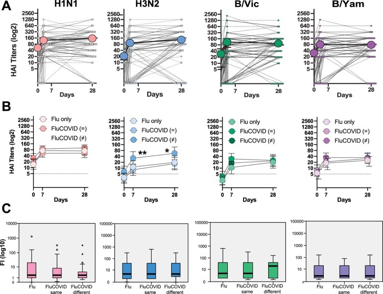
Blood samples were collected longitudinally at the recruitment before single or dual vaccination, and early after (mean, range: 7, 4–12). Another sample was collected after 4 weeks post-vaccination with a mean time of 28 (range 20–34 days). To investigate the induction of antibodies against influenza viruses, we measured hemagglutination–inhibition (HAI) antibodies against all four vaccine reference strains included in the 2022–2023 influenza vaccine: A/Victoria/2570/2019 (H1N1)pdm09-like virus; A/Darwin/9/2021 (H3N2)-like virus; B/Austria/1359417/2021-like virus (B/Victoria lineage); and B/Phuket/3073/2013-like virus (B/Yamagata lineage). Globally, antibody responses were better for influenza A than B viruses in the three groups of vaccination with seroprotection rates of 79.8% for H1N1, 64.5% for H3N2, 56.5% for B/Victoria, 56.5% for B/Yamagata (Figure 1A) and seroconversion rates of 37.5%, 53.1%, 55.5% and 39.1%, respectively at day 28 post-vaccination. Importantly, antibody levels were already high at baseline with Geometric Mean Titers (GMT) (CI 95%) of 54.56 (40.7–68.42), and 22.2 (14.4–30) for influenza A H1N1 and H3N2, respectively; and 30.48 (6.58–54.39) and 31.99 (22.83–41.15) for influenza B from the Victoria and the Yamagata lineages.
Figure 1.
Immune response to influenza in the FluCOVID study. (A) Overall immune response against influenza viruses according to the strains included in the 2022–2023 vaccine. Hemagglutination inhibitory (HAI) antibodies are shown for each patient. Geometric mean titer (GMT, big dots) and confidence interval (CI 95%) are also shown. Serum samples were examined over two independent experiments for HAI assays at baseline, day 5–7 and day 28. (B) Longitudinal profile as GMT and CI 95% of HAI antibodies against influenza viruses according to vaccination groups -Flu only versus FluCOVID in same ( = ) or different (≠) arm are shown. Independent-Samples Kruskal-Wallis test was performed and Bonferroni correction for multiple tests was applied for pairwise comparisons. Day 7: **p = 0.01. FluCOVID ( = ) versus FluCOVID different (≠) (p = 0.004). Day 28: *p = 0.02. FluCOVID ( = ) versus FluCOVID different (≠) (p = 0.006). Flu versus FluCOVID different (p = 0.049). (C) Boxplot diagram of geometric mean fold rise (GMFR) antibody titers against influenza viruses are shown at day 28. Outliers from the observed distribution are shown.
Next, we investigated whether concomitant administration of mRNA COVID-19 vaccines interfere with seasonal influenza vaccine responses. For this, we compared the longitudinal antibody response and determined fold-increase of antibody titers from baseline levels against influenza viruses according to vaccination groups (Figure 1B and C). Generally, no significant differences were found on HAI titers between single or dual vaccination at any time point. However, the FluCOVID vaccine group administered in different arms showed higher antibody levels for the H3N2 virus at day 7 post-vaccination when compared administration in the same arm, showing GMT (CI95%): FluCOVID ( = ) 37.7, (21.7–53.7) versus FluCOVID (≠) 172.7, (83.9–261.5), p = 0.004. Similarly, antibody levels were significantly higher for the H3N2 virus when administered in different arms compared to the same arm and Flu only group at day 28 post-vaccination: FluCOVID ( = ) 83.8 (43.9–123.7) versus FluCOVID (≠) 220.75(118.19–323.31), p = 0.004; and Flu only 114.0(66.79–161.3) versus FluCOVID (≠) 220.75(118.19–323.3), p = 0.049. These differences were found when comparing the geometric mean of the antibody titers to H3N2 in the longitudinal follow up. However, when analyzing the fold induction (FI) from baseline levels at the same time points (day 7 and day 28) we found no significant differences; and fold induction levels were similar between the Flu only and the Flu/SARS-CoV-2 in the same arm group compared to the Flu/SARS-CoV-2 different arm group. Additionally, no differences for the H1N1, nor influenza B Yamagata or Victoria lineages vaccine components were found when we compared fold-increase (expressed as GMR) from baseline antibody levels between the three vaccination groups. Data is shown in Figure 1C. Seroprotection, seroconversion and antibody levels expressed as GMT and GMR at baseline, and days 7 and 28, according to vaccination groups or in the total cohort are also shown in Supplementary Tables 1–3.
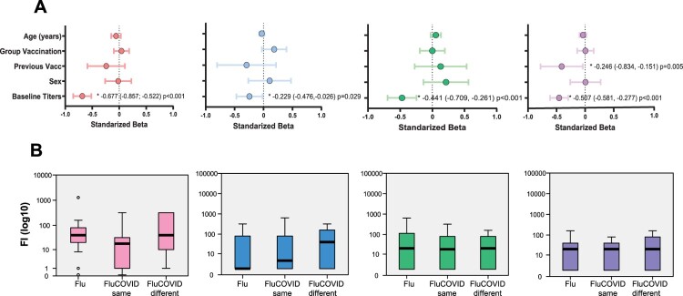
Since many factors such as age, sex or pre-existing immunity can play a role in differential responses to seasonal influenza vaccine among adults [10,17], we next decided to investigate the impact of such factors on the immune response to influenza vaccine. First, we investigated antibody responses against influenza and SARS-CoV-2 according to sex. Only baseline titers to B/Victoria influenza virus as well as B/Yamagata were significantly different in male compared to females (Supplementary Table 4). Additionally, fold induction of the antibody levels according to sex for the different influenza strains included in the vaccine were only significant different to the B/Victoria virus at day 7 post vaccination (Supplementary Table 5). Second, we defined pre-existing immunity as HAI baseline antibody levels higher than 10 and then calculated the frequency of pre-existing immunity among the different vaccination groups (Table 2). Next, we investigated the role of these and other independent factors that might be influencing fold-increase of antibody levels at day 28 after administration of the single -Flu- or dual -FluCOVID- vaccines. A multivariate linear regression for each influenza subtype included in the vaccine was performed, including age, sex, previous history of influenza vaccination (defined as vaccination in 2021–2022 season), baseline HAI titers and vaccination group. Our results indicated that baseline HAI titers significantly impact the induction of anti-influenza antibodies by day 28 for the four vaccine antigens (Figure 2A), but especially marked for the H1N1 vaccine component with a beta coefficient of −0.67 (CI95% −0.86; −0.52) (p < 0.001). Therefore, and to account for any cofounding effect due to presence of baseline antibody levels, we next compared fold-increase at day 28 among all three vaccination groups only in those subjects without influenza virus pre-existing immunity before vaccination. Figure 2B shows no significant differences between groups. However, in the absence of pre-existing immunity, subjects that received the FluCOVID vaccine in different arm showed better fold-increase for influenza A virus H3N2 than those receiving Flu vaccines only.
Table 2.
Frequency of vaccinees with pre-existing immunity on the FluCOVID vaccine study for each vaccine antigen and according to vaccination groups. Pre-existing immunity was defined as baseline hemagglutination–inhibition (HAI) antibody levels higher than 10.
| Flu only N = 46 | FluCOVID ( = ) N = 42 | FluCOVID (≠) N = 40 | Total N = 128 | |
|---|---|---|---|---|
| H1N1∞ | 28 (60.9%) | 28 (66.7%) | 33 (82.5%) | 89 (69.5%) |
| H3N2 | 14 (30.4%) | 10 (23.8%) | 18 (45%) | 42 (32.8%) |
| B/Vic | 10 (21.7%) | 13 (31%) | 9 (22.5%) | 32 (25%) |
| B/Yam | 19 (41.3%) | 21 (50%) | 21 (52.5%) | 61 (47.7%) |
∞p = 0.08. Independent-Samples Kruskal-Wallis test was performed.
Figure 2.
Impact of pre-existing immunity on the immune response to the QIV influenza vaccine. (A) Multivariate linear regression model of factors correlated with fold increase of antibodies on day 28. The model is adjusted by potential confounding factors to estimate the independent effect of baseline antibodies (titer), age (years) and influenza vaccination in the previous season on induction of antibodies (yes versus no) against each influenza virus included in the vaccine. The standardized beta coefficient and CI 95% for each significant covariate are shown. (B) Boxplot diagram of geometric mean fold rise (GMFR) antibody titers against influenza viruses in vaccinees without pre-existing immunity are shown on day 28. Outliers from the observed distribution are shown. Pre-existing immunity was defined as HAI baseline antibody levels higher than 10. Outliers from the observed distribution are shown.
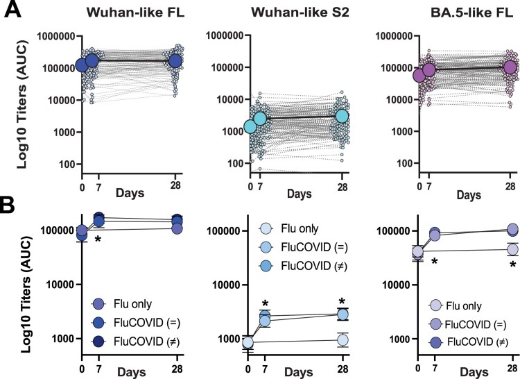
Next, we assessed whether anti-SARS-CoV-2 antibody responses could be different between the FluCOVID vaccination groups. For this we quantified immunoglobulin G (IgG) levels against the spike (S) protein of both Wuhan-like -full length, FL; or S2 domain- and Omicron BA.5 full length S. IgG levels were quantified as area under the curve (AUC) by plotting normalized optical density (OD) values against the reciprocal serum sample dilutions for ELISAs. Then the longitudinal antibody profile of each individual patient together with the GMT (CI 95%) at each time point for AUC ELISA were plotted for the total FluCOVID study; or according to vaccination groups (Figure 3A and B). As expected, no increase on antibody levels against any of the antigens tested were found for the Flu only group, while both FluCOVID vaccination groups showed similar antibody profile for Wuhan-like or Omicron BA.5 antigens, independently of vaccination occurring in the same versus different arm. Antibody levels expressed as GMT and Geometric Mean Ratio (GMR) at baseline, day 7 and day 28 according to groups or in the total cohort are also shown in Supplementary Tables 6, 7 and 8.
Figure 3.
Longitudinal antibody response to SARS-CoV-2 antigens. (A) Longitudinal profile of antibodies against Wuhan-like -full length, FL; or S2 domain- and Omicron BA.5 full length spike (S) proteins in the total cohort. Calculated AUC at each time is shown to quantify changes over time for each individual (small dots) against immunoglobulin G (IgG). Antibody titer was quantified as area under the curve (AUC) after serial serum dilution for each sample. Geometric mean titer (GMT, big dots) and confidence interval (CI 95%) are also shown. (B) Antibody profile as GMT and CI 95% of ELISA titers against SARS-CoV-2 antigens. Independent-Samples Kruskal-Wallis test was performed and Bonferroni correction for multiple tests was applied for pairwise comparisons.
Influenza-specific T-cells and monocytes immune response
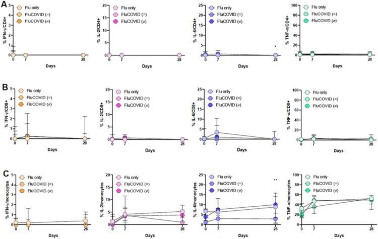
Finally, to evaluate whether the administration of one of these vaccine combinations may impact the cell response to influenza viruses, specific monocytes and T-cells against influenza virus were analysed. As shown in Figure 4, the percentage of CD4 + and CD8+ T cells expressing INF-ɣ, IL-2, IL-6 and TNF-α was similar among the three vaccination groups except for the CD4+ T cells expressing IL-6 at day 28 (p = 0.020): FluCOVID ( = ) versus Flu (p = 0.047), and FluCOVID ( = ) versus FluCOVID (≠) (p = 0.010). Likewise, the percentage of monocytes expressing the selected cytokines was similar between groups with the exception of IL-6 at day 28: FluCOVID (≠) versus the FluCOVID ( = ) (7.84% vs. 3.01%, p = 0.010) and Flu versus FluCOVID ( = ) (5.53% vs. 3.01%, p = 0.047). Next, we analysed the percentage of patients with CD4+, CD8+ T cells and monocytes expressing the selected cytokines. Only minor differences were found (Supplementary Table 9). The percentage of patients with CD4+ T-cells expressing IL-6 at day 7 was higher in FluCOVID (≠) versus FluCOVID ( = ) (71.1% vs. 39%, p = 0.007). However, this difference was not maintained at day 28, and percentage of patients with CD4+ T-cells expressing IL-6 at was higher in the FluCOVID ( = ) group (16.2% vs. 41.5%, p = 0.020). Similarly, the percentage of patients with monocytes expressing IL-6 was higher in the Flu only versus FluCOVID ( = ) (80% vs. 58.5%, p = 0.019) and the FluCOVID (≠) versus FluCOVID ( = ) (86.8% vs. 58.5%, p = 0.010).
Figure 4.
Immune cellular response to influenza in the FluCOVID study. (A) Comparison of the median and IQR of CD4+ T cells percentage expressing INF-ɣ, IL-2, IL-6 and TNF-α. (B) Comparison of the median and IQR of CD8+ T cells percentage expressing INF-ɣ, IL-2, IL-6 and TNF-α. (C) Comparison of the median and IQR of monocytes percentage expressing INF-ɣ, IL-2, IL-6 and TNF-α. The three groups of vaccination are shown at baseline, day 7 and day 28: volunteers vaccinated with FLU vaccine only or vaccinated with both seasonal influenza vaccine and bivalent COVID19 mRNA vaccine in the same arm, FLU/COVID ( = ), or different arms, FLU/COVID (≠). Independent-Samples Kruskal-Wallis test was performed and Mann-Whitney U test was applied for pairwise comparisons: *p = 0.020, Flu versus Flu/COVID ( = ) (p = 0,047) and Flu/COVID ( = ) versus Flu/COVID (≠) (p = 0,010); **p = 0.018, Flu versus Flu/COVID ( = ) (p = 0,047) and Flu/COVID ( = ) versus Flu/COVID (≠) (p = 0,010).
Discussion
The present study examines the immunogenicity and safety of concomitant administration of seasonal influenza and COVID-19 mRNA vaccines. No safety concerns or immune interferences were found. However, our data suggest that dual vaccination could be favored by administering vaccines in different arms since subjects from the FluCOVID vaccine group showed significantly higher antibody titers for the H3N2 vaccine component than those receiving FLU vaccines only, or FluCOVID in the same arm in the longitudinal follow up. However, when analyzing the fold induction (FI) from baseline levels at the same time points (day 7 and day 28) we found no significant differences; and fold induction levels were similar between the Flu only and the Flu/SARS-CoV-2 in the same arm group compared to the Flu/SARS-CoV-2 different arm group. This lack of statistical differences in fold increase levels could be explained by differences on baseline antibody titers among the vaccination groups for H3N2 virus. As indicated in the Supplementary Table 1 the Flu-SARS-CoV-2 different arm group showed higher baseline antibody titers (GMT 6.6) compared to the Flu only (GMT 3.28) and Flu-SARS-CoV-2 same arm group (2.82) (*Kruskal Wallis test p = 0.09). As for the specific cellular immune response, only a few differences were observed at specific timepoints for the three vaccination groups. Specifically, different levels of IL-6 secretion were found for CD4 T-cells and monocytes among the different vaccination groups. It is known that activation of inflammatory responses are crucial for the initiation of innate immunity pathways that can lead to the induction of good adaptative immune responses after vaccination [18]. Therefore, the observed differences on IL-6 cytokine production could potentially influence the antibody mediated immune response after single or dual vaccination with the influenza or COVID-19 vaccine. However, more granular studies are necessary to understand the specific role of IL-6 secretion after influenza vaccination.
Our results support the concomitant administration of seasonal influenza and COVID-19 mRNA vaccines in the upcoming vaccination campaigns. For one side, dual vaccination will ultimately favor vaccine uptake and coverage. On the other side it will provide better protection against both influenza and SARS-CoV-2 infections and severe disease outcomes. Certainly, coadministration of seasonal influenza and other respiratory pathogens vaccines is not an unexplored field. Examples include not only viruses, but also bacterial pathogens such as certain pneumococcal serotypes [19–21]. While some of these studies showed a slight decrease on antibody titers against pneumococcal antigens, the benefits added still overcome the relative reductions observed since administration of both vaccines have proved to reduce the risk of pneumonia complications [22–24]. Similarly, dual administration of influenza and COVID-19 vaccines could ease not only the clinical impact of both diseases at an individual level but also help reduce the burden on national healthcare systems while showing no differences on immune response.
Our study also adds new data on safety and reactogenicity of simultaneous administration of both influenza and COVID-19 vaccines. We found that side effects were lower in the Flu only vaccination group. COVID-19 vaccines alone have shown to cause high frequency of both systemic and local reactogenicity – 76% and 24%, respectively – [25]. In contrast, systemic and local side effects to inactivated influenza vaccines have shown to be less common, with frequencies of 11% and 17.5%, respectively [26]. Therefore, is not surprising that subjects from the FluCOVID vaccine groups showed an increase in the rate of systemic side effects. Nonetheless, only mild symptoms such as fever and chills were self-reported, and no other safety concerns were found consistent with other reports in the literature [12,13,15,25]. Still, our conclusions are limited by the fact that vaccine-related side effects were self-reported by the participants, which could be subjected to potential bias. Additionally, no comparison with a COVID-19 vaccine only group was possible in this study. Nonetheless, some preliminary data showed no significant differences in reactogenicity and immunogenicity between those subjects receiving concomitant vaccination compared to those that received mRNA-COVID19 vaccine alone [12,14]. Further research is needed to investigate the immune responses against SARS-CoV-2 spike protein upon dual vaccination with mRNA COVID-19 and seasonal influenza vaccines.
In conclusion, concomitant administration of the mRNA COVID-19 and seasonal influenza vaccines is safe, produced a mild reactogenicity profile and maintained an adequate immune response, with a better immunological response to influenza A H3N2 when administered in different arms. This study provides population-based evidence to inform decisions about practice and policy for future influenza and COVID-19 vaccination campaigns.
Supplementary Material
Acknowledgements
We thank Richard Cadagan for excellent technical assistance. This work was partly supported by Consejería de Transformación Económica, Industria, Conocimiento y Universidades and FEDER. It is also partly supported by CRIPT (Center for Research on Influenza Pathogenesis and Transmission), a NIAID funded Center of Excellence for Influenza Research and Response (CEIRR, contract # 75N93021C00014) to A.G.-S and T.A.; and by SEM-CIVIC, a NIAID funded Collaborative Influenza Vaccine Innovation Center (contract #75N93019C00051) to A.G.-S. T. A was also funded by the American Lung Association. JSC and EC also were supported by CIBERINFEC – Consorcio Centro de Investigación Biomédica en Red, Instituto de Salud Carlos III, Ministerio de Ciencia e Innovación and Unión Europea – NextGenerationEU (CB21/13/00006).
Funding Statement
This work was supported by American Lung Association: [COVID-1034091]. Centro de Investigación Biomedica en Red de Enfermedades Infecciosas, CIBERINFEC, Instituto de Salud Carlos III, Ministerio de Ciencia e Innovacion and Union Europea – NextGeneration EU (Grant Number CB21/13/00006); Consejeria de Economia, Conocimiento, Empresas y Universidad, Secretaria General de Universidades, Investigacion y Tecnologia, Junta de Andalucia, Spain (P18-RT-3320). SEM-CIVIC National Institute of Allergy and Infectious Diseases: [Grant Number 75N93019C00051]. CEIRR-CRIPT: [75N93021C00014].
Disclosure statement
No potential conflict of interest was reported by the author(s).
Author contributions
T.A, J.S–C, A.G.-S and E.C. conceived, designed and supervised the study. T.A supervised and provided training to A.R. A.R. performed all the antibody quantification experiments: grown influenza viruses for hemagglutination inhibition assays, performed ELISA assays against Wuhan-like and Omicron spikes and calculated antibody titers with help of T.A. C. S-R, MM. M-G, MJ. S–C, E.C included the volunteers and were responsible of their clinical follow-up. M.B processed samples and collected clinical data, performed flow cytometry experiments, analyzed data, wrote manuscript and provided figures. A. E provided influenza virus stocks. T.A analyzed data, wrote the manuscript and prepared figures. All authors reviewed the manuscript.
Declaration of interest statement
The A.G.-S. laboratory has received research support from GSK, Pfizer, Senhwa Biosciences, Kenall Manufacturing, Blade Therapeutics, Avimex, Johnson & Johnson, Dynavax, 7Hills Pharma, Pharmamar, ImmunityBio, Accurius, Nanocomposix, Hexamer, N-fold LLC, Model Medicines, Atea Pharma, Applied Biological Laboratories and Merck, outside of the reported work. A.G.-S. has consulting agreements for the following companies involving cash and/or stock: Castlevax, Amovir, Vivaldi Biosciences, Contrafect, 7Hills Pharma, Avimex, Pagoda, Accurius, Esperovax, Farmak, Applied Biological Laboratories, Pharmamar, CureLab Oncology, CureLab Veterinary, Synairgen, Paratus and Pfizer. A.G.-S. has been an invited speaker in meeting events organized by Seqirus, Janssen, Abbott and Astrazeneca. A.G.-S. is inventor on patents and patent applications on the use of antivirals and vaccines for the treatment and prevention of virus infections and cancer, owned by the Icahn School of Medicine at Mount Sinai, New York.
References
- 1.Zhu N, Zhang D, Wang W, et al. A novel coronavirus from patients with pneumonia in China, 2019. N Engl J Med. 2020;382(8):727–733. doi: 10.1056/NEJMoa2001017 [DOI] [PMC free article] [PubMed] [Google Scholar]
- 2.Morens DM, Fauci AS.. Emerging pandemic diseases: how we got to COVID-19. Cell. 2020;182(5):1077–1092. doi: 10.1016/j.cell.2020.08.021 [DOI] [PMC free article] [PubMed] [Google Scholar]
- 3.Escalera A, Gonzalez-Reiche AS, Aslam S, et al. Mutations in SARS-CoV-2 variants of concern link to increased spike cleavage and virus transmission. Cell Host Microbe. 2022;30(3):373–387 e7. doi: 10.1016/j.chom.2022.01.006 [DOI] [PMC free article] [PubMed] [Google Scholar]
- 4.Krammer F. The human antibody response to influenza A virus infection and vaccination. Nat Rev Immunol. 2019;19(6):383–397. doi: 10.1038/s41577-019-0143-6 [DOI] [PubMed] [Google Scholar]
- 5.Krammer F, Smith GJD, Fouchier RAM, et al. Influenza. Nat Rev Dis Primers. 2018;4(1):3. doi: 10.1038/s41572-018-0002-y [DOI] [PMC free article] [PubMed] [Google Scholar]
- 6.Yap C, Ali A, Prabhakar A, et al. Comprehensive literature review on COVID-19 vaccines and role of SARS-CoV-2 variants in the pandemic. Ther Adv Vaccines Immunother. 2021;9:25151355211059791. [DOI] [PMC free article] [PubMed] [Google Scholar]
- 7.Krammer F. The role of vaccines in the COVID-19 pandemic: what have we learned? Semin Immunopathol. 2023 Jul 12. [DOI] [PMC free article] [PubMed] [Google Scholar]
- 8.Kubale JT, Frutos AM, Balmaseda A, et al. High co-circulation of influenza and severe acute respiratory syndrome coronavirus 2. Open Forum Infect Dis. 2022;9(12):ofac642. doi: 10.1093/ofid/ofac642 [DOI] [PMC free article] [PubMed] [Google Scholar]
- 9.Rao S, Armistead I, Tyler A, et al. Respiratory syncytial virus, influenza, and coronavirus disease 2019 hospitalizations in children in Colorado during the 2021–2022 respiratory virus season. J Pediatr. 2023;260:113491. doi: 10.1016/j.jpeds.2023.113491 [DOI] [PMC free article] [PubMed] [Google Scholar]
- 10.Aydillo T, Escalera A, Strohmeier S, et al. Pre-existing hemagglutinin stalk antibodies correlate with protection of lower respiratory symptoms in flu-infected transplant patients. Cell Rep Med. 2020;1(8):100130. doi: 10.1016/j.xcrm.2020.100130 [DOI] [PMC free article] [PubMed] [Google Scholar]
- 11.Andrews N, Tessier E, Stowe J, et al. Duration of protection against mild and severe disease by COVID-19 vaccines. N Engl J Med. 2022;386(4):340–350. doi: 10.1056/NEJMoa2115481 [DOI] [PMC free article] [PubMed] [Google Scholar]
- 12.Lazarus R, Baos S, Cappel-Porter H, et al. Safety and immunogenicity of concomitant administration of COVID-19 vaccines (ChAdOx1 or BNT162b2) with seasonal influenza vaccines in adults in the UK (ComFluCOV): a multicentre, randomised, controlled, phase 4 trial. Lancet. 2021;398(10318):2277–2287. doi: 10.1016/S0140-6736(21)02329-1 [DOI] [PMC free article] [PubMed] [Google Scholar]
- 13.Hall KT, Stone VE, Ojikutu B.. Reactogenicity and concomitant administration of the COVID-19 booster and influenza vaccine. JAMA Netw Open. 2022;5(7):e2222246. doi: 10.1001/jamanetworkopen.2022.22246 [DOI] [PubMed] [Google Scholar]
- 14.Janssen C, Mosnier A, Gavazzi G, et al. Coadministration of seasonal influenza and COVID-19 vaccines: a systematic review of clinical studies. Hum Vaccin Immunother. 2022;18(6):2131166. doi: 10.1080/21645515.2022.2131166 [DOI] [PMC free article] [PubMed] [Google Scholar]
- 15.Wagenhauser I, Reusch J, Gabel A, et al. Immunogenicity and safety of coadministration of COVID-19 and influenza vaccination. Eur Respir J. 2023 Jan;61(1). [DOI] [PMC free article] [PubMed] [Google Scholar]
- 16.Dulfer EA, Geckin B, Taks EJM, et al. Timing and sequence of vaccination against COVID-19 and influenza (TACTIC): a single-blind, placebo-controlled randomized clinical trial. Lancet Reg Health Eur. 2023;29:100628. doi: 10.1016/j.lanepe.2023.100628 [DOI] [PMC free article] [PubMed] [Google Scholar]
- 17.Cordero E, Aydillo TA, Perez-Ordonez A, et al. Deficient long-term response to pandemic vaccine results in an insufficient antibody response to seasonal influenza vaccination in solid organ transplant recipients. Transplantation. 2012;93(8):847–854. doi: 10.1097/TP.0b013e318247a6ef [DOI] [PubMed] [Google Scholar]
- 18.Aydillo T, Gonzalez-Reiche AS, Stadlbauer D, et al. Transcriptome signatures preceding the induction of anti-stalk antibodies elicited after universal influenza vaccination. NPJ Vaccines. 2022;7(1):160. doi: 10.1038/s41541-022-00583-w [DOI] [PMC free article] [PubMed] [Google Scholar]
- 19.Song JY, Cheong HJ, Hyun HJ, et al. Immunogenicity and safety of a 13-valent pneumococcal conjugate vaccine and an MF59-adjuvanted influenza vaccine after concomitant vaccination in ⩾60-year-old adults. Vaccine. 2017 Jan 5;35(2):313–320. [DOI] [PubMed] [Google Scholar]
- 20.Ofori-Anyinam O, Leroux-Roels G, Drame M, et al. Immunogenicity and safety of an inactivated quadrivalent influenza vaccine co-administered with a 23-valent pneumococcal polysaccharide vaccine versus separate administration, in adults >/=50years of age: results from a phase III, randomized, non-inferiority trial. Vaccine. 2017;35(46):6321–6328. doi: 10.1016/j.vaccine.2017.09.012 [DOI] [PubMed] [Google Scholar]
- 21.Frenck RW, Gurtman A, Rubino J, et al. Randomized, controlled trial of a 13-valent pneumococcal conjugate vaccine administered concomitantly with an influenza vaccine in healthy adults. Clin Vaccine Immunol. 2012;19(8):1296–1303. doi: 10.1128/CVI.00176-12 [DOI] [PMC free article] [PubMed] [Google Scholar]
- 22.Seki M. Strategies for geriatric pneumonia in healthcare facilities – How effective is combined influenza and pneumococcal vaccination? Int J Gen Med. 2020;13:663–666. doi: 10.2147/IJGM.S264835 [DOI] [PMC free article] [PubMed] [Google Scholar]
- 23.Hanafy AS, Seleem WM, Elkattawy HA.. Potential impact of combined influenza and pneumococcal vaccines on the severity of respiratory illness in COVID-19 infection among type 2 diabetic patients. Clin Exp Med. 2023 Feb;23(1):141–150. [DOI] [PMC free article] [PubMed] [Google Scholar]
- 24.Hedlund J, Christenson B, Lundbergh P, et al. Effects of a large-scale intervention with influenza and 23-valent pneumococcal vaccines in elderly people: a 1-year follow-up. Vaccine. 2003;21(25-26):3906–3911. doi: 10.1016/S0264-410X(03)00296-2 [DOI] [PubMed] [Google Scholar]
- 25.Haas JW, Bender FL, Ballou S, et al. Frequency of adverse events in the placebo arms of COVID-19 vaccine trials: A systematic review and meta-analysis. JAMA Netw Open. 2022;5(1):e2143955. doi: 10.1001/jamanetworkopen.2021.43955 [DOI] [PMC free article] [PubMed] [Google Scholar]
- 26.Govaert TM, Dinant GJ, Aretz K, et al. Adverse reactions to influenza vaccine in elderly people: randomised double blind placebo controlled trial. Br Med J. 1993;307(6910):988–990. doi: 10.1136/bmj.307.6910.988 [DOI] [PMC free article] [PubMed] [Google Scholar]
Associated Data
This section collects any data citations, data availability statements, or supplementary materials included in this article.


