It is increasingly becoming clear that the tumor immune microenvironment (TME) plays a critical role in tumor progression and drug resistance. For instance, we and others have identified that the presence of tumor supportive M2-polarized macrophages associates with the poorest prognosis in patients with acute myeloid leukemia (AML).1,2 Functionally, we could show for the first time that M2-polarized macrophages were able to increase homing, self-renewal potential, alter the metabolome, and increase transformation capacity of leukemic blasts in vivo.1 Thus, it would be of clinical interest to target this tumor-supportive cell population in order to improve outcome in AML patients. Given the inherent plasticity of macrophages, several studies have aimed to identify factors that would repolarize tumor-supportive M2 macrophages into tumor-suppressive M1 macrophages. Blockage of TIGIT, as well as knockout of GFI1 or upregulation of IRF7 were shown to promote an M2 to M1 repolarization.3-5 In the context of AML, however, it is possible that macrophages also carry leukemic mutations,1,6 thereby potentially impacting on macrophage function even in case M1 repolarization would be successful. Thus, an alternative to macrophage repolarization would be to target this subpopulation via drug-induced apoptosis. Previously, we identified the NAMPT inhibitor KPT-9274 as well as the antibiotic thiostrepton as potential candidates. Thiostrepton targets the oncogene FOXM1,7 but was recently also suggested to have M2 to M1 macrophage-repolarizing activity.8 Furthermore, thiostrepton was reported to have anti-cancer effects in solid tumors9 ,10 and lymphoid malignancies, such as multiple myeloma.11 Therefore, we investigated the efficacy of thiostrepton in AML and focused on its mode of action.
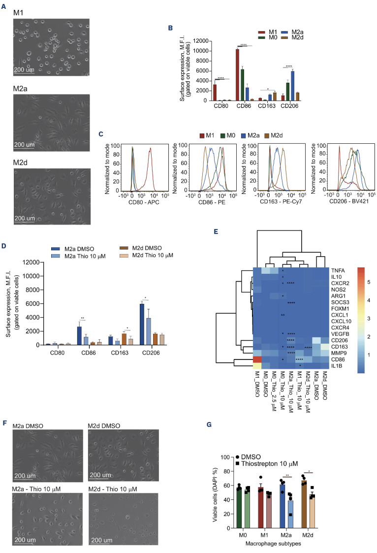
First, we evaluated whether thiostrepton would drive M2 to M1 macrophage repolarization. Human peripheral blood mononuclear cell (PBMC)-derived monocytes were isolated from healthy PBMC by adherence and cultured for 6 days in M-CSF (50 ng/mL). Thereafter, macrophages were either kept in M-CSF as non-polarized M0 macrophages, or they were differentiated towards M1 (20 ng/mL interferon γ [IFNγ] and 100 ng/mL lipoplysaccharide [LPS]) or M2 macrophages (20 ng/mL interleukin [IL] 4 for M2a and 20 ng/mL IL6 for M2d) for 24 hours (hrs). Macrophage phenotypes were characterized morphologically (Figure 1A), by flow cytometry (Figure 1B, C; Online Supplementary Figure 1A; whereby M1-polarized macrophages expressed CD80 and CD86, and M2-polarized macrophages expressed CD163 and CD206) and by gene expression profiles (Figure 1E). Both CD80, CD86 as well as CD163, CD206 are well established markers for M1 and M2 macrophages, respectively, whose expression is induced upon cytokine polarization.12 Upregulation of CD80 and CD86 is associated with bactericidal activities, while increased expression of CD163 and CD206 is linked to immunomodulation and tissue repair.12,13 In order to study the effects of thiostrepton, non-polarized M0 macrophages, or M2a- or M2d-polarized macrophages were treated for 24 hrs and results were compared to vehicle-treated (0.01% dimethyl sulfoxide [DMSO]) macrophages. When non-polarized M0 macrophages were treated with thiostrepton, we observed a slight decrease in CD206 expression in a dose-dependent manner (Online Supplementary Figure S1B). At a dose of 10 mM, thiostrepton did not upregulate any of the M1 markers in M2-polarized macrophages. The only significant differences that were observed were slight reductions in CD86 mean fluorescence intensity (MFI) and CD206 MFI in M2a-polarized macrophages as well as CD163 MFI in M2d-polarized macrophages (Figure 1D; Online Supplementary Figure S1C). Gene expression analysis of a wide range of M1 and M2 markers (16 in total) in macrophages treated with thiostrepton provided no clear indication of an M2 to M1 repolarization. Instead several M1 markers (e.g., CD86, IL1B) as well as M2 markers (e.g., CD206, CD163, MMP9) were downregulated upon thiostrepton treatment (Figure 1E). While we are aware that these repolarization studies were performed on healthy PBMC-derived macrophages and not on AML associated macrophages (AAM) macrophages, similar to previous studies,8 these data do not support the notion that thiostrepton acts as an M1-repolarizing agent.
Next, we wished to study how thiostrepton would impact on M2 macrophage function. AML and lymphoma cell lines were cultured on murine and human M2- polarized macrophages that were pretreated with thiostrepton for 12 hrs or 9 days, to study direct and long-term effects. As controls, leukemic cells were cultured on M1-polarized macrophages, which impaired cell proliferation, in particular of AML cell lines (Online Supplementary Figure S1D). However, thiostrepton pretreatment of M2-polarized macrophages did not convert these into tumor-suppressive macrophages (Online Supplementary Figure S1D). No apoptosis was induced when AML cells were grown on thiostrepton-pretreated human M2d macrophages (Online Supplementary Figure S1E), and proliferation was also not impaired when murine thiostrepton-pretreated macrophages were used (Online Supplementary Figure S1F). Previous studies showed that the secretion of lactate can repolarize M1 to M2 macro-phages.14 Extracellular flux measurements confirmed that lymphoma cell lines were low in oxygen consumption rates (OCR) and tended to be more glycolytic, possibly explaining their increased resistance to M1 macrophages (Online Supplementary Figure S1G).
While we could not find compelling evidence for M2 to M1 macrophage reprogramming by thiostrepton, we did note that the viability of M2 macrophages was affected at the highest dose (10 mM) of thiostrepton (Figure 1F, G). Loss in viability was also accompanied by morphological changes, whereby thiostrepton-treated macrophages displayed a round shaped appearance compared to the long stretched-out macrophages treated with vehicle (Figure 1F). Next, we wondered whether thiostrepton would also exhibit antileukemic potential towards leukemic cells directly.
Figure 1.
Thiostrepton treatment induces macrophage cytotoxicity but not M2 to M1 repolarization. (A) Human macrophages were cultured for 24 hours (hrs) in the presence of lipopolysaccharide (LPS) (100 ng/mL) and interferon γ (IFNγ) (20 ng/mL) for M1 polarization, interleukin (IL)4 (20 ng/mL) for M2a polarization, and IL6 (20 ng/mL) for M2d polarization. Representative pictures of M1, M2a and M2d macrophage morphology. Scale bar: 200 mm. (B) Bar plot comparing the levels (mean fluorescence intensity [MFI]) of M2 (CD163 and CD206) and M1 (CD80 and CD86) markers measured by flow cytometery measurements (fluorescence-activated cell sorting [FACS] in healthy macrophages activated with respective cytokines for 24 hrs. M0 macrophages were generated with 50 ng/ mL of MCSF (upper panels). (C) Representative histograms of macrophage marker expression in different macrophage subtypes. (D) Bar plot comparing the levels (MFI) of M2 (CD163 and CD206) and M1 (CD80 and CD86) markers measured by FACS in healthy activated M2a and M2d macrophages treated with dimethyl sulfoxide (DMSO) or thiostrepton (Thio) (10 mM) for 24 hrs. (E) Heatmap displaying the transcript levels of the indicated genes in M1, M0, M2a and M2d macrophages treated with vehicle (DMSO) or thiost-repton (10 μM) for 24 hrs. **P<0.01, ***P<0.001 indicate significance compared to all other groups. Genes in blue indicate M2-associated genes and genes in red indicate M1-associated genes. (F) Cell morphologies of M2a and M2d activated macrophages treated with vehicle or thiostrepton (10 mM) for 24 hrs. Scale bar: 200 mm. (G) Bar plot comparing the viability (DAPI- cells) of M0, M1, M2a, and M2d macrophages treated with thiostrepton and vehicle for 24 hrs. One-way (F, left panel) or two-way (B-D) analysis of variance (ANOVA); *P<0.05, **P<0.01, ***P<0.001. (F, right panel) Wilcoxon signed rank test (two-sided), *P<0.05.
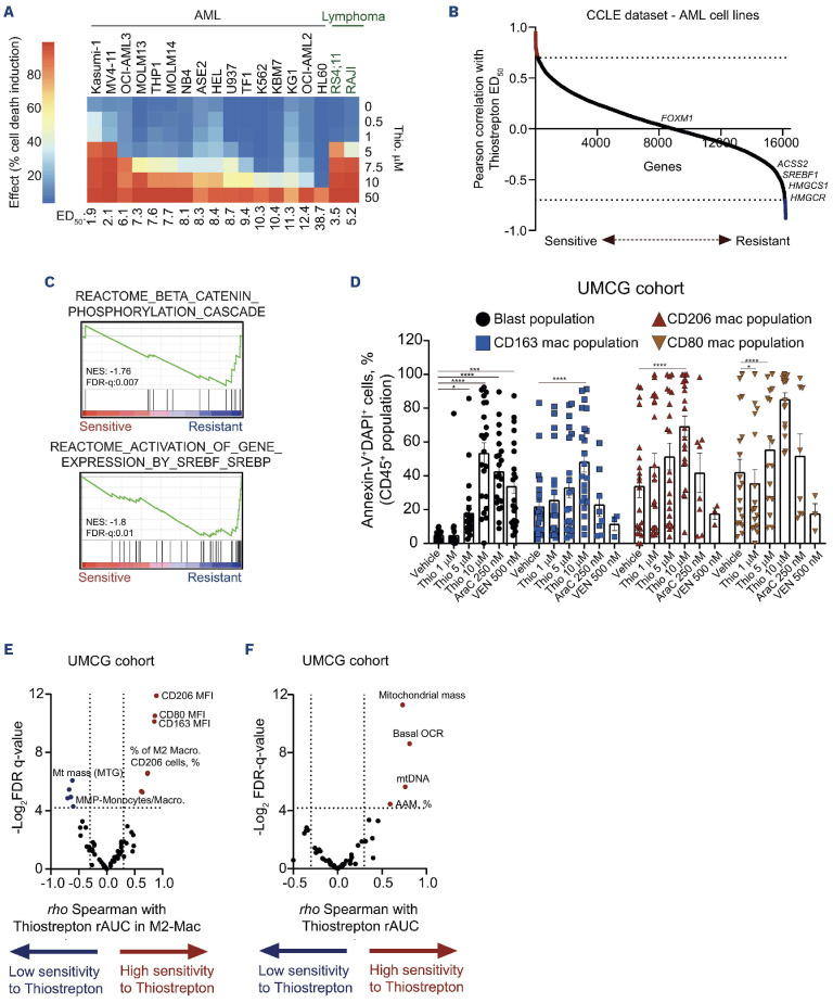
Indeed, thiostrepton also displayed cytotoxic effects in leukemic cell lines with median effective dose (ED50) values ranging from 1.91 mM to 38.67 mM (n=16 AML cell lines; n=2 B-cell acute lymphoblastic leukemia [B-ALL]/lymphoma cell lines; Figure 2A). Similar observations were made by a recent publication showing thiostrepton-mediated apoptosis in a panel of B-pre-ALL cell lines.15 In order to unravel the molecular mechanisms underlying thiostrepton sensitivity in AML, we ranked AML cells treated with thiostrepton from most sensitive to most resistant based on the continuous ED50 values obtained, which we then correlated with the gene expression data retrieved from the CCLE RNA sequencing of the treated AML cell lines (Figure 2B). Gene ontology (GO) and gene set enrichment analysis (GSEA) indicated that cells resistant to thiostrepton displayed increased expression of genes regulated by the SREBP family of transcription factor, whereas sensitivity was linked to increased mitochondrial metabolism, fatty acid metabolism and oxidative phosphorylation (OXPHOS) terms (Online Supplementary Figure S1H; Figure 2C).
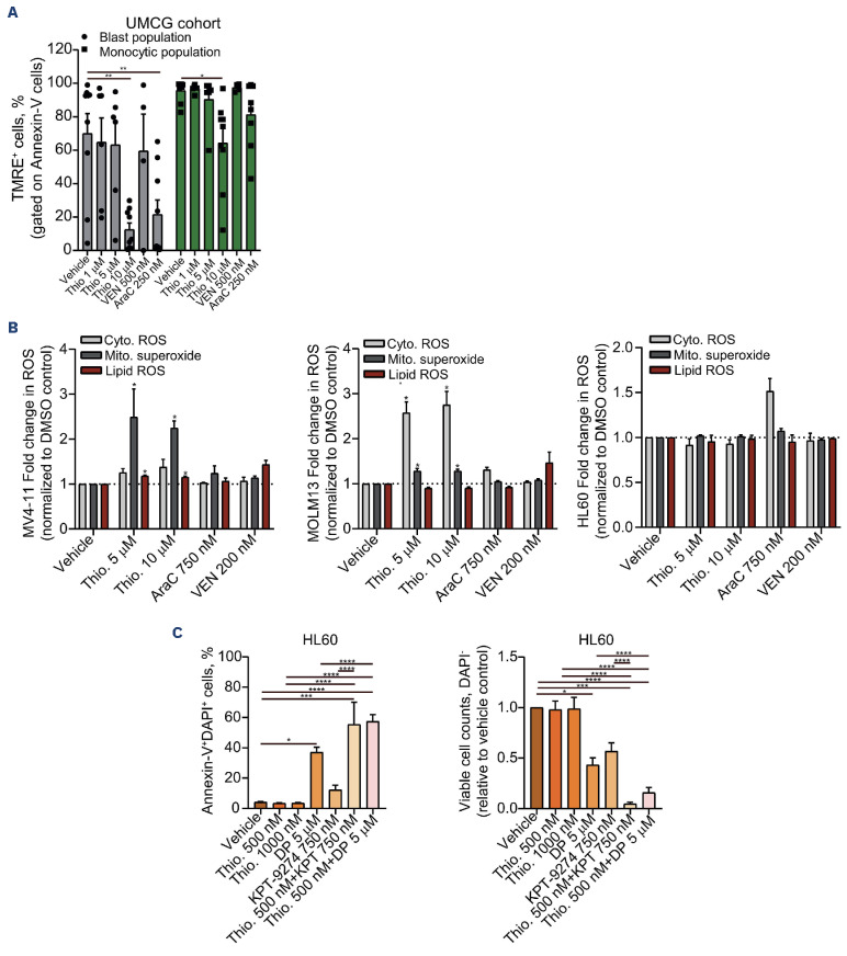
In order to investigate whether thiostrepton treatment would target both the tumor cells as well as their supportive microenvironment, we used our previously published workflow to investigate cytotoxicity in ex vivo treated primary AML samples1 with which efficacy of drugs can be evaluated on all compartments (Online Supplementary Figure S2A). Thiostrepton induced a significant increase in cell death in a dose-dependent manner in ex vivo treated primary AML blast cells as well as in the myeloid/monocytic tumor supportive subpopulation (n=22 patients) (Figure 2D), while cytarabine (AraC) and venetoclax (VEN) mainly targeted the leukemic blast population. Our results indicate that thiostrepton targets both the M2 (CD163+ and CD206+) as well as the M1 (CD80+) macrophage population in AML patients. Ideally, M1 macrophages would be spared to promote tumor suppression. However, patients with a large population of M2 macrophages typically have low levels of M1 macrophages and it is this patient group that displayed the poorest prognosis,1 and we, therefore, propose that in this patient group thiostrepton treatment could provide therapeutic benefits. Evaluation of healthy CD34+ showed no signs of impaired viability upon thiostrepton treatment and progenitor frequencies were not affected, suggesting that thiostrepton treatment might provide an attractive therapeutic approach (Online Supplementary Figure S2B). We wished to identify AML patients that would benefit most from thiostrepton treatment. Therefore, we correlated various AML features such as the presence, size and polarization state of macrophages, as well as metabolic features such as basal oxygen consumption rate (OCR) and the amount of mitochondria, with sensitivity to thiostrepton. The macrophage/monocytic tumor supportive compartment (Figure 2E) as well as the leukemic blasts themselves (Figure 2F) were most sensitive to thiost-repton-induced apoptosis in AML samples with high M2 macrophage content, increased mitochondrial mass and higher OCR, potentially highlighting an increased sensitivity in AML patients that are more OXPHOS-driven.16 These data are in line with our cell line data in which we observed that the most sensitive cell lines like MV4-11, MOLM13 and THP1 are rather high in OCR, while the least sensitive lines like HL60 are glycolytic.17 We then correlated thiostrepton-sensitivity to RNA-sequencing data we generated for the ex vivo treated primary AML at diagnosis, which again revealed that OXPHOS-driven cells were more sensitive to thiostrepton, while terms such ferroptosis control and SREBP-controlled genes were associated with thiostrepton resistance (Online Supplementary Figure S2C). Subsequent analysis of the mitochondrial membrane potential (measured by TMRE staining) indicated that both thiostrepton (10 mM) and AraC decreased the mitochondrial membrane potential in leukemic blasts, but only thiostrepton was also able to reduce the mitochondrial membrane potential of the macrophage/monocytic subpopulation (Figure 3A). Given the link between thiostrepton insensitivity and increased ferroptosis and gene expression control of processes associated with lipid peroxidation, we questioned whether thiostrepton would induce cell death via the induction of reactive oxygen species (ROS), ultimately resulting in lipid ROS formation and induction of ferroptosis.18 In order to study the role of oxidative stress, we measured the level of mitochondrial superoxide, cytoplasmatic ROS, and lipid ROS using compartment-specific and redox-sensitive fluorescent dyes. Short-term exposure to thiostrepton (24 hrs) increased ROS levels in all three compartments in MV4-11 cells, while in MOLM13 cells we observed a particularly strong upregulation of cytoplasmic ROS (Figure 3B). In stark contrast, thiostrepton was not able to induce ROS in thiostrepton-resistant HL60 cells, while treatment with AraC did induce cytoplasmic ROS (Figure 3B). Finally, considering that resistance to thiostrepton was associated with the activation of SREBF/ SREBP pathway, we decided to test if the Food and Drug Administration-approved phosphodiesterase inhibitor dipyridamole (DP) and/or the NAMPT inhibitor (KPT-9274) could promote cytotoxicity in leukemic cells resistant to thiostrepton. Indeed treatment of HL60 cells with thiostrepton in combination with DP and KPT-9274 induced a significant increase in cell death compared to thiostrepton alone (Figure 3C). SREBP act as key transcriptional regulators of genes involved in lipid homeostasis, associated with lipid detoxification by refueling the monosaturated fatty acid pool. Inhibition of such pathways sensitizes cells to thiostrepton-induced cell death.
Figure 2.
Thiostrepton can target both the leukemic cells and the tumor microenvironment in acute myeloid leukemia. (A) Median effective dose (ED50) of thiostrepton in a panel of acute myeloid leukemia (AML) and B-cell acute lymphoblastic leukemia (B-ALL)/lymphoma cell lines. (B) Hockey stick plot displaying the correlation between RNA-sequencing data from AML cell lines (CCLE dataset) and thiostrepton sensitivity. Expression of SREBP target genes was correlated with thiostrepton insensitivity (C) Gene set enrichment analysis (GSEA) using the Pearson correlations depicted in (B) normalized enrichment score (NES) and false discover rate (FDR-q) are indicated. (D) Ex vivo drug-induced apoptosis of thiostrepton in a set of 22 primary AML samples (left panel). Cells were treated for 72 hours, and apoptosis was evaluated by Annexin-V/ DAPI staining by flow cytometry. Leukemic blast cells were analyzed in the CD45dimCD34+CD117+ population, while macrophages were analyzed in the CD45highCD14+CD163+ population. (E, F) Volcano plot demonstrating the Spearman correlation between the ex vivo thiostrepton-induced apoptosis (reported as reverse area under the curve [rAUC]) on M2 macrophage (E) and AML blasts (F) treated ex vivo in our set of primary AML samples (N=22 samples). Blue dots indicate significant negative correlation and red dot indicate a positive correlation. Bar graphs represent the mean ± standard error of the mean of at least 3 independent experiments. *P<0.05, **P<0.01, ***P<0.001. UMCG: University Medical Center Groningen.
Figure 3.
Decreased mitochondrial membrane potential and increased reactive oxygen species upon thiostrepton treatment. (A) Mitochondrial membrane potential was detected by flow cytometry in acute myeloid leukemia (AML) blasts, gated on CD45+CD34+ (or CD117+ for NPM1 mutant AML)/Annexin V- and the myeloid-associated monocytic population (gated on SSChighCD45highCD163+ cells) using the TMRE staining method. Cells were treated with vehicle dimethyl sulfoxide [DMSO], cytarabine (AraC, 250 nM), venetoclax (VEN, 500 nM), or thiostrepton (1-5-10 mM) for 72 hours (hrs). Bar graphs represent the mean ± standard error of the mean, each point represents a patient sample. (B) Fold change in lipid reactive oxygen species (ROS) (C11-BODIPY), cytoplasmic ROS (DCFDA), and mitochondrial superoxide levels (MitoSOXTM) in MV4-11, MOLM13 and HL60 cells treated with thiostrepton (5-10 mM), cytarabine (AraC, 750 nM) or venetoclax (VEN, 200 nM) for 72 hrs; N=4 independent experiments. (C) Apoptosis levels (left panel) and viable cell counts (relative to vehicle control, right panel) measured by flow cytometry in HL60 cells treated with vehicle or with increasing concentrations of thiostrepton (0.5-1 mM) and/or dipyridamole (DP, 5 mM) or KPT-9274 (750 nM) for 72 hrs using an APC-Annexin V/DAPI staining method. Bar graphs represent the mean ± standard error of the mean of at least 3 independent experiments. *P<0.05, **P<0.01, ***P<0.001. UMCG: University Medical Center Groningen.
Therapeutic approaches aimed at eradicating the tumor-supportive microenvironment are gaining interest. Thiostrepton treatment provides such a therapeutic opportunity, by inducing cytotoxicity in the leukemic blasts but also in the supportive macrophage/monocytic compartment of AML patients. While OXPHOS-driven AML subtypes harboring relatively large M2-macrophage compartments are most sensitive, the thiostrepton-resistant AML subtypes can be sensitized by co-treatment with SREBF/SREBP pathway inhibition.
Supplementary Material
Acknowledgments
The authors would like to thank Dr Emmanuel F Griessinger for kindly providing the MS5, RS4;11, MOLM14 and RAJI cells used in the co-culture assays, as well as some of the leukemic lines used in the study.
Funding Statement
Funding: This investigation was supported by Fundação de Amparo à Pesquisa do Estado de São Paulo (FAPESP, grant no. 2013/08135-2). DAP-M received a fellowship from FAPESP (grant no. 2017/23117-1). IW received a fellowship from FAPESP (grant no. 2015/09228-0). IW and DAP-M were sponsored by the Abel Tasman Talent Program (ATTP) of the Graduate School of Medical Sciences of the University of Groningen/ University Medical Center Groningen (UG/UMCG), the Netherlands.
References
- 1.Weinhäuser I, Pereira-Martins DA, Almeida LY, et al. M2 macrophages drive leukemic transformation by imposing resistance to phagocytosis and improving mitochondrial metabolism. Sci Adv. 2023;9(15):eadf8522. [DOI] [PubMed] [Google Scholar]
- 2.Xu ZJ, Gu Y, Wang CZ, et al. The M2 macrophage marker CD206: a novel prognostic indicator for acute myeloid leukemia. Oncoimmunology. 2020;9(1):1683347. [DOI] [PMC free article] [PubMed] [Google Scholar]
- 3.Yang X, Feng W, Wang R, et al. Repolarizing heterogeneous leukemia-associated macrophages with more M1 characteristics eliminates their pro-leukemic effects. Oncoimmunology. 2018;7(4):e1412910. [DOI] [PMC free article] [PubMed] [Google Scholar]
- 4.Brauneck F, Fischer B, Witt M, et al. TIGIT blockade repolarizes AML-associated TIGIT(+) M2 macrophages to an M1 phenotype and increases CD47-mediated phagocytosis. J Immunother Cancer. 2022;10(12):e004794. [DOI] [PMC free article] [PubMed] [Google Scholar]
- 5.Al-Matary YS, Botezatu L, Opalka B, et al. Acute myeloid leukemia cells polarize macrophages towards a leukemia supporting state in a growth factor independence 1 dependent manner. Haematologica. 2016;101(10):1216-1227. [DOI] [PMC free article] [PubMed] [Google Scholar]
- 6.Klco JM, Spencer DH, Miller CA, et al. Functional heterogeneity of genetically defined subclones in acute myeloid leukemia. Cancer Cell. 2014;25(3):379-392. [DOI] [PMC free article] [PubMed] [Google Scholar]
- 7.Kim TH, Hanh BTB, Kim G, et al. Thiostrepton: a novel therapeutic drug candidate for mycobacterium abscessus infection. Molecules. 2019;24(24):4511. [DOI] [PMC free article] [PubMed] [Google Scholar]
- 8.Hu G, Su Y, Kang BH, et al. High-throughput phenotypic screen and transcriptional analysis identify new compounds and targets for macrophage reprogramming. Nat Commun. 2021;12(1):773. [DOI] [PMC free article] [PubMed] [Google Scholar]
- 9.Liu SX, Zhou Y, Zhao L, et al. Thiostrepton confers protection against reactive oxygen species-related apoptosis by restraining FOXM1-triggerred development of gastric cancer. Free Radic Biol Med. 2022;193(Pt 1):385-404. [DOI] [PubMed] [Google Scholar]
- 10.Takeshita H, Yoshida R, Inoue J, et al. FOXM1-mediated regulation of reactive oxygen species and radioresistance in oral squamous cell carcinoma cells. Lab Invest. 2023;103(5):100060. [DOI] [PubMed] [Google Scholar]
- 11.Trasanidis N, Katsarou A, Ponnusamy K, et al. Systems medicine dissection of chr1q-amp reveals a novel PBX1-FOXM1 axis for targeted therapy in multiple myeloma. Blood. 2022;139(13):1939-1953. [DOI] [PubMed] [Google Scholar]
- 12.Takiguchi H, Yang CX, Yang CWT, et al. Macrophages with reduced expressions of classical M1 and M2 surface markers in human bronchoalveolar lavage fluid exhibit pro-inflammatory gene signatures. Sci Rep. 2021;11(1):8282. [DOI] [PMC free article] [PubMed] [Google Scholar]
- 13.Sica A, Mantovani A. Macrophage plasticity and polarization: in vivo veritas. J Clin Invest. 2012;122(3):787-795. [DOI] [PMC free article] [PubMed] [Google Scholar]
- 14.Noe JT, Rendon BE, Geller AE, et al. Lactate supports a metabolic-epigenetic link in macrophage polarization. Sci Adv. 2021;7(46):eabi8602. [DOI] [PMC free article] [PubMed] [Google Scholar]
- 15.Kuttikrishnan S, Prabhu KS, Khan AQ, Alali FQ, Ahmad A, Uddin S. Thiostrepton inhibits growth and induces apoptosis by targeting FoxM1/SKP2/MTH1 axis in B-precursor acute lymphoblastic leukemia cells. Leuk Lymphoma. 2021;62(13):3170-3180. [DOI] [PubMed] [Google Scholar]
- 16.Erdem A, Marin S, Pereira-Martins DA, et al. Inhibition of the succinyl dehydrogenase complex in acute myeloid leukemia leads to a lactate-fuelled respiratory metabolic vulnerability. Nat Commun. 2022;13(1):2013. [DOI] [PMC free article] [PubMed] [Google Scholar]
- 17.Erdem A, Marin S, Pereira-Martins DA, et al. The glycolytic gatekeeper PDK1 defines different metabolic states between genetically distinct subtypes of human acute myeloid leukemia. Nat Commun. 2022;13(1):1105. [DOI] [PMC free article] [PubMed] [Google Scholar]
- 18.Stockwell BR, Friedmann Angeli JP, Bayir H, et al. Ferroptosis: a regulated cell death nexus linking metabolism, redox biology, and disease. Cell. 2017;171(2):273-285. [DOI] [PMC free article] [PubMed] [Google Scholar]
Associated Data
This section collects any data citations, data availability statements, or supplementary materials included in this article.


