

Abstract
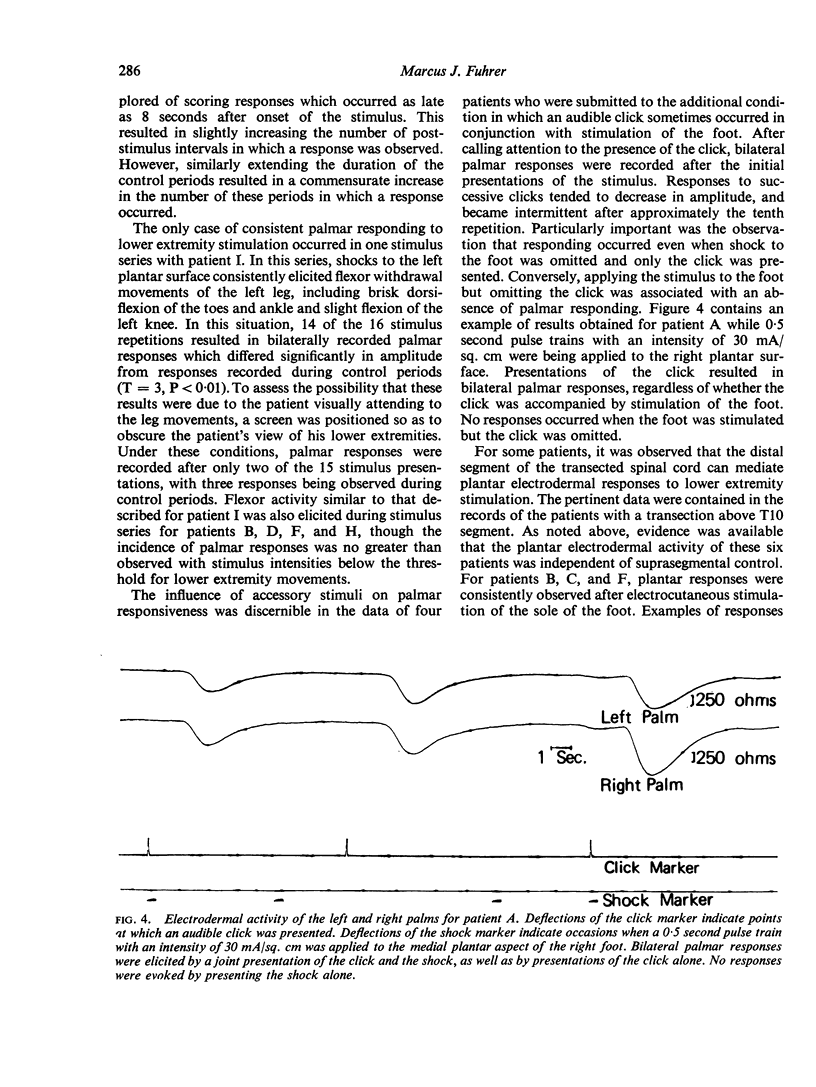
An effort was made to corroborate earlier reports that the central nervous system rostral to a functionally complete transection of the human spinal cord remains responsive to noxious stimulation of sites below the level of the transection. Nine patients were studied who had sustained a functionally complete transection of the thoracic spinal cord between T5 and T11 spinal segments. Noxious electrocutaneous stimulation or intense pressure was applied to a lower extremity while electrodermal activity was recorded concurrently from contralateral palmar sites which were shown to be under normal suprasegmental control. While electrodermal responses were occasionally recorded in the post-stimulus intervals, there was no tendency for these responses to exceed the number or amplitude of responses recorded during stimulus-free control periods. These results were interpreted as suggesting that the few responses observed during the post-stimulus intervals were not evoked by stimulation of the lower extremities and were instead representative of spontaneous electrodermal activity or were related to uncontrolled auditory or visual stimuli accompanying lower extremity stimulation. Additional results highlighted the importance of controlling accessory auditory and visual cues occurring in conjunction with lower extremity stimuli. In some patients with a transection above the sympathetic outflow to the lower extremities, it was shown that electrodermal responses from the plantar aspect of each foot could be elicited reliably by lower extremity stimuli. These results confirmed previously reported evidence that the functionally isolated human spinal cord can reflexly mediate electrodermal responses.
Full text
PDF
Selected References
These references are in PubMed. This may not be the complete list of references from this article.
- BURCH N. R., GREINER T. H. A bioelectric scale of human alertness: concurrent recordings of the EEG and GSR. Psychiatr Res Rep Am Psychiatr Assoc. 1960 Jan;12:183–193. [PubMed] [Google Scholar]
- Bain W. A., Irving J. T., McSwiney B. A. The afferent fibres from the abdomen in the splanchnic nerves. J Physiol. 1935 Jun 18;84(3):323–333. doi: 10.1113/jphysiol.1935.sp003281. [DOI] [PMC free article] [PubMed] [Google Scholar]
- Bloch V. Le contrôle central de l'activité électrodermale. Etude neurophysiologique et psychophysiologique d'un indice sympathique de l'activation réticulaire. J Physiol (Paris) 1965;57 (Suppl 13):1–132. [PubMed] [Google Scholar]
- COOPER K. E., KERSLAKE D. M. Vasoconstriction in the hand during electrical stimulation of the lumbar sympathetic chain in man. J Physiol. 1955 Jan 28;127(1):134–142. doi: 10.1113/jphysiol.1955.sp005243. [DOI] [PMC free article] [PubMed] [Google Scholar]
- DAY J. L., LIPPITT M. W., Jr A LONG-TERM ELECTRODE SYSTEM FOR ELECTROCARDIOGRAPHY AND IMPEDANCE PNEUMOGRAPHY. Psychophysiology. 1964 Oct;1:174–182. doi: 10.1111/j.1469-8986.1964.tb03232.x. [DOI] [PubMed] [Google Scholar]
- Fuhrer M. J., Kilbey M. Effects of spinal-cord transections on electrodermal activity in man. Psychophysiology. 1967 Oct;4(2):176–186. doi: 10.1111/j.1469-8986.1967.tb02756.x. [DOI] [PubMed] [Google Scholar]
- KUHN R. A. Functional capacity of the isolated human spinal cord. Brain. 1950;73(1):1–51. doi: 10.1093/brain/73.1.1. [DOI] [PubMed] [Google Scholar]
- SOUREK K. DIE BEDEUTUNG DES GALVANISCHEN HAUTREFLEXES FUER DIE LOKALISATION VON LAESIONEN IM NERVENSYSTEM DES MENSCHEN. Acta Neurochir (Wien) 1964;11:518–529. doi: 10.1007/BF01413493. [DOI] [PubMed] [Google Scholar]