Abstract
Osteoarthritis (OA) is the most common form of arthritis. However, the exact pathogenesis remains unclear. Emerging evidence shows that N6-methyladenosine (m6A) modification may have an important role in OA pathogenesis. This study aimed to investigate the role of m6A writers and the underlying mechanisms in osteoarthritic cartilage. Among m6A methyltransferases, Wilms tumor 1-associated protein (WTAP) expression most significantly differed in clinical osteoarthritic cartilage. WTAP regulated extracellular matrix (ECM) degradation, inflammation and antioxidation in human chondrocytes. Mechanistically, the m6A modification and relative downstream targets in osteoarthritic cartilage were assessed by methylated RNA immunoprecipitation sequencing (MeRIP-seq) and RNA sequencing, which indicated that the expression of frizzled-related protein (FRZB), a secreted Wnt antagonist, was abnormally decreased and accompanied by high m6A modification in osteoarthritic cartilage. In vitro dysregulated WTAP had positive effects on β-catenin expression by targeting FRZB, which finally contributed to the cartilage injury phenotype in chondrocytes. Intra-articular injection of adeno-associated virus-WTAP alleviated OA progression in a mouse model, while this protective effect could be reversed by the application of a Wnt/β-catenin activator. In summary, this study revealed that WTAP-dependent RNA m6A modification contributed to Wnt/β-catenin pathway activation and OA progression through post-transcriptional regulation of FRZB mRNA, thus providing a potentially effective therapeutic strategy for OA treatment.
Subject terms: Methylation, Osteoarthritis
WTAP: A Game-changer in Osteoarthritis Cartilage Degradation
This research examined the function of an RNA alteration known as WTAP in osteoarthritis (OA - a condition which breaks down the joints). Presently, treatments are limited due to insufficient understanding of the root causes of the disease. The study discovered that WTAP, an enzyme (a substance that speeds up chemical reactions in the body) that changes RNA, was increased in cartilage affected by OA and played a role in the advancement of the disease. This was accomplished through a process known as methylation, influencing the expression of FRZB, a protein that blocks a signaling pathway linked with cartilage growth and joint health. By inhibiting FRZB, WTAP activates this harmful pathway, resulting in cartilage breakdown. The study suggests that focusing on WTAP could be a potential treatment approach for OA.
This summary was initially drafted using artificial intelligence, then revised and fact-checked by the author.
Introduction
Osteoarthritis (OA) is a degenerative disease that is significantly associated with disorders of cartilage metabolism, inflammation, and oxidative stress1–3. It has been estimated that by 2032, the prevalence of OA among individuals over 45 years old will rise to 29.5%4. However, the treatment of OA is still restricted due to an unclear understanding of its pathogenesis. Therefore, it is essential to investigate the pathogenic mechanisms of OA to develop effective therapies.
Recent advances in OA research have revealed that aberrant epigenetic alterations in OA-susceptible genes are linked to the development and progression of the disease, indicating that epigenetic mechanisms play a crucial role in the onset or progression of OA5,6. Common epigenetic modifications, including histone modifications7–9, DNA methylation10–12, and noncoding RNA regulation, have been extensively studied in OA13,14. In addition to reversible chemical modifications on DNA, similar modifications to RNA could be functional mediators of gene expression, and N6-methyladenosine (m6A) modification is one of the most deeply researched modifications15. The m6A modification of RNA is mainly driven by m6A methyltransferases (METTL3, METTL14, WTAP, and VIRMA, termed “writers”), removed by m6A demethylases (FTO and ALKBH5, termed “erasers”), and recognized by m6A binding proteins (IGF2BPs, YTHDCs, and YTHDFs, termed “readers”)16. Although this modification has been gradually discovered in various diseases and affects the development of diseases17–19, only a few studies have shown that m6A methylation in chondrocytes could affect OA progression, such as the methyltransferase METTL320–22. Information on the roles of the remaining m6A methylation in OA is still lacking.
The wingless-type (Wnt) signaling pathway is essential for the development of cartilage, bone, and joints and has also been linked to postnatal joint homeostasis and disease23. The canonical Wnt signaling pathway, which involves β-catenin, has been extensively investigated in OA animal models as well as in OA patients24. Current data strongly suggest that canonical Wnt signaling is essential for joint and bone formation, as well as cartilage maintenance25. This phenotype is distinguished by protracted cell survival and the prevention of hypertrophic differentiation26.
Frizzled-related protein (FRZB) is an inhibitor of the Wnt pathway that restricts the activation of the canonical Wnt pathway27. The classic Wnt/β-catenin signaling pathway is abnormally activated in OA, and FRZB effectively alleviates the severity of OA by inhibiting the Wnt pathway28,29. In addition, the Wnt/β-catenin signaling pathway was reported to be regulated by m6A methylation and thus affected disease progression30,31. However, it remains unclear whether m6A methylation contributes to changes in FRZB expression and influences downstream Wnt pathway activation in OA.
In this study, we investigated the role of m6A writers and explored the underlying mechanisms in human osteoarthritic cartilage. Our results first revealed that WTAP expression was the most significantly upregulated methyltransferase in OA, and it promoted chondrocyte degeneration, inflammation and oxidative stress via its m6A catalytic activity. Importantly, we found that the decreased expression of WTAP inhibited Wnt signaling activation and alleviated OA progression by decreasing m6A-modified FRZB in vitro and in vivo.
Materials and methods
Clinical specimens
All human joint tissues were obtained from patients with detailed clinical characteristics from January 2020 to December 2021. The human samples and the experimental protocols were reviewed and approved by the Ethics Committee of the Nanjing Drum Tower Hospital (2020-156-01). Written informed consent was obtained from the patients before the start of the study.
Animal study
Wild C57BL/6 mice were purchased from the Model Animal Research Center of Nanjing University and then kept at the institution’s animal facility with high standards of animal husbandry for subsequent experiments. Destabilization of the medial meniscus (DMM) surgery was performed on 12-week-old male mice under general anesthesia to generate the osteoarthritic phenotype. One week after the initial surgery, the mice were randomly treated with adeno-associated virus (AAV) negative control virus (NC) or siWTAP (General Biosystems, China), and then, the mice were treated with or without SKL2001. SKL2001 (6 mg/kg, every two days)32 and a total of 10 μl of solution containing AAV-siWTAP and NC (~1 × 10¹¹ vg/ml) were slowly injected into the knees. At 8 weeks post-injury, the mice were euthanized, and the knee joints and major organs were collected. All animal feeding and experimental procedures were approved by the Ethics Committee and the Institutional Animal Care and Use Committee of Drum Tower Hospital, Nanjing University Medical School.
Open field test
The wild-type C57BL/6 mice were divided into three groups: sham, DMM surgery with NC (DMM + NC), and DMM surgery with siWTAP (DMM+siWTAP). Then, each mouse was placed in a square open field, a camera system was used to monitor the activities of the animals for 5 min, and VisuTrack video was used to record and analyze the trajectory.
Footprint analysis
Mice were painted with different ink colors on the paws of the forelimbs and hind limbs and then placed in a transparent tube with white paper on the bottom to record their tracks.
Histological analysis
After fixation and decalcification, mouse knee joints and major organs were embedded in paraffin wax and sectioned into continuous thin slides (5 mm thick) by a microtome (Thermo, Germany). The bone sections were subjected to Safranin O/fast green (S.O.) staining (#G1371, Solarbio, Beijing, China) to evaluate the cartilage lesions using the Osteoarthritis Research Society International (OARSI) grading system. Organ sections were stained with hematoxylin and eosin (H&E) (#C0105S, Beyotime) for structural analysis.
Immunohistochemical (IHC) staining
After deparaffinization and hydration, the histological slides of the mouse knee joint were subjected to antigen retrieval and blocked with 5% bovine serum albumin (BSA) for 1 h at room temperature. Following incubation overnight (4 °C) with primary antibodies against collagen II (#BA0533, Boster, Wuhan, China), ADAMTS5 (#14351, Cell Signaling Technology, USA), MMP13 (#ab219620, Abcam, UK), FRZB (#ab205284, Abcam, UK), and β-catenin (#8814S, Cell Signaling Technology), the slides were incubated with a horseradish peroxidase-conjugated secondary antibody (Biosharp, Shanghai, China) for 30 min at 37 °C. An ultrasensitive DAB Kit (Typing, Nanjing, China) was used to visualize the immunohistochemical staining.
MeRIP-Seq and RNA-Seq analysis
Total RNA was extracted from uninjured and osteoarthritic cartilage tissues (2 females and 1 male with an average age of 69.67 ± 8.083 years), followed by mRNA sequencing and m6A sequencing, which were simultaneously performed (Genesky Biology, Shanghai, China). For mRNA sequencing, mRNAs were single-end sequenced with an Illumina HiSeq 2500. Transcript assembly and differential expression were evaluated by Cufflinks with Refseq mRNAs to guide fragmentation and were then incubated with m6A antibody (Synaptic System, Germa) for immunoprecipitation. Immunoprecipitated RNA was analyzed by high-throughput sequencing. Kyoto Encyclopedia of Genes and Genomes (KEGG) pathway and Gene Ontology (GO) analyses were employed to predict target gene functions.
Cell culture and OA model in vitro
Human chondrocytes were isolated from human cartilage tissue (6 females and 7 males with an average age of 70.31 ± 5.663 years). Briefly, human cartilage pieces were cut into small pieces and digested with type II collagenase. Primary chondrocytes were cultured in DMEM (Gibco, CA, USA) containing 10% FBS (Gibco, CA, USA) and 1% penicillin/streptomycin (HyClone, USA) at 37 °C in a humidified atmosphere with 5% CO2. Experiments with chondrocytes were performed in passages 0–2. With recombinant human TNF-a (PeproTech, Rocky Hill, USA), osteoarthritis was induced in previously extracted human chondrocytes.
Cell transfection
The WTAP interference virus (HBLV-h-WTAP shRNA1-ZsGreen-PURO) was purchased from Hanbio Biotech (Shanghai, China). Human chondrocytes were plated in a six-well plate and amplified to a cell density of 80%, after which a mixture of lentivirus (MOI = 100) and polybrene (6 µg/mL, Solarbio, Beijing, China) was added to the medium. After 48 h, the chondrocytes were screened with puromycin (Meilunbio, Dalian, China). For WTAP overexpression, 80% confluent chondrocytes were transfected with the plasmid pCMV-WTAP neomycin (GK Gene) and the FuGENE®HD transfection reagent (Promega, Madison, Wisconsin, USA) and incubated for 24 h at 37 °C and 5% CO2. Then, chondrocytes were screened using neomycin (Meilunbio, Dalian, China) for one day. All transfection efficiencies were verified by qRT‒PCR and Western blotting analysis.
Immunofluorescence (IF)
Chondrocytes were seeded in 12-well plates at 1.5 × 10⁵ cells, after which the chondrocytes were washed with PBS and fixed with 4% paraformaldehyde for 15 min at room temperature. The cells were then blocked with 5% bovine serum albumin (BSA) for one hour at room temperature. The cells were incubated with the primary antibodies anti-WTAP (#60188-1-Ig, Proteintech, USA), anti-β-catenin (#8814S, Cell Signaling Technology, USA), and anti-FRZB (#ARG58723, Arigo, China) overnight at 4 °C, after which the chondrocytes were washed and incubated with Alexa Fluor 568-conjugated secondary antibody (Invitrogen, Carlsbad, USA) for 1 h in the dark. Finally, chondrocytes were incubated with a DAPI staining solution (AbMole, Houston, USA) for nuclear staining. Fluorescence was examined under a fluorescence microscope (Nikon, Tokyo, Japan).
RNA isolation and quantitative RT‒PCR
Total RNA was isolated from chondrocytes using TRIzol (Invitrogen, Carlsbad, CA, USA) according to the manufacturer’s protocol. Complementary DNA (cDNA) was synthesized by Hiscript II QRT SuperMix for qPCR (+gDNA wiper) (Vazyme, Nanjing, China) with total RNA. Quantitative PCR was performed using ChamQTM SYBR Color qPCR Master Mix (Vazyme, Nanjing, China) with gene-specific primers. The primer sequences are shown in Supplementary Table 1. The β-actin gene was used as an internal control to normalize differences in each sample.
Reactive oxygen species (ROS) staining
The total level of reactive oxygen species in the cell was detected with the H2DCFDA probe (AbMole, Houston, USA). According to the instructions, chondrocytes were seeded in 12-well plates at a 1.5 × 10⁵ cell/ml concentration, after which the adherent chondrocytes were incubated with 5 μM staining solution for 30 min at 37 °C in the dark. They were subsequently washed with PBS three times and immediately analyzed with oxidized H2DCFDA fluorescence under a fluorescence microscope (Nikon, Tokyo, Japan). Signal quantification was performed using ImageJ software (US National Institutes of Health).
Western blotting
Cells were lysed in RIPA buffer with a protease inhibitor cocktail (KeyGEN, Jiangsu, China) to obtain protein lysates, which were then quantified with a BCA quantification kit (Beyotime, Shanghai, China). After SDS‒PAGE electrophoresis, the proteins were transferred to PVDF membranes (Millipore, Massachusetts, USA) and blocked in WB blocking solution at room temperature for 1 h (Thermo Fisher Scientific, USA), followed by incubation with specific antibodies such as anti-ADAMTS4 (#11865-1-AP, Proteintech, USA), anti-ADAMTS5 (#14351, Cell Signaling Technology, USA), anti-MMP13 (#ab219620, Abcam, UK), anti-WTAP (#60188-1-Ig, Proteintech, USA), anti-FRZB (#ab205284, Abcam, UK), anti-β-catenin (#8814S, Cell Signaling Technology, USA) and β-actin (#81115-1-RR, Proteintech, USA) at 4 °C overnight. After washing with 1% TBST for 30 min and incubation with secondary antibodies (1:5000, Abcam) for 2 h, the signal on the membranes was detected by a chemiluminescence system (Tanon 5200 Multi).
m6A RNA methylation assay (colorimetric)
The m6A RNA Methylation Assay Kit (ab185912, Abcam, Shanghai, China) was used to measure the m6A level of mRNA. According to the instructions, 200 ng of mRNA was incubated with 80 μL of binding solution at 37 °C for 90 min, after which 50 μL of diluted capture antibody was added and incubated for 60 min at room temperature, and 50 μL of diluted detection antibody was added to each well for 30 min. After incubation with 100 μL of developing solution in the dark at room temperature for 10 min, stop solution was added to each sample, and the absorbance was measured at 450 nm.
Annexin V/PI staining assay
Live-dead cell staining was identified by an Alexa Fluor® 488 annexin V/Dead Cell Apoptosis Kit (SAB, Maryland, USA) according to the manufacturer’s instructions. Briefly, staining solutions A and B were added to the prepared dyeing solution C at a ratio of 1:1000. After incubation at 4 °C in the dark for 15–20 min, the samples were washed with PBS three times. A fluorescence microscope (Nikon, Tokyo, Japan) was used to detect the results.
Statistical analysis
The results are presented as the mean ± standard error of the mean (SEM). GraphPad Prism 6.02 (GraphPad Software, Inc., USA) was used for statistical analysis. Two-tailed Student’s t test or two-way ANOVA was conducted by Graph-Pad Prism Software. P values < 0.05, P values < 0.01 and P values < 0.001 indicated statistical significance (marked with *, **, ***, respectively).
Results
High expression of WTAP in clinical osteoarthritic cartilage and TNF-α-induced chondrocytes
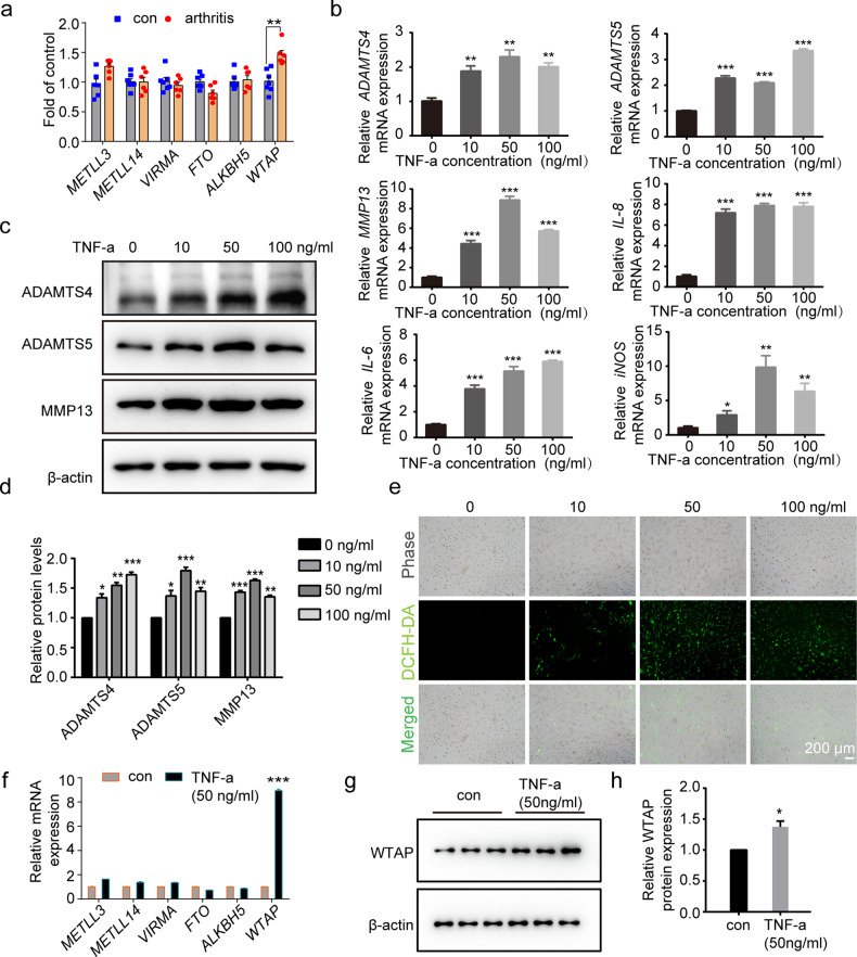
First, we detected the expression of major m6A methylation-related genes (METTL3, METTL14, WTAP, VIRMA, ALKBH5, and FTO) in clinical osteoarthritic cartilage, and our results indicated that WTAP was the only methyltransferase with significantly upregulated expression (Fig. 1a). To induce a cellular OA model, we treated human chondrocytes with the proinflammatory cytokine TNF-a, a crucial inflammatory cytokine in OA33,34. Then, the OA-like phenotype was confirmed with increased mRNA expression of cartilage matrix metabolic genes (ADAMTS4, ADAMTS5, MMP13) and inflammatory genes (IL-6, IL-8, iNOS) (Fig. 1b) and elevated protein expression of ADAMTS4, ADAMTS5, and MMP13 (Fig. 1c, d). Since elevated levels of ROS in cartilage are associated with OA progression4,35,36, we found an increased ROS level in chondrocytes with TNF-α stimulation (Fig. 1e). Notably, chondrocytes treated with 50 ng/ml TNF-α for 24 h showed an obvious OA-like phenotype, which was applied in subsequent experiments.
Fig. 1. WTAP was upregulated in TNF-α-induced chondrocytes and osteoarthritic cartilage.
a Relative mRNA expression of m6A methylases (METLL3, METLL4, VIRMA, FTO, ALKBH5, and WTAP) measured by qRT‒PCR in osteoarthritic (n = 6) vs. uninjured cartilage tissues (n = 7). b Relative mRNA expression of ADAMTS4, ADAMTS5, MMP13, IL-6, IL-8, and iNOS measured by qRT‒PCR in human chondrocytes with or without TNF-α treatment (50 ng/mL). c, d Western blotting (c) and quantitative analysis (d) of ADAMTS4, ADAMTS5, and MMP13 in human chondrocytes with or without TNF-α treatment (50 ng/mL). e DCFH-DA staining for ROS levels in chondrocytes treated with TNF-α (50 ng/mL). Scale bar: 200 µm. f Relative mRNA expression levels of m6A methylases (METLL3, METLL4, VIRMA, FTO, ALKBH5, and WTAP) measured by qRT‒PCR in chondrocytes with or without TNF-α treatment (50 ng/mL). g, h Western blotting (g) and corresponding quantitative analysis (h) of WTAP protein expression in osteoarthritic cartilage. Data are presented as the means ± SEMs. *P < 0.05; **P < 0.01; ***P < 0.001; ns, not significant.
Next, we investigated the expression of the major m6A methylation-related genes in the cellular OA model. In TNF-α (50 ng/ml)-induced chondrocytes, WTAP mRNA expression was the most significantly upregulated (Fig. 1f), and we also confirmed the upregulated WTAP protein expression in the chondrocytes treated with TNF-α (Fig. 1g, h). These results demonstrated that WTAP was highly expressed in osteoarthritic cartilage and may play a role in OA progression.
Involvement of WTAP in the osteoarthritic chondrocyte phenotype
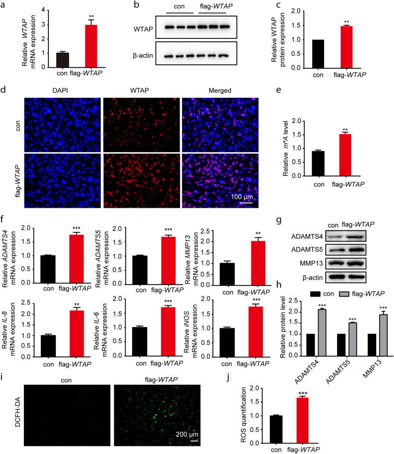
To determine the role of WTAP in OA, we transfected chondrocytes with a plasmid overexpressing WTAP (flag-WTAP). After neomycin screening (10 mg/mL) for 24 h (Supplementary Fig. 1a), increased WTAP mRNA and protein levels were confirmed by qRT‒PCR and Western blotting (Fig. 2a–c). Immunofluorescence detection results also showed that the WTAP protein level was increased in chondrocytes (Fig. 2d), and the m6A methylation level was elevated following WTAP overexpression (Fig. 2e).
Fig. 2. WTAP overexpression aggravated the TNF-α-induced osteoarthritic chondrocyte phenotype.
a–d Overexpression efficiency of WTAP in human chondrocytes. Relative WTAP expression measured by qRT‒PCR (a), Western blotting (b), quantitative analysis (c), and IF staining (d) in chondrocytes after transfection with flag-WTAP. Scale bars: 100 µm. e The m6A level of total RNA in chondrocytes with or without WTAP overexpression. f Relative mRNA expression of ADAMTS4, ADAMTS5, MMP13, IL-6, IL-8, and iNOS was measured by qRT‒PCR in chondrocytes with or without WTAP overexpression. g, h Western blotting (g) and quantitative analysis (h) of ADAMTS4, ADAMTS5, and MMP13 expression in chondrocytes with or without WTAP overexpression. i, j Detection of ROS (i) and quantitative analysis (j) in chondrocytes with or without WTAP overexpression. Scale bars: 200 µm. Data are presented as the means ± SEMs. *P < 0.05; **P < 0.01; ***P < 0.001; ns, not significant.
To evaluate the effect of WTAP on the osteoarthritic chondrocyte phenotype, we determined the mRNA expression levels of ADAMTS4, ADAMTS5, MMP13, IL-6, IL-8, and iNOS (Fig. 2f), along with the protein expression levels of ADAMTS4, ADAMTS5 and MMP13, and found that they were significantly increased in chondrocytes after overexpressing WTAP (Fig. 2g, h). Moreover, the production of active oxygen in the WTAP-overexpressing chondrocytes was significantly increased compared to that in the control chondrocytes (Fig. 2i, j).
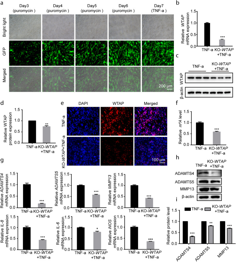
Furthermore, chondrocytes were transfected with lentivirus to knock out WTAP (KO-WTAP), and the optimal puromycin concentration was 2.5 μg/mL after screening the chondrocytes (Supplementary Fig. 1b). TNF-α (50 ng/mL) treatment had no effect on transfection efficiency on the seventh day (Fig. 3a). The KO-WTAP cell model demonstrated significantly reduced levels of WTAP mRNA and protein under TNF-α treatment (Fig. 3b–d). Immunofluorescence results also confirmed the effective transfection of WTAP lentivirus (Fig. 3e). Knocking out WTAP in chondrocytes decreased the m6A level (Fig. 3f) and significantly decreased the mRNA (ADAMTS4, ADAMTS5, MMP13, IL-6, IL-8, iNOS) and protein levels of arthritis-related genes (ADAMTS4, ADAMTS5, MMP13) in the OA cell model (Fig. 3g–i). Additionally, TNF-α-induced ROS levels were reduced in the chondrocytes with WTAP knockout (Supplementary Fig. 2a, b). Taken together, these results demonstrated the involvement of WTAP in the osteoarthritic chondrocyte phenotype.
Fig. 3. Knocking out WTAP alleviated the osteoarthritic chondrocyte phenotype.
a GFP identification in human chondrocytes transfected with WTAP gene knockout lentivirus (KO-WTAP). Scale bars: 200 µm. b–e Knockout efficiency of WTAP in human chondrocytes. Relative WTAP expression measured by qRT‒PCR (b), Western blotting (c), quantitative analysis (d), and IF staining (e) of chondrocytes after transfection with KO-WTAP. f The m6A level of total RNA in chondrocytes with or without KO-WTAP transfection. g Relative mRNA expression of ADAMTS4, ADAMTS5, MMP13, IL-6, IL-8, and iNOS measured by qRT‒PCR in chondrocytes with or without KO-WTAP transfection. h, i Western blotting (h) and quantitative analysis (i) of ADAMTS4, ADAMTS5 and MMP13.
Enhanced m6A modification of FRZB in osteoarthritic cartilage
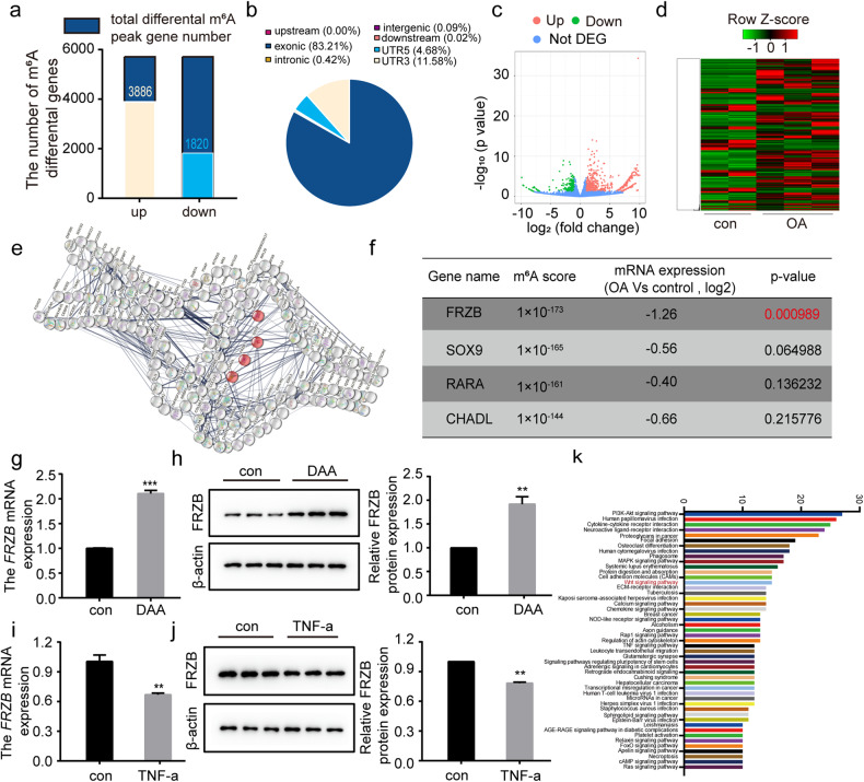
MeRIP-seq was performed in this study to determine the potential downstream targets of m6A modification in clinical osteoarthritic cartilage samples. Compared to uninjured cartilage, we identified 5706 genes that were modified by m6A in osteoarthritic cartilage (Fig. 4a). Among these genes, 3886 were found to have elevated methylation peaks, which were mainly enriched in the exon region (83.21%), followed by the 3’ UTR (11.58%) (Fig. 4b). Moreover, RNA-seq was performed in human osteoarthritic cartilage samples and uninjured cartilage, and there were 675 upregulated genes and 260 downregulated genes in osteoarthritic cartilage (Fig. 4c, d). To identify enriched functional pathways, we performed statistical analysis based on the top 200 upregulated genes according to the m6A score using the STRING database for protein‒protein interaction network functional enrichment analysis (http://string-db.org) (Fig. 4e). The m6A-modified genes associated with osteoarthritic cartilage were enriched in GO:0061037 (negative regulation of cartilage development) according to GO analysis. FRZB had the most significantly downregulated expression with identified high m6A modification in osteoarthritic cartilage (Fig. 4f).
Fig. 4. The Wnt pathway inhibitor FRZB abnormally elevated m6A methylation in OA.
a Statistical analysis of m6A peak gene number. b The distribution of different m6A peaks in different regions of the reference genome. c Volcano plot illustrating the distributions of differentially expressed m6A genes. d Heatmap of differential gene clustering. e Functional analysis of hypermethylated genes based on the STRING database (the genes marked in red are genes related to cartilage development, https://cn.string-db.org/). f Cartilage development-related genes corresponding to the methylation sequencing score, gene expression and P value score. g, h FRZB expression was assessed by qRT‒PCR (g) and Western blotting (h) in human chondrocytes with or without treatment with the m6A methylation inhibitor DAA (50 µM). i, j FRZB expression was assessed in human chondrocytes treated with TNF-α (50 ng/ml). Relative FRZB expression measured by qRT‒PCR (i), Western blotting and quantitative analysis (j) of FRZB protein in human osteoarthritic chondrocytes. k KEGG analysis of m6A difference genes in osteoarthritic (n = 3) vs. normal cartilage tissues (n = 2). Data are presented as the means ± SEMs. *P < 0.05; **P < 0.01; ***P < 0.001; ns, not significant.
To confirm the expression and modification of FRZB in OA, we examined its mRNA and protein levels in chondrocytes treated with DAA (50 µM), a specific m6A methylation inhibitor (Supplementary Fig. 3a). As shown in Fig. 4g, h, FRZB expression was found to be upregulated in response to DAA treatment, and we observed decreased FRZB expression in the cellular OA model (Fig. 4i, j). All of the above results were consistent with the results of our MeRIP-seq and RNA-seq analyses.
FRZB, a frizzled-related protein, is a Wnt antagonist that has been implicated in the regulation of Wnt/β-catenin signaling37. The results of Kyoto Encyclopedia of Genes and Genomes (KEGG) enrichment analysis also showed that the FRZB-related Wnt/β-catenin pathway was significantly enriched in clinical osteoarthritic cartilage (Fig. 4k). Hence, these findings suggest that WTAP may mediate the m6A modification of FRZB cells and regulate the osteoarthritic chondrocyte phenotype via the Wnt/β-catenin pathway.
WTAP activated the Wnt/β-catenin signaling pathway via FRZB
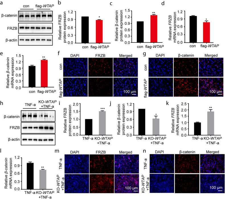
To investigate whether WTAP affected the Wnt/β-catenin pathway, we identified the expression of FRZB and β-catenin in chondrocytes with WTAP overexpression (flag-WTAP) and WTAP knockout (KO-WTAP). In flag-WTAP chondrocytes, we observed a decrease in FRZB expression and an increase in β-catenin expression (Fig. 5a–e). Similar results were also confirmed by immunofluorescence (Fig. 5f, g). In contrast, we found increased FRZB expression and decreased β-catenin expression in the KO-WTAP chondrocytes treated with TNF-a (50 ng/ml) (Fig. 5h–l), which was also confirmed by immunofluorescence results (Fig. 5m, n). Hence, our results suggested that WTAP could activate the Wnt/β-catenin pathway by inhibiting FRZB expression.
Fig. 5. The FRZB-Wnt/β-catenin axis was regulated by WTAP in chondrocytes.
a–c Protein levels of FRZB and β-catenin were analyzed in chondrocytes with or without WTAP overexpression. d, e Relative mRNA expression levels of FRZB (d) and β-catenin (e) were measured by qRT‒PCR in chondrocytes with or without WTAP overexpression. f, g IF staining of intracellular FRZB (f) and β-catenin (g) in TNF-α-pretreated cells with or without WTAP overexpression. Scale bars: 100 µm. h–j Protein levels of FRZB and β-catenin were analyzed in TNF-α-pretreated chondrocytes with or without KO-WTAP transfection. k, l Relative mRNA expression levels of FRZB (k) and β-catenin (l) were measured by qRT‒PCR in TNF-α-pretreated cells with or without KO-WTAP transfection. m, n IF staining of intracellular FRZB (m) and β-catenin (n) expression in TNF-α-pretreated cells with or without KO-WTAP transfection. Scale bars: 100 µm. Data are presented as the means ± SEMs. *P < 0.05; **P < 0.01; ***P < 0.001; ns, not significant.
WTAP-induced the osteoarthritic chondrocyte phenotype through the m6A-FRZB/Wnt/β-catenin signaling pathway
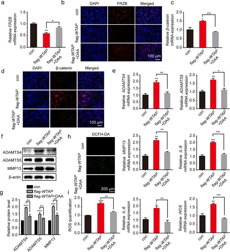
To further determine WTAP-mediated m6A-FRZB in osteoarthritic cartilage, we used the methylation inhibitor DAA to investigate FRZB m6A modification and the activation of Wnt/β-catenin in the flag-WTAP chondrocytes. The qRT‒PCR and Western blotting results indicated that the mRNA and protein expression of FRZB was significantly upregulated in chondrocytes after treatment with DAA (50 µM) (Fig. 6a, b), while the mRNA and protein levels of Wnt/β-catenin were suppressed (Fig. 6c, d). Furthermore, we observed a significant decrease in the osteoarthritic chondrocyte phenotype in the flag-WTAP chondrocytes after DAA treatment, including decreased mRNA levels of arthritis-related genes (ADAMTS4, ADAMTS5, MMP13, IL-6, IL-8, iNOS) and decreased protein levels of ADAMTS4, ADAMTS5, and MMP13 (Fig. 6e–g). ROS staining showed that the level of reactive oxygen species was reduced in the flag-WTAP chondrocytes treated with DAA (Fig. 6h, i). Taken together, our findings suggested that WTAP affected the stability of FRZB by m6A modification to promote osteoarthritic chondrocyte catabolism.
Fig. 6. WTAP regulated Wnt/β-catenin signaling activation and the osteoarthritic chondrocyte phenotype by m6A modification of FRZB.
a, b FRZB expression was analyzed in chondrocytes with WTAP overexpression after DAA (50 µM) treatment by qRT‒PCR (a) and IF staining (b). Scale bars: 100 µm. c, d β-catenin expression was analyzed in chondrocytes with WTAP overexpression after DAA (50 µM) treatment by qRT‒PCR (c) and IF staining (d). Scale bars: 100 µm. e Relative mRNA expression of ADAMTS4, ADAMTS5, MMP13, IL-6, IL-8, and iNOS measured by qRT‒PCR in chondrocytes with WTAP overexpression after DAA (50 µM) treatment. f, g Western blotting (f) and quantitative analysis (g) of ADAMTS4, ADAMTS5 and MMP13 expression in chondrocytes with WTAP overexpression after DAA (50 µM) treatment. h, i Detection of ROS (h) and quantitative analysis (i) in chondrocytes with WTAP overexpression after DAA (50 µM) treatment. Scale bars: 200 µm. Data are presented as the means ± SEMs. *P < 0.05; **P < 0.01; ***P < 0.001; ns, not significant.
Silencing WTAP alleviated OA development through the Wnt/β-catenin signaling pathway
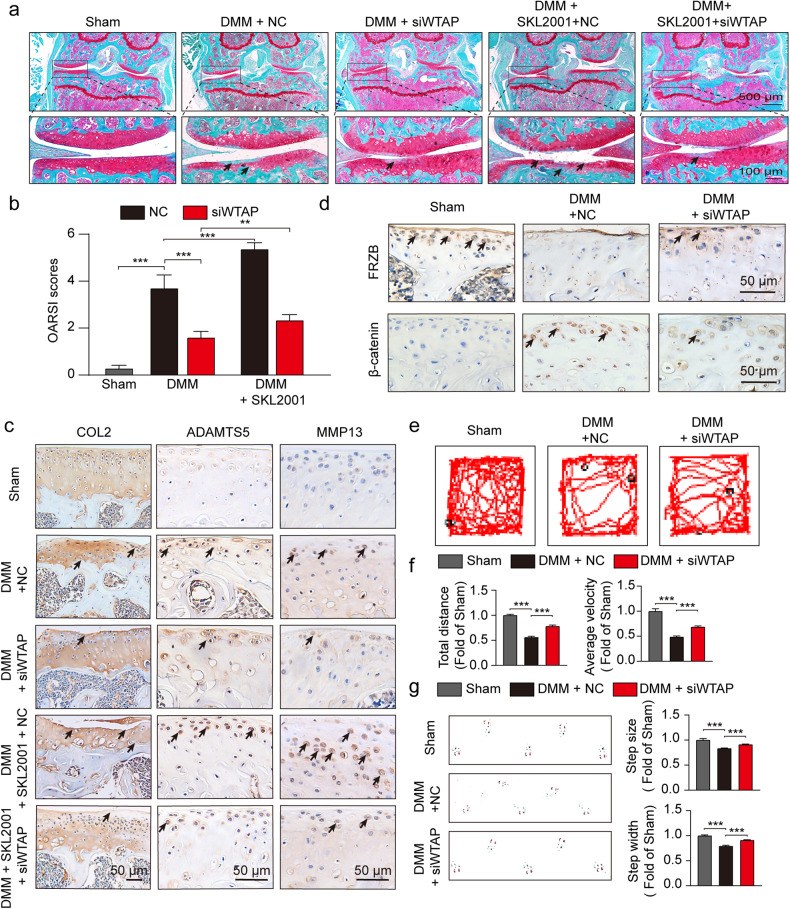
To investigate whether WTAP plays a role in OA progression in vivo, we intra-articularly administered NC or adeno-associated virus (AAV) siWTAP to a DMM-induced OA mouse model. The knockout efficiency was confirmed by IHC (Supplementary Fig. 4a). Safranin O and fast green staining showed an improvement in cartilage surfaces of the mice with DMM-induced OA treated with AAV siWTAP but not NC AAV (Fig. 7a). Quantitative analysis with OARSI scoring showed that NC AAV led to significantly higher OARSI scores, whereas siWTAP treatment decreased OARSI scores (Fig. 7b). The injection of AAV siWTAP alleviated degenerative changes in the cartilage matrix, such as catabolic response and increased extracellular matrix (ECM) composition, in the OA mouse model, as indicated by the IHC results (Fig. 7c). Consistent with the in vitro findings, siWTAP deactivated the Wnt/catenin-β pathway, leading to improved osteoarthritic cartilage degradation by inhibiting the expression of FRZB (Fig. 7d). We also noted that these protective effects of siWTAP on osteoarthritic cartilage and OARSI scores were abolished through activation of the Wnt/β-catenin signaling pathway by SKL2001 (Fig. 7a–d). Moreover, we confirmed that siWTAP inhibits osteoarthritic phenotypes through the open field test and footprint analysis (Fig. 7e–g). Notably, the good biocompatibility of siWTAP and SKL2001 was observed in the tissue organizational structure in major organs (Supplementary Fig. 5a). These results demonstrate that the β-catenin pathway contributes to WTAP-stimulated catabolism changes in osteoarthritic cartilage and development.
Fig. 7. WTAP inhibition alleviated OA progression through Wnt/β-catenin signaling inactivation.
a Safranin O/fast green staining of the cartilage in the indicated groups at 8 weeks after DMM surgery. b OARSI scoring of knee OA was evaluated by Safranin O/fast green staining (n = 6). c, d IHC staining of COL2, ADAMTS5, MMP13, FRZB, and β-catenin in cartilage from the indicated groups at 8 weeks after DMM surgery. e Open field test in different groups. f The corresponding quantitative analysis of (e) (n = 6). g Footprint images in different groups and statistical analysis (n = 6). Data are presented as the means ± SEMs. *P < 0.05; **P < 0.01; ***P < 0.001; ns, not significant.
Discussion
In the present study, we found that the m6A ‘writer’ WTAP had the most significant differential expression in osteoarthritic cartilage. MeRIP-seq and RNA-seq analysis revealed that possible targets of WTAP were associated with FRZB and the Wnt/β-catenin signaling pathway. The negative regulator of WTAP on FRZB expression and Wnt/β-catenin pathway inhibition was mediated by m6A modification, which finally induced degradation of the extracellular matrix, inflammatory reactions, and oxidative stress in osteoarthritic chondrocytes. Our study revealed a novel role of WTAP in OA progression and identified it as a promising therapeutic target for OA treatment (Fig. 8).
Fig. 8.
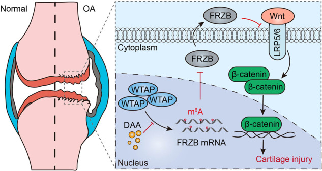
Proposed model for the regulation of WTAP on OA progression through FRZB/β-catenin signaling activation.
Among the numerous post-transcriptional modifications, m6A methylation, the most abundant mRNA modification, has a critical role in various physiological processes and disease progression38. In particular, m6A modification promotes the inflammatory response, chondrocyte apoptosis, and extracellular matrix imbalance in OA, which ultimately leads to OA exacerbation39. M6A-related enzymes determine the function and importance of m6A. For instance, m6A writers (METTL3, METTL14, WTAP, and VIRMA) interact with various binding proteins to regulate the methylation of RNA. In contrast, m6A erasers (FTO and ALKBH5) can catalyze RNA demethylation. Additionally, m6A readers (IGF2BPs, YTHDCs, and YTHDFs) recognize m6A-mediated physiological behavior and influence RNA function. Although much research has focused on m6A writers40 or erasers16 in osteoarthritic chondrocytes, there is a lack of studies on the general profile of m6A-related enzyme expression. Our study demonstrated that WTAP, the only notable methyltransferase, was responsible for osteoarthritic chondrocytes in OA patient samples and in vitro OA cellular models. Moreover, selective silencing of WTAP in articular cartilage effectively attenuated surgically induced cartilage damage and OA progression. These results indicate that the emergence of m6A regulation provides new insights into the molecular mechanisms of OA.
Furthermore, we first confirmed the possible targets of m6A modification in osteoarthritic cartilage in the present study. Our bioinformatic analysis revealed that FRZB had extensive m6A sites in osteoarthritic cartilage compared to the m6A sites of transcribed genes in uninjured cartilage based on m6A sequencing analysis. FRZB, also known as Sfrp-3, is a Wnt signaling pathway antagonist that belongs to the family of secreted frizzled-related proteins (sFRPs). Recent genetic findings suggested that FRZB could affect the cartilage metabolic matrix and cartilage degeneration41,42 and could even be defined as a potential therapeutic target for OA. Hence, it is crucial to understand the molecular basis of m6A regulation of FRZB expression in articular chondrocytes. Furthermore, we found that WTAP could affect the expression stability of FRZB by m6A modification, leading to cartilage degeneration and OA progression. These findings suggested that WTAP-mediated hyperm6A methylation inhibited FRZB expression in chondrocytes, which contributed to OA pathogenesis.
Wnt/β-catenin, as a downstream pathway of FRZB, has been implicated in the maintenance of cartilage homeostasis in OA, such as cartilage matrix catabolism, oxidative stress, and chondrocyte differentiation43,44. Previous studies have shown that m6A methylation-related enzymes, including m6A writers and erasers, regulate proteins in the Wnt/β-catenin signaling pathway, which affects disease progression31,45,46. However, to the best of our knowledge, no relative study on OA has confirmed the role of m6A regulation in Wnt/β-catenin signaling activation. Our study revealed that the genes with the most m6A modification differences, e.g., FRZB, according to m6A sequencing and transcription sequencing in osteoarthritic cartilage, were mostly involved in Wnt/β-catenin signaling. Furthermore, the activation of β-catenin by SKL2001 markedly reversed the protective effect of siWTAP on OA progression, suggesting that WTAP promotes cartilage matrix catabolism through Wnt/β-catenin signaling activation. Our results indicated that WTAP-mediated hyperm6A-methylated conditions were closely associated with Wnt/β-catenin pathway activation in osteoarthritic cartilage, highlighting a promising therapeutic target for OA treatment in the future.
The present study has some limitations. First, the influence of WTAP-induced m6A modification on the FRZB/Wnt/β-catenin axis was not further explored in chondrocytes under TNF-α exposure, which may cause some bias in OA conditions in vitro. Second, the FRZB/Wnt/β-catenin axis is not the only pathway that WTAP regulates, so the detailed regulation of WTAP in biological processes in OA needs further validation. Finally, our findings are based solely on laboratory data, and therefore, prospective clinical studies are needed to confirm the protective effect of WTAP inhibition in human OA progression.
In conclusion, our study revealed that the elevation of WTAP-dependent RNA m6A modification aggravated osteoarthritic cartilage degeneration and progression. Mechanistically, WTAP stabilized FRZB mRNA in an m6A-dependent manner and activated the Wnt/β-catenin signaling pathway in osteoarthritic chondrocytes. Our findings indicated that dysregulated RNA m6A modification mediated by WTAP may be a promising therapeutic target for OA treatment.
Supplementary information
Acknowledgements
This work was supported by the National Natural Science Foundation of China (32271409), Nanjing Distinguished Youth Fund (JQX20001), National Basic Research Program of China (2021YFA1201404), Major Project of NSFC (81991514), Jiangsu Provincial Key Medical Center Foundation, Jiangsu Provincial Medical Outstanding Talent Foundation, Jiangsu Provincial Medical Youth Talent Foundation and Jiangsu Provincial Key Medical Talent Foundation. The Fundamental Research Funds for the Central Universities (14380493, 14380494) also contributed.
Author contributions
X.X., Q.J., and W.Y. conceived the project. X.X. and X.A. designed the experiments. X.A. performed the research, analyzed the data, and wrote the manuscript under the supervision of X.X., Q.J., and W.Y. R.W. refined the experimental results and revised the article. W.W. performed some animal experiments. Z.L., Z.S., and R.W. collected clinical samples for this research. All the authors have read and approved the final manuscript.
Data availability
All data relevant to the study are included in the article or uploaded as supplementary information.
Competing interests
The authors declare no competing interests.
Ethical approval
The human samples and the experimental protocols were reviewed and approved by the Ethics Committee of the Nanjing Drum Tower Hospital (2020-156-01). All animal feeding and experimental procedures were approved by the Ethics Committee and the Institutional Animal Care and Use Committee of Drum Tower Hospital, Nanjing University Medical School.
Footnotes
Publisher’s note Springer Nature remains neutral with regard to jurisdictional claims in published maps and institutional affiliations.
These authors contributed equally: Xueying An, Rongliang Wang.
Contributor Information
Wenjin Yan, Email: 13814533004@163.com.
Qing Jiang, Email: qingj@nju.edu.cn.
Xingquan Xu, Email: xuxingquan@nju.edu.cn.
Supplementary information
The online version contains supplementary material available at 10.1038/s12276-023-01135-5.
References
- 1.Wang KD, et al. Digoxin targets low density lipoprotein receptor-related protein 4 and protects against osteoarthritis. Ann. Rheum. Dis. 2022;81:544–555. doi: 10.1136/annrheumdis-2021-221380. [DOI] [PMC free article] [PubMed] [Google Scholar]
- 2.Xu C, et al. Curcumin primed ADMSCs derived small extracellular vesicle exert enhanced protective effects on osteoarthritis by inhibiting oxidative stress and chondrocyte apoptosis. J. Nanobiotechnol. 2022;20:123–138. doi: 10.1186/s12951-022-01339-3. [DOI] [PMC free article] [PubMed] [Google Scholar]
- 3.Ito Y, et al. Both microRNA-455-5p and -3p repress hypoxia-inducible factor-2alpha expression and coordinately regulate cartilage homeostasis. Nat. Commun. 2021;12:4148–4150. doi: 10.1038/s41467-021-24460-7. [DOI] [PMC free article] [PubMed] [Google Scholar]
- 4.Bolduc JA, Collins JA, Loeser RF. Reactive oxygen species, aging and articular cartilage homeostasis. Free Radic. Biol. Med. 2019;132:73–82. doi: 10.1016/j.freeradbiomed.2018.08.038. [DOI] [PMC free article] [PubMed] [Google Scholar]
- 5.Dai J, et al. Kdm6b regulates cartilage development and homeostasis through anabolic metabolism. Ann. Rheum. Dis. 2017;76:1295–1303. doi: 10.1136/annrheumdis-2016-210407. [DOI] [PubMed] [Google Scholar]
- 6.Rice SJ, Beier F, Young DA, Loughlin J. Interplay between genetics and epigenetics in osteoarthritis. Nat. Rev. Rheumatol. 2020;16:268–281. doi: 10.1038/s41584-020-0407-3. [DOI] [PubMed] [Google Scholar]
- 7.Nunez-Carro C, et al. Histone extraction from human articular cartilage for the study of epigenetic regulation in osteoarthritis. Int. J. Mol. Sci. 2022;23:1–12. doi: 10.3390/ijms23063355. [DOI] [PMC free article] [PubMed] [Google Scholar]
- 8.Sohn DH, et al. Local Joint inflammation and histone citrullination in a murine model of the transition from preclinical autoimmunity to inflammatory arthritis. Arthritis Rheumatol. 2015;67:2877–2887. doi: 10.1002/art.39283. [DOI] [PMC free article] [PubMed] [Google Scholar]
- 9.Iliopoulos D, Malizos KN, Tsezou A. Epigenetic regulation of leptin affects MMP-13 expression in osteoarthritic chondrocytes: possible molecular target for osteoarthritis therapeutic intervention. Ann. Rheum. Dis. 2007;66:1616–1621. doi: 10.1136/ard.2007.069377. [DOI] [PMC free article] [PubMed] [Google Scholar]
- 10.Chen X, et al. Reversal of epigenetic PPARgamma suppression by diacerein alleviates oxidative stress and osteoarthritis in mice. Antioxid Redox Signal. 2022;37:40–53. doi: 10.1089/ars.2021.0219. [DOI] [PubMed] [Google Scholar]
- 11.Kehayova YS, Watson E, Wilkinson JM, Loughlin J, Rice SJ. Genetic and epigenetic interplay within a COLGALT2 enhancer associated with osteoarthritis. Arthritis Rheumatol. 2021;73:1856–1865. doi: 10.1002/art.41738. [DOI] [PubMed] [Google Scholar]
- 12.Alvarez-Garcia O, et al. Increased DNA methylation and reduced expression of transcription factors in human osteoarthritis cartilage. Arthritis Rheumatol. 2016;68:1876–1886. doi: 10.1002/art.39643. [DOI] [PMC free article] [PubMed] [Google Scholar]
- 13.Goldring MB, Marcu KB. Epigenomic and microRNA-mediated regulation in cartilage development, homeostasis, and osteoarthritis. Trends Mol. Med. 2012;18:109–118. doi: 10.1016/j.molmed.2011.11.005. [DOI] [PMC free article] [PubMed] [Google Scholar]
- 14.Meng F, et al. MicroRNA-193b-3p regulates chondrogenesis and chondrocyte metabolism by targeting HDAC3. Theranostics. 2018;8:2862–2883. doi: 10.7150/thno.23547. [DOI] [PMC free article] [PubMed] [Google Scholar]
- 15.Yang C, et al. The role of m(6)A modification in physiology and disease. Cell Death Dis. 2020;11:960. doi: 10.1038/s41419-020-03143-z. [DOI] [PMC free article] [PubMed] [Google Scholar]
- 16.Yang Y, Hsu PJ, Chen YS, Yang YG. Dynamic transcriptomic m(6)A decoration: writers, erasers, readers and functions in RNA metabolism. Cell Res. 2018;28:616–624. doi: 10.1038/s41422-018-0040-8. [DOI] [PMC free article] [PubMed] [Google Scholar]
- 17.Jin S, et al. The m6A demethylase ALKBH5 promotes tumor progression by inhibiting RIG-I expression and interferon alpha production through the IKKepsilon/TBK1/IRF3 pathway in head and neck squamous cell carcinoma. Mol. Cancer. 2022;21:97. doi: 10.1186/s12943-022-01572-2. [DOI] [PMC free article] [PubMed] [Google Scholar]
- 18.Li G, et al. WTAP-mediated m(6)A modification of lncRNA NORAD promotes intervertebral disc degeneration. Nat. Commun. 2022;13:1469. doi: 10.1038/s41467-022-28990-6. [DOI] [PMC free article] [PubMed] [Google Scholar]
- 19.Yadav P, et al. M6A RNA methylation regulates histone ubiquitination to support cancer growth and progression. Cancer Res. 2022;82:1872–1899. doi: 10.1158/0008-5472.CAN-21-2106. [DOI] [PMC free article] [PubMed] [Google Scholar]
- 20.Chen X, et al. METTL3-mediated m(6)A modification of ATG7 regulates autophagy-GATA4 axis to promote cellular senescence and osteoarthritis progression. Ann. Rheum. Dis. 2022;81:87–99. doi: 10.1136/annrheumdis-2021-221091. [DOI] [PubMed] [Google Scholar]
- 21.Yang J, et al. m(6)A-mediated upregulation of AC008 promotes osteoarthritis progression through the miR-328-3pAQP1/ANKH axis. Exp. Mol. Med. 2021;53:1723–1734. doi: 10.1038/s12276-021-00696-7. [DOI] [PMC free article] [PubMed] [Google Scholar]
- 22.Sang W, et al. METTL3 involves the progression of osteoarthritis probably by affecting ECM degradation and regulating the inflammatory response. Life Sci. 2021;278:119528. doi: 10.1016/j.lfs.2021.119528. [DOI] [PubMed] [Google Scholar]
- 23.Lodewyckx L, Lories RJ. WNT Signaling in osteoarthritis and osteoporosis: what is the biological significance for the clinician? Curr. Rheumatol. Rep. 2009;11:23–30. doi: 10.1007/s11926-009-0004-6. [DOI] [PubMed] [Google Scholar]
- 24.Zhu M, et al. Inhibition of beta-catenin signaling in articular chondrocytes results in articular cartilage destruction. Arthritis Rheum. 2008;58:2053–2064. doi: 10.1002/art.23614. [DOI] [PMC free article] [PubMed] [Google Scholar]
- 25.Day TF, Guo X, Garrett-Beal L, Yang Y. Wnt/beta-catenin signaling in mesenchymal progenitors controls osteoblast and chondrocyte differentiation during vertebrate skeletogenesis. Dev. Cell. 2005;8:739–750. doi: 10.1016/j.devcel.2005.03.016. [DOI] [PubMed] [Google Scholar]
- 26.Dell’Accio F, De Bari C, Luyten FP. Molecular markers predictive of the capacity of expanded human articular chondrocytes to form stable cartilage in vivo. Arthritis Rheum. 2001;44:1608–1619. doi: 10.1002/1529-0131(200107)44:7<1608::AID-ART284>3.0.CO;2-T. [DOI] [PubMed] [Google Scholar]
- 27.Lu FI, Thisse C, Thisse B. Identification and mechanism of regulation of the zebrafish dorsal determinant. Proc. Natl Acad. Sci. Usa. 2011;108:15876–15880. doi: 10.1073/pnas.1106801108. [DOI] [PMC free article] [PubMed] [Google Scholar]
- 28.Zhong L, et al. Nitric oxide mediates crosstalk between interleukin 1beta and WNT signaling in primary human chondrocytes by reducing DKK1 and FRZB expression. Int. J. Mol. Sci. 2017;18:2491. doi: 10.3390/ijms18112491. [DOI] [PMC free article] [PubMed] [Google Scholar]
- 29.Lories RJ, Corr M, Lane NE. To Wnt or not to Wnt: the bone and joint health dilemma. Nat. Rev. Rheumatol. 2013;9:328–339. doi: 10.1038/nrrheum.2013.25. [DOI] [PMC free article] [PubMed] [Google Scholar]
- 30.Pang P, et al. Mettl14 attenuates cardiac ischemia/reperfusion injury by regulating Wnt1/beta-catenin signaling pathway. Front Cell Dev. Biol. 2021;9:762853. doi: 10.3389/fcell.2021.762853. [DOI] [PMC free article] [PubMed] [Google Scholar]
- 31.Li J, et al. RNA m(6)A methylation regulates dissemination of cancer cells by modulating expression and membrane localization of beta-catenin. Mol. Ther. 2022;30:1578–1596. doi: 10.1016/j.ymthe.2022.01.019. [DOI] [PMC free article] [PubMed] [Google Scholar]
- 32.Zhang YN, et al. Dexmedetomidine alleviates gut-vascular barrier damage and distant hepatic injury following intestinal ischemia/reperfusion injury in mice. Anesth. Analg. 2022;134:419–431. doi: 10.1213/ANE.0000000000005810. [DOI] [PubMed] [Google Scholar]
- 33.He X, Deng L. Potential of miR-25-3p in protection of chondrocytes: emphasis on osteoarthritis. Folia Histochem. Cytobiol. 2021;59:30–39. doi: 10.5603/FHC.a2021.0004. [DOI] [PubMed] [Google Scholar]
- 34.Lee SG, et al. Anti-inflammatory and anti-osteoarthritis effects of fermented Achyranthes japonica Nakai. J. Ethnopharmacol. 2012;142:634–641. doi: 10.1016/j.jep.2012.05.020. [DOI] [PubMed] [Google Scholar]
- 35.Li Y, et al. Tannic acid/Sr(2+)-coated silk/graphene oxide-based meniscus scaffold with anti-inflammatory and anti-ROS functions for cartilage protection and delaying osteoarthritis. Acta Biomater. 2021;126:119–131. doi: 10.1016/j.actbio.2021.02.046. [DOI] [PubMed] [Google Scholar]
- 36.Deng C, et al. Bioceramic scaffolds with antioxidative functions for ROS scavenging and osteochondral regeneration. Adv. Sci. 2022;9:e2105727. doi: 10.1002/advs.202105727. [DOI] [PMC free article] [PubMed] [Google Scholar]
- 37.Bougault C, et al. Protective role of frizzled-related protein B on matrix metalloproteinase induction in mouse chondrocytes. Arthritis Res. Ther. 2014;16:R137. doi: 10.1186/ar4599. [DOI] [PMC free article] [PubMed] [Google Scholar]
- 38.Jonkhout N, et al. The RNA modification landscape in human disease. RNA. 2017;23:1754–1769. doi: 10.1261/rna.063503.117. [DOI] [PMC free article] [PubMed] [Google Scholar]
- 39.Zhai G, et al. Regulatory role of N6-methyladenosine (m6A) modification in osteoarthritis. Front Cell Dev. Biol. 2022;10:946219. doi: 10.3389/fcell.2022.946219. [DOI] [PMC free article] [PubMed] [Google Scholar]
- 40.Liu Q, Li M, Jiang L, Jiang R, Fu B. METTL3 promotes experimental osteoarthritis development by regulating inflammatory response and apoptosis in chondrocyte. Biochem Biophys. Res. Commun. 2019;516:22–27. doi: 10.1016/j.bbrc.2019.05.168. [DOI] [PubMed] [Google Scholar]
- 41.van den Bosch MH, et al. Brief Report: Induction of matrix metalloproteinase expression by synovial Wnt signaling and association with disease progression in early symptomatic osteoarthritis. Arthritis Rheumatol. 2017;69:1978–1983. doi: 10.1002/art.40206. [DOI] [PubMed] [Google Scholar]
- 42.Schivo S, et al. ECHO, the executable CHOndrocyte: a computational model to study articular chondrocytes in health and disease. Cell Signal. 2020;68:109471. doi: 10.1016/j.cellsig.2019.109471. [DOI] [PubMed] [Google Scholar]
- 43.Rockel JS, et al. Hedgehog inhibits beta-catenin activity in synovial joint development and osteoarthritis. J. Clin. Invest. 2016;126:1649–1663. doi: 10.1172/JCI80205. [DOI] [PMC free article] [PubMed] [Google Scholar]
- 44.Wang S, et al. Bugan Rongjin decoction alleviates inflammation and oxidative stress to treat the postmenopausal knee osteoarthritis through Wnt signaling pathway. Biomed. Eng. Online. 2021;20:103. doi: 10.1186/s12938-021-00939-8. [DOI] [PMC free article] [PubMed] [Google Scholar]
- 45.Zhou S, et al. FTO regulates the chemo-radiotherapy resistance of cervical squamous cell carcinoma (CSCC) by targeting beta-catenin through mRNA demethylation. Mol. Carcinog. 2018;57:590–597. doi: 10.1002/mc.22782. [DOI] [PubMed] [Google Scholar]
- 46.Cui X, et al. Cross talk between RNA N6-methyladenosine methyltransferase-like 3 and miR-186 regulates hepatoblastoma progression through Wnt/beta-catenin signalling pathway. Cell Prolif. 2020;53:e12768. doi: 10.1111/cpr.12768. [DOI] [PMC free article] [PubMed] [Google Scholar]
Associated Data
This section collects any data citations, data availability statements, or supplementary materials included in this article.
Supplementary Materials
Data Availability Statement
All data relevant to the study are included in the article or uploaded as supplementary information.


