Abstract
The RFC5 gene encodes a small subunit of replication factor C (RFC) complex in Saccharomyces cerevisiae and has been shown to be required for the checkpoints which respond to replication block and DNA damage. Here we describe the isolation of RAD24, known to play a role in the DNA damage checkpoint, as a dosage-dependent suppressor of rfc5-1. RAD24 overexpression suppresses the sensitivity of rfc5-1 cells to DNA-damaging agents and the defect in DNA damage-induced Rad53 phosphorylation. Rad24, like Rfc5, is required for the regulation of Rad53 phosphorylation in response to DNA damage. The Rad24 protein, which is structurally related to the RFC subunits, interacts physically with RFC subunits Rfc2 and Rfc5 and cosediments with Rfc5. Although the rad24Δ mutation alone does not cause a defect in the replication block checkpoint, it does enhance the defect in rfc5-1 mutants. Furthermore, overexpression of RAD24 suppresses the rfc5-1 defect in the replication block checkpoint. Taken together, our results demonstrate a physical and functional interaction between Rad24 and Rfc5 in the checkpoint pathways.
The survival of eucaryotes depends on the accurate transmission of genetic information from one generation to the next. Successful mitotic division requires the events of the cell cycle to be ordered such that the initiation of late cycle events is dependent on the completion of early events. The mechanisms that ensure the proper ordering of cell cycle events have been termed checkpoint controls (7). When DNA replication is delayed and DNA damage occurs, checkpoint controls activate cell cycle arrest enough to complete DNA replication and repair DNA damage (4, 18).
In the budding yeast Saccharomyces cerevisiae, checkpoint pathways induce cell cycle arrest in G1 or G2/M and retard S-phase progression in response to DNA damage. Other checkpoints prevent cells with incompletely replicated DNA from exiting the S phase. A number of genes that are involved in the DNA damage checkpoint and/or the replication block checkpoint have been identified elsewhere (4, 18). These include RAD9, RAD17, RAD24, POL2, MEC1/ESR1, RAD53/SPK1/MEC2/SAD1, RFC5, MEC3, and DDC1. Among these genes, RAD9, RAD17, RAD24, MEC3, and DDC1 are involved not only in the G2/M-phase but also in the G1- and S-phase DNA damage checkpoints (11, 12, 17, 22–24, 31–33). POL2, encoding a large subunit of DNA polymerase (Pol) ɛ, is required for the checkpoints responding to replication block and DNA damage in S phase (15, 16). MEC1 and RAD53 are necessary for the checkpoints operating in response to both DNA damage and incomplete DNA replication (1, 33). RAD53 encodes a dual-specificity protein kinase (25), and Mec1 belongs to the phosphatidylinositol kinase family that includes human ataxia-telangiectasia-mutated (ATM) proteins (9, 21). Rad53 is phosphorylated in response to DNA damage and DNA replication block in a MEC1-dependent manner (20, 29).
Replication factor C (RFC) is required for DNA replication and repair and consists of one large and four small subunits. In S. cerevisiae, the large subunit of RFC is encoded by RFC1/CDC44 and the four small subunits are encoded by RFC2, RFC3, RFC4, and RFC5 (3). RFC is a structure-specific DNA-binding protein complex that recognizes the primer-template junction. RFC loads proliferating cell nuclear antigen (PCNA) onto the primer terminus and then Pols δ and ɛ bind to the DNA-RFC-PCNA complex to constitute a processive replication complex (2, 10, 30). A temperature-sensitive mutant of RFC5 whose lethality can be suppressed by overexpression of the Rad53 kinase has been identified (28). We have demonstrated that RFC5 is required for the checkpoints operating in response to DNA replication block and DNA damage in S phase (26, 28). Phosphorylation of Rad53 is reduced in rfc5-1 mutants in response to DNA damage during the S phase, suggesting that RFC5 functions upstream of RAD53. However, it is not yet known how Rfc5 regulates the checkpoint pathway.
To identify genes that interact with RFC5 in the checkpoint pathway, we isolated dosage-dependent suppressors of rfc5-1 mutants. One of the suppressor genes was found to be RAD24, a gene which has been shown to play a role in the DNA damage checkpoint. Overexpression of RAD24 suppresses the DNA damage sensitivity and Rad53 phosphorylation defect in rfc5-1 mutants. RAD24 encodes a protein structurally related to subunits of the RFC complex, and the Rad24 protein associates with the components of RFC. Although rad24Δ alone does not cause a defect in the replication block checkpoint, its introduction does exacerbate the defect in rfc5 mutants. Thus, Rad24 and Rfc5 interact physically and functionally in the checkpoints responding to DNA damage and replication block.
MATERIALS AND METHODS
Strains, media, and general methods.
Yeast strains used in this study are listed in Table 1. DNA was manipulated by standard procedures (19). Standard genetic techniques were used for manipulating yeast strains (8). Media used to maintain selection of TRP1 and URA3 plasmids are synthetic complete media containing 0.5% Casamino Acids and the appropriate supplements.
TABLE 1.
List of strains used in this study
| Straina | Genotype |
|---|---|
| KSC766 | MATa rfc5-1 ade2 his2 trp1 ura3 leu2 lys2 |
| TSY401 | MATa ade1 his2 trp1 ura3 leu2 |
| TSY418 | MATa rad24Δ::LEU2 ade1 his2 trp1 ura3 leu2 |
| TSY437 | MATa rad24Δ::TRP1 ade1 his2 trp1 ura3 leu2 |
| TSY535 | MATa RFC5-HA::LEU2 rad24Δ::TRP1 ade1 his2 trp1 ura3 leu2 |
| TSY601 | MATa rfc5-1::LEU2 ade1 his2 trp1 ura3 leu2 |
| TSY602 | MATa rfc5-1::LEU2 rad24Δ::LEU2 ade1 his2 trp1 ura3 leu2 |
| TSY612 | MATa RFC5-Myc::LEU2 rad24Δ::TRP1 ade1 his2 trp1 ura3 leu2 |
All strains except KSC766 are isogenic. KSC766 is congenic to other strains.
Screening of dosage-dependent suppressors of rfc5-1.
To isolate dosage-dependent suppressors of rfc5-1, rfc5-1 (KSC766) mutants were transformed with an S. cerevisiae YEp13 genomic library. Approximately 40,000 transformants grown at 25°C were replica plated to yeast extract-peptone-dextrose (YEPD) containing 1 mg of hydroxyurea (HU) per ml at 37°C. The plasmids were recovered and retransformed into KSC766 cells. Plasmids that conferred the suppression were subjected to restriction and partial sequence analysis. After elimination of plasmids containing the RFC5 gene, four plasmids were further tested to see whether they suppressed the HU sensitivity of the rad53 mutant (spk1-101 [28]). Two of the plasmids did not suppress the spk1-101 phenotype and were found to contain an overlapping region on chromosome V. Subcloning analysis along with a search of DNA databases localized the suppressor gene to RAD24.
Plasmids.
A 4-kb KpnI-SpeI fragment from pRS416 carrying the RAD24 gene (obtained from T. Weinert) was cloned into KpnI-XbaI-digested YEplac112 and YEplac195 (5), creating YEpT-RAD24 and YEpRAD24, respectively. To create the rad24 disruption construct, the N-terminal and C-terminal region of the RAD24 gene was amplified by PCR with the primers R24N-5′(GGGCTCGAGAGATCATCACAATGCG) and R24N-3′(GCATCTAAAGCTTCTTGTAC) or R24C-5′(CCCGCATGCGGAAAGGGACAGAAGGCT) and R24C-3′(GGGCTCGAGGTAATGTGCATAGATTTGTG). The rad24 disruption plasmid was constructed by a three-part ligation of the XhoI-HindIII-treated PCR-amplified N-terminal fragment and the SphI-XhoI-treated PCR-amplified C-terminal fragment with SphI-HindIII-linearized YIplac128 (5). A null allele for RAD24 (rad24Δ::LEU2) selecting for leucine prototrophy was obtained after sporulation of diploid cells transformed with XhoI-digested rad24 disruption plasmid. Disruption for RAD24 was confirmed by Southern blotting. The rad24Δ::TRP1 strains were obtained by replacing LEU2 with TRP1 in the rad24Δ::LEU2 strains with pLW1 (a gift from M. Shirayama). To construct the rfc5-1 integration plasmid pIR-1, the HindIII-SmaI fragment from the recovered rfc5-1 mutation (28) was cloned into YIplac128 digested with HindIII and SmaI. The rfc5-1::LEU2 strains were obtained by transforming pIR-1 into TSY401 cells after treatment with MluI. The NcoI-SalI fragment from R5HC (28), whose NcoI site was blunted with Klenow fragment, was cloned into YIplac128 cleaved with SmaI and SalI, creating the RFC5-HA integration plasmid pTS-I5H. The EcoRI-BamHI fragment from pTS-I5H and the BamHI-HindIII fragment containing DNA sequences of four Myc epitopes were cloned into YIplac128 cleaved with EcoRI and HindIII, creating the RFC5-Myc integration plasmid pTS-I5M. The RFC5-HA and RFC5-Myc integration strains were obtained by transformation with BglII-digested pTS-I5H and pTS-I5M, respectively. Precise integration was confirmed by PCR. The RFC5-HA- and RFC5-Myc-integrated cells, in which the endogenous RFC5 gene is disrupted, grow as well as do wild-type cells. To create the RAD24-HA and RAD24-Myc plasmids (YCpRAD24-HA and YCpRAD24-Myc), the RAD24 sequence was amplified by PCR with the 5′ primer CTCGAATTCTTTCAGGAATATAACTCT and the 3′ primer CTCGGATCCCGAGTATTTCCAGATCTGAAT, creating a BamHI restriction site at the C-terminal end. The NcoI-BamHI-digested PCR fragment, together with the KpnI-NcoI fragment from the RAD24 gene and the BamHI-SalI fragment containing DNA sequences of two hemagglutinin (HA) epitopes, were subcloned into YCplac33 (5), creating YCpRAD24-HA. The NcoI-BamHI-digested PCR fragment, together with the KpnI-NcoI fragment from the RAD24 gene and the BamHI-HindIII fragments containing DNA sequences of two HA and four Myc epitopes, were subcloned into YCplac33 (5), creating YCpRAD24-Myc. The tagged RAD24 genes complement the sensitivities of the rad24Δ mutant to methyl methanesulfonate (MMS) and UV, indicating that the tagged versions are fully functional. YCpT-RFC5 was constructed by inserting the HindIII-SalI fragment of the RFC5 gene into YCplac22 (5). YCpRFC5, YEpPOL30, YEpT-POL30 and YCp-RAD53-HA were described previously (26, 28).
Immunoblotting.
Immunoblotting analysis was performed as previously described (26). Yeast cells were grown in synthetic complete medium selectable for TRP1 and/or URA3 plasmids. Cells were then diluted in YEPD and allowed to grow for 3 h before cells were treated with MMS. Cells were pelleted, washed with chilled water, and resuspended in sodium dodecyl sulfate (SDS) sample buffer U. An equal volume of glass beads was added, and the cells were lysed by vortexing. Extracts were clarified by 15 min of centrifugation, and 2-mercaptoethanol was added to a final concentration of 1%. The samples were boiled for 5 min and fractionated on SDS-polyacrylamide gels. Proteins were then transferred to nylon membranes; subjected to immunoblot analysis with the mouse monoclonal anti-HA (BAbCO or Boehringer Mannheim), rat monoclonal anti-HA (Boehringer Mannheim), rabbit polyclonal anti-HA (MBL), rabbit polyclonal anti-Myc (MBL), or rabbit anti-Rfc2 serum (a gift from A. Sugino) antibodies, and were detected with the ECL kit (Amersham).
Immunoprecipitation.
Yeast cells were grown in synthetic complete medium selectable for URA3 plasmids. Cells were then diluted in YEPD and allowed to grow for 3 h. Cells (20 U of optical density at 600 nm) were pelleted, washed, and resuspended in 150 μl of lysis buffer (26). An equal volume of glass beads was added, and the cells were lysed by vortexing. Extracts were clarified by 15 min of centrifugation at 4°C. The supernatant was diluted with lysis buffer and incubated at 4°C for 2 h with 30 μl of protein A-Sepharose beads bound with anti-Rfc2, anti-HA, or anti-Myc antibodies. Protein concentration was determined by the Bio-Rad protein assay (Bio-Rad). Immunoprecipitates were washed four times with lysis buffer, twice with another buffer (20 mM HEPES-Na [pH 7.5], 10 mM MgCl2, 4 mM MnCl2), and boiled immediately in 1× SDS-polyacrylamide gel electrophoresis (PAGE) sample buffer (26). The proteins were detected after immunoblotting with antibodies described above.
Sucrose density gradient centrifugation.
Cells were pelleted, washed, and resuspended in lysis buffer. An equal volume of glass beads was added, and the cells were lysed by vortexing. Extracts were clarified by 15 min of centrifugation at 4°C, and 200 μl of the supernatant was separated by sucrose density gradient sedimentation (4 ml of 10 to 40% sucrose gradient in lysis buffer centrifuged in an SW60 rotor at 40,000 rpm for 12 h at 4°C). The gradients were fractionated from the top (200 μl/fraction) and subjected to immunoblotting with antibodies described above.
UV radiation and drug sensitivities.
The UV radiation sensitivity assay was performed as described previously (27). Cells grown at 37°C were plated on YEPD and then irradiated by UV light at 254 nm. After 2 to 3 days of incubation at 37°C, the number of colonies was counted. MMS sensitivity was determined as described elsewhere (27). Cells were incubated with 0 to 0.06% MMS at 37°C for 30 min. Incubation was terminated by addition of sodium thiosulfate to a final concentration of 5%. Aliquots were plated on YEPD, and the number of colonies was counted after incubation at 37°C for 2 to 3 days. The HU sensitivity assay was performed as described previously (28).
Immunofluorescence microscopic analysis.
Yeast cells were grown in YEPD medium at 30°C. To examine spindle elongation at 37°C, the culture was synchronized in the G1 phase by addition of 6 μg of α-factor per ml at 30°C for 1 h. After 1 h, α-factor (6 μg/ml) was subsequently added, and the culture was shifted to 37°C for 1 h. HU was added to a final concentration of 10 mg/ml to the culture during the last 30 min of incubation with α-factor. Cells were then washed to remove α-factor and released into YEPD containing 10 mg of HU per ml at 37°C. To examine spindle elongation at 30°C, the culture was synchronized in the G1 phase by addition of 6 μg of α-factor per ml at 30°C for 2 h. HU was added at 10 mg/ml to the culture during the last 30 min of incubation with α-factor. Cells were then washed to remove α-factor and released into YEPD containing 10 mg of HU per ml at 30°C. Aliquots of cells were removed and processed for DNA flow cytometry analysis, viability assessment, and indirect immunofluorescence microscopy as described previously (26). For analysis of suppression of the rfc5-1 checkpoint defect by RAD24 overexpression, yeast cells were grown in synthetic complete medium selectable for URA3 plasmids, diluted in YEPD, and allowed to grow at 30°C for 3 h. The culture was synchronized in G1 with α-factor and released into YEPD containing 10 mg of HU per ml at 37°C, and aliquots of cells were removed and processed as described above.
RESULTS
Isolation of the RAD24 gene as a dosage-dependent suppressor of rfc5-1.
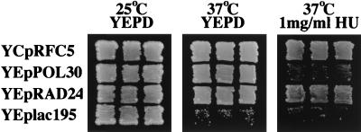
The rfc5-1 mutation is defective for the checkpoints responding to DNA damage and replication block (26, 28). We have shown that the rfc5-1 mutation confers a growth defect and HU sensitivity at the restrictive temperature. Overexpression of POL30, which encodes PCNA, suppresses the growth defect but not the HU sensitivity in rfc5-1 mutants (28) (see Fig. 1). On the other hand, both defects are suppressed by a high dosage of the checkpoint control gene RAD53 (28). To identify genes involved in the checkpoint control, we isolated genes which suppress the HU sensitivity of the rfc5-1 mutant in a dosage-dependent manner. rfc5-1 mutants were transformed with an S. cerevisiae YEp13 genomic library, and transformants grown at 25°C were replica plated to YEPD containing 1 mg of HU per ml at 37°C. In addition to plasmids containing RFC5, four plasmids which suppressed the rfc5-1 growth defect on YEPD containing HU at 37°C were recovered. These plasmids were further tested for whether they suppressed the HU sensitivity of rad53 mutants. RAD53 is considered to function downstream of RFC5 in the checkpoint pathway (26, 28). Two plasmids suppressed the HU sensitivity of rad53 mutants, while the other two did not (data not shown). The suppressor genes on the latter plasmids were chosen for more-detailed analysis, since their function is presumably more closely related to that of RFC5. Restriction and DNA sequence analysis indicated that the plasmids contain an overlapping region on chromosome V. Subcloning analysis and a DNA database search identified the suppressor gene as RAD24 (6, 14). As shown in Fig. 1, a high-copy-number plasmid carrying RAD24 (YEpRAD24) suppressed both the growth defect and HU sensitivity in rfc5-1 mutants.
FIG. 1.
RAD24 overexpression suppresses the rfc5 growth defect. rfc5-1 mutant (KSC766) cells transformed with the YCpRFC5, YEpPOL30, YEpRAD24, or YEp vector (YEplac195) were streaked and incubated on YEPD medium at 25°C or on YEPD medium in the presence or absence of 1 mg of HU per ml at 37°C.
Effect of RAD24 overexpression on the rfc5-1 defect in the replication block checkpoint.
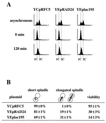
We have shown that rfc5-1 mutants are partially defective for the checkpoint responding to DNA replication block (26). Since overexpression of RAD24 suppressed the growth defect of rfc5-1 mutants on HU-containing medium (Fig. 1), we examined the effect of RAD24 overexpression on the rfc5-1 defect in the replication block checkpoint. rfc5-1 cells carrying the YCpRFC5, YEpRAD24, or YEp vector were synchronized with α-factor and released into medium containing HU at 37°C. Flow cytometric analysis showed that DNA replication was efficiently blocked in those cells until 120 min after release into HU (Fig. 2A). Most (99%) of the RFC5 cells were arrested as large budded cells with short spindles, and 31% of rfc5-1 mutant cells exhibited elongated spindles at 120 min after release into HU. Overexpression of RAD24 decreased the population of rfc5-1 mutants with elongated spindles; 19% of rfc5-1 mutants carrying YEpRAD24 showed elongated spindles at 120 min after release (Fig. 2B). Furthermore, cell survival following HU treatment was higher in rfc5-1 mutants carrying YEpRAD24 than in those carrying the YEp vector (Fig. 2B). Thus, high levels of Rad24 can partially suppress the rfc5-1 checkpoint defect responding to replication block.
FIG. 2.
Suppression of spindle elongation and sensitivity of HU-treated rfc5 mutants by RAD24 overexpression. (A) Flow cytometric analysis of the DNA content of G1-synchronized cells released into medium containing HU. rfc5-1 mutants (TSY601) carrying YCpRFC5, YEpRAD24, or YEplac195 were synchronized in G1 by α-factor treatment and released into YEPD containing 10 mg of HU per ml at 37°C as described in Materials and Methods. Aliquots of cells were collected at 0 and 120 min after release from α-factor and examined for DNA content by flow cytometry. Dotted lines indicate the DNA content of 1C and 2C cells. The top panels represent asynchronous cells not treated with HU at 30°C and are included as a reference. Typical data from at least two independent experiments is presented. (B) Spindle elongation and sensitivity of HU-treated rfc5 mutants. rfc5-1 mutants (TSY601) carrying YCpRFC5, YEpRAD24, or YEplac195 were synchronized in G1 and released into YEPD containing 10 mg of HU per ml as described above. Cells were collected and fixed in formaldehyde at 120 min after release into HU. Nuclear and microtubular structures were visualized with DAPI (4′,6-diamidino-2-phenylindole) and antitubulin antibodies, respectively. At least 200 cells were examined. Viabilities were determined at 120 min after release in HU. Results are means plus or minus standard errors of at least two independent cultures per strain.
rfc5 rad24 double mutants are more defective for the checkpoint responding to replication block than are single rfc5 mutants.
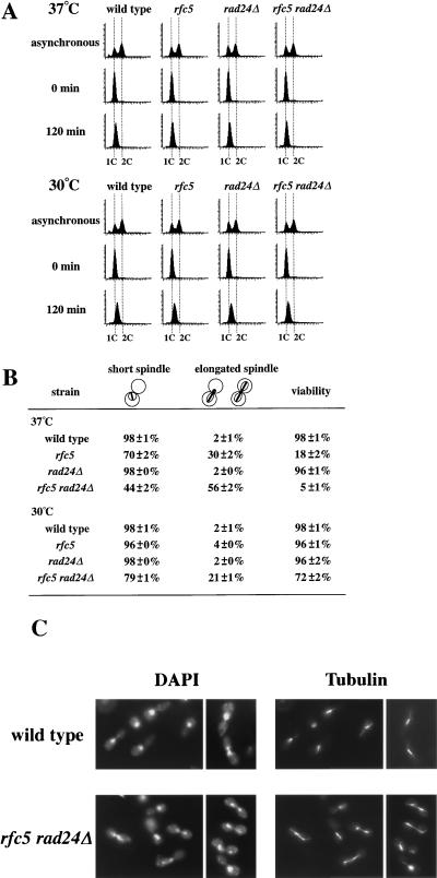
Although the rad24Δ mutant shows no apparent defect in the replication block checkpoint (14, 33), RAD24 overexpression suppresses the checkpoint defect in rfc5-1 mutants. To determine whether RAD24 is involved in the replication block checkpoint, we examined the effect of the rad24Δ mutation on the checkpoint defect in rfc5-1 mutants. DNA content and spindle elongation were analyzed in rfc5-1 and rfc5-1 rad24Δ mutants following the release of α-factor-arrested cells into medium containing HU at 37°C (Fig. 3). If cells are defective for the replication block checkpoint, HU-treated cells should enter into mitosis, as evidenced by spindle elongation prior to completion of DNA replication. Flow cytometric analysis showed that DNA replication was efficiently blocked in wild-type and rfc5-1, rad24Δ, and rfc5-1 rad24Δ mutant cells until 120 min after release into HU (Fig. 3A). Most (98%) of the wild-type and rad24Δ cells were arrested as large budded cells with short spindles, while 30% of rfc5-1 mutant cells exhibited elongated spindles at 120 min after release into HU. However, 56% of rfc5-1 rad24Δ mutants showed elongated spindles at 120 min after release (Fig. 3B and C). Cell survival following HU treatment was lower in rfc5-1 rad24Δ double mutants than in either single mutant (Fig. 3B). Thus, rfc5-1 rad24Δ double mutants show a more pronounced defect in the replication block checkpoint than do single rfc5-1 mutants. Since rfc5-1 mutants are defective for DNA replication at 37°C (28), this exacerbated defect might be a secondary consequence of more perturbed DNA replication by the rad24Δ mutation. To exclude this possibility, we next examined DNA content and spindle elongation in rfc5-1 rad24Δ mutants following the release of α-factor-arrested cells into HU at 30°C (Fig. 3). rfc5-1 rad24Δ mutant cells exhibited no apparent replication defect at 30°C, since they grew as well as did wild-type cells and did not accumulate in the S phase at 30°C. Flow cytometric analysis showed that DNA replication was efficiently blocked in cells until 120 min after release into HU (Fig. 3A). Most of the wild-type, rfc5-1, and rad24Δ cells were arrested as large budded cells with short spindles, while 21% of rfc5-1 rad24Δ mutant cells exhibited elongated spindles at 120 min after release into HU. rfc5-1 rad24Δ mutants became more sensitive to HU treatment even at 30°C (Fig. 3B). These results confirm that the rad24Δ mutation enhanced the checkpoint defect in rfc5-1 mutants.
FIG. 3.
Effects of the rad24Δ mutation on the replication block checkpoint in rfc5-1 mutants. (A) Flow cytometric analysis of the DNA content of G1-synchronized cells released into medium containing HU. Wild-type (TSY401) and rfc5-1 (TSY601), rad24Δ (TSY418), and rfc5-1 rad24Δ (TSY602) mutant cells were synchronized in G1 and released into YEPD containing 10 mg of HU per ml at 30 or 37°C as described in Materials and Methods. Aliquots of cells were collected at 0 and 120 min after release into HU and examined for DNA content by flow cytometry. Dotted lines indicate the DNA content of 1C and 2C cells. The top panels represent asynchronous cells not treated with HU at 30°C and are included as a reference. Typical data from at least two independent experiments is presented. (B) Spindle elongation and viability of cells in the presence of HU. Wild-type (TSY401) and rfc5-1 (TSY601), rad24Δ (TSY418), and rfc5-1 rad24Δ (TSY602) mutant cells were synchronized in G1 and released into YEPD containing 10 mg of HU per ml at 30 or 37°C as described in Materials and Methods. Cells were collected and fixed in formaldehyde. Nuclear and microtubular structures at 120 min after release into medium with HU were visualized with DAPI (4′,6-diamidino-2-phenylindole) and antitubulin antibodies, respectively. At least 200 cells were examined. Viabilities were determined at 120 min after release in HU. Results are means plus or minus standard errors of at least two independent cultures per strain. (C) Photomicrographs of wild-type and rfc5-1 rad24Δ mutant cells at 120 min after release from the G1 block into medium containing HU. Wild-type (TSY401) and rfc5-1 rad24Δ (TSY602) mutant cells were synchronized in G1 and released into YEPD containing 10 mg of HU per ml at 37°C. Cells were collected at 120 min after release and fixed in formaldehyde. Nuclear and microtubular structures were visualized with DAPI and antitubulin antibodies, respectively.
Effects of RAD24 overexpression on the response to DNA damage in rfc5-1 mutants.
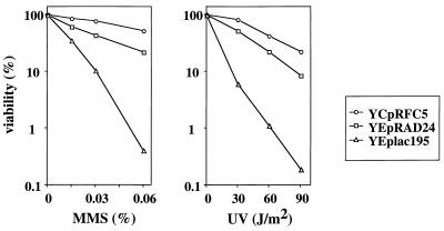
Several lines of evidence have implicated RAD24 in the DNA damage checkpoint (17, 22, 32, 33). The rfc5-1 mutation is also defective for the DNA damage checkpoint and is sensitive to DNA damage (26). To investigate the relationship between RFC5 and RAD24, we examined the effects of RAD24 overexpression on the DNA damage sensitivity of rfc5-1 mutants. Overexpression of POL30 suppresses the growth defect but not the sensitivity to DNA damage in rfc5-1 mutants (26, 28). We therefore examined whether RAD24 overexpression would suppress the DNA damage sensitivity of rfc5-1 mutants carrying YEpT-POL30. RAD24 overexpression restored the ability of rfc5-1 cells carrying YEpT-POL30 to survive exposure to MMS and UV irradiation (Fig. 4).
FIG. 4.
Effects of RAD24 overexpression on DNA damage sensitivity in rfc5 mutants. rfc5-1 mutant (KSC766) cells carrying YEpT-POL30 were transformed with YCpRFC5, YEpRAD24, or YEplac195. The transformants in log-phase culture grown at 37°C were treated with the indicated concentrations of MMS for 30 min or irradiated at the indicated doses with UV light. Viability of cells was estimated as described in Materials and Methods.
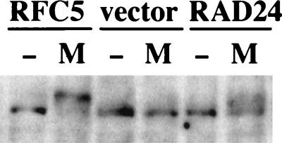
Rad53 is an essential protein kinase that plays a role in the DNA damage checkpoint pathway (1). Exposure of cells to MMS leads to the phosphorylation of Rad53, resulting in the accumulation of a lower-mobility form of Rad53 (20, 29). We have shown that the phosphorylation of Rad53 is reduced in response to MMS treatment in rfc5-1 mutants, providing evidence that Rfc5 is required for the DNA damage-induced phosphorylation of Rad53 (26). Since overexpression of RAD24 can suppress the sensitivity to MMS of rfc5-1 mutants, we expected that its overexpression would also suppress the defect in the MMS-induced Rad53 phosphorylation of rfc5-1 mutants. To test this hypothesis, the phosphorylation state of Rad53 was examined in vivo by immunoblot analysis in cells expressing the Rad53-HA protein. When treated with MMS at 37°C, Rad53-HA in wild-type cells became highly phosphorylated as indicated by the appearance of isoforms with a lower electrophoretic mobility. In contrast, the phosphorylation of Rad53-HA in rfc5-1 mutants was greatly reduced (Fig. 5). However, the DNA damage-induced phosphorylation of Rad53 was partially restored in rfc5-1 mutants by the introduction of YEpT-RAD24, as evidenced by the appearance of smeared, shifted bands corresponding to Rad53 (Fig. 5). Thus, suppression of the DNA damage sensitivity of rfc5-1 mutants by RAD24 overexpression was correlated with the modification of Rad53. These observations suggest that overexpression of RAD24 suppresses the DNA damage sensitivity of the rfc5-1 mutation by activating Rad53. This is consistent with our earlier observation that RAD53 overexpression can suppress the DNA damage sensitivity of rfc5-1 mutants (26).
FIG. 5.
Effects of RAD24 overexpression on modification of Rad53 in rfc5 mutants. rfc5-1 mutant (KSC766) cells were transformed with YCpRAD53-HA and YCpT-RFC5 (RFC5), YEpT-RAD24 (RAD24), or YEplac112 (vector). The transformants grown at 25°C were shifted to 37°C for 1 h and then incubated with YEPD (−) or YEPD containing 0.1% MMS (M) at 37°C for 2 h. The cells were subjected to immunoblotting analysis as described in Materials and Methods.
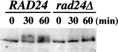
Since overexpression of RAD24 restored the DNA damage-induced phosphorylation of Rad53 in rfc5-1 mutants, it is possible that Rad24 is also required for activation of Rad53 kinase. To examine this possibility, we tested the phosphorylation state of Rad53 in rad24Δ mutants that suffered from DNA damage. When wild-type cells were treated with MMS, Rad53-HA underwent modification. In contrast, the MMS-induced modification of Rad53-HA was reduced in rad24Δ mutants (Fig. 6). This result indicates that Rad24, like Rfc5, is required for the DNA damage-induced phosphorylation of the Rad53 kinase.
FIG. 6.
Modification of Rad53 in rad24Δ mutants. RAD24 (TSY401) and rad24Δ (TSY418) mutant cells carrying YCp-RAD53-HA were grown at 30°C. The cells were incubated with 0.04% MMS for the indicated time and subjected to immunoblotting analysis as described in Materials and Methods.
Rad24 proteins associate with subunits of the RFC complex.
The coding sequence of the RAD24 gene is 1,977 bp in length, and the predicted protein consists of 659 amino acids, corresponding to a molecular mass of 76 kDa, and contains a nucleoside triphosphate binding motif (6, 14). Rad24 is most homologous to the fission yeast Rad17, and they show a well-conserved structural organization. Rad24 is also structurally related to components of the RFC complex (6, 14). RFC subunits contain eight domains termed the RFC boxes (3). Rad24 contains homology to RFC boxes II, III (nucleotide binding motif), and VIII but lacks sequences corresponding to RFC boxes I (the DNA ligase homology domain), IV, V (DEAD box), VI, and VII.
The genetic interaction presented above and sequence similarities between the RFC subunits and Rad24 raised the possibility that Rad24 associates with the RFC complex. To examine the physical interaction between Rad24 and the RFC complex, we tagged the RAD24 gene with the HA or Myc epitope and analyzed its association with the RFC subunits Rfc2 and Rfc5. When extracts from cells harboring a low-copy-number-tagged RAD24 plasmid (YCpRAD24-HA or YCpRAD24-Myc) were subjected to immunoblot analysis, we detected an appropriately sized protein immunoreactive with the anti-HA or anti-Myc antibody (data not shown). Isogenic rad24Δ cells with or without an integrated copy of RFC5-HA (RFC5-HA rad24Δ or RFC5 rad24Δ cells, respectively) were transformed with the YCpRAD24-Myc or YCp vector. Extracts were prepared from the transformed cells and subjected to immunoprecipitation with an antibody to the HA epitope. The immunoprecipitates were then analyzed by immunoblotting with antibodies to the HA epitope, the Myc epitope, and Rfc2. When immunoblotted with the anti-HA antibody, bands migrating at about 40 kDa were detected in the RFC5-HA cells, while no band was detected by the anti-HA antibody in the RFC5 cells (Fig. 7A). When immunoblotted with the anti-Myc antibody, bands corresponding to Rad24-Myc were observed in the immunocomplex from the cells coexpressing Rfc5-HA and Rad24-Myc, while Rad24-Myc proteins were absent in the immunocomplex from the cells expressing only Rad24-Myc or Rfc5-HA (Fig. 7A). Consistent with the previous finding that Rfc2 and Rfc5 are subunits of the RFC complex (3, 28), immunoblotting with the anti-Rfc2 antibody revealed that Rfc5-HA coprecipitated with Rfc2 (Fig. 7A). Extracts were also prepared from rad24Δ mutants carrying YCpRAD24-Myc or the YCp vector and subjected to immunoprecipitation with anti-Rfc2 or control serum. The immunoprecipitates were then analyzed by immunoblotting with antibodies against the Myc epitope and Rfc2. A signal corresponding to the Rad24-Myc proteins was observed in extracts from cells carrying YCpRAD24-Myc after immunoprecipitation with the anti-Rfc2 antibody (Fig. 7B). We next examined coimmunoprecipitation of Rfc2 and Rfc5 with Rad24 in the reciprocal experiment. RFC5-Myc rad24Δ or RFC5 rad24Δ cells were transformed with the YCpRAD24-HA or YCp vector. Extracts were prepared from the transformed cells and subjected to immunoprecipitation with an antibody to the HA epitope. The immunoprecipitates were then analyzed by immunoblotting with antibodies against the HA epitope, the Myc epitope, and Rfc2. Rfc5-Myc was observed in the immunoprecipitates from cells coexpressing Rfc5-Myc and Rad24-HA. Rfc2 was found to coprecipitate with Rad24-HA in a tagged-Rad24-specific manner (Fig. 7C). These results show that the Rad24 protein interacts physically with components of the RFC complex.
FIG. 7.
Association of Rad24 with subunits of RFC. (A) Coimmunoprecipitation of Rad24 and Rfc2 with Rfc5. Cell extracts prepared from RFC5 rad24Δ (TSY418) and RFC5-HA rad24Δ (TSY535) cells carrying YCpRAD24-Myc (+) or YCplac33 (−) were immunoprecipitated (IP) with anti-HA antibody. The immunocomplexes were separated by SDS-PAGE and immunoblotted with anti-HA antibody (top), anti-Myc antibody (middle), or anti-Rfc2 serum (bottom). (B) Coimmunoprecipitation of Rad24 with Rfc2. Cell extracts prepared from rad24Δ (TSY418) cells carrying YCpRAD24-Myc (+) or YCplac33 (−) were immunoprecipitated (IP) with preimmune control serum (c.s.) or anti-Rfc2 serum. The immunocomplexes were separated by SDS-PAGE and immunoblotted with anti-Rfc2 serum (top) or anti-Myc antibody (bottom). (C) Coimmunoprecipitation of Rfc2 and Rfc5 with Rad24. Cell extracts prepared from RFC5 rad24Δ (TSY437) and RFC5-Myc rad24Δ (TSY612) cells carrying YCpRAD24-HA (+) or YCplac33 (−) were immunoprecipitated (IP) with anti-HA antibody. The immunocomplexes were separated by SDS-PAGE and immunoblotted with anti-HA antibody (top), anti-Myc antibody (middle), or anti-Rfc2 serum (bottom).
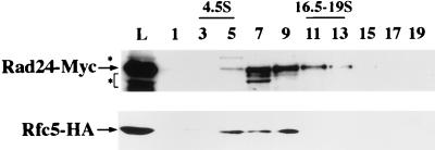
We investigated whether Rad24 associates with the RFC complex or with RFC proteins in smaller complexes. Extracts from cells coexpressing Rfc5-HA and Rad24-Myc were fractionated by sucrose density gradient centrifugation and subjected to immunoblotting with the anti-HA and anti-Myc antibodies. As shown in Fig. 8, Rad24-Myc cosedimented with Rfc5-HA as a 10S particle. It has been shown that the purified yeast RFC complex sediments as an 8.7S particle (34). Altogether, these results strongly suggest that Rad24 proteins associate with the RFC complex.
FIG. 8.
Cosedimentation of Rfc5 and Rad24. A cell extract prepared from RFC5-HA rad24Δ (TSY535) cells carrying YCpRAD24-Myc was separated in a 10 to 40% sucrose gradient, and the load on the gradient (L) and fractions (removed from the top of the gradient) were analyzed by immunoblotting with anti-HA (upper panel) and anti-Myc (lower panel) antibodies. Bovine serum albumin (4.5S) and thyroglobulin (16.5-19S) were separated simultaneously in an independent gradient as markers. The upper band marked with an asterisk is a protein other than Rad24-Myc, which is recognized by the anti-Myc antibody. The lower bands marked with an asterisk are likely proteolytic products of Rad24-Myc.
DISCUSSION
In this paper, we provide evidence demonstrating that the interaction between RFC5 and RAD24 is linked with the checkpoint control in the budding yeast. First, RAD24 overexpression suppressed the rfc5-1 defect in the replication block checkpoint. Second, rfc5-1 rad24Δ mutants showed a more pronounced defect in the replication block checkpoint than did single rfc5-1 mutants. Third, RAD24 overexpression suppressed the DNA damage sensitivity and restored the DNA damage-induced phosphorylation of Rad53 in rfc5-1 mutants. Fourth, Rad24, like Rfc5, was required for MMS-induced Rad53 phosphorylation. Finally, Rad24 proteins were found to interact physically with components of the RFC complex, Rfc2 and Rfc5, and to cosediment with Rfc5. Taken together, these findings strongly support a model in which Rfc5 and Rad24 interact physically and functionally in the checkpoint pathways.
The budding yeast RFC has been purified to homogeneity by assaying replication activity in vitro. The purified RFC complex is composed of five different subunits, each of which is encoded by an essential gene (3). The amino acid sequence of Rad24 has similarities with those of the five subunits of RFC in three of the eight domains termed the RFC boxes. The RAD24 gene encodes a predicted protein of 659 amino acids with a molecular mass of 76 kDa. Although the peptide corresponding to Rad24 is not detected in highly purified fractions of yeast RFC (3), we demonstrated the association of Rad24 with Rfc2 and Rfc5 by immunoblot analysis following immunoprecipitation and the cosedimentation of Rad24 with Rfc5 in sucrose density gradient centrifugation. One likely explanation for these results is that Rad24 proteins may bind unstably or indirectly to the RFC complex and therefore dissociate during the purification steps. Thus, Rad24 appears to associate with the RFC complex but not with RFC proteins in smaller complexes. The physical interaction between Rfc5 and Rad24 was not affected by treatment with MMS or arrest with nocodazole in M phase (data not shown). Therefore, the checkpoint or DNA replication status does not appear to regulate the interaction but rather the other properties, for example, the activity of the Rad24-RFC complex. The RFC complex possesses a structure-specific DNA binding activity, displaying a preference for DNA molecules mimicking DNA replication substrate, and an ATPase activity that is stimulated by DNA (10, 30). The fact that Rad24 contains a nucleotide binding motif raises the possibility that Rad24, like RFC proteins, may possess ATP binding activity. It will be interesting to see whether ATPase activity of the RFC-Rad24 complex can be stimulated by recognizing the primer terminus or aberrant structures resulting from DNA damage and replication delay. RAD24 overexpression appears to suppress the rfc5-1 mutation through the physical interaction between Rad24 and Rfc5, although we cannot exclude the other possibilities, for instance, that high levels of Rad24 could activate checkpoint pathways independently of RFC5.
RAD24 has been suggested to have a role in DNA replication and/or repair, because overexpression of RAD24 strongly reduces the growth rate of mutants that are defective in the DNA replication-repair proteins Rfc1, Pol α, and Pol δ (14). Although it remains possible that an increased dosage of RAD24 could rescue the rfc5-1 defect in DNA replication or DNA repair, the strongest evidence for a functional interaction between RFC5 and RAD24 in the checkpoint comes from the analysis of double mutants. rfc5-1 rad24Δ mutants were more defective for the replication block checkpoint than were single rfc5-1 mutants. Of particular note, at a temperature that does not affect DNA replication, neither single mutant exhibited the checkpoint defect, yet the double mutant was defective for the checkpoint. It is therefore less possible that the observed checkpoint defect results from a general disturbance of the whole DNA replication apparatus.
One plausible explanation of the RFC5-RAD24 interaction is that these two genes function redundantly in the same checkpoint pathways but that the function of RAD24 is modest relative to that of RFC5 in the replication block checkpoint. The additive defect in the replication block checkpoint in the double mutants suggests that rfc5-1 mutants may still have some residual checkpoint activity at the restrictive temperature due to the leakiness of the conditional mutation. Another explanation of the RFC5-RAD24 interaction is that RAD24 and RFC5 function in different but overlapping checkpoint pathways and that an increased dosage of RAD24 can compensate for loss of function of RFC5. For example, the signal that induces the RFC5-mediated checkpoint pathway differs from the signal that induces the RAD24-mediated checkpoint pathway; RFC5 may be involved primarily in recognizing the primer terminus and monitoring stalled DNA replication, whereas RAD24 might be required for recognizing the aberrant DNA structures resulting from DNA replication delay.
RAD24 has been shown to play a role in all known DNA damage checkpoint controls in the G1, S, and G2/M phases. It has been demonstrated that the Rad53 protein kinase is phosphorylated in response to DNA damage, and thus, this biochemical modification correlates with the activation of the checkpoint pathway. We have shown that rfc5-1 mutants are sensitive to DNA damage and defective for the phosphorylation of Rad53 in response to DNA damage. Similar to rfc5-1, rad24Δ was defective for the phosphorylation of Rad53 in response to DNA damage. Overexpression of RAD24 partially suppressed the DNA damage sensitivity and restored the phosphorylation of Rad53 in rfc5-1 mutants. These observations are consistent with our finding that Rfc5 and Rad24 interact physically and regulate the DNA damage checkpoint pathway. Lydall and Weinert (13) showed that the functions of RAD17, RAD24, and MEC3 in response to DNA damage are genetically indistinguishable and proposed that these genes play similar roles in DNA damage processing directly linked to the checkpoint control in S. cerevisiae. We are now examining the interaction of RFC5 with RAD17 and MEC3 in the checkpoint control.
The observations presented here provide evidence indicating that the interaction between RFC5 and RAD24 is linked with the checkpoint pathway in the budding yeast. However, it remains to be precisely determined how RFC5 and RAD24 are involved in the checkpoint signal transduction. Further experiments will be aimed at elucidating the biochemical properties of the RFC-Rad24 complex and its interaction with the other components in the checkpoint pathway.
ACKNOWLEDGMENTS
We thank A. Sugino and T. Weinert for materials and H. Araki, C. Brenner, A. Carr, T. Enoch, M. Lamphier, Y. Nakaseko, and R. Ruggieri for helpful discussions and suggestions. K.S. is especially indebted to Kay Sullivan, who passed away during this work, for encouragement and advice.
This work was supported by a Grant-in-Aid for Scientific Research on Priority Areas and General Research from the Ministry of Education, Science, Sports and Culture of Japan (to K.M. and K.S.).
REFERENCES
- 1.Allen J B, Zhou Z, Siede W, Friedberg E C, Elledge S J. The SAD1/RAD53 protein kinase controls multiple checkpoints and DNA damage-induced transcription in yeast. Genes Dev. 1994;8:2416–2428. doi: 10.1101/gad.8.20.2401. [DOI] [PubMed] [Google Scholar]
- 2.Burgers P M J. Saccharomyces cerevisiae replication factor C. II. Formation and activity of complexes with the proliferating cell nuclear antigen and with DNA polymerase δ and ɛ. J Biol Chem. 1991;266:22698–22706. [PubMed] [Google Scholar]
- 3.Cullmann G, Fien K, Kobayashi R, Stillman B. Characterization of the five replication factor C genes of Saccharomyces cerevisiae. Mol Cell Biol. 1995;15:4661–4671. doi: 10.1128/mcb.15.9.4661. [DOI] [PMC free article] [PubMed] [Google Scholar]
- 4.Elledge S J. Cell cycle checkpoints: preventing an identity crisis. Science. 1996;274:1664–1672. doi: 10.1126/science.274.5293.1664. [DOI] [PubMed] [Google Scholar]
- 5.Gietz R D, Sugino A. New yeast-Escherichia coli shuttle vectors constructed with in vitro mutagenized yeast genes lacking six-base pair restriction sites. Gene. 1988;74:527–534. doi: 10.1016/0378-1119(88)90185-0. [DOI] [PubMed] [Google Scholar]
- 6.Griffiths J F, Barbet N C, McCready S, Lehmann A R, Carr A M. Fission yeast rad17; a homologue of budding yeast RAD24 that shares regions of sequence similarity with DNA polymerase accessory proteins. EMBO J. 1995;14:5812–5823. doi: 10.1002/j.1460-2075.1995.tb00269.x. [DOI] [PMC free article] [PubMed] [Google Scholar]
- 7.Hartwell L H, Weinert T A. Checkpoints: controls that ensure the order of cell cycle events. Science. 1989;246:229–234. doi: 10.1126/science.2683079. [DOI] [PubMed] [Google Scholar]
- 8.Kaiser C, Michaelis S, Mitchell A. Methods in yeast genetics. Cold Spring Harbor, N.Y: Cold Spring Harbor Laboratory Press; 1994. [Google Scholar]
- 9.Kato R, Ogawa H. An essential gene, ESR1, is required for mitotic cell growth, DNA repair, and meiotic recombination in Saccharomyces cerevisiae. Nucleic Acids Res. 1994;22:3104–3112. doi: 10.1093/nar/22.15.3104. [DOI] [PMC free article] [PubMed] [Google Scholar]
- 10.Lee S-H, Kwong A D, Pan Z-Q, Hurwitz J. Studies on the activator 1 protein complex, an accessory factor for proliferating cell nuclear antigen-dependent DNA polymerase δ. J Biol Chem. 1991;266:594–602. [PubMed] [Google Scholar]
- 11.Longhese M P, Fraschini R, Plevani P, Lucchini G. Yeast pip3/mec3 mutants fail to delay entry into S phase and to slow down DNA replication in response to DNA damage, and they define a functional link between Mec3 and DNA primase. Mol Cell Biol. 1996;16:3235–3244. doi: 10.1128/mcb.16.7.3235. [DOI] [PMC free article] [PubMed] [Google Scholar]
- 12.Longhese M P, Paciotti V, Fraschini R, Zaccarini R, Plevani P, Lucchini G. The novel DNA damage checkpoint protein Ddc1p is phosphorylated periodically during the cell cycle and in response to DNA damage in budding yeast. EMBO J. 1997;17:5216–5226. doi: 10.1093/emboj/16.17.5216. [DOI] [PMC free article] [PubMed] [Google Scholar]
- 13.Lydall D, Weinert T. Yeast checkpoint genes in DNA damage processing: implications for repair and arrest. Science. 1995;270:1488–1491. doi: 10.1126/science.270.5241.1488. [DOI] [PubMed] [Google Scholar]
- 14.Lydall D, Weinert T. G2/M checkpoint genes of Saccharomyces cerevisiae: further evidence for roles in DNA replication and/or repair. Mol Gen Genet. 1997;256:638–651. doi: 10.1007/s004380050612. [DOI] [PubMed] [Google Scholar]
- 15.Navas T A, Sanchez Y, Elledge S J. RAD9 and DNA polymerase ɛ form parallel sensory branches for transducing the DNA damage checkpoint signal in Saccharomyces cerevisiae. Genes Dev. 1996;10:2632–2643. doi: 10.1101/gad.10.20.2632. [DOI] [PubMed] [Google Scholar]
- 16.Navas T A, Zhou Z, Elledge S J. DNA polymerase ɛ links the DNA replication machinery to the S phase checkpoint. Cell. 1995;80:29–39. doi: 10.1016/0092-8674(95)90448-4. [DOI] [PubMed] [Google Scholar]
- 17.Paulovich A G, Margulies R U, Garvik B M, Hartwell L H. RAD9, RAD17, and RAD24 are required for S phase regulation in Saccharomyces cerevisiae in response to DNA damage. Genetics. 1997;145:45–62. doi: 10.1093/genetics/145.1.45. [DOI] [PMC free article] [PubMed] [Google Scholar]
- 18.Paulovich A G, Toczyski D P, Harwell L H. When checkpoints fail. Cell. 1997;88:315–321. doi: 10.1016/s0092-8674(00)81870-x. [DOI] [PubMed] [Google Scholar]
- 19.Sambrook J, Fritsch E F, Maniatis T. Molecular cloning: a laboratory manual. 2nd ed. Cold Spring Harbor, N.Y: Cold Spring Harbor Laboratory Press; 1989. [Google Scholar]
- 20.Sanchez Y, Desany B A, Jones W J, Liu Q, Wang B, Elledge S J. Regulation of RAD53 by the ATM-like kinase MEC1 and TEL1 in yeast cell cycle checkpoint pathways. Science. 1996;271:357–360. doi: 10.1126/science.271.5247.357. [DOI] [PubMed] [Google Scholar]
- 21.Savitsky K, Bar-Shira A, Gilad S, Rotman G, Ziv Y, Vanagaite L, Tagle D A, Smith S, Uziel T, Sfez S, Ashkenazi M, Pecker I, Frydman M, Harnik R, Patanajali S R, Simmons A, Clines G A, Sartiel A, Gatti R A, Chessa L, Sanal O, Lavin M F, Jaspers N G J, Taylor A M R, Arlett C F, Miki T, Weissman S M, Lovett M, Collins F S, Shiloh Y. A single ataxia telangiectasia gene with a product similar to PI-3 kinase. Science. 1995;268:1749–1753. doi: 10.1126/science.7792600. [DOI] [PubMed] [Google Scholar]
- 22.Siede W, Friedberg A S, Dianova I, Friedberg E C. Characterization of G1 checkpoint control in the yeast Saccharomyces cerevisiae following exposure to DNA-damaging agent. Genetics. 1994;138:271–281. doi: 10.1093/genetics/138.2.271. [DOI] [PMC free article] [PubMed] [Google Scholar]
- 23.Siede W, Friedberg A S, Friedberg E C. RAD9-dependent G1 arrest defines a second checkpoint for damaged DNA in the cell cycle of Saccharomyces cerevisiae. Proc Natl Acad Sci USA. 1993;90:7985–7989. doi: 10.1073/pnas.90.17.7985. [DOI] [PMC free article] [PubMed] [Google Scholar]
- 24.Siede W, Nusspaumer G, Portillo V, Rodriguez R, Friedberg E C. Cloning and characterization of RAD17, a gene controlling cell cycle responses to DNA damage in Saccharomyces cerevisiae. Nucleic Acids Res. 1996;24:1669–1675. doi: 10.1093/nar/24.9.1669. [DOI] [PMC free article] [PubMed] [Google Scholar]
- 25.Stern D F, Zheng P, Beidler D R, Zerillo C. Spk1, a new kinase from Saccharomyces cerevisiae, phosphorylates proteins on serine, threonine, and tyrosine. Mol Cell Biol. 1991;13:3744–3755. doi: 10.1128/mcb.11.2.987. [DOI] [PMC free article] [PubMed] [Google Scholar]
- 26.Sugimoto K, Ando S, Shimomura T, Matsumoto K. Rfc5, replication factor C component, is required for regulation of Rad53 protein kinase in the yeast checkpoint pathway. Mol Cell Biol. 1997;17:5905–5914. doi: 10.1128/mcb.17.10.5905. [DOI] [PMC free article] [PubMed] [Google Scholar]
- 27.Sugimoto K, Sakamoto Y, Takahashi O, Matsumoto K. HYS2, an essential gene required for DNA replication in Saccharomyces cerevisiae. Nucleic Acids Res. 1995;23:3493–3500. doi: 10.1093/nar/23.17.3493. [DOI] [PMC free article] [PubMed] [Google Scholar]
- 28.Sugimoto K, Shimomura T, Hashimoto K, Araki H, Sugino A, Matsumoto K. Rfc5, a small subunit of replication factor C complex, couples DNA replication and mitosis in budding yeast. Proc Natl Acad Sci USA. 1996;93:7048–7052. doi: 10.1073/pnas.93.14.7048. [DOI] [PMC free article] [PubMed] [Google Scholar]
- 29.Sun Z, Fay D S, Marini F, Foiani M, Stern D F. Spk1/Rad53 is regulated by Mec1-dependent protein phosphorylation in DNA replication and damage checkpoint pathways. Genes Dev. 1996;10:395–406. doi: 10.1101/gad.10.4.395. [DOI] [PubMed] [Google Scholar]
- 30.Tsurimoto T, Stillman B. Replication factors required for SV40 DNA replication in vitro. I. DNA structure specific recognition of a primer-template junction by eucaryotic DNA polymerases and their accessory factors. J Biol Chem. 1991;266:1950–1960. [PubMed] [Google Scholar]
- 31.Weinert T A, Hartwell L H. The RAD9 gene controls the cell cycle response to DNA damage in Saccharomyces cerevisiae. Science. 1988;241:317–322. doi: 10.1126/science.3291120. [DOI] [PubMed] [Google Scholar]
- 32.Weinert T A, Hartwell L H. Cell cycle arrest of cdc mutants and specificity of the RAD9 checkpoint. Genetics. 1993;134:63–80. doi: 10.1093/genetics/134.1.63. [DOI] [PMC free article] [PubMed] [Google Scholar]
- 33.Weinert T A, Kiser G L, Hartwell L H. Mitotic checkpoint genes in budding yeast and the dependence of mitosis on DNA replication and repair. Genes Dev. 1994;8:652–665. doi: 10.1101/gad.8.6.652. [DOI] [PubMed] [Google Scholar]
- 34.Yoder B L, Burgers P M. Saccharomyces cerevisiae replication factor C. I. Purification and characterization of its ATPase activity. J Biol Chem. 1991;266:22689–22697. [PubMed] [Google Scholar]


