Abstract
Ergosterol is essential for fungal cell membrane integrity and growth, and numerous antifungal drugs target ergosterol. Inactivation or modification of ergosterol biosynthetic genes can lead to changes in antifungal drug susceptibility, filamentation and stress response. Here, we found that the ergosterol biosynthesis gene ERG251 is a hotspot for point mutations during adaptation to antifungal drug stress within two distinct genetic backgrounds of Candida albicans. Heterozygous point mutations led to single allele dysfunction of ERG251 and resulted in azole tolerance in both genetic backgrounds. This is the first known example of point mutations causing azole tolerance in C. albicans. Importantly, single allele dysfunction of ERG251 in combination with recurrent chromosome aneuploidies resulted in bona fide azole resistance. Homozygous deletions of ERG251 caused increased fitness in low concentrations of fluconazole and decreased fitness in rich medium, especially at low initial cell density. Dysfunction of ERG251 resulted in transcriptional upregulation of the alternate sterol biosynthesis pathway and ZRT2, a Zinc transporter. Notably, we determined that overexpression of ZRT2 is sufficient to increase azole tolerance in C. albicans. Our combined transcriptional and phenotypic analyses revealed the pleiotropic effects of ERG251 on stress responses including cell wall, osmotic and oxidative stress. Interestingly, while loss of either allele of ERG251 resulted in similar antifungal drug responses, we observed functional divergence in filamentation regulation between the two alleles of ERG251 (ERG251-A and ERG251-B) with ERG251-A exhibiting a dominant role in the SC5314 genetic background. Finally, in a murine model of systemic infection, homozygous deletion of ERG251 resulted in decreased virulence while the heterozygous deletion mutants maintain their pathogenicity. Overall, this study provides extensive genetic, transcriptional and phenotypic analysis for the effects of ERG251 on drug susceptibility, fitness, filamentation and stress responses.
AUTHOR SUMMARY
Invasive infections caused by the fungal pathogen Candida albicans have high mortality rates (20–60%), even with antifungal drug treatment. Numerous mechanisms contributing to drug resistance have been characterized, but treatment failure remains a problem indicating that there are many facets that are not yet understood. The azole class of antifungals targets production of ergosterol, an essential component of fungal cell membranes. Here, we provide insights into the contributions of ERG251, a component of the ergosterol biosynthesis pathway, to increased growth in azoles as well as broad scale effects on stress responses filamentation and pathogenicity. One of the most striking results from our study is that even a single nucleotide change in one allele of ERG251 in diploid C. albicans can lead to azole tolerance. Tolerance, a distinct phenotype from resistance, is the ability of fungal cells to grow above the minimum inhibitory concentration in a drug concentration-independent manner. Tolerance frequently goes undetected in the clinic because it is not observable in standard assays. Strikingly, azole tolerance strains lacking one allele of ERG251 remained virulent in a mouse model of infection highlighting the potential for mutations in ERG251 to arise and contribute to treatment failure in patients.
INTRODUCTION
Candida albicans is the most prevalent human fungal pathogen, affecting millions of people and causing serious and deadly infections in immunocompromised and immunodeficient individuals [1–4]. Invasive infections caused by C. albicans can result in mortality rates nearing ~60% despite the existing antifungal therapies [1,2,5,6]. Antifungal treatment failures and infection recurrences are common [1,2,7,8]. One contribution to treatment failure is drug resistance, which is defined as the ability to grow above the minimum inhibitory concentration (MIC) of a drug-susceptible isolate at rates similar to growth in the absence of drug. However, treatment failure can also occur in strains that are classified as susceptible based on MIC. This highlights the importance of drug tolerance, which is the ability of a fungus to grow slowly above the MIC in a drug concentration-independent manner [7,9,10].
Fungistatic azole drugs target Erg11 in the ergosterol biosynthesis pathway [6,11–13]. Ergosterol is an essential component of fungal cell membranes and acts to maintain cell membrane integrity and fluidity. Azole exposure leads to the depletion of ergosterol and accumulation of a toxic sterol 14-ɑ-methylergosta-8,24(28)-dien-3β,6α-diol (herein referred to dienol) that permeabilizes the plasma membrane, arrests fungal growth, and increases sensitivity to environmental stresses [12,14,15]. During treatment with fungistatic azoles, many Candida species can rapidly evolve drug resistance through various mechanisms including modification or overexpression of the gene encoding the drug target ERG11 and upregulation of drug efflux pumps encoded by CDR1, CDR2, and MDR1 [16,17]. However, these are not the only possible mechanisms. For example, the transcription factor Adr1 has recently been identified as a key regulator of ergosterol biosynthesis and hyperactivation of Adr1 conferred azole resistance in C. albicans [18].
Ergosterol biosynthesis is broadly conserved among the Saccharomycotina phylogeny group which includes Candida species as well as the baker’s yeast Saccharomyces cerevisiae. However, some differences in gene duplication and expression patterns in the more than 20 enzymes along the ergosterol biosynthetic pathway have been identified [19–22]. Ergosterol biosynthesis is divided into three parts: the mevalonate, late, and alternate pathways [21,23]. The mevalonate pathway is responsible for the production of farnesyl diphosphate (FPP), an important ergosterol intermediate. Dephosphorylation of FPP generates farnesol, a quorum-sensing molecule that can regulate the yeast-to-hyphae transition and biofilm formation in C. albicans [24–26]. The late pathway is responsible for using FPP to synthesize ergosterol. The rate-limiting enzyme, lanosterol 14-α-demethylase, Erg11 is the direct target of azoles. Inhibition of Erg11 by azoles activates the alternate pathway that proceeds from lanosterol toward the production of the toxic sterol dienol. Key enzymes in the alternate pathway, Erg6 and Erg3, are respectively responsible for the initial step and last step of dienol generation [14,21,27]. Inactivation or modification of ERG3 or ERG6 impacts drug susceptibility of many Candida species [14,20,22,28–33]. For example, loss of Erg6 function reduces susceptibility to nystatin and polyenes in C. glabrata [28,34,35]. Loss of Erg3 function confers resistance to azoles in C. albicans, C. parapsilosis, C. dubliniensis and resistance to polyenes in C. albicans and C. lusitaniae [22,29–31,36,37]. ERG3 inactivation causes the depletion of ergosterol and accumulation of 14α-methylfecosterol which supports growth in presence of azoles despite altered membrane composition [14,22].
Both the late and alternate pathways of ergosterol biosynthesis utilize C-4 sterol methyl oxidase, the catalytic component of the C-4 demethylation complex that is responsible for removing the two methyl groups from the C-4 position of the sterol molecule [20,21,38]. In Saccharomyces cerevisiae, Erg25 is the solo C-4 sterol methyl oxidase and essential for standard growth [21,39]. However, both Aspergillus fumigatus and C. albicans encode two membrane C-4 sterol methyl oxidases with one of them serving as the primary enzyme during biosynthesis [19,20,38]. In C. albicans, ERG251 encodes the primary C-4 sterol methyl oxidase, but few studies have characterized it independently and the findings are contradictory. For example, erg251∆/∆ exhibited increased fluconazole (FLC) susceptibility and accumulation of eburicol, the direct precursor for 14α-methylfecosterol, in the presence of FLC [20]. However, in a haploid C. albicans strain, transposon insertion into ERG251 resulted in decreased FLC susceptibility [36]. This contradiction highlights the need to understand the effect of growth conditions and genetic background on the relationship between ERG251 and drug susceptibility.
Proper cellular ergosterol levels are crucial for the multiple cellular functions including stress response, nutrient transport, and host-pathogen interactions [21,38]. Deletion or overexpression of the key genes in the ergosterol biosynthetic pathway disrupts ergosterol biosynthesis and results in increased susceptibility to osmotic and cell wall stress [21,40]. Disruption of ERG6 and ERG24 also leads to the reduced transport of potassium, calcium and metal in S. cerevisiae and A. fumigatus [41–43]. Furthermore, C. albicans erg2∆/∆ and erg24∆/∆ mutants exhibit abnormal vacuolar physiology and filamentation defects, and are avirulent in disseminated model of candidiasis [44].
In this report, we determined the effects of heterozygous and homozygous inactivation of ERG251 on drug susceptibility, filamentation, virulence, and response to stress. Heterozygous inactivation of ERG251 in C. albicans resulted in azole tolerance. Strikingly, we identified recurrent heterozygous point mutations in ERG251 in two distinct genetic backgrounds (SC5314 and P75063) in three independent FLC evolution experiments [10] (Zhou et al. 2024, under review). Evolved strains and engineered strains with ERG251 point mutations were either tolerant or resistant to azoles. Azole tolerance occurred with single allele dysfunction of ERG251 in both of the euploid genetic backgrounds. Azole resistance occurred with single allele dysfunction of ERG251 in combination with concurrent aneuploidy of chromosome 3 and chromosome 6. Homozygous deletion of ERG251 resulted in increased fitness in the presence of low concentrations of FLC, but decreased fitness in rich medium, especially at low cell density. During FLC exposure, ERG251 deletion mutants (heterozygous and homozygous) exhibited upregulation of the Zinc finger transporter, ZRT2, and genes in the alternate sterol biosynthesis pathway. Importantly, overexpression of ZRT2 was sufficient to increase azole tolerance, although to a lesser extent than the ERG251 single allele dysfunction mutants. Therefore, we propose that the combination of upregulated ZRT2 and the alternate sterol pathway drive ERG251-mediated azole tolerance. Interestingly, we found that deletion of only the A allele of ERG251 (ERG251-A), not the B allele, resulted in a filamentation defect in SC5314 background. Lastly, erg251∆/∆ showed decreased virulence while both heterozygous deletion mutants maintained their pathogenicity, highlighting the importance of Erg251 in virulence. In summary, we showed the complex and pleiotropic effects of Erg251 on fitness, stress responses, filamentation, and pathogenicity while also highlighting the importance of Erg251-driven azole drug tolerance.
RESULTS
Recurrent point mutations in ERG251 evolve during adaptation to fluconazole
During in vitro evolution of C. albicans in the presence of FLC, ERG251 point mutations were recurrently detected in three independent experiments and within two distinct genetic backgrounds: P75063 and SC5314 (Table 1) [10]. Using whole genome sequencing (WGS), we detected ERG251 point mutations in 16 FLC-evolved strains (Table 1). In P75063 ERG251 is homozygous, while in the type strain SC5314, there are two non-synonymous variants between ERG251-A and ERG251-B, and de novo point mutations were identified in both alleles. The point mutations were all heterozygous and were characterized as missense (A60D, A268D, G62W, W265G, and H274Q), nonsense stop gained (L113* and E273*), frameshift (S27fs), or stop lost (*322Y) (Table 1). Many of these ERG251 point mutations arose in evolved strains with large genomic copy number changes, predominantly whole chromosome trisomies (Table 1 and Fig S1A). The high frequency of mutations in ERG251 during exposure to antifungal drug suggests an important role of ERG251 in response to antifungal drug stress.
Table 1. List of FLC-evolved strains with different ERG251 point mutations.
ERG251 mutations were identified in three independent FLC evolution experiments with two distinct genetic backgrounds (SC5314-derived Sn152 and BWP17, and P75063). Multiple single-colony strains from the same evolved population are organized into the same row. ERG251 point mutations are annotated on the mutated allele, and represented as allele A/allele B, except for AMS5615 where the chromosome region containing ERG251 became homozygous prior to acquisition of the point mutation. ERG251 in the P75063 background is homozygous.
| Strains | Genetic background | ERG251 genotypes | Mutation types | Copy number changes | FLC MIC (24hr) | FLC SMG (48hr) |
|---|---|---|---|---|---|---|
| Strains from evolution experiment 1 (Zhou et al. 2024, under review) | ||||||
| Evolved 1.1/1.2 | Sn152 | ERG251/ERG251 L113* | Stop gained | Chr3x3, Chr6x3, Chr5 LOH | >256µg/ml | N. A |
| Evolved 2.1/2.2 | Sn152 | ERG251 E273* /ERG251 | Stop gained | Chr3x3, Chr6x3 | >256µg/ml | N. A |
| Evolved 3.1/3.3 | Sn152 | ERG251 A60D /ERG251 | Missense | Chr3x3, Chr6x3 | >256 µg/ml | N. A |
| Evolved 3.2 | Sn152 | ERG251/ERG251 *322Y | Stop lost | Chr6x3 | 1 µg/ml | 0.58 |
| Strains from evolution experiment 2 | ||||||
| AMS5615 | BWP17 | ERG251/ERG251 A268D | Missense | Chr4 (partial)x4, Chr7x3, Chr7 LOH | 1µg/ml | 0.79 |
| AMS5617/5618 | BWP17 | ERG251 S27fs /ERG251 | Frame shift | Chr4x3,Chr5 LOH | 1µg/ml | 0.73 |
| AMS5622/5623/5624 | BWP17 | ERG251 G62W /ERG251 | Missense | Chr7x3 | 1µg/ml | 0.54 |
| AMS5625/5626 | BWP17 | ERG251 W265G /ERG251 | Missense | None | 1µg/ml | 0.50 |
| Strains from evolution experiment 3 [10] | ||||||
| AMS4130 | P75063 | ERG251/ERG251 H274Q | Missense | None | 1µg/ml | 0.26 |
Single allele dysfunction of ERG251 leads to azole drug tolerance
To determine the impact of ERG251 point mutations on drug susceptibility, we quantified azole resistance and tolerance in the evolved isolates. All FLC-evolved strains carrying ERG251 point mutations in the SC5314-derived background were either resistant (minimal inhibitory concentration, MIC≥ 256µg/ml) or tolerant (Supra-MIC growth, SMG >0.50) to FLC and other azoles (voriconazole, VOC, and itraconazole, ITC) (Table 1, S1 Table). Similarly, the FLC-evolved strain carrying an ERG251 point mutation in the P75063 background had increased tolerance relative to the progenitor (increase in SMG from 0.13 to 0.26) (Table 1, S1 Table).
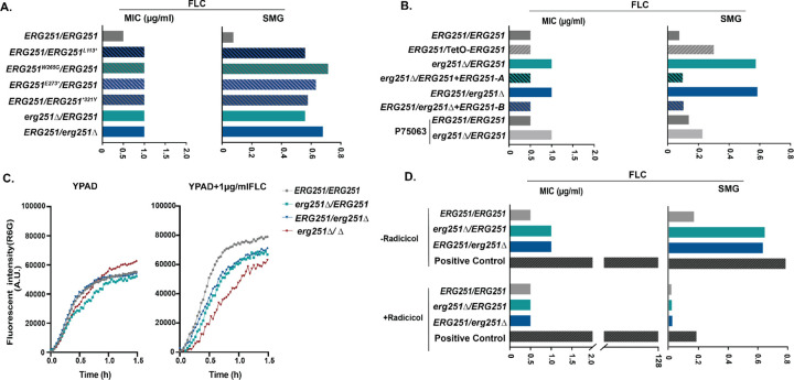
Next, we engineered representative point mutations from the FLC-evolved strains into the wildtype, drug-sensitive SC5314 background and determined azole susceptibility. Four different heterozygous ERG251 point mutations (L113*, W265G, E273*, and *321Y) were selected to represent the range of drug tolerant and resistant phenotypes. The engineered point mutants resulted in a 2-fold increase in MIC and more than an 8-fold increase in tolerance to three different azole drugs (FLC, VOC, ITC) (Fig 1A, S1B and S1 Table).
Fig. 1. The point mutation of ERG251 leads to the partial dysfunction of ERG251 causing acquisition of azole tolerance.
Liquid microbroth drug susceptibility assay. Fluconazole (FLC) resistance quantified as the MIC50 at 24hr in increasing centrations of FLC (left) and FLC tolerance quantified as the Supra-MIC growth at 48hr (SMG, right) which is the average growth above the MIC50 for: A. the wildtype SC5314 (ERG251/ERG251), engineered heterozygous ERG251 point mutations strains in the SC5314 background, and both heterozygous deletion mutants of ERG251 in the SC5314 background; and B. the wildtype SC5314 (ERG251/ERG251), ERG251 overexpression strain, both heterozygous deletion mutants of ERG251 and their corresponding complementation strains, an ERG251 heterozygous deletion in the P75063 background and wildtype P75063 (P75063-ERG251/ERG251) as a control. C. Rhodamine 6G efflux kinetics of two heterozygous deletion mutants and the homozygous deletion of ERG251 in SC5314 with SC5314 (ERG251/ERG251) as the control in YPAD (left) and YPAD+1µg/ml FLC (right). Plots indicate average fluorescence intensity changes of Rhodamine 6G (R6G) from three biology replicates over 90 min. D. 24hr MIC (left, µg/ml) and 48hr SMG (right, tolerance) in FLC with or without radicicol (Hsp90 inhibitor) treatment for two het deletion mutants of ERG251 with SC5314 (ERG251/ERG251) and a positive control strain known to be resistant to FLC as the controls. A&B&D. Each bar represents the average of three technical replicates of a single strain.
Based on these phenotypes we hypothesized that the heterozygous point mutations were due to a loss of gene function. We generated two different heterozygous deletion mutants of ERG251 by deleting either the A or the B allele in the SC5314 background (a∆/B: erg251∆/ERG251 and A/b∆: ERG251/erg251∆). Additionally, we constructed a strain with heterozygous over-expression of ERG251 (ERG251/TetO-ERG251) in the SC5314 background. We validated the deletion mutants using WGS and confirmed that transformation did not introduce off-target effects (Fig S1B). Heterozygous deletion of ERG251 resulted in azole tolerance levels that were the same as all four engineered strains with heterozygous ERG251 point mutations (Fig 1A and S1 Table). Meanwhile, over-expression of ERG251 only resulted in a small increase in FLC tolerance (SMG=0.3, Fig 1B). Complementation of the heterozygous mutants erg251∆/ERG251 and ERG251/erg251∆ with the missing ERG251 allele (erg251∆/ERG251+ERG251-A and ERG251/erg251∆+ERG251-B) eliminated the FLC tolerance (Fig 1B and S1B). In the P75063 genetic background, heterozygous deletion of ERG251 was sufficient to cause the increase in azole tolerance observed for the FLC-evolved strain that carried the ERG251 point mutation (Fig 1B, Table1). Therefore, we conclude that these ERG251 point mutations lead to the single allele dysfunction of ERG251 which causes azole tolerance in C. albicans.
We next tested if ERG251-driven azole tolerance was caused by upregulated drug efflux pumps and if it is dependent on Hsp90, an important mediator for drug-tolerance and stress response [45,46]. Measurement of Rhodamine 6G (R6G) is a useful method for quantifying efflux pump activity [47]. We found that ERG251-driven azole tolerance was independent of drug efflux pumps as indicated by no increase in the rate of efflux of R6G for ERG251 heterozygous deletion mutants compared to ERG251/ERG251 (SC5314) during the exposure to FLC (Fig 1C). We found that ERG251-mediated tolerance depends on Hsp90 function. Addition of an Hsp90 inhibitor (radicicol, 2.5µM) to assays measuring azole resistance (MIC50) and tolerance (SMG) blocked the acquired azole tolerance of ERG251 heterozygous deletion mutants, but did not inhibit growth of a positive control strain known to be resistant to FLC (Fig 1D). Additionally, for susceptible and tolerant strains, inhibition of Hsp90 caused FLC to become fungicidal as no viable cells were recovered from higher FLC concentrations combined with radicicol (Fig S2). Hsp90 regulates cell morphogenesis and cell wall stress through the calcineurin pathway, suggesting that ERG251-mediated tolerance may also alter cell membrane and/or cell wall stress responses [46].
Single allele dysfunction of ERG251 and concurrent aneuploidy leads to azole resistance
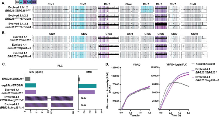
The single allele dysfunction of ERG251 was sufficient to reproduce the azole tolerance phenotype observed in 10/16 of the FLC-evolved strains with ERG251 point mutations. However, 6 of the FLC-evolved strains with ERG251 point mutations acquired bona fide azole resistance (MIC >256 µg/ml FLC, Table 1). All 6 resistant ERG251 mutants also had chromosome (Chr)3 and Chr6 concurrent aneuploidies (Fig 2A, Table 1, Evolved 1.1/1.2, 2.1/2.2 and 3.1/3.3 representing two single colonies from three independent FLC-evolved lineages). Recently, we showed that Chr3 and Chr6 concurrent aneuploidy causes azole tolerance and correlates with elevated expression of drug responsive genes located on these chromosomes, including CDR1, CDR2, MDR1, and MRR1 (Zhou et al. 2024, under review). We hypothesized that heterozygous deletion of ERG251 in the Chr3 and Chr6 aneuploid background would make these tolerant cells resistant. To test this hypothesis, we isolated an azole tolerant strain with Chr3 and Chr6 concurrent aneuploidies and wild-type alleles of ERG251 in the SC5314-derived genetic background (Evolved 4.1: ERG251/ERG251) (Fig 2B). We deleted one copy of ERG251 (on Chr4) from this concurrent aneuploid strain, and confirmed that the mutants maintained the aneuploid chromosomes by whole genome sequencing. Heterozygous deletion of ERG251 in the concurrent aneuploidy background with elevated drug efflux resulted in a 256-fold increase in MIC, reproducing the azole resistance phenotype observed for the FLC-evolved resistant strains (>256µg/ml, Fig 2B, 2C, 2D and S1 Table). We therefore conclude that the combination of the single allele dysfunction of ERG251 and concurrent aneuploidy leads to bona fide drug resistance.
Fig 2. Single allele dysfunction of ERG251 in combination with concurrent aneuploidy causes azole resistance.
A. Representative whole genome sequencing (WGS) data of the FLC-evolved strains 1.1/1.2, 2.1/2.2, and 3.1/3.2 that acquired heterozygous point mutations at ERG251 and Chr3 and Chr6 concurrent aneuploidy. B. WGS data of FLC-evolved strain 4.1 that had wild-type alleles of ERG251/ERG251 and Chr3 and Chr6 concurrent aneuploidy, plus two ERG251 heterozygous deletion mutants engineered in the Evolved 4.1 aneuploid background. A&B WGS data are plotted as the log2 ratio and converted to chromosome copy number (y-axis, 1–4 copies) as a function of chromosome position (x-axis, Chr1-ChrR). The baseline ploidy was determined by propidium iodide staining (S1 Table). Haplotypes relative to the reference genome SC5314 are indicated. C. 24hr MIC (left, µg/ml) and 48hr SMG (right, tolerance) in FLC for SC5314 (ERG251/ERG251), ERG251 heterozygous deletion mutant in the SC5314 background, FLC-evolved strain 4.1, and two ERG251 heterozygous deletion mutants engineered in the Evolved 4.1 aneuploid background (two independent transformations). Each bar represents the average of three technical replicates per strain. D. Rhodamine 6G efflux kinetics of ERG251 heterozygous deletion mutant in evolved strain 4.1 background with evolved strain 4.1 and SC5314 (ERG251/ERG251) as the controls in YPAD (left) and YPAD+1µg/ml FLC (right). Plots indicate fluorescence intensity changes of Rhodamine 6G (R6G) over 90 min.
Erg251 exhibits contrasting effects on fitness in the presence or absence of drug
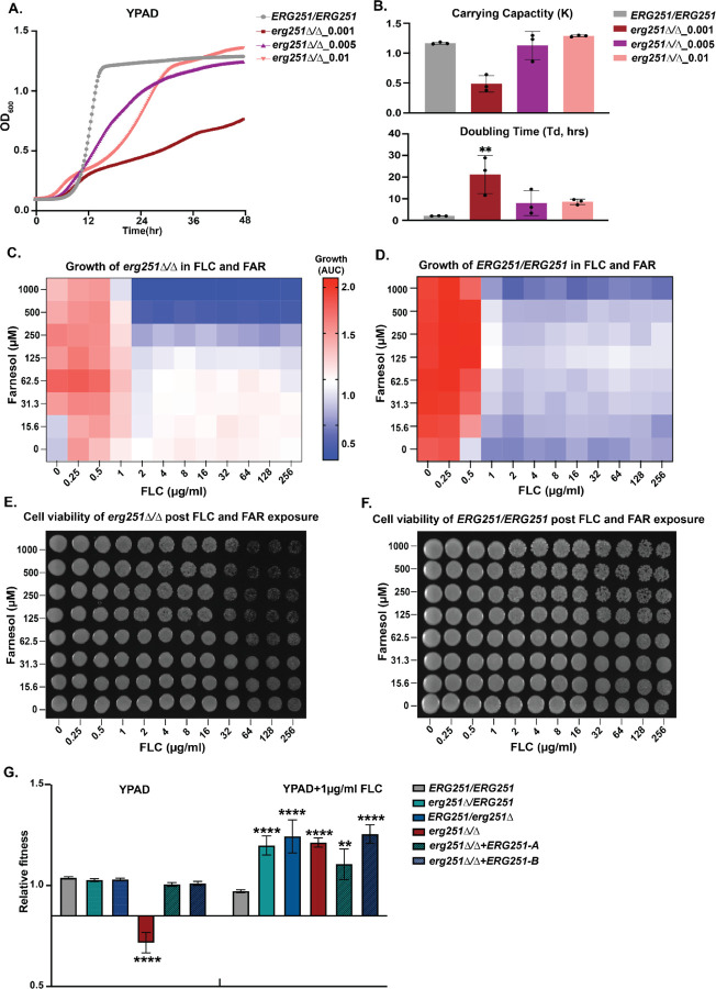
Our results show that disruption of one copy of ERG251 results in tolerance or resistance to azoles in distinct genetic backgrounds. Although we have shown that Hsp90 is required for tolerance, the full range of mechanisms and impact of changing ERG251 are not known. In order to fully understand the function of ERG251 in different cellular processes, two independent homozygous ERG251 deletion mutants were generated in SC5314 background. We confirmed that these deletions did not introduce any large-scale genomic changes (LOH and aneuploidy) (Fig S1B), and two independent erg251∆/∆ mutants (d51 and d70) exhibited identical phenotypes. Both erg251∆/∆ mutants had decreased growth in rich medium with low initial cell density (OD600=0.001) compared to the wild-type control in a 96-well plate format with shaking (Fig 3A and B). Importantly, we found that decreased growth of erg251∆/∆ in YPAD was related to the cell density. The growth defects of erg251∆/∆ were partially rescued by simply increasing initial cell density (from OD600=0.001 to 0.005 or 0.01), which mostly restored carrying capacity but not doubling time (Fig 3A and B). In C. albicans, cell density is communicated and linked with gene expression via the quorum sensing process, and farnesol is a major quorum sensing molecule secreted by C. albicans [24,48,49]. The production of farnesol requires the dephosphorylation of FPP, the precursor for the ergosterol biosynthesis pathway [25,50]. Therefore, we tested the impact of different concentrations of farnesol (0–1000µM) on the growth of erg251∆/∆ mutants with low initial cell density (Fig 3C, Y-axis). We found moderate concentrations of farnesol (62.5–250 µM) improved the growth of erg251∆/∆ in YPAD, while farnesol had no impact on growth of wild-type control (ERG251/ERG251) (Fig 3D, Y-axis). Therefore, homozygous deletion of ERG251 may result in disrupted ergosterol biosynthesis which subsequently provides negative feedback on farnesol production contributing to the growth defect of erg251∆/∆.
Fig 3. Homozygous deletion of ERG251 results in decreased fitness at low initial cell density and increased fitness in the presence of low concentrations of FLC (≤1µg/ml).
A. 48hr growth curve analysis of erg251∆/∆ started at three different initial cell densities (OD600=0.001, 0.005, or 0.01) with ERG251/ERG251 (SC5314, OD600=0.001) as the control. B. Carrying capacity (K) and doubling time (Td, hrs) determined from growth curve analysis in Fig 2A. C&D. X-Y growth curve assay of (C) erg251∆/∆ and (D) ERG251/ERG251 in the presence of increasing concentrations of FLC ( X-axis, 0–256 µg/ml, 2-fold dilutions) and/or increasing concentrations of farnesol (FAR) (Y-axis, 0–1000 µM, 2-fold dilutions). Growth was estimated with the area under the curve (AUC heatmap) of the 48hr growth curve. E&F. Cell viability of (E) erg251∆/∆ and (F) ERG251/ERG251 after 48 hr exposure to FLC or/and FAR. Cells from Fig 3B were plated on YPAD agar and imaged after 24hr incubation. G. Relative fitness calculated from head-to-head competitive assay for erg251∆/ERG251, ERG251/erg251∆, erg251∆/∆, erg251∆/∆+ERG251-A, and erg251∆/∆+ERG251-B compared to the fluorescent control strain (ERG251/ERG251). B&G: Values are mean ± SEM calculated from three technical replicates. Data were assessed for normality by Shapiro-Wilk, and significant differences between the ERG251/ERG251 and mutants were calculated using two-way ANOVA with Dunnett’s multiple comparisons test. ****p<0.0001, **p<0.01. A-G: At least three biological replicates were performed.
We next measured the impact of FLC on erg251∆/∆ strains. The growth defect of the erg251∆/∆ strain prevented us from conducting MIC and SMG assays for resistance and tolerance because these assays are normalized to growth in rich media (no drug) and erg251∆/∆ strains grow poorly in these conditions. Therefore, we tested the impact of FLC on erg251∆/∆ using a growth curve assay. By monitoring the growth of erg251∆/∆ at different concentrations (0–256µg/ml) of FLC, we found that low concentrations of FLC (≤1µg/ml) increased growth of erg251∆/∆ compared to no drug (Fig 3C, X-axis). High concentrations of FLC (>1µg/ml) had minimal impact on erg251∆/∆ growth (Fig 3C, X-axis). In contrast, the wild-type control (ERG251/ERG251) exhibited decreased growth at concentrations at and above the MIC50 (0.5µg/ml) (Fig 3D, X-axis). This suggests that total dysfunction of ERG251 can promote C. albicans growth in FLC but only at low concentrations.
Interestingly, adding either farnesol or FLC only partially restored the growth defect of erg251∆/∆ (Fig 3C). Next, we determined if adding farnesol in combination with FLC could further contribute to restored growth of erg251∆/∆. Growth of erg251∆/∆ from all different concentration combinations showed that low concentrations of FLC (≤1µg/ml) are sufficient to confer increased growth regardless of the concentration of farnesol (Fig 3C). In contrast, high-concentrations of both farnesol (>125µM) and FLC (>1µg/ml) greatly inhibited growth of erg251∆/∆ (Fig 3C). Growth inhibition for the wild-type control (ERG251/ERG215) was solely controlled by the FLC concentration (Fig 3D). Furthermore, high-concentration farnesol (>125µM) combined with high concentrations of FLC (>64µg/ml) exhibited a killing effect on the erg251∆/∆ cells but not on the wild-type control (ERG251/ERG251) (Fig 3E and F). These results suggest that in the absence of Erg251, farnesol can make FLC fungicidal at high concentrations, likely due to more severe inhibition of cell growth and ergosterol production.
Lastly, a head-to-head competition assay validated the fitness trade-off for erg251∆/∆, with a fitness cost in YPAD and a fitness benefit in the presence of a low concentration of FLC (1µg/ml) (Fig 3G). This fitness trade-off was not seen for the two heterozygous deletion mutants (erg251∆/ERG251 or ERG251/erg251∆) and was completely rescued by complementation of the homozygous deletion mutant with either ERG251-A or ERG251-B (erg251∆/∆+ERG251-A or erg251∆/∆+ERG251-B) (Fig 3G). Taken together, we propose that in response to low concentration FLC, erg251∆/∆ may upregulate the alternate sterol production pathway to compensate for ergosterol production and support increased growth (see below).
Pleiotropic effects of Erg251 on cell wall organization, stress response, and biofilm formation
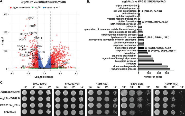
We next explored the mechanisms by which ERG251 affects fitness, drug susceptibility, and other stress responses. Transcriptional analysis was performed for the SC5314 wildtype (ERG251/ERG251), two ERG251 heterozygous deletion mutants, and one homozygous deletion mutant using RNAseq in two different log phase conditions: YPAD and YPAD+1µg/ml FLC. We first focused our analysis on the comparison between erg251∆/∆ and ERG251/ERG251 in YPAD to understand the role of ERG251 in a broad range of cellular processes (Fig 4A, S2 Table). Differential expression analysis was used to identify genes with a significant change in abundance in erg251∆/∆ cells compared to ERG251/ERG251 (913 genes, log2 fold change ≥ 1 or ≤−1 and adjusted p-value < 0.05). Gene Ontology (GO) analyses of differentially expressed genes revealed an overrepresentation of genes associated with cell wall organization, biofilm formation, filamentation growth, metabolic processes, and stress response (Fig 4B, S3 Table).
Fig 4. Homozygous deletion of ERG251 leads to increased sensitivity to cell wall and osmotic stress but decreased sensitivity to oxidative stress.
A. Volcano plot for differentially expressed genes (log2 fold change ≥ 1 or ≤-1 and adjusted p-value < 0.05) in the erg251∆/∆ mutant compared to ERG251/ERG251 in YPAD with both fold change and p-value indicated. B. Gene Ontology (GO) terms for differentially expressed genes (log2 fold change ≥ 1 or ≤ −1 and adjusted p-value < 0.05) in the erg251∆/∆ mutant compared to ERG251/ERG251 in YPAD. C. Spot plates growth of ERG251/ERG251, erg251∆/ERG251, ERG251/erg251∆, and erg251∆/∆ on YPAD (30°C), YPAD (37°C), 1.2M NaCl, 0.03% SDS and 7.5mM H2O2 agar plates. A-C: At least three biological replicates were performed.
We identified many down-regulated genes in erg251∆/∆ cells involved in cell membrane, filamentation, and stress response. Genes that regulate cell membrane structure including lipid metabolism, ergosterol and sphingolipid biosynthesis were down-regulated, including ERG11, ERG1, LIP1, PLB1, UPC2, ARE2, and SCS7 (Fig 4A and 4B) [36,51]. Genes that regulate filamentation and biofilm formation were also down-regulated, including FGR23, HYR1, ALS2, and ALS4 (Fig 4A and 4B) [52–54]. Additionally, the osmotic stress related gene AQY1 and heat stress related genes HSP70 and HSP12 were also down-regulated (Fig 3A) [54–56]. In contrast, genes that are involved in oxidative stress response like SOD5 and AOX2 were up-regulated (Fig 3A and 3B) [57,58]. These changes in lipid metabolism and stress response may affect metabolism and nutrient availability more broadly including carbon and amino acid metabolism [20,59].
GO analysis of biological process identified enrichment of 26 genes in erg251∆/∆ cells that encode proteins with GlycosylPhosphatidylInositol (GPI)-anchored motifs. GPI anchors attach proteins to the cell surface contributing to cell-wall integrity, cell-cell interaction, and hyphal formation [60,61]. These genes include HYR1, FGR23, and SOD5 as well as cell wall specific genes in the PGA and ALS families. Overall, genes encoding GPI-anchored motifs were down-regulated in erg251∆/∆ cells (20 out of 26) (Fig 4A and S4 Table) consistent with prior work demonstrating cross-talk between the ergosterol and GPI biosynthesis pathways [62,63].
Phenotypic analysis was consistent with transcriptional analysis for the pleiotropic effects of ERG251 on cell wall organization and stress responses. Compared to wildtype (ERG251/ERG251), erg251∆/∆ exhibited no change in response to increased temperature (37°C). However, erg251∆/∆ exhibited decreased growth in the presence of osmotic (1.2M NaCl) and cell membrane (0.03% SDS) stress, but increased resistance to H2O2 (Fig 4C). These changes in stress response were not observed for the two ERG251 heterozygous deletion mutants at either the transcriptional or phenotypic levels (Fig 4C, Fig S3A, and S3B). Taken together, this indicates that the total disruption of ERG251 results in a dramatic physiological response that impacts cell membrane/cell wall compositions, and osmotic/oxidative stress responses.
ERG251-A exhibits dominant regulation of filamentation
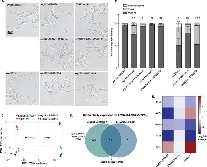
Given that genes related to filamentation and biofilm formation were downregulated in erg251∆/∆, we next quantified filamentation in all three deletion mutants (homozygous and two heterozygous). Using an in vitro filamentation assay, we found that erg251∆/ERG251 had a ~25% decrease in the proportion of hyphae, while ERG251/erg251∆ exhibited almost no change compared to wild-type ERG251/ERG251 (Fig 5A and 5B). Complementation of erg251∆/ERG251 with the ERG251-A allele restored wild-type filamentation (Fig 5A and 5B). This indicates that ERG251-A plays a dominant role in regulating filamentation, while ERG251-B is not required for filamentation in C. albicans. Additionally, a more severe filamentation defect (~50%) was observed in erg251∆/∆ compared to ERG251/ERG251 (Fig 5A and 5B). Complementation of erg251∆/∆ with the ERG251-A allele, not the ERG251-B allele, was able to partially restore filamentation (Fig 5A and 5B). This data supports a dominant role of ERG251-A in regulating filamentation.
Fig 5. Deletion of ERG251-A but not ERG251-B leads to decreased filamentation.
A. Representative filamentation images of wildtype ERG251/ERG251, erg251∆/ERG251, erg251∆/ERG251+ERG251-A, ERG251/erg251∆, ERG251/erg251∆+ERG251-B, erg251∆/∆, erg251∆/∆+ERG251-A, and erg251∆/∆+ERG251-B. Cells were induced in RPMI supplemented with 10% FBS for 4 hrs. Scale bar, 20 μm. B. Quantification of the yeast (<6μm), pseudohyphae (15–36 μm), and hyphae (>36 μm) from genotypes in Fig 5A. 150 to 500 cells were counted for each strain, and at least two biological replicates were performed. Error bars indicate SEM. Statistical significance for filamentation was compared to ERG251/ERG251 and assessed using two-way ANOVA with uncorrected Fisher’s LSD, ***P <0.001, **P <0.01,* P ≤ 0.05, ns: P >0.05. C. Principal component analysis of transcriptional data in YPAD and YPAD+FLC (1µg/ml) for ERG251/ERG251, erg251∆/ERG251, ERG251/erg251∆, and erg251∆/∆. D. Venn diagrams comparing the genes that are differentially expressed in erg251∆/ERG251 and ERG251/erg251∆ (log2 fold change ≥ 0.5 or ≤-0.5 and adjusted p-value < 0.1) relative to ERG251/ERG251 in YPAD. E. The relative expression level (log2 fold change) of genes associated with filamentation in erg251∆/ERG251, ERG251/erg251∆, and erg251∆/∆compared to ERG251/ERG251 in YPAD.
ERG251-A regulation of filamentation might be caused by the control of genes that are involved in the yeast-to-hyphae transition. Transcriptional analysis revealed that deletion of ERG251-A (erg251∆/ERG251) resulted in a greater impact on overall gene expression than deletion of ERG251-B (ERG251/erg251∆) (Fig 5C, S3A–D). In the YPAD condition, deletion of ERG251-A resulted in 357 differentially expressed genes (log2 fold change ≥ 0.5 or ≤-0.5 and adjusted p-value < 0.1, S5 Table) compared to deletion of ERG251-B which altered expression of 26 genes (log2 fold change ≥ 0.5 or ≤-0.5 and adjusted p-value < 0.1, S6 Table)(Fig 5D). Only 11 genes were significantly differentially expressed in both heterozygous mutants including ERG6, ERG251 and CRZ2 (Fig 5D). Notably, ERG6 had increased expression in both heterozygous mutants which may contribute to the activation of the alternate pathway for ergosterol biosynthesis (Fig S3A, S3B, and Fig 5D). This suggests there is redundancy of the ERG251-A and ERG251-B alleles in ergosterol biosynthesis, which is consistent with the same azole tolerance phenotypes we observed upon loss of function of either allele (Fig 1). Furthermore, in the SC5314 background ERG251-A and ERG251-B had similar RNA abundance (Fig S3E), and both of the tagged proteins, Erg251-A-GFP and Erg251-B-GFP, localized to the endoplasmic reticulum (ER) in both yeast and hyphal phases (Fig S3F). This indicates that the divergent function of the two ERG251 alleles is not caused by allelic expression or subcellular translocation. Among the 346 genes that were differentially expressed only in erg251∆/ERG251, GO analysis revealed an enrichment of genes that regulate filamentation (Fig S3C, S7 Table). Genes that positively regulate filamentation, including HYR1 and HWP1, and their up-stream transcription factor SFL2 were all down-regulated in erg251∆/ERG251 (Fig S3A and 5E) [53,54]. Transcription factor EFH1 was up-regulated in erg251∆/ERG251 and its overexpression may lead to pseudohyphal formation (Fig S3A and 5E) [64,65]. Finally, we found that in YPAD, both erg251∆/ERG251 and erg251∆/∆ have largely conserved regulation of this subset of genes involved in filamentation: SFL2, HWP1, HWP2, HYR1, HYR3, and EFH1 (Fig 5E).
Homozygous deletion of ERG251 leads to downregulation of ergosterol biosynthesis genes and upregulation of alternate sterol biosynthesis genes
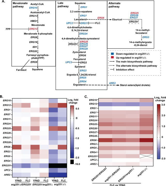
Homozygous deletion of ERG251 results in a diverse set of phenotypic effects that may be directly related to disrupted ergosterol biosynthesis and lipid metabolism genes. To more comprehensively analyze the impact of deleting ERG251 on ergosterol biosynthesis, we analyzed transcription of genes involved in ergosterol biosynthesis from all three pathways: mevalonate, late and alternate (Fig 6A). In YPAD, erg251∆/∆ had decreased expression relative to wild-type for 11 ERG genes, and increased expression of ERG12, ERG25, and ERG6 (Fig 6A and B). Among the 11 down-regulated ERG genes, ERG1 and ERG11 had the most significant decreases (log2 fold change= −1.5 and −1.3 respectively). These two genes represent two rate-limiting steps in the ergosterol biosynthesis pathway (Veen M et al., 2003). Two additional key genes were down regulated in erg251∆/∆ compared to wildtype: UPC2, encoding a transcription factor that activates ERG genes, and ARE2, encoding a sterol acyltransferase that regulates the storage and decomposition of ergosterol [66–68].
Fig 6. Homozygous deletion of ERG251 leads to the downregulation of ergosterol biosynthesis genes.
A. Overview of the ergosterol biosynthetic pathway in C. albicans, including the mevalonate, late ergosterol, and alternate pathways. Genes that were down-regulated (blue) and up-regulated (red) in the erg251∆/∆ under YPAD conditions relative to SC5314 [14,21,69–71]. B. The relative gene expression levels (log2-fold change) for all ERG genes in the heterozygous and homozygous mutants erg251∆/ERG251, ERG251/erg251∆, and erg251∆/∆ grown in YPAD or YPAD+1µg/ml FLC conditions, compared to the wildtype ERG251/ERG251 in the same condition. C. The relative expression level (log2 fold change) of ERG genes in the wildtype ERG251/ERG251, and mutants erg251∆/ERG251, ERG251/erg251∆, and erg251∆/∆ grown in YPAD+1µg/ml FLC compared to YPAD condition.
Unlike most ergosterol biosynthesis genes, ERG25, the paralog of ERG251, had increased expression across both the heterozygous and homozygous ERG251 deletion mutants (log2 fold change ~0.5, Fig 6B). ERG25 is expressed at low levels relative to ERG251 [20], and increased expression of ERG25 improved growth and filamentation of the erg251∆/∆ null mutant (Xiong et al, co-submitted manuscript). We therefore hypothesized that Erg25 and Erg251 can compensate for each other during ergosterol biosynthesis despite showing significant sequence divergence (Fig S4A). We were unable to generate a double homozygous deletion of ERG251 and ERG25 after multiple attempts. Instead, we generated a heterozygous deletion of ERG25 in the wild-type and in the erg251∆/∆ mutant, and neither ERG25 mutant had an effect on drug susceptibility (Fig S4B and S4C). Transcriptional data also supported that the increased abundance of ERG25 transcripts in erg251∆/∆ was still lower than ERG251 transcripts in the wild-type background (Fig S4D). Together, our data directly show the paralog compensation between ERG251 and ERG25 at the transcriptional level and high-expression level of ERG251 combined with observed phenotypic changes again supports ERG251 is the major player for methyl sterol synthesis and can impact drug susceptibility.
When comparing the transcriptional abundance of ERG genes between the two growth conditions (FLC vs YPAD), we found that almost all ERG genes had increased expression in response to FLC exposure across all four strains, with or without ERG251 deletion (Fig 6C). Importantly, ERG genes that were down-regulated in erg251∆/∆ in YPAD were partially restored with growth in FLC (1µg/ml), which may explain the improved growth of erg251∆/∆ in low concentrations of FLC (≤1µg/ml) (Fig 6C and 3E). Strikingly, ERG6 had 8-fold increased expression in erg251∆/∆ in the presence of FLC relative to ERG251/ERG251 (Fig 6C), and over-expression of ERG6 can result in accumulation of the alternative sterols leading to cell survival in the presence of FLC [21,27,69]. This is consistent with a recent report showing that with FLC exposure, erg251∆/∆ has a depletion of ergosterol and an accumulation of the sterol precursors for the toxic dienol (Lu et al 2023). These results support the homozygous deletion of ERG251 can lead to upregulation of alternative sterols that promote survival in presence of FLC. However, the heterozygous ERG251 mutants had minimal differences in ERG gene expression (other than ERG251 itself) compared to the wild-type in the presence of FLC (Fig 6B). This suggests that the azole tolerance of the heterozygous ERG251 mutants is caused by mechanisms independent from ergosterol biosynthesis gene expression changes.
Dysfunction of ERG251 turns on Zinc transporter contributing to decreased azole susceptibility
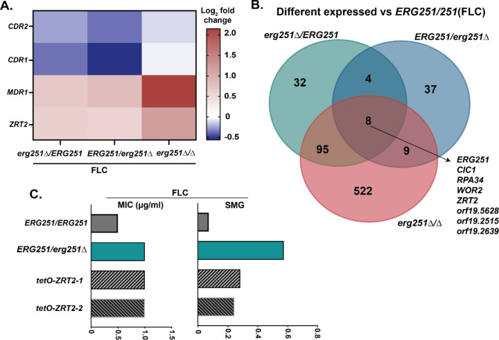
To determine the mechanism driving decreased drug susceptibility, we further compared the transcriptional analysis of all three ERG251 deletion mutants during growth in FLC. No significant change in expression was observed for genes encoding the drug efflux pumps CDR1, CDR2, and MDR1 across the ERG251 deletion mutants, compared to wild-type (log2 fold change ≥ 0.5 or ≤−0.5 and adjusted p-value < 0.1), with an exception for MDR1 in erg251∆/∆ that had a 2-fold increase (adjusted p-value=9.6x10⁻⁷) (Fig 7A, S9–S11 Table). We next determined whether there was a conserved transcriptional response across the three ERG251 mutants after FLC exposure. In YPAD+1ug/ml FLC, only 8 genes were significantly differentially expressed in all three ERG251 deletion mutants relative to ERG251/ERG251 (heterozygous deletion mutants: log2 fold change ≥ 0.5 or ≤ −0.5 and adjusted p-value < 0.1, homozygous deletion mutant: log2 fold change ≥ 1 or ≤ −1 and adjusted p-value < 0.05) (Fig 5E). Based on the predicted and characterized functions of these 8 genes, we focused on ZRT2 which encodes a zinc transporter. In C. albicans, Zrt2 localizes to the plasma membrane and is essential for Zinc uptake and growth at acidic pH [72]. Interestingly, ZRT2 was upregulated in both heterozygous and homozygous ERG251 deletion mutants during FLC exposure. This supports that the dysfunction of ERG251 can upregulate the Zinc transporter.
Fig 7. Dysfunction of ERG251 activates a Zinc transporter contributing to decreased azole susceptibility.
A. The relative expression level (log2 fold change) of CDR1, CDR2, MDR1 and ZRT2 in erg251∆/ERG251, ERG251/erg251∆, and erg251∆/∆ compared to ERG251/ERG251 under YPAD+1µg/ml FLC condition. B. Venn diagrams comparing the genes that differentially expressed in erg251∆/ERG251, ERG251/erg251∆ and erg251∆/∆ related to ERG251/ERG251 under YPAD+1µg/ml FLC condition. C. 24hr MIC (left, µg/ml) and 48hr SMG (right, tolerance) in FLC for two ZRT2 overexpression strains (tetO-ZRT2-1 and tetO-ZRT2-2) in SC5314 background together with SC5314 (ERG251/ERG251) and erg251∆/ERG251 as the controls. At least three biological replicates were performed.
To determine if elevated ZRT2 contributes to the azole tolerance in the ERG251 mutants, we constructed a ZRT2 overexpression strain. Overexpression of ZRT2 in the SC5314 background (ERG251/ERG251) resulted in 2-fold increase in FLC MIC50 and increased FLC tolerance, but resulted in less tolerance than the erg251∆/ERG251 mutant (Fig 7C). We propose that Zrt2 may contribute to the drug efflux activities distinct from the ATP-dependent drug efflux pumps such as CDR1 to increase drug tolerance.
Single allele dysfunction of ERG251 maintains pathogenicity in a murine model
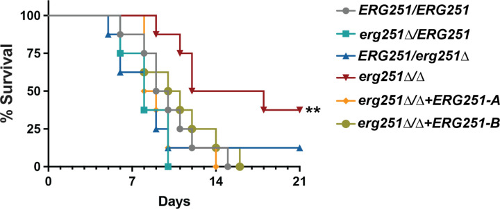
We next explored the effects of ERG251 mutations on pathogenicity to better understand the roles of these mutations during infection. The pleiotropic effects of ERG251 on varied cellular responses, especially decreased resistance to superoxide and reduced filamentation raise the question of whether dysfunction of ERG251 would also lead to a pathogenicity defect. We tested the two heterozygous and homozygous ERG251 deletion mutants in the standard mouse tail-vein injection model of disseminated candidiasis [73]. There was no difference in survival between the wild-type control SC5314 and the heterozygous mutants (erg251∆/ERG251 and ERG251/erg251∆) (Fig 8). However, mice infected with erg251∆/∆ had significantly longer survival compared to the wild-type control or either heterozygous deletion mutant with a mean time of survival of 15 days for erg251∆/∆ compared to 8–10 days for all other strains (Fig 8). The statistical analysis showed that the survival curve for erg251∆/∆ is significantly different compared to the wildtype (P = 0.0015, Long-rank (Mantel-Cox) test) (Fig 8). We also tested the survival of two complementation strains of erg251∆/∆, which also had a mean time of survival of 10 days indicating the restoration of virulence (Fig 8). Taken together, this indicates that homozygous deletion of ERG251 leads to attenuated virulence, which supports the importance of ERG251 in varied cellular responses essential for pathogenicity. However, the partial dysfunction of ERG251 has no impact on virulence. The lack of a virulence defect in the heterozygous mutants emphasizes that these mutations, which result in high levels of azole tolerance, remain infectious.
Fig 8. Heterozygous deletion of ERG251 maintains pathogenicity in a murine model.
A. ICR mice were injected via the tail vein with 5x10⁵ cells of ERG251/ERG251 (SC5314), erg251∆/ERG251, ERG251/erg251∆, and erg251∆/∆+ERG251-A and erg251∆/∆+ERG251-B and survival was presented over the time. The erg251∆/∆ mutant survival curves were significantly attenuated from that of the ERG251/ERG251 (Log-rank (Mantel-Cox) test; **, p = 0.0015). Eight mice per strain were used.
DISCUSSION
Changes in ERG251 impact the susceptibility of C. albicans to azoles
Antifungal tolerance varies among C. albicans clinical isolates and correlates with the inability to clear an infection. The tolerance phenotype is stable even in the absence of antifungal drug stress [9]. Despite this, the molecular mechanisms causing antifungal tolerance are not known. We found that ERG251 is a hotspot for point mutations during adaptation to antifungal drug stress, and that heterozygous deletion of ERG251 can drive azole tolerance in diverse clinical isolates of C. albicans. This highlights the novel finding that an ERG251 point mutation alone can drive azole tolerance and contribute to the adaptation to FLC.
Aneuploidy is associated with the rapid evolution of azole tolerance [74,75] (Zhou et al. 2024, under review). We recently found that the Chr3 and Chr6 concurrent aneuploidies conferred multi-azole tolerance via elevated drug efflux pumps (Zhou et al. 2024, under review). Here, we report that recurrent single nucleotide point mutations in ERG251 (Chr4) often occur together with aneuploidies of smaller chromosomes (Chr3-Chr7) during adaptation to FLC (Table 1). Therefore, we dissected the impact of aneuploidy and/or ERG251 point mutations on drug susceptibility by engineering the ERG251 mutations within euploid or aneuploid backgrounds. Heterozygous point mutations at either allele of ERG251 in euploid backgrounds led to the single allele dysfunction of ERG251 which conferred azole tolerance. Heterozygous deletion of ERG251 in a Chr3 and Chr6 aneuploid background was sufficient to cause very high levels of azole resistance (Fig. 2). In other words, the combination of two mechanisms that independently cause azole tolerance can dramatically increase the growth of fungal cells in drug, resulting in bona fide azole resistance. Multiple simultaneous mutations are common in cancer cells and often lead to treatment failure [76–79]. Our results highlight the diverse trajectories that C. albicans can take during adaptation to antifungal drugs and support that two independent mechanisms of tolerance (point mutation and aneuploidy) can evolve within the same cell resulting in drug resistance.
The impact of mutations is also influenced by the genetic background of an organism. Across different genetic backgrounds, ERG251 had conserved roles in causing azole tolerance and driving adaptation to FLC; however, variation in tolerance levels was observed. We identified point mutations in ERG251 within two distinct genetic backgrounds (SC5314 and P75063) from three independent evolution experiments across multiple labs. All ERG251 point mutations conferred multi-azole tolerance. However, in the P75063 background, ERG251-driven tolerance (SMG=0.26) was lower than in SC5314 background (SMG>0.5). In the P75063 background, ERG251 is homozygous (no allelic variation) despite the heterozygosity across the rest of Chr4. Additionally, the distinct alleles of ERG251 in the SC5314 background did not affect azole tolerance (Fig 1A), therefore we hypothesize that genetic variations of ERG251 between different genetic backgrounds are not causing the differences in tolerance phenotype. Instead, it is more likely to be caused by the genetic variation at other loci in the genome [80].
Drug susceptibility in heterozygous vs homozygous ERG251 deletion mutants
One of the particularly striking observations from our study was the range of different heterozygous point mutations that phenocopied the heterozygous deletion of ERG251. The numerous possible mutation sites at either allele of the ERG251 combined with a strong fitness advantage for all characterized ERG251 mutations in FLC explains why ERG251 mutations were recurrent in three independent in vitro evolution experiments. The probability of a single-nucleotide mutation in ERG251 that has a phenotype appears to be higher than other types of point mutations that cause increased growth in azoles. For example, point mutations in ergosterol-related genes like ERG11 and UPC2 that cause drug resistance in Candida species are all homozygous in diploid organisms or the only allele present in haploid organisms [81–85]. Comparatively, all of the de novo ERG251 point mutations identified here were heterozygous. Although the homozygous deletion of ERG251 led to a similar fitness advantage as the heterozygous deletion mutants in the presence of low concentrations of FLC (<1µg/ml), the homozygous deletion strain exhibited a fitness cost in rich medium and at higher concentrations of FLC, supporting why only heterozygous mutants were identified during in vitro evolution experiments.
Prior studies of the relationship of ERG251 to azole susceptibility in C. albicans have found conflicting results. In some experiments, loss of ERG251 through genetic manipulation or pharmacological inhibition resulted in increased susceptibility to azoles whereas in other experiments, disruption of ERG251 decreased susceptibility to azoles [20,36]. Our findings help to explain these disparate results. We observed substantial phenotypic differences between heterozygous and homozygous deletion mutants indicating that the amount of Erg251 remaining in the cell can affect ergosterol biosynthesis and other metabolic processes. Another group recently demonstrated that erg251∆/∆ phenotypes are dependent on other growth conditions, including oxygen availability (Xiong et al. 2024, co-submitted manuscript). We also found that the growth of erg251∆/∆ was dependent on cell density and azole concentration. The erg251∆/∆ mutants had decreased susceptibility to low concentrations of FLC, but increased susceptibility to high concentrations of FLC, especially in combination with the quorum sensing molecule farnesol.
Partial differentiation of ERG251-A vs ERG251-B function
In the SC5314 background, the A and B alleles of ERG251 differ at two non-synonymous SNPs. When comparing the heterozygous mutations from the evolution experiments and engineered strains, we did not observe any differences between the alleles in growth and azole-related phenotypes. GFP-tagging of both alleles showed similar localization patterns, and RNAseq analysis showed that both alleles are expressed at similar levels in rich media and FLC. However, we observed striking differences between the two alleles when considering overall gene expression patterns and filamentation (Fig. 5). In the heterozygous background (SC5314), ERG251 exhibited allelic differences in controlling filamentation with ERG251-A as the dominant player. Phenotypic analysis showed that deletion of ERG251-A alone can lead to filamentation defects while deletion of ERG251-B had no effect on filamentation. The filamentation defect was correlated with altered gene expression of filamentation-related genes like HYR1 and EFH1 [53,54,64,65]. ERG251 is another rare example of a gene containing a gain-of-function allele that regulates filamentation in C. albicans, similar to the transcription factor Rob1 [86]. However, future work is needed to understand the molecular basis of the functional distinctions between diverse ERG251 alleles.
Sterol composition changes when ERG251 is inactivated
Homozygous deletion of ERG251 had a global impact on the expression of sterol biosynthetic genes resulting in down-regulation of ergosterol biosynthetic genes and upregulation of alternate sterol biosynthetic genes. Together with data from a previous study and the co-submitted manuscript by Xiong et al., we propose that homozygous deletion can lead to sterol composition changes that include reduced ergosterol and an accumulation of alternative sterols [20](Xiong et al. 2024). These changes in sterol composition are further supported by additional phenotypic changes we observed, including increased cell wall sensitivity of erg251∆/∆.
We also observed a connection between farnesol and ergosterol biosynthesis. We found moderate concentrations of farnesol (62.5–250 µM) improved the growth of erg251∆/∆ in rich media, while farnesol had no impact on growth of wild-type control. Therefore, homozygous deletion of ERG251 may result in disrupted ergosterol biosynthesis which subsequently provides negative feedback on farnesol production contributing to the growth defect of erg251∆/∆. One ERG gene, ERG12, had decreased expression in response to fluconazole - the opposite trend from all other ERG genes (Fig 6). ERG12 encodes mevalonate kinase and converts mevalonate into 5-phosphomevalonate, the precursor for farnesyl pyrophosphate (FPP). One hypothesis is that the FLC-induced repression of ERG12 is caused by the inhibition of the farnesol or FPP via negative feedback. We also found that homozygous deletion of ERG251 caused increased expression of ERG12. The increased expression of ERG12 combined with farnesol-related growth phenotypes in the erg251∆/∆ mutant suggests that ERG12 expression is negatively regulated by farnesol or its precursor, FPP, and that both deletion of ERG251 and FLC exposure impact ERG12 expression possibly via farnesol production.
When ERG251 was inactivated, its paralog ERG25 was upregulated to compensate for the loss of the primary enzyme. The shared enzymatic function and regulatory networks of paralogs supports a model where compensation between ERG251 and ERG25 occurs on the level of gene expression [38,87]. However, we found that the upregulated ERG25 didn’t reverse the phenotypic changes observed for ERG251 deletion mutants because ERG25 had much lower expression (Fig S4D). Other studies also showed ectopic over-expression of ERG25 only partially recovered the phenotype changes of erg251∆/∆ [20](Xiong et al. 2024). In conclusion, in C. albicans both Erg251 and Erg25 function as the essential C-4 sterol methyl oxidase with Erg251 serving as the primary role controlling drug susceptibility, filamentation, biofilm formation and other stress responses [20](Xiong et al. 2024).
Upregulated Zinc transporter contributing azole tolerance
We also found transcriptional upregulation of the Zinc transporter, Zrt2, in all ERG251 deletion mutants (heterozygous and homozygous) during exposure to fluconazole. The upregulation of this plasma membrane transporter could be caused by alterations in cell membrane structure that coincide with the down-regulation of ergosterol biosynthesis in ERG251 deletion mutants [41–43]. Compared to ERG251 heterozygous deletion mutants, over-expression of ZRT2 led to the same 2-fold increase in MIC and a significant, but smaller increase in azole tolerance (Fig. 7). Therefore, we propose that ERG251-mediated changes in sterol metabolism and upregulated Zinc transport together promote decreased drug susceptibility observed for the ERG251 deletion mutants.
In summary, this study identified recurrent heterozygous point mutations in the methyl sterol oxidase ERG251 during adaptation to antifungal drug stress and characterized for the first time point mutation-driven azole tolerance. We utilized genetic, transcriptional and phenotypic analyses to understand the effects of inactivating ERG251 both partially and completely. Increased azole tolerance was observed in two distinct genetic backgrounds, and heterozygous mutations of ERG251 promote multi-azole tolerance while maintaining virulence in a mouse model of systemic infection. Future experiments will explore ERG251 genetic diversity and phenotypes across a broad range of clinical isolates in Candida albicans and in related Candida species.
MATERIALS AND METHODS
Yeast isolates and culture conditions
All strains used in this study are listed in S1 Table including FLC evolved isolates and engineered yeast and bacteria strains. Strains were stored at −80℃ in 20% glycerol. Isolates were grown in YPAD media (20 g/L peptone, 10 g/L yeast extract, 2% dextrose, and 15 g/L agar for plates) supplemented with 40 µg/ml adenine and 80 µg/ml uridine.
Strain construction
All engineered strains in this study were generated in the SC5314 background, except one ERG251 heterozygous deletion in the P75063 background. Strains were constructed by lithium acetate transformation using PCR products with at least 140 bp of homology to the target locus. Primers used in this study are listed in S12 Table.
-
ERG251 heterozygous deletion
The FLIP-NAT construct was PCR amplified from the plasmid pJK863 [88] using primer sets 1630+1631 and transformed into background strains SC5314 and P75063. NAT-resistant transformants were PCR screened for correct integration of the FLIP-NAT construct at the ERG251 locus using primer pairs 1652+1045 (left of integration) and 1636+1653 (right of integration). Transformants were validated by whole genome sequencing for correct integration.
-
ERG251 homozygous deletion
To promote FLIP-mediated excision of FLIP-NAT, correct heterozygous deletion strains erg251∆/ERG251 and ERG251/erg251∆ were inoculated in YNB+BSA from frozen stocks and incubated at 30℃, 220rpm, for 48 hrs. Cultures were diluted and 100 cells were plated on YPAD agar, then incubated at 30°C for 24 hrs. Recovered colonies were patched to both YPAD and YPAD+150 µg/ml NAT. Colonies growing on only YPAD were screened for correct FLIP-mediated excision of FLIP-NAT using primer pairs 1574+1575 (inside NAT) and 1652+1653 (across ERG251). Colonies that correctly excised FLIP-NAT were re-transformed with the FLIP-NAT construct (PCR amplified from the plasmid pJK863 [88] using primer sets 1630+1631). NAT-resistant transformants were PCR screened for correct integration of the FLIP-NAT construct at the remaining ERG251 locus using primer pairs 1652+1045 (left of integration), 1636+1653 (right of integration), and 1632+1633 (inside ERG251). Transformants were validated by whole genome sequencing for correct integration.
-
Construct ERG251-NAT plasmid
To generate ERG251 mutant complementation and point mutation, we built up an ERG251-NAT plasmid by fusing ERG251 upstream plus gene (1644+1645), NAT (1574+1575), and ERG251 downstream (1646+1647) into the pUC19 backbone (1578+1579). PCR amplified fragments were aligned using NEBuilder HiFi DNA Assembly Cloning Kit following the manufacturer’s instructions and transferred into E. coli. Ampicillin-resistant transformants were screened using primer pairs 1352+1353 and saved in frozen stocks as pAS3118.
-
ERG251 mutant complementation
The wild-type ERG251 upstream region and genes (A or B) were PCR amplified from heterozygous deletion strains using primer pair 1652+1645. The NAT gene and downstream ERG251 region were PCR amplified from pAS3118 using 1574+1653 primers. SOEing PCR was performed using primer pair 1652+1653. The subsequent ERG251-NAT construct was transformed into the erg251∆/ERG251, ERG251/erg251∆, and erg251∆/∆ mutants that had previously excised FLIP-NAT as described above. NAT-resistant transformants were PCR screened for correct integration of the ERG251-NAT construct using primer pairs 1634+1154 (left integration), 1636+1635 (right integration), and 1634+1635 (across integration). Transformants were validated by whole genome sequencing for correct integration.
-
ERG251 point mutation
Site-directed mutagenesis using double-primer PCR was used to generate ERG251 point mutation construct. Primers with the desired mutations were paired with ERG251-NAT upstream (1652) or downstream (1653) primer to amplify mutated ERG251-NAT construct from pAS3118. Four different point mutations were engineered in this study: L113*(1652+1649/1648+1653), W265G(1652+1655/1654+1653), E273*(1652+1651/1650+1653), and *321Y(1652+1657/1656+1653). The amplified two fragments were fused using SOEing PCR and transformed into the SC5314 background. Transformants were first PCR screened using primers 1652+1575 (left integration) and 1636+1653 (right integration) for correct integration and then validated by whole genome sequencing for base substitution and mutated allele.
-
ERG251 overexpression
The TetO promoter replacement construct was PCR amplified using primer pair 1679+1680 from plasmid pLC605 [89] and transformed into the SC5314 background strain. NAT-resistant transformants were PCR screened for correct integration of the TetO promoter replacement using primer pairs 1652+1176 (left integration) and 1177+1633 (right integration).
-
ERG251-GFP
The c-terminal GFP-NAT construct was PCR amplified from plasmid pMG2120 [90] using the 1925+1926 primer pairs and transformed into the SC5314 background strain . NAT-resistant colonies were PCR screened for correct integration of the GFP-NAT construct at the c-terminal end of the ERG251 locus using primer pairs 1632+1927 (left integration) and 1636+1653 (right integration). Transformants were validated by Sanger sequencing for tagged alleles.
-
ERG25 heterozygous deletion
The FLIP-NAT construct was PCR amplified from plasmid pJK863 using primer pairs 1921+1922 and transformed into the SC5314 background strain. NAT-resistant transformants were PCR screened for correct integration of FLIP-NAT at the ERG25 locus using primer pairs 1923+1045 (left integration) and 1636+1924 (right integration).
Filamentation
Strains were inoculated in 2% dextrose YPAD from frozen stocks and incubated at 30℃, 220 rpm for 16 hrs. Strains were diluted 1:100 into RPMI+10% FBS, then incubated at 37℃ for 4 hrs. Cells were harvested, washed once with PBS, and resuspended in PBS before microscopy. Images were captured using an Olympus IX83 microscope and analyzed using ImageJ v1.54d.
Microscopy
Erg251-GFP tagged strains were struck on YPAD agar plates from frozen stocks and incubated at 30℃ for 24 hrs. Cultures were inoculated in 2% YPAD and incubated at 30℃, 220 rpm for 16 hrs. Cultures were diluted 1:100 in fresh 2% YPAD or RPMI+10% FBS, then incubated at 30℃, 220 rpm for 4 hrs. Cells were spun down, washed once with PBS, and resuspended in PBS before microscopy. Images were captured using an Olympus IX83 microscope.
Spot plate assay
Strains were inoculated in 2% dextrose YPAD from glycerol stocks and incubated at 30℃, 220 rpm for 16 hrs. Cultures were normalized to 106 cells/ml, then 10-fold serially diluted. 10µl of each (106-103) dilutions were spotted onto YPAD agar with and without drugs. All spot plates were performed in triplicates. Plates were incubated for 48 hrs at 30℃ and imaged using a BioRad GelDoc XR+ imaging system.
RNA sequencing
RNA extraction: For RNA extraction, all 4 strains (wildtype and 3 ERG251 deletion mutants) were struck on YPAD agar plates from frozen stocks and incubated at 30℃ for 24 hrs. Cultures were then inoculated in 2% YPAD (50 ml) and incubated at 30℃, 220 rpm for 16 hrs. Overnight cultures were then diluted 1:100 into 50 ml YPAD or YPAD+1µg/ml FLC and grown at 30℃, 220 rpm for 5–6 hrs to OD600 of 0.5. Cells were harvested by centrifugation and frozen in liquid nitrogen. RNA were prepared according to the manufacturer’s instructions for the Qiagen RNeasy Mini kit (Qiagen, US) using the mechanical disruption method. Removal of DNA was performed with a DNase (Qiagen RNase-free DNase set, US) 1 hr incubation at room temperature on column. Three independent cultures of each strain were grown to provide three biological replicates for RNA-seq experiments.
RNA-Seq: Library preparation was performed by SeqCenter (Pittsburgh, PA) using Illumina’s Stranded mRNA preparation and 10bp unique dual indices (UDI). Sequencing was done on a NovaSeq X Plus, producing 150bp paired end reads. Demultiplexing, quality control, and adapter trimming was performed with bcl-convert (v4.1.5) (BCL Convert).
RNA-Seq data analysis: C. albicans transcriptome (SC5314_version_A21-s02-m09-r10_orf_coding, downloaded from http://www.candidagenome.org/download/sequence/C_albicans_SC5314/Assembly21/current/?C=S;O=A on 2023/08/17) was indexed using salmon (v1.10.2) [91]. All samples were quasi-mapped to transcriptome index using salmon resulting in quantification of reads mapped to each transcript. The output quantification files were imported into R (v4.1.2) using tximport (v1.22.0) [92] and DESeq2 (1.34.0) [93] was used to model gene expression. PCA analysis was performed using DESeq2 and used to identify any outliers amongst the replicates. We identified one wild-type control grown in FLC as an outlier and excluded this sample from all further analyses (Fig 5C). The DESeq2 ‘contrast’ wrapper was then used to estimate log2 fold changes for each mutant relative to the wild-type control in YPAD and YPAD+1µg/ml FLC conditions and identify differentially expressed genes. We also estimated log2 fold changes for each strain grown in FLC relative to the same strain in YPAD and identified differentially expressed genes. The threshold for differentially expressed genes was an absolute value log2 fold change ≥ 0.5 and adjusted p-value < 0.1 for heterozygous deletion mutants. Because the homozygous deletion mutant is predicted to have stronger effects on global gene expression, we used stricter thresholds for the homozygous deletion mutant of an absolute value log2 fold change ≥ 1 and adjusted p-value < 0.05. Differentially expressed genes in ERG251 mutants or after FLC exposure are listed in supplementary tables.
Gene Ontology Analysis
GO slim mapper from Candida Genome Database (http://www.candidagenome.org/) [94] was conducted on the set of genes that were different expressed in ERG251 mutants grown in YPAD relative to wild-type controls in YPAD. Process Ontology was performed for all three ERG251 deletion mutants and output files are included in supplementary tables.
Rhodamine 6G efflux assay
Drug efflux was measured using an adapted protocol [29,47]. Strains were struck on YPAD agar from frozen stocks and incubated at 30℃ for 24 hrs. Recovered cells were inoculated into 2% dextrose YPAD or YPAD+1 µg/ml FLC. Cultures were incubated at 30℃, 220 rpm, for 16 hrs. Cultures were diluted 1:100 into fresh media of the same condition, then incubated 30℃, 220 rpm, for 3 hrs. Subcultures were harvested and washed once with room temperature PBS, then resuspended in PBS and incubated at 30℃ for 1 hr. Rhodamine 6G (Sigma) was added to a final concentration of 10 µg/ml. Cells were incubated at 30℃ for 1 hr. Following incubation, cells were washed twice with 4℃ PBS, then resuspended in room temperature PBS. Immediately, OD600 and baseline fluorescence were measured (excitation 344 nm, emission 555 nm) for 5 minutes in 1-minute intervals using a BioTek Synergy H1 plate reader. Following initial measurements, dextrose was added to a final concentration of 1%. Fluorescence was measured for 90 minutes in 2-minute intervals using a BioTek Synergy H1 plate reader. All strains were conducted in three independent replicates and tested with and without dextrose.
Growth curve assay
Strains were inoculated in 2% dextrose YPAD from frozen stocks and incubated at 30℃, 220 rpm for 16 hrs. Cultures were diluted in fresh 1% dextrose YPAD to a final OD600 of 0.01. Normalized cultures were diluted 1:10 into a 96-well NUNC plate containing 1% dextrose YPAD with or without drug. Cells were incubated at 30℃ in a BioTek Epoch 2 microplate spectrophotometer shaking in a double orbital (237rpm) with OD600 readings taken every 15 minutes for 48 hrs. Each isolate was conducted in triplicates.
Growth assay for FLC and farnesol
Strains were inoculated in 2% dextrose YPAD from frozen stocks and incubated at 30℃, 220 rpm for 16 hrs. Cultures were diluted in fresh 1% dextrose YPAD to a final OD600 of 0.01. Normalized cultures were diluted 1:10 into a 96-well NUNC plate containing 1% dextrose YPAD supplemented with or without drug. Both FLC and farnesol were diluted in a two-fold serial: FLC concentration (x-axis, 2x dilution for each column) ranged from 0 to 256µg/ml, and farnesol concentration (y-axis, 2x dilution for each row) ranged from 0 to 1000µM. Cells were incubated at 30℃ in a BioTek Epoch 2 microplate spectrophotometer shaking in a double orbital (237rpm) with OD600 readings taken every 15 minutes for 48hrs. Plates were conducted in triplicates. 48 hrs later, 10 µl cells from each well were plated onto YPAD agar plate to monitor viability, and plate images were taken after 24 hrs incubation at 30℃.
Relative fitness assay
Isolates were inoculated in 2% dextrose YPAD from frozen stocks and incubated at 30℃, 220 rpm, for 16hrs. Cultures were diluted in fresh 1% dextrose YPAD to a final OD600 of 0.01. Normalized cultures from the sample of interest and the fluorescent control strain (same fitness as the WT) were then combined at a 1:1 ratio, and the combined culture was diluted 1:10 into a 96-well NUNC plate containing 1% dextrose YPAD or YPAD+1 µg/ml FLC (initial OD600, N0=0.001). Cells were incubated at 30℃ in a BioTek Epoch 2 microplate spectrophotometer with double-orbital (237rpm) shaking. OD600 readings were taken every 15 minutes for 48 hrs to monitor cell growth and OD600 at the endpoint. 20 μl culture was removed from one of the triplicates for flow cytometry at 24 hrs. Cultures were diluted in PBS and 10,000 singlets were gated and analyzed at each time point using a Cytek Aurora flow cytometer (R0021). After 24 hrs the population reached the stationary phase and OD600 was about 1.3 (Nt), therefore there were a total 10 generations for the competition assay estimated using equation generations=[log10 (Nt/N0)]/0.3. Proportions of sample interest were indicated by the proportion of non-fluorescent cells while fluorescent control was indicated by blue fluorescent cells. All competitive assays were conducted in three independent replicates. Relative fitness was estimated using natural log regression analysis of the proportion of sample of interest and fluorescent control against the generations (10 generations): ln (proportion of sample of interest/ proportion of fluorescent control)/generations.
Microdilution MIC and SMG assays
Isolates were inoculated in 2% dextrose YPAD from frozen stocks and incubated at 30 °C, 220 rpm, for 16 hrs. Cultures were diluted in fresh 1% dextrose YPAD to a final OD600 of 0.01. Normalized cultures were diluted 1:10 into 1% dextrose YPAD media containing either a two-fold serial dilution of drug or a no-drug control. Drug concentrations ranged from 0.5μg/ml to 256 μg/ml FLC and 0.0625 μg/ml to 32 μg/ml itraconazole and voriconazole. Triplicates of each isolate were set up using flat-bottom 96-well plates and incubated in a humidified chamber at 30°C. Cells were resuspended at the 24 hrs and 48 hrs time points and OD600 readings were taken using a BioTek Epoch 2 microplate spectrophotometer. The MIC50 of each strain was determined as the drug concentration at which ≥ 50% of growth was inhibited relative to the no-drug control at 24 hrs post-inoculation. The supra-MIC growth (SMG) was measured as the average growth above the MIC50 when standardized to the no-drug control at 48 hrs post-inoculation [9]. To measure the impact of Hsp90 inhibition, 2.5 µM radicicol (Cayman Chemicals) was added to the 1% dextrose YPAD in the microdilution MIC and SMG assay plate. To determine cell viability, 5µl was removed from the assay plate after the 48 hr time point and plated onto YPAD agar plates without any drugs. Plate images were taken after 24 hrs incubation at 30 °C.
Ploidy analysis (DNA-PI staining)
Cells were prepared as described previously [95]. Strains were inoculated in 2% dextrose YPAD from frozen stocks and incubated at 30℃, 220 rpm, for 16 hrs (cell density ~1x10⁷ cells/ml). Cultures were spun down and the supernatant was removed. Cell pellets were resuspended in 70% ethanol, and then washed twice with 50 mM sodium citrate. Cells were then treated with RNAse A at 37℃ for at least 2 hrs, and then stained with 25 µg/ml propidium iodide (PI) at 37℃ in the dark for 16 hrs. Samples were diluted in 50 mM sodium citrate and at least 10,000 singlets were gated and analyzed using a Cytek Aurora flow cytometer (R0021). 488-nm lasers were used to excite the PI-staining and 618/24 filters were used to detect the PI-staining emission signals. Data were analyzed using FlowJo v10.8.1.
Illumina whole genome sequencing
Genomic DNA was isolated using a phenol-chloroform extraction as described previously [96]. Libraries were prepared using the Illumina DNA Prep kit and IDT 10bp UDI indices, and sequenced on an Illumina NextSeq 2000, producing 2x151bp reads. Demultiplexing, quality control, and adapter trimming were performed with bcl-convert (https://support.illumina.com/sequencing/sequencing_software/bcl-convert.html)(v3.9.3). Adapter and quality trimming were performed with BBDuk (BBTools v38.94) [97]. Trimmed reads were aligned to the C. albicans reference genome (SC5314_version_A21-s02-m09-r08) using BWA-MEM (v0.7.17) with default parameters [98,99]. Aligned reads were sorted, duplicate reads were marked and the resulting BAM file was indexed with Samtools (v1.10) [99]. Quality of trimmed FASTQ and BAM files was assessed for all strains with FastQC (v0.11.7), Qualimap (v2.2.2-dev) and MultiQC (v1.16) [100–102].
Visualization of whole genome sequencing data
Chromosomal copy number changes were visualized using the Yeast Mapping Analysis Pipeline (YMAP v1.0). Aligned BAM files were uploaded to YMAP and read depth was determined and plotted as a function of chromosome position using the reference genome C. albicans SC5314 (A21-s02-m09-r08). Read depth was corrected for GC-content and chromosome-end bias. WGS data were plotted as the log2 ratio and converted to chromosome copy number (y-axis, 1–4 copies) as a function of chromosome position (x-axis, Chr1-ChrR) using the Yeast Mapping Pipeline (YMAP) [103]. The baseline chromosome copy number (ploidy) was determined by flow cytometry (S1 Table). Haplotypes are indicated by color: gray is heterozygous (AB), magenta is homozygous B, and cyan is homozygous A.
Variant calling
De novo variant calling and preliminary filtering were performed with Mutect2 and FilterMutectCalls (GATK v4.1.2), both with default parameters as previously described [104]. Variant calling was run separately for 3 groups of strains corresponding to different progenitors. The first group called the Sn152 progenitor as “normal” and Sn152-evolved strains as “tumor”. The second group called BWP17 as “normal” and BWP17-evolved strains as “tumor”. The third group called P75063 as “normal” and P75063-evolved strains as “tumor”. Additional VCF filtering was performed with bcftools (v1.17) [99]. Individual VCF files were subset to remove the progenitor strain, and filtered for calls with a quality status of “PASS”. A merged VCF file was created for each progenitor group. Merged VCF files were subset to exclude repeat regions (as marked in the SC5314 A21-s02-m09-r08 GFF) and 5000 bp subtelomeric regions, and additional hard filtering was performed (minimum 5 supporting reads, at least one supporting read in both forward and reverse direction, minimum alternate allele frequency of 0.2 for diploid, single colony cultures). Identical variants found in at least half of all progeny were considered to be present in the progenitor strain and were removed [10]. Variants were annotated with SnpEff (v5.0e, database built from SC5314 version A21-s02-m09-r08, with alternate yeast nuclear codon table) and visually verified in IGV [105,106]. All variants of ERG251 were compiled into S13 Table.
Murine model
C. albicans strains were serially passaged three times in YPD broth, grown in a shaking incubator at 30°C for 16–24h at each passage. To prepare C. albicans for infection, yeast cells were collected by centrifugation, washed in PBS, and counted using a hemocytometer. Male, 5–6 weeks old ICR mice (Envigo) were infected with 2x10⁵ C. albicans yeast cells via the lateral tail vein. Mice were monitored three times daily for survival for 21 days. Moribund mice were humanely euthanized.
Ethics statement
The mouse experiments were approved by the Institutional Animal Care and Use Committee of the Lundquist Institute for Biomedical Innovation at Harbor-UCLA Medical Center.
Supplementary Material
ACKNOWLEDGEMENTS
We are grateful to Berman lab, Cowen lab and Köhler lab for the plasmids used for strain engineering: pMG2120, pLC605 and pJK863. We thank Luke Dragseth, Maicy Vossen and Hanaa Alhosawi for technical assistance with the evolution and sequencing of some of the evolved strains where ERG251 mutations were initially identified. We are grateful to Petra Vande Zande for helpful discussions and feedback on the manuscript. Funding for this work was provided by the National Institutes of Health (R01 AI143689) and Burroughs Wellcome Fund Investigator in the Pathogenesis of Infectious Diseases Award (#1020388) to AS, the Swanson-Holcomb Undergraduate Research Fund at Gustavus Adolphus College to TB and LSB, and the First Year Research Experience Award at Gustavus Adolphus College to BH.
Data availability
All whole genome sequences and RNA sequences are available in the Sequence Read Archive repository under BioProject PRJNA1068093 and BioProject PRJNA1068582.
REFERENCES
- 1.Pfaller MA, Diekema DJ, Turnidge JD, Castanheira M, Jones RN. Twenty Years of the SENTRY Antifungal Surveillance Program: Results for Candida Species From 1997–2016. Open Forum Infect Dis. 2019;6: S79–S94. [DOI] [PMC free article] [PubMed] [Google Scholar]
- 2.Pfaller MA. Antifungal drug resistance: mechanisms, epidemiology, and consequences for treatment. Am J Med. 2012;125: S3–13. [DOI] [PubMed] [Google Scholar]
- 3.Perea S, López-Ribot JL, Kirkpatrick WR, McAtee RK, Santillán RA, Martínez M, et al. Prevalence of molecular mechanisms of resistance to azole antifungal agents in Candida albicans strains displaying high-level fluconazole resistance isolated from human immunodeficiency virus-infected patients. Antimicrob Agents Chemother. 2001;45: 2676–2684. [DOI] [PMC free article] [PubMed] [Google Scholar]
- 4.Pappas PG, Kauffman CA, Andes DR, Clancy CJ, Marr KA, Ostrosky-Zeichner L, et al. Clinical Practice Guideline for the Management of Candidiasis: 2016 Update by the Infectious Diseases Society of America. Clin Infect Dis. 2015;62: e1–e50. [DOI] [PMC free article] [PubMed] [Google Scholar]
- 5.Andes DR, Safdar N, Baddley JW, Playford G, Reboli AC, Rex JH, et al. Impact of treatment strategy on outcomes in patients with candidemia and other forms of invasive candidiasis: a patient-level quantitative review of randomized trials. Clin Infect Dis. 2012;54: 1110–1122. [DOI] [PubMed] [Google Scholar]
- 6.Cowen LE. The evolution of fungal drug resistance: modulating the trajectory from genotype to phenotype. Nat Rev Microbiol. 2008;6: 187–198. [DOI] [PubMed] [Google Scholar]
- 7.Berman J, Krysan DJ. Drug resistance and tolerance in fungi. Nat Rev Microbiol. 2020;18: 319–331. [DOI] [PMC free article] [PubMed] [Google Scholar]
- 8.Sanglard D. Emerging Threats in Antifungal-Resistant Fungal Pathogens. Front Med. 2016;3: 11. [DOI] [PMC free article] [PubMed] [Google Scholar]
- 9.Rosenberg A, Ene IV, Bibi M, Zakin S, Segal ES, Ziv N, et al. Antifungal tolerance is a subpopulation effect distinct from resistance and is associated with persistent candidemia. Nat Commun. 2018;9: 2470. [DOI] [PMC free article] [PubMed] [Google Scholar]
- 10.Todd RT, Soisangwan N, Peters S, Kemp B, Crooks T, Gerstein A, et al. Antifungal Drug Concentration Impacts the Spectrum of Adaptive Mutations in Candida albicans. Mol Biol Evol. 2023;40: msad009. [DOI] [PMC free article] [PubMed] [Google Scholar]
- 11.White TC, Marr KA, Bowden RA. Clinical, cellular, and molecular factors that contribute to antifungal drug resistance. Clin Microbiol Rev. 1998;11: 382–402. [DOI] [PMC free article] [PubMed] [Google Scholar]
- 12.Shapiro RS, Robbins N, Cowen LE. Regulatory circuitry governing fungal development, drug resistance, and disease. Microbiol Mol Biol Rev. 2011;75: 213–267. [DOI] [PMC free article] [PubMed] [Google Scholar]
- 13.Cowen LE, Steinbach WJ. Stress, drugs, and evolution: the role of cellular signaling in fungal drug resistance. Eukaryot Cell. 2008;7: 747–764. [DOI] [PMC free article] [PubMed] [Google Scholar]
- 14.Kelly SL, Lamb DC, Kelly DE, Manning NJ, Loeffler J, Hebart H, et al. Resistance to fluconazole and cross-resistance to amphotericin B in Candida albicans from AIDS patients caused by defective sterol delta5,6-desaturation. FEBS Lett. 1997;400: 80–82. [DOI] [PubMed] [Google Scholar]
- 15.Lupetti A, Danesi R, Campa M, Del Tacca M, Kelly S. Molecular basis of resistance to azole antifungals. Trends Mol Med. 2002;8: 76–81. [DOI] [PubMed] [Google Scholar]
- 16.Sanglard D, Kuchler K, Ischer F, Pagani JL, Monod M, Bille J. Mechanisms of resistance to azole antifungal agents in Candida albicans isolates from AIDS patients involve specific multidrug transporters. Antimicrob Agents Chemother. 1995;39: 2378–2386. [DOI] [PMC free article] [PubMed] [Google Scholar]
- 17.Revie NM, Iyer KR, Robbins N, Cowen LE. Antifungal drug resistance: evolution, mechanisms and impact. Curr Opin Microbiol. 2018;45: 70–76. [DOI] [PMC free article] [PubMed] [Google Scholar]
- 18.Manjari Shrivastava, Gaëlle S. Kouyoumdjian, Eftyhios Kirbizakis, Daniel Ruiz, Manon Henry, Antony T. Vincent, et al. The Adr1 transcription factor directs regulation of the ergosterol pathway and azole resistance in Candida albicans. MBio. 2023;14: e01807–23. [DOI] [PMC free article] [PubMed] [Google Scholar]
- 19.Blosser SJ, Merriman B, Grahl N, Chung D, Cramer RA. Two C4-sterol methyl oxidases (Erg25) catalyse ergosterol intermediate demethylation and impact environmental stress adaptation in Aspergillus fumigatus. Microbiology. 2014;160: 2492–2506. [DOI] [PMC free article] [PubMed] [Google Scholar]
- 20.Lu H, Li W, Whiteway M, Wang H, Zhu S, Ji Z, et al. A Small Molecule Inhibitor of Erg251 Makes Fluconazole Fungicidal by Inhibiting the Synthesis of the 14α-Methylsterols. MBio. 2023;14: e0263922. [DOI] [PMC free article] [PubMed] [Google Scholar]
- 21.Somanon Bhattacharya, Brooke D. Esquivel, Theodore C White. Overexpression or Deletion of Ergosterol Biosynthesis Genes Alters Doubling Time, Response to Stress Agents, and Drug Susceptibility in Saccharomyces cerevisiae. MBio. 2018;9: 10.1128/mbio.01291-18. [DOI] [PMC free article] [PubMed] [Google Scholar]
- 22.Martel CM, Parker JE, Bader O, Weig M, Gross U, Warrilow AGS, et al. Identification and characterization of four azole-resistant erg3 mutants of Candida albicans. Antimicrob Agents Chemother. 2010;54: 4527–4533. [DOI] [PMC free article] [PubMed] [Google Scholar]
- 23.Jordá T, Puig S. Regulation of Ergosterol Biosynthesis in Saccharomyces cerevisiae. Genes . 2020;11. doi: 10.3390/genes11070795 [DOI] [PMC free article] [PubMed]
- 24.Hornby JM, Jensen EC, Lisec AD, Tasto JJ, Jahnke B, Shoemaker R, et al. Quorum sensing in the dimorphic fungus Candida albicans is mediated by farnesol. Appl Environ Microbiol. 2001;67: 2982–2992. [DOI] [PMC free article] [PubMed] [Google Scholar]
- 25.Hornby JM. Quorum sensing and the regulation of morphology in the dimorphic fungus Candida albicans. 2003. Available: https://search.proquest.com/openview/0b2bfd0efe29f67bc0222719435cd8b2/1?pq-origsite=gscholar&cbl=18750&diss=y
- 26.Ramage G, Saville SP, Wickes BL, López-Ribot JL. Inhibition of Candida albicans biofilm formation by farnesol, a quorum-sensing molecule. Appl Environ Microbiol. 2002;68: 5459–5463. [DOI] [PMC free article] [PubMed] [Google Scholar]
- 27.Sanglard D, Ischer F, Parkinson T, Falconer D, Bille J. Candida albicans mutations in the ergosterol biosynthetic pathway and resistance to several antifungal agents. Antimicrob Agents Chemother. 2003;47: 2404–2412. [DOI] [PMC free article] [PubMed] [Google Scholar]
- 28.Jensen-Pergakes KL, Kennedy MA, Lees ND, Barbuch R, Koegel C, Bard M. Sequencing, disruption, and characterization of the Candida albicans sterol methyltransferase (ERG6) gene: drug susceptibility studies in erg6 mutants. Antimicrob Agents Chemother. 1998;42: 1160–1167. [DOI] [PMC free article] [PubMed] [Google Scholar]
- 29.Vale-Silva LA, Coste AT, Ischer F, Parker JE, Kelly SL, Pinto E, et al. Azole resistance by loss of function of the sterol Δ5,6-desaturase gene (ERG3) in Candida albicans does not necessarily decrease virulence. Antimicrob Agents Chemother. 2012;56: 1960–1968. [DOI] [PMC free article] [PubMed] [Google Scholar]
- 30.Young LY, Hull CM, Heitman J. Disruption of ergosterol biosynthesis confers resistance to amphotericin B in Candida lusitaniae. Antimicrob Agents Chemother. 2003;47: 2717–2724. [DOI] [PMC free article] [PubMed] [Google Scholar]
- 31.Pinjon E, Moran GP, Jackson CJ, Kelly SL, Sanglard D, Coleman DC, et al. Molecular mechanisms of itraconazole resistance in Candida dubliniensis. Antimicrob Agents Chemother. 2003;47: 2424–2437. [DOI] [PMC free article] [PubMed] [Google Scholar]
- 32.Morio F, Pagniez F, Lacroix C, Miegeville M, Le Pape P. Amino acid substitutions in the Candida albicans sterol Δ5,6-desaturase (Erg3p) confer azole resistance: characterization of two novel mutants with impaired virulence. J Antimicrob Chemother. 2012;67: 2131–2138. [DOI] [PubMed] [Google Scholar]
- 33.Ksiezopolska E, Schikora-Tamarit MÀ, Beyer R, Nunez-Rodriguez JC, Schüller C, Gabaldón T. Narrow mutational signatures drive acquisition of multidrug resistance in the fungal pathogen Candida glabrata. Curr Biol. 2021;31: 5314–5326.e10. [DOI] [PMC free article] [PubMed] [Google Scholar]
- 34.Vandeputte P, Tronchin G, Larcher G, Ernoult E, Bergès T, Chabasse D, et al. A nonsense mutation in the ERG6 gene leads to reduced susceptibility to polyenes in a clinical isolate of Candida glabrata. Antimicrob Agents Chemother. 2008;52: 3701–3709. [DOI] [PMC free article] [PubMed] [Google Scholar]
- 35.Carolus H, Sofras D, Boccarella G, Sephton-Clark P, Romero CL, Vergauwen R, et al. Acquired amphotericin B resistance and fitness trade-off compensation in Candida auris. Research Square. 2023. doi: 10.21203/rs.3.rs-3621420/v1 [DOI]
- 36.Gao J, Wang H, Li Z, Wong AH-H, Wang Y-Z, Guo Y, et al. Candida albicans gains azole resistance by altering sphingolipid composition. Nat Commun. 2018;9: 1–14. [DOI] [PMC free article] [PubMed] [Google Scholar]
- 37.Rybak JM, Dickens CM, Parker JE, Caudle KE, Manigaba K, Whaley SG, et al. Loss of C-5 Sterol Desaturase Activity Results in Increased Resistance to Azole and Echinocandin Antifungals in a Clinical Isolate of Candida parapsilosis. Antimicrob Agents Chemother. 2017;61. doi: 10.1128/AAC.00651-17 [DOI] [PMC free article] [PubMed]
- 38.Kennedy MA, Johnson TA, Lees ND, Barbuch R, Eckstein JA, Bard M. Cloning and sequencing of the Candida albicans C-4 sterol methyl oxidase gene (ERG25) and expression of an ERG25 conditional lethal mutation in Saccharomyces cerevisiae. Lipids. 2000;35: 257–262. [DOI] [PubMed] [Google Scholar]
- 39.Kim SH, Steere L, Zhang Y-K, McGregor C, Hahne C, Zhou Y, et al. Inhibiting C-4 Methyl Sterol Oxidase with Novel Diazaborines to Target Fungal Plant Pathogens. ACS Chem Biol. 2022;17: 1343–1350. [DOI] [PMC free article] [PubMed] [Google Scholar]
- 40.Kodedová M, Sychrová H. Changes in the Sterol Composition of the Plasma Membrane Affect Membrane Potential, Salt Tolerance and the Activity of Multidrug Resistance Pumps in Saccharomyces cerevisiae. PLoS One. 2015;10: e0139306. [DOI] [PMC free article] [PubMed] [Google Scholar]
- 41.Li Y, Dai M, Zhang Y, Lu L. The sterol C-14 reductase Erg24 is responsible for ergosterol biosynthesis and ion homeostasis in Aspergillus fumigatus. Appl Microbiol Biotechnol. 2021;105: 1253–1268. [DOI] [PubMed] [Google Scholar]
- 42.Gupta SS, Ton V-K, Beaudry V, Rulli S, Cunningham K, Rao R. Antifungal activity of amiodarone is mediated by disruption of calcium homeostasis. J Biol Chem. 2003;278: 28831–28839. [DOI] [PubMed] [Google Scholar]
- 43.Barreto L, Canadell D, Petrezsélyová S, Navarrete C, Maresová L, Peréz-Valle J, et al. A genomewide screen for tolerance to cationic drugs reveals genes important for potassium homeostasis in Saccharomyces cerevisiae. Eukaryot Cell. 2011;10: 1241–1250. [DOI] [PMC free article] [PubMed] [Google Scholar]
- 44.Luna-Tapia A, Peters BM, Eberle KE, Kerns ME, Foster TP, Marrero L, et al. ERG2 and ERG24 Are Required for Normal Vacuolar Physiology as Well as Candida albicans Pathogenicity in a Murine Model of Disseminated but Not Vaginal Candidiasis. Eukaryot Cell. 2015;14: 1006–1016. [DOI] [PMC free article] [PubMed] [Google Scholar]
- 45.Kim SH, Iyer KR, Pardeshi L, Muñoz JF, Robbins N, Cuomo CA, et al. Genetic Analysis of Candida auris Implicates Hsp90 in Morphogenesis and Azole Tolerance and Cdr1 in Azole Resistance. MBio. 2019;10. doi: 10.1128/mBio.02529-18 [DOI] [PMC free article] [PubMed]
- 46.Robbins N, Cowen LE. Roles of Hsp90 in Candida albicans morphogenesis and virulence. Curr Opin Microbiol. 2023;75: 102351. [DOI] [PMC free article] [PubMed] [Google Scholar]
- 47.Maesaki S, Marichal P, Vanden Bossche H, Sanglard D, Kohno S. Rhodamine 6G efflux for the detection of CDR1-overexpressing azole-resistant Candida albicans strains. J Antimicrob Chemother. 1999;44: 27–31. [DOI] [PubMed] [Google Scholar]
- 48.Mehmood A, Liu G, Wang X, Meng G, Wang C, Liu Y. Fungal Quorum-Sensing Molecules and Inhibitors with Potential Antifungal Activity: A Review. Molecules. 2019;24. doi: 10.3390/molecules24101950 [DOI] [PMC free article] [PubMed]
- 49.Nickerson KW, Atkin AL, Hornby JM. Quorum sensing in dimorphic fungi: farnesol and beyond. Appl Environ Microbiol. 2006;72: 3805–3813. [DOI] [PMC free article] [PubMed] [Google Scholar]
- 50.Yu L-H, Wei X, Ma M, Chen X-J, Xu S-B. Possible inhibitory molecular mechanism of farnesol on the development of fluconazole resistance in Candida albicans biofilm. Antimicrob Agents Chemother. 2012;56: 770–775. [DOI] [PMC free article] [PubMed] [Google Scholar]
- 51.Song J, Liu X, Li R. Sphingolipids: Regulators of azole drug resistance and fungal pathogenicity. Mol Microbiol. 2020;114: 891–905. [DOI] [PubMed] [Google Scholar]
- 52.Kadosh D, Johnson AD. Induction of the Candida albicans filamentous growth program by relief of transcriptional repression: a genome-wide analysis. Mol Biol Cell. 2005;16: 2903–2912. [DOI] [PMC free article] [PubMed] [Google Scholar]
- 53.Villa S, Hamideh M, Weinstock A, Qasim MN, Hazbun TR, Sellam A, et al. Transcriptional control of hyphal morphogenesis in Candida albicans. FEMS Yeast Res. 2020;20. doi: 10.1093/femsyr/foaa005 [DOI] [PMC free article] [PubMed]
- 54.Znaidi S, Nesseir A, Chauvel M, Rossignol T, d’Enfert C. A comprehensive functional portrait of two heat shock factor-type transcriptional regulators involved in Candida albicans morphogenesis and virulence. PLoS Pathog. 2013;9: e1003519. [DOI] [PMC free article] [PubMed] [Google Scholar]
- 55.Carbrey JM, Cormack BP, Agre P. Aquaporin in Candida: characterization of a functional water channel protein. Yeast. 2001;18: 1391–1396. [DOI] [PubMed] [Google Scholar]
- 56.Gong Y, Li T, Yu C, Sun S. Candida albicans Heat Shock Proteins and Hsps-Associated Signaling Pathways as Potential Antifungal Targets. Front Cell Infect Microbiol. 2017;7: 520. [DOI] [PMC free article] [PubMed] [Google Scholar]
- 57.Yan L, Li M, Cao Y, Gao P, Cao Y, Wang Y, et al. The alternative oxidase of Candida albicans causes reduced fluconazole susceptibility. J Antimicrob Chemother. 2009;64: 764–773. [DOI] [PubMed] [Google Scholar]
- 58.Martchenko M, Alarco A-M, Harcus D, Whiteway M. Superoxide dismutases in Candida albicans: transcriptional regulation and functional characterization of the hyphal-induced SOD5 gene. Mol Biol Cell. 2004;15: 456–467. [DOI] [PMC free article] [PubMed] [Google Scholar]
- 59.Burgain A, Tebbji F, Khemiri I, Sellam A. Metabolic Reprogramming in the Opportunistic Yeast Candida albicans in Response to Hypoxia. mSphere. 2020;5. doi: 10.1128/mSphere.00913-19 [DOI] [PMC free article] [PubMed]
- 60.Plaine A, Walker L, Da Costa G, Mora-Montes HM, McKinnon A, Gow NAR, et al. Functional analysis of Candida albicans GPI-anchored proteins: roles in cell wall integrity and caspofungin sensitivity. Fungal Genet Biol. 2008;45: 1404–1414. [DOI] [PMC free article] [PubMed] [Google Scholar]
- 61.Richard M, de Groot P, Courtin O, Poulain D, Klis F, Gaillardin C. GPI7 affects cell-wall protein anchorage in Saccharomyces cerevisiae and Candida albicans. Microbiology. 2002;148: 2125–2133. [DOI] [PubMed] [Google Scholar]
- 62.Victoria GS, Yadav B, Hauhnar L, Jain P, Bhatnagar S, Komath SS. Mutual co-regulation between GPI-N-acetylglucosaminyltransferase and ergosterol biosynthesis in Candida albicans. Biochem J. 2012;443: 619–625. [DOI] [PubMed] [Google Scholar]
- 63.Yadav B, Bhatnagar S, Ahmad MF, Jain P, Pratyusha VA, Kumar P, et al. First step of glycosylphosphatidylinositol (GPI) biosynthesis cross-talks with ergosterol biosynthesis and Ras signaling in Candida albicans. J Biol Chem. 2014;289: 3365–3382. [DOI] [PMC free article] [PubMed] [Google Scholar]
- 64.Doedt T, Krishnamurthy S, Bockmühl DP, Tebarth B, Stempel C, Russell CL, et al. APSES proteins regulate morphogenesis and metabolism in Candida albicans. Mol Biol Cell. 2004;15: 3167–3180. [DOI] [PMC free article] [PubMed] [Google Scholar]
- 65.White SJ, Rosenbach A, Lephart P, Nguyen D, Benjamin A, Tzipori S, et al. Self-regulation of Candida albicans population size during GI colonization. PLoS Pathog. 2007;3: e184. [DOI] [PMC free article] [PubMed] [Google Scholar]
- 66.MacPherson S, Akache B, Weber S, De Deken X, Raymond M, Turcotte B. Candida albicans zinc cluster protein Upc2p confers resistance to antifungal drugs and is an activator of ergosterol biosynthetic genes. Antimicrob Agents Chemother. 2005;49: 1745–1752. [DOI] [PMC free article] [PubMed] [Google Scholar]
- 67.Silver PM, Oliver BG, White TC. Role of Candida albicans transcription factor Upc2p in drug resistance and sterol metabolism. Eukaryot Cell. 2004;3: 1391–1397. [DOI] [PMC free article] [PubMed] [Google Scholar]
- 68.Lv Q-Z, Yan L, Jiang Y-Y. The synthesis, regulation, and functions of sterols in Candida albicans: Well-known but still lots to learn. Virulence. 2016;7: 649–659. [DOI] [PMC free article] [PubMed] [Google Scholar]
- 69.Revie NM, Iyer KR, Maxson ME, Zhang J, Yan S, Fernandes CM, et al. Targeting fungal membrane homeostasis with imidazopyrazoindoles impairs azole resistance and biofilm formation. Nat Commun. 2022;13: 1–20. [DOI] [PMC free article] [PubMed] [Google Scholar]
- 70.Pierson CA, Eckstein J, Barbuch R, Bard M. Ergosterol gene expression in wild-type and ergosterol-deficient mutants of Candidaalbicans. Med Mycol. 2004;42: 385–389. [DOI] [PubMed] [Google Scholar]
- 71.Veen M, Stahl U, Lang C. Combined overexpression of genes of the ergosterol biosynthetic pathway leads to accumulation of sterols in Saccharomyces cerevisiae. FEMS Yeast Res. 2003;4: 87–95. [DOI] [PubMed] [Google Scholar]
- 72.Crawford AC, Lehtovirta-Morley LE, Alamir O, Niemiec MJ, Alawfi B, Alsarraf M, et al. Biphasic zinc compartmentalisation in a human fungal pathogen. PLoS Pathog. 2018;14: e1007013. [DOI] [PMC free article] [PubMed] [Google Scholar]
- 73.Sanchez AA, Johnston DA, Myers C, Edwards JE Jr, Mitchell AP, Filler SG. Relationship between Candida albicans virulence during experimental hematogenously disseminated infection and endothelial cell damage in vitro. Infect Immun. 2004;72: 598–601. [DOI] [PMC free article] [PubMed] [Google Scholar]
- 74.Kukurudz RJ, Chapel M, Wonitowy Q, Adamu Bukari A-R, Sidney B, Sierhuis R, et al. Acquisition of cross-azole tolerance and aneuploidy in Candida albicans strains evolved to posaconazole. G3 . 2022;12. doi: 10.1093/g3journal/jkac156 [DOI] [PMC free article] [PubMed]
- 75.Feng Yang, Eduardo F. C. Scopel, Hao Li, Liu-liu Sun, Nora Kawar, Yong-bing Cao, et al. Antifungal Tolerance and Resistance Emerge at Distinct Drug Concentrations and Rely upon Different Aneuploid Chromosomes. MBio. 2023;14: e00227–23. [DOI] [PMC free article] [PubMed] [Google Scholar]
- 76.Pompei S, Lagomarsino MC. A fitness trade-off explains the early fate of yeast aneuploids with chromosome gains. Proceedings of the National Academy of Sciences. 2023;120: e2211687120. [DOI] [PMC free article] [PubMed] [Google Scholar]
- 77.Taylor AM, Shih J, Ha G, Gao GF, Zhang X, Berger AC, et al. Genomic and Functional Approaches to Understanding Cancer Aneuploidy. Cancer Cell. 2018;33: 676–689.e3. [DOI] [PMC free article] [PubMed] [Google Scholar]
- 78.Kolodner RD, Cleveland DW, Putnam CD. Cancer. Aneuploidy drives a mutator phenotype in cancer. Science. 2011. pp. 942–943. [DOI] [PMC free article] [PubMed]
- 79.Yona AH, Manor YS, Herbst RH, Romano GH, Mitchell A, Kupiec M, et al. Chromosomal duplication is a transient evolutionary solution to stress. Proc Natl Acad Sci U S A. 2012;109: 21010–21015. [DOI] [PMC free article] [PubMed] [Google Scholar]
- 80.Hirakawa MP, Martinez DA, Sakthikumar S, Anderson MZ, Berlin A, Gujja S, et al. Genetic and phenotypic intra-species variation in Candida albicans. Genome Res. 2015;25: 413–425. [DOI] [PMC free article] [PubMed] [Google Scholar]
- 81.Flowers SA, Barker KS, Berkow EL, Toner G, Chadwick SG, Gygax SE, et al. Gain-of-function mutations in UPC2 are a frequent cause of ERG11 upregulation in azole-resistant clinical isolates of Candida albicans. Eukaryot Cell. 2012;11: 1289–1299. [DOI] [PMC free article] [PubMed] [Google Scholar]
- 82.Flowers SA, Colón B, Whaley SG, Schuler MA, Rogers PD. Contribution of clinically derived mutations in ERG11 to azole resistance in Candida albicans. Antimicrob Agents Chemother. 2015;59: 450–460. [DOI] [PMC free article] [PubMed] [Google Scholar]
- 83.Rybak Jeffrey M., Cheshta Sharma, Doorley Laura A., Barker Katherine S., Palmer Glen E., David Rogers P.. Delineation of the Direct Contribution of Candida auris ERG11 Mutations to Clinical Triazole Resistance. Microbiology Spectrum. 2021;9: e01585–21. [DOI] [PMC free article] [PubMed] [Google Scholar]
- 84.Burrack LS, Todd RT, Soisangwan N, Wiederhold NP, Selmecki A. Genomic Diversity across Candida auris Clinical Isolates Shapes Rapid Development of Antifungal Resistance In Vitro and In Vivo. MBio. 2022;13: e0084222. [DOI] [PMC free article] [PubMed] [Google Scholar]
- 85.Li J, Aubry L, Brandalise D, Coste AT, Sanglard D, Lamoth F. Upc2-mediated mechanisms of azole resistance in Candida auris. Microbiol Spectr. 2024; e0352623. [DOI] [PMC free article] [PubMed]
- 86.Glazier Virginia E., Juraj Kramara, Tomye Ollinger, Norma V. Solis, Robert Zarnowski, Wakade Rohan S., et al. The Candida albicans reference strain SC5314 contains a rare, dominant allele of the transcription factor Rob1 that modulates filamentation, biofilm formation, and oral commensalism. MBio. 2023;14: e01521–23. [DOI] [PMC free article] [PubMed] [Google Scholar]
- 87.Vande Zande P, Siddiq MA, Hodgins-Davis A, Kim L, Wittkopp PJ. Active compensation for changes in TDH3 expression mediated by direct regulators of TDH3 in Saccharomyces cerevisiae. PLoS Genet. 2023;19: e1011078. [DOI] [PMC free article] [PubMed] [Google Scholar]
- 88.Shen J, Guo W, Köhler JR. CaNAT1, a heterologous dominant selectable marker for transformation of Candida albicans and other pathogenic Candida species. Infect Immun. 2005;73: 1239–1242. [DOI] [PMC free article] [PubMed] [Google Scholar]
- 89.Veri AO, Miao Z, Shapiro RS, Tebbji F, O’Meara TR, Kim SH, et al. Tuning Hsf1 levels drives distinct fungal morphogenetic programs with depletion impairing Hsp90 function and overexpression expanding the target space. PLoS Genet. 2018;14: e1007270. [DOI] [PMC free article] [PubMed] [Google Scholar]
- 90.Gerami-Nejad M, Forche A, McClellan M, Berman J. Analysis of protein function in clinical C. albicans isolates. Yeast. 2012;29: 303–309. [DOI] [PMC free article] [PubMed] [Google Scholar]
- 91.Patro R, Duggal G, Love MI, Irizarry RA, Kingsford C. Salmon provides fast and bias-aware quantification of transcript expression. Nat Methods. 2017;14: 417–419. [DOI] [PMC free article] [PubMed] [Google Scholar]
- 92.Soneson C, Love MI, Robinson MD. Differential analyses for RNA-seq: transcript-level estimates improve gene-level inferences. F1000Res. 2015;4: 1521. [DOI] [PMC free article] [PubMed] [Google Scholar]
- 93.Love MI, Huber W, Anders S. Moderated estimation of fold change and dispersion for RNA-seq data with DESeq2. Genome Biol. 2014;15: 550. [DOI] [PMC free article] [PubMed] [Google Scholar]
- 94.Skrzypek MS, Binkley J, Binkley G, Miyasato SR, Simison M, Sherlock G. The Candida Genome Database (CGD): incorporation of Assembly 22, systematic identifiers and visualization of high throughput sequencing data. Nucleic Acids Res. 2017;45: D592–D596. [DOI] [PMC free article] [PubMed] [Google Scholar]
- 95.Todd RT, Braverman AL, Selmecki A. Flow Cytometry Analysis of Fungal Ploidy. Curr Protoc Microbiol. 2018;50: e58. [DOI] [PMC free article] [PubMed] [Google Scholar]
- 96.Selmecki A, Forche A, Berman J. Aneuploidy and isochromosome formation in drug-resistant Candida albicans. Science. 2006;313: 367–370. [DOI] [PMC free article] [PubMed] [Google Scholar]
- 97.Bushnell B. BBTools software package. 2014. Available online: http:/sourceforgenet/projects/bbmap.
- 98.Li H. Aligning sequence reads, clone sequences and assembly contigs with BWA-MEM. arXiv [q-bio.GN]. 2013. Available: http://arxiv.org/abs/1303.3997
- 99.Danecek P, Bonfield JK, Liddle J, Marshall J, Ohan V, Pollard MO, et al. Twelve years of SAMtools and BCFtools. Gigascience. 2021;10. doi: 10.1093/gigascience/giab008 [DOI] [PMC free article] [PubMed] [Google Scholar]
- 100.Andrews S. FastQC: A quality control analysis tool for high throughput sequencing data. Github; Available: https://github.com/s-andrews/FastQC
- 101.Okonechnikov K, Conesa A, García-Alcalde F. Qualimap 2: advanced multi-sample quality control for high-throughput sequencing data. Bioinformatics. 2016;32: 292–294. [DOI] [PMC free article] [PubMed] [Google Scholar]
- 102.Ewels P, Magnusson M, Lundin S, Käller M. MultiQC: summarize analysis results for multiple tools and samples in a single report. Bioinformatics. 2016;32: 3047–3048. [DOI] [PMC free article] [PubMed] [Google Scholar]
- 103.Abbey DA, Funt J, Lurie-Weinberger MN, Thompson DA, Regev A, Myers CL, et al. YMAP: a pipeline for visualization of copy number variation and loss of heterozygosity in eukaryotic pathogens. Genome Med. 2014;6: 100. [DOI] [PMC free article] [PubMed] [Google Scholar]
- 104.Van der Auwera GA, O’Connor BD. Genomics in the Cloud: Using Docker, GATK, and WDL in Terra. “O’Reilly Media, Inc.”; 2020. [Google Scholar]
- 105.Cingolani P, Platts A, Wang LL, Coon M, Nguyen T, Wang L, et al. A program for annotating and predicting the effects of single nucleotide polymorphisms, SnpEff: SNPs in the genome of Drosophila melanogaster strain w1118; iso-2; iso-3. Fly . 2012;6: 80–92. [DOI] [PMC free article] [PubMed] [Google Scholar]
- 106.Robinson JT, Thorvaldsdóttir H, Wenger AM, Zehir A, Mesirov JP. Variant Review with the Integrative Genomics Viewer. Cancer Res. 2017;77: e31–e34. [DOI] [PMC free article] [PubMed] [Google Scholar]
- 107.Hornby JM, Nickerson KW. Enhanced production of farnesol by Candida albicans treated with four azoles. Antimicrob Agents Chemother. 2004;48: 2305–2307. [DOI] [PMC free article] [PubMed] [Google Scholar]
Associated Data
This section collects any data citations, data availability statements, or supplementary materials included in this article.
Supplementary Materials
Data Availability Statement
All whole genome sequences and RNA sequences are available in the Sequence Read Archive repository under BioProject PRJNA1068093 and BioProject PRJNA1068582.


