Abstract
Psychogenic fugue is a disorder of memory that occurs following emotional or psychological trauma and results in a loss of one’s personal past including personal identity. This paper reports a case of psychogenic fugue in which the individual lost access not only to his autobiographical memories but also to his native German language. A series of experiments compared his performance on a variety of memory and language tests to several groups of control participants including German–English bilinguals who performed the tasks normally or simulated amnesia for the German language. Neuropsychological, behavioral, electrophysiological and functional neuroimaging tests converged on the conclusion that this individual suffered an episode of psychogenic fugue, during which he lost explicit knowledge of his personal past and his native language. At the same time, he appeared to retain implicit knowledge of autobiographical facts and of the semantic or associative structure of the German language. The patient’s poor performance on tests of executive control and reduced activation of frontal compared to parietal brain regions during lexical decision were suggestive of reduced frontal function, consistent with models of psychogenic fugue proposed by Kopelman [The Handbook of Memory Disorders, 2nd ed., Wiley, Chichester, 2002, p. 451] and Markowitsch [Memory, Consciousness, and the Brain, Psychology Press, Philadelphia, 2000, p. 319].
Keywords: Functional amnesia, Dissociative amnesia, Autobiographical memory, Implicit memory, Memory disorders
Psychogenic fugue is a disorder of memory that appears suddenly and is thought to be attributable to an emotional or psychological trauma rather than to an organic cause. It is usually associated with loss of memory for the whole of one’s personal past including personal identity. Beyond this general level of description, however, the few reports in the literature of dissociative or psychogenic fugue indicate variable characteristics (for reviews, see Kihlstrom & Schacter, 2000; Kopelman, 1995, 2002). As the name of the disorder implies, there is often a period of flight or wandering of variable duration during which time a person may be unaware of any problem (Kopelman, Christensen, Puffett, & Stanhope, 1994; Schacter, Wang, Tulving, & Freedman, 1982). After becoming aware, patients frequently remain relatively unconcerned by their discovery and often show a generalized flat affect (Dalla Barba, Mantovan, Ferruzza, & Denes, 1997). The retrograde amnesia often resolves within a short period of time—a few days or weeks (Schacter et al., 1982)—but sometimes appears to last for years (Dalla Barba et al., 1997). The resolution of the problem may come in response to a particular cue (e.g., Schacter et al., 1982) but often occurs spontaneously without any obvious precipitating event.
Clinically these cases sometimes appear to be associated with unusual personality characteristics (Barbarotto, Laiacona, & Cocchini, 1996; Kopelman, 1995; Kopelman et al., 1994) and sometimes occur not only without evidence of brain damage but also without any indication of emotional trauma (Dalla Barba et al., 1997). Other cases present following depression and contemplation of suicide (Kopelman, 2002) or a mild head injury, often without loss of consciousness, and no detectable injury to the brain (De Renzi, Lucchelli, Muggia, & Spinnler, 1995, 1997). In many of these instances, the etiology of the problem is unclear and the characteristics of the disorder even more variable. Often there is evidence of physical or emotional trauma some time in the more distant past (Markowitsch, 2000). Our review here will focus primarily on those cases that have not involved an immediately preceding physical injury of any sort and for which a diagnosis of psychogenic amnesia or fugue seems reasonable.
On the basis of neuropsychological investigations, it appears that there is usually no or only a mild anterograde memory deficit (Dalla Barba et al., 1997; Kopelman et al., 1994; Schacter et al., 1982). New information about oneself can be acquired in the ongoing present and standard tests of episodic memory appear close to normal. Findings with respect to memory for semantic information are somewhat more mixed, however, although loss of personal semantics—overlearned facts or generic knowledge about one’s personal past such as memory for family members or places one has lived, for example—is one of the hallmarks of the disorder, general knowledge about the world such as famous people or places or past public events is usually spared (Dalla Barba et al., 1997; Schacter et al., 1982) although there are exceptions (Kopelman et al., 1994; Kritchevsky, Zouzounis, & Squire, 1997). Findings with respect to other cognitive functions are even less clear. Some studies have reported relative stability of intellectual function, particularly in the verbal domain (Kopelman et al., 1994; Schacter et al., 1982), whereas others have suggested at least some drop-off, particularly in the performance domain (Kaszniak, Nussbaum, Berren, & Santiago, 1988; Schacter et al., 1982). Although tests of executive function have rarely been reported, Kopelman et al. (1994) observed a sharply reduced verbal fluency in one patient with an FAS score of only 11.
More extensive experimental investigation of these individuals is relatively sparse, but one issue that has received some attention concerns whether these people, while lacking explicit memory for their personal past, demonstrate implicit autobiographical memory. There have been some clinical observations of implicit memory—distress on certain related projective tests (Kaszniak et al., 1988), for example—but little in the way of controlled experiments. Gudjonsson (1979) reported heightened skin conductance responses to some personally relevant information. Kopelman et al. (1994), however, found no word-stem completion priming for surnames and place names from a patient’s personal past, although forced choice recognition for the same materials was above chance.
Recently, with the advent of more sophisticated brain imaging technologies, it has become possible to examine whether these functional amnesias, which are not accompanied by any obvious brain trauma or neuropathology, are associated with functional changes in brain activity. In a study using single photon emission tomography (SPECT), Markowitsch, Calabrese, et al. (1997) reported reduced perfusion in inferior prefrontal and anterior temporal regions in the right hemisphere in a case of probable psychogenic amnesia. In another case study, using positron emission tomography (PET), Markowitsch, Fink, Thöne, Kessler, & Heiss (1997) reported differences between a dissociative fugue patient and control subjects in the patterns of activation observed during attempts to retrieve autobiographical memories. Whereas normal control subjects showed a widespread activation of temporal and frontal regions of the right hemisphere, the fugue patient exhibited more restricted activations in left temporal and frontal regions, consistent with a pattern found for retrieval of impersonal semantic memories. Markowitsch (1999) suggested that changes in metabolic activity in right anterior temporal and inferolateral prefrontal cortex might represent a “blocking” of access to autobiographical memories. In a similar vein, a number of other studies of patients and normal individuals have implicated the right temporal pole and right prefrontal cortex in the retrieval of autobiographical memories (Costello, Fletcher, Dolan, Frith, & Shallice, 1998; Fink et al., 1996).
Finally, an issue that is often raised when people claim to have forgotten their past in the absence of any detectable brain damage concerns the possibility of simulation or malingering (Barbarotto et al., 1996). As noted earlier, dissociative fugues often occur in unstable personalities (Kopelman, 1995) following stressful experiences or unpleasant circumstances, from which people may have a powerful motive to escape. So it is often difficult to distinguish between an amnesia that might be “real” and one that is feigned, just as it is often impossible to be certain whether an amnesia is organic or psychogenic. It may even be the case that an apparent dissociative amnesia has both real and fabricated aspects (Barbarotto et al., 1996; Kopelman, 2000; Kopelman et al., 1994). To be absolutely certain of any diagnosis of dissociative amnesia or fugue is thus, in most cases, extremely problematic.
The case that we report in the present paper has many characteristics consistent with a diagnosis of dissociative fugue as well as one characteristic that has rarely been documented in the literature, namely loss of native language. Language difficulties have not usually been reported in the context of functional amnesias although Kritchevsky et al. (1997) noted that one patient with functional retrograde amnesia reported having to relearn English in the first week following onset of his memory problem. In the present case, in addition to a detailed history and extensive neuropsychological data, we present results from three experimental studies that used behavioral, electrophysiological, and neuroimaging procedures to investigate the nature of this apparent dissociative fugue incident. Two of the studies explored F.F.’s knowledge of the German language and the third examined an implicit measure of his autobiographical memory. In each of the experiments we compared F.F.’s performance to groups of control participants including German–English bilinguals who simulated amnesia for the German language.
1. Case history
Patient F.F. was a 33-year-old male, who after walking along unfamiliar streets for an indeterminate length of time one evening, entered a motel and asked a clerk to call police, stating that he believed he had been pushed out of a van by two men. He claimed not to know who or where he was and he had no identification on him. The police took him to the University Medical Center, where he was seen by emergency room personnel and admitted to the psychiatric ward of the hospital. The patient spoke English with an accent that was later determined to be German but he claimed to have no knowledge of German and did not respond to any German instructions. He gave medical staff a name, which he thought (correctly) might be his first name. Shortly after admission to the hospital, F.F. was given sodium amytal and asked various questions about his past. Although some information that he produced may have been accurate, other things he reported clearly were false, and so the amytal test could not be considered reliable.
A photograph of F.F. was shown on TV and within a few days he was identified by two women who had reportedly dated him. These women came to the hospital and provided considerable information about his past including the fact that he had come to the United States from Germany a little more than 3 months previously and that he had a considerable sum of money as well as expensive clothes and luggage. A roommate was also found and asked to come to the hospital to identify him. F.F. was clearly afraid of this roommate and thought that the roommate might have shot at him at one time. (During the amytal interview, F.F. reported that someone had shot at him, but the information could not be verified.) The roommate reported that they had had a disagreement and that he had asked F.F. to leave; he stated that he had thrown all of F.F.’s luggage and other possessions out of the house. As far as we are aware, none of these possessions nor any of the money was ever recovered.
F.F.’s passport was found when his living quarters were searched and it was confirmed that he was a German citizen and that his temporary visa into the United States had expired almost a month previously. When we first interviewed F.F. 9 days after he was admitted to the hospital, he had acquired considerable information about his past, some from the results of the amytal procedure (some of which later turned out to be incorrect) and some from the people who had come to the hospital to identify him. He claimed, however, to have no memories of his personal past, for his childhood, his family members, any schools he attended, any jobs he had held or any people he may have known. Most remarkably, he claimed to have no knowledge of the German language; he could neither speak nor understand it. In the course of our interview on this initial occasion and on subsequent discussions with him over the next few days, he reported mostly vague images of things that might have been related to his past life; he had considerable knowledge of computers, however, and thought that he might have been involved with technology. He was able to report only one clear incident from his personal past. When we mentioned the possibility of doing a brain scan using magnetic resonance imaging (MRI), he recalled having been in an MRI scanner, which he was able to describe in considerable detail. He also recalled that this procedure followed a motorcycle accident, although he was unable to describe any details of the accident. The MRI scan that we subsequently administered showed no evidence of brain insult.
After finding out his citizenship, F.F. contacted the German consulate for assistance. Subsequently, his brother was located in Germany and offered to send him a plane ticket home. We also contacted his brother and discovered that F.F. had disappeared suddenly from his home in Germany some 4 months previously. His brother provided us with a few other details of F.F.’s life including the fact that he had owned a computer business that may not have been doing well financially at the time of his departure from Germany. After F.F. talked to his brother, he told us that his family members were upset because he had disappeared without explanation and that he thought he may have done something wrong although he was unsure what that might have been. From the information that we were able to gather, it does not appear that F.F. was amnesic during his 4-month stay in the United States prior to the time that he entered the hospital, although information that he had given the women he dated—that he had a successful business in Germany, that his parents were dead—was not entirely true.
We were able to test the patient over a period of 4 days at which time he was discharged from the hospital. F.F. returned voluntarily to Germany within a week and we subsequently learned from his father that he was arrested and put in jail immediately on disembarking from the plane. Although we were unable to discover the exact nature of his crime, we do know that it concerned his business, and that he was given 18 months probation. His father also reported that he had communicated with his son in German after his return, although F.F.’s German was somewhat dysfluent.
F.F. contacted us a little more than a year later, asking for some help in understanding what had led to his “denying everything” during the period of time that he was in hospital in Tucson. We were able to query him further about the events of the previous year and his state of mind during the dissociative episode. We report some of his responses in the final discussion.
2. Neuropsychological profile
We administered three types of neuropsychological tests to F.F.—anterograde memory tests, retrograde memory tests, and tests of frontal function. We also administered an abbreviated test of intellectual function, the Wechsler Abbreviated Scale of Intelligence (WASI; Psychological Corporation, 1999). F.F.’s full scale IQ was 113. His performance IQ, however, was 133, indicating intelligence in the superior range. His score of 95 on the verbal scale, based on vocabulary and similarities, was only in the average range and likely reflected the fact that English was his second language.
2.1. Anterograde memory tests
The results of tests of anterograde memory are shown in Table 1. On the California Verbal Learning Test (Delis, Kramer, Kaplan, & Ober, 1987), F.F.’s recall performance was 1–2 standard deviations below the mean although his recognition was normal. This below average performance reflected F.F.’s lack of knowledge of some of the words and their category membership, particularly the spices and herbs. A more accurate indicator of verbal memory was obtained from the Wechsler Memory Scale-III (Wechsler, 1997). On free recall of the 12-item unrelated word list, across 4 trials F.F. achieved a score of 47 out of a possible 48 with a scaled score of 19, indicating an extremely high level of performance. Overall on the WMS-III, F.F. demonstrated a pattern of performance showing superior verbal/auditory memory—at the 95th percentile and above—compared to only average levels of performance on visual memory, with performance ranging from the 42nd percentile on immediate visual recall to the 66th percentile on delayed visual recall.
Table 1.
F.F.’s performance on anterograde memory tests
| Wechsler Memory Scale-III | Memory index | Scaled score |
Percentile |
|---|---|---|---|
| Auditory immediate | 142 | 16.5 | 99.7 |
| Auditory delayed | 124 | 14 | 95 |
| Visual immediate | 97 | 9.5 | 42 |
| Visual delayed | 106 | 11 | 66 |
| California Verbal Learning Test | Number correct | ||
| Trials 1–5 | 48/60 | ||
| Recognition hits | 16/16 | ||
| False positives | 3/28 |
We also gave F.F. a test of implicit memory, for which we had control data from a previous study (Glisky & Delaney, 1996). The test consisted of a study list of 24 items, each beginning with a different first 3 letters, and a test list of 81 three-letter word stems, each of which had at least 10 possible completions. The test list consisted of word stems for the 24 targets and for 24 baseline items normed to be equivalent to the targets, and 33 filler items placed at the beginning to disguise the relation between the study and the test list. F.F. heard each of the 24 items once during study and rated them on a 1–5 scale for pleasantness. He was then given the word-stem completion test with instructions to produce the first word that came to mind. F.F. produced only one of the 24 baseline items (4%) but five of the target items (21%) indicating a 17% priming effect. His performance compared favorably to our previously tested control group, who showed baseline performance of 4% and a priming effect of 27%. An interesting side observation occurred during the distractor task between the study and test phases. F.F. was presented with each letter of the alphabet in succession and was asked to generate a proper noun beginning with that letter. F.F. chose to generate place names, and had little trouble with any of the letters except “G” at which point he paused for a very long time and then finally produced the country Greece.
2.2. Retrograde memory tests
Results of tests of retrograde memory are shown in Table 2. Here we administered the Crovitz task (Crovitz & Schiffman, 1974) as described by Schacter et al. (1982). F.F. was given 24 different words, 8 that described an event such as “holiday” or “trip,” 8 that described a feeling such as “happy” or “sad,” and 8 that described an object or person such as “airplane” or “child.” F.F. was asked to produce a memory for an event from his personal past related to the word and to date that memory. As can be seen in the table, all of the memories that he retrieved were from the past few days since his fugue episode. Some of these, particularly in the event category, reflected information from his recent past (2–3 months previous), but he stated that one of the two girls that had visited him in the hospital related the information to him in the past few days. F.F. appeared to be remembering the visit from the girls and had no feeling that the memories he was reporting were real memories of his personal past. He retrieved memories about objects and people somewhat faster than those about feelings and events, but the latency differences were small.
Table 2.
F.F.’s performance on retrograde memory tests
| Crovitz task | Mean (S.D.) age of memory (days) |
Mean (S.D.) retrieval latency (s) |
|---|---|---|
| Eight events | 5.9 (2.2) | 11.4 (6.3) |
| Eight feelings | 2.9 (3.3) | 10.6 (7.6) |
| Eight objects/people | 5.1 (4.0) | 4.9 (3.4) |
| Famous names | Number identified | |
| 1990s | 5/7 | |
| 1980s | 3/5 | |
| 1970s | 4/5 | |
| 1960s | 6/6 | |
| 1950s | 4/6 | |
| Pre-1950s | 3/6 | |
| World events | Intact | |
| Geographical locations | Intact |
We also administered an ad hoc test of famous names, spread roughly equally across the past five decades. Although F.F. was slow to retrieve much of the information, 30 of the 34 names were familiar to him and he provided identifying information for 25 of them. There was no indication of a temporal gradient. He was also able to produce the names of numerous well-known sports figures when given the sport as a cue, including Lance Armstrong, Pele, Magic Johnson, Mohammed Ali (and his former name Cassius Clay), Alberto Tombo, Pete Sampras, Tiger Woods, and Dale Earnhardt. He had knowledge of many significant world events including the Gulf War, the Vietnam War, WWI and WWII, the Korean War, and the Russian invasion of Afghanistan. He was also very familiar with many geographical locations. His semantic knowledge thus seemed to be largely intact.
2.3. Frontal-lobe dependent tests
Because of recent neuroimaging evidence implicating the frontal lobes (particularly on the right) in autobiographical memory (Fink et al., 1996; Markowitsch, Fink, et al., 1997) we conducted some standard tests of executive function along with tests of working memory from the WMS-III, which are thought to depend on the integrity of the frontal lobes. Results of these tests are shown in Table 3. Although F.F.’s working memory index from the WMS-III was 115 (85th percentile), F.F. showed a discrepancy between verbal and spatial working memory tests. His performance was well above average on the two verbal working memory tasks, letter–number sequencing and digit span (scaled scores of 15 and 17, respectively), but his performance was only average on spatial span (scaled score of 10). His performance on mental control, which involves tasks such as saying numbers, days and months of the year forwards and backwards as quickly as possible, was also only average, although this may have been attributable to the fact that English was F.F.’s second language. His performance on the Wisconsin Card Sorting Test was borderline impaired on perseverative errors and he was severely deficient on the FAS test as well as on Trails B, scoring below the 10th percentile on the latter (Spreen & Strauss, 1991).
Table 3.
F.F.’s performance on tests of working memory and executive function
| Wechsler Memory Scale-III | Raw score | Scaled score |
|---|---|---|
| Mental control | 27 | 10 |
| Letter-number sequence | 15 | 15 |
| Digit span | F = 14 B = 12 | 17 |
| Spatial span | F = 8 B = 9 | 10 |
| WCST | ||
| Categories | 6 | |
| Preservative errors | 11 | |
| FAS total | 34 | |
| Trails | ||
| A | 22 s, 0 errors | |
| B | 109 s, 2 errors |
2.4. Summary
Neuropsychological testing revealed that F.F. was of above average intelligence—in the superior range based on performance IQ. His episodic memory based on anterograde tests was extremely good particularly in the verbal domain, although he experienced some difficulties with low frequency English words that he did not know. His memory for visual information was notably lower than for verbal. He showed normal priming on a test of implicit memory and his general knowledge of famous names and significant world events appeared intact, although he was slow retrieving it. On working memory tasks, he showed a similar discrepancy between verbal and visuospatial tasks, performing at very high levels on digit span but only at an average level on spatial span. On other tests of frontal function, he performed below expectations given his performance on the IQ and memory tests.
3. Experiment 1
Experiment 1 investigated F.F.’s ability to learn and remember German–English paired associates compared to English–English pairs and German non-word–English pairs. If, as he claimed, F.F. had no explicit knowledge of German, then it should be the case that he would have more difficulty learning German–English pairs of words than English–English pairs. Further, we reasoned that someone with no knowledge of German (i.e., a non-German speaker) would perform similarly on German–English word pairs and German non-word–English pairs, and would likely show no advantage for semantically related German–English pairs compared to unrelated German–English pairs. We were unsure how F.F. would perform, although we speculated that he might show some implicit knowledge of German and thus learn German–English pairs more quickly and accurately than a non-German speaker. There was some concern that F.F. might be feigning his amnesia for reasons unknown to us. To control for this possibility, we included other German–English bilingual individuals in the experiment, some who were asked to perform the tasks normally and others who were asked to simulate amnesia for the German language.
3.1. Method
3.1.1. Control participants
Three groups of five individuals served as controls and participated in this study. One group consisted of English monolingual subjects, who spoke no German. The other two groups were German–English bilinguals, whose first language was German and who had been in the United States for varying lengths of time. The control participants ranged in age from 23 to 36. All were students at the University of Arizona and were offered monetary compensation for their participation.
3.1.2. Materials and procedure
The stimulus materials consisted of 6 lists of 10 paired associates. Half of the pairs in each list were related and half were unrelated. Two lists were English–English pairs (e.g., cow-dime), two lists were German–English pairs (e.g., boden-ceiling) and two lists were German non-word–English pairs (e.g., pflonge-lake). German non-words were constructed by two of the authors who were native German speakers (O.H. and A.H.). They consisted of pronounceable letter strings that were similar in length to the German words used in the study and were judged likely to be real German words by one of the English-speaking authors.
The 10 pairs from each list were presented at study on index cards for approximately 2 s per pair, followed by an immediate cued recall test, in which the first member of the pair was presented alone and the subject’s task was to recall the second member of the pair. No feedback was provided. If all 10 pairs were not recalled correctly, they were all presented a second time followed by another cued recall test. Trials continued until all 10 pairs were recalled correctly on a single trial. The index cards were shuffled at both study and test so that across subjects and trials, pairs and cues were presented in a different random order. Lists were presented in the same fixed order for all participants: the two English–English lists, followed by the two German–English lists, followed by the two German non-word–English lists. All participants were given the same intentional learning instructions for the English–English and German non-word–English lists. For the German–English lists, however, the simulators were instructed to act as if they were trying to convince us that they had lost all memory for the German language.
3.2. Results
Fig. 1 illustrates the number of related and unrelated pairs recalled on Trial 1 for F.F. and the three control groups. A 2 (language of pair) × 2 (relatedness) × 3 (subject group) ANOVA on the number of pairs recalled for the three control groups indicated three main effects and a marginally significant three-way interaction, F(2, 12) = 3.56, M.S.E. = 0.016, P = 0.06. A number of follow-up analyses explored the comparisons of interest. The left graph in Fig. 1 shows that all three control groups, with the exception of the group of German speakers that performed at ceiling, showed a relatedness effect for the English–English pairs, recalling more related than unrelated pairs F(1, 12) = 17.02, M.S.E. = 0.008. There were no significant differences across groups F(2, 12) = 2.69, M.S.E. = 0.014, and F.F. performed well within the range of control performance.
Fig. 1.
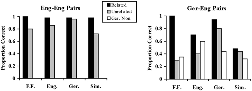
Number of related and unrelated pairs recalled on Trial 1 for F.F. and three control groups, English speakers (Eng.), German–English bilingual speakers (Ger.), and German bilingual speakers simulating lack of knowledge of German (Sim.). The figure on the left shows performance on the English–English pairs and the figure on the right shows performance on the German–English pairs.
The right graph in Fig. 1 shows performance on the German–English pairs in the two leftmost bars of each group and performance on the German non-word–English pairs in the rightmost bar of each group. An analysis of these data indicated a significant relatedness effect, F(1, 12) = 5.33, M.S.E. = 0.036 and an effect of group, F(2, 12) = 5.57, M.S.E. = 0.083. What is clearly evident in this figure is that F.F.’s performance does not resemble that of any of the control groups. On the German–English pairs, the German speakers performed much as they did in English showing a small non-significant relatedness effect, t(4) = 2.06. The English speakers performed more poorly than they did on the English–English pairs, although they too exhibited a relatedness effect, t(4) = 3.87, probably attributable to the similarity of words across languages (e.g., könig-queen). The simulators, however, performed even more poorly than the English speakers and importantly showed no difference in memory between related and unrelated pairs t(4) = 0.22. F.F., on the other hand, demonstrated an exaggerated relatedness effect, recalling all 10 of the related pairs but only 3 of the 10 unrelated pairs.
On the German non-word–English pairs, the English speakers performed much like they did on the German–English pairs (0.60 compared to 0.55, t(4) = 1.41). The German–English bilingual subjects performed more poorly on the non-word materials than on the German–English pairs, 0.44 compared to 0.87 for the honest group and 0.32 compared to 0.46 for the simulators. This difference was significant for the honest German speakers, t(4) = 3.31, but not for the simulators t(4) = 1.24. F.F. again performed unlike the other groups. Although his performance on the German non-word–English pairs was different overall from his performance on the real German–English pairs (0.35 compared to 0.65), this effect was entirely attributable to his excellent performance on the related German pairs. His performance on the German unrelated pairs (0.30). was not different from his performance on the non-words (0.35).
We also looked at number of trials to criterion for each type of list. The pattern of results was identical to that found for performance on the first trial.
3.3. Discussion
The results of the paired associate learning task suggest that, although F.F. denied explicit knowledge of German, he nevertheless appeared to have some knowledge, perhaps at an implicit level, of the semantic or associative properties of the German language. This enabled him to perform perfectly on the related German–English pairs (although he claimed not to understand the German) but provided no advantages on the unrelated German–English pairs, on which he performed much like he did on the German nonsense–English pairs. His performance on the German–English pairs was unlike that of the real German speakers, who showed a small relatedness effect and performed overall at a much higher level than F.F., and also unlike that of the non-German speakers, who performed more poorly than F.F. on the related pairs but similarly on the unrelated pairs. F.F.’s performance was also unlike that of the German speakers who were instructed to simulate amnesia for the German language. The simulators performed equally poorly on the related and unrelated pairs, showing no hint of a relatedness effect, whereas F.F. showed an exaggerated difference between the related and unrelated pairs. In fact, none of the simulators exhibited the pattern of performance shown by F.F. These findings suggest that F.F. was not malingering but that he was truly unable to gain explicit access to German; nevertheless, it appeared that when the German–English pairs were highly associated, he was able to make use of implicit knowledge of some properties of the German language to facilitate his learning performance.
4. Experiment 2
In this experiment, we attempted to use an electrophysiological measure—skin conductance response (SCR)—to gather evidence for implicit knowledge of autobiographical information in the absence of explicit knowledge. The use of autonomic measures to indicate implicit knowledge has been demonstrated in a number of organic memory disorders, notably in cases of prosopagnosia in which people who are unable to recognize familiar faces nevertheless show an elevated SCR to those faces known to them relative to unfamiliar faces (Bauer, 1984; Tranel & Damasio, 1985). Also in organic amnesia and in some cases of frontal lobe damage, patients have been found to display normal autonomic discrimination between targets and distractors despite impaired recognition (Diamond, Mayes, & Meudell, 1996; Rapcsak et al., 1998). In a patient with a dissociative disorder, Gudjonsson (1979) demonstrated heightened electrodermal responses to items of personal relevance of which the patient denied knowledge.
In the present experiment, the patient listened to statements about his personal past and responded as to their truth or falsity as far as he knew. Some of the true statements, F.F. had recently come to know from conversations with friends and family. Other information that we obtained from his brother, he had not been told. We measured skin conductance responses that occurred while he listened to these statements, and we compared his performance to that of control participants who were asked to tell the truth to some sentences and to lie to others.
4.1. Method
4.1.1. Control participants
Four individuals, two English speakers (one female and one male) and two German–English bilingual speakers (one female and one male), participated in this study. They were between the ages of 25 and 35 and were either graduate students at the University of Arizona or spouses or friends of a graduate student.
4.1.2. Materials and procedure
The stimulus materials consisted of 15 sets of 10 sentences, all in English, each set relating to a particular piece of information concerning the individual’s personal past. These materials were constructed initially for F.F. after which parallel sets of questions were constructed for the other participants. The 10 sentences in each set were identical except for the final word in the sentence, which was the target word. For example, one set of sentences was of the form: “My brother’s name is …. ” One of the 10 sentences was completed with the correct information and the other 9 were completed with incorrect information. The sets of sentences included information about family and friends’ names, about geographical locations in which people had grown up, about streets where they had lived, about significant events that had happened in their lives, about schools they had attended, about places they had worked. The correct target sentence always occurred in the sixth to nineth position in the set. This positioning was used to allow stability in the SCR to each statement before the target sentence appeared. The novelty of an initial statement usually creates an elevated SCR. By waiting until later in the set, we could be sure that the SCR response was relatively stable when the target sentence was read.
Sentences were read aloud by the experimenter and F.F. was asked to respond “yes” to a sentence that he believed to be correct and “no” to all other sentences. After the reading of each statement, there was a pause of approximately 12 s, which allowed time for the SCR to occur and return to baseline before the next sentence was read. Electrodermal activity was recorded at 120 Hz at a gain of 10 μS per volt using the Biopac MP100 system with GSR100 amplifier (Biopac Systems, Inc., Santa Barbara, CA, USA). Two 6 mm Ag/AgCl leads were filled with a 0.9% M NaCl in Unibase paste (Lykken and Venables, 1971) and then placed on the palmar surface of the middle phalanges of the first and second fingers of one hand, which had been briefly swabbed with distilled water. A constant voltage of 0.5 V was applied across the two electrodes. Off-line, artifact-free signals were low-pass filtered (cut-off frequency = 0.7 Hz) and down-sampled to 20 Hz before parameter extraction in order to maximize the signal-to-noise ratio. The experimenter computed each SCR manually by measuring the rise in amplitude between a minimum and maximum point following the reading of a sentence. The first response within a 6 s period following the reading of the statement was the response recorded.
Sentences for control participants were constructed in exactly the same way as those for F.F. A close friend or relative was asked to provide the same kind of personal information that was available for F.F. Correct target sentences occurred in exactly the same ordinal position in each set as they had for F.F. and control participants were asked to make the same responses as F.F. made. For statements to which F.F. had given a correct answer, we asked controls to respond honestly. For statements to which F.F. had given an incorrect answer (i.e., responded “no” to the correct sentence), we instructed control subjects to do the same. Specifically, we instructed them at the start of each set of 10 sentences whether they were to tell the truth or to lie. We further told them that when they were asked to lie, they should say “no” to the correct answer as well as “no” to all of the other answers, as if they had no knowledge of the correct answer. This manipulation was intended to control for the possibility that F.F. was malingering. If he were intentionally feigning amnesia and trying to deceive us by saying “no” to true statements about his personal past, we expected that his SCR to those sentences should look similar to that of control subjects who were intentionally lying to the correct answer.
4.2. Results and discussion
The differences between the minimum and maximum amplitudes in SCR for each sentence were calculated. Because of the elevated SCR to the first sentence in each set, the adaptation of the SCR over time, and its sensitivity to things such as movement, deep breathing, and coughing, as well as the possibility that some of our distractors might also have had some personal significance unknown to us, it was decided a priori to compare the change in amplitude of the target SCR in each set to that of the response to the sentence immediately preceding the target. The mean changes in SCR amplitudes for target and non-target sentences across the 15 sets are illustrated in Fig. 2. F.F.’s data were analyzed across items whereas control data were analyzed across subjects.
Fig. 2.
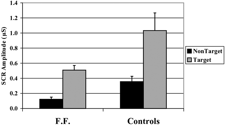
Mean changes in skin conductance response (SCR) amplitudes for target and non-target sentences for F.F. and controls.
F.F., like controls, showed a significantly enhanced SCR to the target sentences compared to the non-target sentences, t(14) = 7.34 for F.F. and t(3) = 3.74 for controls. We also analyzed the SCR data as a function of the behavioral response. F.F. responded correctly (i.e., said “yes”) to 6 of the 15 target sentences and incorrectly (i.e., said “no”) to 9 of the target sentences. He made only one false positive response to which he showed no elevated SCR. The difference between “yes” and “no” responses is shown in Fig. 3. F.F. showed a significantly greater elevation of the SCR to sentences that he denied recognizing compared to those that he correctly acknowledged, t(13) = 2.15. Control participants, on the other hand, showed no differences in SCR to those sentences that they answered honestly compared to those to which they lied, t(3) = 1.39. In fact, they showed a non-significant tendency towards a greater SCR to their truthful responses.
Fig. 3.
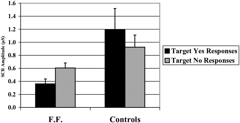
Mean changes in skin conductance response (SCR) amplitudes for targets to which F.F. and controls subjects responded “yes” (i.e., told the truth) and responded “no” (i.e., lied).
The results suggest that, although F.F. denied explicit knowledge of many aspects of his personal past, he nevertheless had knowledge of that information at some level. Although it is difficult to know how to interpret the difference between his autonomic responsiveness to facts that he explicitly acknowledged and those that he denied, the pattern of his performance did not resemble that of control participants who were intentionally lying. It could be that he was more invested in the lie than the control subjects. Alternatively, the elevated SCR to the negative responses could reflect implicit knowledge—knowledge that somehow was inaccessible to him at a conscious level.
5. Experiment 3
Experiment 3 used functional magnetic resonance imaging (fMRI) to examine brain regions associated with a lexical decision task using German and English words and non-words. During scanning, F.F. was presented with German words, English words, German non-words, and English non-words. We compared the pattern of activation obtained from F.F. with activation patterns obtained from native German speakers who were also fluent in English. We asked the bilingual German–English speakers to perform the lexical decision task in two ways. During one fMRI scan, they completed the task normally (i.e., classifying words based on their actual knowledge of German and English). During a separate scan, they were asked to perform the task while pretending to have no knowledge of German. This manipulation allowed us to assess whether F.F.’s activation patterns mirrored those of German speakers who were simulating the loss of their language. Two possible outcomes were considered that focused on frontal lobe activations. First, Markowitsch, Calabrese, et al. (1997) and Markowitsch, Fink, et al. (1997) observed decreased activity in prefrontal cortex, particularly during attempts at memory retrieval, in cases of probable psychogenic amnesia. F.F. may therefore show decreased frontal activation compared to controls. Alternatively, if F.F. were feigning his loss of language, the activation pattern might be similar to that of the bilingual German speakers who were simulating. Recent studies on the brain correlates of deception have demonstrated increased activation in bilateral prefrontal and ventrolateral frontal regions when subjects are lying (Spence et al., 2001) or feigning memory impairment (Lee et al., 2002), compared to truthful controls. Specifically, we expected that simulating the loss of German might increase frontal activations, reflecting an increased degree of response monitoring and suppression of relatively automatic motor responses to well-known lexical items.
5.1. Methods
5.1.1. Control participants
Four bilingual German–English speakers (three male and one female) participated as controls. The bilingual participants were native German speakers who, like F.F., had spent a year or less in the USA, but who considered themselves to be fluent in English. Participants ranged in age from 23 to 36 years old, were right-handed, with normal vision and hearing, and no history of significant head injury or medical conditions that might affect cognitive functioning. All participants were screened for contraindications to MRI and were offered monetary compensation for their participation.
5.1.2. Materials and procedures
German and English words and non-words were five to eight letters in length. English words were drawn from Kucera and Francis (1967) norms, with a range of frequency counts of 20–300 words per million (moderate to high frequency words). English non-words were created by exchanging two letters of the word so that the non-words remained pronounceable. German words were translated directly from the English word list and German non-words were created using the same criteria as English non-words.
Two test lists were constructed, each consisting of 350 items; 100 English Words, 100 German words, 50 English non-words, 50 German non-words, and 50 control stimuli, presented in random order. The control stimulus consisted of a series of number signs (#####). Presentation order of test lists was counterbalanced across participants. Stimuli were presented in the scanner on MR Vision 2000 goggles (Resonance Technology, Inc.), which were mounted to the head coil so that they rested comfortably over the subject’s eyes. All stimuli were presented centered on the computer screen in bright green 80-point font on a black background. Presentation was self-paced; the item remained on the screen until a response was recorded, followed by a 500 ms blank screen, followed immediately by the next list item. Response times were collected using a computer mouse modified for use in the scanner and placed in the individual’s right hand. Participants were instructed to classify each item as a word or non-word, as quickly as possible, by pressing the index finger button for a word, and the ring finger button when they saw a non-word. When a control stimulus appeared, participants responded by pressing the non-word button.
Following a brief practice session, subjects completed the two test lists in the scanner, each lasting approximately 12 min. The controls participated in two separate scanning sessions. In one session, they were instructed to perform normally, pressing the word button for both German and English words, and the non-word button for German and English non-words. During the other session, they were instructed to “respond as if you do not know any German at all; the only language that you speak is English.” No specific instructions were given as to what their responses should be for various item types. Two of the German participants completed the simulation instruction session first; the other two German participants completed the normal instruction session first.
5.1.3. Image acquisition
Images were collected on a 1.5 T GE Horizon whole-body echo speed magnet using single-shot spiral sequence (Glover & Lee, 1995), TR = 2000, TE = 40, matrix 64 × 64. Sections (19 sections, 6 mm, no skip) were collected in the axial plane and covered the whole brain. After all functional scans were completed, a T1 weighted set of images (256 × 256, TE = min full, TR = 500, FOV = 22, using the same slice selection as the functional data set) and a high resolution SPGR series (whole brain 1.5 mm sections, 256 × 256, flip = 30, TE = min full, TR = 22, FOV = 25 cm) were also collected in order to locate anatomical regions of activation and to overlay functional images for re-registration in Talairach space.
5.1.4. Data analysis
Images were corrected for minor head movement using Analysis of Functional NeuroImages (AFNI; Cox, 1995), normalized, and transformed into standard space (Talairach & Tournoux, 1988). Images were analyzed using a rapid presentation event-related technique developed and validated by Dale (1999). The linear slope was removed on a voxel-by-voxel basis and spatial filtering was accomplished using a Hanning filter with a 1.5 voxel radius. Following detrending and filtering, epochs of a 16-s post-stimulus onset time window with a 4-s pre-stimulus baseline were modeled as a linear combination of a time-invariant hemodynamic response (HDR) with Gaussian noise. An estimate of the HDR and variance with the mean signal intensity removed for each condition was modeled using simultaneous least squares fitting of the original MR signal across the time windows. These included the German non-words, English non-words, German words, and English words. Control items were treated as the baseline condition. Images were tested on a voxel-by-voxel basis using a t-statistic weighted for an ideal HDR, modeled as a gamma function with a 2.25 s onset time and a tau of 1.25 s. Maps of active voxel clusters were created by calculating the covariance between the estimated signal response and the ideal HDR function and representing the maximal t-value at a significance of P < 0.0001. A minimum of three contiguous voxels at the P < 0.0001 level were required for a cluster to survive. Anatomically overlapping clusters were identified across participants using Tailarach coordinates, and the extent of activation was measured individually as the number of active voxels within each cluster. Individual cluster data were imported into SPSS for further analysis. To compare the fMRI signal across conditions, for each individual, HDR estimates (0–16 s post-stimulus onset) were calculated for each cluster by averaging all significantly active voxels within that region and baselining them to pre-stimulus baseline (−4.0 to 0 s). The HDR peak amplitude was determined by averaging the values at 2, 4, 6, and 8 s post-stimulus onset.
5.2. Results
5.2.1. Reaction times
F.F. classified all English words as words, and all English and German non-words as non-words. He classified 2.5% of German words as words; the remainder were classified as non-words. Control participants in the simulation condition also classified between 2 and 5.5% of German words as words and the remainder as non-words. Under normal instructions, controls classified all German and English words as words.
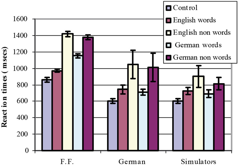
Reaction times (RT) for lexical decision are shown in Fig. 4. RTs from the four control participants were analyzed using a repeated-measures ANOVA, comparing instruction (normal, simulation), language (English, German), and word type (word, non-word). Results indicated a marginal main effect of word type, F(1, 3) = 4.83, M.S.E. = 83929, P = 0.11, where responses to non-words were longer on average than responses to words. The interaction between word type and instruction condition was also significant, F(1, 3) = 10.25, M.S.E. = 4810, P < 0.05. Non-word responses were slower in the normal condition than in the simulation condition, while responses to real words were similar across instruction conditions. Because so many of the trials in the simulation condition were to be classified as “non-word,” participants, when they were simulating, may have adopted a simple strategy of identifying English words as “yes” responses, and all other trials as “no,” making the non-word responses faster than under normal conditions. RTs for control items were significantly faster than all other word and non-word conditions, as indicated by a least significant difference t-test, t(3) = 3.654, P < 0.05.
Fig. 4.
Mean (S.E.M.) reaction times for lexical decision for F.F., German bilingual controls (German), and the same German bilingual controls simulating a lack of knowledge of the German language (simulators). Standard errors for the control conditions were obtained from the control group analysis; standard errors for F.F. were obtained from the item analysis.
Compared to controls, F.F. took considerably longer to respond to all categories of stimuli, even the control items. Generalized slowing was typical of his performance on all neuropsychological tests, and we observed the same slowness in his regular conversational speech as well. Unlike controls, F.F.’s responses to German words took an average of 183 ms longer to classify compared to English words, although the German words still showed an RT advantage over both German and English non-words. An item analysis of F.F.’s data confirmed these results, indicating that RTs for German words were significantly slower than English words, t(199) = 7.18, P < 0.0001, but significantly faster than German and English non-words, t(199) = 6.46, P < 0.0001.
5.2.2. fMRI results
F.F.’s results showed significant activation in brain regions that were consistent with those identified during other lexical decision studies (e.g., Schnyer, Ryan, Trouard, & Forster, 2002), and similar to the active regions observed in the control participants. These brain regions included left and right superior parietal lobe (Brodmann areas 7, 40), left and right posterior lateral prefrontal cortex (BA 6, 44), left anterior lateral prefrontal cortex (BA 45, 46), left middle temporal gyrus (BA 21), and bilateral extrastriate regions including fusiform gyrus. The one regional difference between F.F. and controls was observed in subcortical structures. All controls showed activation within multiple striatal regions, including bilateral caudate and putamen. In contrast, no striatal activations were observed in F.F.’s data.
5.2.3. Extent of activation
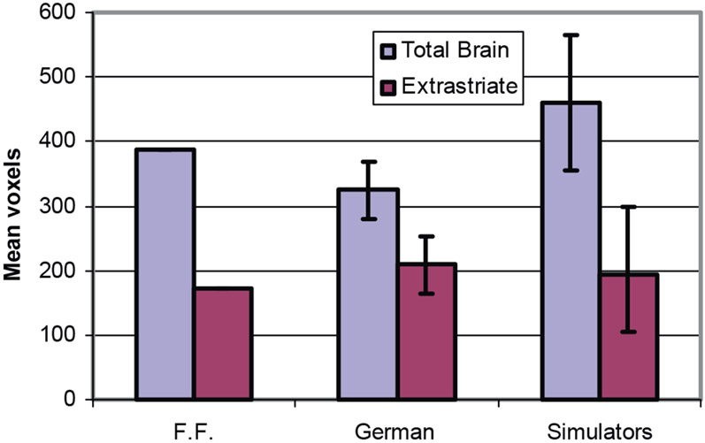
To begin, we compared F.F. and controls on a global measure of activation extent in order to ensure that any regional differences in activation were not confounded with a difference in overall activation. We also assessed the extent of activation within a region that we did not expect to differ across conditions, namely extrastriate cortex. Fig. 5 shows the mean number of active brain voxels and the number of active voxels within extrastriate regions for F.F. and the controls in both instruction conditions. Data for the control group were analyzed using a repeated-measures ANOVA, comparing region (total brain versus extrastriate) and instruction type (normal, simulation). As expected, extent of activation was greater across the total brain than within the extrastriate region alone (P < 0.001). More importantly, region interacted with instruction type, F(1, 3) = 24.16, P < 0.016. As depicted in Fig. 5, the number of total brain voxels increased during simulation compared to normal instructions, but this increase did not occur within extrastriate regions, which remained constant across instruction conditions. F.F. showed a similar degree of activation within the extrastriate region as controls. On total brain activation, F.F.’s results appear to be midway between the two control conditions.
Fig. 5.
Extent of activation as measured by the mean (S.E.M.) number of significant active voxels over the whole brain and within posterior extrastriate regions that included bilateral fusiform gyrus. Data are presented for F.F., German bilingual controls (German), and the same German bilingual controls simulating lack of knowledge of the German language (simulators). Active voxels were determined using a criterion of P < 0.0001 per voxel and a cluster size of >3 contiguous voxels. Standard errors for the control conditions were obtained from the control group analysis.
5.2.4. Frontal lobes
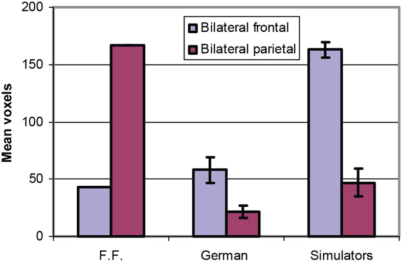
Of interest was whether the increased activation for controls during simulation instructions occurred primarily within frontal regions, consistent with previous literature on deception (Lee et al., 2002; Spence et al., 2001), and whether F.F.’s data continued to resembled the simulators’ data. We were also interested in whether F.F. showed relatively less frontal activation than controls, as suggested by Markowitsch (1999). To investigate these issues, we compared extent of activation within bilateral frontal regions (BA 6, 4, 45, and 46 combined) to activation within bilateral parietal cortex (BA 7 and 40 combined). Fig. 6 depicts the mean number of significant voxels in frontal and parietal regions for F.F. and controls. Data from controls were analyzed using a repeated-measures ANOVA comparing region (frontal, parietal) and instruction (normal, simulation). Results indicated significantly greater activation in frontal regions than parietal regions, F(1, 3) = 163.49, P < 0.001. Additionally, controls showed more active voxels in these two regions overall during simulation compared to normal instructions, indicated by a main effect of instruction, F(1, 3) = 19.02, P < 0.05. Importantly, region and instruction type showed a significant interaction, F(1, 3) = 24.23, P < 0.01. For simulators, the increase in activation in frontal regions (mean increase = 105) was significantly greater than the increase in parietal regions (mean increase = 25.5).
Fig. 6.
Extent of activation as measured by the mean (S.E.M.) number of significant active voxels in bilateral frontal and bilateral parietal regions. Data are presented for F.F., German bilingual controls (German), and the same German bilingual controls simulating lack of knowledge of the German language (simulators). Active voxels were determined using a criterion of P < 0.0001 per voxel and a cluster size of >3 contiguous voxels. Standard errors for the control conditions were obtained from the control group analysis.
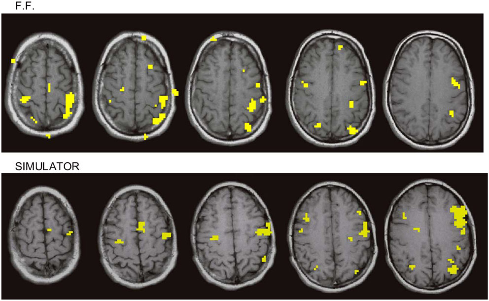
Greater frontal activation compared to parietal activation was consistently observed in every control participant, and this difference was particularly evident during simulation. This pattern contrasts sharply with F.F.’s data (see Fig. 6), which showed nearly four times greater extent of activation in parietal regions than frontal regions (167 voxels versus 43 voxels, respectively). Fig. 7 compares five axial sections of F.F.’s brain and similar sections from a typical control participant during simulation. The figure illustrates greater parietal activation relative to frontal activation in F.F., and the opposite pattern in the simulator.
Fig. 7.
Five axial sections of F.F.’s brain and similar sections from a typical control participant during simulation, showing activations in frontal and parietal regions during lexical decision. The figure illustrates greater parietal activation relative to frontal activation in F.F., and the opposite pattern in the simulator.
5.2.5. Peak hemodynamic response amplitudes
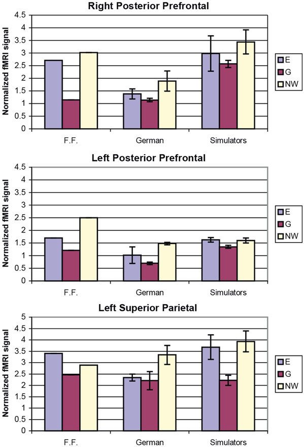
Mean HDR amplitudes were compared in left and right posterior lateral prefrontal cortex, left anterior lateral prefrontal cortex, and left and right superior parietal regions. Mean amplitudes for each region were analysed for the control participants using a repeated-measures ANOVA comparing instruction (normal, simulation) and three word types (English, German, non-words). Note that the two non-word conditions were combined, since activation was similar in all regions for the two non-word conditions. A general pattern emerged that is exemplified in Fig. 8, which depicts mean amplitudes within the left and right posterior prefrontal regions and left superior parietal lobe. All regions showed two main effects. First, there was a main effect of word type (P-values ranging from 0.029 to 0.003). Amplitudes were always least for German words and significantly greater for either English words, non-words, or both. Second, amplitudes increased for all three word types under simulation instructions, as indicated by a main effect of instruction type (P-values ranging from 0.049 to 0.019). The interactions between instruction and word type were not significant.
Fig. 8.
Mean (S.E.M.) amplitudes of the hemodynamic response across all active voxels in three cortical regions: right posterior lateral prefrontal; left posterior lateral prefrontal; left superior parietal. Data from three word types, English (E), German (G), and non-words (NW) are presented for F.F., German bilingual controls (German), and the same German bilingual controls simulating lack of knowledge of the German language (simulators).
F.F’s data resemble the amplitude pattern across word types in the control participants. As with controls, the amplitude for F.F.’s data in all regions was less for German words than English and non-words (see Fig. 8). In terms of overall mean amplitude, F.F.’s data are mixed; amplitudes sometimes fall within the range of the control participants under normal instructions, but sometimes appear more similar to controls under simulation instructions. This overall similarity in the amplitudes of F.F.’s and control subjects’ activations contrasts with the clear differences between F.F. and controls that were evident in the pattern and extent of activations observed in these same brain regions (see Figs. 6 and 7).
5.3. Discussion
In summary, bilingual German–English controls showed increased activation when simulating a lack of knowledge for the German language compared to their normal performance on a bilingual lexical decision task. The increase was greatest in frontal regions, consistent with previous studies on deception (Lee et al., 2002; Spence et al., 2001), along with a smaller but still significant increase in parietal cortex. In contrast, F.F. exhibited a striking difference in the ratio of frontal to parietal activation, with more extensive activation in parietal than frontal regions. It is important to note that within extrastriate cortex that served as a control region, F.F., simulating controls, and normal instruction controls all showed a similar extent of activation.
The results in the simulation condition were interesting in their own right. Consistent with our expectations and prior literature, simulating the loss of a language probably required greater monitoring of stimuli and responses, inhibition of prepotent responses, and selection of alternative classification responses. This increase in strategic control during simulation was reflected in substantial increases in both the extent and amplitude of frontal activations along with a modest increase in bilateral superior parietal lobule (BA 7, 40). Two frontal regions that showed consistent increases across all controls included posterior left prefrontal cortex (BA 6, 44) and anterior left prefrontal cortex (BA 45, 46). The posterior prefrontal region is commonly observed during relatively pure lexical tasks such as lexical decision or stem completion, suggesting that it plays a role in access to lexical codes. Left anterior prefrontal cortex has been associated with semantic processing of lexical items and is active during such tasks as verb generation or semantic associative tasks (for review, see Wagner, Koutstaal, Maril, Schacter, & Buckner, 2000). In the simulation condition, fluent bilingual speakers classified English words as words, but German words as non-words. Thus, the task may have been similar to a source monitoring task, first requiring the identification of meaningful English and German words (relying on anterior prefrontal regions), and then the categorization of those words as word and non-word, respectively, based on lexical codes (relying on posterior prefrontal cortex). In all regions, the mean amplitude data are consistent with the expectation that identification of German words by native German speakers should be easier than identifying either English or non-words, even under simulation instructions. HDR response amplitudes are often related to fluency or ease with which stimuli are processed (Schnyer et al., 2002). The finding that mean amplitudes across word types for F.F.’s data were similar to controls (less for German words compared to English words and non-words) demonstrates, at least at a non-conscious level, his fluency with the German language.
The most striking difference in F.F.’s data compared to controls was the observation of greater parietal activation relative to frontal activation. Although the exact reason for this apparent anterior to posterior processing shift is unclear, two points are relevant. First, other researchers have suggested that the parietal lobule in the vicinity of the angular gyrus participates in aspects of phonological or lexical processing, as indicated by cross-modality priming studies (Badgaiyan, Schacter, & Alpert, 1999; Schacter, Badgaiyan, & Alpert, 1999). Second, Markowitsch (1999) has highlighted decreased activity in prefrontal cortex as a consistent correlate of functional amnesia, possibly related to an increase in glucocorticoids present as a result of sustained stress, and reflected in frontal dysfunction. Taken together, these points suggest that F.F. may have relied to a greater degree on processing of phonological and lexical aspects of the stimuli in order to classify them. Decreased frontal resources, consistent with the prior literature on psychogenic amnesia, may have forced F.F. to compensate by increasing the lexical analysis of the items, rather than relying on access to the semantic representation of the words. F.F.’s data are not consistent with the pattern of activation obtained when bilingual controls simulated the loss of their primary language. Indeed, simulation instructions only emphasized the preponderance of frontal activation compared to posterior parietal and extrastriate cortical regions, rather than decreased it.
6. General discussion
Patient F.F. exhibits many of the classical characteristic behaviors associated with psychogenic fugue as documented in previous reports. His amnesic episode was triggered by a traumatic event, apparently involving a robbery and shooting of which he was the victim. His memory for the details of these occurrences and those immediately following is still unclear. As we were able to learn from him recently, events of the previous year had also been extremely stressful psychologically and physically, including a serious motorcycle accident resulting in minor head trauma and other physical injuries, problems with his business, and impending divorce from his wife. He decided to get away from all of these problems by coming to the United States and starting anew. He was in the process of upgrading his tourist visa to a 6-year work visa when, for reasons unknown to him now, he decided instead to obtain false papers and run away again. It was at this point that he was driven to an unknown location by his roommate, robbed of his money and all other possessions, and shot at as he fled. This general pattern of physical and emotional disturbances followed by a specific precipitating stressor is quite consistent with other reports of psychogenic fugue in the literature (Kopelman, 2002; Markowitsch, 2000).
Many aspects of F.F.’s neuropsychological profile were also consistent with previous reports: He had a normal I.Q. and was able to retrieve general knowledge of the world and information about famous people and events of the past several decades. His ability to acquire new episodic memories was intact, although his performance on the Wechsler Memory Scale-III showed an unusual verbal/visual split. On immediate testing, his auditory/verbal memory was at the 99.7th percentile, 45 points higher than his visual memory, which was at only the 42nd percentile. A similar discrepancy, although smaller, was evident on tests of delayed memory, and on tests of working memory. These findings could reflect some form of right hemisphere dysfunction or depression, perhaps in right prefrontal regions, which have been associated with retrieval of visuospatial information in both long-term and working memory (see Fletcher & Henson, 2001). F.F.’s relatively poor performance on tests of executive function is also suggestive of some frontal dysfunction. Kopelman (2002) has proposed that psychological stressors affect frontal executive control systems, which may lead to inhibition of autobiographical memories. It is possible that such stressors cause a more general dysfunction of the frontal lobes as suggested by Markowitsch, Calabrese, et al.’s (1997) findings of reduced perfusion in right inferior prefrontal regions in a patient with psychogenic amnesia (see also Costello et al., 1998). In the present paper, the neuroimaging results of Experiment 3 are also suggestive of a depression of frontal processes. In the lexical decision task, all participants except F.F. showed greater extent of activation in frontal regions than in parietal regions, whereas F.F. showed the opposite pattern: fewer active frontal lobe voxels but a larger number of active voxels in parietal regions. This finding suggests that F.F. may have relied to a greater extent on non-frontal processes to perform the lexical decision task.
In the present study, we also found evidence that F.F. retained implicit knowledge of autobiographical information that he was unable to recollect explicitly and aspects of the German language that he claimed not to know. Although in these studies, as in the lexical decision study, it is impossible to be certain that F.F. was not feigning his amnesia, the patterns of his performance did not resemble that of simulators who were instructed to lie. The comparison to simulators is not without problems given that it is not clear that the processes engaged by simulators and malingerers would necessarily be the same. Nevertheless, the finding of differences across three different paradigms, which could not have been predicted a priori, provides some confidence that this patient was experiencing a true psychogenic amnesia. Particularly convincing, we believe, were the findings from the paired associate study. It is hard to imagine how someone pretending not to know any German would show perfect performance on the highly related German pairs and very poor performance on the unrelated pairs. Indeed, none of the simulators produced such an outcome. Instead, this finding suggests that F.F. had implicit knowledge of the associative structure of German, which facilitated his learning of related pairs, but provided no benefit for unrelated pairs. Similarly, in the lexical decision task, F.F. also demonstrated some knowledge of the German language. First, he classified 2.5% of the German words as words. Second, although he responded to all other German words as if they were non-words, he showed a clear differentiation in reaction times between the German words and non-words, an outcome that would be unexpected if he had no knowledge of German.1 Third, the finding of an increase in extent of activation in the frontal lobes of German–English bilingual individuals when they were simulating loss of knowledge of the German language, a pattern that was not in evidence in F.F, provides further suggestive evidence against malingering.
Some of F.F.’s comments to us in recent communication are also instructive and consistent with a view of psychogenic amnesia as a temporary state of disrupted consciousness, which in F.F.’s case was probably beginning to resolve by the end of our testing. F.F. stated that “I just neglected my whole life … The break even point, where not knowing the answers and ignoring the knowledge of the answers is difficult to find … I was aware of knowing German in written and spoken form somehow after about 10 days … It was a part of my life I just wanted to lock away in a dark chamber. I can’t even say if it was an active will or passive defense … The point where this disorientation was replaced by neglecting the truth is not easy to find and somehow undefined … In the last test, the lie detector, some of the things were common (familiar) and my mother’s name went through my ‘personal barrier’.” He also indicated that after he returned to Germany, he “had big problems speaking German fluidly for about 5 weeks … the language was mixed up with a lot of American phrases and English words.”
These remarks are consistent with descriptions of other cases of psychogenic amnesia, which have often reported a mixture of fantasy and reality, some islands of memory, and awareness of some aspects of the past. Kopelman states that “subjects often show some degree of ‘knowledge’ or ‘recognition’ of certain memories without explicit recollection” (Kopelman, 2002, p. 466) and “may manifest varying levels of awareness for differing memories, possibly simulating amnesia for certain items, having only ‘knowledge’ or familiarity judgements’ for other items, and being ‘unaware’ of certain other memories” (Kopelman et al., 1994, p. 688; see also Kopelman, 2002). This seems a very apt description of F.F. He appeared unable to recall explicitly much of his personal past but demonstrated an implicit awareness and feeling of familiarity of several facts of his life. In addition, some of the most important things—his mother’s name, for example—may have penetrated his awareness, yet he chose not to disclose that knowledge at the time, suggesting at least an element of conscious simulation. He also demonstrated implicit knowledge of some properties of his native language, and reported that a more complete understanding of German began to return approximately 10 days after the initiating episode, although some confusion persisted for weeks. He described a disorientation and a feeling that he was not able to access the truth because of a “blocking” or “barrier.” It appears that although the immediate precipitating cause of the amnesia was the robbery and shooting that occurred in Tucson, much of the emotional turmoil that plagued F.F. had occurred in Germany. This may have accounted for the extension of the amnesia into the language domain. Finally, it is worth noting that F.F.’s voluntary return to Germany even though he risked incarceration is perhaps the best indicator that he was not malingering. All cases of psychogenic fugue are associated with emotional trauma that provides a powerful motive for psychological and often physical escape as well. The present study documents such a case in which the amnesia covered virtually all of the individual’s personal past including his native language. The findings are consistent with models proposed by Kopelman (2000, 2002) and Markowitsch (2000) suggesting that emotional stressors lead to a depression of some aspects of frontal function, which inhibit or block access to autobiographical memories. In the present study, F.F. showed reduced levels of performance on some tests of frontal function and reduced frontal activation in the fMRI study of lexical decision. In addition, his unexpectedly low performance on the visuospatial episodic and working memory tasks could have reflected impaired retrieval processes dependent on right prefrontal brain regions. These ideas, however, are speculative, and other explanations are possible. For example, depression could contribute to decreased performance on frontal/executive tasks (Grant, Thase, & Sweeney, 2001) and to reduced activation in prefrontal brain regions (Drevets, 2001), and although we had no direct measures of depression in the present study, depression is commonly associated with psychogenic fugue (Kopelman, 2000). More studies of psychogenic amnesia that include measures of executive function and brain imaging are needed before the role of frontal function in these patients will be determined with any certainty.
Acknowledgements
We gratefully acknowledge the assistance of the medical personnel in the psychiatric unit at the University Medical Center and of research assistants Pamela Perschler and Andrea Soule.
Footnotes
In fact, when two monolingual English speakers performed the same lexical decision task, the RTs for German words and non-words were indistinguishable.
References
- Badgaiyan RD, Schacter DL, & Alpert NM (1999). Auditory priming within and across modalities: Evidence from positron emission tomography. Journal of Cognitive Neuroscience, 11, 337–348. [DOI] [PubMed] [Google Scholar]
- Barbarotto R, Laiacona M, & Cocchini G (1996). A case of simulated, psychogenic or focal pure retrograde amnesia: Did an entire life become unconscious? Neuropsychologia, 34, 575–585. [DOI] [PubMed] [Google Scholar]
- Bauer RM (1984). Autonomic recognition of names and faces in prosopagnosia: A neuropsychological application of the guilty knowledge test. Neuropsychologia, 22, 457–469. [DOI] [PubMed] [Google Scholar]
- Costello A, Fletcher PC, Dolan RJ, Frith CD, & Shallice T (1998). The origins of forgetting in a case of isolated retrograde amnesia following a haemorrhage: Evidence from functional imaging. Neurocase, 4, 437–446. [Google Scholar]
- Cox RW (1995). Afni: Software for analysis and visualization of functional magnetic resonance neuroimages. Computers and Biomedical Research, 29, 162–173. [DOI] [PubMed] [Google Scholar]
- Crovitz HF, & Schiffman H (1974). Frequency of episodic memories as a function of their age. Bulletin of the Psychonomic Society, 4, 517–518. [Google Scholar]
- Dale AM (1999). Optimal experimental design for event-related fMRI. Human Brain Mapping, 8, 109–114. [DOI] [PMC free article] [PubMed] [Google Scholar]
- Dalla Barba G, Mantovan MC, Ferruzza E, & Denes G (1997). Remembering and knowing the past: A case study of isolated retrograde amnesia. Cortex, 33, 143–154. [DOI] [PubMed] [Google Scholar]
- Delis DC, Kramer J, Kaplan E, & Ober BA (1987). The California Verbal Learning Test. San Antonio, TX: Psychological Corporation. [Google Scholar]
- De Renzi E, Lucchelli F, Muggia S, & Spinnler H (1995). Persistent retrograde amnesia following a minor trauma. Cortex, 31, 531–542. [DOI] [PubMed] [Google Scholar]
- De Renzi E, Lucchelli F, Muggia S, & Spinnler H (1997). Is memory loss without anatomical damage tantamount to a psychogenic deficit? The case of pure retrograde amnesia. Neuropsychologia, 35, 781–794. [DOI] [PubMed] [Google Scholar]
- Diamond BJ, Mayes AR, & Meudell PR (1996). Autonomic and recognition indices of memory in amnesic and healthy control subjects. Cortex, 32, 439–459. [DOI] [PubMed] [Google Scholar]
- Drevets WC (2001). Neuroimaging and neuropathological studies of depression: Implications for the cognitive-emotional features of mood disorders. Current Opinion in Neurobiology, 11, 240–249. [DOI] [PubMed] [Google Scholar]
- Fink GR, Markowitsch HJ, Reinkemeier M, Bruckbauer T, Kessler J, & Heiss WD (1996). Cerebral representation of one’s own past: Neural networks involved in autobiographical memory. Journal of Neuroscience, 16, 4275–4282. [DOI] [PMC free article] [PubMed] [Google Scholar]
- Fletcher PC, & Henson RNA (2001). Frontal lobes and human memory. Insights from functional neuroimaging. Brain, 124, 849–881. [DOI] [PubMed] [Google Scholar]
- Glisky EL, & Delaney SM (1996). Implicit memory and new semantic learning in posttraumatic amnesia. Journal of Head Trauma Rehabilitation, 11, 31–42. [Google Scholar]
- Glover GH, & Lee AT (1995). Motion artifacts in fMRI: Comparison of 2dft with pr and spiral scan methods. Magnetic Resonance in Medicine, 33, 624–635. [DOI] [PubMed] [Google Scholar]
- Grant MM, Thase ME, & Sweeney JA (2001). Cognitive disturbance in outpatient depressed younger adults: Evidence of modest impairment. Biological Psychiatry, 50, 35–43. [DOI] [PMC free article] [PubMed] [Google Scholar]
- Gudjonsson GH (1979). The use of electrodermal responses in a case of amnesia (a case report). Medicine, Science, and the Law, 19, 138–140. [DOI] [PubMed] [Google Scholar]
- Kaszniak AW, Nussbaum PD, Berren MR, & Santiago J (1988). Amnesia as a consequence of male rape: A case report. Journal of Abnormal Psychology, 97, 100–104. [DOI] [PubMed] [Google Scholar]
- Kihlstrom JF, & Schacter DL (2000). Functional amnesia. In Boller F & Grafman J (Eds.), Handbook of neuropsychology (2nd ed., pp. 409–427). Amsterdam: Elsevier. [Google Scholar]
- Kopelman MD (1995). The assessment of psychogenic amnesia. In Baddeley AD, Wilson BA, & Watts FN (Eds.), Handbook of memory disorders (pp. 427–448). Chichester: Wiley. [Google Scholar]
- Kopelman MD (2000). Focal retrograde amnesia and the attribution of causality: An exceptionally critical review. Cognitive Neuropsychology, 17, 585–621. [DOI] [PubMed] [Google Scholar]
- Kopelman MD (2002). Psychogenic fugue. In Baddeley AD, Kopelman MD, & Wilson BA (Eds.), The handbook of memory disorders (2nd ed., pp. 451–471). Chichester: Wiley. [Google Scholar]
- Kopelman MD, Christensen H, Puffett A, & Stanhope N (1994). The great escape: A neuropsychological study of psychogenic amnesia. Neuropsychologia, 32, 675–691. [DOI] [PubMed] [Google Scholar]
- Kritchevsky M, Zouzounis J, & Squire LR (1997). Transient global amnesia and functional retrograde amnesia: Contrasting examples of episodic memory loss. Philosophical Transactions of the Royal Society of London, B, 352, 1747–1754. [DOI] [PMC free article] [PubMed] [Google Scholar]
- Kucera H, & Francis W (1967). Computational analysis of present-day American English. Providence, RI: Brown University Press. [Google Scholar]
- Lee TMC, Liu H-L, Tan L-H, Chan CCH, Mahankali S, & Feng C-M et al. , (2002). Lie detection by functional magnetic resonance imaging. Human Brain Mapping, 15, 157–164. [DOI] [PMC free article] [PubMed] [Google Scholar]
- Markowitsch HJ (1999). Functional neuroimaging correlates of functional amnesia. Memory, 7, 561–583. [DOI] [PubMed] [Google Scholar]
- Markowitsch HJ (2000). Repressed memories. In Tulving E (Ed.), Memory, consciousness, and the brain (pp. 319–330). Philadelphia: Psychology Press. [Google Scholar]
- Markowitsch HJ, Calabrese P, Fink GR, Durwen HF, Kessler J, & Härting C et al. , (1997). Impaired episodic memory retrieval in a case of probable psychogenic amnesia. Psychiatry Research Neuroimaging, 74, 119–126. [DOI] [PubMed] [Google Scholar]
- Markowitsch HJ, Fink GR, Thöne A, Kessler J, & Heiss W-D (1997). A pet study of persistent psychogenic amnesia covering the whole life span. Cognitive Neuropsychiatry, 22, 135–158. [DOI] [PubMed] [Google Scholar]
- Wechsler abbreviated scale of intelligence. (1999). San Antonio, TX: Psychological Corporation. [Google Scholar]
- Rapcsak SZ, Kaszniak AW, Reminger SL, Glisky ML, Glisky EL, & Comer JF (1998). Dissociation between verbal and autonomic measures of memory following frontal lobe damage. Neurology, 50, 1259–1265. [DOI] [PubMed] [Google Scholar]
- Schacter DL, Badgaiyan RD, & Alpert NM (1999). Visual word stem completion priming within and across modalities. NeuroReport, 10, 2061–2065. [DOI] [PubMed] [Google Scholar]
- Schacter DL, Wang PL, Tulving T, & Freedman M (1982). Functional retrograde amnesia: A quantitative case study. Neuropsychologia, 20, 523–532. [DOI] [PubMed] [Google Scholar]
- Schnyer D, Ryan L, Trouard T, & Forster K (2002). Masked word repetition results in increased fMRI signal: A framework for understanding signal changes in priming. NeuroReport, 13, 281–284. [DOI] [PubMed] [Google Scholar]
- Spence SA, Farrow TFD, Herford AE, Wilkinson ID, Zheng Y, & Woodruff PWR (2001). Behavioural and functional anatomical correlates of deception in humans. NeuroReport, 12, 2849–2853. [DOI] [PubMed] [Google Scholar]
- Spreen O, & Straus E (1991). A compendium of neuropsychological tests. Oxford: Oxford University Press. [Google Scholar]
- Talairach J, & Tournoux P. (1998). Co-planar stereotaxic atlas of the human brain. New York: Times Medical. [Google Scholar]
- Tranel D, & Damasio AR (1985). Knowledge without awareness: An autonomic index of facial recognition by prosopagnosics. Science, 228, 1453–1454. [DOI] [PubMed] [Google Scholar]
- Wagner AD, Koutstaal W, Maril A, Schacter DL, & Buckner RL (2000). Task-specific repetition priming in left inferior prefrontal cortex. Cerebral Cortex, 10, 1176–1184. [DOI] [PubMed] [Google Scholar]
- Wechsler D (1997). Wechsler Memory Scale-III. San Antonio, TX: Psychological Corporation. [Google Scholar]


