Persoonia : Molecular Phylogeny and Evolution of Fungi logoLink to Persoonia : Molecular Phylogeny and Evolution of Fungi
. 2023 Dec 30;51:280–417. doi: 10.3767/persoonia.2023.51.08

Fungal Planet description sheets: 1550–1613

PW Crous 1,2, MM Costa 1, H Kandemir 1, M Vermaas 1, D Vu 1, L Zhao 1, E Arumugam 3, A Flakus 4, Ž Jurjević 5, M Kaliyaperumal 3, S Mahadevakumar 6,7, R Murugadoss 3, RG Shivas 8, YP Tan 9, MJ Wingfield 2, SE Abell 10, TS Marney 9, C Danteswari 11, V Darmostuk 4, CM Denchev 12, TT Denchev 12, J Etayo 13, J Gené 14, S Gunaseelan 3, V Hubka 15,16, T Illescas 17, GM Jansen 18, K Kezo 3, S Kumar 7, E Larsson 19, KT Mufeeda 7, M Piątek 4, P Rodriguez-Flakus 4, PVSRN Sarma 11, M Stryjak-Bogacka 4, D Torres-Garcia 14, J Vauras 20, D A Acal 21, A Akulov 22, K Alhudaib 23,24, M Asif 25, S Balashov 5, H-O Baral 26, A Baturo-Cieśniewska 27, D Begerow 28, A Beja-Pereira 29,30, MV Bianchinotti 31, P Bilański 32, S Chandranayaka 33, N Chellappan 3, DA Cowan 34, FA Custódio 35, P Czachura 4, G Delgado 36, NI De Silva 37, J Dijksterhuis 1, M Dueñas 38, P Eisvand 39, V Fachada 40,41, J Fournier 42, Y Fritsche 43, F Fuljer 44, KGG Ganga 45, MP Guerra 43, K Hansen 46, N Hywel-Jones 47, AM Ismail 23,24,48, C R Jacobs 49, R Jankowiak 32, A Karich 50, M Kemler 28, K Kisło 51, W Klofac 52, I Krisai-Greilhuber 53, KPD Latha 45, R Lebeuf 54, ME Lopes 43, S Lumyong 37, J G Maciá-Vicente 55,56, G Maggs-Kölling 57,58, D Magistà 59,60, P Manimohan 45, MP Martín 38, E Mazur 4, M Mehrabi-Koushki 39,61, AN Miller 62, A Mombert 63, E A Ossowska 64, K Patejuk 65, OL Pereira 35, S Piskorski 66, M Plaza 67, AR Podile 11, A Polhorský 68, W Pusz 65, M Raza 69, M Ruszkiewicz-Michalska 66, M Saba 25, RM Sánchez 31, R Singh 70, L Śliwa 4, ME Smith 71, VM Stefenon 43, D Strasiftáková 72, N Suwannarach 37, K Szczepańska 73, MT Telleria 38, DS Tennakoon 37, M Thines 74,75,76, RG Thorn 77, J Urbaniak 73, M van der Vegte 78, V Vasan 3, C Vila-Viçosa 29,41, H Voglmayr 53, M Wrzosek 51, J Zappelini 43, JZ Groenewald 1
PMCID: PMC11041897  PMID: 38665977

Abstract

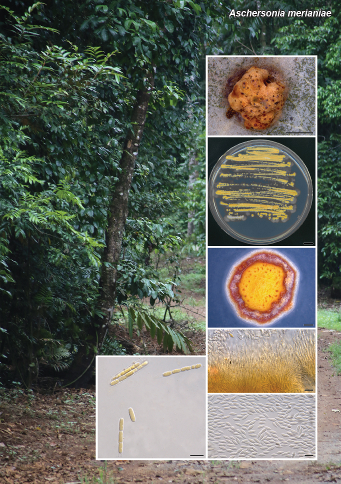
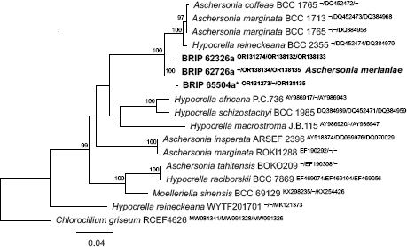
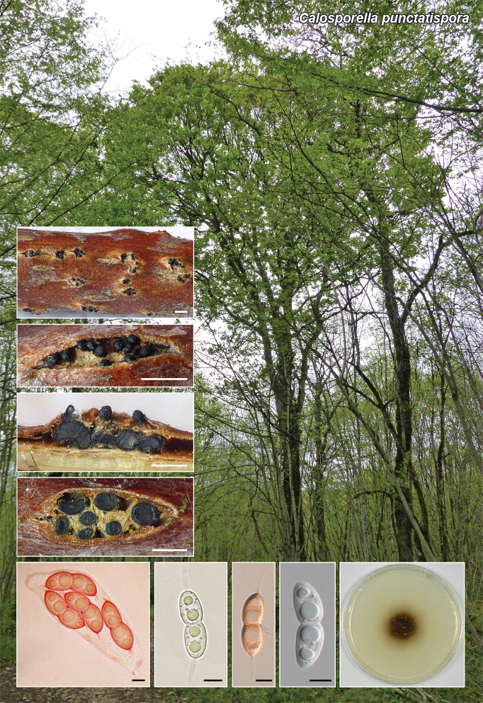
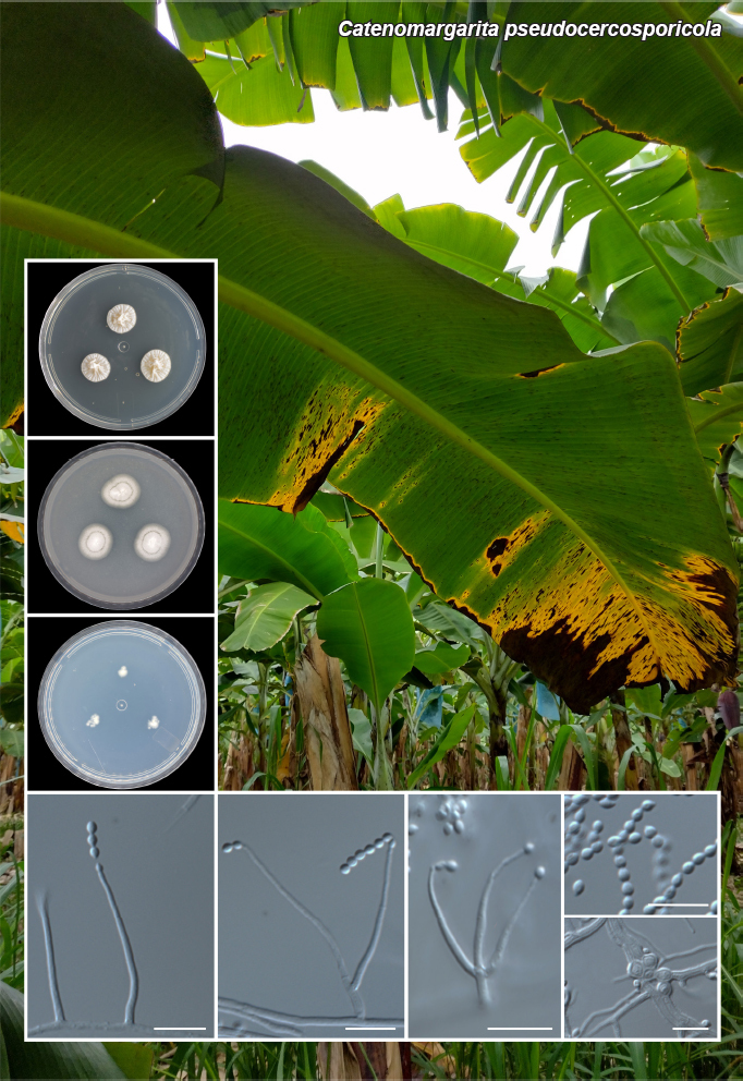
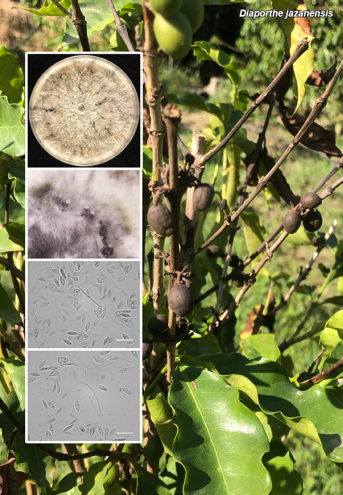
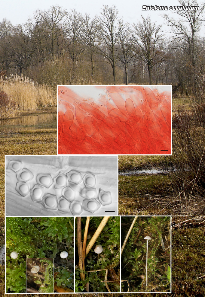
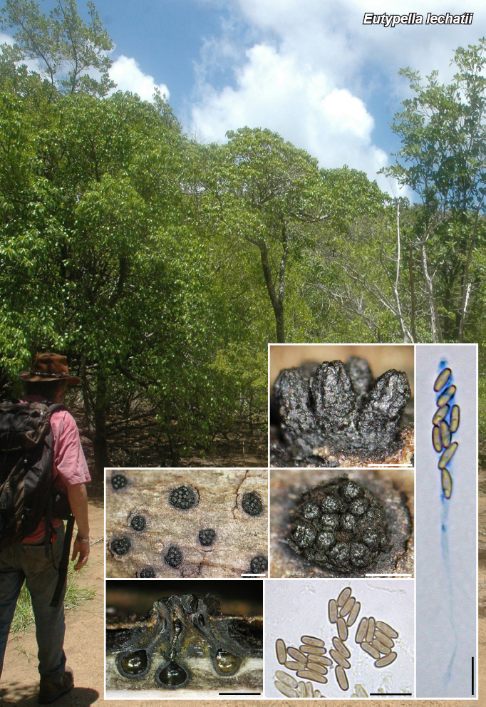
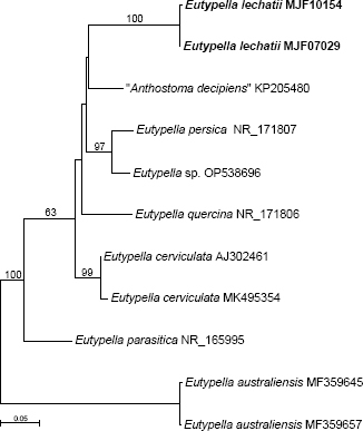
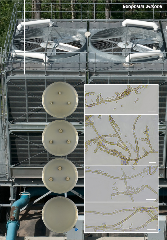
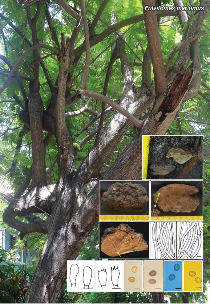
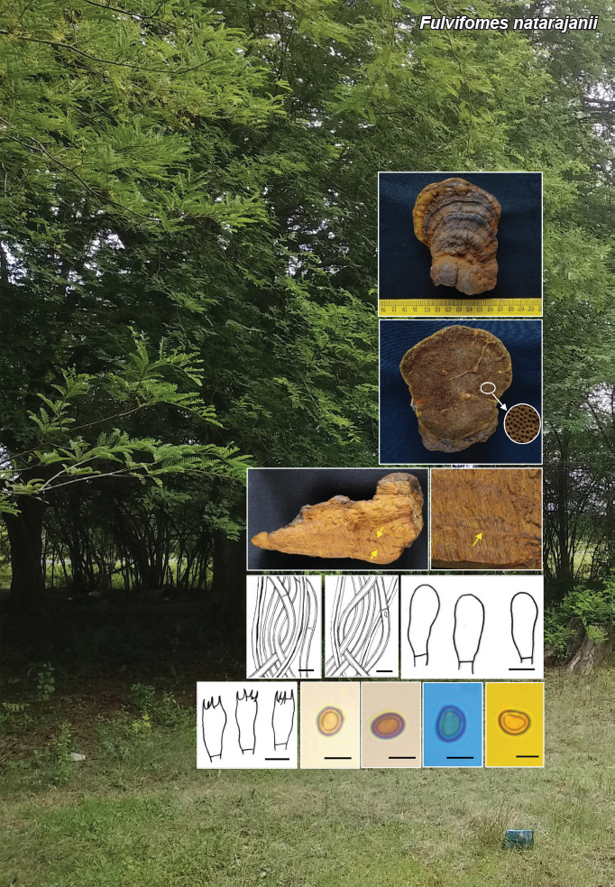
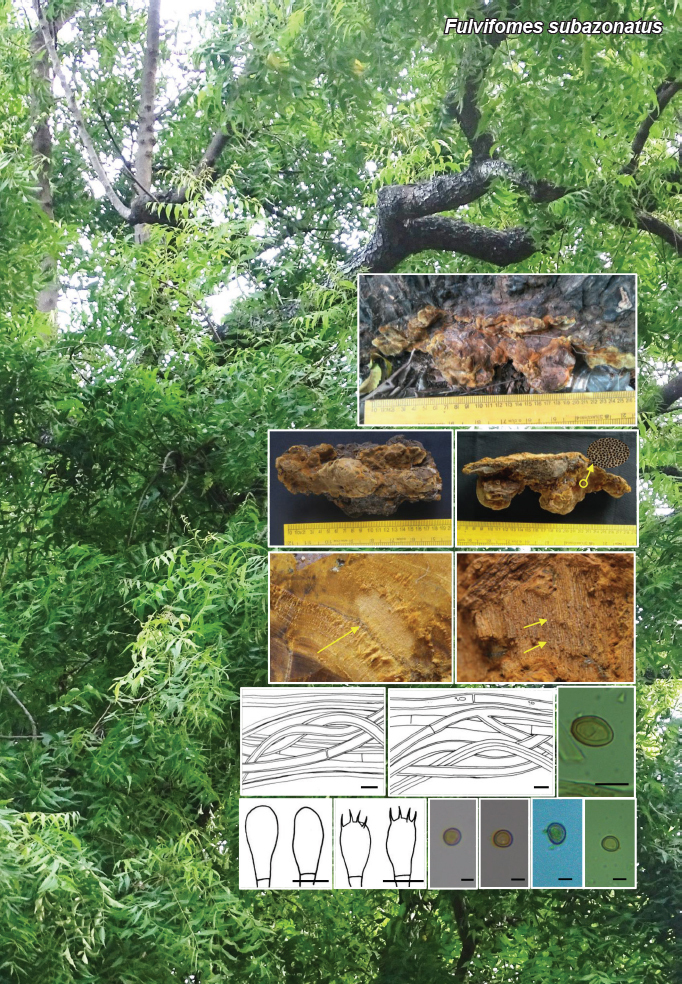
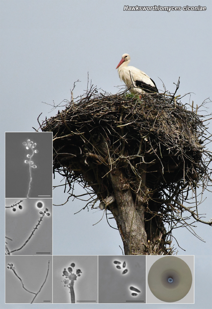
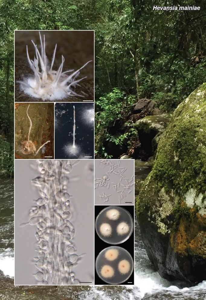
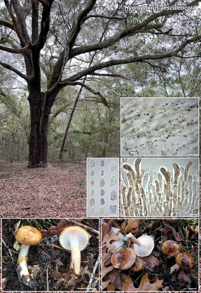
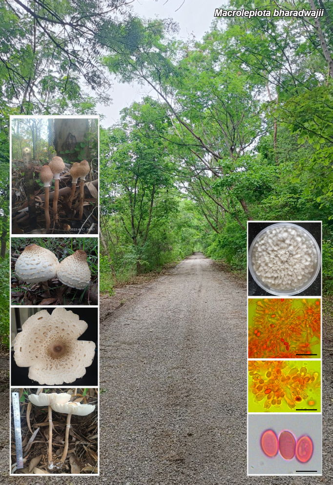
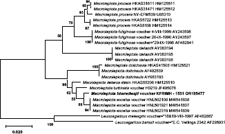
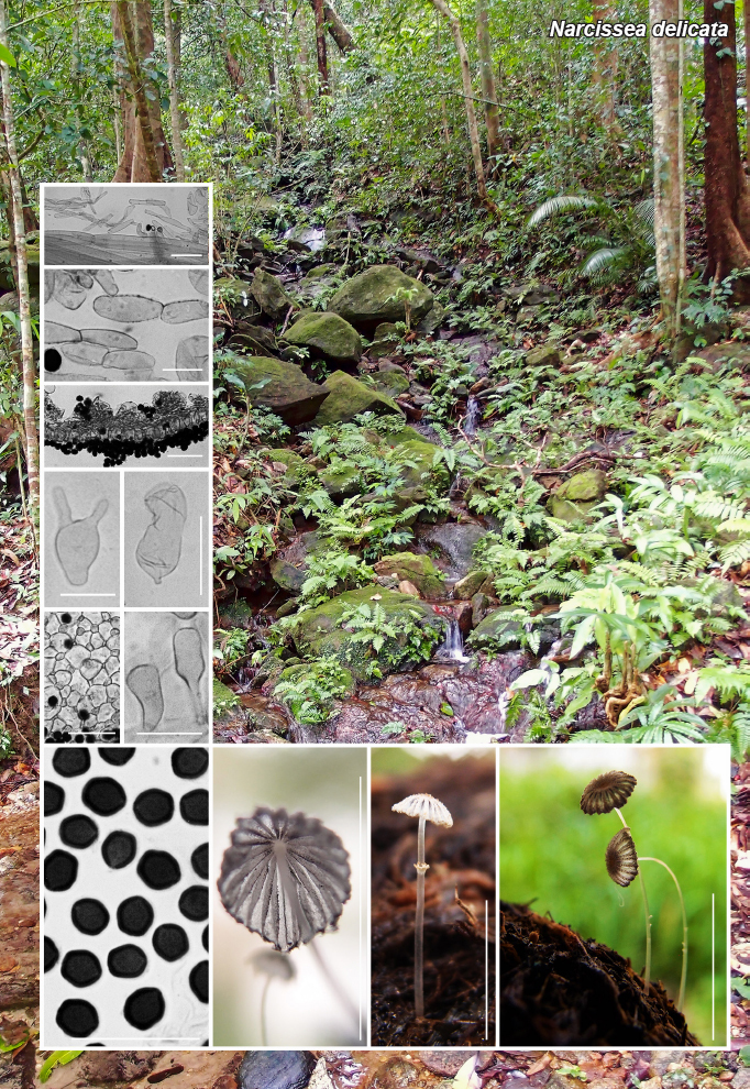
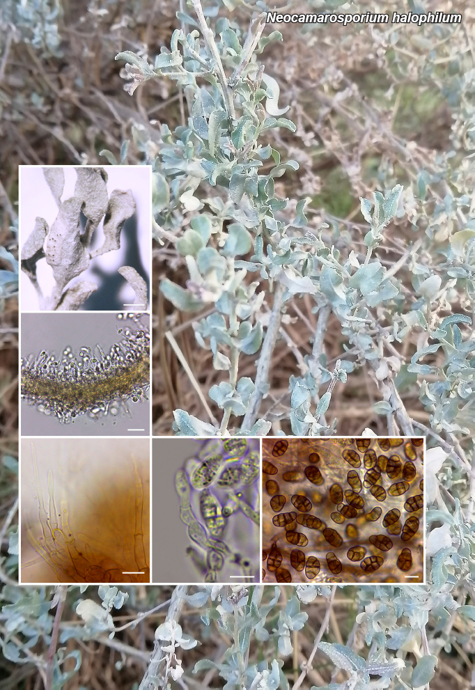
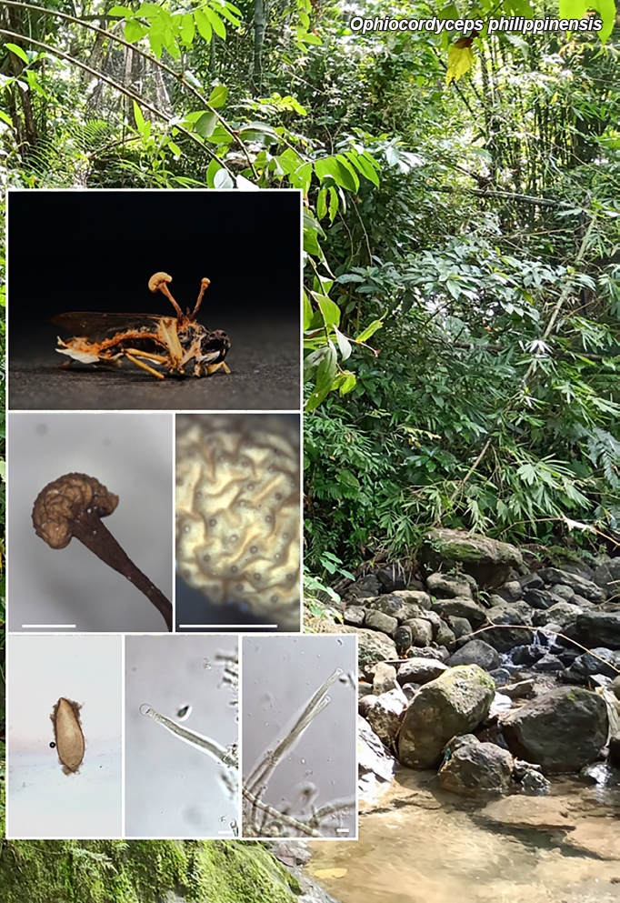
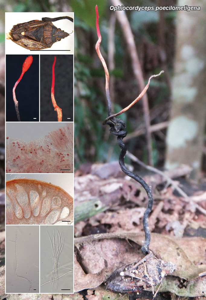
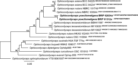
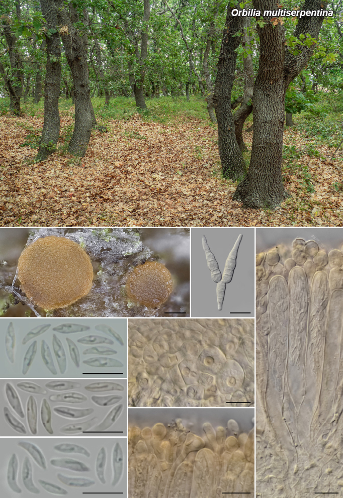
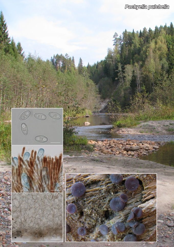
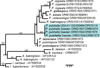
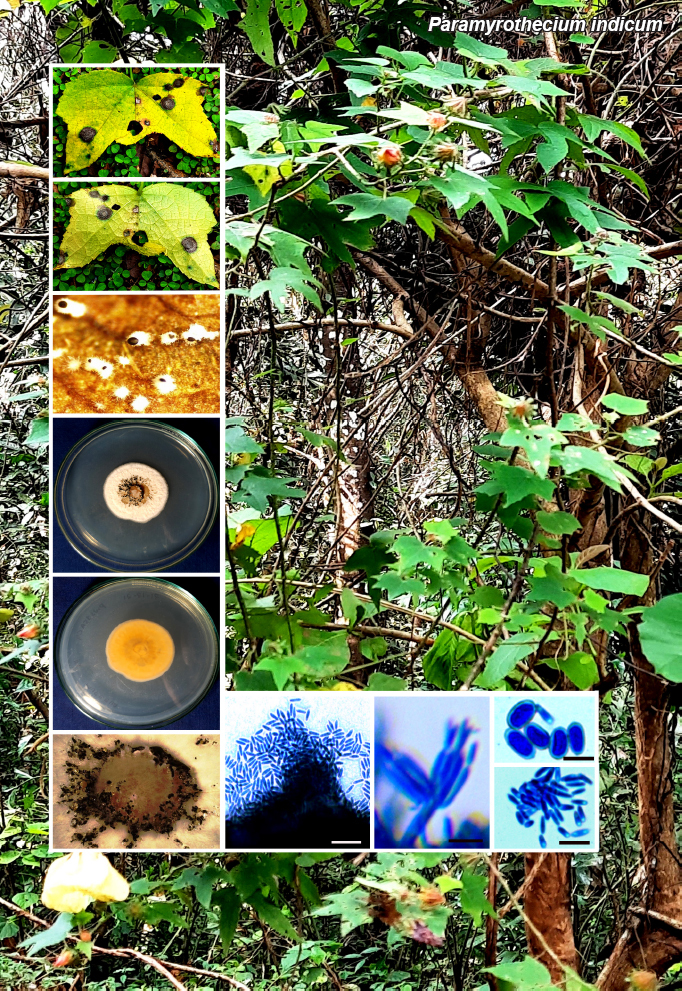
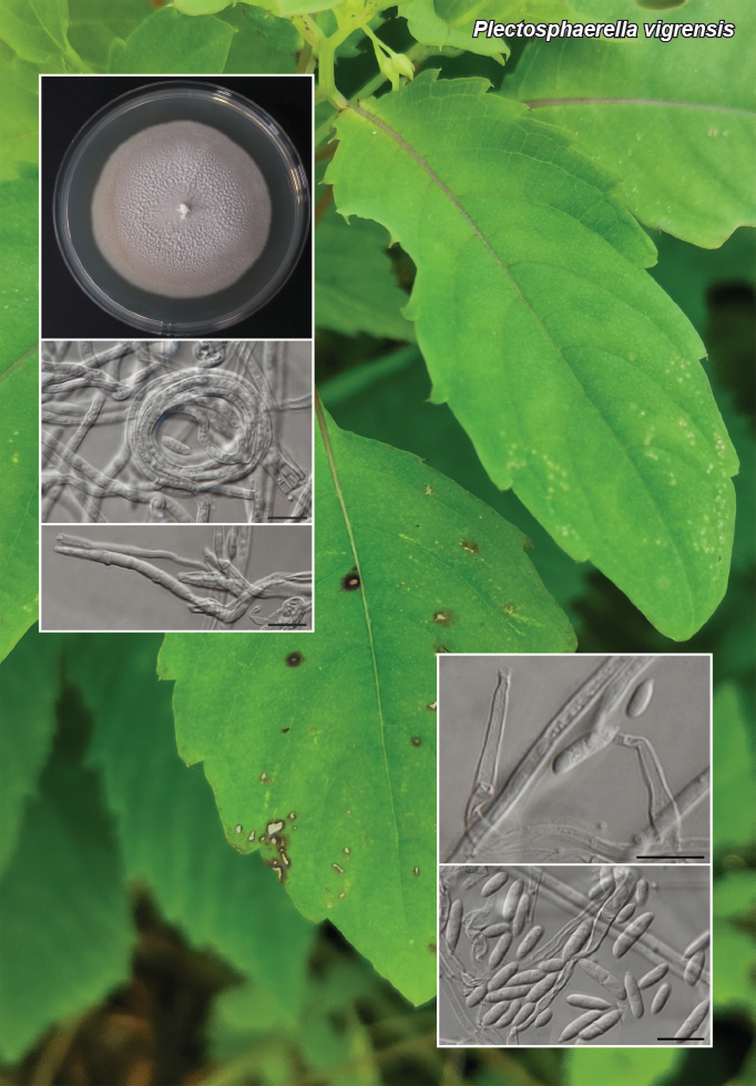
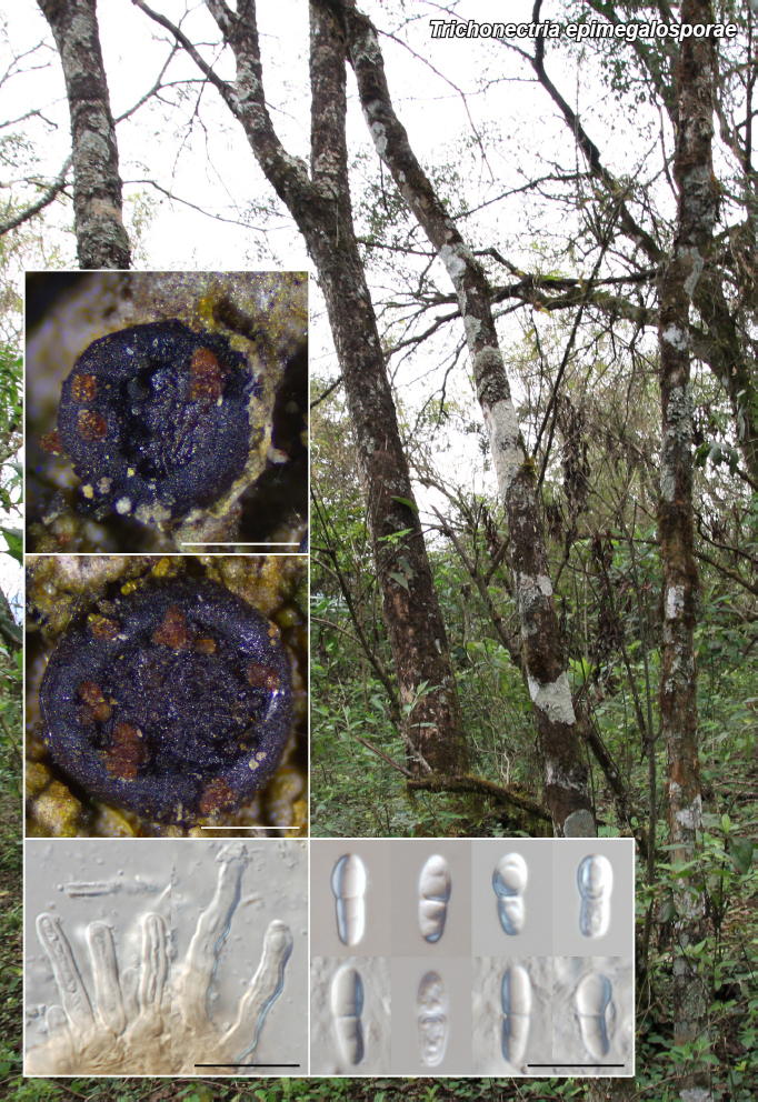
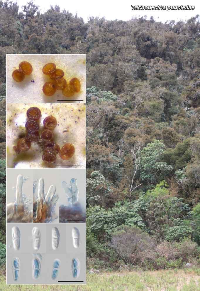
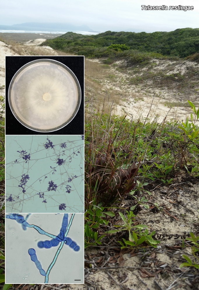
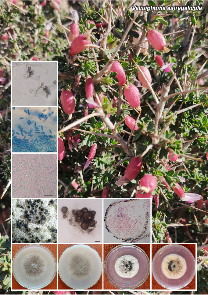
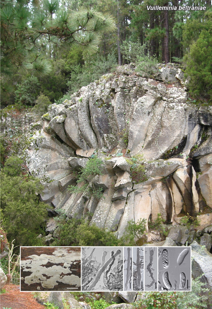
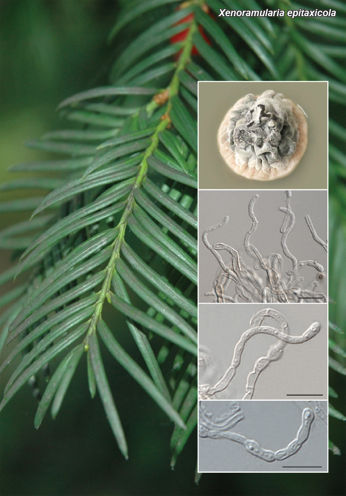
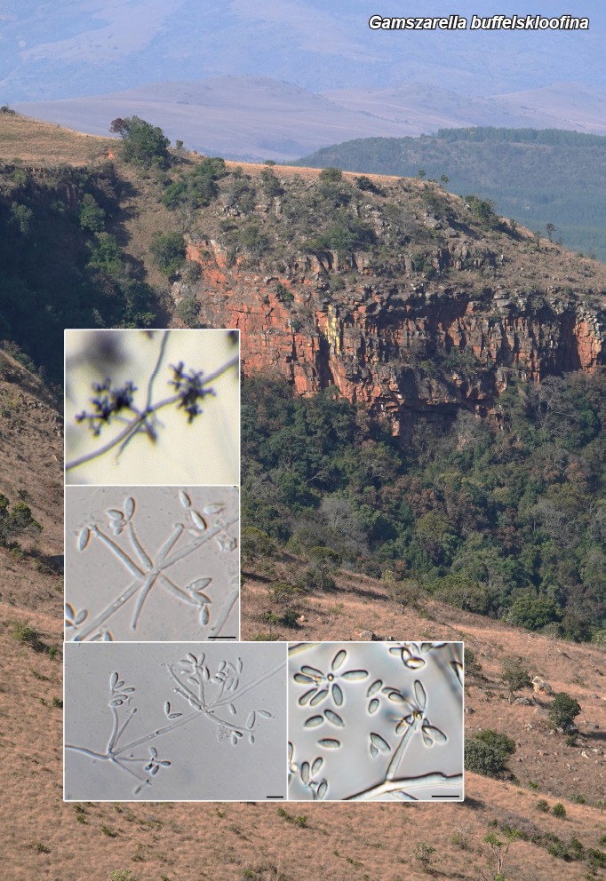
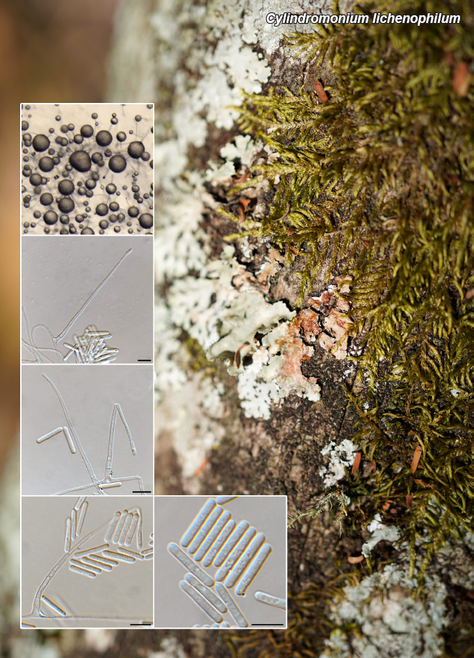
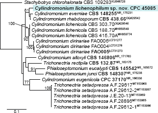
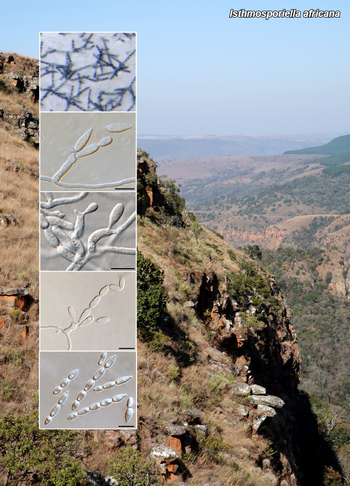
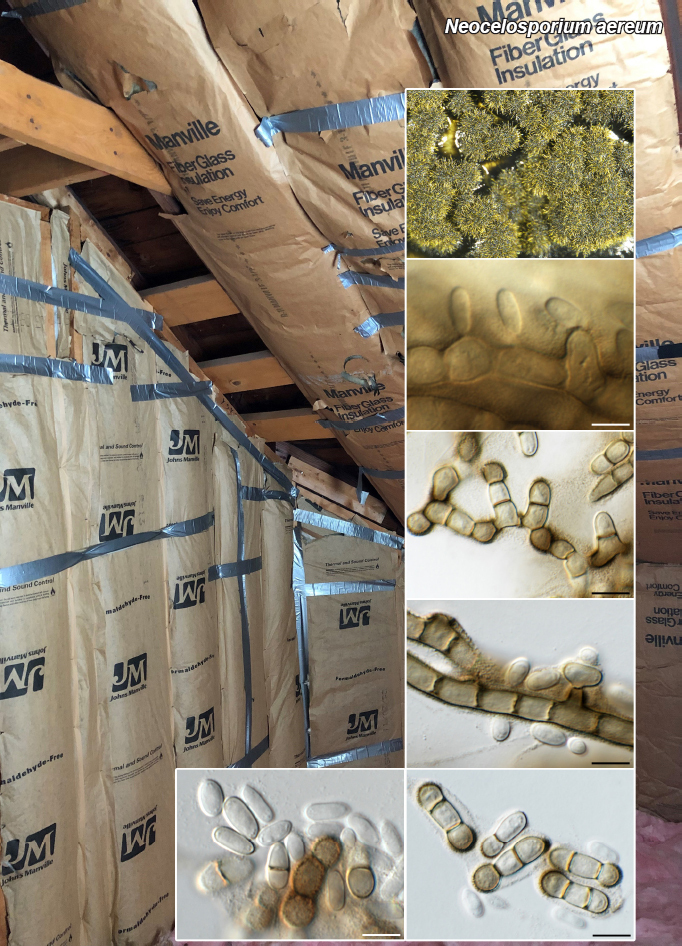
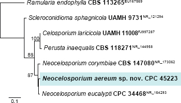
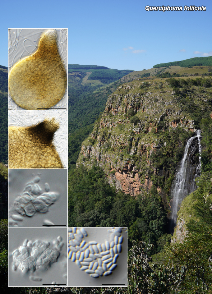
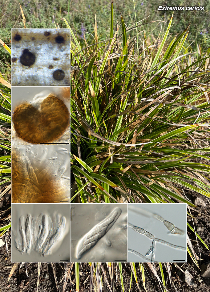
Novel species of fungi described in this study include those from various countries as follows: Argentina, Neocamarosporium halophilum in leaf spots of Atriplex undulata. Australia, Aschersonia merianiae on scale insect (Coccoidea), Curvularia huamulaniae isolated from air, Hevansia mainiae on dead spider, Ophiocordyceps poecilometigena on Poecilometis sp. Bolivia, Lecanora menthoides on sandstone, in open semi-desert montane areas, Sticta monlueckiorum corticolous in a forest, Trichonectria epimegalosporae on apothecia of corticolous Megalospora sulphurata var. sulphurata, Trichonectria puncteliae on the thallus of Punctelia borreri. Brazil, Catenomargarita pseudocercosporicola (incl. Catenomargarita gen. nov.) hyperparasitic on Pseudocercospora fijiensis on leaves of Musa acuminata, Tulasnella restingae on protocorms and roots of Epidendrum fulgens. Bulgaria, Anthracoidea umbrosae on Carex spp. Croatia, Hymenoscyphus radicis from surface-sterilised, asymptomatic roots of Microthlaspi erraticum, Orbilia multiserpentina on wood of decorticated branches of Quercus pubescens. France, Calosporella punctatispora on dead corticated twigs of Aceropalus. French West Indies (Martinique), Eutypella lechatii on dead corticated palm stem. Germany, Arrhenia alcalinophila on loamy soil. Iceland, Cistella blauvikensis on dead grass (Poaceae). India, Fulvifomes maritimus on living Peltophorum pterocarpum, Fulvifomes natarajanii on dead wood of Prosopis juliflora, Fulvifomes subazonatus on trunk of Azadirachta indica, Macrolepiota bharadwajii on moist soil near the forest, Narcissea delicata on decaying elephant dung, Paramyrothecium indicum on living leaves of Hibiscus hispidissimus, Trichoglossum syamviswanathii on moist soil near the base of a bamboo plantation. Iran, Vacuiphoma astragalicola from stem canker of Astragalus sarcocolla. Malaysia, Neoeriomycopsis fissistigmae (incl. Neoeriomycopsidaceae fam. nov.) on leaf spots on flower Fissistigma sp. Namibia, Exophiala lichenicola lichenicolous on Acarospora cf. luederitzensis. Netherlands, Entoloma occultatum on soil, Extremus caricis on dead leaves of Carex sp., Inocybe pseudomytiliodora on loamy soil. Norway, Inocybe guldeniae on calcareous soil, Inocybe rupestroides on gravelly soil. Pakistan, Hymenagaricus brunneodiscus on soil. Philippines, Ophiocordyceps philippinensis parasitic on Asilus sp. Poland, Hawksworthiomyces ciconiae isolated from Ciconia ciconia nest, Plectosphaerella vigrensis from leaf spots on Impatiens noli-tangere, Xenoramularia epitaxicola from sooty mould community on Taxus baccata. Portugal, Inocybe dagamae on clay soil. Saudi Arabia, Diaporthe jazanensis on branches of Coffea arabica. South Africa, Alternaria moraeae on dead leaves of Moraea sp., Bonitomyces buffels-kloofinus (incl. Bonitomyces gen. nov.) on dead twigs of unknown tree, Constrictochalara koukolii on living leaves of Itea rhamnoides colonised by a Meliola sp., Cylindromonium lichenophilum on Parmelina tiliacea, Gamszarella buffelskloofina (incl. Gamszarella gen. nov.) on dead insect, Isthmosporiella africana (incl. Isthmosporiella gen. nov.) on dead twigs of unknown tree, Nothoeucasphaeria buffelskloofina (incl. Nothoeucasphaeria gen. nov.), on dead twigs of unknown tree, Nothomicrothyrium beaucarneae (incl. Nothomicrothyrium gen. nov.) on dead leaves of Beaucarnea stricta, Paramycosphaerella proteae on living leaves of Protea caffra, Querciphoma foliicola on leaf litter, Rachicladosporium conostomii on dead twigs of Conostomium natalense var. glabrum, Rhamphoriopsis synnematosa on dead twig of unknown tree, Waltergamsia mpumalanga on dead leaves of unknown tree. Spain, Amanita fulvogrisea on limestone soil, in mixed forest, Amanita herculis in open Quercus forest, Vuilleminia beltraniae on Cistus symphytifolius. Sweden, Pachyella pulchella on decaying wood on sand-silt riverbank. Thailand, Deniquelata cassiae on dead stem of Cassia fistula, Stomiopeltis thailandica on dead twigs of Magnolia champaca. Ukraine, Circinaria podoliana on natural limestone outcrops, Neonematogonum carpinicola (incl. Neonematogonum gen. nov.) on dead branches of Carpinus betulus. USA, Exophiala wilsonii water from cooling tower, Hygrophorus aesculeticola on soil in mixed forest, and Neocelosporium aereum from air in a house attic. Morphological and culture characteristics are supported by DNA barcodes.

Citation: Crous PW, Costa MM, Kandemir H, et al. 2023. Fungal Planet description sheets: 1550–1613. Persoonia 51: 280–417. doi: 10.3767/persoonia.2023.51.08.

Keywords: ITS nrDNA barcodes, LSU, new taxa, systematics

Acknowledgments

The work of P.W. Crous and colleagues benefitted from funding by the European Union’s Horizon 2020 research and innovation program (RISE) under the Marie Sklodowska-Curie grant agreement No. 101008129, project acronym ‘Mycobiomics’, and the Dutch NWO Roadmap grant agreement No. 2020/ENW/00901156, project ‘Netherlands Infrastructure for Ecosystem and Biodiversity Analysis – Authoritative and Rapid Identification System for Essential biodiversity information’ (acronym NIEBA-ARISE). M. Plaza and T. Illescas are grateful to the Junta de Andalucía for facilitating their vehicle access to areas of special protection; and to both their daughters, C. Plaza and M. Illescas, for revising the English text, and the Asociación Botánica y Micológica de Jaén, for funding part of the DNA sequences included in this study. The study of T.T. Denchev & C.M. Denchev was supported by the Bulgarian National Science Fund (Grant no. KP-06-N51/10/16.11.2021). K. Karasungur is thanked for technical help with the Austrian material of Arrhenia sequenced within the Austrian Barcode of Life project, supported by the Austrian Federal Ministry of Education, Science and Research. Y.P. Tan and colleagues acknowledge The Australian Biological Resources Study for funding. F.A. Custódio and O.L. Pereira are thankful to the Coordenação de Aperfeiçoamento de Pessoal de Níve Superior, Brazil (CAPES), finance code 001, Fundação de Amparo à Pesquisa do Estado de Minas Gerais (FAPEMIG), and Conselho Nacional de Desenvolvimiento Científico e Tecnológico (CNPq) for their financial support. The authors acknowledge K. Bensch for the help with the Latin name of the new fungus. A. Miller would like to thank the Roy J. Carver Biotechnology Center at the University of Illinois for Sanger sequencing. J. Fournier is thankful to R. Courtecuisse (Lille University) for having organised field trips to Guadeloupe and Martinique from 2003 to 2016 and special thanks go to P.-A. Moreau who attended several of these forays and shared his image of C. Lechat at La Caravelle. V. Vasan, N. Chellappan, E. Arumugam, R. Murugadoss and M. Kaliyaperumal thank the Director, Centre of Advanced Studies in Botany, University of Madras for the laboratory facilities. M. Kaliyaperumal and E. Arumugam acknowledge the Tamil Nadu State Council for Higher Education (RGP/2019-20/MU/HECP-0040) for providing financial aid. The authors acknowledge C. Janagar Dhas, Centre of Advanced Studies in Botany, University of Madras for assistance with photography. R. Murugadoss would like to acknowledge CSIR – Junior Research Fellowship (JRF-NET), New Delhi, India (09/0115(13300)/2022-EMR-I) for the financial assistance. M. Kaliyaperumal and co-authors thank Dr. J. Bhat, for his suggestion with the nomenclature. M. Kaliyaperumal, E. Arumugam and S. Gunaseelan would like to thank Prof. N. Mathivanan, Director, Centre for Advanced Studies in Botany, University of Madras, Chennai, for providing laboratory facilities. The study of R. Jankowiak was funded by the Ministry of Science and Higher Education of the Republic of Poland (SUB/040013/ D019) and by the National Science Centre, Poland (contract no. 2021/41/B/ NZ8/03456). R.G. Thorn and co-authors thank the Walpole Island First Nation for providing permission to search for and collect Hygrophorus aesculeticola at Bkejwanong. Participation of M.E. Smith was supported by the US National Science foundation grant DEB-2106130 and NIFA-USDA award FLA-PLP-005289. Y. Lamoureux is acknowledged for microscopical work on his collection of Hygrophorus paludosoides. G. Delgado thanks W. Colbert and S. Ward (Eurofins Built Environment) for the provision of laboratory facilities. J.G. Maciá-Vicente acknowledges the support of the Landes-Offensive zur Entwicklung Wissenschaftlich-ökonomischer Exzellenz (LOEWE) of the state of Hesse within the framework of the Cluster for Integrative Fungal Research (IPF) of Goethe University Frankfurt. Both authors thank H.-O. Baral for providing sequence data and his feedback on the phylogeny of Hymenoscyphus. V. Fachada and co-authors would like to thank C. Vieira (MHNC-UP) for managing the voucher collections in the PO herbarium. We are grateful for the valuable assistance provided by K. Bensch and S. Pennycook in determining the correct taxonomic nomenclature. E. Larsson acknowledges the Swedish Taxonomy Initiative, SLU Artdatabanken, Uppsala, Sweden. G.M. Jansen is grateful to J. Vauras for making collections from TUR available and for his valuable ideas about neighbouring species, to M. Gotink, M. van der Vegte and M. Plekkenpol for their collections and their help in financing the sequencing, to D. Bandini for supplying a gill of the isotype of Inocybe mytiliodora for sequencing, to P.B. Matheny for making his description and sequence of his collection PBM1572 available and for sharing his ideas for rooting the tree, and to the Ger van Zaanen Fund for financing the sequencing of L4343844. The description of Inocybe pseudomytiliodora was critically reviewed by J. Vauras and P.B. Matheny, and their valuable suggestions and improvements are gratefully acknowledged. E. Mazur acknowledges the staff of the Herbario Nacional de Bolivia, Instituto de Ecología, Universidad Mayor de San Andrés, La Paz, for their generous long-term cooperation with lichenologists in the W. Szafer Institute of Botany, Polish Academy of Sciences, the collectors: A. Flakus, K. Wilk and B. Cykowska for making their material available for the study, and L. Sliwa who supervised her doctoral thesis. E. Mazur was supported by statutory funds from the W. Szafer Institute of Botany, Polish Academy of Sciences, and the National Science Centre, Poland, project 2016/21/B/NZ8/02463. A.R. Podile acknowledges the Department of Science and Technology, Govt. of India for the award of the JC Bose Fellowship (Grant No. JCB/2017/000053) and the Ministry of Education, Govt. of India and Institution of Excellence Directorate, University of Hyderabad for the award of the project (Grant No.UOH-IOE-RC3-21-065). S. Mahadevakumar thanks the Director, KSCSTE - Kerala Forest Research Institute & Head of Office Botanical Survey of India, Andaman and Nicobar Regional Centre, Port Blair, M. Madappa, Department of Studies in Botany, University of Mysore and K.T. Mufeeda Forest Patho-logy Department, Kerala Forest Research Institute, Peechi, Thrissur for their kind support and technical assistance. K.G.G. Ganga acknowledges support from the University Grants Commission (UGC), India, in the form of a UGC research fellowship (Ref No. 20/12/2015(ii) EU-V). K.G.G. Ganga and co-authors also thank the authorities of the University of Calicut for providing facilities for this study. R.M. Sanchez and M.V. Bianchinotti acknowledge the National Council for Scientific and Technical Research (CONICET) for providing the funds for this research (PIP11220130100280CO). D.A. Acal would like to thank the Department of Science and Technology (DOST-SEI), Republic of Philippines for the financial support during the collection of material and the Department of Environmental and Natural Resources of Biodiversity Management Bureau (DENR-BMB), Republic of Philippines for the issuance of Wildlife Gratuitous Permits (no. 319). F. Fuljer was funded by the Operational Program of Integrated Infrastructure, co-financed with the European Fund for Regional Development (EFRD) ITMS2014+313021W683: ‘DNA barcoding of Slovakia (SK-BOL), as a part of international initiative International Barcode of Life (iBOL)’. The authors would also like to thank I. Kušan and N. Matočec for conveying valuable information about bioclimatic zones of localities. K. Hansen is thankful to Juan Carlos Zamora, Ibai Olariaga and Timo Kosonen for their help on field, microscopic and molecular work, to Henning Knudsen for nomenclatural advice, Donald H. Pfister for discussions on the new species, and to C, CUP, NICE and UPS for arranging loans of specimens, and the Swedish Taxonomy Initiative, SLU Artdatabanken, Sweden for providing funding for this research. S. Kumar and co-authors are grateful to the Director, KSCSTE-Kerala Forest Research Institute, Peechi for providing library and laboratory facilities. They also acknowledge the Science & Engineering Research Board (SERB), Department of Science & Technology (DST), Govt. of India for financial support (CRG/2019/005014). The research of K. Patejuk, W. Pusz and A. Baturo-Cieśniewska (in part) was funded by the Forest Fund under the agreement concluded between the State Forests National Forest Holding and the Wigry National Park (contracts no. EZ.0290.1.21.2021 and EZ.0290.1.21.2022). E.A. Ossowska and co-authors are grateful to the members of Herbario Nacional de Bolivia, Instituto of Herbario Nacional de Bolivia, Instituto de Ecología, Universidad Mayor de San Andrés, La Paz, for the generous cooperation and M. Kukwa for help with the description. This research received support from the SYNTHESYS Project (DE-TAF-8180) http://www.synthesys.info/ which is financed by European Community Research Infrastructure Action under the FP7 ‘Capacities’ Programme and University of Gdansk, granted to EAO. V. Darmostuk and co-authors acknowledge our colleagues and all staff of the Herbario Nacional de Bolivia, Instituto de Ecología, Universidad Mayor de San Andrés, La Paz, for their generous long-term cooperation. They would also like to thank the SERNAP (http://sernap.gob.bo), and all protected areas staff, for providing permits for scientific studies, as well as their assistance and logistical support during the field works. This research was financially supported by the National Science Centre (NCN) in Poland (grant number DEC-2013/11/D/NZ8/03274). The study of P. Eis-vand and M. Mehrabi-Koushki was financially supported by grant (SCU. AP1401.294) from the Research Council of Shahid Chamran University of Ahvaz. Financial support to M. Dueñas and colleagues was provided by Plan Nacional I+D+I project no. CGL2009-07231 and Ref. 202030E059. They also acknowledge M. Glenn (Seton Hall University, USA) for her kind English revision, and M. Ghobad-Nejhad for sending the ITS nrDNA alignment with sequences published in Ghobad-Nejhad & Duhem (2014). The study of M. Piątek and co-authors was funded by the National Science Centre, Poland, under the project 2017/27/B/NZ9/02902. V. Hubka was supported by a Czech Academy of Sciences Long-term Research Development Project (RVO: 61388971). A. Ismail and co-authors thank the Deanship of Scientific Research, Vice Presidency for Graduate Studies and Scientific Research, King Faisal University, Saudi Arabia, for supporting this research for work through grant number (GRANT 4597). M. Thines acknowledges funding by the LOEWE excellence initiative of the government of Hesse, in the framework of the Centre for Translational Biodiversity Genomics (TBG).

REFERENCES

  1. Aalto M. 1974. Amanita magnivolvata sp. nova (Agaricales). Karstenia 14: 93–96. [Google Scholar]
  2. Acharius E. 1810. Lichenographia Universal is. Frid. Danckwerts., Gottingen. [Google Scholar]
  3. Ajitomi A, Takushi T, Sato T, et al. 2017. First report of flyspeck of mango caused by Stomiopeltis sp. in Japan. Journal of General Plant Pathology 83: 299–303. [Google Scholar]
  4. Alidadi A, Kowsari M, Javan-Nikkham M, et al. 2019. Deniquelata quercina sp. nov.; a new endophyte species from Persian oak in Iran. Phytotaxa 405: 187–194. [Google Scholar]
  5. Almaraz T. 2002. Bases corológicas de Flora Micológica Ibérica. Numeros 1766–1932. In: Pando F, Hernández JC. (eds), Cuadernos de trabajo de Flora Micológica Ibérica 17: 1–124. [Google Scholar]
  6. Amandeep K, Atri NS, Munruchi K. 2015. Taxonomic study on the coprophilous mushrooms from Punjab, India: new records of family Agaricaceae. Current Research in Environmental & Applied Mycology 5: 27–45. [Google Scholar]
  7. Ariyawansa HA, Maharachchikumbura SS, Karunarathne SC, et al. 2013. Deniquelata barringtoniae gen. et sp. nov., associated with leaf spots of Barringtonia asiatica. Phytotaxa 105: 11–20. [Google Scholar]
  8. Ariyawansa HA, Thambugala KM, Manamgoda DS, et al. 2015. Towards a natural classification and backbone tree for Pleosporaceae. Fungal Diversity 71: 85–139. [Google Scholar]
  9. Armand A, Bundhun D, Bhunjun CS, et al. 2023. Paramyrothecium amorphophalli sp. nov., a causal agent of leaf blight on elephant foot yam in northern Thailand. Current Research in Environmental & Applied Mycology 13: 57–67. [Google Scholar]
  10. Arnolds E. 1990. Genus Hygrophorus. In: Bas C, Kuyper TW, Noordeloos ME, et al. (eds), Flora Agaricina Neerlandica 2: 115–133. Balkema, Lisse. [Google Scholar]
  11. Arup U, Ekman S, Grube M, et al. 2007. The sister group relation of Parmeliaceae (Lecanorales, Ascomycota). Mycologia 99: 42–49. [DOI] [PubMed] [Google Scholar]
  12. Aveskamp MM, De Gruyter J, Woudenberg JHC, et al. 2010. Highlights of the Didymellaceae: a polyphasic approach to characterise Phoma and related pleosporalean genera. Studies in Mycology 65: 1–60. [DOI] [PMC free article] [PubMed] [Google Scholar]
  13. Bandini D, Huijser H. 2017. Vezelkoppen (Inocybe) van Ameland, Inocybegriseotarda Poirier. Coolia 60: 243–247. [Google Scholar]
  14. Bandini D, Oertel B, Eberhardt U. 2021. A fresh outlook on the smooth-spored species of Inocybe: type studies and 18 new species. Mycological Progress 20: 1019–1114. [Google Scholar]
  15. Baral HO, Weber E, Marson G. 2020. Monograph of Orbiliomycetes (Ascomycota) based on vital taxonomy. Part I + II. National Museum of Natural History Luxembourg. [Google Scholar]
  16. Bas C. 1969. Morphology and subdivision of Amanita and a monograph of its section Lepidella. Persoonia 5: 285–579. [Google Scholar]
  17. Bas C, Kuyper TW, Noordeloos ME, et al. 1995. Flora Agaricina Neerlandica 3. Critical monographs on families of agarics and boleti occurring in the Netherlands. Balkema, Rotterdam Brookfield. [Google Scholar]
  18. Bellanger JM, Lebeuf R, Sesli E, et al. 2021. Hygrophorus sect. Olivaceoumbrini: new boundaries, extended biogeography and unexpected diversity unravelled by transatlantic studies. Persoonia 46: 272–312. [DOI] [PMC free article] [PubMed] [Google Scholar]
  19. Berkeley MJ, Broome CE. 1873. ‘1875. ’. Enumeration of the Fungi of Ceylon. Part II, containing the remainder of the Hymenomycetes, with the remaining established tribes of Fungi. Botanical Journal of the Linnean Society 14: 29–140. [Google Scholar]
  20. Bizio E, Ferisin G, Dovana F. 2017. Note sul campo di variabilità di Inocybe griseotarda. Rivista di Micologia 60: 59–70. [Google Scholar]
  21. Błońska E, Lasota J, Jankowiak R, et al. 2021. Biological and physicochemical properties of the nests of White Stork Ciconia ciconia reveal soil entirely formed, modified and maintained by birds. Science of the Total Environment 763: 43020. [DOI] [PubMed] [Google Scholar]
  22. Boldt-Burisch K, Douanla-Meli C. 2023. First report of Querciphoma minuta causing branch and stem canker in Platanus × hispanica in Germany. New Disease Reports 47: e12153. [Google Scholar]
  23. Bon M. 1978. Amanitopsis pachyvolvata. Documents Mycologiques 8: 36. [Google Scholar]
  24. Bon M. 1990. Flore mycologique du littoral - 05 - Inocybe. Documents Mycologiques 20: 61–66. [Google Scholar]
  25. Bouckaert R, Heled J, Kühnert D, et al. 2014. BEAST 2: A software platform for bayesian evolutionary analysis. PLoS Computational Biology 10: e1003537. [DOI] [PMC free article] [PubMed] [Google Scholar]
  26. Bresadola G. 1903. Fungi polonici. Annales Mycologici 1: 97–131. [Google Scholar]
  27. Calduch M, Gené J, Guarro J, et al. 2002. Hyphomycetes from Nigerian Rain Forests. Mycologia 94: 127–135. [PubMed] [Google Scholar]
  28. Candusso M. 1997. Fungi Europaei 6: Hygrophorus s.l. Edizioni Candusso, Alassio, Italia. [Google Scholar]
  29. Castresana J. 2000. Selection of conserved blocks from multiple alignments for their use in phylogenetic analysis. Molecular Biology and Evolution 17: 540–552. [DOI] [PubMed] [Google Scholar]
  30. Chater AO. 1980. Carex L. In: Tutin TG, Heywood VH, Burges NA, et al. (eds), Flora Europaea. Vol. 5: 290–323. Cambridge University Press, Cambridge. [Google Scholar]
  31. Chernomor O, Von Haeseler A, Quang Minh B. 2016. Terrace aware data structure for phylogenomic inference from supermatrices. Systematic Biology 65: 997–1008. [DOI] [PMC free article] [PubMed] [Google Scholar]
  32. Chhetri PK, Pradhan BK, Chhetri B. 2019. First record of Ophiocordyceps dipterigena Berk. & Broome (Ophiocordycipitaceae) in the Himalayas. National Academy Science Letters 43: 367–369. [Google Scholar]
  33. Clemençon H. 1982. European Omphalinoid Tricholomataceae. Zeitschrift für Mykologie 48: 195–237. [Google Scholar]
  34. Coker TLR, Rozsypálek J, Edwards A, et al. 2019. Estimating mortality rates of European ash (Fraxinus excelsior) under the ash dieback (Hymenoscyphus fraxineus) epidemic. Plants, People, Planet 1: 48–58. [Google Scholar]
  35. Consiglio G, Contu M. 1999. Amanita dryophila (Amanitaceae) spec. nov. and the species of the section Vaginatae with a semifriable universal veil and ellipsoid spores. Persoonia 17: 287–190. [Google Scholar]
  36. Contu M. 1984. Novitates (2). Documents Mycologiques 14: 26. [Google Scholar]
  37. Contu M. 1999. Appunti sul genere Amanita IX Nuove specie e studi tassonomico-nomenclaturali nella sezione Vaginatae. Bollettino dell’Associazione Micologica ed Ecologica Romana 46: 3–22. [Google Scholar]
  38. Crous PW, Akulov A, Balashov S, et al. 2023. New and interesting fungi. 6. Fungal Systematics and Evolution 11: 109–156. [DOI] [PMC free article] [PubMed] [Google Scholar]
  39. Crous PW, Cowan DA, Maggs-Kölling G, et al. 2020. Fungal Planet description sheets: 1112-1181. Persoonia 45: 251–409. [DOI] [PMC free article] [PubMed] [Google Scholar]
  40. Crous PW, Cowan DA, Maggs-Kölling G, et al. 2021a. Fungal Planet description sheets: 1182-1283. Persoonia 46: 313–528. [DOI] [PMC free article] [PubMed] [Google Scholar]
  41. Crous PW, Groenewald JZ. 2017. The genera of fungi - G4: Camarosporium and Dothiora. IMA Fungus 8: 131–152. [DOI] [PMC free article] [PubMed] [Google Scholar]
  42. Crous PW, Groenewald JZ, Shivas RG, et al. 2011. Fungal Planet description sheets: 69–91. Persoonia 26: 108–156. [DOI] [PMC free article] [PubMed] [Google Scholar]
  43. Crous PW, Hernández-Restrepo M, Schumacher RK, et al. 2021b. New and interesting fungi. 4. Fungal Systematics and Evolution 7: 255–343. [DOI] [PMC free article] [PubMed] [Google Scholar]
  44. Crous PW, Luangsa-ard JJ, Wingfield MJ, et al. 2018a. Fungal Planet description sheets: 785–867. Persoonia 41: 238–417. [DOI] [PMC free article] [PubMed] [Google Scholar]
  45. Crous PW, Mohammed C, Glen M, et al. 2007. Eucalyptus microfungi known from culture. 3. Eucasphaeria and Sympoventuria genera nova, and new species of Furcaspora, Harknessia, Heteroconium and Phacidiella. Fungal Diversity 25: 19–36. [Google Scholar]
  46. Crous PW, Osieck ER, Jurjević Ž, et al. 2021c. Fungal Planet description sheets: 1284–1382. Persoonia 47: 178–374. [DOI] [PMC free article] [PubMed] [Google Scholar]
  47. Crous PW, Schoch CL, Hyde KD, et al. 2009. Phylogenetic lineages in the Capnodiales. Studies in Mycology 64: 17–47. [DOI] [PMC free article] [PubMed] [Google Scholar]
  48. Crous PW, Schumacher RK, Wingfield MJ, et al. 2018b. New and interesting fungi. 1. Fungal Systematics and Evolution 1: 169–215. [DOI] [PMC free article] [PubMed] [Google Scholar]
  49. Crous PW, Shivas RG, Quaedvlieg W, et al. 2014. Fungal Planet description sheets: 214–280. Persoonia 32: 184–306. [DOI] [PMC free article] [PubMed] [Google Scholar]
  50. Crous PW, Wingfield MJ, Burgess TI, et al. 2016. Fungal Planet description sheets: 469–557. Persoonia 37: 218–403. [DOI] [PMC free article] [PubMed] [Google Scholar]
  51. Crous PW, Wingfield MJ, Guarro J, et al. 2013. Fungal Planet description sheets: 154–213. Persoonia 31: 188–296. [DOI] [PMC free article] [PubMed] [Google Scholar]
  52. Crous PW, Wingfield MJ, Guarro J, et al. 2015. Fungal Planet description sheets: 320–370. Persoonia 34: 167–266. [DOI] [PMC free article] [PubMed] [Google Scholar]
  53. Crous PW, Wingfield MJ, Lombard L, et al. 2019. Fungal Planet description sheets: 951–1041. Persoonia 43: 223–425. [DOI] [PMC free article] [PubMed] [Google Scholar]
  54. Darriba D, Posada D, Kozlov AM, et al. 2020. ModelTest-NG: A new and scalable tool for the selection of DNA and protein evolutionary models. Molecular Biology and Evolution 37: 291–294. [DOI] [PMC free article] [PubMed] [Google Scholar]
  55. Darriba D, Taboada GL, Doallo R, et al. 2012. jModel test 2: more models, new heuristics and parallel computing. Nature Methods 9: 772. [DOI] [PMC free article] [PubMed] [Google Scholar]
  56. Davydov EA, Yakovchenko LS, Hollinger J, et al. 2021. The new genus Pulvinora (Lecanoraceae) for species of the ‘Lecanora pringlei’ group, including the new species Pulvinora stereothallina. Bryologist 124: 242–256. [Google Scholar]
  57. Dayarathne MC, Jones EBG, Maharachchikumbura SSN, et al. 2020. Morphomolecular characterization of microfungi associated with marine based habitats. Mycosphere 11: 1–188. [Google Scholar]
  58. De Beer ZW, Marincowitz S, Duong TA, et al. 2016. Hawksworthiomyces gen. nov. (Ophiostomatales), illustrates the urgency for a decision on how to name novel taxa known only from environmental nucleic acid sequences (ENAS). Fungal Biology 120: 1323–1340. [DOI] [PubMed] [Google Scholar]
  59. Denchev CM. 2001. Class Ustomycetes (Orders Tilletiales, Ustilaginales, and Graphiolales). In: Fakirova V. (ed.), Fungi of Bulgaria. Vol. 4: 1–286. Editio Academica ‘Prof. Marin Drinov’ & Editio Pensoft, Sofia. [Google Scholar]
  60. Denchev TT, Denchev CM, Begerow D, et al. 2023. Anthracoidea obtusatae (Anthracoideaceae, Ustilaginales), a new smut fungus on Carex obtusata (Cyperaceae) from Central Asia. Phytotaxa 595: 139–148. [Google Scholar]
  61. Denchev TT, Denchev CM, Koopman J, et al. 2021. Host specialization and molecular evidence support a distinct species of smut fungus, Anthracoidea hallerianae (Anthracoideaceae), on Carex halleriana (Cyperaceae). Willdenowia 51: 57–67. [Google Scholar]
  62. Denchev TT, Denchev CM, Michikawa M, et al. 2013. The genus Anthracoidea (Anthracoideaceae) in Japan and some adjacent regions. Mycobiota 2: 1–125. [Google Scholar]
  63. Devadatha B, Sarma VV, Ariyawansa HA, et al. 2018. Deniquelata vittalii sp. nov., a novel Indian saprobic marine fungus on Suaeda monoica and two new records of marine fungi from Muthupet mangroves, East coast of India. Mycosphere 9: 565–582. [Google Scholar]
  64. Dos Santos LA, Aptroot A, Lücking R, et al. 2023. Lecanora s.lat. (Ascomycota, Lecanoraceae) in Brazil: DNA barcoding coupled with phenotype characters reveals numerous novel species. Journal of Fungi 9: 415. [DOI] [PMC free article] [PubMed] [Google Scholar]
  65. Du P, Cao TX, Wu YD, et al. 2021. Two new species of Hymenochaetaceae on Dracaena cambodiana from tropical China. MycoKeys 80: 1–17. [DOI] [PMC free article] [PubMed] [Google Scholar]
  66. Edgar RC. 2004. MUSCLE: multiple sequence alignment with high accuracy and high throughput. Nucleic Acids Research 32: 1792–1797. [DOI] [PMC free article] [PubMed] [Google Scholar]
  67. Edler D, Klein J, Antonelli A, et al. 2021. raxmlGUI 2.0: A graphical interface and toolkit for phylogenetic analyses using RAxML. Methods in Ecology and Evolution 12: 373–377. [Google Scholar]
  68. Edler F, Miccoli I, Pfnur H, et al. 2019. Space charge layer effects in silicon studied by in situ surface transport. Journal of Physics: Condensed Matter 31: 1–8. [DOI] [PubMed] [Google Scholar]
  69. Egidi E, De Hoog GS, Isola D. et al. 2014. Phylogeny and taxonomy of meristematic rock-inhabiting black fungi in the Dothideomycetes based on multi-locus phylogenies. Fungal Diversity 65: 127–165. [Google Scholar]
  70. Egorova TV. 1999. The sedges (Carex L.) of Russia and adjacent states (within the limits of the former USSR). Saint Petersburg State Chemical-Pharmaceutical Academy, Saint Petersburg, Russia & Missouri Botanical Garden Press, St. Louis, USA. [Google Scholar]
  71. Ekanayaka AH, Hyde KD, Jones EBG, et al. 2017. A new species of Trichoglossum (Geoglossales, Ascomycota) from Thailand. Phytotaxa 316: 11–170. [Google Scholar]
  72. Enderle M, Krieglsteiner GJ, Bender H. 1986. Studien zur Gattung Coprinus (Pers.: Fr.) SF Gray in der Bundesrepublik Deutschland. III. Zeitschrift für Mykologie 52: 101–132. [Google Scholar]
  73. Etayo J. 2001. Hongos liquenícolas de Ecuador. I. Dos especies nuevas del orden Hypocreales (Ascomycota): Pronectria parmotrematis y Trichonectria leptogiicola. Anales del Jardín Botánico de Madrid 58: 219–222. [Google Scholar]
  74. Etayo J, Sancho LG. 2008. Hongos liquenícolas del Sur de Sudamérica, especialmente de Isla Navarino (Chile). Bibliotheca Lichenologica 98: 1–302. [Google Scholar]
  75. Etayo J, Van den Boom PPG. 2005. Contribution to the lichen flora of the Canary Islands. VIII. Some lichenicolous fungi. Nova Hedwigia 81: 157–162. [Google Scholar]
  76. Fathima M, Usman M, Khalid AN. 2023. Fulvifomes aurantiacus sp. nov. (Basidiomycota; Hymenochaetaceae) from Pakistan. Phytotaxa 599: 078–088. [Google Scholar]
  77. Favre J. 1955. Les champignons supérieurs de la zone alpine du Parc National Suisse. Ergebnisse der Wissensnschaftlichen Untersuchungen des Schweizerrischen National Parks 5: 1–212. [Google Scholar]
  78. Felsenstein J. 1985. Confidence limits on phylogenies: An approach using the bootstrap. Evolution 39: 783–791. [DOI] [PubMed] [Google Scholar]
  79. Ferrari E. 2010. Inocybe dai litorali alla zona alpina. Fungi Non Delineati Pars LIV–LV: 1–212. [Google Scholar]
  80. Fletcher A, Purvis OW, Coppins BJ. 2009. Aspicilia A. Massal. In: Smith CW, Aptroot A, Coppins BJ, et al. (eds), The lichens of Great Britain and Ireland: 181–188. The British Lichen Society, London. [Google Scholar]
  81. Fournier J, Lechat C. 2011. Eutypella phaeospora, a new species on Chenopodiaceae. Mycotaxon 118: 441–446. [Google Scholar]
  82. Fraiture A. 1993. Les Amanitopsis d’Europe (genre Amanita, Agaricales, Fungi) Synthèse critique de la littérature. Opera Botanica Belgica 5: 1–128. [Google Scholar]
  83. Friedrich RC, Shrestha B, Salvador-Montoya CA, et al. 2018. Ophiocordyceps neonutans sp. nov., a new neotropical species from O. nutans complex (Ophiocordycipitaceae, Ascomycota). Phytotaxa 344: 215–27. [Google Scholar]
  84. Fujimori S, Abe JP, Okane I, et al. 2019. Three new species in the genus Tulasnella isolated from orchid mycorrhiza of Spiranthes sinensis var. amoena (Orchidaceae). Mycoscience 60: 71–81. [Google Scholar]
  85. Galtier N, Gouy M, Gautier C. 1996. SEAVIEW and PHYLO_WIN: two graphic tools for sequence alignment and molecular phylogeny. Bioinformatics 12: 543–548. [DOI] [PubMed] [Google Scholar]
  86. Gams W. 1971. Cephalosporium-artige Schimmelpilze (Hyphomycetes). Fischer Publishing, Stuttgart, Germany. [Google Scholar]
  87. Gams W, Holubová-Jechová V. 1976. Chloridium and some other dematiaceous hyphomycetes growing on decaying wood. Studies in Mycology 13: 1–99. [Google Scholar]
  88. Gams W, Stielow B, Gräfenhan T, et al. 2019. The ascomycete genus Niesslia and associated monocillium-like anamorphs. Mycological Progress 18: 5–76. [Google Scholar]
  89. Gams W, Zare R. 2001. A revision of Verticillium sect. Prostrata. III. Generic classification. Nova Hedwigia 72: 47–55. [Google Scholar]
  90. Gao H, Yin J, Li Y. et al. 2022. Introducing Querciphoma styphnolobii sp. nov., the first sexual morph of Querciphoma (Leptosphaeriaceae, Pleosporales). Phytotaxa 555: 279–290. [Google Scholar]
  91. Ghobad-Nejhad M, Duhem B. 2014. Novelties in the Corticiales: Vuillemina nilsii sp. nov. and Dendrominia gen. nov. (Basidiomycota). Mycological Progress 13: 1–11. [Google Scholar]
  92. Gibbs T. 1908. A new Coprinus. The Naturalist 614: 100. [Google Scholar]
  93. Giraldo A, Crous PW. 2019. Inside Plectosphaerellaceae. Studies in Mycology 92: 227–286. [DOI] [PMC free article] [PubMed] [Google Scholar]
  94. Giraldo A, Hernández-Restrepo M, Crous PW. 2019. New plectosphaerellaceous species from Dutch garden soil. Mycological Progress 18: 1135–1154. [Google Scholar]
  95. Glenn MG, Gomez-Bolea A, Orsi EV. 1997. Effects of thallus damage on interactions of lichens with non-lichenized fungi under natural and laboratory conditions. The Lichenologist 29: 51–65. [Google Scholar]
  96. Glynou K, Ali T, Buch A-K, et al. 2016. The local environment determines the assembly of root endophytic fungi at a continental scale. Environmental Microbiology 18: 2418–2434. [DOI] [PubMed] [Google Scholar]
  97. Glynou K, Thines M, Maciá-Vicente JG. 2018. Host species identity in annual Brassicaceae has a limited effect on the assembly of root-endophytic fungal communities. Plant Ecology & Diversity 11: 569–580. [Google Scholar]
  98. Goettel MS, Koike M, Kim JJ, et al. 2008. Potential of Lecanicillium spp. for management of insects, nematodes and plant diseases. Journal of Invertebrate Pathology 98: 256–261. [DOI] [PubMed] [Google Scholar]
  99. Gomes RR, Glienke C, Videira SI, et al. 2013. Diaporthe: a genus of endophytic, saprobic and plant pathogenic fungi. Persoonia 31: 1–41. [DOI] [PMC free article] [PubMed] [Google Scholar]
  100. Gonçalves MFM, Aleixo A, Vicente TFL, et al. 2019. Three new species of Neocamarosporium isolated from saline environments: N. aestuarinum sp. nov., N. endophyticum sp. nov. and N. halimiones sp. nov. Mycosphere 10: 608–621. [Google Scholar]
  101. Gouy M, Guindon S, Gascuel O. 2010. SeaView version 4: A multiplatform graphical user interface for sequence alignment and phylogenetic tree building. Molecular Biology and Evolution 27: 221–224. [DOI] [PubMed] [Google Scholar]
  102. Grube M, Baloch E, Arup U. 2004. A phylogenetic study of the Lecanora rupicola group (Lecanoraceae, Ascomycota). Mycological Research 108: 506–514. [DOI] [PubMed] [Google Scholar]
  103. Guindon S, Dufayard JF, Lefort V, et al. 2010. New algorithms and methods to estimate maximum-likelihood phylogenies: assessing the performance of PhyML 3.0. Systematic Biology 59: 307–321. [DOI] [PubMed] [Google Scholar]
  104. Hagestad OC, Hou L, Andersen JH, et al. 2021. Genomic characterization of three marine fungi, including Emericellopsis atlantica sp. nov. with signatures of a generalist lifestyle and marine biomass degradation. IMA Fungus 12: 21. [DOI] [PMC free article] [PubMed] [Google Scholar]
  105. Hall TA. 1999. BioEdit: a user-friendly biological sequence alignment editor and analysis program for Windows 95/98/NT. Nucleic Acids Symposium Series 41: 95–98. [Google Scholar]
  106. Hanss JM, Moreau PA. 2020. Une révision des Amanites «vaginées» (Amanita sect. Vaginatae) en Europe, 1re partie: quelques Amanites argentées. Bulletin de la Société Mycologique de France 133: 67–141. [Google Scholar]
  107. Hattori T, Ota Y, Sotome K. 2022. Two new species of Fulvifomes (Basidiomycota, Hymenochaetaceae) on threatened or near threatened tree species in Japan. Mycoscience 63: 131–141. [DOI] [PMC free article] [PubMed] [Google Scholar]
  108. Hattori T, Sakayaroj J, Jones EBG, et al. 2014. Three species of Fulvifomes (Basidiomycota, Hymenochaetales) associated with rots on mangrove tree Xylocarpus granatum in Thailand. Mycoscience 55: 344–354. [Google Scholar]
  109. Hendrichs M, Begerow D, Bauer R, et al. 2005. The genus Anthracoidea (Basidiomycota, Ustilaginales): a molecular phylogenetic approach using LSU rDNA sequences. Mycological Research 109: 31–40. [DOI] [PubMed] [Google Scholar]
  110. Heredia G, Li DW, Wendt L, et al. 2020. Natonodosa speciosa gen. et sp. nov. and rediscovery of Poroisariopsis inornata: neotropical anamorphic fungi in Xylariales. Mycological Progress 19: 15–30. [Google Scholar]
  111. Hesler LR, Smith AH. 1963. North American species of Hygrophorus. University of Tennessee Press, Knoxville. [Google Scholar]
  112. Hietala AM, Agan A, Nagy NE, et al. 2022. The native Hymenoscyphus albidus and the invasive Hymenoscyphus fraxineus are similar in their necrotrophic growth phase in ash leaves. Frontiers in Microbiology 13: 892051. [DOI] [PMC free article] [PubMed] [Google Scholar]
  113. Hobart C, Henrici A. 2011. Inocybe mytiliodora new to Britain. Field Mycology 12: 77–80. [Google Scholar]
  114. Hongsanan S, Hyde KD, Phookamsak R, et al. 2020. Refined families of Dothideomycetes: orders and families incertae sedis in Dothideomycetes. Fungal Diversity 105: 17–318. [Google Scholar]
  115. Hosen MI, Song ZP, Gates G, et al. 2017. Two new species of Xanthagaricus and some notes on Heinemannomyces from Asia. Mycokeys 28: 1–18. [DOI] [PMC free article] [PubMed] [Google Scholar]
  116. Hosen MI, Song ZP, Gates G, et al. 2018. Xanthagaricus caeruleus, a new species with ink-blue lamellae from southeast China. Mycoscience 59: 188–192. [Google Scholar]
  117. Hosen MI, Yang ZL. 2013. Coniolepiota spongodes (Agaricaceae, Basidiomycota) in Bangladesh and China. Mycotaxon 124: 341–347. [Google Scholar]
  118. Hosoya T, Maruyama K. 2004. Pachyella globispora sp. nov. (Pezizaceae) from Japan. Mycoscience 45: 112–115. [Google Scholar]
  119. Hou LW, Giraldo A, Groenewald JZ, et al. 2023. Redisposition of acremonium-like fungi in Hypocreales. Studies in Mycology 105: 23–203. [DOI] [PMC free article] [PubMed] [Google Scholar]
  120. Hou LW, Groenewald JZ, Pfenning LH, et al. 2020. The phoma-like dilemma. Studies in Mycology 96: 309–396. [DOI] [PMC free article] [PubMed] [Google Scholar]
  121. Huelsenbeck JP, Ronquist F. 2001. MrBayes: Bayesian inference of phylogeny. Bioinformatics 17: 754–755. [DOI] [PubMed] [Google Scholar]
  122. Hussain S, Afshan NS, Ahmad H, et al. 2018. Xanthagaricus pakistanicus sp. nov. (Agaricaceae), the first report of the genus from Pakistan. Turkish Journal Botany 42: 123–133. [Google Scholar]
  123. Hyde KD, Chaiwan N, Norphanphoun C, et al. 2018. Mycosphere notes: 169–224. Mycosphere 9: 271–430. [Google Scholar]
  124. Hyde KD, Jeewon R, Chen YJ, et al. 2020. The numbers of fungi: is the descriptive curve flattening? Fungal Diversity 103: 219–271. [Google Scholar]
  125. Illescas T. 2022. Amanita dryophila f. grisea, f. nov., una forma no descrita de una Amanita poco conocida. Boletín Informativo de la Sociedad Micológica Extremeña 22: 13–19. [Google Scholar]
  126. Illescas T, Plaza M. 2022. Amanita calida sp. nov., una nueva especie europea de Amanita sect. Vaginatae. Fungi Iberici 2: 41–54. [Google Scholar]
  127. Jaklitsch WM, Voglmayr H. 2012. Phylogenetic relationships of five genera of Xylariales and Rosasphaeria gen. nov. (Hypocreales). Fungal Diversity 52: 75–98. [Google Scholar]
  128. Jayasiri SC, Hyde KD, Jones EBG, et al. 2019. Diversity, morphology and molecular phylogeny of Dothideomycetes on decaying wild seed pods and fruits. Mycosphere 10: 1–186. [Google Scholar]
  129. Jayawardena RS, Hyde KD, Wang S, et al. 2022. Fungal diversity notes 1512–1610: taxonomic and phylogenetic contributions on genera and species of fungal taxa. Fungal Diversity 117: 1–272. [DOI] [PMC free article] [PubMed] [Google Scholar]
  130. Ji XH, Wu F, Dai YC, et al. 2017. Two new species of Fulvifomes (Hymenochaetales, Basidiomycota) from America. MycoKeys 22: 1–13. [Google Scholar]
  131. Johnston PR, Quijada L, Smith CA, et al. 2019. A multigene phylogeny toward a new phylogenetic classification of Leotiomycetes. IMA Fungus 10: 1. [DOI] [PMC free article] [PubMed] [Google Scholar]
  132. Kalyaanamoorthy S, Minh BQ, Wong TKF, et al. 2017. ModelFinder: Fast model selection for accurate phylogenetic estimates. Nature Methods 14: 587–589. [DOI] [PMC free article] [PubMed] [Google Scholar]
  133. Karatygin IV, Simonyan SA. 1985. Species Ustilaginalium pro URSS novae et rarae in Armenia et Georgia inventae. Novosti Sistematiki Nizshikh Rastenii 22: 119–121. [In Russian.] [Google Scholar]
  134. Kärcher R. 1988. Une variété nouvelle d’Amanitopsis sous Fagaceae: Amanita vaginata (Bull. : Fr.) Quél. var. elongata v. nov. Documents Mycologiques 19: 53–55. [Google Scholar]
  135. Kärcher R, Contu M. 1999. Amanita praelongipes Kärcher & Contu, nom. nov. e un parametron micromorfologico rilevante per la taxificazione nel genere Amanita sect. Vaginatae. Bollettino Gruppo Micologico G. Bresadola 42: 351–359. [Google Scholar]
  136. Katoh K, Misawa K, Kuma K, et al. 2002. MAFFT: a novel method for rapid multiple sequence alignment based on fast Fourier transform. Nucleic Acids Research 30: 3059–3066. [DOI] [PMC free article] [PubMed] [Google Scholar]
  137. Katoh K, Rozewicki J, Yamada KD. 2019. MAFFT online service: multiple sequence alignment, interactive sequence choice and visualization. Briefings in Bioinformatics 20: 1160–1166. [DOI] [PMC free article] [PubMed] [Google Scholar]
  138. Katoh K, Standley DM. 2013. MAFFT Multiple Sequence Alignment Software version 7: improvements in performance and usability. Molecular Biology and Evolution 30: 772–780. [DOI] [PMC free article] [PubMed] [Google Scholar]
  139. Katoh K, Toh H. 2010. Parallelization of the MAFFT multiple sequence alignment program. Bioinformatics 26: 1899–1900. [DOI] [PMC free article] [PubMed] [Google Scholar]
  140. Keirle MR, Hemmes DE, Desjardin DE. 2004. Agaricales of the Hawaiian Islands. 8. Agaricaceae: Coprinus and Podaxis; Psathyrellaceae: Coprinopsis, Coprinellus and Parasola. Fungal Diversity 15: 33–124. [Google Scholar]
  141. Kelly KL, Judd DB. 1976. COLOR. Universal language and dictionary of names. National Bureau of Standards, Special Publication; 440. [Google Scholar]
  142. Kepler RM, Luangsa-ard JJ, Hywel-Jones NL, et al. 2017. A phylogenetically-based nomenclature for Cordycipitaceae (Hypocreales). IMA Fungus 8: 335–353. [DOI] [PMC free article] [PubMed] [Google Scholar]
  143. Khao-ngam S, Mongkolsamrit N, Rungjjndamai W, et al. 2021. Ophiocordyceps asiana and Ophiocordyceps tessaratomidarum (Ophiocordycipitaceae, Hypocreales), two new species on stink bugs from Thailand. Mycological Progress 20: 341–353. [Google Scholar]
  144. Kimura M. 1980. A simple method for estimating evolutionary rate of base substitutions through comparative studies of nucleotide sequences. Journal of Molecular Evolution 16: 111–120. [DOI] [PubMed] [Google Scholar]
  145. Kirika P, Parnmen S, Lumbsch T. 2012. Two new species of Lecanora sensu stricto (Lecanoraceae, Ascomycota) from east Africa. MycoKeys 3: 37–47. [Google Scholar]
  146. Kondratyuk S, Popova LP, Khodosovtsev OY, et al. 2021. The fourth checklist of Ukrainian lichen-forming and lichenicolous fungi with analysis of current additions. Acta Botanica Hungarica 63: 97–163. [Google Scholar]
  147. Kornerup A, Wanscher JH. 1961. Methuen handbook of colour, 1st edn. Eyre Methuen, London. [Google Scholar]
  148. Kornerup A, Wanscher JH. 1978. Methuen handbook of colour, 3rd ed. Eyre Methuen, London. [Google Scholar]
  149. Kosonen T, Huhtinen S, Hansen K. 2021. Taxonomy and systematics of Hyaloscyphaceae and Arachnopezizaceae. Persoonia 46: 26–62. [DOI] [PMC free article] [PubMed] [Google Scholar]
  150. Koukol O. 2012. A new species of Infundichalara from pine litter. Mycotaxon 120: 343–352. [Google Scholar]
  151. Kozlov AM, Darriba D, Flouri T, et al. 2019. RAxML-NG: a fast, scalable and user-friendly tool for maximum likelihood phylogenetic inference. Bioinformatics 35: 4453–4455. [DOI] [PMC free article] [PubMed] [Google Scholar]
  152. Kristiansen R. 2011. Pachyella hydrophila (Pezizales) – the first finding in Europe. Agarica 30: 97–102. [Google Scholar]
  153. Kück P, Meusemann K. 2010. FASconCAT: Convenient handling of data matrices. Molecular Phylogenetics and Evolution 56: 1115–1118. [DOI] [PubMed] [Google Scholar]
  154. Kühner R. 1988. Diagnoses de quelques nouveaux Inocybes récoltés en zone alpine de la Vanoise (Alpes françaises). Documents Mycologiques 19: 1–27. [Google Scholar]
  155. Kühner R, Lamoure D. 1972. Agaricales de la zone alpine. Pleurotacées. Le Botaniste 55: 7–37. [Google Scholar]
  156. Kukkonen I. 1963. Taxonomic studies on the genus Anthracoidea (Ustilaginales). Annales Botanici Societatis Zoologicae Botanicae Fennicae ‘Vanamo’ 34: 1–122. [Google Scholar]
  157. Kumar S, Stecher G, Li M, et al. 2018. MEGA X: Molecular evolutionary genetics analysis across computing platforms. Molecular Biology and Evolution 35: 1547–1549. [DOI] [PMC free article] [PubMed] [Google Scholar]
  158. Kumla J, Suwannarach N, Sringernyuang K, et al. 2018. Xanthagaricus thailandensis sp. nov. (Agaricales, Basidiomycota), from northern Thailand. Phytotaxa 348: 109–117. [Google Scholar]
  159. Kumla J, Suwannarach N, Wannathes N. 2021. Hymenagaricus saisamornae sp. nov. (Agaricales, Basidiomycota) from Northern Thailand. Chiang Mai Journal of Science 48: 827–836. [Google Scholar]
  160. Kuyper TW. 1986. A revision of the genus Inocybe in Europe 1. Subgenus Inosperma and the smooth-spored species of subgenus Inocybe. Persoonia Supplement 3: 1–247. [Google Scholar]
  161. Lanfear R, Calcott B, Ho SYW, et al. 2012. Partitionfinder: combined selection of partitioning schemes and substitution models for phylogenetic analyses. Molecular Biology and Evolution 29: 1695–1701. [DOI] [PubMed] [Google Scholar]
  162. Lanfear R, Frandsen PB, Wright AM, et al. 2016. PartitionFinder 2: New methods for selecting partitioned models of evolution for molecular and morphological phylogenetic analyses. Molecular Biology and Evolution 34: 772–773. [DOI] [PubMed] [Google Scholar]
  163. Lawrence DP, Gannibal PB, Peever TL, et al. 2013. The sections of Alternaria: Formalizing species-groups concepts. Mycologia 105: 530–546. [DOI] [PubMed] [Google Scholar]
  164. Lebeuf R, Alexandrova AV, Cerna-Mendoza A, et al. 2021. Fungal Systematics and Evolution: FUSE 8. Sydowia 74: 193–249. [Google Scholar]
  165. Loizides M, Bellanger JM, Yiangou Y. et al. 2018. Preliminary phylogenetic investigations into the genus Amanita (Agaricales) in Cyprus, with a review of previous records and poisoning incidents. Documents Mycologiques 37: 201–218. [Google Scholar]
  166. Lu L, Karunarathna SC, Dai DQ, et al. 2022. Description of four novel species in Pleosporales associated with coffee in Yunnan, China. Journal of Fungi 8: 1113. [DOI] [PMC free article] [PubMed] [Google Scholar]
  167. Ludwig E. 2001. Pilzkompendium Band 1. Beschreibungen. IHW Verlag. Eching. [Google Scholar]
  168. Lumbsch HT, Elix J. 2004. Lecanora. Flora Australia 56A: 10. [Google Scholar]
  169. Lumyong S. 2020. Xanthagaricus siamensis sp. nov. (Agaricaceae), a new species with dull green lamellae from northern Thailand. Phytotaxa 437: 14–22. [Google Scholar]
  170. Luo H, Tao YQ, Fan XY, et al. 2018. Identification and characterization of Alternaria iridiaustralis causing leaf spot on Iris ensata in China. Mycobiology 46: 168–171. [DOI] [PMC free article] [PubMed] [Google Scholar]
  171. Lutzoni FM. 1997. Phylogeny of lichen- and non-lichen forming omphalinoid mushrooms and the utility for testing for combinability among multiple data sets. Systematic Biology 46: 373–406. [DOI] [PubMed] [Google Scholar]
  172. Magain N, Sérusiaux E. 2015. Dismantling the treasured flagship lichen Sticta fuliginosa (Peltigerales) into four species in Western Europe. Mycological Progress 14: e97. [Google Scholar]
  173. Malícek J, Berger F, Palice Z, et al. 2017. Corticolous sorediate Lecanora species (Lecanoraceae, Ascomycota) containing atranorin in Europe. The Lichenologist 49: 431–455. [Google Scholar]
  174. Mark K, Cornejo C, Keller C, et al. 2016. Barcoding lichen-forming fungi using 454 pyrosequencing is challenged by artifactual and biological sequence variation. Genome 59: 685–704. [DOI] [PubMed] [Google Scholar]
  175. Martinez M, Salvador-Montoya CA, De Errasti A, et al. 2023. Fulvifomes wrightii (Hymenochaetales), a new species related to F. robiniae from Argentina and Paraguay. Fungal Systematics and Evolution 12: 47–57. [DOI] [PMC free article] [PubMed] [Google Scholar]
  176. Mayfield DA, Karakaya A, Batzer JC, et al. 2013. Diversity of sooty blotch and flyspeck fungi from apples in northeastern Turkey. European Journal of Plant Pathology 135: 805–815. [Google Scholar]
  177. Medeiros ID, Mazur E, Miadlikowska J, et al. 2021. Turnover of lecanoroid mycobionts and their Trebouxia photobionts along an elevation gradient in Bolivia highlights the role of environment in structuring the lichen symbiosis. Frontiers in Microbiology 12: 774839. [DOI] [PMC free article] [PubMed] [Google Scholar]
  178. Meyer B, Printzen C. 2000. Proposal for a standardized nomenclature and characterization of insoluble lichen pigments. The Lichenologist 32: 571–583. [Google Scholar]
  179. Miller MA, Pfeiffer W, Schwartz T. 2010. Creating the CIPRES Science Gateway for inference of large phylogenetic trees. In: Proceedings of the Gateway Computing Environments Workshop (GCE), New Orleans, LA: 1–8. [Google Scholar]
  180. Minh BQ, Nguyen MA, Von Haeseler A. 2013. Ultrafast approximation for phylogenetic bootstrap. Molecular Biology and Evolution 30: 1188–1195. [DOI] [PMC free article] [PubMed] [Google Scholar]
  181. Minh BQ, Schmidt HA, Chernomor O, et al. 2020. IQ-TREE 2: New Models and Efficient Methods for Phylogenetic Inference in the Genomic Era. Molecular Biology and Evolution 37: 1530–1534. [DOI] [PMC free article] [PubMed] [Google Scholar]
  182. Moncada B. 2012. El género Sticta (Schreb.) Ach. en Colombia, Taxonomía, Ecogeografía e Importancia. Doctoral thesis, Universidad Nacional de Colombia, Bogotá. [Google Scholar]
  183. Mongkolsamrit S, Noisripoom W, Tasanathai K, et al. 2022. Comprehensive treatise of Hevansia and three new genera Jenniferia, Parahevansia and Polystromomyces on spiders in Cordycipitaceae from Thailand. MycoKeys 91: 113–149. [DOI] [PMC free article] [PubMed] [Google Scholar]
  184. Munsell A. 2009. Munsell book of soil color charts 2009 revision. X-Rite, Incorporated. Grand Rapids. Michigan. [Google Scholar]
  185. Munsell AH. 1919. A color notation. Munsell Color Company. [Google Scholar]
  186. Neville P, Poumarat S. 2009. Quelques espèces nouvelles ou mal délimitées d'Amanita de la sous-section Vaginatinae. 1er complément à Amaniteae, Fungi Europaei 9. Fungi non Delineati 51–52: 1–197. [Google Scholar]
  187. Nguyen LT, Schmidt HA, Von Haeseler A, et al. 2015. IQ-TREE: A fast and effective stochastic algorithm for estimating maximum likelihood phylogenies. Molecular Biology and Evolution 32: 268–274. [DOI] [PMC free article] [PubMed] [Google Scholar]
  188. Nishikawa J, Nakashima C. 2020. Japanese species of Alternaria and their species boundaries based on host range. Fungal Systematics and Evolution 5: 197–281. [DOI] [PMC free article] [PubMed] [Google Scholar]
  189. Nordin A, Savic S, Tibell L. 2010. Phylogeny and taxonomy of Aspicilia and Megasporaceae. Mycologia 102: 1339–1349. [DOI] [PubMed] [Google Scholar]
  190. Ohmaki A, Okane I, Crous PW, et al. 2023. Cylindromonium dirinariae sp. nov. (Ascomycota, Hypocreales), a new nectrioid lichenicolous species on Dirinaria applanata in Japan. Fungal Systematics and Evolution 11: 1–10. [DOI] [PMC free article] [PubMed] [Google Scholar]
  191. Olou BA, Ordynets A, Langer E. 2019. First new species of Fulvifomes (Hymenochaetales, Basidiomycota) from tropical Africa. Mycological Progress 18: 1383–1393. [Google Scholar]
  192. Ortega A, Contu M. 2003. Sobre algunas especies interesantes del género Amanita Sección Vaginatae en Andalucía (España). Revista Catalana de Micología 25: 71–77. [Google Scholar]
  193. Orton PD, Watling R. 1979. British Fungus Flora, Agarics and Boleti. 2 Coprinaceae part 1: Coprinus. Her Majesty’s Stationery Office. Edinburgh. [Google Scholar]
  194. Ossowska EA. 2021. First records of Sticta weigelii s.str. from Bolivia confirmed by molecular data. Folia Cryptogamica Estonica 58: 65–72. [Google Scholar]
  195. Ossowska EA, Kosecka M, Jaskólska J, et al. 2022b. Two taxa of the genus Sticta (Peltigerales, Ascomycota), S. andina and S. scabrosa subsp. scabrosa, new to Bolivia confirmed by molecular data. Plant and Fungal Systematics 67: 45–54. [Google Scholar]
  196. Ossowska EA, Moncada B, Kukwa M, et al. 2022a. New species of Sticta (lichenised Ascomycota, lobarioid Peltigeraceae) from Bolivia suggest a high level of endemism in the Central Andes. MycoKeys 92: 131–160. [DOI] [PMC free article] [PubMed] [Google Scholar]
  197. Owe-Larsson B, Nordin A, Tibell L. 2007. Aspicilia. In: Nash TH, III, Ryan BD, Diederich P. et al. (eds), Lichen flora of the Greater Sonoran Desert Region, Vol. 3: 61–108. Tempe, Lichens Unlimited, Arizona State University. [Google Scholar]
  198. Papizadeh M, Wijayawardene NN, Amoozegar MA, et al. 2018. Neocamarosporium jorjanensis, N. persepolisi, and N. solicola spp. nov. (Neocamarosporiaceae, Pleosporales) isolated from saline lakes of Iran indicate de possible halotolerant nature for the genus. Mycological Progress 17: 661–679. [Google Scholar]
  199. Papong K, Boonpragob K, Parnmen S, et al. 2012. Molecular phylogenetic studies on tropical species of Lecanora sensu stricto (Lecanoraceae, Ascomycota). Nova Hedwigia 96: 1–13. [Google Scholar]
  200. Perera RH, Hyde KD, Jones EBG, et al. 2023. Profile of Bionectriaceae, Calcarisporiaceae, Hypocreaceae, Nectriaceae, Tilachlidiaceae, Ijuhyaceae fam. nov., Stromatonectriaceae fam. nov. and Xanthonectriaceae fam. nov. Fungal Diversity 118: 95–271. [Google Scholar]
  201. Persoon CH. 1828. Mycologia Europaea 3. Palmius, Erlangen. [Google Scholar]
  202. Petch T. 1931. Notes on entomogenous fungi. Transactions of the British Mycological Society 16: 55–75. [Google Scholar]
  203. Pfister DH. 1973. The psilopezioid fungi. IV. The genus Pachyella (Pezizales). Canadian Journal of Botany 51: 2009–2023. [Google Scholar]
  204. Pfister DH. 1979. Type studies in the genus Peziza. VI. Species described by C.H. Peck. Mycotaxon 8: 333–338. [Google Scholar]
  205. Pfister DH. 1995. The psilopezioid fungi IX. Pachyella habrospora, a new species from Brazil. Mycotaxon 54: 393–396. [Google Scholar]
  206. Pfister DH, Candoussau F. 1981. The psilopezioid fungi. VIII. Additions to the genus Pachyella. Mycotaxon 13: 457–464. [Google Scholar]
  207. Pfister DH, Matocec N, Kušan I. 2008. ‘2009. ’. Integrated studies in the classification of the Pezizaceae. Re-evaluation of the genus Pachyella with a new segregate genus Adelphella. Mycologia Montenegrina 11: 7–17. [Google Scholar]
  208. Piątek M, Lutz M, Nobis M, et al. 2015. Phylogeny and morphology of Anthracoidea pamiroalaica sp. nov. infecting the endemic sedge Carex koshewnikowii in the Pamir Alai Mts (Tajikistan). Mycological Progress 14: 120. [Google Scholar]
  209. Poirier J. 2002. Notes sur le genre Inocybe - 1. Documents Mycologiques 31: 3–13. [Google Scholar]
  210. Prematunga C, Boonmee S, Jones EBG, et al. 2023. Neocamarosporium aquaticum (Neocamarosporiaceae, Dothideomycetes), a novel fungus from salt marsh habitats. Botanica Marina 66: 271–279. [Google Scholar]
  211. Price MN, Dehal PS, Arkin AP. 2009. FastTree: Computing large minimum-evolution trees with profiles instead of a distance matrix. Molecular Biology and Evolution 26: 1641–1650. [DOI] [PMC free article] [PubMed] [Google Scholar]
  212. Price MN, Dehal PS, Arkin AP. 2010. FastTree 2: Approximately maximum-likelihood trees for large alignments. PLoS ONE 5: e9490. [DOI] [PMC free article] [PubMed] [Google Scholar]
  213. Pusz W, Patejuk K, Kaczmarek-Pieńczewska A. 2022. Mycobiota of infected organs of small balsam (Impatiens parviflora DC.) seeds in Wigry National Park. Progress in Plant Protection 62: 37–43. [Google Scholar]
  214. Qiao M, Zheng H, Guo J-S, et al. 2021. Two new asexual genera and six new asexual species in the family Microthyriaceae (Dothideomycetes, Ascomycota) from China. MycoKeys 85: 1–30. [DOI] [PMC free article] [PubMed] [Google Scholar]
  215. Quaedvlieg W, Binder M, Groenewald JZ, et al. 2014. Introducing the Consolidated Species Concept to resolve species in the Teratosphaeriaceae. Persoonia 33: 1–40. [DOI] [PMC free article] [PubMed] [Google Scholar]
  216. Quan Y, Deng S, Prenafeta-Boldủ FX. et al. 2023. The origin of human pathogenicity and biological interactions in Chaetothyriales. Fungal Diversity 10.1007/s13225-023-00518-3. [DOI]
  217. Quan Y, Muggia L, Moreno LF, et al. 2020. A re-evaluation of the Chaetothyriales using criteria of comparative biology. Fungal Diversity 103: 47–85. [Google Scholar]
  218. Quélet L. 1886. Enchiridion Fungorum in Europa media et praesertim in Gallia Vigentium.
  219. Quijada L, Huhtinen S, Beltrán-Tejera E. 2015. Studies in Hyaloscyphaceae associated with major vegetation types in the Canary Islands I: Cistella and Hyphodiscus. Willdenowia 45: 131–146. [Google Scholar]
  220. Rafiqi M, Kosawang C, Peers JA, et al. 2023. Endophytic fungi related to the ash dieback causal agent encode signatures of pathogenicity on European ash. IMA Fungus 14: 10. [DOI] [PMC free article] [PubMed] [Google Scholar]
  221. Rambaut A. 2009. FigTree ver. 1.4.3. http://tree.bio.ed.ac.uk/software/figtree [accessed 4 Oct. 2016].
  222. Rannala B, Yang Z. 1996. Probability distribution of molecular evolutionary trees: a new method of phylogenetic inference. Journal of Molecular Evolution 43: 304–311. [DOI] [PubMed] [Google Scholar]
  223. Rappaz F. 1987. Taxonomie et nomenclature des Diatrypacées à asques octosporus. Mycologia Helvetica 2: 285–648. [Google Scholar]
  224. Rappaz F. 1992. Anthostoma decipiens et sa position systématique. Mycologia Helvetica 5: 21–32. [Google Scholar]
  225. Rashmi M, Kushveer JS, Sarma VV. 2019. A worldwide list of endophytic fungi with notes on ecology and diversity. Mycosphere 10: 798–1079. [Google Scholar]
  226. Rayner RW. 1970. A mycological colour chart. CMI and British Mycological Society. Kew, Surrey, UK. [Google Scholar]
  227. Réblová M, Gams W, Štěpánek V. 2011. The new hyphomycete genera Brachyalara and Infundichalara, the similar Exochalara and species of ‘Phialophora sect. Catenulatae’ (Leotiomycetes). Fungal Diversity 46: 67–86. [Google Scholar]
  228. Réblová M, Štěpánek V. 2018. Introducing the Rhamphoriaceae, fam. nov. (Sordariomycetes), two new genera, and new life histories for taxa with Phaeoisaria- and Idriella-like anamorphs. Mycologia 110: 750–770. [DOI] [PubMed] [Google Scholar]
  229. Rico VJ. 1999. Aspicilia crespiana, a new lichen species from southern Europe. Lichenologist 31: 129–139. [Google Scholar]
  230. Ridgway R. 1912. Color standards and color nomenclature. Ridgway, Washington, DC. [Google Scholar]
  231. Roalson EH, Jiménez-Mejías P, Hipp AL, et al. 2021. A framework infrageneric classification of Carex (Cyperaceae) and its organizing principles. Journal of Systematics and Evolution 59: 726–762. [Google Scholar]
  232. Ronquist F, Huelsenbeck JP. 2003. MrBayes 3: Bayesian phylogenetic inference under mixed models. Bioinformatics 19: 1572–1574. [DOI] [PubMed] [Google Scholar]
  233. Ronquist F, Teslenko M, Van der Mark P, et al. 2012. MrBayes 3.2: Efficient bayesian phylogenetic inference and model choice across a large model space. Systematics Biology 61: 539–542. [DOI] [PMC free article] [PubMed] [Google Scholar]
  234. Rossman AY. 1983. The phragmosporous species of Nectria and related genera. Mycological Papers 150: 1–164. [Google Scholar]
  235. Rossman AY, Samuels GJ, Rogerson CT, et al. 1999. Genera of Bionectriaceae, Hypocreaceae and Nectriaceae (Hypocreales, Ascomycetes). Studies in Mycology 42: 1–248. [Google Scholar]
  236. Roux C, Bertrand M, Nordin A. 2016. Aspicilia serenensis Cl. Roux et M. Bertrand sp. nov., espèce nouvelle de lichen (groupe d’A. calcarea, Megasporaceae). Bulletin de la Societe Linneenne de Provence 67: 165–182. [Google Scholar]
  237. Ryberg M, Larsson E, Jacobsson S. 2010. An evolutionary perspective on morphology and ecological characters in the mushroom family Inocybaceae (Agaricomycotina, Fungi). Molecular Phylogenetics and Evolution 55: 431–442. [DOI] [PubMed] [Google Scholar]
  238. Ryvarden L. 2004. Neotropical polypores Part 1 – Introduction, Ganodermataceae and Hymenochaetaceae. Synopsis Fungorum 19: 1–227, Fungiflora, Oslo, Norway. [Google Scholar]
  239. Saitou N, Nei M. 1987. The neighbor-joining method: A new method for reconstructing phylogenetic trees. Molecular Biology and Evolution 4: 406–425. [DOI] [PubMed] [Google Scholar]
  240. Salgado-Neto G, Sanz-Veiga PA, Braz Vaz MA. 2018. First record of Ophiocordyceps dipterigena (Ascomycota: Hypocreales: Ophiocordycipitaceae) infecting adults of Melanagromyza sojae (Diptera: Agromyzidae) in Brazil. Ciência Rural 48.
  241. Salvador-Montoya CA, Popoff OF, Reck M, et al. 2018. Taxonomic delimitation of Fulvifomes robiniae (Hymenochaetales, Basidiomycota) and related species in America: F. squamosus sp. nov. Plant Systematics and Evolution 304: 445–459. [Google Scholar]
  242. Samson RA, Brady BL. 1982. Akanthomyces novoguineensis sp. nov. Transactions of the British Mycological Society 79: 571–572. [Google Scholar]
  243. Schoch CL, Seifert KA, Huhndorf S, et al. 2012. Nuclear ribosomal internal transcribed spacer (ITS) region as a universal DNA barcode marker for Fungi. Proceedings of the National Academy of Sciences of the USA 109: 6241–6246. [DOI] [PMC free article] [PubMed] [Google Scholar]
  244. Seifert K, Morgan-Jones G, Gams W, et al. 2011. The genera of Hyphomycetes. CBS Biodiversity Series no. 9: 1-997. CBS-KNAW Fungal Biodiversity Centre, Utrecht, Netherlands. [Google Scholar]
  245. Silvestro D, Michalak I. 2012. raxmlGUI: a graphical front-end for RAxML. Organisms Diversity & Evolution 12: 335–337. [Google Scholar]
  246. Simmons EG. 2007. Alternaria. An identification manual. CBS Biodiversity Series 6. CBS Fungal Biodiversity Centre, Utrecht, The Netherlands. [Google Scholar]
  247. Sohrabi M, Stenroos S, Myllys L, et al. 2013. Phylogeny and taxonomy of the ‘manna lichens’. Mycological Progress 12: 231–269. [Google Scholar]
  248. Stamatakis A. 2006. RAxML-VI-HPC: maximum likelihood-based phylogenetic analyses with thousands of taxa and mixed models. Bioinformatics 22: 2688–2690. [DOI] [PubMed] [Google Scholar]
  249. Stamatakis A. 2014. RAxML version 8: a tool for phylogenetic analysis and post-analysis of large phylogenies. Bioinformatics 30: 1312–1313. [DOI] [PMC free article] [PubMed] [Google Scholar]
  250. Stangl J. 1989. Die Gattung Inocybe in Bayern. Hoppea 46: 5–388. [Google Scholar]
  251. Sung G, Hywel-Jones NL, Sung J-M, et al. 2007. Phylogenetic classification of Cordyceps and the clavicipitaceous fungi. Studies in Mycology 57: 5–59. [DOI] [PMC free article] [PubMed] [Google Scholar]
  252. Swofford DL. 2002. PAUP: Phylogenetic AnalysisUusing Parsimony (and other methods). Version 4.0b10. Sunderland, MA, Sinauer Associates. [Google Scholar]
  253. Swofford DL. 2003. PAUP* 4.0b10. Phylogenetic Analysis Using Parsimony (*and other methods). Version 4. Sinauer Associates, Sunderland, MA, USA. [Google Scholar]
  254. Syed MF, Saba M, Chattha SM, et al. 2023. Hymenagaricus pakistanicus (Agaricaceae, Agaricales), a new species from Pakistan based on morphological and molecular evidence. Phytotaxa 594: 292–300. [Google Scholar]
  255. Tamura K, Stecher G, Kumar S. 2021. MEGA11: Molecular evolutionary genetics analysis version 11. Molecular Biology and Evolution 38: 3022–3027. [DOI] [PMC free article] [PubMed] [Google Scholar]
  256. Tamura K, Stecher G, Peterson D, et al. 2013. MEGA6: molecular evolutionary genetics analysis version 6.0. Molecular Biology and Evolution 30: 2725–2729. [DOI] [PMC free article] [PubMed] [Google Scholar]
  257. Tan YP, Bishop-Hurley SL, Shivas RG, et al. 2022. Fungal Planet description sheets: 1436-1477. Persoonia 49: 261–350. [DOI] [PMC free article] [PubMed] [Google Scholar]
  258. Tan YP, Shivas R.G. 2023. Index of Australian Fungi 8: 1–14. [Google Scholar]
  259. Tan YP, Shivas RG. 2022. Nomenclatural novelties. Index of Australian Fungi 1: 1–18. [Google Scholar]
  260. Tasanathai K, Noisripoom W, Chaitika T, et al. 2019. Phylogenetic and morphological classification of Ophiocordyceps species on termites from Thailand. MycoKeys 56: 101–129. [DOI] [PMC free article] [PubMed] [Google Scholar]
  261. Tchoumi JMT, Coetzee MPA, Rajchenberg M, et al. 2020. Poroid Hymenochaetaceae associated with trees showing wood-rot symptoms in the Garden Route National Park of South Africa. Mycologia 112: 722–741. [DOI] [PubMed] [Google Scholar]
  262. Tibuhwa DD, Mwanga ZN. 2014. A comprehensive study on Agaricus-like mushrooms from Mwalimu JK Nyerere Mlimani Campus. Tanzania Journal of Biology, Agriculture and Healthcare 4: 70–78. [Google Scholar]
  263. Torres-Garcia D, García D, Réblová M, et al. 2023. Diversity and novel lineages of black yeasts in Chaetothyriales from freshwater sediments in Spain. Persoonia 51: 194–228. [DOI] [PMC free article] [PubMed] [Google Scholar]
  264. Trifinopoulos J, Nguyen L-T, Von Haeseler A, et al. 2016. W-IQ-TREE: a fast online phylogenetic tool for maximum likelihood analysis. Nucleic Acids Research 44(W1): W232–W235. [DOI] [PMC free article] [PubMed] [Google Scholar]
  265. Trimbach J. 1990. Pachyella lazzariana, espèce subalpine nouvelle. Rivista di Micologia 33: 341–345. [Google Scholar]
  266. Tulloss RE. 1994. Type studies in Amanita section Vaginatae I: Some taxa described in this Century (studies 1-23) with notes on description of spores and refractive hyphae in Amanita. Mycotaxon 52: 305–396. [Google Scholar]
  267. Tulloss RE, Yang ZL. 2023. Amanitaceae studies. http://www.amanitaceae.org [accessed 5 Aug. 2023].
  268. Uljé CB. 2005. Coprinus. In: Noordeloos ME, Kuyper TW, Vellinga EC. (eds), Flora agaricina neerlandica 6: 22–109. Taylor & Francis, Boca Raton. [Google Scholar]
  269. Valenzuela-Lopez N, Cano-Lira JF, Guarro J, et al. 2018. Coelomycetous Dothideomycetes with emphasis on the families Cucurbitariaceae and Didymellaceae. Studies in Mycology 90: 1–69. [DOI] [PMC free article] [PubMed] [Google Scholar]
  270. Van der Aa HA. 1967. A new species of Curvularia. Persoonia 5: 45–46. [Google Scholar]
  271. Vandepol N, Liber J, Desirò A. et al. 2020. Resolving the Mortierellaceae phylogeny through synthesis of multi-gene phylogenetics and phylogenomics. Fungal Diversity 104: 267–289. [DOI] [PMC free article] [PubMed] [Google Scholar]
  272. Vánky K. 1979. Species concept in Anthracoidea (Ustilaginales) and some new species. Botaniska Notiser 132: 221–231. [Google Scholar]
  273. Vánky K. 1985. Carpathian Ustilaginales. Symbolae Botanicae Upsalienses 24: 1–309. [Google Scholar]
  274. Vánky K. 1994. European smut fungi. Fischer Verlag, Stuttgart, Jena, New York. [Google Scholar]
  275. Vánky K. 2011. Smut fungi of the world. APS Press, St. Paul, Minnesota, USA. [Google Scholar]
  276. Vellinga EC, Sysouphanthong P, Hyde KD. 2011. The family Agaricaceae: phylogenies and two new white-spored genera. Mycologia 103: 494–509. [DOI] [PubMed] [Google Scholar]
  277. Videira SIR, Groenewald JZ, Braun U, et al. 2016. All that glitters is not Ramularia. Studies in Mycology 83: 49–163. [DOI] [PMC free article] [PubMed] [Google Scholar]
  278. Videira SIR, Groenewald JZ, Nakashima C, et al. 2017. Mycosphaerellaceae – chaos or clarity? Studies in Mycology 87: 257–421. [DOI] [PMC free article] [PubMed] [Google Scholar]
  279. Vizzini A, Zotti M, Traverso M, et al. 2016. Variability, host range, delimitation and neotypification of Amanita simulans (Amanita section Vaginatae): collections associated with Helianthemum grasslands, and epitypification of A. lividopallescens. Phytotaxa 280: 1–22. [Google Scholar]
  280. Voglmayr H, Jaklitsch WM. 2014. Stilbosporaceae resurrected: generic reclassification and speciation. Persoonia 33: 61–82. [DOI] [PMC free article] [PubMed] [Google Scholar]
  281. Voitk A, Saar I, Lücking R, et al. 2020. Surprising morphological, ecological and ITS sequence diversity in the Arrhenia acerosa complex (Basidiomycota: Agaricales: Hygrophoraceae). Sydowia 73: 133–162. [Google Scholar]
  282. Wanasinghe DN, Hyde KD, Jeewon R, et al. 2017. Phylogenetic revision of Camarosporium (Pleosporinae, Dothideomycetes) and allied genera. Studies in Mycology 87: 207–256. [DOI] [PMC free article] [PubMed] [Google Scholar]
  283. Watling R. 1998. Heinemannomyces: a new lazuline-spored agaric genus from South East Asia. Belgian Journal of Botany 131: 133–138. [Google Scholar]
  284. Widhelm TJ, Bertoletti FR, Asztalos MJ, et al. 2018. Oligocene origin and drivers of diversification in the genus Sticta (Lobariaceae, Ascomycota). Molecular Phylogenetics and Evolution 126: 58–73. [DOI] [PubMed] [Google Scholar]
  285. Wirth V, Hauck M, Schultz M. 2013. Die Flechten Deutschlands. Eugen Ulmer KG, Stuttgart, 2 Band. [Google Scholar]
  286. Woudenberg JHC, Groenewald JZ, Binder M, et al. 2013. Alternaria redefined. Studies in Mycology 75: 171–212. [DOI] [PMC free article] [PubMed] [Google Scholar]
  287. Wu F, Zhou LW, Vlasák J, et al. 2022. Global diversity and systematics of Hymenochaetaceae with poroid hymenophore. Fungal Diversity 113: 1–192. [Google Scholar]
  288. Wu W, Diao Y. 2023. The chalara-like anamorphs of Leotiomycetes. Fungal Diversity 119: 213–490. [Google Scholar]
  289. Xiao Y-P, Hongsanan S, Hyde KD, et al. 2019. Two new entomopathogenic species of Ophiocordyceps in Thailand. MycoKeys 47: 53–74. [DOI] [PMC free article] [PubMed] [Google Scholar]
  290. Yang XQ, Ma SY, Peng ZX, et al. 2021. Diversity of Plectosphaerella within aquatic plants from southwest China, with P. endophytica and P. sichuanensis spp. nov. MycoKeys 80: 57–75. [DOI] [PMC free article] [PubMed] [Google Scholar]
  291. Zhang D, Gao F, Jakovlic I, et al. 2020. PhyloSuite: An integrated and scalable desktop platform for streamlined molecular sequence data management and evolutionary phylogenetics studies. Molecular Ecology Resources 20: 348–355. [DOI] [PubMed] [Google Scholar]
  292. Zhang ZF, Zhou SY, Eurwilaichitr L. et al. 2021. Culturable mycobiota from Karst caves in China II, with descriptions of 33 new species. Fungal Diversity 106: 29–136. [Google Scholar]
  293. Zhang ZY, Chen WH, Zou X, et al. 2019. Phylogeny and taxonomy of two new Plectosphaerella (Plectosphaerellaceae, Glomerellales) species from China. MycoKeys 57: 47–60. [DOI] [PMC free article] [PubMed] [Google Scholar]
  294. Zhao R, Karunarathna S, Raspé O, et al. 2011. Major clades in tropical Agaricus. Fungal Diversity 51: 279–296. [Google Scholar]
  295. Zhao RL, Desjardin DE, Soytong K, et al. 2010. A monograph of Micropsalliota in Northern Thailand based on morphological and molecular data. Fungal Diversity 45: 33–79. [Google Scholar]
  296. Zhao X, Leavitt SD, Zhao ZT, et al. 2016. Towards a revised generic classification of lecanoroid lichens (Lecanoraceae, Ascomycota) based on molecular, morphological and chemical evidence. Fungal Diversity 78: 293–304. [Google Scholar]
  297. Zheng HF, Huang FC, Liu B, et al. 2021. Fulvifomes nonggangensis and F. tubogeneratus (Hymenochaetales, Basidiomycota): Two new species from Southern China based on morphological and molecular evidence. Mycobiology 49: 213–222. [DOI] [PMC free article] [PubMed] [Google Scholar]
  298. Zhou LW. 2014. Fulvifomes hainanensis sp. nov. and F. indicus comb. nov. (Hymenochaetales, Basidiomycota) evidenced by a combination of morphology and phylogeny. Mycoscience 55: 70–77. [Google Scholar]
  299. Zhou LW. 2015. Fulvifomes imbricatus and F. thailandicus (Hymenochaetales, Basidiomycota): Two new species from Thailand based on morphological and molecular evidence. Mycological Progress 14: 89. [Google Scholar]
  300. Zwetko P, Blanz P. 2004. Die Brandpilze Österreichs. Doassansiales, Entorrhizales, Entylomatales, Georgefischeriales, Microbotryales, Tilletiales, Urocystales, Ustilaginales. In: Ehrendorfer F. (ed.), Catalogus Florae Austriae 3(3), Biosystematics and Ecology Series 21. Verlag der Österreichischen Akademie der Wissenschaften, Vienna, Austria. [Google Scholar]
Persoonia. 2023 Dec 30;51:284–285.

Fungal Planet 1550 – 30 December 2023

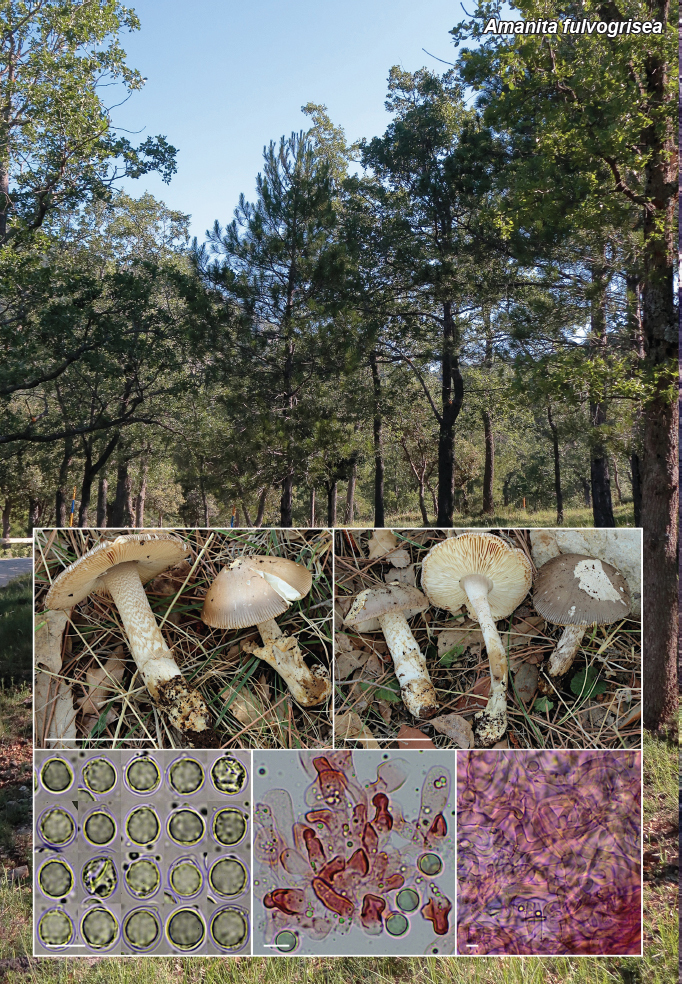
Amanita fulvogrisea Illescas, sp. nov.

T Illescas 1

Etymology. From the Latin adjectives fulvus (brownish, tawny) and griseus (grey), in reference to its basidiomatal colours.

Classification — Amanitaceae, Agaricales, Agaricomycotina.

Basidiomata with isolated to gregarious growth, medium-sized, with stipe base deeply inserted in the substrate. Pileus 60–80 mm diam, initially conical-convex, later convex, finally flatconvex, without umbon, smooth, not viscid in dry weather, grey (5Y 6/1), bistre (10YR 5/2), or tan (10YR 7/4) (Munsell 2009), usually with the umbon and the marginal zone slightly darker, presenting large patches of general veil; margin strongly striate, occupying 1/7 to 1/4 of the radius of the pileus, and strongly cleft at several points. Lamellae white to cream (7.5Y 9/2), moderately crowed, free, arcuate, sometimes bifurcate, with edges crenulate, concolourous or slightly darker; lamellulae numerous to very numerous, truncate at right angles, or reduced to a denticle at margin. Stipe cylindrical, slightly broadened towards the base, 80–100 × 9–15 mm, striated at the junction with the lamellae, white or whitish, sub-smooth or more frequently with floccose, and brownish to greyish remains of the partial veil along almost its entire length, forming meandering bands. Volva adherent (type III; Fraiture 1993), membranous but fragile, externally spotted with brown well as the pileus veil. Context whitish, with banal fungal odour. Spore deposit white. Basidiospores (8–)9–12(–13.5) × (8–)8.5–10.5(–11.5) μm (av. = 10.4 × 9.6; Q = 1.00–1.16; Qm = 1.09), globose, subglobose or broadly ellipsoid, with rounded, slightly prominent apicula, thin-walled, hyaline, inamyloid; in fresh material with abundant vacuolar content, in dry material agglutinated forming a large guttule. Basidia claviform, tetrasporic, rarely bisporic or monosporic, 47–68 × 12–15 μm, thin-walled, with frequently curved sterigmata up to 8 μm long, and abundant yellow-green vacuolar content. Lamellar edge partially sterile, especially in young specimens. Marginal cells spherical, sphaeropedunculate or ellipsoid, with somewhat thickened wall, 25–42 × 21–31 μm. Lamellar trama bilateral, irregularly divergent, 150–250 μm wide. Subhymenium predominantly ramose, composed of cylindrical to truncated cone-shaped cells, some with lateral projections, and frequently cellular at the base of the basidia, 9–16 × 6–10 μm, Qm = 1.6. Mediostratum composed of almost parallel, septate hyphae, 3–7.5 μm wide, sometimes with thickened walls, showing some physaloid elements. Hymenopodium formed by chains of prismatic or subspherical cells, with terminal elements frequently very thick-walled, up to 4 μm, and irregularly sharp ends, 24–45 × 10–22 μm, and presence of some sphaeropedunculate acrophysalids, diverging diagonally to the lamina axis, 25–50 × 16–28 μm. Pileipellis 420–525 μm wide. Suprapellis with a first layer in cutis 110–125 μm wide, with gelatinised hyphae 1.3–3.4 μm wide, and a lower layer with greenish yellow pigmented hyphae, parallel in the first layers, then progressively more interwoven, 2.6–5.3 μm wide. Subpellis with some physaloid and intricate elements, less pigmented hyphae, 3.7–9.7 μm wide. Context under cuticle with hyphae similar to subcutis but wider, with intraparietally and extraparietally pigmented physaloid elements, and some spherocytes, 25–36 × 15–21 μm. Volva 375–550 μm wide, with an outer layer consisting of septate hyphae, mostly parallel to the stipe surface on the outer side, intertwined and wider on the inner side, 4.0–7.3 μm wide, with sparse spherocytes, 28–52 × 17–41 μm. Stipitipellis formed by parallel hyphae, 4.2–7.5 μm wide, showing some emergent elements, replaced towards the interior of the context by thick-walled, yellow-pigmented, physaloid elements. Oleiferous hyphae ramificate, 12–17.5 μm wide, mostly present on the pileipellis and volva. Clamps absent in all examined structures.

Habit, Habitat & Distribution — Collected so far only in two nearby locations in the Baetic System, in mid-mountain areas, in early October (although it probably also appears in late spring), growing in small groups on limestone soils in forests of Quercus rotundifolia, Quercus faginea and Pinus nigra subsp. salzsmanii, probably in association with Quercus spp.

Typus. Spain, Hornos, N38°13' W02°10', 1 310 m a.s.l., on limestone soil, in mixed forest of Quercus faginea, Quercus rotundifolia and Pinus nigra subsp. salzmanii, 5 Oct. 2019, T. Illescas & C. Morente (holotype in Herbarium of the Junta de Andalucía: JA-Cussta 9346; ITS and LSU sequences GenBank OR420077 and OR416232; MycoBank MB 849798).

Additional material examined. Spain, Hornos, N38°14' W02°39', 1 325 m a.s.l., on limestone soil, in mixed forest of Quercus rotundifolia, Pinus nigra subsp. Salzmanii and Quercus faginea, 5 Oct. 2019, T. Illescas & C. Morente, JA-Cussta 9345, ITS and LSU sequences GenBank OR420078 and OR416233.

Notes — Amanita fulvogrisea is characterised by a robust habit such as Amanita fulvoides basidiomata, but with possible greyish colouration, subglobose spores, an almost exclusively filamentous veil, and especially their characteristic hymenopodium cells. It forms a subclade with good support (0.98/99), forming together with the A. exilis sister subclade, an independent clade within Amanita stirps Lvidopallescens, also well supported (1/100). Amanita fulvogrisea has an affinity of 98.1 % with the phylogenetically closest species, A. exilis, comparing the ITS+LSU sequences obtained from their respective holotypes. However, this species produces “tall and slender basidiomata usually with a stipe twice as long as the width of the pileus” and ellipsoid spores (Crous et al. 2021c). Another difference with A. fulvogrisea would be its epicutis in trichoderm. Amanita lividopallescens is a robust species like A. fulvogrisea that could grow in the same habitats, but in its more typical forms, differs in its cream to ochraceous pileus, spores usually with Qm 1.2–1.3, fertile lamellar edge and volva usually broad and membranous (Tulloss 1994, Vizzini et al. 2016). Our phylogenetic analysis includes an Amanita not yet described in the A. lividopallescens clade, which we have given the working name Amanita tigrinipes nom. prov., and which appeared as Amanita lividopallescens subclade ‘A’ in Loizides et al. (2018); we have collected this species growing in Mediterranean Quercus forests, with or without Cistus spp., and it has darker brown and grey colourations than A. lividopallescens, as well as a very marked zigzag banding decoration on the stipe and concolourous to that of the pileus, which makes it morphologically very similar to A. fulvogrisea.

(Notes continued on Supplementary page)

Colour illustrations. Holotype collection area at Hornos. Holotype collection JA-Cussta 9346; collection JA-Cussta 9345; basidiospores; hymenopodium cells; hyphae of the inner layer of the volva. Scale bars = 50 mm (specimens in situ), 10 μm (microscopy).

graphic file with name per-2024-51-8-F1.jpg

Amanita fulvogrisea

Supplementary material

FP1550

Phylogenetic tree.

per-2024-51-8-SF1.jpg (205.9KB, jpg)
Persoonia. 2023 Dec 30;51:286–287.

Fungal Planet 1551 – 30 December 2023

Amanita herculis Plaza & Illescas, sp. nov.

M Plaza 1, T Illescas 2

Etymology. Herculis, genitive of Hercules, in reference to the robustness of most of its specimens and to the mythological character who opened the Strait of Gibraltar, ‘columns of Hercules’, where some of these specimens were found.

Classification — Amanitaceae, Agaricales, Agaricomycotina.

Basidiomata medium to large sized. Pileus 47–115 mm diam, ash grey (10P 8/1), silver grey (10RP 7/1), greyish brown (5RP 8/1) or bistre (2.5Y 5/2), often with a brownish border (5R 5/1) at margin (Munsell 2009), convex to planoconvex, sometimes depressed at centre with maturity; usually without umbon. Striated margin between 1/5 and 1/3 of the radius of the pileus, often with broad patches of general veil remnants, white with ferruginous macules. Lamellae dense, not very thick, some bifurcate, with numerous lamellulae, white to light cream, with crenulate edge greyish brown pigmented at maturity. Stipe 73–170 × 10–28 mm, cylindrical, widening towards the base, although in adult specimens it may narrow slightly at the centre, without annulus; longitudinally striated surface even along its entire length, with slightly differentiated remnants of the partial veil, which may take on shades of the pileus at maturity, and form a slight scaly band at the point of junction with the volva. Volva thick and membranous, externally white with numerous ferruginous spots, and internally whitish or with slight tints of the colour of the pileus, up to 90 × 50 × 6 mm, usually type IV (Fraiture 1993), irregularly shaped with few lobes. Context white, cavernous in the stipe, with agreeable fungal odour and taste. Spore deposit white. Basidiospores (8.9–)10.2–12.8(–14.8) × (8.6–)9.5–12.2(–14.4) μm (av. = 11.5 × 10.8; Q = 1.01–1.14; Qm = 1.07), globose to subglobose, with rounded or troncoconic, slightly prominent apicula, often sublateral; thin-walled, hyaline, inamyloid; in fresh material with abundant vacuolar content, in dry material agglutinated forming a large guttule. Basidia claviform, tetrasporic, rarely mono- or bisporic, 50–70 × 14–19 μm, thin-walled, with sterigmata 3–12(–15.5) μm length, and abundant yellow-green vacuolar content. Lamellar edge sterile. Marginal cells spherical or sphaeropedunculata, with slightly thickened wall, 22–48 × 16–35 μm. Lamellar trama bilateral, irregularly divergent. Subhymenium cellular, ramose in primary branches (Bas 1969), composed by cylindrical cells widened at the top, truncated cone-shaped or rounded cells, often with lateral projections, 8–19 × 5–10 μm. Mediostratum composed of nearly parallel hyphae, 4–12 μm wide, with frequent inflated articles. Hymenopodium with subspherical or broadly claviform terminal elements, with thickened wall, disorderly disposed, 21.5–54 × 14.5–27 μm. Suprapellis with a first layer in cutis, with gelified hyphae, 1.7–4.3 μm wide, and a lower layer with greenish yellow pigmented hyphae with a slightly thickened wall, usually parallel to the surface of the pileus, 3.3–8.0 μm wide. Subpellis formed by similar but more intertwined hyphae. Volva with an outer layer formed by hyphae parallel to the stipe axis, with slightly thickened wall, 3.7–9.0 μm wide; a visually distinguishable intermediate layer consisting of interwoven hyphae with abundant greenish yellow intracellular pigment, and an inner layer with the side next to the stipe slightly gelified, consisting of laxly interwoven hyphae of variable thickness; in the last two layers interspersed, with relative abundance, physaloid elements and globose to broadly claviform spherocytes, 34–65 × 21–49 μm. Stipitipellis formed by parallel hyphae, the outer layer thinner, 1.2–6.4 μm wide, replaced towards the interior of the context by thick-walled, yellow-pigmented, physaloid elements. Oleiferous hyphae mainly present on the pileipellis and volva. Clamps absent in all examined structures.

Habit, Habitat & Distribution — Isolated to gregarious fruitlets located in several provinces of Andalusia at low altitudes, in acid or neutral soils, always under the influence of Quercus spp. Collected mostly in late spring, with high temperatures, although also in early autumn

Typus. Spain, La Puebla de los Infantes, N37°49' W05°24', 272 m a.s.l., on sandy soil, in open Quercus rotundifolia forest with Cistus spp., 15 May 2022, T. Illescas & C. Morente (holotype in the Fungarium of the Junta de Andalucía: JA-Cussta 9355; ITS and LSU sequences GenBank OR427365 and OR428566; MycoBank MB 849920).

Additional materials examined. Spain, Castellar de la Frontera, La Almoraima, in Quercussuber forest, 21 May 2022, M. Plaza & C.M. Jiménez, JA-Cussta 9357, ITS and LSU sequences GenBank OR427366 and OR428567; ibid., 25 May 2022, JA-Cussta 9358, ITS sequence GenBank OR501895; Córdoba, Conejeras, in Quercus suber, Quercus rotundifolia and Pinus pinea mixed forest, 9 Oct. 2021, T. Illescas, JA-Cussta 9353, ITS and LSU sequences GenBank OR427363 and OR428564; ibid., 19 June 2023, JA-Cussta 9356; La Puebla de los Infantes, La Goyorita, under Quercus rotundifolia with Cistus spp., 24 Apr. 2022, C. Morente & T. Illescas, JA-Cussta 9354, ITS and LSU sequences GenBank ITS OR427364 and OR428565.

Notes — Species of Amanita sect. Vaginatae with a large to medium-sized sacciform and membranous volva (type IV) are phylogenetically all found, for the most part, in clade 1 of the Section (Hanss & Moreau 2020). Among the European species of this clade, A. herculis is characterised by its robust habit, relatively short stipe, silver-grey or bistre coloured and occurrence in thermophilous Quercus spp. forests. Amanita calida, the phylogenetically closest species (97.4 % identity in the combined ITS+LSU matrix of their holotypes), which may share the same spots and times of occurrence, has much lighter colourations with a mixture of light brownish and greyish tones, a distinct umbon and a much more stylised habit (Illescas & Plaza 2022). Amanita praelongipes differs from the previous two species as it has no marginal cells on the lamellar edge (Kärcher 1988, Kärcher & Contu 1999); in addition, the partial ITS sequence of its typus is only 90 % identical to the same region of the holotype of A. herculis. These three species are characterised by having the smallest spores within the clade (Lm < 12 μm), but whereas A. calida and A. praelongipes have globose spores, A. herculis has subglobose spores. In a sister clade to A. herculis and A. calida are two species typical of Eurosiberian coniferous or mixed forests, of large size, also with broad, membranous volva: Amanita magnivolvata, which has a grey pileus with an olive tinge, darker at the margin, and broadly ellipsoid spores, with Qm = 1.2 (Aalto 1974), and Amanita pachyvolvata, with ochraceous to yellowish ochraceous tones in the pileus, initially olivaceous (Bon 1978, Tulloss 1994).

(Notes continued on Supplementary page)

Colour illustrations. Holotype collection area at La Puebla de los Infantes. Collections JA-Cussta 9354, JA-Cussta 9355 (holotype), JA-Cussta 9357, JA-Cussta 9356; basidiospores; subhymenium; hyphae of the outer layer of the volva. Scale bars = 20 mm (specimens in situ), 10 μm (microscopy)

graphic file with name per-2024-51-8-F2.jpg

Amanita herculis

Supplementary material

FP1551

Phylogenetic tree.

per-2024-51-8-SF2.jpg (140.1KB, jpg)
Persoonia. 2023 Dec 30;51:288–289.

Fungal Planet 1552 – 30 December 2023

Anthracoidea umbrosae T. Denchev, Denchev, Begerow & Kemler, sp. nov.

TT Denchev 1, CM Denchev 1, D Begerow 2, M Kemler 2

Etymology. The epithet is derived from the host plant, Carex umbrosa.

Classification — Anthracoideaceae, Ustilaginales, Ustilaginomycetes.

Infection local. Sori in some female flowers, around aborted nuts as subglobose to ovoid hard bodies, 1.5–2.0 × 1.2–1.8 mm, initially covered by a thin, greyish peridium that later flakes away exposing a black spore mass, powdery on the surface. Spores small-sized, flattened, in plane view irregularly rounded, suborbicular, broadly elliptical, elliptical or ovate in outline, (14.5–) 15.5–20.5(–22) × (13–)14–17.5(–18.5) (17.9 ± 1.1 × 15.9 ± 1.0) μm (n/6 = 600), in side view 10–13 μm thick, medium to dark reddish brown; wall unevenly thickened, 1.0–2.0(–2.3) μm thick, sometimes with 1–2, usually inconspicuous internal swellings, light refractive areas rarely present, protuberances absent; minutely verruculose to almost smooth, spore profile not affected. In scanning electron microscopy (SEM), warts up to 0.2 μm high, sometimes partly confluent, forming short rows or small groups. Spore germination unknown.

Host & Distribution — On Cyperaceae: Carex, Rhomboidales-Mitrata Clade of C. subg. Carex: C. umbrosa subsp. umbrosa, C. umbrosa subsp. huetiana, and hybrids of C. umbrosa subsp. huetiana; Europe (Andorra, Austria, Bulgaria, Czechia, Spain) and Asia (Armenia, Georgia).

Typus. Bulgaria, Mt Vitosha, below Golyam Rezen Peak, near the trail Aleko Chalet – Kupena Chalet, N42°33'25.4" E23°17'50.5", 1 983 m a.s.l., on Carex umbrosa subsp. huetiana (Cyperaceae), 6 July 2016, C.M. Denchev & T.T. Denchev, 1643 (holotype SOMF 30500; LSU sequence GenBank OQ801120; MycoBank MB 848431).

Additional materials examined. Anthracoidea caryophylleae: Austria, Steiermark, Graz, Bezirk Mariatrost, bei Bildunghaus Mariatrost SSE unterhalb der Basilika, N47°06'19" E15°29'30", 450 m a.s.l., on C. caryophyllea, 13 May 2013, C. Scheuer, s.n. (GZU 000306019, LSU sequence GenBank OQ801115). – Bulgaria, Sredna Gora Mts, Mt Lozenska Planina, above St. Spas Monastery, N42°35'08.8" E23°31'11.1", 1043 m a.s.l., on C. caryophyllea, 1 June 2016, T.T. Denchev & C.M. Denchev, 1611 (SOMF 30791, LSU sequence GenBank OQ801116); Mt Konyavska Planina, above Tsar-venyano village, Kyustendil Province, N42°21'12.2" E22°47'55.4", 863 m a.s.l., on C. caryophyllea, 5 June 2021, T.T. Denchev & C.M. Denchev, 2122 (SOMF 30792, LSU sequence GenBank OQ801113); the Rhodopes, near Trigrad, Smolyan Province, N41°35'14.8" E24°24'04.8", 1300 m a.s.l., on C. caryophyllea, 23 June 2021, T.T. Denchev & C.M. Denchev, 2129 (SOMF 30793, LSU sequence GenBank OQ801114). — Anthracoidea umbrosae: Andorra, Circo de Pessons, en bosques subalpinos, 2 000–2 600 m a.s.l., on C. umbrosa subsp. huetiana, 21 July 1986, C. Gómez, M. Luzeño & P. Vargas, 10138 GL (paratype MA-Fungi 37689). – Armenia, Shirak Province, Ashotsk District, near the border to Georgia, NE of Bavra, SE from former village Saragyugh, unnamed mountain, S of Mt Akchala, N of top, 3 065 m a.s.l., on C. umbrosa subsp. huetiana, N41°08'24" E43°57'19", 1 Sept. 2005, M. Oganesyan, H. Ter-Voskanyan & E. Vitek, 05-2290 (paratype W 2006-0011768). – Bulgaria, Mt Vitosha, above Goli Vrah Lift Station, N42°35'19.9" 23°17'16.1", 1 845 m a.s.l., on C. umbrosa subsp. huetiana, 31 July 2021, T.T. Denchev & C.M. Denchev, 2146 (paratype SOMF 30794, LSU sequence GenBank OQ801117); Mt Vitosha, SE of Cherni Vrah Peak, N42°33'36.7" E23°17'03.0", 2 240 m a.s.l., on C. umbrosa subsp. huetiana, 1 Aug. 2021, T.T. Denchev & C.M. Denchev, 2150 (paratype SOMF 30796, LSU sequence GenBank OQ801119); Mt Vitosha, below Golyam Rezen Peak, near the trail Aleko Chalet – Kupena Chalet, N42°33'15.5" 23°17'57.1", 2 230 m a.s.l., on C. umbrosa subsp. huetiana, 6 July 2016, T.T. Denchev & C.M. Denchev, 1646 (paratype SOMF 30501); Mt Vitosha, below Golyam Rezen Peak, near the trail Kupena Chalet – Cherni Vrah Peak, N42°33'14.4" E23°17'46.6", 2 097 m a.s.l., on C. umbrosa subsp. huetiana, 6 July 2016, T.T. Denchev & C.M. Denchev, 1644 (paratype SOMF 30797); Mt Vitosha, above Kumata Chalet, 1 800 m a.s.l., on C. umbrosa subsp. huetiana, 7 July 1985, C.M. Denchev, s.n. (paratype SOMF 18498); Mt Vitosha, above Kumata Chalet, N42°35'13.3" E23°14'49.6", 1 792 m a.s.l., on hybrid of C. umbrosa subsp. huetiana, 12 July 2019, T.T. Denchev & C.M. Denchev, 1942 (paratype SOMF 30795, LSU sequence GenBank OQ801118); Rila Mts, below Ribno Yakoruda Lake, N42°06'04.5" E23°35'48.7", 2 149 m a.s.l., on C. umbrosa subsp. huetiana, 3 Aug. 2021, T.T. Denchev, 2154 (paratype SOMF 30798).

Notes — Anthracoidea species are restricted to host plants belonging to the same or closely related sections of Carex (Kukkonen 1963, Vánky 1979). Carex umbrosa has been traditionally included in Carex sect. Mitratae (Chater 1980, Egorova 1999). For this reason, the smut fungus on Carex umbrosa was referred to A. caryophylleae (Vánky 1994, 2011). Anthracoidea caryophylleae on C. umbrosa subsp. umbrosa has been previously reported from Austria (Zwetko & Blanz 2004) and Czech Republic (Vánky 1985), and on C. umbrosa subsp. huetiana – from Andorra (Almaraz 2002), Bulgaria (Denchev 2001), Georgia (Karatygin & Simonyan 1985) and Spain (Almaraz 2002). Currently, the hosts of A. caryophylleae are reduced to carices in C. sect. Mitratae, in its traditional circumscription (Denchev et al. 2013). A recently proposed classification of Carex places C. umbrosa within the Rhomboidales-Mitrata Clade of Carex subg. Carex (Roalson et al. 2021). The aim of the present study was to clarify the taxonomic status of the Anthracoidea species on C. umbrosa. A combined approach, using host specialisation, comparative morphology, and molecular data, revealed that the smut fungus on C. umbrosa is a distinct species from A. caryophylleae. Anthracoidea umbrosae can be distinguished from A. caryophylleae by having a thinner spore wall (up to 3 μm thick for A. caryophylleae). The phylogeny inferred from the nLSU sequences demonstrated that the new species A. umbrosae is not closely related to A. caryophylleae but is sister group to a clade containing A. hallerianae, A. pamiroalaica, A. rupestris, A. baldensis, A. caricis-albae, A. cf. rupestris, A. globularis, A. irregularis and A. caricis. Based on a megablast search of NCBIs GenBank nucleotide database, the closest hits using the LSU sequence are A. hallerianae (voucher SOMF 30201, GenBank MT628657; Identities = 95.93 %, 10 gaps (0.9 %)), A. hallerianae (strain SOMF 30199, GenBank MT628660; Identities = 95.93 %, 11 gaps (0.9 %)) and A. hallerianae (strain SOMF 30000, GenBank MT628661; Identities = 95.98 %, 11 gaps (1 %)).

Colour illustrations. Mount Vitosha, near Sofia City – habitat. Habit; spores in LM; spores in SEM. Scale bars = 0.2 cm (sori), 10 μm (spores in LM), 5 μm (spores in SEM).

graphic file with name per-2024-51-8-F3.jpg

Anthracoidea umbrosae

Supplementary material

FP1552-1

Phylogenetic tree.

per-2024-51-8-SF3.jpg (168.5KB, jpg)
FP1552-2

Table.

per-2024-51-8-ST1.jpg (150.8KB, jpg)
Persoonia. 2023 Dec 30;51:290–291.

Fungal Planet 1553 – 30 December 2023

Arrhenia alcalinophila Karich, Krisai & Klofac, sp. nov.

A Karich 1, I Krisai-Greilhuber 2, W Klofac 3

Etymology. Named after its base-rich habitat.

Classification — Hygrophoraceae, Agaricales, Agaricomycetes.

Basidiomata omphalinoid. Pileus 4–15 mm wide, when fresh greyish cream and translucently striate with brownish striation, centrally small umbilicate and brownish, hygrophanous, becoming light grey when drying, glabrous, margin straight to somewhat wavy and slightly involute. Lamellae slightly to strongly decurrent, developed normally, sometimes slightly thickish, not clearly anastomosing, greyish, lighter than fresh pilei and darker than dried ones. Stipe central, sometimes slightly eccentric, 10–25 mm long, 1–2 mm wide, concolorous to pileus, smooth, slightly pruinose at apex. Smell inconspicuous, occasionally pelargonium-like when drying; taste not tested. Spore print white. Basidia 2-spored 26–33 × 7–8 μm. Sterigmata 6–8 μm. Basidiospores variable in size and shape (n = 67, 2 collections, 3 basidiomata): 9.0–12.0(–14.0) × (4.5–)5.0–6.5(–7.5) μm, Q = (1.4–)1.5–2.4(–2.9), QAV = 1.8–2.1, subglobose to broadly ellipsoid or (sub)lacrymoid to pyriform and then centrally constricted. Hymenophoral trama subirregular, consisting of 2–8 μm wide hyphae, indistinctly incrusted. Pileipellis a cutis of (sub)-parallel 3–13 μm wide hyphae. Pileipellis and pileitrama hyphae distinctly incrusted. Stipitipellis hyphae 2–9 μm wide, incrusted. Stipititrama hyphae not incrusted. Cystidia absent. Clamp connections abundant in all tissues.

Sociability, Habitat & Distribution — Basidiomata growing solitary or in small clusters on the ground in unimproved grassland on loamy to calcareous soil. Known from Germany, Austria, France and the USA (from one environmental sample).

Typus. Germany, Mittelherwigsdorf, ‘Katzenlehne’, N50°55,51' E14°44,71', 280 m a.s.l., orchard meadow in unimproved grassland on loamy soil, 2 Oct. 2022, A. Karich (holotype GLM-F135687; ITS and LSU sequences GenBank OR258065 and OR258067; MycoBank MB 849350).

Additional materials examined. Austria, Lower Austria, Pyhra: Hinterholz-Schauching, N48°8.65' E15°39.65', mapping grid square 7859/4, 400 m a.s.l., in meadow, 10 Nov. 2012, W. Klofac, WU-Myc 32560, ITS sequence GenBank MW113705. – Germany, Zittau, ‘Kaiserfelder’, N50°52.44' E14°47.58', 283 m a.s.l., on bare soil in a sheep pasture, 10 Oct. 2021, A. Karich, GLM-F135688, ITS and LSU sequences GenBank OR258064 and OR258066.

Notes — A recent study showed high morphological and molecular genetic diversity in the Arrhenia acerosa complex (Voitk et al. 2020). This species complex includes pleurotoid species, e.g., A. acerosa and A. glauca, as well as species with omphalinoid habit. Arrhenia alcalinophila was treated as ‘G-3’ in Voitk et al. (2020). Due to the limited number of specimens and the limited information about the specimens available the authors decided not yet to describe ‘G-3’ as new species. In a phylogenetic tree based on ITS-sequences, A. alcalinophila belongs to a separate clade within the acerosa-complex and is quite distant from another species with omphalinoid habit, i.e., ‘G-2’. In contrast to A. alcalinophila, however, all examined specimens of ‘G-2’ have four-spored basidia, while the spore size is roughly in the same range (Voitk et al. 2020). Phylogenetically, A. acerosa s.str. is the closest species, but has pleurotoid basidiomata, at least, when young.

From a look at the morphological descriptions of A. griseopallida in the literature (Quelét 1886, Bas et al. 1995, Ludwig 2001) it is obvious that it could be confused with A. alcalinophila. However, according to Kühner & Lamoure (1972), A. griseopallida can develop pleurotoid basidiomata and has four-spored basidia. Furthermore, the colour of fresh basidiomata of A. griseopallida is dark greyish brown and therefore seems to represent another group within the acerosa-complex (Clemençon 1982, Ludwig 2001). Persoon (1828), who examined Desmazière’s original material of Agaricus griseopallidus, mentions an upright pileus margin, thicker lamellae and a slightly reddish colouration when drying. All these features were not observed in specimens of A. alcalinophila. Lutzoni (1997) published an ITS/LSU sequence of Phaeotellus griseopallidus (= A. griseopallida) generated of a specimen which had been approved by Lamoure (GenBank U66436). It can be concluded that this specimen represents A. griseopallida s.str.

Colour illustrations. Orchard meadow at the holotype location. Basidiomata of holotype and GLM-F135688 (photo credit A. Karich); line drawings of spores from holotype (top) and GLM-F135688 (bottom) and two-spored basidia from holotype (drawing A. Karich). Scale bars = 1 cm (basidiomata), 10 μm (line drawings).

graphic file with name per-2024-51-8-F4.jpg

Arrhenia alcalinophila

Supplementary material

FP1553

Phylogenetic tree.

per-2024-51-8-SF4.jpg (123.7KB, jpg)
Persoonia. 2023 Dec 30;51:292–293.

Fungal Planet 1554 – 30 December 2023

Aschersonia merianiae Y.P. Tan, Marney, Abell & R.G. Shivas, sp. nov.

YP Tan 1, TS Marney 1, SE Abell 2, RG Shivas 3

Etymology. Named after Maria Sibylla Merian (1647–1717), who was a German naturalist and artist. Maria Merian made several trips to South America from the sale of her illustrations of plants and insects, in an age when science was largely the pursuit of wealthy men. Maria Merian documented the nature of metamorphosis with illustrated life cycles for nearly 200 species of insects and amphibians.

Classification — Clavicipitaceae, Hypocreales, Sordariomycetes.

Hosts are scale insects (Hemiptera: Coccoidea) attached to upper and lower leaf surfaces in tropical and sub-tropical rainforests. Mycelium covers host, orange, with a membranous yellow to orange hypothallus. Stromata 1–3 mm diam, luteus, globose to nodular, with several ostioles visible as brown dots, outer tissue composed of thin-walled collapsed cells, internal tissue composed of thick-walled textura epidermoidea. Perithecia immersed in stroma, obpyriform, 350–450 × 125–160 μm. Asci cylindrical, 130–160 × 5–10 μm, capitate, 8-spored. Ascospores pale yellow, smooth, filiform, multiseptate; part-ascospores disarticulate at septa, doliiform to subcylindrical, 6–10 × 2–3 μm. Conidiomata scattered in stroma, cavities or convolutions, exude luteus conidial masses. Conidiophores reduced to conidiogenous cells. Conidiogenous cells phialides, cylindrical, 15–25 × 2–2.5 μm, narrowed at the apex, lateral on hyphae or densely crowed on the hymenium. Paraphyses not seen. Conidia fusoid, 7.5–10 × 2.5–3 μm, straight, smooth-walled, hyaline to pale yellow.

Culture characteristics (25 °C, 4 wk, in darkness) — On potato dextrose agar (PDA) colonies 7–10 mm diam and 2 mm high, compact, partly immersed, low convex, luteus coloured due to abundant conidial ooze, with a distinct sienna to rustcoloured slightly undulating margin 1–2 mm wide; reverse sienna becoming paler towards the margin. In densely spread-plate cultures colonies less than 1 mm diam, luteus to orange, coral in reverse; after 8 wk colonies 15–18 mm diam.

Typus. Australia, Queensland, Lacey Creek, on scale insect (Coccoidea), Mar. 2017, S.E. Abell, Y.P. Tan, M.D.E. Shivas & R.G Shivas (holotype preserved as metabolically inactive culture BRIP 65504a; ex-type culture BRIP 65504a; LSU and tef1 sequences GenBank OR131273 and OR138131; MycoBank MB 850245).

Additional materials examined. Australia, Queensland, Babinda, on scale insect (Coccoidea), 5 Apr. 2015, T.S. Marney, S.E. Abell, M.D.E. Shivas & R.G Shivas, culture BRIP 62326a, LSU, rpb2 and tef1 sequences GenBank OR131274, OR138132 and OR138133; Lacey Creek, on scale insect (Coccoidea), 7 June 2015, T.S. Marney, S.E. Abell, M.D.E. Shivas & R.G Shivas, culture BRIP 62726a, rpb2 and tef1 sequences GenBank OR138134 and OR138135.

Notes — Aschersonia merianiae was found on scale insects on leaves of unidentified trees in a lowland rainforest in northern Queensland. The stromata from specimens collected in the field contained perithecia. Pure cultures were generated from mycelium taken internally from the stromata. These cultures were very slow-growing and produced abundant conidial ooze after 4 wk.

Based on a megablast of NCBIs GenBank nucleotide database, the closest relevant hits using the LSU sequence are Hypocrella reineckeana (strain ZZHBL201707, GenBank MK121374, Identities 832/834 (99 %)), Moelleriella sinensis (culture CGMCC 3.18912, GenBank KT963554, Identities 824/826 (99 %)) and M. epiphylla (strain I93-813, GenBank EU392583, Identities 795/838 (95 %), 11 gaps). The closes hits using the rpb2 sequence are H. reineckeana (strain BCC 2355, GenBank DQ452474, Identities 832/866 (96 %)), A. marginata (strain BCC 1713, GenBank DQ452473, Identities 801/835 (96 %)) and A. coffeae (strain BCC 1765, Identities 792/828 (96 %)). The closest hits using the tef1 sequence are H. reineckeana (strain ZZHBL201707, GenBank MK121375, Identities 879/905 (97 %)), Aschersonia marginata (strain BCC 1713, GenBank DQ384968, Identities 862/902 (96 %)) and M. sinensis (culture CGMCC 3.18912, GenBank KT987415, Identities 794/815 (97 %)).

Phylogenetic tree based on a maximum likelihood analysis of a concatenated alignment (rpb2, tef1 and LSU) of selected species of Aschersonia. The maximum likelihood analysis was done on the IQ-TREE web server (Trifinopoulos et al. 2016) based on the GTR substitution model with gamma-distribution rate variation. Bootstrap support values greater than 70 % are given at the nodes. Chlorocillium griseum (strain RCEF4626) was used as the outgroup. GenBank accession numbers are indicated (superscript LSU/rpb2/tef1). Novel taxon is shown in bold. Ex-type strains indicated with asterisks (*). The alignment and trees are publicly available in Zenodo (doi: 10.5281/zenodo.8046093).

graphic file with name per-2024-51-8-F5.jpg

Colour illustrations. Dirt track through tropical rainforest, northern Queensland. Stromata; spread-plate culture after 4 wk on PDA; colony after 4 wk on PDA; conidiophores and conidia; conidia; part-ascospores. Scale bars (top to bottom) = 1 mm, 1 cm, 1 mm, all others 10 μm.

graphic file with name per-2024-51-8-F6.jpg

Aschersonia merianiae

Persoonia. 2023 Dec 30;51:294–295.

Fungal Planet 1555 – 30 December 2023

Calosporella punctatispora Voglmayr & Mombert, sp. nov.

H Voglmayr 1, A Mombert 2

Etymology. The name refers to the punctate ascospore wall due to its verruculose ornamentation.

Classification — Sydowiellaceae, Diaporthales, Sordariomycetes.

Pseudostromata c. 0.5–1.5 mm diam, corticolous, immersed, inconspicuous, elevating and rupturing the bark, pustulate in face view, circular to elongate, without a black line, containing up to 15 perithecia. Ectostromatic disc 0.2–1 mm diam, erumpent, reduced, fusoid to circular, brownish, containing 1–15 distinctly protruding black ostioles. Entostroma inconspicuous, reduced, crumbly, brownish. Ostioles necks 140–200 μm long, 90–130 μm diam, erumpent, cylindrical, black, rounded at tips. Perithecia black, 350–500 μm diam. Asci broadly fusoid, 55–75 × 14–25 μm, containing four obliquely biseriate to irregularly arranged ascospores, with an indistinct, not congophilic, inamyloid apical apparatus. Ascospores 21–29.5 × 7–9.5 μm, hyaline, fusoid to oblong, straight to slightly curved, uniseptate, distinctly constricted at septum, inequilateral with one cell commonly wider, 2–4 guttulate, with broadly subacute to rounded ends and hyaline tapering appendages at both ends projecting for 6–11 μm, with distinctly verruculose ascospore wall ornamentation in water and Congo red, appearing smooth in 3 % KOH. Asexual morph unknown.

Culture characteristics (22 °C after 4 wk) — Colonies on 2 % cornmeal dextrose agar (CMD) reaching 50 mm diam, first dull yellowish, in centre turning umber with age, floccose in centre, smooth at margins, margins irregular, indistinct; reverse umber in centre, dull yellowish towards margin.

Ecology & Distribution — On thin (1.5–4.5 mm diam) dead corticated twigs of Acer opalus still attached to the trees; only known from eastern and southern France.

Typus. France, Jura (39), Port-Lesney, Bélvedère Edgar Faure, on dead corticated twigs of Acer opalus still attached to the tree, N47.003502° E5.808043°, 348 m a.s.l., 11 Apr. 2020, A. Mombert, AM2004111 (holotype WU-MYC 0050080; ex-type culture D336 = CBS 149660; ITS-LSU sequence GenBank OR143132; MycoBank MB 850243).

Additional materials examined. France, Doubs (25), Quingey, on dead twigs of a young Acer opalus still attached to the tree, N47.095852° E5.85464°, 495 m a.s.l., 13 Apr. 2020, A. Mombert, AM2004134 (WU-MYC 0050081); Doubs (25), Chassagne-st-Denis, Les Dents du Lério, on dead twigs of Acer opalus still attached to the tree, N47.079621° E6.104152°, 9 Jan. 2021, A. Mombert, AM2101092 (WU-MYC 0050082); Alpes-de-Haute-Provence (04), Le Castellard-Mélan, near to the grotte St-Vincent, on dead twigs of Acer opalus still attached to the tree, N44.220441° E6.125649°, 1511 m a.s.l., 24 June 2023, A. Mombert, AM2306241 (WU-MYC 0050583).

Notes — Nucleotide BLAST searches of the ITS and LSU at the NCBI GenBank nucleotide database revealed species of Sydowiellaceae (Diaporthales) as closest matches; the ITS sequence had highest similarity to Calosporella innesii (strain AR 3831, GenBank JF681964.1, Identities = 486/524 (92.75 %), 13 gaps), strain AR 3639, GenBank JF681965.1, Identities = 485/524 (92.56 %), 13 gaps), followed by Alborbis galericulata (strain AR 3890, GenBank JF681967.1, Identities = 434/506 (85.77 %), 33 gaps) and Paragnomonia fragariae (e.g., strain CBS 146.64, EF212844.1, Identities = 375/428 (87.62 %), 20 gaps) while the LSU sequence again had highest similarity to Calosporella innesii (e.g., strain AR 3639, EU683071.1, Identities = 846/852 (99.30 %), no gaps), followed by Rossmania ukurunduensis (strain AR 3484, EU683075.1, Identities = 839/852 (98.47 %), no gaps) and then various species from Sydowiellaceae (all with Identities = 836/852 (98.12 %), no gaps), e.g., Cainiella johansonii (voucher Kruys 731, JF701920.1), Paragnomonia fragariae (e.g., strain F133.1, MK524450.1), Sillia ferruginea (e.g., strain CBS 125581), Sydowiella centaureae (e.g., strain MFLU 16-2858, KY52350.1), Sydowiella depressula (strain CBS 813.79, MK524445.1) and S. fenestrans (e.g., strain CBS 125530, MK524444).

The phylogeny inferred from a matrix of ITS and nLSU sequences of Sydowiellaceae revealed a highly supported sister group relationship of the new species Calosporella punctatispora with the generic type of Calosporella, C. innesii (100 % ML and MP BS). Morphologically, C. punctatispora and C. innesii share similar pustulate stromata with reduced crumbly entostroma, broadly fusoid asci with an indistinct apical apparatus and hyaline, guttulate ascospores with distinct tapering appendages at each end; ecologically, they share occurrence on dead corticated thin twigs of the same host genus, Acer (Voglmayr & Jaklitsch 2014). However, C. punctatispora differs markedly from C. innesii by four-spored (vs eight-spored) asci, 1-septate, strongly constricted, inequilateral, straight to curved ascospores with verruculose walls (vs 3-septate, not to slightly constricted, equilateral, straight ascospores with smooth walls) (Voglmayr & Jaklitsch 2014). While C. innesii is only known from Acer pseudoplatanus, C. punctatispora appears to be restricted to Acer opalus. The verruculose ascospore walls and ascospore appendages in C. punctatispora are only evident in water and Congo red; they disappear in 3 % KOH.

Colour illustrations. Acer opalus forest in spring, where the holotype was collected. Overview and detail of dead corticated twig showing brownish ectostromatic disks and ostioles; pseudostroma in vertical section; pseudostroma in horizontal section; four-spored ascus (in Congo red); ascospore with appendages (in water); ascospore with verruculose wall and appendages (in Congo red); ascospore with apparently smooth wall (in 3 % KOH); culture on CMD after 4 wk. Scale bars = 500 μm (pseudostromata), 5 μm (ascus and spores).

graphic file with name per-2024-51-8-F7.jpg

Calosporella punctatispora

Supplementary material

FP1555-2

Phylogenetic tree.

per-2024-51-8-SF5.jpg (144.9KB, jpg)
FP1555-1

Table.

per-2024-51-8-ST2.jpg (139.3KB, jpg)
Persoonia. 2023 Dec 30;51:296–297.

Fungal Planet 1556 – 30 December 2023

Catenomargarita F.A. Custódio & O.L. Pereira, gen. nov.

FA Custódio 1, OL Pereira 1

Etymology. Name derived from the combination of catena and margarita, meaning chain and pearls, respectively, in Latin, referring to the long conidial chains produced by the fungus.

Classification — Niessliaceae, Hypocreales, Sordariomycetes.

Mycelium septate, branched, smooth- or rough-walled, forming bundles, hyaline. Conidiophores erect, cylindrical straight or flexuous, arising from hyphae or the from ropes formed by the mycelium, unbranched, poorly branched, septate, some times reduced to conidiogenous cells, hyaline. Conidiogenous cells monophialidic, terminal or intercalary, smooth-walled, cylindrical to subulate, flexuous, hyaline. Conidia limoniform, papillated, thin- and smooth-walled, formed in long basipetal chains, aseptate, hyaline. Chlamydospores ellipsoid, globose, thick- and smooth-walled, intercalated, hyaline. Sexual morph was not observed.

Type species. Catenomargarita pseudocercosporicola F.A. Custódio & O.L. Pereira

MycoBank MB 850039.

graphic file with name per-2024-51-8-F8.jpg

Catenomargarita pseudocercosporicola

Persoonia. 2023 Dec 30;51:297.

Fungal Planet 1556 – 30 December 2023

Catenomargarita F.A. Custódio & O.L. Pereira, gen. nov.


Catenomargarita pseudocercosporicola F.A. Custódio & O.L. Pereira, sp. nov.

Etymology. Name refers to Pseudocercospora, the fungal genus from which this fungus was isolated.

Mycelium septate, branched, smooth- or rough-walled, forming bundles, hyaline, 0.85–2.8 μm diam hyphae. Conidiophores solitary or aggregated, erect, arising from hyphae, with 1–3 septa, sometimes reduced to conidiogenous cells, unbranched, poorly branched, bearing 1–3 phialides, flexuous, hyaline, (18–)28.5–52.5 × (1.3–)1.5–2.0 μm. Conidiogenous cells monophialidic, terminal or intercalary, smooth-walled, cylindrical to subulate, flexuous, hyaline, (12–)16.5–33(–40) × 1–2 μm. Conidia limoniform, papillate, thin- and smooth-walled, aseptate, hyaline, 2.0–2.9 × 1.5–2.0(–2.5) μm, with a germ pore, arranged in dry chains. Chlamydospores ellipsoid, globose, thick- and smooth-walled, intercalated, hyaline, 3.5–6.2 × 3.2–5.3 μm.

Culture characteristics — Colonies on potato dextrose agar (PDA) umbonate, radially striate with lobate edge, aerial mycelium scarce to moderate, dense, cottony, buff colour (45) (Rayner 1970) in the centre to white on the periphery surface, buff on reverse, reaching 16 mm diam after 2 wk at 25 °C with a photoperiod of 12 h. Colonies on oatmeal agar (OA) flat with entire edge, aerial mycelium moderate, cottony, aerial sporulation, white to vinaceous buff (86) surface, reverse buff, reaching 22 mm diam after 2 wk at 25 °C with a photoperiod of 12 h. Colonies on synthetic nutrient-poor agar (SNA) flat, fimbriate, aerial mycelium scarce, buff surface and reverse, reaching 6 mm diam after 2 wk at 25 °C with a photoperiod of 12 h.

Typus. Brazil, Maranhão, Itinga do Maranhao, hyaline hyperparasite on Pseudocercospora fijiensis growing on leaves of Musa acuminata (Musaceae), 15 Dec. 2021, F.A. Custódio (holotype VIC 49479; culture ex-type COAD 3662; ITS, LSU, SSU, rpb2 and tef1-a sequences GenBank OR558367, OR558365, OR558366, OR557609 and OR557608; MycoBank MB 850040).

Notes — The LSU and SSU sequences of Catenomargarita pseudocercosporicola have similarity with sequences of Niessliaceae and Clavicipitaceae. Phylogenetic analyses indicate that the genus belongs to Niessliaceae, with Trichosphaerella ceratophora being the closest species. Catenomargarita differs from Trichosphaerella by producing monophialidic conidiogenous cells and smooth-walled conidia. The family includes acremonium-like fungi that produce monophialides and hyaline conidia, such as the genera Acremoniopsis, Collarina, Monocillium and Niesslia (Hou et al. 2023). Catenomargarita differs from the other Niessliaceae genera by producing conidial chains, branched conidiophores and chlamydospores. Niesslia catenata and N. indica produce conidial chains but differs from Catenomargarita pseudocercosporicola in the absence of branched conidiophores and chlamydospores (Gams et al. 2019).

Based on a megablast search of NCBIs GenBank nucleotide database, the closest hits using the ITS sequence had highest similarity to the sequence of an ‘uncultured fungus’ hyperparasitic of Hemileia vastratrix in Puerto Rico (OTU 79, GenBank KT328841; Identities = 545/546 (99 %), one gap (0 %)), ‘Colletotrichum sp.’ (strain PT0920, GenBank ON155281; Identities = 611/640 (95 %), 10 gaps (1 %)) and Trichoderma longibrachiatum (strain 0501G, GenBank MF276664; Identities = 595/644 (92 %), 19 gaps (2 %)). Closest hits using the LSU sequence are Eucasphaeria capensis (strains CBS 120027 and CBS 120028, GenBank EF110619 and EF110618; Identities = 1 028/1 059 (97 %), one gap (0 %)), Keithomyces cf. carneus (strain CBS 126563, GenBank MT078856; Identities = 980/1 009 (97 %), three gaps (0 %)) and ‘Clavicipitaceae sp.’ (strain MEXU 26354, GenBank JQ811556; Identities = 1 005/1 036 (97 %), four gaps (0 %)). Closest hits using the SSU sequence has highest similarity to an ‘uncultured fungus’ (strain nco61b01c1, GenBank KC672415; Identities = 934/939 (99 %), one gap (0 %)), ‘Hypocreales sp. MR-2023e’ (strains CGMCC 3.22396 and CGMCC 3.22401, GenBank OQ758192 and OQ758191; Identities = 925/939 (99 %), one gap (0 %)). For the sequences of rpb2 and tef1-a on a megablast search of NCBIs GenBank nucleotide database, no significant hits were obtained.

Colour illustrations. Black Sigatoka disease symptoms on Musa acuminata (Musaceae) in Itinga do Maranhao, state of Maranhao, Brazil. Colony on PDA after 2 wk at 25 °C; colony on OA after 2 wk at 25 °C; colony on SNA after 2 wk at 25 °C; conidiophores and conidiogenous cells giving rise to conidia; conidia in chains; chlamydospores. Scale bars = 10 μm.

Supplementary material

FP1556

Phylogenetic tree.

per-2024-51-8-SF6.jpg (280.1KB, jpg)
Persoonia. 2023 Dec 30;51:298–299.

Fungal Planet 1557 – 30 December 2023

Circinaria podoliana Szczepanska, Rodr. Flakus, Urbaniak & Sliwa, sp. nov.

P Rodriguez-Flakus 1, L Sliwa 1, K Szczepańska 2, J Urbaniak 2

Etymology. The epithet refers to the geographical region of Ukraine where the species was first collected.

Classification — Megasporaceae, Pertusariales, Lecanoromycetes.

Thallus lichenised, saxicolous, crustose, squamulose-areolate, contiguous rarely divided into small groups, 300–600 μm thick. Squamules thick, flexuose, irregular, (0.4–2.0 mm diam), partially erected and overlapping, less often rounded and slightly convex. Prothallus absent. Upper surface smooth, olive brown to chocolate brown, mat, pruinose on the margins. Upper cortex 30–50 μm thick, hyaline, with a thin, brown epinecral layer, 7–20 μm thick; medulla hyaline, with many tiny crystals, partially dissolving in N, not dissolving in K. Photobiont green, chlorococcoid, cells 7–16 μm diam; photobiont layer 50–70 μm thick, continuous but uneven in some parts. Apothecia immersed, rounded to irregular, 1 per areole, 0.3–1.2 mm diam; thalline margin not apparent, formed by radially cracked and white pruinose thalline areole; disc flat, circular to irregular, black, densely white pruinose; proper margin indistinct; hymenium hyaline, 150–170 μm high, I+ greenish blue; paraphyses septate and anastomosing, thin at the base, c. 2 μm wide, swollen at tips; submoniliform (2 – 3 globose apical cells), epithecium, c. 20 μm high, olive brown, N+ clearly green, K+ orange brown (Caesiocinerea-green pigment; Meyer & Printzen 2000), with a thin, uneven crystal layer, c. 10 μm thick (crystals colourless, dissolving in N and not dissolving in K); hypothecium hyaline, 40–50 μm high, I+ blue. Asci cylindrical in an immature state, 110–120 × 20–25 μm, clavate when mature, Aspicilia-type, ascospores 4–6 per ascus, hyaline, subglobose to broadly el-lipsoid (20–)24–28(–32) μm diam (n = 20). Pycnidia immersed, c. 200 μm diam, rounded, ostiolar region projected, rounded, white pruinose. Conidia filiform, straight, 6–10 × 1 μm (n = 30). Chemistry. Spot tests: cortex K–, C–, P–, medulla K–, C–, P–. TLC (solvent A and C): all of the analysed samples contained aspicilin as a secondary metabolite.

Habit, Habitat & Distribution — Circinaria podoliana grows on natural limestone outcrops in open area, rarely on artificial substrata (mortar), in sunny, warm and exposed places.

Typus. Ukraine, Podilya Upland, Khmelnytskyi Oblast (province), Podil’s’ki Tovtry National Park, Kitaygorod village, 15 km SE of Kamyanets’ Podil’s’kyi, canyon of Ternava River, N48°38'25" E26°46'59", 140 m a.s.l., limestone outcrops on south and southwest facing slope with Crataegus sp., Rosa sp. and young trees of Juglans regia, Morus sp. and Populus sp., 24 June 2003, L. Sliwa, 1878 (holotype KRAM L-49384; ITS and mcm7 sequences GenBank OR436503 and OR441081; MycoBank MB 849796).

Additional materials examined. Ukraine, Podilya Upland, Khmelnytskyi Oblast (province), Podil’s’ki Tovtry National Park, Ustya village at the junction of Smotrich River and the Dniester, N48°33'54" E26°39'24", 200 m, 26 June 2003, L. Sliwa, 1981 (KRAM L-49477; ITS, mtSSU and mcm7 sequences GenBank OR436500, OR436507 and OR441080); Privorot’e near Makiv village, 10 km N of Kamyanets’ Podil’s’kyi, N48°47'27" E26°38'41", 223 m, 25 June 2003, M. Kukwa, 1880 (KRAM L-48743; ITS and mtSSU sequences GenBank OR436501 and OR457667); Chotyry Kavalery near Verbka village (Tovtra Verbets’ka nature reserve), 13 km N of Kamyanets’ Podil’s’kyi, N48°48'24" E26°35'54", 289 m, 26 June 2003, A. Zalewska (KRAM L-49261; ITS sequence GenBank OR436502); Kitaygorod village, 15 km SE of Kamyanets’ Podil’s’kyi, canyon of Ternava River, N48°38'25" E26°46'59", 140 m, 24 June 2003, K. Czyzewska (KRAM L-48907; ITS and mtSSU sequences GenBank OR436505 and OR436509, KRAM L-48920; ITS and mtSSU sequences GenBank OR436504 and OR436508); Chernivtsi Oblast (province), right bank of the Dniester River in Khotyn city, the Khotyn Fortress, 15 Aug. 2005, J. Kiszka (KRAP; ITS, mtSSU and mcm7 sequences GenBank OR436506, OR436510 and OR441082).

Notes — Circinaria is heteromorphic genus, including crustose, subfruticose and subfoliose species (Sohrabi et al. 2013). Currently, there are 13 taxa of the genus known to occur in Ukraine (Kondratyuk et al. 2021). The new species, C. podoliana, is a member of Circinaria contorta/calcarea complex and presents typical features of the genus, such as: broadly ellipsoid spores reduced to four or six per ascus, short conidia and the presence of aspicilin. Specimens of C. podoliana were housed in fungaria under the current name Circinaria contorta (Nordin et al. 2010). Circinaria contorta is a very common and widely distributed lichen, occurring on calcareous rocks and other base-rich substrata (Fletcher et al. 2009, Wirth et al. 2013). The species has a crustose thallus consisting of separate or partly aggregated, white pruinose, rounded, slightly convex areoles with immersed crater-like apothecia (Rico 1999, Owe-Larsson et al. 2007). The main feature that distinguishes C. podoliana from C. contorta is the habit of the thallus, which is distinctly thicker and composed of overlapping, flexuose, olive brown to chocolate brown squamules that are pruinose only on the margins. Some specimens of the new species were labelled as C. hoffmanniana (Roux et al. 2016), which is also a common species occurring on calcareous rocky substrates. The most characteristic features of C. hoffmanniana are: grey to brownish grey, thin, cracked-areolate but continuous thallus producing immersed to sessile, prominent apothecia. Circinaria podoliana can be distinguished from the species mainly by a thicker and clearly squamulose-areolate thallus. A third calcareous lichen that may be confused with C. podoliana is C. calcarea. However, the latter species differs in having circular, chalk-white, smooth, cracked-areolate thallus and a grey prothallus.

In our analyses C. podoliana is nested within Megasporaceae and forms a distinct and highly supported lineage within the Circinaria as a sister to the C. contorta clade. Based on a megablast search of NCBIs GenBank nucleotide database, the closest hit using the ITS sequence had highest similarity to Circinaria sp. (voucher Leavitt 19096 (SL-2021a), GenBank MZ922119; Identities = 92.80 %, nine gaps (1 %)). The closest hit using the mtSSU sequence is Aspicilia calcarea (voucher Wedin 6500 (UPS), GenBank AY853310; Identities = 96.89 %, three gaps (0 %)). The closest hit using the mcm7 sequence is Aspicilia contorta (voucher Wetmore (MIN 808806), GenBank GU980989; Identities = 99.42 %, no gaps).

Colour illustrations. Podilya Upland in Ukraine, canyon of Ternava River near Kitaygorod village (locus classicus). A habit of thallus with ascomata (holotype, KRAM L-49384); cross-section through thallus and apothecium with mature spores. Scale bars = 2 mm (a habit), 50 μm (apothecium), 100 μm (thallus).

graphic file with name per-2024-51-8-F9.jpg

Circinaria podoliana

Supplementary material

FP1557-1

Phylogenetic tree.

per-2024-51-8-SF7.jpg (158KB, jpg)
FP1557-2

Table.

per-2024-51-8-ST3.jpg (434.3KB, jpg)
Persoonia. 2023 Dec 30;51:300–301.

Fungal Planet 1558 – 30 December 2023

Curvularia huamulaniae Y.P. Tan & R.G. Shivas, sp. nov.

YP Tan 1, RG Shivas 2

Etymology. Named after Hua Mulan, a legendary folk heroine from the period of Chinese history called the Northern and Southern Dynasties (386–581 AD).

Classification — Pleosporaceae, Pleosporales, Dothideomycetes.

Asexual morph abundant on wheat straw agar after 2 wk at 25 °C in the dark. Conidiophores single, erect, straight to flexuous, with many geniculations, brown, cylindrical, smooth, 70–150 μm long, 6–8 μm wide, septate, with prominent scars, unbranched or sparingly branched. Conidiogenous cells intercalary and terminal, brown, smooth, with darkened scars. Conidia straight to slightly curved on one side and strongly curved on the other, hemispherical to obpyriform, 28–42 × 14–22 μm, rounded at the apex and flattened to rounded at the base, 3–4-distoseptate, reddish brown, smooth.

Typus. Australia, Queensland, Brisbane, isolated from air, 12 Feb. 1964, R.G. Rees (holotype BRIP 10936a permanently preserved in a metabolically inactive state; culture ex-type BRIP 10936a; ITS, gapdh and tef1 sequences GenBank OR130931, OR135531 and OR135532; MycoBank MB 849552; isotype IMI 108517).

Notes — Curvularia huamulaniae is morphologically indistinguishable from C. papendorfii (Van der Aa 1967). Curvularia huamulaniae differs from C. papendorfii (ex-type strain CBS 308.67) by sequence comparison of the ITS region (GenBank KJ909774; Identities 647/653 (99 %), three gaps; unique nucleotide at positions 188(C), 258(T), 512(T)), gapdh (GenBank KM083617; Identities 414/430 (96 %); unique nucleotide at positions 25(T), 28(T), 37(G), 47(G), 49(T), 54(T), 65(C), 78(G), 177(T), 189(C), 273(T), 291(A), 297(T), 348(C), 375(C), 426(C)), and tef1 (GenBank KM196594; Identities 895/899 (99 %); unique nucleotide at positions 156(C), 276(T), 366(C), 393(C)).

Based on a megablast search of NCBIs GenBank nucleotide database, the closest relevant hits using the ITS sequence are C. papendorfii (strain CBS 308.67, GenBank KJ909774, Identities 647/653 (99 %), three gaps), C. sporobolicola (strain BRIP23040b, GenBank MH414908, Identities 646/652 (99 %), two gaps) and C. siddiquii(strain CBS 196.62, Identities 580/590 (98 %), six gaps). The closest relevant hits using the gapdh sequence are C. papendorfii (strain CBS 308.67, GenBank KM083617, Identities 414/430 (96 %)), C. sporobolicola (strain BRIP23040b, GenBank MH433652, Identities 423/446 (95 %)) and C. siddiquii (strain CBS 196.62, GenBank MN688850, Identities 431/461 (93 %)). The closest relevant hits using the tef1 sequence are C. papendorfii (strain CBS 308.67, GenBank KM196594, Identities 895/899 (99 %)), C. eragrostidis (isolate CD15, GenBank MK886803, Identities 966/989 (98 %)) and C. dactyloctenicola (strain M0307, GenBank OM714560, Identities 922/944 (98 %)).

Phylogenetic tree based on maximum likelihood analysis of the alignment of the combined gapdh and tef1α sequences from species of Curvularia. Analyses were performed on the Geneious Prime 2023 platform using RAxML v. 8.2.11 and MrBayes v. 3.2.6, both based on the GTR substitution model with gamma-distribution rate variation. Bootstrap support (bs) values greater than 70 % and Bayesian posterior probabilities (pp) greater than 0.8 are given at the nodes (bs/pp). Curvularia eragrostidis (strain CBS 189.48 was used as the outgroup. GenBank accession numbers are indicated (superscript gapdh/tef1). Novel taxon is shown in bold. Ex-type strains are marked by an asterisk (*). The alignment and phylogeny are publicly available in Zenodo (https://doi.org/10.5281/zenodo.8042668).

graphic file with name per-2024-51-8-F10.jpg

Colour illustrations. Moonrise on Sunrise Beach, Queensland, Australia. Colony on wheat straw agar; conidiophores; conidia. Scale bars = 100 μm (colony), 10 μm (others).

graphic file with name per-2024-51-8-F11.jpg

Curvularia huamulaniae

Persoonia. 2023 Dec 30;51:302–303.

Fungal Planet 1559 – 30 December 2023

Deniquelata cassiae Tennakoon & Suwannar., sp. nov.

DS Tennakoon 1, N Suwannarach 1

Etymology. Named after the host genus from which it was collected, Cassia.

Classification — Didymosphaeriaceae, Pleosporales, Dothideomycetes.

Ascomata separate, immersed to semi-immersed, partly erumpent, solitary or scattered, globose to sub-globose, unilocular, dark brown to black, 200 μm diam, with central papillate ostiole; wall of 3–4 layers of brown, darkly pigmented cells of textura angularis. Pseudoparaphyses cellular, hyaline, smooth, branched, septate, anastomosing, 1–2 μm diam. Asci bitunicate, 8-spored, cylindrical-clavate, fissitunicate, slightly curved, rounded apex, with low ocular chamber, 1–3 μm diam, 1–2 μm high (visible only in young asci), with a short pedicel, 60–80 × 10–12 μm. Ascospores bi- to tri-seriate, ellipsoidal to fusoid, widest in middle of second cell from apex, developing a central septum and then becoming muriformly septate, slightly constricted at the septa, straight to curved, hyaline when immature and reddish brown at maturity, guttulate, 14–16 × 6–8 μm.

Typus. Thailand, Chiang Rai, on dead stem of Cassia fistula (Fabaceae), 12 Aug. 2021, D.S. Tennakoon, CMD012A (holotype CMUB 40013, isotype CMUB 40015; ITS, LSU, SSU and tef1 sequences GenBank OR500092-OR500093, 0R500088-0R500089, 0R500090-0R500091 and OR501827-OR501828; MycoBank MB 849873).

Notes — Deniquelata was introduced by Ariyawansa et al. (2013) to accommodate D. barringtoniae as the type species. Subsequently, D. hypolithi, D. macphersoniae, D. quercina, D. vittalii and D. yunnanensis were added by various researchers (Devadatha et al. 2018, Alidadi et al. 2019, Crous et al. 2021a, Lu et al. 2022, Tan & Shivas 2022). Deniquelata species have been reported as pathogens, saprophytes, and endophytes (Ariyawansa et al. 2013, Crous et al. 2021a, Tan & Shivas 2022). The phylogeny inferred from the ITS, LSU, SSU and tef1 sequences demonstrated that the new species D. cassiae nested in the Deniquelata clade and forms an independent lineage sister to D. yunnanensis with 80 % statistical support. Deniquelata cassiae differs from D. yunnanensis in their ellipsoidal to fusoid ascospores, whereas D. yunnanensis has ellipsoidal to oblong ascospores (Lu et al. 2022). Furthermore, D. cassiae can be distinguished from D. macphersoniae based on ITS nucleotide differences (14/698 bases). Deniquelata cassiae can be distinguished from D. quercina in the distinct size differences of asci (85–155 × 13–18 μm vs 60–80 × 10–12 μm) and ascospores (9–11 × 18–21 μm vs 14–16 × 6–8 μm) (Alidadi et al. 2019). In addition, D. cassiae differs from D. vittalii and D. barringtoniae in the distinct size differences in asci (68–80 × 10–15 μm and 75–135 × 10–19 μm) and ascospores (14–16 × 5–7 μm and 18–26 × 7.5–13.5 μm) (Ariyawansa et al. 2013, Devadatha et al. 2018).

Based on a megablast search of NCBIs GenBank nucleotide database, the closest hits using the ITS sequence had highest similarity to Deniquelata yunnanensis (strain ZHKUCC:22-0198, GenBank OP297803; Identities = 785/794 (98.86 %), two gaps (0.8 %)), D. yunnanensis (strain ZHKUCC:22-0199, GenBank OP297804; Identities = 802/813 (98.64 %), two gaps (0.2 %)), D. macphersoniae (strain BRIP 72538b, GenBank NR_182616; Identities = 839/847 (99.1 %), one gap (0.1 %)), Didymosphaeriaceae sp. (strain A1S3-D34, GenBank KJ780753; Identities = 709/745 (95.1 %), 12 gaps (1.6 %)). Closest hits using the LSU sequence are D. yunnanensis (strains ZHKUCC:22-0199 and ZHKUCC:22-0198, GenBank OP297774 and OP297773; Identities = 916/919 (99.78 %), two gaps (0.2 %)), D. macphersoniae (strain BRIP 72538b, GenBank OP598061; Identities = 890/890 (99.6 %), one gap (0.1 %)), Neokalmusia arundinis (strain MFLUCC 14-0222, GenBank KX954400; Identities = 948/953 (4.2 %), seven gaps (0.1 %)), Phaeodothis winteri (strain AFTOL-ID 1590, GenBank DQ678073; Identities = 947/953 (99.4 %), six gaps (0.1 %)), Bimuria novae-zelandiae (strain CBS 107.79, GenBank OM337538; Identities = 1 702/1 707 (99.7 %), four gaps (0.1 %)).

Colour illustrations. Holotype collection area in Chiang Rai, Thailand. Appearance of ascomata on dead stem of Cassia fistula; vertical section of ascoma; peridium; asci; ascospores. Scale bars = 100 μm (section), 10 μm (peridium), 30 μm (asci), 8 μm (ascospores).

graphic file with name per-2024-51-8-F12.jpg

Deniquelata cassiae

Supplementary material

FP1559-1

Phylogenetic tree.

per-2024-51-8-SF8.jpg (218.6KB, jpg)
FP1559-2

Table.

per-2024-51-8-ST4.jpg (188.6KB, jpg)
Persoonia. 2023 Dec 30;51:304–305.

Fungal Planet 1560 – 30 December 2023

Diaporthe jazanensis Alhudaib & A.M. Ismail, sp. nov.

K Alhudaib 1, AM Ismail 2, D Magistà 3

Etymology. The name refers to the collection locality (Jazan, Saudi Arabia).

Classification — Diaporthaceae, Diaporthales, Sordariomycetes.

Conidiomata pycnidial, mostly solitary, pale to dark brown, deeply embedded in potato dextrose agar (PDA) erumpent, irregular globose or sub-globose to depressed, exuding dark brown to black conidial masses, when aged bordered with thick-walled cells of textura angularis. Conidiophores hyaline, smooth, densely aggregated. Conidiogenous cells densely phialidic, hyaline, terminal, subcylindrical, tapered towards the apex. Paraphyses not observed. Alpha conidia hyaline, smooth, fusoid or ellipsoidal, straight to curved, aseptate, tapered at both ends, 7.0–9.8 × 2.5–3.3 μm (av. = 8.4 ± 0.69 × 2.9 ± 0.19 μm). Beta conidia spindle-shaped, smooth, acute at apex, slightly truncate at base, frequently curved, 20.9–36.3 × 0.7–2.2 (av. = 30.6 ± 4.3 × 1.6 ± 0.41 μm). Gamma conidia not observed.

Culture characteristics — Colonies covered the dish after 10 d at 25 °C. Colony on PDA at the beginning white to beige becoming pale brown, flattened, distinctly imbricated like tree branches. Reverse side pale brown, turning dark brown with age.

Typus. Saudi Arabia, Jazan Region, N17°23'07.9" E43°09'01.5", on branches of Coffea arabica (Rubiaceae), 12 Oct. 2022, A.M. Ismail (holotype KSA-28R-2023; culture ex-type PPDU28R; ITS, tef1 and tub2 sequences GenBank OQ586408, OR137582 and OR137586; MycoBank MB 849121).

Additional materials examined. Saudi Arabia, Jazan Region, N17°23'07.9" E43°09'01.5", on branches of C. arabica, 12 Oct. 2022, A.M. Ismail, culture PPDU28S, ITS, tef1 and tub2 sequences GenBank OQ586409, OR137583 and OR137587; ibid., culture PPDU28U, ITS, tef1 and tub2 sequences GenBank OQ586410, OR137584 and OR137588; ibid., culture PPDU28Z, ITS, tef1 and tub2 sequences GenBank OQ586411, OR137585 and OR137589.

Notes — Diaporthe jazanensis is phylogenetically closely related but well-separated from D. caatingaensis CBS 141542 and D. brasiliensis CBS 133183. Furthermore, D. jazanensis can be distinguished by its somewhat wider alpha conidia (2.5–3.3 μm) than those of D. caatingaensis (1.5–2.5 μm) (Crous et al. 2016) and D. brasiliensis (2–3 μm) (Gomes et al. 2013). Additionally, beta conidia are present in D. jazanensis, but absent in D. caatingaensis and D. brasiliensis. Colonies of D. jazanensis on PDA covered the entire surface of Petri dishes after 10 d, which is faster than observed in D. caatingaensis (14 d) and D. brasiliensis (14 d) at 25 °C. Moreover, the colony morphology of D. jazanensis is white to beige, becoming pale brown with age, flattened, distinctly imbricated like a tree branch while, the colonies of D. caatingaensis have white, fluffy aerial mycelium and colonies of D. brasiliensis are flat, with an entire edge, dense, felty and buff, grey olivaceous.

Based on a megablast search of NCBIs GenBank nucleotide database, the closest hits using the ITS sequence had highest similarity to D. caatingaensis (strain 11, GenBank OP955669; Identities = 513/513 (100 %), no gaps), D. caatingaensis (strain 21, GenBank OP959918; Identities = 513/514 (99 %), one gap (0 %)) and D. caatingaensis (strain 16, GenBank OP956980; Identities = 513/514 (99 %)), no gaps (0 %)). Closest hits using the tef1 sequence are D. caatingaensis (strain URM7486, GenBank KY115603; Identities = 336/347 (97 %), three gaps (0 %)), D. brasiliensis (strain CBS 133183, GenBank KC343768; Identities = 321/345 (93 %), one gap (0 %)) and D. brasiliensis (strain LGMF926; GenBank KC343769; Identities = 321/346 (93 %), one gap (0 %)). Further, the closest hits using the tub2 gene sequence are D. brasiliensis (strain CBS 133183, GenBank KC344010; Identities = 353/359 (98 %), no gaps (0 %)), D. caatingaensis (strain URM7486, GenBank KY115600; Identities = 374/385 (97 %), four gaps (1 %)) and D. monospora (strain CFCC 55362, GenBank MZ577286; Identities = 409/444 (92 %), two gaps (0 %)).

Colour illustrations. Coffea arabica in coffee plantation in the Jazan Region, Saudi Arabia. Colony morphology and sporulation on PDA medium; alpha and beta conidia. Scale bars = 10 μm.

graphic file with name per-2024-51-8-F13.jpg

Diaporthe jazanensis

Supplementary material

FP1560-2

Phylogenetic tree.

per-2024-51-8-SF9.jpg (143.1KB, jpg)
per-2024-51-8-SF9a.jpg (111.5KB, jpg)
FP1560-1

Table.

per-2024-51-8-ST5.jpg (325.3KB, jpg)
per-2024-51-8-ST5a.jpg (117.9KB, jpg)
Persoonia. 2023 Dec 30;51:306–307.

Fungal Planet 1561 – 30 December 2023

Entoloma occultatum M. van der Vegte & G.M. Jansen, sp. nov.

GM Jansen 1, GM van der Vegte 2

Etymology. Refers to the easily overlooked hidden basidiomata.

Classification — Entolomataceae, Agaricales, Agaricomycetes.

Basidiomata small-sized. Pileus 2–6 mm, hemispherical, centre slightly depressed; young: not striate, a little micaceous, tomentose with little scales at the centre, white; mature: striate for 3/4 of the radius, smooth, light cream, more beige towards the centre, margin involute. Lamellae L = 8–12, l = 1–3, intermediate gills present, adnate with decurrent tooth, ventricose, pallid white; edge entire, concolorous. Stipe 13–40 × 0.5–0.6 mm, cylindrical, smooth; pallid, pale cream with lengthwise paler fibres. Smell inconspicuous. Spores 7.5–10.8 × 5.9–7.7 μm (av. 8.6 × 6.6 μm), Q = 1.1–1.5 (Qav = 1.27) (n = 97 from three collections), heterodiametrical, 5–6 angled. Basidia 24–41 × 6.5–11 μm, clavate, 4-spored, clamped. Lamellae edge fertile. Pleurocystidia not seen. Cheilocystidia not seen. Caulocystidia not seen. Pileipellis cutis with a transition to a trichoderm, terminal cells clavate up to 85 μm long and 6–13 μm wide, intracellular pigment hardly visible (somewhat granular pigment in the terminal hyphen at centre, incrusted pigment not seen. Stipititrama cylindrical hyphen 7–10.5 μm wide, intracellular pigment. Trama hyphen rather short 50–100 × 10–11 μm. Clamps all tissues with clamps. All measurements in 5 % KOH.

Habitat & Distribution — Gregarious between mosses on peaty soil, unfertilised hayfield on loamy ground with calcareous and iron rich seepage. So far only known from the type location.

Typus. The Netherlands, Gelderland Province, Groesbeek, De Bruuk, 51.762 5.958, 22 July 2019, M. van der Vegte (holotype L 0608197; ITS sequence GenBank OR417331; MycoBank MB 849735).

Additional materials examined. The Netherlands, Gelderland Province, Groesbeek, De Bruuk, 51.761 5.957, 25 Sept. 2022, M. van der Vegte (L 4343915, ITS sequence GenBank OR417332); ibid., 51.762 5.958, 23 Aug. 2019, M. van der Vegte (L 4343928, not sequenced).

Notes — This minute fungus thrives inconspicuously amidst mosses, its diminutive stature making it prone to being easily disregarded. It bears a resemblance to Hemimycena or Mycena species. However, under microscopic observation, the significant portion of pentagonal spores stands out. Superficially, E. occultatum exhibits similarities to E. olorinum, a species that is also small but substantially less petite in its growth habit. Another species, E. percandidum, bears some resemblance but differs in having spores with 6–10 angles in side view. Additionally, E. cephalotrichum comes close in resemblance, but it can be distinguished by its larger spores and pileipellis and stipitipellis both featuring capitate terminal elements and lacking clamps.

Based on a megablast search of NCBIs GenBank nucleotide database, all hits using the ITS sequence showed a percentage identity of less than 85 %. Interestingly, in the phylogenetic analysis, E. occultatum shows an isolated position in the tree. The extremely minute basidiomata are easily overlooked, necessitating meticulous and close examination even when the location is known, to locate these diminutive fruiting bodies. Entoloma occultatum is characterised by its small size, with a significant proportion of pentagonal spores, and is likely associated with uncultured hay land featuring a calcareous and iron rich seepage.

graphic file with name per-2024-51-8-F14.jpg

Phylogenetic tree with FastTree support values based on nrITS1-5.8S-ITS2 sequence data for Entoloma occultatum using maximum likelihood analysis with FastTree v. 2.1.12 (Price et al. 2009, 2010); MAFFT alignment (MAFFT G–INS–i, v. 7.490 (Katoh et al. 2002, Katoh & Standley 2013).

Colour illustrations. The Netherlands, Groesbeek, de Bruuk, unfertilised hayfield, type locality. Spores, pileipellis, (all from holotype); basidiomata in situ. Scale bars = 10 μm (pileipellis), 5 μm (spores).

graphic file with name per-2024-51-8-F15.jpg

Entoloma occultatum

Persoonia. 2023 Dec 30;51:308–309.

Fungal Planet 1562 – 30 December 2023

Eutypella lechatii J. Fourn. & A.N. Mill., sp.

AN Miller 1, J Fournier 2

Etymology. Name refers to our friend Christian Lechat († 7 January 2022) who first collected this species.

Classification — Diatrypaceae, Xylariales, Xylariomycetidae, Sordariomycetes.

Stromata corticolous, pustulate, not raising the periderm individually, regularly scattered, rarely in contact or coalescent, broadly conical with flat base, 1–1.7 mm diam at base, containing 5–15(–25) perithecia, appearing on host surface as discoid to ellipsoid structures 0.5–1.5 mm diam, roughened by prominent clusters of ostioles. Ectostroma black, well-developed around stromata, 150–350 μm thick, flaky to crumbly, loosely prosenchymatous and spreading horizontally between stromata, not reaching the woody host tissues. Entostroma present above perithecia between ostiolar necks, composed of yellowish to olivaceous yellow necrotic host tissue. Perithecia subglobose to depressed-spherical, 350–480 μm diam, with strongly convergent ostiolar necks 400–550 μm long. Ostioles collectively erumpent, more or less shiny black, tightly packed, slightly diverging above the disk, 300–850 μm high, 150–325 μm diam, cylindrical to most often attenuated at apex, coarsely sulcate lengthwise, apically rounded to flattened and reticulately-cracked. Paraphyses copious, hyphal, hyaline, remotely septate, obscurely guttulate, 4–5 μm wide at base, gradually tapering to 1 μm wide above asci, embedded in a dense gelatinous matrix. Asci long-pedicellate, 8-spored, the spore-bearing part spindle-shaped at maturity with ascospores 2–3-seriate in central part, 32–42 × 4.5–6 μm, stipes 40–50 μm long; ascus apex poorly differentiated, without visible apical ring, not bluing in Melzer’s reagent after 3 % KOH pre-treatment. Ascospores (5.2–)5.8–7.6(–8.7) × (1.9–)2.2–2.6(–2.8) μm, Q = (2.0–)2.3–3.2(–4.3) μm, N = 180 (Me = 6.5 × 2.4 μm, Qe = 2.8), pale brown, narrowly ellipsoid to oblong, with broadly rounded ends, occasionally faintly bent, smooth-walled. The three collections CLL7200, MJF07029 and MJF10154 respectively feature ascospores 6.5 × 2.3 μm, 6.8 × 2.4 μm and 6.3 × 2.4 μm on average.

Habitat & Distribution — Coastal meso- to xerophilic forest. Known only from Martinique.

Typus. French West Indies, Martinique, Les Anses d’Arlet, Anse Noire, N14.5263 W61.0839, costal meso- to xerophilic forest, on dead corticated palm stem, 22 Aug. 2007, J. Fournier, MJF07029 (holotype ILLS00122397 (ILLS); ITS sequence GenBank OR616540; MycoBank MB 850272).

Additional materials examined. French West Indies, Martinique, La Trinité, Caravelle peninsula, Pointe-Rouge, N14.7605 W60.9344, costal meso- to xerophilic forest, on dead corticated palm stem, 12 Aug. 2007, C. Lechat (CLL7200, ILLS00122398), ITS2 sequence GenBank OR616541; N14.7189 W60.9318W, costal meso- to xerophilic forest, on dead corticated palm stem, 26 Aug. 2010, J. Fournier (MJF10154, ILLS00122399), ITS sequence GenBank OR616542.

Notes — This diatrypaceous fungus features the main characters of the genus Eutypella in having small pustulate stromata with ostiolar necks converging and protruding through a common disc (Rappaz 1987). It is characterised by rounded, separate stromata piercing the persistent periderm, long, stout ostioles that are deeply sulcate, a thick layer of blackened host tissue above the deeply immersed perithecia, olivaceous entostromatic tissue between the ostiolar necks and perithecia, inconspicuous and inamyloid apical ring and light brown, narrowly ellipsoid to occasionally suballantoid ascospores, 6.5 × 2.4 μm on average. It is morphologically distinct from the three north temperate species E. grandis, E. parasitica and E. phaeospora, which possess similar dark-coloured and mostly straight ascospores, by an olivaceous entostroma vs white for the two former and absent for the latter (Rappaz 1987, Fournier & Lechat 2011). Ascospores of E. grandis and E. parasitica are larger, respectively 8.5–11.2 × 3.0–3.2 μm and 6.8–11.2 × 2.2–2.5 μm, whereas those of E. phaeospora are more broadly ellipsoid, 6–7.5 × 3.4–4 μm. Although Anthostoma decipiens could likewise be considered because of its pale brown ascospores, (5–)6–9(–10) × (2.5–)2.8–4 μm, it differs by its widely spread diatrypoid to eutypoid stromata with an abundant brownish grey entostroma and shorter ostioles (Rappaz 1992). None of the ITS sequences of A. decipiens in GenBank originate from a verifiable sexual morph and are better assigned to Eutypella sp.

graphic file with name per-2024-51-8-F16.jpg

Colour illustrations. Christian Lechat entering a dry coastal forest in Caravelle peninsula, La Trinite, Martinique, French West Indies (photo credit P.-A. Moreau). Ostioles; stromatic discs on host surface; stroma in vertical section; ascospores in water; ascus in diluted blue ink. Scale bars = 1 mm (ostioles), 500 μm (stromatic disks, stroma section), 250 μm (stromatic disk), 10 μm (ascospores and ascus) (photos J. Fournier).

Maximum Likelihood tree generated using PhyML in SeaView v. 5.0.4 (Gouy et al. 2010). Eutypella lechatii is in bold. Numbers above branches refer to bootstrap support values from 1 000 bootstrap replicates. GenBank accession numbers for the ITS region are given after taxon names. The alignment and trees are publicly available in TreeBASE (study ID: TB2: S30694).

graphic file with name per-2024-51-8-F17.jpg

Eutypella lechatii

Persoonia. 2023 Dec 30;51:310–311.

Fungal Planet 1563 – 30 December 2023

Exophiala wilsonii Jurjevic, Torres-Garcia, Gené & Hubka, sp. nov.

Z Jurjevic 1, D Torres-Garcia 2, J Gene 2, V Hubka 3

Etymology. Named in honour of David Merl Wilson for his significant contribution to Mycotoxin and Plant Pathology science.

Classification — Herpotrichiellaceae, Chaetothyriales, Chaetothyriomycetidae, Eurotiomycetes.

Micromorphology (on malt extract agar; MEA): Hyphae light olive brown, moniliform, smooth-walled, septate, 2–3 μm diam. Conidiophores poorly differentiated from vegetative hyphae, micronematous or short erect. Conidiogenous cells variable in shape, cylindrical, flask-shaped, ovoid to elongate. Conidia obovoid to cylindrical, usually aseptate, smooth, 3.5–7 × 2–4 μm diam. Yeast-like budding cells not observed. Chlamydospores not observed. Sexual morph unknown.

Culture characteristics — (in darkness, 25 °C after 14 d): Colonies MEA 10–12 mm diam, floccose, mycelium smoke grey to dark greyish olive in the centre (R46; Ridgway 1912), abruptly rising 3–4 mm, margins submerged (3–5 mm broad zone), good sporulation, soluble pigments absent, reverse olivaceous black (R46). Colonies on Czapek yeast autolysate agar (CYA) 7–9 mm diam, floccose to velvety, mycelium greyish olive to dark greyish olive (R46), centrally crateriform, poor sporulation, soluble pigments absent, reverse deep greyish olive to dark greyish olive (R46). Colonies on potato dextrose agar (PDA) 11–12 mm diam, floccose to slightly funiculose centrally, mycelium smoke grey to greyish olive (R46), poor sporulation, soluble pigments absent, reverse dark greyish olive (R46). Colonies on oatmeal agar (OA) 10–11 mm diam, floccose, mycelium dark greyish olive (R46), heavy sporulation, soluble pigments absent, reverse dark greyish olive (R46). Colony diam (in mm after 14 d) at 20 °C / 30 °C / 35 °C; MEA 9–10 / 11–12 / germinate; CYA 5–6 / 8–9 / no growth; PDA 5–8 / 12–13 / 2–3; OA 7–9 / 10–12 / no growth; no growth at 37 °C on MEA, CYA, PDA and OA.

Typus. USA, Indiana, water from cooling tower, 28 Jan. 2015, Ž. Jurjevic (holotype CBS H-25008, culture ex-type CCF 5674 = CBS 142811 = EMSL 2678; ITS, LSU, SSU, mcm7, tub2 and RPB1 sequences GenBank OR552118, OR555859, OR555748, OR581281, OR581282 and OR581283; MycoBank MB 843824).

Notes — A BLAST analysis of the ITS sequence of E. wilsonii showed the greatest similarity with sequences derived from the ex-types of Cladophialophora nyingchiensis, Capronia acutiseta and Capronia rubiginosa with similarities ranging from 88.9–89.8 %. The LSU region showed greater similarity with Capronia fungicola, Capronia kleinmondensis and Exophiala dermatitidis with similarities ranging from 97.8–98.1 %, while the SSU region was most similar to Exophiala halophila, Cladophialophora devriesii and Rhinocladiella phaeophora with similarities ranging from 99.2–99.3 %. The similarities of mcm7, tub2 and RPB1 to known taxa were < 85 %.

In the phylogeny based on combined data from the ITS and LSU regions of the nrDNA, E. wilsonii most closely clustered with Exophiala alcalophila and Valentiella maceioensis. These species differ from E. wilsonii by having slimy, smooth colonies and by the shape of their conidia which are spherical, ellipsoidal to slightly reniform in E. alcalophila, and ellipsoid, subglobose to globose in V. maceioensis.

The phylogeny of the family Herpotrichiellaceae (black yeasts and allies) has been analysed repeatedly using sets of different genes; however, the statistical support of the backbone of the trees is minimal (Quan et al. 2020), similar to our phylogeny. Many morphologically defined genera appear to be polyphyletic, but the re-definition of generic limits is postponed until a more complete overview of species and a better-supported phylogeny is obtained. For this reason, we temporarily describe our new species in the genus Exophiala based on morphological similarities. At the same time, it is highly probable that it will be reclassified in the future, as evidenced for instance by very high genetic distances from its closest relatives and a high phylogenetic distance from the type species of the genus Exophiala, E. salmonis.

Colour illustrations. Cooling tower where the novel species was collected. Fourteen-day-old cultures of Exophiala wilsonii on MEA (top to bottom 20, 25, 30 and 35 °C); conidia and conidiophores on MEA. Scale bars = 10 μm.

graphic file with name per-2024-51-8-F18.jpg

Exophiala wilsonii

Supplementary material

FP1563

Phylogenetic tree.

per-2024-51-8-SF10.jpg (183.2KB, jpg)
Persoonia. 2023 Dec 30;51:312–313.

Fungal Planet 1564 – 30 December 2023

Fulvifomes maritimus V. Vasan, N. Chellappan & M. Kaliyaperumal, sp. nov.

V Vasan 1, N Chellappan 1, E Arumugam 1, R Murugadoss 1, M Kaliyaperumal 1

Etymology. The species epithet ‘maritimus’ derived from Latin word representing the coastal area.

Classification — Hymenochaetaceae, Hymenochaetales, Agaricomycetes.

Basidiomata perennial, solitary, pileate, sessile, hard when fresh, and light in weight when dry. Pilei applanate, projecting up to 8 cm in length, 13.5 cm wide and 6.5 cm thick near attachment. Pileal surface greyish brown (6F3; Kornerup & Wanscher 1978) near the margin to greyish brown (6F2) towards the attachment, crusted, azonate, meagerly cracked, scrupose. Margin greyish brown (6F2), acute to obtuse, 4 mm in thickness. Pore surface brown (6D7). Pores regular, round, 5–7 per mm. Context brown (6E6) homogenous, 2.5 cm thick. Tube layer brown (6D7), 3.7 cm, stratified, each stratum up to 5 mm. Hyphal system dimitic; tissue darkening in KOH without hyphal swelling. Context generative hyphae thin- to thick-walled, pale yellow to brown, simple septate, rarely branched, 3.1–4.9 μm; skeletal hyphae, thick-walled with narrow to wide lumen, pale yellow, aseptate, unbranched, 3–4.2 μm wide. Trama generative hyphae thin- to thick-walled, hyaline to dark brown, simple septate, rarely branched, 2.8–4.6 μm wide. Skeletal hyphae thick-walled with narrow to wide lumen, brown, aseptate, unbranched, 3.4–4.6 μm. Setae and cystidioles absent. Basidia clavate to broadly clavate, with four sterigmata, 8–12.9 × 5–9 μm. Basidioles clavate, 3–10 × 3.5–5 μm. Basidiospores smooth, subglobose to broadly ellipsoid, thick-walled, pale yellow to golden yellow in water, turning golden brown to brown in KOH, (4.9–)5.2–5.4(–5.7) × (4.4–)4.6–4.9(–5.2) μm (n = 30/2), Q = 1.12 (Q range 1.1–1.17), CB̄ ; IKĪ7

Typus. India, Tamil Nadu, Chennai district, Besant Nagar, E80°26'28.31" N13°00'08.34", on living Peltophorum pterocarpum (Fabaceae), 8 Sept. 2022, V. Vasan (holotype MUBL1095; ITS and LSU sequences GenBank OR520888 and OR512084; MycoBank MB 849926).

Notes — Fulvifomes maritimus is a typical species of Fulvifomes. Phylogenetic analysis inferred from ITS and LSU sequence data indicates that F. maritimus forms a distinct lineage with F. grenadensis, F. thailandicus, F. aurantiacus and three Indian species viz., F. malaiyanurensis, F. thiruvannamalaiensis and F. jawadhuvensis (63 % ML, 1.00 BPP). Fulvifomes maritimus is similar to F. jawadhuvensis and F. malaiyanurensis in having homogenous context, dimitic hyphal system, absence of cystidioles but varies in basidiome and basidiospore characters. Fulvifomes maritimus resembles F. thiruvannamalaiensis and F. grenadensis in having a cracked basidiome, homogenous context and dimitic hyphal system, but our Indian specimen differs microscopically in the absence of cystidioles and basidiospore size (Ryvarden 2004, Jayawardena et al. 2022).

Fulvifomes maritimus and F. thailandicus have a similar dimitic hyphal system and crusted pileus, but differ significantly in other morphological features (Zhou 2015). Our Indian species, F. aurantiacus and F. hainanensis have a dimitic hyphal system and lack cystidioles. Nevertheless, F. maritimus differs in having a meagerly cracked crusted pilear surface, a homogenous context and smaller spores (Zhou 2014, Fathima et al. 2023). Fulvifomes maritimus differs from F. caligoporus, F. cedrelae, F. coffeatoporus, F. dracaenicola, F. eleodendri and F. subindicus in having a dimitic hyphal system, compared to the mono-dimitic hyphal system in the other species (Hattori et al. 2014, Salvador-Montoya et al. 2018, Tchoumi et al. 2020, Du et al. 2021, Wu et al. 2022).

Based on a BLAST search of NCBI (GenBank nucleotide database), the closest hits using the ITS sequence had highest similarity with Phellinus sp. (YD-2015 isolate NZ13, GenBank KP658515; Identities = 641/642 (99 %), no gaps), Phellinus sp. (YD-2015 isolate KP31, GenBank KP658591; Identities = 640/642 (99 %), no gaps) and Phellinus sp. (YD-2015 isolate NZ7, GenBank KP658510; Identities = 640/642 (99 %), no gaps). Closest hits using the LSU sequence are Fulvifomes jawadhuvensis (SKM-JMR5, GenBank MW048886; Identities = 862/864 (99 %), no gaps), Fulvifomes thailandicus (LWZ 20140731-1, GenBank NG068761; Identities = 858/864 (99 %), no gaps) and Fulvifomes thailandicus (LWZ 20140731-1, GenBank KR905665; Identities = 858/864 (99 %), no gaps).

Colour illustrations. Holotype collection area, Besant Nagar, Chennai district. Habitat; pilear surface; pore surface; transverse section of basidiomata; camera lucida drawing of holotype: contextual hyphae, tramal hyphae; basidioles; basidia and basidiospores. Scale bars = 5 μm.

graphic file with name per-2024-51-8-F19.jpg

Fulvifomes maritimus

Supplementary material

FP1564-2

Phylogenetic tree.

per-2024-51-8-SF11.jpg (163.8KB, jpg)
FP1564-1

Table.

per-2024-51-8-ST6.jpg (184.7KB, jpg)
Persoonia. 2023 Dec 30;51:314–315.

Fungal Planet 1565 – 30 December 2023

Fulvifomes natarajanii E. Arumugam, S. Gunaseelan, & M. Kaliyaperumal, sp. nov.

E Arumugam 1, M Kaliyaperumal 1, S Gunaseelan 1

Etymology. The species epithet ‘natarajanii’ refers to the Indian mycologist, Krishnamoorthy Natarajan, in recognition for his contributions to Indian mycology, especially on taxonomic studies in Agaricomycetes.

Classification — Hymenochaetaceae, Hymenochaetales, Agaricomycetes.

Basidiomata perennial, solitary, pileate, applanate to meagrely ungulate, sessile, woody hard when fresh, light in weight when dry. Pileus dimidiate, projecting up to 9.5 cm, 7 cm wide and 3.6 cm thick near the base. Pilear surface brown (6E7; Kornerup & Wanscher 1978) to greyish brown (6F3) near the attachment, glabrous, narrowly zonate, with a few irregular cracks and without any distinct crust. Margin brown (6D7), entire, obtuse, 3 mm in thickness. Pore surface dark brown (6F7). Pores round to angular, regular, 4–6 per mm. Context brown (6D7), homogenous, zonate, up to 3 cm in thickness. Tube brown (6E7), distinctly stratified with intermittent context layers, each stratum up to 5 mm long. Hyphal system dimitic, tissue darkening in KOH without hyphal swelling. Context generative hyphae thin– to thick-walled, pale yellow to brown, simple septate, frequently branched, 3 – 4.6 μm wide, skeletal hyphae, thick-walled with narrow to wide lumen, pale yellow, aseptate, unbranched, 3–4.2 μm wide. Trama generative hyphae thin- to thick-walled, hyaline to dark brown, simple septate, rarely branched, 2.6 – 4.7 μm wide, skeletal hyphae, thick-walled with narrow to wide lumen, pale yellow to brown, aseptate, unbranched, 3.0–4.4 μm wide, Cystidioles and setae absent. Basidioles clavate, 5–18 × 3–7 μm. Basidia clavate to broadly clavate, with four sterigmata, 6–17 × 3–8 μm. Basidiospores smooth, thick-walled, subglobose to broadly ellipsoid, pale yellow to golden yellow in water, turning golden brown to brown in KOH, (5.4 –)6.0 – 6.5(–6.8) × (4.9–)5.2 – 5.4(– 5.7) μm (n = 30/2), Q = 1.14; (Q range 1.10–1.25), CB-, IKI -.

Typus. India, Tamil Nadu, Villupuram district, Nedimozhiyanur, N12°12'74.45" E78°73'28.05", on dead wood of Prosopis juliflora (Fabaceae), 14 Sept. 2021, E. Arumugam (holotype MUBL1047; LSU sequence GenBank OQ608722; MycoBank MB 847912).

Additional material examined. India, Tamil Nadu, Villupuram district, Nedi-mozhiyanur, N12°12'74.45" E78°73'28.05", on dead wood of P. juliflora, 14 Sept. 2021, E. Arumugam (paratype MUBL1048, LSU sequence GenBank OQ608723).

Notes — Fulvifomes natarajanii is similar to F. malaiyanurensis (Jayawardena et al. 2022) in sharing a zonate pilear surface, dimitic hyphal system, stratified tube layer and in the absence of cystidioles. However, the former varies by having an applanate basidiome, glabrous pilear surface, tube layer with intermittent context and in the size of its basidiospores (5.4–6.8 × 4.9–5.7 μm), whereas F. malaiyanurensis is distinct by having ungulate to triquetrous basidiomata, a velutinate pilear surface and 4.2–5.7 × 3.9–5.2 μm basidiospores. Fulvifomes jawadhuvensis is similar to F. natarajanii in having a stratified tube layer with a thin layer of context in between the stratum, a dimitic hyphal system and the absence of cystidioles, but the former varies in having meagrely warted pilei and in its basidiospore size (4.8–6.9 × 4.1–5.2 μm) (Jayawardena et al. 2022). Fulvifomes thiruvannamalaiensis resemblances F. natarajanii by sharing a glabrous pilear surface with a stratified tube layer and dimitic hyphal system, yet F. thiruvannamalaiensis differs in having a radially sulcate pilear surface with round to obtuse margin, the presence of cystidioles and in its basidiospore size (5.3–6.9 × 4.6–5.5 μm) (Jayawardena et al. 2022). The Maximum Likelihood (ML) and Bayesian Inference (BI) analyses revealed that F. natarajanii formed a distinct lineage (100 % ML, 1.0 BI) from other Indian Fulvifomes spp. (F. jawadhuvensis, F. malaiyanurensis and F. thiruvannamalaiensis) with 82 % ML, 0.9 BI.

Fulvifomes natarajanii is similar to F. thailandicus (Zhou 2015) in sharing distinctly stratified tube layers with intermittent context layers, a dimitic hyphal system and in the absence of cystidioles, but the latter differs in having nodulose, concentrically sulcate, uncracked basidiomes with a duplex context, smaller pores (8–9 per mm) and cyanophilic basidiospores (4.4–5.3 × 3.5–4.5 μm). Fulvifomes natarajanii shares similar characteristics with F. grenadensis by having a homogeneous context, lacking cystidioles and stratified tubes with intermittent context, but the latter differs by having sulcate, cracked basidiomes, and in basidiospore size (4–6 × 3–4 μm) (Ryvarden 2004).

Fulvifomes natarajanii shares similar microscopic characters with F. hainanensis by having a dimitic hyphal system and in the absence of cystidioles, but the latter differs in the presence of an uncracked pilear surface, duplex context, larger pores (3–4 / mm) and slightly smaller spores (5–6.4 × 4–5.1 μm) (Zhou 2014). Fulvifomes natarajanii is similar to F. elaeodendri and F. yoroui in having triquetrous to ungulate basidiomata, and number of pores per mm, but differs in other characters (Olou et al. 2019, Tchoumi et al. 2020).

Fulvifomes natarajanii varies from F. caligoporus, F. cedrelae, F. coffeatoporus, F. dracaenicola, F. elaeodendri, F. floridanus, F. robinae, F. siamensis, F. squamosus and F. subindicus in having a dimitic hyphal system, whereas the other species have a mono-dimitic hyphal system (Hattori et al. 2014, Salvador-Montoya et al. 2018, Tchoumi et al. 2020, Du et al. 2021, Wu et al. 2022).

Based on a BLAST search of NCBI (GenBank nucleotide database), the closest hits using the LSU sequence had highest similarity with Fulvifomes thailandicus IFP (LWZ 20140731-1, GenBank NG_068761; Identities = 857/870 (99 %), one gap (0 %)), Fulvifomes thailandicus (LWZ 20140731-1, GenBank KR905665; Identities = 857/870 (99 %), one gap (0 %)) and Phellinus robiniae (CBS 211.36, GenBank AY059038; Identities = 853/871 (98 %), two gaps (0 %)).

Colour illustrations. Holotype collection area. Habitat; pilear surface; pore surface; transverse section of basidiomata; microscope illustrations and camera lucida drawing of holotype: contextual hyphae, tramal hyphae; cystidioles; basidioles; basidia and basidiospores: basidiospore in water; basidiospore in KOH; basidiospore in cotton blue; basidiospore in Melzer’s reagent. Scale bars = 5 μm.

graphic file with name per-2024-51-8-F20.jpg

Fulvifomes natarajanii

Supplementary material

FP1565-2

Phylogenetic tree.

per-2024-51-8-SF12.jpg (115.9KB, jpg)
FP1565-1

Table.

per-2024-51-8-ST7.jpg (185.7KB, jpg)
Persoonia. 2023 Dec 30;51:316–317.

Fungal Planet 1566 – 30 December 2023

Fulvifomes subazonatus R. Murugadoss, S. Gunaseelan & M. Kaliyaperumal, sp. nov.

R Murugadoss 1, S Gunaseelan 1, K Kezo 1, E Arumugam 1, M Kaliyaperumal 1

Etymology. The species epithet ‘subazonatus’, signifies the close phylogenetic relationship with Fulvifomes azonatus.

Classification — Hymenochaetaceae, Hymenochaetales, Agaricomycetes.

Basidiomata perennial, resupinate to pileate, applanate, sessile, broadly attached to the substrate by the whole width, wood corky when dry. Pilei dimidiate, projecting up to 10.3 cm, 22 cm wide and 9 cm thick at the base. Pilear surface yellowish brown (5E8; Kornerup & Wanscher 1978), velutinate when fresh, smooth, azonate, lacks crust, at maturity turning brownish grey (7F2). Margin yellowish brown (5E6), round to obtuse, 4.2 mm thick. Pore surface yellowish brown (5F7). Pores regular, round, smooth, 3–5 per mm. Context dark brown (6F6), duplex with discontinuous black line, up to 6 mm thick. Tube layer brown (6E6), stratified with intermittent context, each stratum up to 0.3 cm thick. Hyphal system mono-dimitic; tissue darkening in KOH without hyphal swelling. Context, monomitic with thin-walled generative hyphae frequently septate, branched, pale yellow to brown 2.6–3.9 μm wide; thick-walled generative hyphae rarely septate, brown, 4.6–5.7 μm wide. Trama dimitic, hyphae loosely interwoven, generative hyphae dominant, thin- to thick-walled, rarely septate, infrequently branched, thickwalled generative hyphae rarely encrusted, 2.8–5.2 μm wide. Skeletal hyphae golden yellow, narrow lumen, unbranched, aseptate 2.8–3.4 μm wide. Setae and cystidioles absent. Basidioles clavate, 9–12 × 3.2–3.7 μm. Basidia clavate with four sterigmata, 10–12 × 3.2–4 μm. Basidiospores smooth, thick-walled, globose to broadly ellipsoid, golden yellow (5.2–)5.4–5.7(–5.9) × 4.6–4.9(–5.2), Q = 1.08, (n = 30/2), Q = 1.0–1.17, CB̄, IKĪ.

Typus. India, Tamil Nadu, Chennai district, Guindy, N13°00'45.65" E80°14'10.90", basal trunk of Azadirachta indica (Meliaceae), 20 Oct. 2021, R. Murugadoss (holotype MUBL1046; ITS and LSU sequences GenBank OQ608642 and OQ608643; MycoBank MB 847919).

Additional material examined. India, Tamil Nadu, Chennai district, Guindy, N13°00'45.65" E80°14'10.90", basal trunk of A. indica, 15 Oct. 2021, R. Murugadoss (paratype AT03, ITS and LSU sequences GenBank OR502858 and OR502859).

Notes — Fulvifomes subazonatus resembles F. azonatus in having an azonate pilear surface, and duplex context, but differs in having larger pores (3–5 / mm), a mono-dimitic hyphal system, in the absence of cystidioles and larger basidiospore size. The phylogenetic analysis inferred from ITS and LSU reveal that F. subazonatus formed a sister clade to F. azonatus and F. nonggangensis (92 % MLBS, 1 BI). Fulvifomes subazonatus differs from F. nonggangensis in basidiome characters, hyphal system, absence of cystidioles and larger basidiospores (F. subazonatus, 5.4–5.7 × 4.6–4.9 μm vs F. nonggangensis, 4.2–5.5 × 3.1–4.2 μm) (Zheng et al. 2021).

Although F. subazonatus is similar to F. dracaenicola, F. elaeodendri, F. floridanus, F. robiniae, F. siamensis, F. squamosus, F. subindicus and F. wrightii in having a mono-dimitic hyphal system, it differs significantly in its morphological characters (Hattori et al. 2014, Salvador-Montoya et al. 2018, Tchoumi et al. 2020, Du et al. 2021, Wu et al. 2022, Martinez et al. 2023). Fulvifomes subazonatus differs significantly from other Fulvifomes spp. having a dimitic hyphal system (F. aurantiacus, F. centroamericanus, F. costaricense, F. fastuosus, F. hainanensis, F. halophilus, F. imazekii, F. imbricatus, F. inermis, F. jouzaii, F. krugiodendri, F. merrillii, F. nakasoneae, F. thailandicus, F. tubogneratus and F. xylocarpicola (Hattori et al. 2014, 2022, Zhou 2014, 2015, Ji et al. 2017, Zheng et al. 2021, Fathima et al. 2023).

Based on a BLAST search of NCBI (GenBank nucleotide data-base), the closest hits using the ITS sequence had highest similarity with Fulvifomes nonggangensis (GXU2254, GenBank MT571503; Identities = 662/693 (96 %), 11 gaps (1 %)), Fulvifomes azonatus (Dai 17470, GenBank MH390418; Identities = 661/693 (95 %), ten gaps (1 %)) and Fulvifomes azonatus (Cui 8452, GenBank MH390417; Identities = 661/693 (95 %), ten gaps (1 %)). Closest hits using the LSU sequence are Fulvifomes azonatus (Dai 17203, GenBank MH390397; Identities = 825/857 (96 %), eight gaps (0 %)), Fulvifomes nonggangensis (GXU1127, GenBank MT571502; Identities = 824/857 (96 %), eight gaps (0 %)) and Fulvifomes nonggangensis (GXU2254, GenBank MT571501; Identities = 824/857 (96 %), eight gaps (0 %)).

Colour illustrations. Holotype collection area, Guindy, Chennai district. Habitat; pilear surface; pore surface; transverse section of basidiomata; camera lucida drawing of holotype: contextual hyphae, tramal hyphae; basidioles; basidia and basidiospores. Scale bars = 5 μm.

graphic file with name per-2024-51-8-F21.jpg

Fulvifomes subazonatus

Supplementary material

FP1566-2

Phylogenetic tree.

per-2024-51-8-SF13.jpg (128.4KB, jpg)
FP1566-1

Table.

per-2024-51-8-ST8.jpg (222.5KB, jpg)
Persoonia. 2023 Dec 30;51:318–319.

Fungal Planet 1567 – 30 December 2023

Hawksworthiomyces ciconiae R. Jankowiak, sp. nov.

R Jankowiak 1, P Bilahski 1

Etymology. Name refers to the Ciconia ciconia nest where the ex-type strain was collected.

Classification — Ophiostomataceae, Ophiostomatales, Sordariomycetes.

Sexual morph not observed. Conidiophores hyaline, mononematous, micronematous or semi-macronematous, cylindrical, straight or flexuous, unbranched, smooth-walled, reduced to conidiogenous cells. Conidiogenous cells polyblastic, integrated or discrete, terminal or intercalary, cylindrical, straight or curved, sympodial with multiple apical and subapical denticles, (2–)17–59(–121) μm long, (0.5–)1–1.5(–1.5) μm wide at base. Swollen apical part with denticles (1.5–)2–4(–6.5) μm long and (2–)2.5–4(–5) μm wide. Conidia subhyaline to slightly pigmented, aseptate, diverse in shape varying from slightly pyriforme, broadly ellipsoidal to subglobose, tapering towards the base, base truncate, sometimes formed singly on denticles directly emerging from vegetative hyphae, (2.5–)3.5–5(–7.5) × (1–)1.5–3.5(–4.5) μm.

Culture characteristics — Colonies with optimal growth at 25 °C on 2 % malt extract agar (MEA), reaching 31 mm (± 0.52 mm) diam in 14 d, with a radial growth rate of 1.12 (± 0.02) mm/d. Colonies flattened, brownish grey (4D2; Kornerup & Wanscher 1978) to greyish beige (4C2) towards the margin, surface slightly velvet, margin smooth, reverse brownish grey (4D2). Hyphae submerged in agar with little aerial mycelium, yellowish white (2A2), smooth, (0.5–)1–2(–3.5) μm wide.

Cardinal temperature for growth — Minimum 5 °C, optimum 25 °C, maximum 25 °C.

Typus. Poland, north-eastern Poland, isolated from Ciconia ciconia nest, Góra Stokowa, Oct. 2018, R. Jankowiak (holotype O-F-261729; culture ex-type CBS 147964 = CMW = 57296; ITS, LSU, βT, CAL and TEF1-a sequences GenBank OR195108, OR195108, OR195095, OR195096 and OR195097; MycoBank MB 849341).

Notes — Phylogenetically, H. ciconiae resides in a strongly supported clade in the genus Hawksworthiomyces. In a multigene phylogeny of the ITS and LSU gene regions, H. ciconiae differs from H. hibbettii (CMW 37663) by 11.94 %, H. riparius (FMR 19083) by 13.29 %, Hawksworthiomyces sp. (INBio_ 4506O) by 6.03 %, and from Hawksworthiomyces sp. (INBio_ 4506N) by 6.04 %.

The genus Hawksworthiomyces established by De Beer et al. (2016) presently includes six formally described species that are associated with decaying wood or other plant material, and/or soil. Similarly, H. ciconiae was found in a White Stork nest that was built with decaying organic matter, e.g., leaf litter or wood debris (Błońska et al. 2021). In addition, a recently described species, H. riparius has been found in fluvial sediments in Spain (Tan et al. 2022). Members of the Hawksworthiomyces are characterised by mononematous conidiophores with integrated or discrete, polyblastic, and apically denticulate conidiogenous cells, and hyaline, aseptate, ellipsoidal to cylindrical conidia (De Beer et al. 2016). In addition, H. riparius produces synnemata in culture (Tan et al. 2022). Hawksworthiomyces ciconiae is like other Hawksworthiomyces species in the conidial dimensions and shape, except H. riparius that has distinctly smaller conidia (2.5–3.5 × 1–2.5 μm). The new Hawksworthiomyces species differs from other species in having conidiogenous cells with apical as well as subapical denticles. In addition, H. ciconiae does not produce secondary conidia as H. crousii and does not form synnemata as seen in H. riparius. Furthermore, the optimum growth temperature for H. hibbettii and H. crousii is 30 °C (De Beer et al. 2016), while in H. ciconiae it is 25 °C. Hawksworthiomyces riparius grows at an optimum temperature of 25 °C, to a maximum of 37 °C, while H. ciconiae is unable to grow above 25 °C.

Based on a megablast search of NCBIs GenBank nucleotide database, the closest hit of H. ciconiae (CBS 147964) using the ITS sequence is Hawksworthiomyces sp. (INBio4515N, GenBank KM242369, from beetle gut, Costa Rica; identities = 392/422 (93 %), eight gaps (1 %)). The closest hits using the LSU sequence are Sporothrix stylites (CBS 118848, GenBank MH874594, from pine utility poles, South Africa, identities = 869/896 (97 %), one gap (0 %)) and S. narcissi (CBS 138.50, GenBank NG_067354, from Narcissus sp., Netherlands, Identities = 865/893 (97 %), one gap (0 %)). The closest hits using the βT, CAL and TEF1-α sequences are different members of the Ophiostomatales, as there are no protein-coding sequence data in GenBank for Hawksworthiomyces species.

Colour illustrations. Ciconia ciconia nest, northern-eastern Poland. Conidiogenous cells and conidia; colony on MEA. Scale bars = 10 μm.

graphic file with name per-2024-51-8-F22.jpg

Hawksworthiomyces ciconiae

Supplementary material

FP1567-1

Phylogenetic tree.

per-2024-51-8-SF14.jpg (268.7KB, jpg)
FP1567-2

Table.

per-2024-51-8-ST9.jpg (362.2KB, jpg)
per-2024-51-8-ST9a.jpg (249.1KB, jpg)
Persoonia. 2023 Dec 30;51:320–321.

Fungal Planet 1568 – 30 December 2023

Hevansia mainiae Y.P. Tan, R.G. Shivas, Abell, Hywel-Jones & Marney, sp. nov.

YP Tan 1, TS Marney 1, SE Abell 2, N Hywel-Jones 3, RG Shivas 4

Etymology. Named after Barbara York Main (1929–2019), an Australian arachnologist who became an expert on the taxonomy, biogeography and ecology of trapdoor and funnel-web spiders. Barbara Main collected specimens across Australia and described 34 species and seven new genera of arachnids. Barbara Main was awarded the Medal of the Order of Australia in 2011 for her service to science, conservation, and to the community of Western Australia, as a researcher and educator in the field of arachnology. The genera Bymainiella and Mainosa (spiders), as well as Barbarella (pseudoscorpion) were named in her honour.

Classification — Cordycipitaceae, Hypocreales, Sordariomycetes.

Hosts are spiders attached to lower leaf surfaces in tropical rainforests. Mycelium covers host, off-white, comprised of hyphae 1.5–3 μm wide, smooth, hyaline and septate. Synnemata numerous, simple, filiform, 3–5 mm long and 50–150 μm wide, composed of textura porrecta. Conidiophores abundant on middle and upper portions of synnemata, reduced to conidiogenous cells, lateral or terminal on hyphae. Conidiogenous cells phialidic globose to lageniform or obovoid, 4–11 × 4–6 μm, with a short cylindrical neck 2.5–5 × 1 μm, hyaline, thin-walled. Conidia cylindrical to fusoid, rounded at apex, narrowed near the base, with a minute refractive hilum, 7–12 × 1.5–2 μm, hyaline.

Culture characteristics (25 °C, 4 wk, in darkness) — On potato dextrose agar (PDA) up to 3 cm diam, white, flat, with abundant synnemata at the centre, powdery due to abundant sporulation, growth sparse at margin, pale pink pigment produced in the agar; reverse vinaceous at centre, rosy vinaceous towards the margin.

Typus. Australia, Queensland, Kuranda, on dead spider, 6 June 2015, T.S. Marney, K.L. Bransgrove, M.D.E. Shivas & R.G. Shivas (holotype BRIP 62570a permanently preserved in a metabolically inactive state; culture ex-type BRIP 62570a; LSU, SSU, rpb2 and tef1α sequences GenBank OQ025237, OQ025232, OQ054472 and OQ054473; MycoBank MB 849551).

Additional materials examined. Australia, Queensland, Julatten, on dead spider, 4 Apr. 2015, T.S. Marney, S.E. Abell, M.D.E. Shivas & R.G. Shivas, culture BRIP 62358a, LSU, SSU, rpb2 and tef1α sequences GenBank OQ025236, OQ025231, OQ054470 and OQ054471; ibid., on dead spider, 6 June 2015, T.S. Marney, M.D.E. Shivas & R.G. Shivas, culture BRIP 62738a, rpb2 and tef1α sequences GenBank OR161069 and OR161070; Kuranda, on dead spider, 5 June 2015, T.S. Marney, K.L. Bransgrove, M.D.E. Shivas & R.G. Shivas, culture BRIP 62571a, SSU, rpb2 and tef1α sequences GenBank OQ025233, OQ054474 and OQ054475; ibid., on dead spider, 5 June 2015, T.S. Marney, K.L. Bransgrove, M.D.E. Shivas & R.G. Shivas, culture BRIP 62587a, SSU, rpb2 and tef1α sequences GenBank OQ025234, OQ054476 and OQ054477; Mission Beach, on dead spider, 8 June 2015, T.S. Marney, S.E. Abell, M.D.E. Shivas & R.G. Shivas, culture BRIP 62590a, SSU, rpb2 and tef1α sequences GenBank OQ025235, OQ054478 and OQ054479; ibid., on dead spider, 8 June 2015, T.S. Marney, S.E. Abell, M.D.E. Shivas & R.G. Shivas, culture BRIP 62591a, LSU, rpb2 and tef1α sequences GenBank OQ025238, OQ054480 and OQ054481; ibid., on dead spider, 8 June 2015, T.S. Marney, S.E. Abell, M.D.E. Shivas & R.G. Shivas, culture BRIP 62592a, rpb2 and tef1α sequences GenBank OQ054482 and OQ054483; Tully, 29 Apr. 2021, T.S. Marney, Y.P. Tan, K.L. Bransgrove, M.J. Ryley, S.M. Thompson, M.D.E. Shivas & R.G. Shivas, culture BRIP 72771a, LSU, rpb2 and tef1α sequences GenBank OR140733, OR161071 and OR161072.

Notes — Hevansia mainiae is the tenth species described in this genus of spider infecting fungi (Mongkolsamrit et al. 2022). Hevansia mainiae is known only as an asexual morph from several Australian specimens that are morphologically indistinguishable from the description of the holotype of Akanthomyces novoguineensis collected in Papua New Guinea (Samson & Brady 1982). Akanthomyces novoguineensis was transferred to H. novoguineensis (Kepler et al. 2017) as a consequence of the ending of the dual nomenclatural system for pleomorphic fungi in 2011. Hevansia was established for the least inclusive genus-level clade that includes H. novoguineensis and H. nelumboides, with H. novoguineensis as the type species (Kepler et al. 2017). In our phylogenetic analysis, H. mainiae was sister to the ex-type strain (CBS 610.80) of H. novoguineensis. Based on a megablast search of NCBIs GenBank nucleotide database, the closest relevant hits using the LSU sequence are H. novoguineensis (strain EPF020, GenBank JX192737, Identities 803/803 (100 %)), H. nelumboides (strain BCC 41864, GenBank JN201873, Identities 803/803 (100 %)) and A. arachnophilus (strain BCC 17655, GenBank GQ249977, 803/803 (100 %)). The closest relevant hits using the SSU sequence are A. arachnophilus (strain BCC 17655, GenBank GQ249949; Identities 473/473 (100 %)), H. novoguineensis (strain EPF020, GenBank JX192767, Identities 472/473 (99 %)) and H. nelumboides (strain BCC 2190, GenBank MF416584, Identities 471/472 (99 %)). The closest relevant hits using the rpb2 sequence are H. novoguineensis (ex-type strain CBS 610.80, GenBank MH521844, Identities 887/895 (99 %)), H. nelumboides (strain BCC 2093, GenBank MF416437, Identities 878/889 (99 %)) and H. minuta (strain MY06537.02, GenBank MZ707834, Identities 794/820 (97 %)). The closest relevant hits using the tef1α sequence are H. novoguineensis (strain EPF046, GenBank JX192799, Identities 911/918 (99 %)), H. nelumboides (strain BCC41864, GenBank JN201867, Identities 896/910 (98 %)) and Jenniferia cinerea strain EPF155, GenBank JX192812, Identities 876/918 (95 %)).

Colour illustrations. Creek running through tropical rainforest, northern Queensland. On infected spider in tropical rainforest (top and middle left); on PDA (middle right); synnema with conidiophores; conidia; colonies on PDA (upper surface and reverse). Scale bars = 1 mm (macromorphology), 10 μm (micromorphology), 1 cm (colonies on PDA).

graphic file with name per-2024-51-8-F23.jpg

Hevansia mainiae

Supplementary material

FP1568

Phylogenetic tree.

per-2024-51-8-SF15.jpg (123.8KB, jpg)
Persoonia. 2023 Dec 30;51:322–323.

Fungal Planet 1569 – 30 December 2023

Hygrophorus aesculeticola Lebeuf, Thorn & M.E. Sm., sp. nov.

R Lebeuf 1, RG Thorn 2, ME Smith 3, CR Jacobs 4

Etymology. From the Latin noun aesculetum, an oak grove, and adjectival suffix -icola (not declined), dweller.

Classification — Hygrophoraceae, Agaricales, Agaricomycetes.

Pileus 30–70 mm broad, hemispherical to convex and eventually plane, covered with a thick (1–2 mm deep) clear yellow-brown gluten over a yellowish brown to deep brown surface, in one older basidiome the gluten receding to the centre, leaving 8–10 mm of the margin glabrous, grey-brown; margin at first incurved, then slightly upturned, acute. Lamellae subdistant, from arcuate to segmentiform, white to cream, or pale yellow especially near pileus margin, waxy, sometimes partly forked or laterally attached, with 1–2 tiers of lamellulae. Stipe 40–70 × 8–20 mm, equal or tapering to apex from clavate base, solid, sheathed from base to 10–15 mm below apex with brown fibrils overlain with clear, yellow-brown glutinous universal veil, in upper part covered with pale yellow squamules turning brown in exsiccata, on a white, dry background. Context white in stipe. Odour absent. Basidiospores (128 spores, 3 basidiomata, 2 collections) 7–10(–10.5) × 4–5.5(–6.0) μm, av. 8.5 × 4.9 μm, Q = 1.54–2.25, Qav = 1.75, ellipsoid, oblong, amygdaliform, inamyloid, smooth; also present were infrequent larger basidiospores, variable in shape (ellipsoid, heart-shaped, triangular), measuring 12.5–16 × 5.5–7.5 μm, borne on 1- or 2-spored basidia. Basidia 48–63 × 7–10.5 μm, mostly 4-spored, also 1-, 2- or 3-spored, narrowly clavate. Hymenial cystidia absent. Lamellar trama divergent, made of thin-walled, interwoven, cylindrical hyphae 3–8 μm broad. Pileipellis a thick ixotrichoderm, 600–800 μm thick in KOH, filled with abundant pale yellow to yellowish brown extracellular crystals (granular, rod-shaped, ellipsoid), free in matrix or adhering to the hyphae, and less abundant small to large amorphous crystals, dark blue in KOH, always adhering to the hyphae. Upper hyphae ± repent to interwoven, branched, colourless or with intracellular pale yellow pigment or intraparietal amber-like pigment, 2–4.5 μm diam. Lower hyphae ascending, branched, colourless or with intracellular pale yellow pigment, 2–4(–7) μm diam, rather compact below, more loose above. Pileitrama hyphae subparallel to interwoven, smooth, colourless, 2–8 μm diam. Stipitipellis a mostly thick gelatinous matrix 100–1 000 μm wide, filled with abundant extracellular pale yellow crystals (granular, ellipsoid, rod-shaped), free or adhering to the hyphae, and less abundant small to large amorphous crystals, dark blue in KOH, always adhering to the hyphae. Hyphae in matrix repent, interwoven or ascending, branched, cylindrical, 3–7 μm broad, colourless or with a spirally-incrusting or intracellular pale yellow pigment. Stipititrama of parallel, cylindrical or slightly inflated hyphae 2– 12 μm wide. Upper stipe squamules made of compact masses of erect, branched, parallel, cylindrical hyphae, colourless or with a yellowish brown intracellular pigment, 3–5 μm wide; terminal hyphae cylindrical, mostly rounded at apex, also narrowly clavate, tapered or rostrate, measuring 26–90 × 3– 8 μm. Clamp connections present in all tissues, including frequent medallion clamps in pileipellis and stipitipellis.

Habitat & Distribution — Solitary or gregarious under Quercus, in late fall (Ontario) or mid-winter (Florida). Known from Ontario, in Canada, and from Florida, in the USA. Also found on root samples collected in Mississippi, in the USA.

Typus. USA, Florida, Putnam County, Ordway-Swisher Biological Station, N29.705482° W81.967156°, in a mixed forest dominated by Quercus (mostly Q. virginiana and Q. laurifolia) with some Pinus mixed in (P. elliottii and P. palustris) near the classroom, 5 Feb. 2018, M.E. Smith, MES-3095 (holotype DAOM 984968, isotype FLAS-F-62822; ITS sequence GenBank MK020098; MycoBank MB 849951).

Additional material examined. Canada, Ontario, Wallaceburg, Walpole Island First Nation, approx. N42.635° W82.500°, on soil in oak savannah with Quercus alba and Sassafras albidum, 3 Nov. 2013, S.N. Allan, RGT131109/02 (UWO-F-3092, ITS sequences GenBank MN752314, MN752315).

Notes — Hygrophorus aesculeticola was first recognised as an undescribed species in section Limacini by Bellanger et al. (2021, as Hygrophorus sp. 3). It is characterised by its medium size, yellowish brown to dark brown glutinous pileus and stipe, upper part of stipe dry, covered with pale yellow squamules turning brown in dried specimens, and growth with Quercus. Microscopically, the presence of very large variously-shaped spores borne on 1- or 2-spored basidia and the abundant crystals seen in the pileipellis, particularly the amorphous crystals turning dark blue in KOH, are distinctive. Hygrophorus glutinifer is very similar macroscopically and also produces extracellular pigments, some of them turning blue-green in alkali, but the gluten on the stipe forms dark brown bands or spots on a whitish background when drying, its basidia are 4-spored, sometimes 2-spored, and it is restricted to Europe (Arnolds 1990, Candusso 1997, Bellanger et al. 2021). Hygrophorus paludosoides, described from Michigan, in the USA, is also similar, but it differs in its paler pileus and association with Fagus (Hesler & Smith 1963). Even though spores are said by Hesler & Smith to measure 7–9 × 5–5.5 μm, in the Quebec collection sequenced by Bellanger et al. (2021), they measured 9.2–11.5 × 6–6.5 μm. The pileipellis of that collection also contained extracellular crystals turning blue in KOH (Y. Lamoureux, pers. comm.).

Colour illustrations. Collection site of the holotype in Florida. Lower inserts show holotype (left) and collection RGT131109/02 (right). Middle inserts show spores (left) and floccules at stipe apex (right). Upper insert shows pileipellis hyphae incrusted with amorphous crystals turning blue in KOH. Scale bars = 20 mm (basidiomata), 10 μm (all others).

graphic file with name per-2024-51-8-F24.jpg

Hygrophorus aesculeticola

Supplementary material

FP1569

Phylogenetic tree.

per-2024-51-8-SF16.jpg (172.7KB, jpg)
Persoonia. 2023 Dec 30;51:324–325.

Fungal Planet 1570 – 30 December 2023

Hymenagaricus brunneodiscus M. Asif, Saba & M. Raza, sp. nov.

M Asif 1, M Saba 1, M Raza 2

Etymology. The name refers to the brown disc on the pileus of the specimen.

Classification — Agaricaceae, Agaricales, Agaricomycetes.

Basidiomata large in size. Pileus 2.8–4.3 cm diam, plane to convex, light-grey (10YR8/1; Munsell 1975) to brownish grey (10YR4/1), slightly umbo, covered with very dark brown (7.5YR2/3), rarely depressed around umbo, thin context, initially covered with villose or velvety threads, later greyish brown patches (7.5YR6/2), crowded at centre, scattered towards margins, dry and dull, margins appendiculate or hairy, raptured radially at maturity. Lamellae greyish brown (5YR4/2), crowded, free to adnex, narrow, edges even, margins entire, lamellulae present in 3 tiers of different lengths and alternating with lamellae. Stipe 3.5–8.6 × 0.5–0.9 cm, greyish white (N8/0), centrally attached, tapering upwards, curved from the middle or upper half, reddish brown (5YR2/4) on bruising, cylindrical, hollow, thin whitish context, covered with whitish cottony flacks, dry and shiny, base slightly bulbous with pseudorrhiza. Annulus present, pendant, whitish, superior, disappear at maturity. Volva absent. Smell and taste are not recorded. Basidiospores (50/2/2), (4.1–)5.1–6.2(–6.4) × (3.0–)3.3–3.9(–4.0) μm, avl × avw = 5.6 × 3.6 μm; Q = 1.3–1.8; Qav = 1.6; ellipsoid to oblong or subcylindrical, rarely lacrymoid, thick-walled, smooth, apiculate, hyaline in 5 % KOH, non-dextrinoid, congophilous. Basidia (12.2–)13.4–16.1(–18.2) × (5.7–)6.0–6.9(–7.3) μm, avl × avw = 13.8 × 6.1 μm, clavate to broadly clavate, frequently bisporic, rarely tetrasporic, rarely with long sterigmata up to 5 μm, thick-walled, congophilous. Cheilocystidia (9.1–)9.5–14.1(–15.4) × (4.7–)4.8–6.2(–6.4) μm, avl × avw = 12.2 × 5.5 μm, clavate to broadly clavate or rarely sphaeropedunculate, thick-walled, tiny oil droplets present. Pleurocystidia absent. Pileipellis a trichoderm, made up of cylindrical and long and irregular hyphae with 1.6–3.3 μm diam, avw = 2.2 μm, thick-walled, septate, rarely constricted at septa, smooth, rarely branched. Pileocystidia absent. Stipitipellis made up of long, narrow cylindrical hyphae with 1.4–2.5 μm diam, avw = 2 μm, thick-walled, septate, regularly arranged. Caulocystidia absent. Clamp connections are absent in all tissues.

Habitat — Saprotrophic, caespitose (in clusters), or connate (fused at base), rarely in solitary, on fallen leaf debris in nutrient-rich loamy soil under mixed angiospermic vegetation dominated by Eucalyptus.

Typus. Pakistan, Punjab, Pirowal Reseve Forest, District Khanewal N30°34' E71°98', 136 m a.s.l., in nutrient-rich soil on fallen leaves of mixed angiospermic vegetation dominated by Eucalyptus (Myrtaceae), 27 July 2022, M. Asif, SP38 (holotype LAH37956; ITS and LSU sequences GenBank OR510862 and OR510864; MycoBank MB 849872); Fungi of Pakistan FOP: UPID_0008. Figshare: 10.6084/m9.figshare.24211023.

Additional material examined. Pakistan, Punjab, Pirowal Reserve Forest, Distruct Khanewal N30°34' E71°98', 136 m a.s.l., in nutrient-rich soil on fallen leaves of mixed angiospermic vegetation dominated by Eucalyptus, 27 July 2022, M. Asif, SP40 (isotype LAH37955; ITS and LSU sequences GenBank OR510863 and OR510865).

Notes — Previously, 21 species of the genus Hymenagaricus have been reported globally (Index Fungorum, accessed 3 Sept. 2023) including only one species (H. pakistanicus) from Pakistan (Syed et al. 2023). Here, we describe a new species named H. brunneodiscus, growing in nutrient-rich soil on fallen leaves of Eucalyptus, and is characterized by its large basidiomata (up to 9 cm diam), dark-brown disc, velvety threads on the surface, raptured margins, stipe pseudorrhiza, rarely lacrymoid basidiospores, clavate to sphaeropedunculate cheilocystidia, and pileipellis made of cylindrical and long hyphae. In the phylogenetic analyses, based on nrlTS and combined nrlTS-LSU datasets, H. brunneodiscus belongs to a well-supported clade of Hymenagaricus and is closely related to H. pakistanicus (GenBank OP082406), H. saisamornae (GenBank MW345912), and H. ardosiicolor (GenBank KM360160), with a strong bootstrap and Bayesian posterior probability value (100 % / 1). Apart from nrlTS and LSU (28S) sequences, H. pakistanicus differs from H. brunneodiscus by its small basidiomata, striate margins, dark-olive black squamules, absence of annulus, subglobose basidiospores, and parallel interwoven pileal cells (Syed et al. 2023). Hymenagaricus saisamornae differs due to its unique combination of morpho-anatomical features such as smaller hemispherical pileus (up to 2.5 cm diam), pinkish white surface, stipe turned pastel red when bruised, broadly ellipsoidal to elongate basidiospores, various forms of cheilocystidia, and pileipellis made up of radially parallel hyphae (Kumla et al. 2021). Hymenagaricus ardosiicolor has medium-sized basidiomata, convex pileus, brown scales, and incurved margins (Tibuhwa & Mwanga 2014).

Hymenagaricus brunnodiscus is also closely allied to Heinemannomyces splendidissimus (Watling 1998). However, both genera show noticeable differences in their morphological characters, as Heinemannomyces splendidissimus has a pileus surface with woolly-arachnoid veil remnants composed of cylindrical hyphae; lamellae that are dark blue to bluish grey, and delicate and fugacious annulus (Watling 1998, Hosen et al. 2017).

Based on a megablast search of NCBIs GenBank nucleotide database, the closest hits using the ITS sequence had the highest similarity to Hymenagaricus cf. kivuensis (voucher BR6089, GenBank KM982454; Identities = 615/674, (90 %), nine gaps (1 %), Heinemannomyces splendidissimus (voucher Q. Zhao 2591, GenBank KY039571; Identities = 612/667 (90 %), ten gaps (1 %)) and Hymenagaricus sp. (voucher Q. Zhao 2591, GenBank KM982450; Identities = 613/668 (90 %), nine gaps (1 %)). The closest hits using the LSU sequence are Hymenagaricus sp. (voucher SDBR-CMUWP038, GenBank OP836600; Identities = 945/968 (96 %), three gaps (0 %)) and Hymena garicus sp. (voucher SDBR-CMUNK1508, GenBank OP836385; Identities = 950/965 (98 %), four gaps (0 %).

Colour illustrations. Pakistan, Punjab, District Khanewal, Pirowal Reserve Forest, in nutrient-rich soil on fallen leaves of mixed angiospermic vegetation dominated by Eucalyptus (photo credit M. Asif). Basidiomata of Hymenagaricus brunneodiscus in natural habitat; line drawings of basidia, basidiospores, cheilocystidia, pileus, and stipe elements; basidiospores in 5 % KOH, Melzer’s reagent, and Congo red. Scale bars = 1 cm (basidiomata), 10 μm (micromorphology).

graphic file with name per-2024-51-8-F25.jpg

Hymenagaricus brunneodiscus

Supplementary material

FP1570-2

Phylogenetic ITS tree.

per-2024-51-8-SF17.jpg (141.5KB, jpg)
FP1570-3

Phylogenetic ITS-LSU tree.

per-2024-51-8-SF18.jpg (180.6KB, jpg)
FP1570-1

Table.

per-2024-51-8-ST10.jpg (194.2KB, jpg)
Persoonia. 2023 Dec 30;51:326–327.

Fungal Planet 1571 – 30 December 2023

Hymenoscyphus radicis G. Delgado & Macia-Vicente, sp. nov

G Delgado 1, JG Macia-Vicente 2

Etymology. Named after the isolation source of the fungus, plant roots.

Classification — Helotiaceae, Helotiales, Leotiomycetes.

Root endophyte isolated on culture media from surface-sterilised roots of living plants. Mycelium composed of branched, septate, smooth, hyaline, thin-walled hyphae, 1.5–3(–3.5) μm wide, brown to dark brown in mass, sometimes aggregated in tightly packed, brown cords, up to 27 μm wide; spherical or subspherical, brown blobs of mucilage 6–10 μm diam and produced terminally or intercalary were present in older cultures.

Culture characteristics — Colonies on potato dextrose agar (PDA) moderately slow growing, reaching 12–15 mm diam after 2 wk at 23–24 °C, velvety, flat, cream colour, darker and slightly raised at the centre, with one white concentric ring toward the edge, margin entire, reverse brown, paler brown around the edge. On malt extract agar (MEA) reaching 8–11 mm diam, velvety, cream colour, convex and raised 3–4 mm, margin entire, reverse dull yellow. Cultures sterile.

Habitat & Distribution — Root endophyte. Known so far from Croatia, Germany and France.

Typus. Croatia, Zadar, Novigrad, Karinsko more, N44°11'26.3" E15°30'58.1", 0 m a.s.l., isolated from surface-sterilised, asymptomatic roots of a plant of Microthlaspierraticum (Brassicaceae), 4 May 2013, coll. T. Ali, isol. K. Glynou, P1180 (holotype and culture ex-type permanently preserved in a metabolically inactive state CBS 147105; ITS, LSU, tef1 and RPB2 sequences GenBank KT268497, OR398665, OR400762 and OR400761; MycoBank MB 849824).

Additional materials examined. Croatia, Zadar, Jasenice, N44°14'30.1" E15°32'08.0", 35 m a.s.l., isolated from surface-sterilised, asymptomatic roots of a plant of M. perfoliatum, 4 May 2013, coll. T. Ali, isol. K. Glynou, P1539, ITS sequence GenBank KT268833. – France, Lorraine, Fleisheim, N48°48'0.0" E7°10'48.0", 310 m a.s.l., isolated from surface-sterilised, asymptomatic roots of a plant of Brassica napus (Brassicaceae), 26 May 2014, coll. K. Glynou & J.G. Maciá-Vicente, isol. K. Glynou, P6332, ITS sequence GenBank MH063617. – Germany, Baden-Württemberg, Buchen, N49°31'12.0" E9°24'00.0", 380 m a.s.l., isolated from surface-sterilised, asymptomatic roots of a plant of Capsella bursa-pastoris (Brassicaceae), 26 May 2014, coll. K. Glynou & J.G. Maciá-Vicente, isol. K. Glynou, P6291, ITS sequence GenBank MH063781.

Notes — Hymenoscyphus is a large helotiaceous genus which members were previously considered saprotrophic decomposers or non-pathogenic endophytes until H. fraxineus, the causal agent of the ash dieback and a harmless leaf endophyte in its native Asia, devastated the populations of European ash trees across the continent (Coker et al. 2019, Hietala et al. 2022, Rafiqi et al. 2023). However, despite its extensive diversity and importance, reports of Hymenoscyphus species as root endophytes are practically non-existent (Rashmi et al. 2019). The novel species introduced in this work, H. radicis, was isolated in two distinct studies focusing on root endophytic fungi of Brassicaceae across Europe (Glynou et al. 2016, 2018). The recovered isolates did not produce any of the structures observed in culture for other members of the genus, including apothecial ascomata or chalara-like phialides with conidia, and they remained sterile in all the media used. Intraspecific pairwise comparisons of the ITS sequences belonging to the four strains of H. radicis show they are identical except for one A-G transition between the pairs CBS 147105/P6291 and P1539/ P6332 at position 73 of their 462 aligned positions, and one C-A transversion between CBS 147105 and the remaining strains at position 75. Phylogenetically, the four strains of H. radicis grouped together in a strongly supported monophyletic clade (100 % BS/1 BPP) sister to two strains of H. peruni without support. They were placed within the strongly supported ‘Hymenoscyphus’ 1 clade (100 % BS/1 BPP) of Lebeuf et al. (2021) which includes several terrestrial species such as H. aurantiacus, H. calyculus, H. menthae, H. repandus and the cleistothecial, strictly marine fungus Amylocarpus encephaloides (Hagestad et al. 2021). This varied group is distant from the species-rich Hymenoscyphus s.str. lineage containing a representative strain of the generic lectotype H. fructigenus. We momentarily retain our root endophyte species within Hymenoscyphus s.lat. as the taxonomy of the genus is clearly in need of revision and members of this clade distant from Hymenoscyphus s.str. will be most likely rearranged in a new or another genus in the future.

Based on a megablast search of NCBIs GenBank nucleotide database, the closest hits using the ITS sequence had highest similarity to Hymenoscyphus aurantiacus (strain HMAS 264143, GenBank NR_154907.1; Identities = 486/508 (96 %), five gaps (0 %)), H. peruni (voucher H.B. 9329, GenBank OQ940286.1; Identities = 486/510 (95 %), four gaps (0 %)) and H. menthae (strain HB_5846, GenBank KM114537.1; Identities = 443/476 (93 %), six gaps (1 %)). Closest hits using the LSU sequence are Hymenoscyphus peruni (voucher H.B. 9329, GenBank OQ940286.1; Identities = 749/765 (98 %), eight gaps (1 %)), H. aurantiacus (strain HMAS 264143, GenBank NG_059509.1; Identities = 770/789 (98 %), eight gaps (1 %)) and H. repandus (voucher H.B. 9057, GenBank KT876975.1; Identities = 770/789 (98 %), eight gaps (1 %)). Closest hits using the rpb2 sequence are Amylocarpus encephaloides (strain TRa-018bII, GenBank OL634969.1; Identities = 614/682 (90 %), no gaps), Tricladium obesum (strain CCM F-14598, GenBank MK241440.1; Identities = 554/690 (80 %), 10 gaps (1 %)) and Mycofalcella calcarata (strain CCM F-10289, GenBank MK241439.1; Identities = 548/687 (80 %), four gaps (0 %)). No significant hits were obtained using the tef1 sequence.

Colour illustrations. Rapeseed field verge near Buchen, Germany, where one of the specimens studied was isolated. Colonies on PDA surface and reverse views; mycelium with hyphae and cords; blob of mucilage encasing hypha. Scale bars = 10 μm (hyphal cords), 5 μm (blob of mucilage).

graphic file with name per-2024-51-8-F26.jpg

Hymenoscyphus radicis

Supplementary material

FP1572-2

Phylogenetic tree.

per-2024-51-8-SF19.jpg (252.6KB, jpg)
FP1571-1

Table.

per-2024-51-8-ST11.jpg (211.4KB, jpg)
Persoonia. 2023 Dec 30;51:328–329.

Fungal Planet 1572 – 30 December 2023

Inocybe dagamae Fachada, Beja-Pereira & Vila-Viçosa, sp. nov.

V Fachada 1, A Beja-Pereira 2, C Vila-Viçosa 3

Etymology. Named after Sebastião da Gama, poet and ecologist who dedicated his life to promote and preserve the mountain range harbouring this species.

Classification — Inocybaceae, Agaricales, Agaricomycetes.

Basidiomata stocky, solitary or in small groups. Pileus 10–27 mm, first hemispherical later becoming hemispheric-convex to plano-convex, retaining incurved margin, without or with inconspicuous low umbo, cuticle silky-fibrillose to finely tomen-tose (especially at sub-appendiculate margin), colour pale hazelnut-brown with yellowish ochraceous hues (7.5YR 5/8, 5YR 5/8; Munsell 1919) to darker caramel-brown (5YR 3/6, 4/8), streaked or mottled radially from fibrillosity, often developing a dark brown centre (5YR 1/2, 2/2), abundant greyish white velipellis when young often remaining until late stages. Cortina visible in young specimens, remaining as appendiculate margin, and eventually lost. Lamellae crowded (L = 50–70; l = 1) whitish beige, adnate to broadly adnexed, sinuose, edge entire to finely fimbriate. Stipe 10–20 × 5–7 mm, cylindrical, straight, and rarely curved, slightly enlarged at the base, whitish beige to pale ochraceous pink (especially in upper third), longitudinally lined with whitish fibrils, pruinose only in the upper third. Context whitish beige. Smell spermatic. Taste indistinctive. Basidiospores (6.1–)7.8–8.9–9.9(–11.0) × (5.2–)5.8–6.6–7.4(–8.3) μm, Q = (1.1–)1.2–1.4–1.6(–1.7) (n = 125; N = 2), smooth, highly variable in shape and between basidiomata, short-ovoid (mostly from 4-spored basidia) to elongated-ellipsoid (mostly from 2-spored basidia), rarely subamygdalyform, apex often sub-conical, rarely obtuse. Basidia 20–34 × 8–11 μm, Q = 2.4–4 (n = 23; N = 2), clavate, 2- and 4-spored. Pleurocystidia (45–)54–61–68(–77) × (11–)13–16–19(–24) μm, Q = (2.4–) 2.9–4.0–4.8(–6.5) (n = 32; N = 2), common to somewhat scarce, rather variable in shape but mostly of fusiform tendency with rather short pedicel and neck, occasionally subcylindrical, subutriform, subclavate or ventricose, rarely sublageniform, often with rounded and enlarged apex, sometimes subcapitate, mucronation common and often strong but easily lost, walls most often thin (< 2 μm), sometimes moderately thick at neck (2–3 μm), rarely thicker, very slightly greenish yellow in KOH 4 %. Cheilocystidia (38–)43–55–68(–71) × 12–16–19 μm, Q = (2.1–)2.7– 3.9–5.0(–5.3) (n = 16; N = 2), common to abundant, similar to pleurocystidia but shorter and yellower, in contents and walls, sometimes sided by clavate paracystidia. Caulocystidia (28–)39–49–60(–67) × (10–)12–15–21(–67) μm, Q = (1.9–)2.5–3.3–4.0(–5.3) (n = 61; N = 2), abundant in very apex, rare or absent elsewhere, mostly shaped like hymenial cystidia but often more contorted and with a more clavateutriform tendency, accompanied by numerous clavate paracystidia. Pileipellis a intertwined cutis with slight incrustating brown pigment (especially in subpellis made of short, inflated cells < 15 μm thick), suprapellis made of thinner (3 < 10 μm thick) and more hyaline hyphae. Lamellar trama subregular, made of inflated hyaline cells, most often < 15 μm, thick. Clamp connections present in all tissues.

Habitat & Distribution — On clay soil above limestones. Likely associated with Pinus halepensis and/or Cistus mon-speliensis, sometimes having Quercus coccifera and Juniperus turbinata close by. Only known from two localities in Serra da Arrábida, mainland Portugal. No similar ITS sequences were found in GenBank or UNITE.

Typus. Portugal, Sesimbra, Serra da Achada, Parque Natural da Arrábida, WGS84: 38.440402, -9.082391, 214 m a.s.l., on clay-limestone, near Juniperus turbinata and Cistus monspeliensis, with Pinus halepensis in the area, 27 Dec. 2020, V. Fachada, VF271220IS1 (holotype PO-F2611; ITS sequence GenBank OR439020; MycoBank MB 849790).

Additional material examined. Portugal, Sesimbra, Serra da Achada, Parque Natural da Arrábida, WGS84: 38.445083, -9.085083, 203 m a.s.l., on clay over limestone, near Pinushalepensis, Quercus coccifera and Salvia rosmarinus, 16 Dec. 2022, V. Fachada, VF171222IS1, PO-F2740, ITS+LSU sequence GenBank OR439021.

Notes — Our ITS+LSU analysis shows that Inocybe dagamae has strong support (ufb = 0.96, pp = 1) within the Inocybe psammobrunnea group (Bandini et al. 2021). Although the psammobrunnea/griseotarda complex may hide several cryptic species, I. dagamae is well separated from the latter with at least 20 stable base pair differences (∼2.6 %) in the ITS region. Correspondingly, I. dagamae is morphologically very close to I. psammobrunnea and equally as polymorphic (Bon 1990, Poirier 2002, Bandini & Huijser 2017, Bizio et al. 2017). However, its habitat in calcareous clay together with the shorter and wider spores on average (much lower Q value), set it apart from I. psammobrunnea.

Colour illustrations. Atlantic cliffs of the karstic and clayey Serra da Arrábida, Portugal. Pleurocystidia; cheilocystidia; caulocystidia; basidiospores; basidiomata (PO-F2611, PO-F2740). Scale bars = 10 mm (basidiomata), 20 μm (micrographs). Photography & micrography by Vasco Fachada.

graphic file with name per-2024-51-8-F27.jpg

Inocybe dagamae

Supplementary material

FP1572

Phylogenetic tree.

per-2024-51-8-SF20.jpg (213.2KB, jpg)
Persoonia. 2023 Dec 30;51:330–331.

Fungal Planet 1573 – 30 December 2023

Inocybe guldeniae Vauras & E. Larss., sp. nov.

J Vauras 1, E Larsson 2

Etymology. In honour of Gro Gulden, Norway, for her contributions to the knowledge of fungi in the arctic and alpine zones.

Classification — Inocybaceae, Agaricales, Agaricomycetes.

Pileus 8–32 mm diam, as young hemisphaerical, later planoconvex, not umbonate or indistinctly umbonate, margin inflexed, pale brown to brown, often with abundant whitish velipellis, subtomentose-smooth around centre, towards margin coarsely fibrillose to subsquamulose. Cortina pale greyish, rather abundant in young basidiomata. Lamellae moderately crowded, interspersed with lamellulae, up to 7 mm broad, ventricose, adnexed to narrowly adnate, first pale greyish brown, then pale brown to brown, edge fimbriate, whitish to concolorous. Stipe 20–50 × 2–6 mm, equal to subbulbous, base often tapering, pale brown, greyish brown, brown, not pruinose, woolly-fibrillose. Context in pileus yellowish to brownish white, in stipe pale brown. Smell indistinct. Basidiospores (10.8–)11.3–12.6–14.4(–15.8) × (5.6–)6.0–7.1–8.4(–9.5) μm, Q = (1.4–)1.45–2.15(–2.2), Q mean = 1.77, n = 80, smooth, subamygdaloid, subellipsoid, subboletoid, some somewhat angular, with obtuse apex, often with suprahilar depression, dark yellowish brown. Basidia (27–) 29–35(–36) × 10–13(–14) μm, n = 40, subclavate to clavate, 4-spored, hyaline to pale yellow-brown. Pleurocystidia (54–) 60–69–82(–94) × (11–)13–19–23(–26) μm, Q mean = 3,7, n = 60, mainly fusiform, crystalliferous at apex, crystals often abundant, thick-walled, wall up to 2 μm thick, yellowish. Cheilocystidia (42–)44–64–88(–90) × (12–)13–16–17(–18) μm, n = 40, rather variable, some mucronate, often yellow-brown. Paracystidia 12–21–30 × 8–11–14 μm, n = 40, oval to pyriform, rather scarce. Caulocystidia only at extreme apex of stipe, (44–)48–64–86(–100) × (11–)12–16–20(–21) μm, n = 40, oblong to misshaped subfusiform, cauloparacystidia 14–20–31 × 8–11–15 μm, n = 30, intermixed with hyphoid elements. Clamp connections frequent.

Ecology & Distribution — The species occurs from timberline to the arctic and alpine zone growing on calcareous soil possibly with Salix spp., Betula spp. and Dryas octopetala. Known from Sweden, Norway and Svalbard. Blast search of NCBIs GenBank and the UNITE database recovered no additional data.

Typus. Norway, Sør-Trøndelag, Oppdal commune, S of Kongsvoll, NNW of the mountain Hjerkinnhö, SSW of Grønbakken, between Dovrevegen and the river Svone, alluvial site, near Betula pubescens subsp. czerepanovii, B. nana and Salix spp., on calcareous soil, about 950 m a.s.l., 10 Aug. 1991, J. Vauras, 5713F (holotype TUR-A 171545, isotype GB-0207672; ITS-LSU sequence GenBank OR435297; MycoBank MB 849874).

Additional material examined. Svalbard and Jan Mayen, Svalbard, near Longyearbyen, with Salix polaris, Dryas octopetala and Bistorta vivipara, 12 Aug. 2009, G. Gulden & E. Larsson, EL51-09, GB-0207671, TUR-A 209467, ITS-LSU sequence GenBank OR435298. - Sweden, Lule lappmark, Jokkmokk, Padjelanta NP, Unna Duvgge, moist site on calcareous soil near Betula nana and Salix spp., 15 Aug 2016, G. Gulden, EL157-16, GB-0207670, TUR-A 209468, ITS-LSU sequence GenBank OR435299.

Notes — Inocybe guldeniae belongs in the I. lacera group that earlier was treated as a smooth-spored species (Kuyper 1986, Stangl 1989), but molecular phylogenetic analyses show that it is closely related with species in section Cortinatae (e.g., Ryberg et al. 2010), characterised by nodulose spores and caulocystidia only at extreme apex of stipe or not at all, and by having a cortina at early stages of basidiocarp development. Some species in the I. lacera group have clearly nodulose to angular spores as I. curvipes and I. polytrichi-norvegici, but other species have somewhat undulating spores as I. helobia and I. pluppiana, and others have more boletoid to smooth ellipsoid spores as I. lacera, I. moravica, I. impexa, I. norvegica, and I. guldeniae. Like I. impexa, I. guldeniae has rather large and dark spores. It is a medium-sized species characterised by ochraceous brown plano-convex pileus, having a whitish velipellis when young, on average broader spores (av. Q 1.77) and longer pleurocystidia than the other ± smooth-spored species of the I. lacera group. A blast search of the ITS sequence in NCBIs GenBank nucleotide databae shows about 95 % similarity of I. guldeniae with I. impexa, I. curvipes and I. moravica.

graphic file with name per-2024-51-8-F28.jpg

Colour illustrations. Inocybe guldeniae habitat in the subalpine zone, the type locality in Norway, Oppdal and Dovre area. Basidiomata of the holotype (TUR-A171545); photos of pleuro-, caulo-, cheilocystidia and basidiospores; drawing of pleurocystidia (left), basidiospores, caulocystidia (right). Scale bars = 10 μm (spores and drawing), 20 μm (pleuro, caulo- and cheilocystidia).

Phylogram obtained using PAUP v. 4.0a (Swofford 2003) based on ITS and LSU data showing the position of I. guldeniae within the I. lacera group. Heuristic searches with 1 000 random-addition sequence replicates and tree bisection-reconnection (TBR) branch swapping were performed. Relative robustness of clades was assessed by the bootstrap method using 1 000 heuristic search replicates with 100 random taxon addition sequence replicates and TBR branch swapping. Bootstrap support values are indicated on branches. Inocybe guldeniae is marked in bold and a green box, the holotype indicated. The tree and alignment were deposited in TreeBASE.

graphic file with name per-2024-51-8-F29.jpg

Inocybe guldeniae

Persoonia. 2023 Dec 30;51:332–333.

Fungal Planet 1574 – 30 December 2023

Inocybe pseudomytiliodora G.M. Jansen, sp. nov.

GM Jansen 1

Etymology. pseudomytiliodora, not the real mytiliodora.

Classification — Inocybaceae, Agaricales, Agaricomycetes.

Basidiomata small-sized. Pileus surface and stipe surface darkening after collection. Pileus 8–25(–40) mm, when young hemispherical, convex then flattened, centre with wide low umbo; with a strongly in-rolled margin, ochreous or pale yellow which may change to brownish; covered with brown scales or radially fibrillose covering forming concolorous non-erect scales, which change towards the margin in brown fibrils. Veil pale yellowish brownish silky. Lamellae intermediate gills present, narrowly adnate, young pale yellow, then yellowish to brownish, edge whitish then partially brownish in older specimen. Stipe 20–40 × 2–4 mm, cylindrical, slightly enlarged towards the base, concolorous with the cap, paler to whitish at the apex, covered with distinctly brown squamules in irregular girdles. Smell like rotten mushrooms, fish or mussels, not pleasant; exsiccati still with unpleasant smell after 2 yr. Spores 7.6–10.1 μm (av. 8.8 μm, sd. 0.6 μm) × 4.4–5.7 μm (av. 5.1 μm, sd. 0.3 μm); Q = 1.5–2.0 (Qav = 1.75, sd. 0.15) (n = 60 from 4 collections); ellipsoid, amygdaloid, smooth, thick-walled. Basidia 24–38 × 6.5– 9.5 μm, clavate, 4-spored, clamped. Lamellae edges composed of cheilocystidia and colourless and occasionally brown, (sub)clavate, thin-walled paracystidia. Pleurocystidia 29–48 μm (av. 38 μm, sd. 5 μm) × 12–22 μm (av. 17 μm, sd. 2 μm) (n = 61 from 3 collections), clavate, pyriform, narrowly utriform with crystals, walls 1.25–2.0 μm thick. Cheilocystidia 22–71 μm (av. 47 μm, sd. 12 μm) × 9–23 μm (av. 16 μm, sd. 3.5 μm) (n = 66 from 4 collections), clavate, narrowly utriform with crystals (sometimes a few without crystals), thin-walled, walls 0.7–1.4 μm thick. Caulocystidia present or not, if present narrowly utriform, intermixed with pseudocaulocystidia clavate. Pileipellis cylindrical hyphen 2–16 μm, yellow brown intracellular pigment in addition finely incrusted pigment, subpellis cylindrical hyphen 2.5– 10 μm wide, pigment hardly visible. Clamps all tissues with clamps. All measurements in 5 % KOH.

Habit & Distribution — Solitary or gregarious with deciduous trees such as Tilia, Fagus, Quercus or Betula on loamy soil. Basidiomata known from the Netherlands and Estonia. The ITS sequences obtained from soil samples available in the public databases, further indicate the presence of I. pseudomytiliodora mycelium in Latvia, Georgia and Iran.

Typus. Netherlands, Gelderland Province, Vorden, Estate Wildenborgh, unpaved path with Tilia on loamy soil, 52.121,6.384, 11 Oct. 2020, M. Gotink, MG481 (holotype L 4343913; ITS sequence GenBank OR387050; MycoBank MB 849561).

Additional materials examined. Estonia, Hiiumaa, Sarve Nature Reserve (FL 2 1, Grid 27° E: 653:27), 16 Sept. 2001, J. Vauras (TUR 156412, GenBank OR387052). - The Netherlands, South Holland Province, Rotterdam, Kralingen, 16 Sept. 2017, T. de Graaf-Matulessy (L 4343914, GenBank OR387056); Gelderland Province, Rheden, Graveyard Heiderust, 30 Aug. 2021, M. Plekkenpol (L 4343844, GenBank OR387051).

Notes — Initially, all the studied collections of I. mytiliodora and I. pseudomytiliodora were identified as I. mytiliodora based on the characteristic smell resembling mussels or fish, the presence of thin-walled cystidia, and the absence of caulocystidia. However, upon studying the collections, it became evident that both the presence and absence of caulocystidia varied within and between collections. Additionally, caulocystidia were found in some basidiomata of the isotype material of I. mytiliodora (L 0054127; ITS sequence GenBank OR387053). Therefore, the presence or absence of caulocystidia proved to be an unreliable distinguishing feature. Fortunately, the difference in spore sizes and Qav can be helpful to separate I. mytiliodora and I. pseudomytiliodora, but the limited number of sequenced collections available do not allow for a highly reliable differentiation. If Qav ≈ 1.75, it indicates I. pseudomytiliodora, whereas Qav = 1.6 indicates I. mytiliodora. Spores of I. mytiliodora from two collections including the isotype show Qav ≈ 1.6, sd. 0.14; n = 65). The sequences of I. pseudomytiliodora and I. mytiliodora form two sister branches in the /Ochroalba clade. Their ITS1-5.8S-ITS2 sequences differ at 7 indels and 22 point mutations.

Another closely related species is I. pedemontana. Morphological examination of the syntype (TUR190735; ITS sequence GenBank OR387049) revealed few distinguishing traits separating it from I. mytiliodora. Hobart & Henrici (2011) provide an overview of the naming history of I. pedemontana and, based on morphological descriptions, conclude that I. pedemontana is synonymous with their collection identified as I. mytiliodora. All collections of I. pedemontana including the syntype, from which the ITS sequence was obtained, cluster with I. appendiculata in the /Ochroalba clade. Based solely on morphological traits, Kuyper (1986) considers I. ovoideicystis (nom. inval.), I. pedemontana and I. piricystis as synonyms of I. appendiculata. Despite the absence of the ITS type sequence from I. appendiculata, it seems reasonably certain to consider I. pedemontana as a synonym of I. appendiculata. The identity of the Scottish collection by Hobart & Henrici remains uncertain. Considering the Qav = 1.75 of that collection, I. pseudomytiliodora could potentially be the correct name.

The main traditional differences between I. appendiculata and the I. mytiliodora - I. pseudomytiliodora complex lie in the presence of an appendicular veil and distinct odours, more like rotten flesh, but spermatic when cut or bruised in I. appendiculata and resembling mussels or fish in I. mytiliodoraI. pseudomytiliodora. However, it is important to note that mycologists may perceive odours differently. All I. pedemontana collections had a distinctive mussel-fish smell apparently very different from I. appendiculata, however, that difference turned out to be incorrect to separate those collections from I. appendiculata. Both I. mytiliodora and I. pseudomytiliodora are species that rarely produce basidiomata. The studied collections were all found in association with deciduous trees, whereas I. appendiculata has a preference for coniferous trees.

Colour illustrations. Inocybe pseudomytiliodora habitat in Tilia lane on loamy soil on the estate Wildenborch, The Netherlands, Gelderland Province, Vorden. In situ basidiomata of the holotype (Photo M. Gotink); studio photo of the holotype (L43443913) collection (Photo M. Gotink); gill edge with cheilocystidia collection L4343914; cheilocystidia above and pleurocystidia below from holotype; spores from holotype. Scale bars = 5 μm (spores), 10 μm (cystidia), 20 μm (gill edge).

graphic file with name per-2024-51-8-F30.jpg

Inocybe pseudomytiliodora

Supplementary material

FP1574

Phylogenetic tree.

per-2024-51-8-SF21.jpg (313.9KB, jpg)
Persoonia. 2023 Dec 30;51:334–335.

Fungal Planet 1575 – 30 December 2023

Inocybe rupestroides E. Larss. & Vauras, sp. nov

E Larsson 1, J Vauras 2

Etymology. Refers to the similarity with Inocybe rupestris.

Classification — Inocybaceae, Agaricales, Agaricomycetes.

Pileus 8–35 mm diam, conical to convex, umbonate with obtuse to broad umbo, later plano-convex to plane with or without a broad umbo, as young with slightly incurved margin, dry, rather uniformly coloured ochraceous brown, darker brown with age, smooth to matted fibrillose, with age breaking up to fibrillose scaly at the centre and rimose towards the margin, cortina absent, velipellis thin, fugacious, white, visible in young basidio-mata at pileus margin, soon disappearing. Lamellae moderately crowded, interspersed with lamellulae, L = 30–40, adnexed to almost free, first buff white, later ochraceous brown, edge concolourous to dark ochraceous brown. Stipe 10–40 × 3–5 mm, cylindrical with a slightly rounded base, first pale ochraceous brown, white fibrillose, with age dark ochraceous brown with white stipe base, pruinose for the entire length. Context in pileus pale ochraceous brown, in stipe ochraceous brown, base white. Smell spermatic. Taste indistinct. Basidiospores (7.8–)9.0–9.6–10.0(–11.2) × (4.6–)5.1–5.3–5.6(–6.4) μm, n = 140, Q = 1.68–1.80–1.84, smooth, subamygdaliform, sometimes with an apical papilla and/or suprahilar depression, distinct apiculus, ochraceous brown. Basidia 26–28–32 × 7–9–10 μm, n = 20, clavate, 4-spored, hyaline, sterigmata 5.0–7.3 μm. Pleurocystidia 52–66–85 × 11–14 –17 μm, n = 60, lageniform to utriform to fusiform with short pedicel, thick-walled 2.5–4.5 μm, hyaline to slightly yellow in KOH solution, with crystals. Cheilocystidia similar to pleurocystidia but somewhat shorter, 38–57–65 × 11–15–22 μm, n = 60, with short pedicel or rounded base, thick-walled 2.0–3.5 μm, ochraceous-brown in mature specimens, mixed with clavate to pyriform paracystidia. Caulocystidia over the entire length, dense at stipe apex and few in the lower part, similar to pleurocystidia but longer at stipe apex, fusiform to cylindrical, with crystals, less so in the lower part, 43–70–87 × 11–15–26 μm, n = 54, thick-walled 1.5–3.5 μm, hyaline to slightly yellow in KOH solution. Cauloparacystidia 23–44 × 9–20 μm, n = 30, clavate to pyriform and cylindrical, abundant at apex. Pileipellis a cutis of cylindrical to inflated hyphae with 5–15 μm wide, encrusted, yellowish brown pigmented. Clamp connections present.

Ecology & Distribution — Occurs in the alpine and subalpine zone growing in moist areas on gravelly ground associated with Salixlanata, S. reticulata, S. herbacea, Betula pubescens and B. nana. Collections known from Sweden, Norway and Finland, but Blast search of NCBIs GenBank and the UNITE database recovered one ITS sequence (GenBank AB669653) from ectomycorrhiza of Pyrola sp. on Hokkaido, Japan, suggesting the species to have a wider distribution.

Colour illustrations. Inocybe rupestroides habitat in the subalpine zone, the type locality in Norway, Troms, 2 km west of Helligskogen. Basidiomata of the holotype (GB-0207662); photos of pleuro-, caulo-, cheilocystidia and basidiospores; drawing of pleurocystidia (left), basidiospores, caulocystidia (right). Scale bars = 10 μm (spores and drawing), 20 μm (pleuro, caulo- and cheilocystidia).

Typus. Norway, Troms, Storfjord, 2 km west of Helligskogen, in subalpine area on gravelly soil with Salix spp., Betula pubescens subsp. czerepanovii and B. nana amongst mosses and lichens, 21 Aug. 2017, E. Larsson & J. Vauras, EL198-17 (holotype GB-0207662, isotype TUR-A 206599; ITS- LSU sequence GenBank OR471433; MycoBank MB 849999).

Additional materials examined. Finland, Ostrobotnia ultima, Rovaniemi, Sinettä around Bear Lodge, in sand under Salix spp. and Betula pubescens, 3 Sept. 2013, M. Sasa & E. Larsson, EL359-13, GB-0207663 (ITS-LSU sequence GenBank OR471434). - Norway, Troms, Storfjord, 2 km west of Helligskogen, in subalpine area on gravelly soil with Salix spp., Betula pubescens subsp. czerepanovii and B. nana, 20 Aug. 2017, E. Larsson & J. Vauras, EL186-17, GB-0207664 (ITS-LSU sequence GenBank OR471432); Nordland, Tysfjord, along roadside under Salix spp. and Betula pubescens, 12 July 2006, E. Larsson, EL2-06, GB-0207665 (ITS-LSU sequence GenBank OR471436); ibid., EL3-06, GB-0207666 (ITS-LSU sequence GenBank FN550900). – Sweden, Torne lappmark, Jukkasjärvi, Latnjavagge in alpine zone with Salix reticulata, 11 Aug. 2007, E. Larsson, EL85-07, GB-0207667; Lule lappmark, Jokkmokk, Padjelanta, Arralåbbdå in alpine zone with Salix reticulata and S. herbacea, 11 Aug. 2016, E. Larsson, EL45-16, GB-0207668 (ITS sequence GenBank OR471437); Lycksele lappmark, Tärna, Laisaliden in alpine zone with Salix spp. and Betula nana, 19 Aug. 2015, E. Larsson, EL113-15, GB-0207669 (ITS-LSU sequence GenBank OR471435); Pite Lappmark, Arjeplog, Gabsjetjåhkkå in alpine zone with Salix spp., 13 Aug. 2018, J. Vauras, JV32512, TUR-A 208243 (ITS-LSU sequence GenBank OR471431).

Notes — Inocybe rupestroides is characterised by the ochraceous brown fibrose pileus with obtuse broad umbo, first with pale buff lamella that become dark ochraceous-brown in mature specimens, rather thick-walled hyaline to slightly yellow pleuro- and caulocystidia and brown cheilocystidia. Both I. rupestroides and I. rupestris are found in alpine ecosystems on gravelly ground and they often co-occur, however I. rupestris seems to be favoured by more calcareous ground and was described associated with Salix reticulata and Dryas (Favre 1955). In macro-morphology they are both rather variable species and since Favre (1955) described I. rupestris it has also been described by Kühner (1988) as I. auricomella and by Ferrari (2010) as I. iseranensis. Inocybe rupestris has in general smaller basidiomata with a fine scaly convex pileus, a yellowish ochraceous tone in both pileus, stipe and the lamellae, where I. rupestroides become dark ochraceous brown. Also, I. rupestris has less dense lamellae (L = 25–30) than I. rupestroides (L = 30–40). In micro-morphology I. rupestroides can be discriminated by having on average longer spores than I. rupestris, 9.6 μm vs 8.7 μm (n = 100). Also, I. rupestroides has lageniform cystidia and brown cheilocystidia and I. rupestris has more cylindrical cystidia. Another smooth-spored reddish brown species that can be found in the alpine zones on gravelly ground is I. tjallingiorum (Kuyper 1986), that differs from I. rupestroides by having slightly shorter and ovoid to subamygdaliform spores with almost obtuse apex, no lageniform pleurocystidia and only slightly yellowish cheilocystidia.

Blast searches of the ITS and LSU sequences in NCBIs GenBank show 94 % similarity of I. rupestroides with I. tjallingiorum; the genetic difference to I. rupestris is larger.

graphic file with name per-2024-51-8-F31.jpg

Inocybe rupestroides

Supplementary material

FP1575

Phylogenetic tree.

per-2024-51-8-SF22.jpg (96.1KB, jpg)
Persoonia. 2023 Dec 30;51:336–337.

Fungal Planet 1576 – 30 December 2023

Lecanora menthoides Mazur, sp. nov.

E Mazur 1

Etymology. The epithet refers to the unique colour of the thallus (mentha = mint, -ides = suffix meaning similarity).

Classification — Lecanoromycetes, Lecanorales, Lecanoraceae.

Thallus mint-green, olive or yellowish, conspicuous, thick, areolate, irregular in the outline, and without pruinosity; surface areolate, areoles undulate and irregular (0.1–0.9 mm diam) without vegetative propagules; prothallus not visible or present, black or inky, with filamentous structure; photobiont green, Trebouxia clade C (Medeiros et al. 2021), cells 7–12 μm diam, abundant, photobiont layer 60–75 μm thick, continuous but uneven in some parts; apothecia scarce to abundant, scattered, adnate or sessile, 0.4–1.5 mm diam, circular and densely pruinose, discs concave or convex, brown grey, inky, blue grey or yellow, without blackness; apothecium margin concolourous with thallus, continuous, prominent, below disc level or level with the disc; amphithecium 120–150 μm high, with large crystals (insoluble in K and N); amphithecial cortex amorphous, not well delimited; parathecium not visible; epihymenium brown (HCl‒, K‒, N‒); hymenium hyaline, 80–95 μm high; hypothecium hyaline, pigmented yellow-orange, 280–300 μm high; subhymenium not visible; paraphyses simple, septate, robust, and unthickened at the top — apical and basal parts measure 5 μm; asci clavate with 8 spores; ascospores simple, hyaline, broadly ellipsoid, (11.0–)11.9–13(–13.3) length (L) × (7.0–)7.4–8(–8.2) μm width (W), and spores ratio L/W = 1.6 μm (n = 40); pycnidia present. TLC (A and C solvents) revealed two chemotypes: only zeorin present (AF 27602, AF 27515); atranorin, zeorin, usnic acid, and traces of an unknown substance (AF 27192, AF 27119).

Habit, Habitat & Distribution — Lecanora menthoides grows on sandstone, in open semi-desert montane areas, open areas with Asteraceae, Puya, table mountains of sandstone and rocks, as well as in windy and foggy places (on the fringes of cloud forest).

Typus. Bolivia, Tarija (department), Burnet O’Connor (province), la Cumbre close to the gas pipeline, old road between Entre Ríos and Tarija, S21°26'11" W64°24'42", 2 769 m a.s.l., on sandstone, open area with shrubs and sandstone rocks, 1 Aug. 2015, leg. A. Flakus 27515 (holotype KRAM, isotype LPB; mycobiont ITS, mtSSU and rpb2 sequences GenBank OL603991, OL604072 and OK665590, photobiont ITS OL625036; MycoBank MB 850007).

Additional materials examined. See supplementary page.

Notes — Lecanora (Acharius 1810) is placed in the Lecanoraceae and is the most extended genus in the family, including approximately 550 species worldwide. The genus Lecanora s.lat. includes taxa with lecanorine apothecia, crustose, rarely lobate thallus containing green-algal photobionts of the genus Trebouxia. It is characterised by hyaline, aseptate ascospores, and Lecanora-type asci. Thallus may be adnate, areolate, crustose, granular, placodioid, or peltate, rarely immersed in the substratum. Prothallus is usually blackish brown, whitish to whitish grey, or not visible. Isidia and cephalodia are absent; soredia are present or absent (Lumbsch & Elix 2004). The tropics are poorly explored in the context of crustose lichens, especially of problematic genera, as is Lecanora in its broad sense. The first studies on the genus in the neotropical part of the zone, undoubtedly one of the wealthiest areas in biodiversity, date back to the 19th century. However, only a single or up to dozens of species of the genus have been recorded in particular countries of South America (e.g., 26 in Bolivia). Lecanora menthoides presents typical features for the genus, which are lecanorine apothecia and clavate asci containing eight simple hyaline ascospores. A distinguishing feature of a new species is the colour of the thallus – fresh material is in a mint colour, and with time it fades to shades of olive and yellow (sand) tint. The geographical location – the Andes –, and the substrate – sandstone – are also characteristic for the new species. The species cannot be confused with any of the known saxicolous lecanoroid lichens due to the composition of morphological characters such as the structure of the thallus, pruinosity of apothecia, and chemistry in addition to the colour of the thallus. The closely related species L. fulvastra is epiphytic and differs in the thalli structure, the colour of apothecia and the chemistry.

In the conducted phylogenetic analyses Lecanora menthoides is nested in the clade incorporating the species L. flavidomarginata, L. fulvastra, L. pangerangoensis, L. ulrikii and L. wilsonii. The new species forms a distinct and highly supported lineage as a sister clade to L. fulvastra. Based on a megablast search of NCBIs GenBank nucleotide database, the closest hits using the mtSSU sequence are L. flavidomarginata (Flakus 28943, GenBank OL604077, identities = 96.9 %, 13 gaps (1 %)); L. fulvastra (Flakus 26717, GenBank OL604089, identities = 96.5 %, nine gaps (0 %)); L. saxigena (Landemer 49057, GenBank NC_042183, identities = 92.0 %, 38 gaps (2 %)). The closest hits using ITS sequence had the highest similarity to L. barkmaniana (Vondrak 24669, GenBank OQ717893, identities = 91.60 %, eight gaps (1 %)); L. fulvastra (Sakata 3591, GenBank LC269720, identities = 90.1 %, 11 gaps (1 %)); L. flavidomarginata (Flakus 28943, GenBank OL603996, identities = 85.16 %, 16 gaps (2 %)). The closest hits using the rpb2 sequence are L. flavidomarginata (Flakus 29951, GenBank OK665640, identities = 90.4 %, four gaps (0 %)); Palicella glaucopa (PRF 3174, GenBank KJ152497, identities = 82.0 %, four gaps (0 %)); L. rupicola (Flakus 29527, GenBank OK665608, identities = 81.0 %, no gaps).

Colour illustrations. Bolivia, Tarija, Burnet O’Connor, la Cumbre close to the gas pipeline, old road between Entre Ríos and Tarija (locus classicus) (photo credit A. Flakus). A habit of the thallus (Flakus 9288); the holotype (Flakus 27515); cross-section of apothecium in standard and polarized light (Flakus 27515). Scale bars = 1 cm (a habit), 1 mm (thallus), 100 μm (apothecium).

graphic file with name per-2024-51-8-F32.jpg

Lecanora menthoides

Supplementary material

FP1576-1

Phylogenetic tree.

per-2024-51-8-SF23.jpg (146KB, jpg)
FP1576-2

Table.

per-2024-51-8-ST12.jpg (222.5KB, jpg)
Persoonia. 2023 Dec 30;51:338–339.

Fungal Planet 1577 – 30 December 2023

Macrolepiota bharadwajii Sarma, C. Danteswari, Mahadevak., Chandran. & A.R. Podile, sp. nov.

PVSRN Sarma 1, C Danteswari 1, AR Podile 1, S Mahadevakumar 2, S Chandranayaka 3

Etymology. Name refers to Pullabhotla Bharadwaja Sarma, who collected the holotype specimen and provided it for further scientific investigation.

Classification — Agaricaceae, Agaricales, Agaricomycetidae, Agaricomycetes.

Basidiomata medium sized to large, up to 32 cm tall, pileus 12.8–28.3 cm diam, globose to subglobose, expanding to campanulate when young, and convex at maturity; umbo is distinct and acute; as the pileus expands the umbo slightly flattens, whitish, covered with brownish squamules concentrated towards the centre, becoming minute and sparse towards margin; margins incurved, hymenophore lamellate, lamellulae intercalated when young, margin brownish to whitish. Pileus covered with brownish granular squamules. Cheilocystidia (20–)33 × 11(–15) μm clavate to broadly clavate, hyaline, thin-walled. Pleurocystidia not observed. Squamules on pileus composed of a short chain of palisade cells, and frequently branched, subcylindrical to subfusiform, 8–17 μm diam, hyaline or with yellowish vacuolar pigment, thin- to slightly thick-walled. Clamp connections frequent at the base of basidia and cheilocystidia. Stipe cylindrical (7–)28 × 1.2(–2.9) cm, attenuating upwards, slightly widening towards the base, completely covered with brown tomentose-velvety light brown to brown covering which breaks open into small fine horizontal bands, hollow with central white cottony strand protruding into pileus. Annulus ascending, simple, whitish, membranous. Context whitish, sometimes becoming orange at the base of the stipe when cut. Basidia (28–)33 × 12(–16) μm, clavate, thin-walled, hyaline, four-spored, sterigmata reaching up to 5.2 μm tall. Basidiospores (8.7–)12.2 × (7.4–)11.8 μm, ellipsoid to oblong, slightly thick-walled, smooth, hyaline, with a germ pore caused by an interruption in the episporium on the rounded apex and covered with a hyaline cap.

Culture characteristics — Colonies on potato dextrose agar (PDA) reaching 54 mm diam after 7 d at 28 °C. Surface velvety, with wavy margin, circular. Reverse cream yellowish to pale and buff towards the centre. Sporulation not observed.

Typus. India, Telangana, Hyderabad, on the Campus of University of Hyderabad, Gachibowli, associated with leaf litter under Eucalyptus tree, basidiomata growing solitary on moist soil near the forest region of Hyderabad Central University Campus, 17 June 2022, P.B. Sarma (holotype KFRIMH1501; ITS and LSU sequences GenBank OR185477 and OR238483; MycoBank MB 845642).

Notes — Macrolepiota bharadwajii closely resembles M. excelsa morphologically and phylogenetically. Morphologically, M. bharadwajii is distinct since M. excelsa has a distinct basidiocarp colour (completely brown to dark brown) and its basidiospores are larger in size compared to M. bharadwajii. Based on a megablast search of NCBIs GenBank nucleotide database, the closest hits using the ITS sequence of KFRIMH1501 had highest similarity to Macrolepiota excelsa (vouchers HNL502108 & HNL501921; GenBank MW541838 & MW541837; Identities = 634/636 (98.39 %), one gap (0 %)), Macrolepiota excelsa (voucher HNL502108, GenBank MW541838; Identities = 615/ 623 (98.72 %), two gaps (0 %)), Macrolepiota sp. (H-603, Gen-Bank GQ254066; Identities = 621/635 (97.80 %), seven gaps (1 %)), M. detersa (MFLU121784, GenBank MN128538; Identities = 619/634 (97.63 %), seven gaps (1 %)) and M. detersa (voucher CCMSSC04769, GenBank MT658754; Identities = 616/631 (97.62 %), eight gaps (1 %)). Further, the closest hits using the LSU sequence of KFRIMH1501 had highest similarity to Macrolepiota procera (voucher TAA147834, GenBank AM946456; Identities = 811/814 (99.63 %), two gaps (0 %)), M. procera (voucher GLM45957, GenBank AY207233; Identities = 811/814 (99.63 %), two gaps (0 %)), M. turbinata (voucher FBT2105H, GenBank OL652992; Identities = 810/814 (99.51 %), two gaps (0 %)) and M. fuliginosa (voucher MF-IS2, GenBank OM238168; Identities = 810/814 (99.51 %), two gaps (0 %)) respectively.

graphic file with name per-2024-51-8-F33.jpg

Colour illustrations. University of Hyderabad Campus, mushroom rock road through eastern campus where the holotype specimen was collected. Left panel: immature basidiomata before opening of pileus; basidiocarp showing party expanded pileus; mature basidiocarp with fully expanded pileus; side view of full expanded basidiocarp showing the convex nature of expanded pileus; right panel: pure culture of Macrolepiota bharadwajii; basidium with sterigmata with immature basidiospores; basidium with sterigmata and spores; and basidiospores. Scale bars = 20 μm.

Phylogenetic tree of Macrolepiota bharadwajii constructed using MEGA v. X (Kumar et al. 2018) of the ITS-nrDNA sequence alignment by Neighbour-Joining Method (Saitou & Nei 1987). The percentage of replicate trees in which the associated taxa clustered together in the bootstrap test (1 000 replicates) are shown next to the branches (Felsenstein 1985). The evolutionary distances were computed using the Kimura 2-parameter method (Kimura 1980) and are in the units of the number of base substitutions per site. This analysis involved 27 nucleotide sequences. All ambiguous positions were removed for each sequence pair (pairwise deletion option). There were 811 positions in the final dataset. The phylogenetic position of Macrolepiota bharadwajii is indicated in bold. The trees and alignment were deposited in 10.6084/m9.figshare.24216099.

graphic file with name per-2024-51-8-F34.jpg

Macrolepiota bharadwajii

Persoonia. 2023 Dec 30;51:340–341.

Fungal Planet 1578 – 30 December 2023

Narcissea delicata K.G.G. Ganga, Manim. & K.P.D. Latha, sp. nov.

KGG Ganga 1, P Manimohan 1, KPD Latha 1

Etymology. Name refers to its delicate basidiocarps.

Classification — Psathyrellaceae, Agaricales, Agaricomycetes.

Basidiocarps very small, fragile. Pileus < 1 mm diam when young, finally 2–4 mm diam at maturity, initially ellipsoid to cylindrical, becoming broadly convex to conical, sometimes with a shallow central depression at maturity; surface light brown (6D6; Kornerup & Wanscher 1978) to yellowish brown (5D6) all over when young, becoming brown (6D7) to dark brown (5F5) at the centre, grey (6C1) elsewhere when mature, initially completely covered with a powdery veil, which later becomes granulose with erect, conical flocks at the centre, plicate-striate towards the margin, deliquescent; margin incurved, crenate. Lamellae adnate to adnexed, L = 18–19, subdistant to distant, initially white, becoming grey (6A5) with age, with lamellulae in 1 tier; edge even to the naked eye, finely pruinose under a lens, concolourous with the sides. Stipe 6–10 × 0.1–0.4 mm, central, terete, equal, filiform, hollow; surface translucent, smooth, often with a minute inferior annulus; base slightly enlarged. Odour and taste not distinctive. Pleurocystidia 40–57 × 18–27 μm, abundant, subcylindrical, lageniform or rarely clavate to sphae-ropedunculate, hyaline, thin-walled. Lamella-edge heterogeneous. Cheilocystidia 20–26 × 10–12 μm, abundant, lageniform or clavate with cylindrical apical branches, hyaline, thin-walled. Pileipellis an undifferentiated cutis overlaid with scattered or chains of ellipsoid to subglobose velar elements; hyphae 6–20 μm wide, hyaline, thin-walled; velar elements 25–76 × 6–26 μm, thin-walled, with a pale brown plasmatic pigment and hyaline, granular encrustations dissolving in 3 % KOH. Stipitipellis a cutis rarely with scattered velar elements; hyphae 20–25 μm wide, hyaline, slightly thick-walled; velar elements 10–52 × 4–7 μm, cylindrical, hyaline, thin-walled. Clamp connections not observed on any tissues. Basidia 11–24 × 6–8 μm, clavate or pedicellate-clavate, hyaline, slightly thickwalled, surrounded by 3–5(–6) pseudoparaphyses, 4-spored; sterigmata up to 3 μm long. Basidiospores 6–8.5 × 6–8 × 4–5 μm, on an average 7.18 × 6.92 × 4.5 μm, Q1 = 0.85–1.16, Q1avg = 1.09, Q2 = 1.2–2.0, Q2avg = 1.52, lenticular, rounded-pentagonal often with an apical projection in face view, oblong or ellipsoid or rarely subcylindrical in side view, dark brown to black, thick-walled, with a central germ-pore up to 1.5 μm wide.

Habit, Habitat & Distribution — In small groups, on decaying elephant dung. Known only from the type locality in Kerala State, India.

Typus. India, Kerala State, Kozhikode District, on decaying elephant dung, substratum collected from Kakkayam forest and incubated in the lab at the University of Calicut, N11°33'04.7" E75°55'33.6", 27 Oct. 2016, K.G.G. Ganga (holotype G124 at CALI; ITS and LSU sequences GenBank OR420069 and OR420071; MycoBank MB 849770).

Additional materials examined. India, Kerala State, Kozhikode District, on decaying elephant dung, substratum collected from Kakkayam forest and incubated in the lab at the University of Calicut, N11°33'04.7" E75°55'33.6", 7 Oct. 2016, K.G.G. Ganga, G111 (ITS sequence GenBank OR420068); ibid., 8 Oct. 2016, K.G.G. Ganga, G112 (ITS sequence GenBank OR420070).

Notes — Narcissea delicata is characterised by tiny basidiocarps; a broadly convex to conical pileus with erect, conical velar flocks and plicate-striations; adnate to adnexed lamellae; a stipe with a minute inferior annulus; rounded-pentagonal basidiospores with an apical projection; a hymenium with abundant pleurocystidia; a lamella-edge with lageniform to clavate cheilocystidia with cylindrical apical branches; an undifferentiated cutis-type pileipellis with velar elements and a cutis-type stipitipellis with cylindrical velar elements.

Narcissea cordispora, a species originally described from England as Coprinus cordisporus (Gibbs 1908) and also reported from the Hawaiian Islands and India (Keirle et al. 2004, Amandeep et al. 2015), resembles N. delicata in having fragile basidiocarps with granular velar remnants on the pileus, basidiospores of somewhat similar size and shape, a pileipellis with scattered or chains of ellipsoid to subglobose velar elements and the absence of clamp connections. However, N. cordispora can be distinguished from N. delicata in having larger basidiocarps (pileus > 4 mm diam and stipe 35–50(–75) × 0.5–2 mm) with greater number (> 20) of lamellae, a stipe with a pseudovolva, a lamella-edge with lageniform cheilocystidia and a pileipellis with smooth velar elements. Narcissea cardiaspora, a German species originally described as Coprinus cardiasporus (Enderle et al. 1986) and also recorded from the Netherlands (Uljé 2005), seems to be close to N. delicata in having delicate basidiocarps with granular velar remnants on the pileus, a translucent stipe with a slightly enlarged base, each basidium surrounded by 3–6 pseudoparaphyses and a pileipellis with encrusted velar elements. Narcissea cardiaspora, however, differs from N. delicata in having larger basidiocarps (pileus 10 mm diam and stipe 15–35 × 0.8–1.5 mm), a stipe devoid of an inferior, complete or incomplete annulus, triangular to heart-shaped and narrow basidiospores (6–8.4 × 4–4.7 × 5.3–6(–6.5) μm) and a lamella-edge with subglobose to utriform cheilocystidia.

Narcissea patouillardii, a species originally described as Coprinus patouillardii from France (Orton & Watling 1979), shares a few characters such as delicate basidiocarps with granular velar remnants on the pileus, a translucent stipe with a slightly enlarged base and a pileipellis with granular, encrusted velar elements. However, N. patouillardii has larger basidiocarps (pileus 10–30 mm diam, and stipe 40–70 × 1–2 mm), a stipe devoid of an annulus, lageniform to globose cheilocystidia devoid of cylindrical apical branches and a habitat on vegetable refuse. Except for the description of N. patouillardii based on a collection from Germany (Enderle et al. 1986), any other descriptions available on N. patouillardii, including the description from the type material, do not mention the apical branches of cheilocystidia.

Colour illustrations. India, Kerala State, Kozhikode District, Kakkayam forest, type locality. Basidiocarps, basidiocarp showing lamellae; photos of basidiospores; basidia, pseudoparaphyses; cheilocystidia; pleurocystidia; pileipellis; velar hyphae on pileipellis; stipitipellis. Scale bars = 5 mm (basidio-carps), 20 μm (basidiospores, basidia, cheilocystidia, velar elements on the pileipellis), 50 μm (all others).

(Notes continued on Supplementary page)

graphic file with name per-2024-51-8-F35.jpg

Narcissea delicata

Supplementary material

FP1578

Phylogenetic tree.

per-2024-51-8-SF24.jpg (176.9KB, jpg)
Persoonia. 2023 Dec 30;51:342–343.

Fungal Planet 1579 – 30 December 2023

Neocamarosporium halophilum R.M. Sánchez & Bianchin., sp. nov.

RM Sánchez 1, MV Bianchinotti 1

Etymology. Name refers to the high salinity of the environment where the fungus occurs.

Classification — Neocamarosporiaceae, Pleosporales, Pleosporomycetidae, Dothideomycetes.

Sexual morph not observed. Conidiomata solitary to aggregated, pycnidial, 200–600 μm diam, brown to black, immersed, papillae becoming erumpent, globose with papillate apex and central ostiole, up to 160 μm diam; wall of 3–6 layers of brown textura angularis. Paraphyses sometimes present, up to 113 μm long, 3–6 septate, with a barrel to globose basal cell 6–10 × 8.5–15.5 μm. Conidiophores reduced to cubic or barrel, hyaline, smooth cells 5–9 × 3–8 μm. Conidiogenous cells lining the inner layer of conidioma, separate, sometimes alternating with paraphyses, hyaline, smooth, ampulliform, 4–12.5 × 3–11 μm; proliferating several times percurrently. Conidia solitary, initially hyaline, aseptate, thick-walled, developing a central septum and then becoming muriformly septate, shape variable from globose to obovoid to irregularly ellipsoid, brown, smooth, (12–)13.5–22(–24) × 7–12(–13.5) μm.

Culture characteristics — Colonies after 2 wk at 25 °C, flat, spreading, with moderate aerial mycelium and folded surface. On potato dextrose agar (PDA) surface with a white centre of 50–60 mm and an outer ring umber to greyish sepia (Rayner 1970), reverse black, reaching 60–70 mm diam. On oatmeal agar (OA) surface greyish sepia to olivaceous, reverse black, reaching 55 mm diam. On malt extract agar (MEA) surface pale olivaceous grey to pale mouse grey, reverse black, 20 mm diam.

Habit, Habitat & Distribution — Pathogenic on Atriplex undultata, a halophyte plant found in saline soils from the north-western region of Patagonia Argentina to the centre of the country. Currently known only from the type location in south-west of Buenos Aires province, Argentina.

Typus. Argentina, Buenos Aires, Villarino, Salitral de la Vidriera, S38°44' O62°33', 1 m a.s.l., in necrotic spots on leaves of Atriplexundulata (Chenopodiaceae), 16 Sept. 2019, R.M. Sánchez, M4-2019 (holotype BBB:Atr4-19; culture ex-type BBB:RMS70; ITS, LSU and TEF1 sequences GenBank OR242722, GenBank OR242721 and OR289930; MycoBank MB 849420).

Additional material examined. Argentina, Buenos Aires, Villarino, Salitral de la Vidriera, S38°44' O62°33', 1 m a.s.l., on leaves of A. undulata, 16 Sept. 2019, R.M. Sánchez, M1-2019, BBB:Atr1-19, cultures BBB:RMS56, ITS, LSU and TEF1 sequences GenBank OR297949, OR297951, OR339880 and BBB:RMS57, ITS, LSU and TEF1 sequences GenBank OR297950, OR297952 and OR339881.

Notes — Neocamarosporium halophilum is the first species of the genus found in Argentina and on this host. Phylogenies inferred from concatenated ITS and LSU sequences place the fungus in a clade along with N. aestuarinum, N. aquaticum, and N. jorjanensis, all species characterised for being found in saline environments. Neocamarosporium halophilum was found causing necrotic spots on living halophyte plants near the sea coastline, N. aestuarinum growing in sea water or endophytic in a halophyte plant of the Amaranthaceae, Halimiones portulacoides (Portugal), N. aquaticum was found on lignocellulosic debris of the shoreline (England) and N. jorjanensis in soils of saline lakes (Iran). The new species differs from N. aestuarinum (12.1 ± 2 × 10.7 ± 2.5 μm) and N. jorjanensis (9–12(–13) × 4–8 μm) in having darker, smooth and larger conidia. Besides, it has the largest conidiomata of the four species here mentioned and it is the only one with differentiated conidiophores. Furthermore, N. aquaticum and N. halophilum have a similar conidial length but they are much wider in the former species (18–24 × 14–19 μm) and as N. aestuarinum, both also differ from the new species by the absence of paraphyses. On the other hand, the paraphyses in N. jorjanensis (up to 30 μm long) are much shorter than those of N. halophilum (Papizadeh et al. 2018, Gonçalves et al. 2019, Prematunga et al. 2023).

Based on a megablast search of NCBIs GenBank nucleotide database, the closest hits using the ITS sequence had highest similarity to N. aquaticum (strain MFLUCC:22-0063, GenBank OP730890; Identities = 503/521 (97 %), three gaps (0 %)), N. aestuarinum (strain CMG4, GenBank MH397366; Identities = 504/525 (96 %), six gaps (1 %)) and Neocamarosporium sp. (strain CF-287455, GenBank MG065836; Identities = 503/522 (96 %), four gaps (0 %)). Closest hits using the LSU sequence are N. aquaticum (strain MFLUCC:22-0062, GenBank OP730886; Identities = 845/848 (99 %), one gap (0 %)), N. aquaticum (strain MFLUCC:22-0063, GenBank OP730885; Identities = 845/848 (99 %), one gap (0 %)) and N. chichastianum (strain IBRC-M:30126, GenBank KP004483; Identities = 845/848 (99 %), one gap (0 %)). Closest hits using the TEF1 sequence are Pleosporaceae sp. (strain M303, GenBank KJ443210; Identities = 896/921 (97 %), one gap (0 %)), N. halimiones (strain AFTOL-ID 1240, GenBank DQ677895; Identities = 926/958 (97 %), one gap (0 %)), N. phragmitis (strain MFLUCC 17-0756, GenBank MG844351; Identities = 894/926 (97 %), one gap (0 %)), N. salsolae (strain MFLUCC 17-0826, GenBank MF434456; Identities = 893/925 (97 %), one gap (0 %)) and Pleosporaceae sp. (strain M305, Genbank KJ443212; Identities = 889/921 (97 %), one gap (0 %)).

Colour illustrations. Atriplex undulata growing in Salitral de la Vidriera, south of Buenos Aires province, Argentina. Foliar symptom with visible conidiomata; portion of peridium with conidiogenous cells alternating with paraphyses; paraphyses; percurrent conidiogenous cell; conidia. Scale bars = 0.5 cm (foliar symptom), 10 μm (all others)

graphic file with name per-2024-51-8-F36.jpg

Neocamarosporium halophilum

Supplementary material

FP1579-2

Phylogenetic tree.

per-2024-51-8-SF25.jpg (131.5KB, jpg)
FP1579-1

Table.

per-2024-51-8-ST13.jpg (164.9KB, jpg)
Persoonia. 2023 Dec 30;51:344–345.

Fungal Planet 1580 – 30 December 2023

Ophiocordyceps philippinensis Piskorski, Kisto & Ruszk.-Mich., sp. nov.

S Piskorski 1, M Ruszkiewicz-Michalska 1, K Kisto 2, M Wrzosek 2, DA Acal 3

Etymology. The epithet refers to the Philippines, where the specimen was collected.

Classification — Ophiocordycipitaceae, Hypocreales, Sordariomycetes.

Sexual morph capitated. Stromata 5–6 mm long × 1–2 mm diam, several from the host, stipitate, branching, ochre to brown. Stipe 4–5 mm long × 0.5–0.6 mm diam, cylindrical, dark brown, with a fertile head. Head 2–3 mm long × 2–2.5 mm diam, ochre to brown, single, hemispherical to allantoid. Perithecia 236–256 × 98.5–113 μm, immersed, thick-walled, ovate to elongated pear-shaped, set upright with ostioles on the upper surface of the head. Peridium hyaline, of textura oblita to textura intricata to textura angularis. Asci immature, 140–160 × 3–4.5 μm, hyaline, filiform, with a thick apex; apical cap 4–4.5 × 5 μm, thick, with a small channel in the centre. Ascospores immature. Asexual morph undetermined.

Typus. Philippines, Mindanao Island, Northern Mindanao region, Lanao del Norte province, Iligan City, Mount Agad-Agad, vegetation on the banks of a mountain stream, N08°13'12.4" E124°16'04.080", parasitic on Asilus sp. (Asilidae), 28 Jan. 2022, D.A. Acal (holotype LOD PF 4565, specimen preserved in ethanol; ITS, LSU, SSU and tef1 sequences GenBank OQ641807, OQ641808, OQ660303, OQ641806; MycoBank MB 848172).

Notes — The genus Ophiocordyceps was first described in 1931 as a taxon grouping parasites of invertebrates and having ascospores that do not disintegrate into part-spores (Petch 1931). Disarticulation of ascospores was thereafter not considered a crucial character, and the molecular data were prioritised in the generic delimitation (Sung et al. 2007). Species in this genus are characterised by darkly pigmented, tough to supple stromata that are present in O. philippinensis (stromata branched, dark brown, with hemispherical to allantoid and ochre to brown fertile heads). Phylogenetically, O. philippinensis is closely related to Ophiocordyceps sp. reported from China (GenBank MT248272.1; 3 bp ITS differences), O. globiceps, and O. dipterigena.

Based on a megablast search of NCBIs GenBank nucleotide database, the closest hits using the ITS sequence are Ophiocordyceps sp. (culture MFLU 20-0264, GenBank MT248272.1; Identities 200/203 (99 %), one gap), O. globiceps (culture CBS 756.96, GenBank MN431394; Identities 646/652 (99 %), two gaps) and O. globiceps (culture MFLU:22-0267, GenBank OM100734; Identities 169/170 (99 %), one gap). The closest hits using the LSU sequence are Ophiocordyceps sp. (culture FY-2020a, GenBank MT248289.1; Identities 542/568 (95 %), two gaps), O. globiceps (culture MFLUCC 18-0495, GenBank NG_068274; Identities 500/565 (88 %), 15 gaps) and O. globiceps (culture MFLUCC 18-0495, GenBank NG_068274; Identities 502/568 (88 %), 15 gaps). The closest hits using the SSU sequence are O. globiceps (strain MFLU 18-0661, GenBank MH725812.2; Identities 959/963 (99 %), no gaps), O. globiceps (strain MFLUCC 18-0495, GenBank MH725811.2; Identities 959/963 (99 %), no gaps) and O. globiceps (isolate MFLUCC 18-0495, GenBank NG_068397.1; Identities 959/963 (99 %), no gaps). The closest hits using the tef1 sequence are O. globiceps (culture MFLU:18-066, GenBank H727388.2; Identities 552/571 (97 %), no gaps), O. globiceps (culture MFLUCC:18-0495, GenBank MH727387.1; 544/563 (97 %), no gaps) and O. globiceps (culture MFLU:22-0267, GenBank OQ186383.1; Identities 542/561 (97 %), no gaps).

Ophiocordyceps philippinensis is best distinguished from two related species (O. globiceps and O. dipterigena) by DNA data (tef1, LSU and ITS). From O. globiceps, it differs in some morphological features of the stromata. They are single, unbranched, and cinnamon to yellow in O. globiceps (Xiao et al. 2019), while in O. philippinensis, as many as three ochre to brown stipes grow from the host. Differences are also observed in dimensions: O. globiceps stromata are shorter than in O. philippinensis (4-8 mm long × 0.5–1 mm diam vs 5–6 mm long × 1–2 mm diam). It is visible also in both parts of the stromata: in specimens of O. globiceps the stipe is shorter (3.5–7.5 mm long × 0.2–0.5 mm diam vs 4–5 mm long × 0.5–0.6 mm diam), and the head is smaller (2–3 mm long × 2–2.5 mm diam vs 1–1.5 mm long × 1–1.2 mm diam) than in O. philippinensis. The comparison of O. philippinensis and O. dipterigena is greatly limited due to the incomplete original species description (Berkeley & Broome 1873) and the characteristics given in the subsequent reports (cf. Xiao et al. 2019).

The ecological data on the host spectrum of O. globiceps and O. philippinensis also support separating the two species. Both species attack insects from the order Diptera but from different families: O. globiceps parasitises true big flies of Muscidae (Xiao et al. 2019), and O. philippinensis was collected on a robberfly of Asilidae. The third species, O. dipterigena, was reported on diverse dipteran taxa (e.g., Agromyzidae; Asilidae; Muscidae; Salgado-Neto et al. 2018, Chhetri et al. 2019, Tasanathai et al. 2019). Most probably, O. dipterigena is a species complex that is also expressed by the location of sequences from its specimens on the phylogenetic tree, and some of its records may represent O. philippinensis.

Colour illustrations. Sampling site at Mount Agad-Agad, Philippines. Stromata emerging from infected robberfly; fertile head and details of the upper surface with perithecial apices; single perithecium; asci; apical cap. Scale bars = 10 μm.

graphic file with name per-2024-51-8-F37.jpg

Ophiocordyceps philippinensis

Supplementary material

FP1580

Phylogenetic tree.

per-2024-51-8-SF26.jpg (335.6KB, jpg)
Persoonia. 2023 Dec 30;51:346–347.

Fungal Planet 1581 – 30 December 2023

Ophiocordyceps poecilometigena Y.P. Tan, Marney, Abell & R.G. Shivas, sp. nov.

YP Tan 1, TS Marney 1, SE Abell 2, RG Shivas 3

Etymology. Named after the insect host, Poecilometis sp. (Hemiptera: Pentatomidae).

Classification — Ophiocordycipitaceae, Hypocreales, Sordariomycetes.

Hosts are adult stinkbugs (Poecilometis sp.) on leaf litter in tropical rainforest. Stromata stipitate, solitary, arising from between the head and thorax of host, filiform, flexuous, 5–10 cm long × 1–1.5 mm wide, sometimes bifurcate in the apical part, black, with a pink to bright red cylindrical clava, 10–20 × 2–4 mm, hymenostilbe-like morph present in sub-apical part and perithecial morph in apical part of clava. Conidiophores 25–50 μm long, septate, branched at apex, basal cell 20–25 × 4–7 μm, narrower at the base, hyaline, in palisade layers, with 2–3 apical conidiogenous cells. Conidiogenous cells are phialides, subcylindrical, 14–25 × 3.5–6 μm, hyaline to pink, with 2–5 apical pegs. Conidia aseptate, tear-shaped, 7–10 × 3–4 μm, hyaline or with reddish granulations. Perithecia immersed, ovoid, 500–600 × 150–200 μm, ostiolate. Asci cylindrical, 8-spored, up to 550 × 5–7 μm, breaking into part spores 8–14 × 1.5–2 μm, with hemispherical apical caps, 5–6 × 4–5 μm.

Typus. Australia, Queensland, Mission Beach, on Poecilometis sp., 7 June 2015, T.S. Marney, S. Abell, M.D.E. Shivas & R.G. Shivas (holotype specimen BRIP 62634a; ITS, LSU, SSU and tef1 sequences GenBank OR167124, OP941138, OP941140 and OP948910; MycoBank MB 849554).

Additional specimen examined. Australia, Queensland, Mission Beach, on Poecilometis sp., 7 July 2014, TS. Marney, M.A. Shivas, M.D.E. Shivas & R.G. Shivas, voucher specimen BRIP 61312a, LSU, SSU and tef1 sequences GenBank OP941139, OP941141 and OP948911.

Notes — Ophiocordyceps poecilometigena is a member of the Ophiocordyceps nutans species complex (Friedrich et al. 2018). Its discovery provides further evidence of cryptic speciation within this complex, along with the recent descriptions of O. asiana and O. tessaratomidarum (Khao-ngam et al. 2021). Ophiocordyceps poecilometigena has only been found at one location in northern Queensland on adult Poecilometis sp. DNA sequences were amplified from nucleic acids extracted directly from fresh stromata. Attempts to obtain the fungus into artificial culture medium were unsuccessful.

Based on a megablast search of NCBIs GenBank nucleotide database, the closest relevant hits using the ITS region are O. nutans (strain UoO-113/78, GenBank AJ786583, Identities 346/413 (84 %), 35 gaps) and O. asiana (isolate MY11878, GenBank MW285719, Identities 207/215 (96 %), four gaps). The closest relevant hits using the LSU sequence are O. myr-mecophila (strain BCC82258, GenBank MH028159, Identities 746/785 (95 %), six gaps), O. sphecocephala (strain 4169, GenBank AF327390, Identities 749/798 (93 %), six gaps) and O. irangiensis (strain 3450, GenBank AF327389, Identities 782/852 (92 %), 19 gaps). The closest relevant hits using the SSU sequence are O. nutans (strain NBRC 101749, GenBank JN941712, Identities 1 030/1 039 (99 %), one gap), O. sphecocephala (strain OSC 110998, GenBank DQ522551, Identities 1 019/1 039 (98 %), one gap) and O. tricentri (culture collection NBRC 106968, GenBank AB968393, Identities 1 015/1 039 (98 %), one gap). The closest relevant hits using the tef1 sequence are O. nutans (strain OSC 110994, GenBank AB968588, Identities 940/978 (96 %), no gaps), O. asiana (isolate BCC84234, GenBank MW292438, Identities 814/850 (96 %), no gaps) and O. tessaratomidarum (isolate MY10829, GenBank MW292435, Identity 838/880 (96 %), no gaps).

graphic file with name per-2024-51-8-F38.jpg

Colour illustrations. Ophiocordyceps poecilometigena on adult Poecilometis sp., northern Queensland. Adult host with stroma emergent from between head and thorax; clava; conidiophores, phialides and conidia; vertical section through perithecia; ascus; ascospores. Scale bars = 1 cm (host), 1 mm (clava), 100 μm (vertical section), 10 μm (all others).

Phylogenetic tree of selected Ophiocordyceps species based on maximum likelihood analysis of the combined ITS, LSU, and tef1 gene regions. Analyses were performed on the IQ-TREE web server (Trifinopoulos et al. 2016). Bootstrap support values greater than 70 % are given at the nodes. GenBank accession numbers are indicated (superscript ITS/LSU/tef1). Ophiocordyceps aphrophoridarum (voucher YTS19061907) was used as outgroup. Novel taxon is indicated in bold. Ex-type strains indicated with asterisk (*). The alignment and phylogeny are publicly available in Zenodo (doi: 10.5281/zenodo.8176474).

graphic file with name per-2024-51-8-F39.jpg

Ophiocordyceps poecilometigena

Persoonia. 2023 Dec 30;51:348–349.

Fungal Planet 1582 – 30 December 2023

Orbilia multiserpentina Fuljer, Polhorský & Baral, sp. nov.

F Fuljer 1, A Polhorsky 2, D Strasiftäkovä 3

Etymology. The name refers to the 32-spored asci and the ascospores similar to O. serpentina.

Classification — Orbiliaceae, Orbiliales, Orbiliomycetes.

Apothecia rehydrated 0.1–0.4(–0.5) mm diam (av. 0.25 mm), 0.15–0.22 mm high, slightly translucent, bright to deep orange when dry, round, scattered to subgregarious; disc slightly concave or ± flat, margin smooth to finely rough, indistinct, 0–8 μm protruding; broadly sessile, superficial. Asci *(64–)73–97 (–113) × (6.8–)7.8–9.7(–10.2) μm {5}, †(63–)67–78(–87) × (6–)7–9(–10.8) μm {5}, 32-spored, cylindric-clavate, spores *3–4-seriate, lower spores inverted (usually mixed), pars sporifera *29–64 μm long; apex (†) slightly to medium truncate, sometimes laterally inflated, thin-walled; base with short to long, medium to thick flexuous stalk, T-, L- or Y-shaped. Ascospores *(5.8–)6.5–11.2(–12.5) × (2–)2.1–2.7(–3.1) μm {10}, †(5.5–)6.3–10.5(–10.9) × 1.9–2.5(–2.6) μm {2}, fusoid to fusiform (narrowly amygdaliform), also ellipsoid-fusoid or fusiform-clavate, apex subacute to acute (to acuminate), base very slightly up to strongly attenuated (rarely tail-like), straight to slightly inequilateral, often slightly to medium curved at base. Spore bodies *(3.3–)3.6–7.8(–8.5) × (0.4–)0.5–1.1(–1.3) μm {10}, vermiform to subulate (rarely curved below like an inverted question mark), base often slightly to medium inflated (bulbous), mostly slightly to strongly flexuous. Paraphyses apically slightly to strongly capitate-clavate, rarely spathulate or uninflated, terminal cells *(6.7–)8–22(–29) × (2–)2.7–6.5(–7) μm {6}, †(1.5–) 2.3–5.4 μm wide {2}, lower cells *(3.5–)6.5–20.5(–23) × (1.1–)1.3–2.6(–3) μm {6}, †(0.9–)1.2–1.9 μm wide {2}; sometimes branched near apex and anastomosing near base. Medullary excipulum pale orange, 30–40 μm thick in centre, of loose to dense texturaintricata(-globulosa), at flanks sharply delimited from ectal excipulum. Ectal excipulum subhyaline to light orange, of irregularly oriented textura angularis, 25–65 μm thick, cells *(4.6–)5.7–16(–19.6) × (2.3–)4.3–12.2(–15.8) μm. Anchoring hyphae sparse to medium abundant, *(1.3–)1.7–3.7(–4.6) μm wide, walls 0.3–0.9 μm thick. SCBs abundant in paraphyses and ectal excipulum (from base up to margin), globose, faintly refractive, hyaline, and crystalloid, strongly refractive, rod- or ring- to keyhole-shaped {6}, subhyaline to pale orange, (1–) 2.5–4(–6) × (0.4–)1–4(–5.2) μm. Exudate over paraphyses 0.3–1 μm thick, granular, cloddy to cap-like, pale yellow-orange, at margin and flanks 1–2.3 μm thick, yellow-orange. Asexual morph: trinacrium-like (presumed, from natural substrate) {6}. Conidiophores not observed. Conidia Y-shaped, total size *(27–)28.9–45.3(–50) × (13–)14–30(–32) μm, stipe *(10.3–) 11–20 × (3.2–)3.8–5.1 μm, 1–2-septate (proximal septa not counted), arms medium to strongly tapering and lageniform, with obtuse ends, proximal arm cells often inflated and constricted at septa, *(15.8–)19–27(–32) × (3.6–)4.2–5.3(–6) μm, 2 – 4 (– 5)-septate (proximal septum counted).

Distribution & Habitat — Known from Croatia, probably more widespread but overlooked. Growing on rotten, periodically dry, decorticated xeric branches of Quercus ilex {4}, Q. pubescens {7}, Morus nigra {1}, Myrtus communis {1}, Pistacia lentiscus {1}, Prunus mahaleb {1} and Vitex agnus-castus {1}, typically in maquis vegetation or in oak forests. Desiccation tolerance: fully tolerant for at least 6 m. Altitude: 1–64 m a.s.l. Phenology: Jan., July–Aug. (probably throughout the year).

* = living state; t = dead state; SCBs = KOH-soluble cytoplasmic bodies.

Typus. Croatia, Pag Island, Pag town, Nature Reserve of Dubrava – Hanzina, 4.8 km NW of Pag town, 44.473000N 15.010041E, 20 m a.s.l., attached xeric branch of Quercus pubescens (Fagaceae), on wood, 17 July 2021, F. Fuljer (holotype BRACR38493; ITS and LSU sequences GenBank OR610771 and OR610774; Mycobank MB850242).

Additional materials examined. See supplementary page.

Notes — Orbilia multiserpentina is characterised by 32-spored asci, mainly fusoid to fusiform ascospores, vermiform to subulate spore bodies, abundant presence of crystalloid SCBs in ectal excipulum and paraphyses and occurrence on decorticated, superficially rotten, xeric deciduous branches in mesomediterranean, submediterranean and thermomediterranean thermophilous forests and maquis (north of Dalmatia). Although our records are only from Pag and Rab Islands, we assume, that it is a more widespread species across the islands and coastal areas in the Adriatic and Mediterranean Sea. A trinacrium-like asexual morph was repeatedly observed among the apothecia, but its connection to the sexual morph is only presumed. The remarkable shape of the conidia is unknown in any other orbiliaceous trinacrium-like asexual morphs: they show a constriction at the base of the two arms and partly also between the arm cells, with an often very short septum at the attachment to the stipe. They resemble tridentaria-like asexual morphs (e.g., O. quaestiformis), which are 3- or sometimes more-armed. Morphologically closest species are from series Serpentinae and Ellipsospermae. The very similar O. serpentina differs in 16-spored asci and in smaller trinacrium-like conidia, which are not constricted at the septa. The 32-spored O. multidelphinus with partly similar ascospore characteristics lacks crystalloid SCBs. The 32-spored O. multicercocarpi differs in shorter ascospores with rounded to obtuse ends. The phylogenetically closest species O. multiurosperma concurs in having 32-spored asci and in crystalloid SCBs, but differs in shorter and wider ascospores with shorter spore bodies and basally more curved tail-like ends. Another related taxon, O. filiformis, is somewhat similar in fusiform ascospores with acute apex and attenuated base and in long, sometimes subulate and flexuous spore bodies, but differs in 8-spored asci, absence of crystalloid SCBs in ectal excipulum, and in a more temperate distribution (Baral et al. 2020).

Colour illustrations. Croatia, Pag Island, Nature Reserve of Dubrava - Hanzina, submediterranean forest with dominant oak (holotype locality; photo credit F. Fuljer). Top left to bottom right. Rehydrated apothecia (BRACR38487); conidium (BRACR38493); ascospores (from top to bottom BRACR38492, BRACR38499, BRACR38490); ectal excipulum with mainly crystalloid SCBs (BRACR38493); paraphyses with globose and crystalloid SCBs (BRACR38487) and asci (BRACR38492). All tissues mounted in H2O and living. Scale bars = 100 μm (apothecia), 10 μm (all others).

graphic file with name per-2024-51-8-F40.jpg

Orbilia multiserpentina

Supplementary material

FP1582

Phylogenetic tree.

per-2024-51-8-SF27.jpg (223.6KB, jpg)
Persoonia. 2023 Dec 30;51:350–351.

Fungal Planet 1583 – 30 December 2023

Pachyella pulchella K. Hansen, sp. nov.

K Hansen 1

Etymology. Named for the beauty and small size of the apothecia.

Classification — Pezizaceae, Pezizales, Pezizomycetes.

Apothecia gregarious, shallowly concave when young, soon flat, regularly disc-shaped, sessile, centrally attached to the substrate, flanks attached to the substrate or free, margin amply free, up to 7 mm diam when fresh, thick-fleshed; hymenium smooth, dark violet brown to dark violet (9F4 to 10F4, 11F4; Kornerup & Wanscher 1961) when fresh, becoming dark brown to almost black with violaceous tinge when dried; outside tuberculate to slightly tufted, light greyish yellow, pale violet when fresh, cream-coloured when dried, margin seen as a distinct line (sharp colour change). Excipulum c. 1 500 μm thick when fresh. Ectal excipulum of textura globulosa to textura angularis, c. 600 μm thick, four to seven cells wide at flanks, nine or more cells wide at the base; cells 13–41 × 8–26 μm (longer than wide), with the long axes perpendicular to the outside, with some terminal cells ending in short, ± interwoven, hyphoid hairs, 15–45 × 3–4.5 μm, forming a layer of few hyphae on the outer surface; without gelatinous matrix. Medullary excipulum of dense textura intricata with large inflated cells, lacking a clear boundary with ectal excipulum and subhymenium, cells towards (sub)hymenium less inflated. Asci distinctly, but diffusely amyloid, strong, but evenly blue over the apex, 350–400 × 18–20 μm, arising from croziers. Ascospores uniseriate, hyaline, thin-walled, (21.7–)22.2–25.9(–27.1) × (11.1–)11.4–13.9(–14.9) μm (n = 69), ellipsoid to slightly fusoid, Q = (1.6–)1.7–2.2, with very low, fine, rounded, irregular, cyanophilic warts, visible on the surface, not or barely seen in outline; cytoplasm slightly granular, with two large guttules c. 7.5 μm diam when fresh, often persisting in dried material, but sometimes fusing into a single large guttule; deBary bubbles sometimes present. Paraphyses simple, straight, 3–6 μm broad, in upper part gradually enlarged to 7.3–8.8 μm broad, clavate, septate, uppermost cells 75–90 μm long, exceeding the asci, some embedded in or covered with a thin gelatinous matrix; filled with amorphous, orange brown pigment when fresh, except at the very apex, when dry with large, not well-delimited brownish vacuoles, more abundant towards the apex and especially in the uppermost cell, only slightly darkening in Melzer’s reagent.

Typus. Sweden, Medelpad, Höglandsbodarna, Mjällådalen Nature Reserve, river Mjällån, on sand-silt bank in the river, with Salix, Betula and Alnus, on water-soaked decaying deciduous wood, 14 Sept. 2014, K. Hansen & I. Olariaga, KH.14.72 (holotype S-F470616; ITS and LSU sequences GenBank OR601588 and OR601575; MycoBank MB 850276).

Additional material examined. Sweden, Hälsingland, Bergsjö, Bjåsta, on branches of deciduous trees, laying on very moist soil at the edge of a stream, 14 Aug. 2018, J. Myhrer(UPS F-923036; ITS and LSU sequences GenBank OR601589 and OR601576); in the same place as the holotype, 13 Sept. 2014, K. Hansen & S. Kyrk, KH.14.66 (S-F470617; ITS and LSU sequences GenBank OR601587 and OR601574).

Notes — The placement of this new species in the genus Pachyella is supported by the diffusely blueing asci in Melzer’s reagent, hymenial gel, large globose cells in the ectal excipulum, broadly clavate paraphyses filled with amorphous orange brown pigment, the occurrence on moist wood that is regularly inundated with flowing water, and molecular phylogenetic analyses. The genus Pachyella is, however, currently recognised by globose excipular cells which, on the outside, terminate in flexuous hyphoid hairs embedded in a gelatinous matrix, and often presence of a gel in the medullary excipulum (Pfister 1973, Pfister et al. 2008). Herewith, the circumscription of Pachyella is emended to also include species without excipular gel. In addition to lacking excipular gel, P. pulchella is distinguished from other species of Pachyella by the combination of small, perfectly disc-shaped, centrally attached, dark violet apothecia, and large, very finely warty spores. It naturally represents an early diverging lineage in the genus, together withP. punctispora. These two species are sisters to the rest of the Pachyella species (see ITS-LSU phylogram), i.e., P. adnata, P. clypeata, P. globispora, P. habrospora and P. violaceonigra that produce apothecia with hyphoid hairs forming a palisade layer at the base (Pfister & Candoussau 1981, Pfister 1995, Hosoya & Maruyama 2004). The apothecia of P. punctispora do not have a discrete palisade layer of hyphoid hairs, but have shorter outer excipulum hairs embedded in a gel and a gelatinous matter is present in the medullary excipulum. One additional species, P. hydrophila, produces small apothecia, 4-8 mm diam, with sparse excipular gel that are difficult to see in dried material (type studies by Pfister (1979) and Kristiansen (2011)). Pachyella pulchella differs clearly from P. hydrophila that has reddish brown apothecia, broader paraphyses, reaching a diam of 15 μm, and excipular gel.

graphic file with name per-2024-51-8-F41.jpg

Colour illustrations. The type locality and habitat for Pachyella pulchella in Mjalladalen Nature Reserve in Sweden. In situ apothecia of the paratype (S-F470617); ectal excipulum, asci and paraphyses in MLZ (note the thin gel layer covering the hymenium) and ascospores of holotype (S-F470616). Scale bars = 20 μm (spores), 25 μm (excipulum, asci and paraphyses).

Phylogram of a ML analysis of ITS and LSU rDNA sequences, showing the position of P. pulchella (highlighted with a coloured box). Two species of Iodophanus were used for rooting purpose. ITS sequences of Adelphella, Boudiera and Iodophanus were too divergent to be aligned, and only LSU rDNA sequences were used. The RAxML v. 7.2.6 search (Stamatakis 2006), used a GTR-GAMMA model, with all free model parameters estimated by the program and 1 000 bootstrap replicates (shown by the branches). The terminals originating from holo- or isotypes are marked in bold. The tree and alignment are available in TreeBASE, as no. S30801.

graphic file with name per-2024-51-8-F42.jpg

Pachyella pulchella

Persoonia. 2023 Dec 30;51:352–353.

Fungal Planet 1584 – 30 December 2023

Paramyrothecium indicum Sh. Kumar, Mufeeda, Raghv. Singh & Mahadevak., sp. nov.

S Kumar 1, KT Mufeeda 1, R Singh 2, S Mahadevakumar 3

Etymology. Name refers to India, the country from where this fungus was collected.

Classification — Stachybotryaceae, Hypocreales, Soradariomycetes.

Phytopathogenic, foliicolous, associated with leaf spots, amphigenous, circular to sub-circular, necrotic, brown, 5–20 μm, forming many sporodochia. Colonies amphigenous, brown to blackish in centre and whitish at periphery. Sexual morph: not found. Asexual morph: Conidiomata sporodochial, stromatic, superficial, cupulate, scattered or gregarious; outline oval or irregular in outline, 45–400 μm diam, 50–225 μm deep with a white setose fringe surrounding an olivaceous green to dark green slimy mass of conidia. Setae developing from sporodochia, thin-walled, hyaline, 1–3-septate, smooth, unbranched, straight to flexuous, 35–125 μm long, 2–3 μm wide, terminating in an acute rounded apex. Conidiophores developing from the basal stroma, consisting of a stipe and a penicillately branched conidiogenous apparatus, stipes unbranched, hyaline, septate, smooth, 15–38 × 2–3 μm, primary branches aseptate, unbranched, thin-walled, smooth, 10–20 × 2–2.5 μm; secondary branches aseptate, unbranched, smooth, 10–14 × 2–3 μm; terminating in a single whorl of 3–5 conidiogenous cells arising apically. Conidiogenous cells integrated, terminal, phialidic, cylindrical to sub-cylindrical, hyaline, smooth, straight to slightly curved, 10–15 × 2.5–3.5 μm, conspicuous collarettes and periclinal thickenings. Conidia solitary to catenate, acrogenous, simple, aseptate, hyaline, smooth, thin-walled, straight, conidia two types: cylindrical conidia abundant, 6–8 × 1.5–2.5 μm (av. 6 × 2 μm, n = 20); ellipsoidal conidia observed, 6–10 × 3–4 μm (av. 6 × 3.5 μm, n = 10), rounded at both ends, scars unthickened.

Culture characteristics — See supplementary page.

Typus. India, Kerala, Thrissur, Peechi, near Peechi-Vazhani Wildlife Sanctuary on living leaves of Hibiscus hispidissimus (Malvaceae), 27 July 2022, S. Kumar (holotype as voucher specimen KFRIMH 445; ex-type living culture KFRIMCC 445 as metabolically inactive culture; ITS, LSU, cmdA, tub2 and rpb2 sequences GenBank OR405967, OR419836, OR484298, OR484297 and OR484303; MycoBank MB 849844).

Notes — Based on a megablast search of NCBIs GenBank nucleotide database, the closest hits using the ITS sequence of Paramyrothecium indicum (KFRIMH 445) had highest similarity to Paramyrothecium roridum (GenBank AB823653; Identities = 536/537 (99.81 %), one gap (0 %)), Paramyrothecium sp. (isolate MC-BHU36, GenBank ON620079, Identities = 533/533 (100 %), no gaps), Paramyrothecium sp. (isolate MC-BHU-32, GenBank OL505438, Identities = 536/537 (99.81 %)), one gap (0 %)), Paramyrothecium sp. (strain CRC143, GenBank MZ373255, Identities = 535/536 (99.81 %), one gap (0 %)) and Paramyrothecium sp. (strain CRC9, GenBank MZ373248, Identities = 535/536 (99.81 %), one gap (0 %)). The LSU sequence data of P. indicum had highest similarity to P. roridum (culture CBS 372:50, GenBank MH868182; Identities = 862/862 (100 %), no gaps), Paramyrothecium sp. (isolate TBRC, GenBank MT974029; Identities = 864/865 (99.8 %), no gaps), P. cupuliforme (culture CBS 127789, GenBank MH876139; Identities = 860/862 (99.77 %), no gaps), Xenomyrothecium tongaense (culture CBS 598.80, GenBank MH873062; Identities = 861/863 (99.77 %), two gaps (0 %)) and P. foeniculicola (culture CBS 331.51, GenBank MH868404; Identities = 860/862 (99.77 %), two gaps (0 %)). The cmdA gene sequence had highest similarity to P. eichhorniae (voucher CRC148, GenBank OM810415; Identities = 564/592 (95.27 %), five gaps (0 %)), P. eichhorniae (voucher CRC3, GenBank OM810411; Identities = 562/592 (94.93 %), five gaps (0 %)), P. amorphophalli (strain MFLUCC 22-0085, GenBank OP434479; Identities = 561/592 (94.76 %), six gaps (1 %)), P. eichhorniae (voucher CRC11, GenBank OM81416; Identities = 560/593 (94.44 %), six gaps (1 %)) and P. foliicola (strain MU9, GenBank MN593634; Identities = 540/570 (94.74 %), seven gaps (1 %)). The tub2 gene sequence shared highest similarity to P. amorphophalli (strain MFLUCC 22-0085, GenBank OP434480; Identities = 262/266 (98.5 %), one gap (0 %)), P. eichhorniae (voucher CRC8, GenBank ON033772; Identities = 262/266 (98.5 %), one gap (0 %)), P. eichhorniae (voucher CRC148, GenBank ON033768; Identities = 262/266 (98.5 %), one gap (0 %)), P. eichhorniae (voucher CRC143, GenBank ON033769; Identities = 256/266 (96.24 %), one gap (0 %)), P. foliicola (strain 9_1, GenBank MH824737; Identities = 255/266 (95.86 %), one gap (0 %)) and P. foliicola (strain MU1, GenBank MN398051; Identities = 254/266 (95.49 %), one gap (0 %)). Further, the rpb2 gene sequence shared highest similarity to P. eichhorniae (voucher CRC3, GenBank ON033781; Identities = 850/861 (98.72 %), no gaps), P. eichhorniae (voucher CRC8, GenBank ON033782; Identities = 849/861 (98.61 %), no gaps), P. eichhorniae (voucher CRC148, GenBank ON033783; Identities = 847/861 (98.37 %), no gaps), P. amorphophalli (culture MFLUCC 220085, GenBank OP434481; Identities = 688/694 (98.5 %), no gaps), P. breviseta (voucher CRC13, GenBank ON033774; Identities = 790/861 (91.75 %), no gaps) and P. foliicola (strain HS01, GenBank MW116070; Identities = 650/662 (98.19 %), one gap (0 %)). Paramyrothecium indicum is phylogenetically close to P. amorphophalli (MFLUCC 22 0085T; Armand et al. 2023). Morphologically, P. indicum differs from P. amorphophalli in its longer conidiophores (up to 38 μm long). The conidia of P. indicum are slightly larger (cylindrical conidia 6–8 × 1.5–2.5 μm, ellipsoidal conidia 6–10 × 3–4 μm), than those of P. amorphophalli (cylindrical conidia 5–6.4 × 1.5–2 μm, ellipsoidal conidia 6–7 × 3–3.5 μm).

Colour illustrations. Hibiscus hispidissimus habit near Peechi-Vazhani Wildlife Sanctuary, Thrissur, Kerala. Left panel from top: upper and lower surface of infected leaves, enlarged view of symptoms showing fruiting bodies (sporodochia), top and reverse view of PDA plate; bottom from left: enlarged view of fungus culture showing sporodochia, sporodochia with embedded conidiophores and conidia, conidiophore attached with conidia, cylindrical and ellipsoidal conidia (in lactophenol-cotton blue mixture). Scale bars = 10 μm.

graphic file with name per-2024-51-8-F43.jpg

Paramyrothecium indicum

Supplementary material

FP1584-1

Phylogenetic tree.

per-2024-51-8-SF28.jpg (191.6KB, jpg)
FP1584-2

Table.

per-2024-51-8-ST14.jpg (285.7KB, jpg)
Persoonia. 2023 Dec 30;51:354–355.

Fungal Planet 1585 – 30 December 2023

Plectosphaerella vigrensis Piątek, Patejuk, Baturo-Cieśniewska, Stryjak-Bogacka & Pusz, sp. nov.

M Piqtek 1, M Stryjak-Bogacka 1, K Patejuk 2, W Pusz 2, A Baturo-Ciesniewska 3

Etymology. Name refers to the Wigry National Park (from Latin vigrensis = Wigry), located in northeast Poland, where the fungus was collected.

Classification — Trichosphaeriaceae (syn. Plectosphaerellaceae), Trichosphaeriales, Sordariomycetes.

Hyphae branched, septate, hyaline, smooth, straight, thin-walled, with oil guttules, 1.5–3.0 μm wide, sometimes coiled. Conidiophores monophialidic, emerged laterally or terminally, subcylindrical or ampulliform, hyaline, smooth. Conidiogeneous cells subcylindrical, hyaline, smooth, 13.5–32 × 2–3.5 μm, with conspicuous cylindrical collarette at the conidiogenous locus. Conidia subcylindrical, with rounded apex and slightly truncate base, hyaline, smooth, aseptate or 1-septate, 6–13 × 2.5–3(–3.5) μm, single. Sexual morph not observed.

Culture characteristics — Colonies on malt extract agar (MEA) and potato dextrose agar (PDA) flat, membranaceous, buff white, reaching 55 mm (on MEA) or 45 mm (on PDA) diam after 2 wk growth at 25 °C; surface papillate, with sparse aerial mycelium, margin entire, fimbriate. Reverse yellowish buff.

Typus. Poland, Podlaskie Province, Suwatki County, Wigry National Park (between Wieś and Królówek), N54°07'11" E23°04'43", mixed forest, isolated from leaf spots on Impatiens noli-tangere (Balsaminaceae), 6 June 2021, W. Pusz (holotype KRAM F-59983; culture ex-type I-2 WPN = CBS 150722; ITS, LSU, cmdA, rpb2, tef1 and tub2 sequences GenBank OR539695, OR539696, OR553647, OR540198, OR540197 and OR553648; MycoBank MB 850291).

Notes — The genus Plectosphaerella includes 23 sequenced species of soilborne, plant pathogenic and endophytic fungi (e.g., Giraldo & Crous 2019, Giraldo et al. 2019, Zhang et al. 2019, Crous et al. 2021b, Yang et al. 2021, Tan & Shivas 2023). Plectosphaerella vigrensis is phylogenetically distinct from all previously described species. The most closely related species resolved in phylogenetic analyses are Plectosphaerella nauculispora and P. verschoorii. Morphologically, P. vigrensis is distinct from P. nauculispora in having longer conidia (conidia, both aseptate and 1-septate, 3–7 × 1–2 μm in P. nauculispora; Zhang et al. 2019). It differs from P. verschoorii in having somewhat longer conidia and common production of aseptate conidia. The conidia (both aseptate and 1-septate) in P. verschoorii are 3–11.5 × 2–3 μm while aseptate conidia are scarce (Giraldo et al. 2019). Plectosphaerella vigrensis was isolated from leaf spots occurring on native Impatiens noli-tangere. It was not observed on alien and invasive Impatiens parviflora occurring in the same area of Wigry National Park (Pusz et al. 2022).

Based on a megablast search of NCBIs GenBank nucleotide database, the closest hits using the ITS sequence are Plectosphaerella pauciseptata (culture CBS 144924, GenBank LR590240; Identities = 469/475 (99 %), no gaps), Plectosphaerella niemeijerarum (culture GBC-Fungus 109, GenBank MN077480; Identities = 469/475 (99 %), no gaps) and Plecto-sphaerella cucumerina (culture Ppf5, GenBank EF495236; Identities = 469/475 (99 %), no gaps). The closest hits using the LSU sequence are Plectosphaerella cucumerina (culture HHAUF 210510, GenBank MZ209396; Identities = 962/965 (99 %), no gaps), Plectosphaerella endophytica (culture YMF 1.04701, GenBank MW024052; Identities = 960/966 (99 %), one gap (0 %)) and Plectosphaerella plurivora (culture GZUIFR-H26.5.2, GenBank MK880437; Identities = 955/965 (99 %), no gaps). The closest hits using the cmdA sequence are Plectosphaerella plurivora (culture CBS 131742, GenBank KY416922; Identities = 273/299 (91 %), eight gaps (2 %)), Plectosphaerella ramiseptata (culture CBS 131861, GenBank KY416915; Identities = 261/287 (91 %), seven gaps (2 %)) and Plectosphaerella melonis (culture CBS 489.96, GenBank KY416904; Identities = 262/303 (86 %), twelve gaps (3 %)). The closest hits using the rpb2 sequence are Plectosphaerella cucumerina (culture JW:191043, GenBank LR594804; Identities = 555/563 (99 %), no gaps), Plectosphaerella slobbergiarum (culture NL1930002, GenBank MW890074; Identities = 553/569 (97 %), no gaps) and Plectosphaerella cucumerina (culture CBS 139.60, GenBank LR026200; Identities = 557/578 (96 %), no gaps). The closest hits using the tef1 sequence are Plectosphaerella cucumerina (culture JW:62006, GenBank LR594776; Identities = 896/902 (99 %), no gaps), Plectosphaerella melonis (culture CBS 525.93, GenBank LR026508; Identities = 768/787 (98 %), no gaps) and Plectosphaerella slobbergiarum (culture NL1930002, GenBank MW890114; Identities = 806/527 (97 %), no gaps). The closest hits using the tub2 sequence are Plecto-sphaerella alismatis (culture CBS 113362, GenBank KY421304; Identities = 182/202 (90 %), nine gaps (4 %)), Plectosphaerella oligotrophica (culture LC2510, GenBank KY421310; Identities = 183/209 (88 %), six gaps (2 %)) and Plectosphaerella ramiseptata (culture CBS 131861, GenBank KY421305; Identities = 183/210 (87 %), eight gaps (3 %)).

Colour illustrations. Leaves of Impatiens noli-tangere with leaf spots caused by Plectosphaerella vigrensis, Poland. Colony on MEA; hyphae with hyphal coil; conidiophores; conidia. Scale bars = 10 μm.

graphic file with name per-2024-51-8-F44.jpg

Plectosphaerella vigrensis

Supplementary material

FP1585

Phylogenetic tree.

per-2024-51-8-SF29.jpg (197.9KB, jpg)
Persoonia. 2023 Dec 30;51:356–357.

Fungal Planet 1586 – 30 December 2023

Sticta monlueckiorum Ossowska, Flakus & Rodr.Flakus, sp. nov.

EA Ossowska 1, A Flakus 2, P Rodriguez-Flakus 2

Etymology. Named in honour of the lichenologists, Bibiana Moncada and Robert Lücking, for their important contributions to the taxonomy of the genus Sticta.

Classification — Peltigeraceae, Peltigerales, Lecanoromycetes.

Photobiont nostoc. Stipe absent. Thallus coriaceous, irregular to suborbicular, up to 10 cm diam, moderately branched, with 3–5 branches per 5 cm radius, branching anisotomous to polytomous; lobes laciniate to flabellate, adjacent to interspaced, canaliculate to involute, apices obtuse or truncate and involute, margins sinuous, slightly thickened; lobe internodes 5–20 mm long, 7–20 mm broad. Upper surface pitted to scrobiculate or rugose towards the centre, olive grey to brown with darker apices, glabrous; marginal cilia white to pale brown, fasciculate, up to 0.5 mm long, but also with extension of the lower tomentum. Apothecia principally laminal, dispersed, subpedicellate, with pronounced invagination on lower side, up to 1.5 mm diam; disc brown, shiny; margin entire to crenate, hirsute-ciliate, but in young apothecia glabrous, hairs usually white. Pycnidia visible as brown dots, immersed, up to 400 μm diam. Lower surface uneven, beige or yellowish to dark brown towards the centre; primary tomentum dense and scarce towards the margin, thick but thinner towards the margin, fasciculate in marginal part to spongy in central part of thalli, soft, white to dark brown; secondary tomentum absent. Rhizines absent. Cyphellae 41–60 per cm2 towards the thallus centre and more than 100 per cm2 towards the margin, scattered to aggregated, circular or elongated, urceolate with wide pore to cupuliform, prominent to sessile or suprasessile, in younger parts of thalli above the level of the primary tomentum, with the margin raised and involute, raised and circinate or erect in old cyphellae, beige to brown-coloured, with tomentum only in basal parts; pore (0.25–)0.5–1(–1.5) mm diam; basal membrane scabrous, beige, K–, C–, KC–, P–. Medulla compact, white, K–, C–, KC–, P–. Epihymenium K+ carmine-red. No substances detected by TLC. Upper cortex paraplectenchymatous, up to 40 μm thick. Photobiont layer up to 75 μm thick, cells 6–10 μm diam. Medulla up to 130 μm thick, very compact, without crystals. Lower cortex paraplectenchymatous, up to 40 μm thick. Primary tomentum up to 400 μm long, in fascicles of 12–20, unbranched or rarely sparsely branched in upper part of hyphae, with free apices. Cyphellae cavity up to 600 μm deep; cells of basal membrane very rarely with one papilla. Apothecia up to 650 μm high, without distinct stipe. Excipulum up to 200 μm broad, laterally with projecting hairs, up to 350 μm long, in lower part of excipular medulla with few photobiont cells. Hymenium up to 120 μm high. Epihymenium up to 5 μm high, orange brown, gelatinous upper layer present and with orange granules at top, granules K+ carmine-red dissolving. Asci 6–8-spored. Ascospores predominantly 1-septate, rarely 3-septate, 31–40 × 8–10 μm. Conidia 3.5–5 × 1 μm.

Habit, Habitat & Distribution — Sticta monlueckiorum is known from two localities in Parque Nacional Carrasco in Department Cochabamba, where it grows on tree bark at altitude 2 220 m and 3 520 m a.s.l.

Typus. Bolivia, Dept. Cochabamba, Prov. Tiraque, Parque Nacional Carrasco, Camino de los Nubes, Antenas Sillar-Villa Tunari old road, S17°12'32" W65°41'52", 3 520 m a.s.l., upper montane Yungas cloud forest, Ceja de Monte Inferior (Altimontano), corticolous, 30 Nov. 2014, M. Kukwa, 15234 (holotype UGDA L-62116, isotype LPB; ITS sequence GenBank OR142412; MycoBank MB 849838).

Additional material examined. See supplementary page.

Notes — The new species is closely related to Sticta bi-cellulata and S. pseudoimpressula, as they all have a pitted to rugose upper surface and epihymenium reacting with K. However, those two species differ in having additional K positive reactions of the ring around the apothecial disc, medulla and membrane of the cyphellae, which in S. monlueckiorum is negative. The primary tomentum in S. bicellulata is white to dark brown and secondary tomentum is absent, in S. pseudo-impressula it is greyish brown to black and the secondary tomentum is present, whereas S. monlueckiorum has white to dark brown primary tomentum, but the secondary tomentum is absent. There are also differences in the shape of the cyphellae. In the new species they are circular to elongated, with raised and involute, raised and circinate or erect margins, whereas in S. bicellulata the cyphellae are rounded to irregular with raised and involute margins, and in S. pseudoimpressula irregular with erect margins (Ossowska et al. 2022a). Both species have recently been described from Bolivia, where they were found only in the Department La Paz (Ossowska et al. 2022a), while S. monlueckiorum is reported here from two localities in the Department Cochabamba.

Sticta monlueckiorum belongs to clade I. sensu Widhelm et al. (2018) together with S. sylvatica, to which it is closely related. However, this species is characterised by different, undulate to slightly scrobiculate or faveolate, reddish brown upper surface, without true cilia, but with abundant laminal isidia. The density of cyphellae is lower than in S. monlueckiorum and is 1–10 per cm2 towards the centre and 41– 60 per cm2 towards the margins, and they are rounded to irregular, cupuliform to urceolate with wide pore and with raised and involute margins (Moncada 2012). Sticta sylvatica is a common species (Magain & Sérusiaux 2015, Widhelm et al. 2018), although it has not yet been found in Bolivia (see also Ossowska 2021, Ossowska et al. 2022a, b).

Based on a megablast search of NCBIs GenBank nucleotide database, the closest hits using the ITS sequence had highest similarity to Sticta sylvatica (voucher Lg3837, GenBank KT281736; Identities = 512/517 (99 %), one gap (0 %)), S. peltigerella (voucher MON4410, GenBank MT132763; Identities = 509/517 (89 %), one gap (0 %)), S. bicellulata (voucher 14859, GenBank OP250129; Identities = 507/513 (99 %), no gaps) and S. pseudoimpressula (voucher 14752, GenBank OP250128; Identities = 480/484 (99 %), no gaps).

Colour illustrations. Camino de los Nubes Inca road in Yungas cloud forest, Nacional Park Carrasco, Cochabamba department, Bolivia (photo credit A. Flakus). Cross-section of apothecial margin (mounted in LPCB), cross-section of cyphellum (mounted in LPCB), a habit of thallus with ascomata, lower side of thallus with tomentum and cyphellae. Scale bars = 50 μm (exciple and cyphellum sections), 1 mm (a habit and lower side of thallus).

graphic file with name per-2024-51-8-F45.jpg

Sticta monlueckiorum

Supplementary material

FP1586

Phylogenetic tree.

per-2024-51-8-SF30.jpg (112.4KB, jpg)
Persoonia. 2023 Dec 30;51:358–359.

Fungal Planet 1587 – 30 December 2023

Stomiopeltis thailandica N.I. de Silva & Lumyong, sp. nov.

NI De Silva 1, S Lumyong 1

Etymology. Name refers to the country where it was collected, Thailand.

Classification — Phaeothecoidiellaceae, Mycosphaerellales, Dothideomycetes.

Thyriothecia black, solitary, gregarious, superficial, rounded, 180–230 μm diam, easily removed from the host surface, ostiolate, wall of dark brown textura angularis cell layers, 40–60 μm thickness. Lacking pseudoparaphyses. Asci 8-spored, bitunicate, fissitunicate, oblong to subglobose, with a minute pedicel, apical region of asci usually with a thick opaque region, 45–55 × 11–14 μm. Ascospores uniseriate, hyaline, obovoid to ellipsoid, 1-septate, strongly constricted at the septum, with two different length cells, lower cell slightly broader, guttulate, 10–18 × 4–6 μm.

Typus. Thailand, Chiang Mai Province, on dead twigs of Magnolia champaca (Magnoliaceae), 5 Feb. 2021, N.I. de Silva, NI101 (holotype CMUB 40014; ITS and LSU sequences GenBank OR498908 and OR498907; MycoBank MB 849884).

Notes — The new collection CMUB 40014 resembles the genus Stomiopeltis in having superficial, orbicular, unilocular thyriothecia with pseudoparenchyma cell wall, bitunicate asci with hyaline ascospores with a transverse septum (Ajitomi et al. 2017, Jayasiri et al. 2019). Stomiopeltis species have been reported as pathogens on fruits (Mayfield et al. 2013, Ajitomi et al. 2017), as saprobes on decaying wild fruits (Jayasiri et al. 2019), and on leaves of Syzygium cordatum (Crous et al. 2019). Stomiopeltis thailandica forms a sister clade to Stomiopeltis spp. strains (GA3-23C2b, S1-2-G2, S1-10-G3, S1-14-G3 and S1-5-G4) with high statistical support (93 % ML, 0.99 BYPP). Stomiopeltis thailandica can be distinguished from S. syzygii in having 10–18 × 4–6 μm, hyaline, obovoid to ellipsoid ascospores that are strongly constricted at the septum. Stomiopeltis syzygii has 5 μm wide ascospores that become brown, verruculose, and are only slightly constricted at the septum (Crous et al. 2019). Furthermore, in terms of ascospore dimensions, Stomiopeltis thailandica differs from S. sinensis (19–21 × 5–7 μm) and S. phyllanthi (15–17 × 4–5 μm) (Jayasiri et al. 2019).

Based on a megablast search of NCBIs GenBank nucleotide database, the closest hits using the ITS sequence had highest similarity to Stomiopeltis sp. RS5.1 (strain GA3_23C2b, GenBank FJ438375.1; Identities = 463/517 (90 %), 12 gaps (2 %)), Stomiopeltis sp. RS5.2 (strain NC1_18C1d, GenBank FJ438376.1; Identities = 463/518 (89 %), 13 gaps (2 %)) and Stomiopeltis syzygii (CPC 36323, GenBank NR_166321.1; Identities = 494/567 (87 %), 26 gaps (4 %)). Closest hits using the LSU sequence are Fungal sp. RS3.2 (strain KY4_11.2F2b, GenBank FJ147161.1; Identities = 830/835 (99 %), no gaps), Fungal sp. (culture CBS 119228, GenBank MH874607.1; Identities = 828/836 (99 %), one gap (0 %)) and Fungal sp. (culture CBS 118941, GenBank MH874597.1; Identities = 828/836 (99 %), one gap (0 %)).

Colour illustrations. Flower of Magnolia champaca from which Stomiopeltis thailandica was isolated. Appearance of thyriothecia on host surface; squash mount of thyriothecium; section through thyriothecium; asci; ascospores. Scale bars = 100 μm (thyriothecia, section), 10 μm (asci), 5 μm (ascospores).

graphic file with name per-2024-51-8-F46.jpg

Stomiopeltis thailandica

Supplementary material

FP1587-2

Phylogenetic tree.

per-2024-51-8-SF31.jpg (208.1KB, jpg)
FP1587-1

Table.

per-2024-51-8-ST15.jpg (94.6KB, jpg)
Persoonia. 2023 Dec 30;51:360–361.

Fungal Planet 1588 – 30 December 2023

Trichoglossum syamviswanathii Mahadevak., Mufeeda, C. Danteswari, Sarma & Sh. Kumar, sp. nov.

S Mahadevakumar 1, KT Mufeeda 2, S Kumar 2, C Danteswari 3, PVSRN Sarma 3

Etymology. Named in honour of Dr. Syam Viswanath, Director of the Kerala Forest Research Institute, for his outstanding contribution to the field of forestry in India.

Classification — Geoglossaceae, Geoglossales, Geoglossomycetes.

Saprobic on soil. Sexual morph: Apothecia 25–40 mm in height, scattered, stipitate, slender, clavate – oval, hirsute, dry, shiny when wet, blackish, velvety, ascogenous portion and sterile portion are clearly distinguishable. Ascogenous portion flattened clavate and sometimes curved, black, densely hirsute with setae. Sterile portion cylindrical, densely hirsute from setae, concolourous with fertile part. Ascomatal core made up of brownish cells. Hymenial setae 350–550 × 12–20 μm, blackish brown, acuminate, tapered at the apex and base, thick-walled, septate, sometimes protruding above hymenium. Paraphyses 3–5 μm wide at the apices, numerous, filiform, septate, apically enlarged and curved, light brown at the base and darker at the apex, walls granulate. Asci (220–)265–310(–335) × (21–)24–38(–45) μm, unitunicate, consistently 8-spored, clavate, fusiform, with rounded apex, narrowed below. Ascospores (133–)156–191(–202) × (4–)6–7(–9) μm, guttulate, pale brown when young and greenish brown to dark brown at maturity, fusoid to fusoid-clavate, immature spores aseptate, mature spores with 7- or 15-septate; a single ascus can have spores with both variations in septation number.

Typus. India, Kerala, Thrissur, Palappilly, Field Research Station, Bambusetum, KFRI, basidiomata growing solitary on moist soil near the base of bamboo plantation, 29 June 2022, S. Mahadevakumar (holotype KFRI MH1517; ITS sequence GenBank OR214966; MycoBank MB 849768).

Notes — Based on a megablast search of NCBIs GenBank nucleotide database, the closest hits using the ITS sequence of T. syamviswanathii(KFRIMH 1517) had the highest similarity to T. septatum (voucher MFLU 17-0283, GenBank KY747526; Identities = 530/547 (96.89 %), 10 gaps (1 %)), T. aff. variabile (voucher MICH340328, GenBank OM985846;Identities=496/576 (86.11 %), 31 gaps (5 %)), T. hirsutum (voucher ILLS:61045, GenBank JQ256428; Identities = 501/593 (84.49 %), 40 gaps (6 %)), Trichoglossum variabile (isolate DM289, GenBank MT773330; Identities = 495/587 (84.33 %), 37 gaps (6 %)) and Trichoglossum octopartitum (voucher HKAS:82228, GenBank KY747525; Identities = 451/525 (85.90 %), 30 gaps (5 %)). Trichoglossum syamviswanathii is closely related to T. septatum. However, in T. syamviswanathii immature ascospores are hyaline whereas immature ascospores of T. septatum are pale brown and aseptate (Ekanayaka et al. 2017). Furthermore, they also differ in ascomata, asci, and ascospore size.

graphic file with name per-2024-51-8-F47.jpg

Colour illustrations. Bambusetum of FRC, Palappilly, KFRI. Left panel: apothecia; right panel: top – close view of hymenial setae, septate unbranched paraphyses; middle – immature, cylindrical asci (in KOH); bottom – elongated fusiform mature ascospores. Scale bars = 100 μm.

Phylogenetic tree of Trichoglossum syamviswanathii constructed using MEGA v. X (Kumar et al. 2018) of the ITS-nrDNA sequence alignment by Neighbour-Joining Method (Saitou & Nei 1987). The percentage of replicate trees in which the associated taxa clustered together in the bootstrap test (1 000 replicates) are shown next to the branches (Felsenstein 1985). The evolutionary distances were computed using the Kimura 2-parameter method (Kimura 1980) and are in the units of the number of base substitutions per site. This analysis involved 18 nucleotide sequences. All positions containing gaps and missing data were eliminated (complete deletion option). There were a total of 360 positions in the final dataset. The phylogenetic position of Trichoglossum syamviswanathii is indicated in bold. The tree and alignment were deposited in 10.6084/m9.figshare.24214410.

graphic file with name per-2024-51-8-F48.jpg

Trichoglossum syamviswanathii

Persoonia. 2023 Dec 30;51:362–363.

Fungal Planet 1589 – 30 December 2023

Trichonectria epimegalosporae Darmostuk, Etayo & Flakus, sp. nov.

V Darmostuk 1, A Flakus 1, J Etayo 2

Etymology. Named after the host, Megalospora.

Classification — Nectriaceae, Hypocreales, Sordariomycetes.

Ascomata situated on the apothecial disk and margin or rarely on the host thallus, solitary or forming small groups, orange-brown, subglobose, sessile, irregularly collapsing when dry, with a central ostiole, (105–)110–125(–130) μm diam (n = 20), with tiny hyaline, thick-walled setae at the upper part of ascomata; setae (24–)28–32(–36) × (4.2–)4.5–5.5(–5.7) μm (n = 20). Ascomatal wall 10–12 μm thick, composed of several rows of radially compressed cells, with thin and hyaline cells inside to pale orange wall in external zones, superficially irregular cells with wall unevenly thickened and sinuose, 7–10 × 4–5 μm; outer part with some interwoven hyphae; ostiole central, 20–25 μm diam; periphyses 3–4 × 1.5–2 μm. Asci subcylindrical to narrowly clavate, (7.4–)8.2–9.8(–10.6) × (2.2–)2.5–3.5(–4.0) μm (n = 10), 8-spored. Ascospores biseriate, narrowly ellipsoid, smooth, without perisporium, straight to slightly curved, hyaline, with round ends, sometimes with broader upper cell, 1-septate, usually slightly constricted at the septum, (7.4–)8.2–9.8(–10.6) × (2.2–)2.5–3.5(–4.0) μm (n = 40). Asexual morph not observed.

Habit, Habitat & Distribution — Trichonectria epimegalosporae is known from a few specimens in the Boliviano-Tucumano forest, where it was found on the apothecia and rarely on the thallus of Megalospora sulphurata on tree bark.

Typus. Bolivia, Department Tarija, Burnet O’Connor Province, old road between Tarija and Entre Ríos, S21°27'35" W64°13'28", 1917 m a.s.l., Boliviano-Tucumano forest with Podocarpus, on apothecia of corticolous Megalospora sulphurata var. sulphurata (Megalosporaceae), 30 July 2015, A. Flakus, 27667 (holotype KRAM-L-74376, isotypes LPB; ITS, LSU, SSU, tef1, rpbl sequences GenBank OR514877, OR514879, OR517232, OR525298 and OR529405; MycoBank MB 849952).

Additional material examined. Bolivia, Department Tarija, Burnet O’Connor Province, close to gasoduct, old road between Tarija and Entre Ríos, S21°25'33" W64°19'13", 2 256 m a.s.l., Boliviano-Tucumano forest with Podocarpus and Alnus acuminata, on corticolous Megalospora sulphurata, 1 Aug. 2015, J. Etayo, 32917 (LPB, hb. Etayo).

Notes — The genus Trichonectria, with the generic type Trichonectria hirta, comprises fungi associated with lichens and bryophytes. These fungi are characterised by superficial ascomata that collapse when dry, accompanied by thickwalled setae, and (0-)1-septate to multi-septate ascospores (Rossman 1983, Rossman et al. 1999). The hyphomycetous genus Cylindromonium, with the generic type C. eugeniicola, is often considered an asexual morph of Trichonectria, and this connection has been confirmed for a few species (Glenn et al. 1997, Ohmaki et al. 2023). However, molecular data for Trichonectria hirta is unavailable, and the circumscription of the genus remains unclear. Recent studies have indicated that Cylindromonium and Trichonectria form separate clades within Hypocreales, but their phylogenetic position remains unresolved and requires further sampling (Hou et al. 2023, Perera et al. 2023). The newly described species T. epimegalo-sporae is characterised by sessile, subglobose, orange-brown ascomata, (105–)110–125(–130) μm diam, with tiny hyaline hairs, 8-spored asci and 1-septate, ellipsoid ascospores (7.4–) 8.2–9.8(–10.6) × (2.2–)2.5–3.5(–4.0) μm. Phylogenetically, the new species shows a well-supported sister relationship with Trichonectria pertusariae. The latter species was described on Lepra ophthalmiza (Pertusariaceae) from the Canary Islands and this fungus can be distinguished by having longer fusiform ascospores, (8–)10–11(–15) μm, and different host selection (Etayo & Van den Boom 2005). Morphologically, the new species resembles Trichonectria australis, which was described on Usnea sp. from Argentina and Chile. However, the latter species can be differentiated by its somewhat larger, 100–170 μm orange ascomata, ellipsoid ascospores without constriction at the septum, and different host selection (Etayo & Sancho 2008). Based on a megablast search of NCBIs GenBank nucleotide database, the closest hit using the ITS sequence had the highest similarity to Cylindromonium lichenicola (strain CBS 188.70, GenBank MH859549; Identities = 95 %, four gaps (0 %)). The closest hit using the LSU sequence is C. lichenicola (strain CBS 415.70A, GenBank MH871536; Identities = 99.06 %, two gaps (0 %)). The closest hit using the SSU sequence is Neoacremonium vitellinum (strain CBS 792.69, GenBank HQ232212; Identities = 93.56 %, 59 gaps (2 %)). The closest hit using the tef1 sequence is C. lichenicola (strain CBS 188.70, GenBank OQ470834; Identities = 96.92 %, no gaps). The closest hit using the rpb1 sequence is Metapochonia bulbillosa (strain JCM 18596, GenBank AB758663; Identities = 84.27 %, two gaps (0 %)).

Colour illustrations. Boliviano-Tucumano vegetation, Tarija department, Bolivia (photo credit A. Flakus). Habits of the ascomata on the host apothecia; setae (mounted in water), ascospores (mounted in water). Scale bars = 500 μm (habits), 15 μm (setae), 5 μm (ascospores).

graphic file with name per-2024-51-8-F49.jpg

Trichonectria epimegalosporae

Supplementary material

FP1589 & FP1590-2

Phylogenetic tree.

per-2024-51-8-SF32.jpg (198.4KB, jpg)
FP1589 & FP1590-1

Table.

per-2024-51-8-ST16.jpg (274KB, jpg)
Persoonia. 2023 Dec 30;51:364–365.

Fungal Planet 1590 – 30 December 2023

Trichonectria puncteliae Darmostuk, Etayo & Flakus, sp. nov.

V Darmostuk 1, A Flakus 1, J Etayo 2

Etymology. Named after the host lichen.

Classification — Nectriaceae, Hypocreales, Sordariomycetes.

Ascomata situated on the upper surface of the host thallus, solitary or forming groups, brownish orange, subglobose, sessile, cup-like collapsing when dry, with a central ostiole, (100–)110–125(–135) μm diam (n = 15), with scattered hyaline setae at the upper part of ascomata; setae relatively thick-walled, (18–)20–22(–24) × (2.8–)3.0–3.5(–3.8) μm (n = 10). Asco-matal wall 11–13 μm thick, composed of several rows of radially compressed cells, with a thin and hyaline wall near the centrum and thicker and pale orange (KOH–), at the surface of the perithecial wall, superficially irregular cells with wall unevenly thickened and sinuose, 8.5–12 × 5–6.5 μm; outer part with some interwoven hyphae; ostiole central, 22–27 μm diam; periphyses 4–5 μm length. Asci subcylindrical to narrowly clavate, (26.4–)27.0–32.4(–34.8) × (5.8–)6.0–8.2(–9.5) μm (n = 10), 8-spored. Ascospores biseriate, narrowly ellipsoid, variable in shape, smooth, without perisporium, straight to slightly curved, hyaline, with round ends, 1-septate, sometimes slightly constricted at the septum, (7.4–)8.0–9.4(–10.8) × (2.2–)2.6–3.0(–3.5) μm (n = 60). Asexual morph not observed.

Habit, Habitat & Distribution — Trichonectria puncteliae is known from numerous specimens from the Boliviano-Tucumano forest with altitudinal range 1 535–2 560 m a.s.l., where it was found on the thallus of corticolous Punctelia species (Punctelia borreri and P. subpraesignis).

Typus. Bolivia, Department Tarija, Burnet O’Connor Province, old road between Tarija and Entre Ríos, S21°29'13" W64°11'42", 1535 m a.s.l., Boliviano-Tucumano forest with Podocarpus, on the thallus of Punctelia borreri (Parmeliaceae), 31 July 2015, A. Flakus, 27667 (holotype KRAM-L-74377, isotypes LPB; ITS, LSU, SSU, tef1, rpb1 sequences GenBank OR514876, OR514878, OR517231, OR525297 and OR529406; MycoBank MB 849953).

Additional materials examined. Bolivia, Department Chuquisaca, Hernando Siles Province, 15 km west of Monte Agudo, S19°48'57" W64°05'60", 1 815 m, subandino, disturbed Tucumano-Boliviano forest, on Punctelia borreri, 20 July 2015, J. Etayo, 30321 (LPB, hb. Etayo); Department Tarija, Burnet O’Connor Province, old road between Tarija and Entre Ríos, S21°27'35" W64°13'28", 1917 m a.s.l., Boliviano-Tucumano forest with Podocarpus, on the thallus of Punctelia cf. subpraesignis (Parmeliaceae), 30 July 2015, A. Flakus, 27714 (KRAM-L-74378, LPB); ibid., S21°27'50" W64°12'51", 1 924 m a.s.l., Boliviano-Tucumano forest with epiphytes exposed NW, on Punctelia cf. subpraesignis, 30 July 2015, A. Flakus, 27675 (LPB); ibid., J. Etayo 29977 (LPB, hb. Etayo); ibid., 112 km from Tarija on the way to Entre Ríos, near San Diego, S21°26'28" W64°14'37", 1 620 m a.s.l., Tucumano-Boliviano montano forest, on corticolous Punctelia constantimontium, 9 Aug. 2012, J. Etayo, 28596 (LPB, hb. Etayo); Aniceto Arce Province, Reserva Nacional de Flora y Fauna Tariquía, between la Cumbre and camamento los Alisos, S22°00'41" W64°36'02", 2 560 m a.s.l., Boliviano-Tucumano forest with Alnus acuminata and Polylepis, on Punctelia subpraesignis, 22 July 2015, A. Flakus, 26992 (KRAM-L-74379, LPB); ibid., J. Etayo, 33000 (LPB, hb. Etayo).

Notes — Trichonectria puncteliae is characterised by brownish orange ascomata, that appear darker and cup-like when dry, 8-spored asci and relatively small ascospores, (7.4–)8.0–9.4 (–10.8) × (2.2–)2.6–3.0(–3.5) μm. Based on our multi-gene phylogeny, it forms a well-supported sister relationship to recently described Cylindromonium dirinariae. Although, C. dirinariae is morphologically similar to Trichonectria puncteliae, it can be distinguished by its smaller pale-orange ascomata without setae, 80–100 μm, smaller asci, (21.4–)22.2–26.8(–29.1) × (3.9–)4.2–5.8(–6.5) μm, and different host selection (Ohmaki et al. 2023). Trichonectria leptogiicola is also characterised by relatively small ascospores, 8–10.5 × 3–4 μm, but it can be distinguished by bigger pink to orange coloured ascomata 150–200 μm, larger asci, 45–55 × 7–10 μm, and ascospores ornamented with line-like structure (Etayo 2001).

Based on a megablast search of NCBIs GenBank nucleotide database, the closest hit using the ITS sequence had the highest similarity to Cylindromonium lichenicola (strain CBS 188.70, GenBank MH859549; Identities = 96.70 %, one gap (0 %)). The closest hit using the LSU sequence is C. lichenicola (strain CBS 415.70A, GenBank MH871536; Identities = 98.59 %, two gaps (0 %)). The closest hit using the SSU sequence is Neoacremonium vitellinum (strain CBS 792.69, GenBank HQ232212; Identities = 93.69 %, 48 gaps (2 %)). The closest hit using the tef1 sequence is C. lichenicola (strain CBS 188.70, GenBank OQ470834; Identities = 96.18 %, no gaps). The closest hit using the rpb1 sequence is Metapochonia variabilis (strain CGMCC:3.17926, GenBank KY883215; Identities = 83.51 %, two gaps (0 %)).

Colour illustrations. Montane Boliviano-Tucumano forest, Tarija department, Bolivia (photo credit A. Flakus). Habit of the wet and dry ascomata on the host thallus; setae (mounted in LPCB), ascospores (mounted in water and LPCB (lower line)). Scale bars = 250 μm (habits), 10 μm (setae and ascospores).

graphic file with name per-2024-51-8-F50.jpg

Trichonectria puncteliae

Supplementary material

FP1589 & FP1590-2

Phylogenetic tree.

per-2024-51-8-SF32.jpg (198.4KB, jpg)
FP1589 & FP1590-1

Table.

per-2024-51-8-ST16.jpg (274KB, jpg)
Persoonia. 2023 Dec 30;51:366–367.

Fungal Planet 1591 – 30 December 2023

Tulasnella restingae Fritsche, M.E. Lopes, Zappelini, Ornellas, Stefenon & M.P. Guerra, sp. nov.

Y Fritsche 1, ME Lopes 1, J Zappelini 1, VM Stefenon 1, MP Guerra 1

Etymology. Named after the vegetation of its host plant occurrence, the Restinga.

Classification — Tulasnellaceae, Cantharellales, Agaricomycetes.

Hyphae hyaline, septate and thin-walled, lacking clamp connections; (1.9–)2.2–2.9(–3.2) μm in width. Hyphal anastomosis frequent. The septa often show slight constrictions, and the hyphae exhibit branching, typically at right angles. The base of a branch is frequently narrowed, and the terminal cells of the hyphae are long and often have a bulbous appearance. The monilioid cells are catenulate and arranged in clusters of up to eight cells. Each cell has a globose appearance, but the wall separating each monilioid cell is typically flat. The monilioid cells are (5.6–)6.9–10.0(–12.5) × (7.8–)8.8–13.9(–16.5) μm, and are produced abundantly in long ramified chains of 3–8 cells. No sexual structures have been observed.

Culture characteristics — Colonies on potato dextrose agar (PDA) are cream-colored, with rapid growth rate (up to 6 cm diam after 1 wk of culture at 25 °C, in the dark), margins irregular and translucent with aerial mycelium mostly composed of chains of monilioid cells. On MMN medium colonies are white and produce much less aerial mycelium.

Typus. Brazil, Santa Catarina state, Florianópolis, S27°34'58.6" W48°30'18.7", at sea level, on Epidendrum fulgens(Orchidaceae) protocorms and roots, Sept. 2018, Y Fritsche (holotype TRP4R, culture preserved in a metabolically inactive state at the LFDGV Collection from the Federal University of Santa Catarina, Florianópolis SC, Brazil; culture ex-type TRP4R; ITS and LSU sequences GenBank MT192319 and OR417032; MycoBank MB 849750).

Additional material examined. Brazil, Santa Catarina state, Florianópolis, from protocorms, seedling roots and adult plant roots of E. fulgens, 2018, Y Fritsche, isolate TRS2, ITS and LSU sequences GenBank MT192312 and OR417033.

Notes — Based on a megablast search of NCBIs GenBank nucleotide database, the closest hits using the ITS sequence are from an uncultured Tulasnella (GenBank KC478558; Identity (99 %)), obtained from Phragmipedium longifolium (Orchidaceae). The closest hit from type material was from Tulasnella cumulopuntioides (GenBank LC175323, Identity (93.89 %)), isolated from Spiranthes sinensis (Orchidaceae) in Japan (Fujimori et al. 2019). The closest hit using the LSU sequence had highest similarity to an uncultured Tulasnella sequence (GenBank EU909268; Identity (94.96 %)) obtained from the liverwort Aneura pinguis. The closest hit from type material was from Tulasnella dendritica (GenBank LC175335; Identity (97.35 %)), isolated from Spiranthes sinensis (Orchidaceae) in Japan. It differs from Tulasnella dendritica and T. cumulopuntioides by the size and shape of monilioid cells and also from producing them in mass clusters, which was not observed for these two species.

graphic file with name per-2024-51-8-F51.jpg

Colour illustrations. Epidendrum fulgens growing next to the Atlantic Ocean in the Restinga vegetation, at the Florianópolis Island. Colony on MMN medium; monilioid cells clusters and monilioid cell details. Scale bars = 10 μm.

Maximum likelihood tree obtained using a maximum likelihood analysis with a general time reversible (GTR) substitution model on the complete ITS and partial LSU sequence alignment, as implemented in RAxML-NG v. 1.2.0 (Kozlov et al. 2019). Novel species are indicated in a bold font. The scale bar indicates the expected number of substitutions per site and the bootstrap (BS) support values (based on 1 000 replicates) are shown above each branch. Values less than 70 % are not shown. The tree was rooted to a sequence of Serendipita restingae (GenBank MN595219) and the alignment and tree were deposited in TreeBASE (study ID: TB2:S30664).

graphic file with name per-2024-51-8-F52.jpg

Tulasnella restingae

Persoonia. 2023 Dec 30;51:368–369.

Fungal Planet 1592 – 30 December 2023

Vacuiphoma astragalicola Eisvand & M. Mehrabi, sp. nov.

P Eisvand 1, M Mehrabi-Koushki 2

Etymology. Species epithet refers to the host genus Astragalus from which it was isolated.

Classification — Didymellaceae, Pleosporales, Dothideomycetes.

Morphology on oatmeal agar (OA): Hyphae white to pale olivaceous grey, septate, branched. Conidiomata pycnidial, scattered, mostly solitary, occasionally aggregated to complex structures, immersed or partly immersed in the agar, rarely on aerial mycelium, pale brown to dark brown, with age becoming black, mostly globose to sub-globose but also ovoid or flask-shaped, glabrous but sometimes with some hyphal outgrowths, ostiolate, 61–393 × 50.5–329.5 μm, 95 % confidence limits = 192.5– 220 × 155.5–180 μm, (av. ± SD = 206 ± 76.5 × 167.5 ± 69 μm, n = 100), sometimes developing 1(–3) short necks, 17 –118 × 27.5–102(–128) μm, 95 % confidence limits = 52.5–66 × 58.5– 70 μm, (av. ± SD = 59 ± 24.5 × 64.5 ± 21.5 μm, n = 50). Ostioles 1–3(–4), papillate or non-papillate. Pycnidial wall pseudoparenchymatous, composed of oblong to isodiametric cells, 3–5 layers, 8–24 μm thick, outer 2–3 cell layers pigmented. Conidiogenous cells phialidic, hyaline, smooth, ampulliform to doliiform, 5–8 × 4–7 μm. Conidia smooth- and thin-walled, cylindrical to oblong, erect or sometimes slightly curved, rounded in both ends, 0-septate, guttulate, commonly with several scattered guttules, sometimes with two large polar guttules, 3.5–7(–8) × 1.3–3.1 μm, 95 % confidence limits = 5.1–5.4 × 1.96–2 μm, (av. ± SD = 5.3 ± 0.82 × 2 ± 0.4 μm, n = 100). Conidial matrix cream to buff. Chlamydospores unicellular or multicellular (pseudosclerotioid), solitary or in chain, intercalary or terminal, smooth, brownish grey to grey, where multicellular variable in shape and size.

Culture characteristics — Colonies on OA reaching 53–57 mm diam after 12 d of incubation at 25 ± 0.5 °C, and 20–24 mm diam at 30 ± 0.5 °C, circular with filiform margin, pale olivaceous grey, floccose, pycnidia abundant and scattered all over the colony except active-growing edge; reverse pale olivaceous. Colonies on potato dextrose agar (PDA) reaching 49–53 mm diam after 12 d of incubation at 25 ± 0.5 °C, and 15–19 mm diam at 30 ± 0.5 °C, circular with a regular and white-felted margin, white to pale olivaceous grey, floccose to cottony with abundant woolly aerial mycelium, pycnidia scattered abundantly all over the colony and covered partly by aerial mycelium, sometimes pycnidia obviously aggregated near the centre; reverse pale rosy buff with lighter margin, dark brown to black in zones with abundant pycnidia.

Typus. Iran, Khuzestan Province, Dehdez, isolated from stem canker of Astragalussarcocolla (Leguminosae), Apr. 2018, P. Eisvand (holotype IRAN 18301 F; culture ex-type IRAN 4859C = SCUA-Is-A8; ITS, tub2 and rpb2 sequences GenBank OR286122, OR286203 and OR286201; MycoBank MB 849478).

Additional material examined. Iran, Khuzestan Province, Dehdez, isolated from stem canker of Astragalus sarcocolla (Leguminosae), Apr. 2018, P. Eisvand, culture SCUA-Is-A8-2, ITS, tub2 and rpb2 sequences GenBank OR286123, OR286204 and OR286202.

Notes — Vacuiphoma astragalicola is introduced here as a new species based on molecular phylogeny, together with morphological features. In the phylogenetic tree, both strains of V. astragalicola clustered in a fully-supported clade (MLBS 100 %, MPBS 100 %, BPP 1.0), closely related to the species V. bulgarica and V. ferulae (MLBS 59 %, BPP 0.78). This new species and the ex-type strain of V. bulgarica (CBS 357.84) showed 8 bp difference (1.8 %) across 449 nucleotides of the ITS region, 18 bp difference (5.4 %) across 332 nucleotides of the tub2 region, and 23 bp difference (5.3 %) across 433 nucleotides of the rpb2 region. Morphologically, V. bulgarica was poorly characterised due to the production of sterile pycnidial conidiomata (Aveskamp et al. 2010). Moreover, V. astragalicola differs from V. ferulae (CBS 353.71) in 1.1 %, 6.3 % and 9.7 % nucleotide diversity in ITS, tub2 and rpb2, respectively. Vacuiphoma astragalicola can be morphologically distinguished from V. ferulae by smaller aseptate conidia (3.5–7 × 1.3–3.1 vs 6.5–17 × 3.5–6.5 μm) lacking septa (Hou et al. 2020). Vacuiphoma was established by Valenzuela-Lopez et al. (2018) to accommodate both the new combination V. bulgarica (basionym Phoma bulgarica) and new species V. oculihominis, which formed a well-supported clade distinct from other genera in Didymellaceae, Later, two new species have been introduced, namely V. ferulae and V. laurina (Hou et al. 2020).

Colour illustrations. Astragalus sarcocolla. From bottom to top: colonies on OA (left) and PDA (right) in top and reverse views, respectively; pycnidia; section of pycnidium; conidia; chlamydospores. Scale bars = 70 μm (pycnidia), 20 μm (microscopic section and chlamydospores), 7 μm (conidia mounted in lactophenol cotton blue), 20 μm (conidia mounted in lactophenol).

graphic file with name per-2024-51-8-F53.jpg

Vacuiphoma astragalicola

Supplementary material

FP1592-2

Phylogenetic tree.

FP1592-1

Table.

per-2024-51-8-ST17.jpg (92.8KB, jpg)
Persoonia. 2023 Dec 30;51:370–371.

Fungal Planet 1593 – 30 December 2023

Vuilleminia beltraniae M. Dueñas, Telleria & M.P. Martín, sp. nov.

M Dueñas 1, MP Martín 1, MT Telleria 1

Etymology. Named after Esperanza Beltrán-Tejera, mycologist, botanist, colleague and friend, for her contribution to the knowledge of Macaronesian mycobiota.

Classification — Corticiaceae, Corticiales, Agaricomycetes.

Basidioma resupinate, adnate, at first orbicular, forming small patches, then confluent and effused, crustaceous to ceraceous when dry; hymenophore smooth, pruinose under the lens, finely cracking in older parts, white to yellowish (86.I.Y; Kelly & Judd 1976), with age darker (83.brill. Y–82.V.Y); margin finely fibrillose in young specimens, later abrupt, not especially differentiated. Hyphal system monomitic; hyphae thin to thickwalled, richly branched, with clamps, 3–4 μm wide, those of the subiculum mostly horizontal, loosely interwoven, those of the subhymenium vertical, densely interwoven; abundant crystals especially in the old fruitbodies. Hyphidia numerous, sparsely branched, thin to thick-walled, encrusted with small crystals in the apex. Cystidia of two kinds: 1) tubular, capitate, thin- to moderately thick-walled, projecting in the hymenium, 70–130 × 9–13 μm; 2) cylindrical to fusiform cystidia, thin- to moderately thick-walled, normally included, 80–160 × (6–)8–14 μm. Basidia claviform, more or less pedunculated, sinuous, (55–)65–100 × 6–12 μm, with four sterigmata and basal clamp. Spores allantoid, adaxially concave, (12–)14–23 × (4.5–)5–7 μm, Q = 2.7, thin-walled, smooth, non-amyloid, non-cyanophilous, often with adventitious septa, with oil drops in the protoplasm.

Habitat & Distribution — Most of the specimens examined were found on species of Cistus. Studied samples are from France, Iberian Peninsula, Canary Islands, and Morocco.

Typus. Spain, Canary Islands, Tenerife, La Orotava, La Corona Forestal Natural Park, Caldera de Aguamansa, forest track to Chimoche, N28°21'18.01" W16°29'59.37", 1 240 m a.s.l., on Cistus symphytifolius (Cistaceae), 1 Feb. 2007, M.T. Telleria, 17205Tell. (holotype MA-Fungi 92857; ITS sequence GenBank OR146623; MycoBank MB 849062).

Additional material examined. France, Pyrénées-Orientales, Languedoc-Rosillon, Massif des Albères, Lavall, N42°30'27" E03°00'18", 225 m a.s.l., on Cistus, 5 Nov. 2008, M. T. Telleria, 17851Tell. (MA-Fungi 78451, ITS sequence GenBank OR127125). - Morocco, Marrakech-Tensift-El Haouz, Essaouira, Arganeraie Biosphere Reserve, R 301 road, N31°35'46.3" W09°39'09.4", 145 m a.s.l., on Tetraclinis articulata, 22 Apr. 2010, M.T. Telleria, 18884Tell (MA-Fungi 82460, ITS sequence GenBank JX892944); Marrakech-Tensift-El Haouz, N 9 road, Marrakech to Ouarzazate, next to Touflihte, N31°27'46.9" W07°24'19.9", 1 420 m a.s.l., on C. monspeliensis, 14 Apr. 2010, M.T. Telleria, 18400Tell. (MA-Fungi 92147, ITS sequence GenBank OR127123). - Spain, Ávila, Piedralaves, road to La Adrada, on unidentified twigs, 14 May 1984, M.T. Telleria, 5853Tell. (MA-Fungi 37129, ITS sequence GenBank ON972396); Cantabria, Potes, Monte Tolibes, 30TUN6878, 450 m a.s.l., on C. salvifolius burned, 1 Apr. 1985, M. Dueñas, 3639MD (MA-Fungi 11811, ITS sequence GenBank ON972400); ibid., 3605MD (MA-Fungi 11810, ITS sequence GenBank ON972398); Cantabria, Toyo, Naroba to Toyo road, ravine of Naroba, N43°06'57.7" W04°37'20.6", 605 m a.s.l., on C. salvifolius, 25 Apr. 2015, M.T. Telleria, 20616Tell. (MA-Fungi 98702, ITS sequence GenBank ON972399); Huelva, Doñana Biological Reserve, Laguna de Santa Olaya, next to gatehouse, 29 Apr. 1983, M.T. Telleria, 2513Tell. (MA-Fungi 23575, ITS sequence GenBank OR127128); Teruel, port of Noguera de Albarracín, N40°30'43.4" W01°39'52.6", 1675 m a.s.l., on Cistus, 5 June 2019, M. Dueñas, 15702MD (MA-Fungi 98799, ITS sequence GenBank OR127124); Toledo, Almorox, 30TUK8641,720 m a.s.l., on C. ladanifer twigs, 8 Mar. 2008, J. Hernanz & J.C. Zamora (MA-Fungi 75662, ITS sequences GenBank JX892942, JX892943); Toledo, Corral de Cantos, 30SUJ7282, 1300 m a.s.l., on Cistus, 15 May 1988, M. Dueñas, 4651MD (MA-Fungi 23536, ITS sequence GenBank OR127119); Toledo, Marjaliza, Lituero Alto, 30SVJ1278, 870 m a.s.l., on C. ladanifer, 12 May 1988, M. Dueñas, 4485MD (MA-Fungi 23370, ITS sequence GenBank OR127120); Canary Islands, La Gomera, Degollada de Peraza, on C. monspeliensis twigs, 13 Jan. 1990, M.P. Núñez García (MA-Fungi 35477, ITS sequence GenBank ON972397); Canary Islands, Tenerife, La Orotava, La Corona Forestal Natural Park, Caldera de Aguamansa, forest track to Chimoche, N28°21'18.01" W16°29'59.37", 1240 m a.s.l., on C. symphytifolius, 1 Feb. 2007, M. Dueñas, 11592MD (MA-Fungi 92820, ITS sequence GenBank OR127126).

Additional material examined of Vuilleminia macrospora. Morocco, Marrakech-Tensift-Al Haouz, road R 203, Tzi-n-Test, N30°54'53.9" W08°18'44.1", 1 875 m a.s.l., on Quercus ilex subsp. ballota, 16 Apr. 2010, M. Dueñas, 1 3038MD (MA-Fungi 98696, ITS sequence GenBank ON972398); ibid., 1 3042MD (MA-Fungi 98700, ITS sequence GenBank ON972393); ibid., 1 3048MD (MA-Fungi 98697, ITS sequence GenBank ON972391); ibid., 1 3049MD (MA-Fungi 98698, ITS sequence GenBank ON972392); ibid., on Cistus albidus, M.T. Telleria, 18429Tell. (MA-Fungi 98701, ITS sequence GenBank ON972395); ibid., 18435Tell. (MA-Fungi 98699, ITS sequence GenBank ON972394). – Poland, ad ramas Cornus sanguinea, Eichler 47, (type, F258095, herbarium S!). - Spain, Madrid, Aranjuez, road to Ontígola, 30TUK8969, 580 m a.s.l., on Quercus coccifera, 24 May 1994, M. Dueñas, 7553MD (MA-Fungi 41594, ITS sequence GenBank OR127117).

Notes — Analysis of combined ITS + LSU nrDNA previously published by Ghobad-Nejhad & Duhem (2014), grouped sequences of Vuilleminia macrospora in two clades. Phylogenetic analyses (ITS nrDNA), based on specimens of the MA-Fungi herbarium and previously published data confirm these results. Sequences of Vuilleminia beltraniae cluster together as the sister clade of nine sequences of V. macrospora, six from Morocco, and three from Spain, France and Sweden. Both species have capitate cystidia, but V. beltraniae also has cylindrical to fusiform cystidia, and spores, (12–)14–23 × (4.5–)5–7 μm, narrower than V. macrospora, 12–21 × 6–10.5 μm.

Bresadola (1903) described Corticium acerinum var. macrospora (= V. macrospora) based on Eichler’s material on Cornus sanguinea from Poland, with spores 15–20 × 6–9 μm. Ghobad-Nejhad & Duhem (2014) noted that the type of V. macrospora was lacking and selected the specimen Bourdot 3786 (PC, France, Allier, Saint Priest, 19 Dec. 1904, on Prunus spinosae) as lectotype. We have studied the original specimen (F258095, herbarium S!) and we could not obtain a DNA sequence from it, but its spores agree with the Bresadola description, 17–20 × 7–9 μm. Ghobad-Nejhad & Duhem (2014) considered the specimens MA-Fungi 75662, 18884Tell. (MA-Fungi 82460) and Duhem 4860 (PC) as V. macrospora, but in this study we confirm that they belong to the new species V. beltraniae.

Colour illustrations. Canary Islands, Tenerife, La Corona Forestal Natural Park, Piedra La Rosa viewpoint, where the type was collected (photo credit: M.T. Telleria). From left to right: basidioma, hymenium with cystidia and basidia, capitate cystidia, cylindrical cystidia, basidium and spores (17205Tell., MA-Fungi 92857). Scale bars = 2 mm (basidiome), 20 μm (cystidia and basidia), 10 μm (spores).

graphic file with name per-2024-51-8-F54.jpg

Vuilleminia beltraniae

Supplementary material

FP1593

Phylogenetic tree.

per-2024-51-8-SF34.jpg (174.6KB, jpg)
Persoonia. 2023 Dec 30;51:372–373.

Fungal Planet 1594 – 30 December 2023

Xenoramularia epitaxicola Piątek, Stryjak-Bogacka & Czachura, sp. nov.

M Piqtek 1, M Stryjak-Bogacka 1, P Czachura 1

Etymology. Name refers to the occurrence on the surface of Taxus baccata needles.

Classification — Mycosphaerellaceae, Mycosphaerellales, Dothideomycetes.

Mycelium composed of branched, septate, hyaline, straight to curved, thin-walled hyphae, 1.5–3 μm wide; hyphae disintegrate into arthroconidia. Arthroconidia subcylindrical, rarely ellipsoid, hyaline, smooth, aseptate or rarely 1-septate, 7.5–17 × 1.5–3.5 μm, sometimes constricted at septa, produced by fragmenting hyphae, intercalary or terminally, usually in chains.

Culture characteristics — Colonies on malt extract agar (MEA) and potato dextrose agar (PDA) erumpent, spreading, pale grey, reaching 8 mm diam after 2 wk growth at 25 °C and 14 mm diam after 4 wk growth at 25 °C, surface cerebriform, with moderate aerial mycelium, margin smooth and entire (on MEA) or undulate (on PDA), rosaceous. Reverse dirty rosaceous.

Typus. Poland, Podkarpackie Province, Rzeszów County, Rzeszów-Kmity, municipal greenery, isolated from sooty mould community on Taxus baccata (Taxaceae) needles, 17 Sept. 2018, M. Piatek, W. Bartoszek & P. Czachura (holotype KRAM F-59982; culture ex-type G537 = CBS 150719; ITS, LSU and rpb2 sequences GenBank OR343510, OR343511 and OR344426; MycoBank MB 850289).

Notes — The genus Xenoramularia accommodates ramularia-like hyphomycetes having hyaline, reduced and mostly solitary conidiophores and thickened but undarkened conidial hila and conidiogenous loci (Videira et al. 2016). Three already described species are phytopathogens on angiosperms: X. arxii on Acorus calamus (Acoraceae) in the Netherlands, Xenoramularia neerlandica on Iris pseudoacorus (Iridaceae) and Sparganium ramosum (Typhaceae) in the Netherlands and Xenoramularia polygonicola (type species) on Polygonum (Polygonaceae) in South Korea (Videira et al. 2016). Xenoramularia epitaxicola is phylogenetically related to X. neerlandica and X. polygonicola but differs in having only hyphae disintegrating into arthroconidia. No conidiophores and conidia were observed in cultures. This is the first member of Xenoramularia known on gymnosperms since it was isolated from the sooty mould community covering needles of Taxus baccata (Taxaceae).

Based on a megablast search of NCBIs GenBank nucleotide database, the closest hits using the ITS sequence are Zymoseptoria verkleyi (culture SFC20220920-D398, GenBank OP598057; Identities = 493/498 (99 %), one gap (0 %)), Xenoramularia neerlandica (culture CBS 113615, GenBank KX287553; Identities = 517/529 (98 %), two gaps (0 %)) and Zymoseptoria tritici (culture StA161, GenBank KP852517; Identities = 534/548 (97 %), six gaps (1 %)). The closest hits using the LSU sequence are Xenoramularia neerlandica (culture CPC 18377, GenBank NG_066370; Identities = 879/881 (99 %), one gap (0 %)), Xenoramularia polygonicola (culture CPC 10853, GenBank KX287262; Identities = 874/880 (99 %), no gaps) and Zymoseptoria verkleyi (culture CBS 133618, GenBank NG_042756; Identities = 854/863 (99 %), three gaps (0 %)). The closest hits using the rpb2 sequence are Xenoramularia neerlandica (culture CPC 18378, GenBank KX348108; Identities = 916/1045 (88 %), no gaps), Zymoseptoria verkleyi (culture CBS 133618, GenBank KX348113; Identities = 795/913 (87 %), three gaps (0 %)) and Zymoseptoria tritici (culture ST99CH_3D1, GenBank LT854276; Identities = 927/1070 (87 %), no gaps).

graphic file with name per-2024-51-8-F55.jpg

Phylogenetic tree of Xenoramularia and Zymoseptoria species obtained from a maximum likelihood analysis of the combined multi-locus alignment (2 232 characters, including gaps: ITS/LSU/rpb2). The maximum likelihood analysis was performed using RAxML-NG v. 1.1.0 (Kozlov et al. 2019) and the Bayesian inference was performed using MrBayes v. 3.2.6 (Ronquist et al. 2012). The position of Xenoramularia epitaxicola is indicated in bold and marked by a coloured block. Ex-type cultures are indicated with superscript T. Numbers above branches indicate maximum likelihood bootstrap (MLB) support values > 70 % and Bayesian posterior probabilities (BPP) > 0.8, respectively (MLB/BPP). Ramularia eucalypti and Ramularia proteae were used as an outgroup. The scale bar represents the expected number of changes per site. The alignment was deposited at figshare.com (https://doi.org/10.6084/m9.figshare.23725806.v1).

Colour illustrations. Needles of Taxus baccata with sooty mould communities, Poland. Colony on MEA; hyphae and arthroconidia. Scale bars = 10 μm.

graphic file with name per-2024-51-8-F56.jpg

Xenoramularia epitaxicola

Persoonia. 2023 Dec 30;51:374–375.

Fungal Planet 1595 – 30 December 2023

Nothomicrothyrium Crous, gen. nov.

PW Crous 1, JZ Groenewald 1

Etymology. Name refers to its morphological similarity to Microthyrium.

Classification — Microthyriaceae, Microthyriales, Dothideomycetes.

Saprobic on dead leaves. Hyphae 2-3 μm diam, superficial, medium brown, septate. Ascomata thyrothecial, superficial, circular, medium brown, solitary, connected by hyphae, loose on host tissue; upper wall medium brown, composed of radiating rows of textura prismatica, slightly darker around the ostiole; margin smooth, lobate, slightly darker brown. Hamathecium consisting of asci arranged in a rosette from the rim towards the ostiole, with thin, branched, septate, cellular pseudoparaphyses. Asci 8-spored, bitunicate, fissitunicate, obovoid. Ascospores multiseriate, submedianly 1-septate, upper cell longer and wider than basal cell, not constricted at septum, guttulate, with 2 large guttules in upper cell, smooth-walled, upper cell with central perpendicular linear zone giving rise to flexuous hair-like appendages.

Type species. Nothomicrothyrium beaucarneae Crous

MycoBank MB 850600

graphic file with name per-2024-51-8-F57.jpg

Nothomicrothyrium beaucarneae

Persoonia. 2023 Dec 30;51:375.

Fungal Planet 1595 – 30 December 2023

Nothomicrothyrium beaucarneae Crous, sp. nov.


Etymology. Name refers to the host genus Beaucarnea from which it was isolated.

Saprobic on dead leaves. Hyphae 2–3 μm diam, superficial, medium brown, septate. Ascomata thyrothecial, 70–120 μm diam, superficial, circular, medium brown, solitary, connected by hyphae, loose on host tissue; upper wall medium brown, composed of radiating rows of textura prismatica, slightly darker around the ostiole, 5–15 μm diam; margin smooth, lobate, slightly darker brown. Hamathecium consisting of asci arranged in a rosette from the rim towards the ostiole, with thin, branched, septate, 2–2.5 μm diam cellular pseudoparaphyses. Asci 8-spored, bitunicate, fissitunicate, obovoid, apical chamber not well developed, 20–27 × 8–10 μm. Ascospores (9–)10–11(–12) × 3(–4) μm, multiseriate, submedianly 1-septate, upper cell longer and wider than basal cell, not constricted at septum, guttulate, with 2 large guttules in upper cell, smooth-walled, upper cell with central perpendicular linear zone giving rise to flexuous hair-like appendages, up to 16, 8–16 μm long.

Culture characteristics — Colonies erumpent, spreading, with moderate aerial mycelium and smooth, lobate margin, reaching 5 mm diam after 2 wk at 25 °C. On MEA surface and reverse isabelline, on PDA surface isabelline, reverse olivaceous grey, on OA surface olivaceous grey. Colonies forming spermatogonia in culture.

Typus. South Africa, Western Cape Province, Stellenbosch Botanical Garden, on dead leaves of Beaucarnea stricta (Asparagaceae), 4 Mar. 2023, P.W. Crous, HPC 4112 (holotype CBS H-25289; culture ex-type CPC 45780 = CBS 150084, CPC 45781; ITS sequence GenBank OR680761.1; MycoBank MB 850601).

Notes — Nothomicrothyrium is morphologically similar to Pseudomicrothyrium, which was introduced as a new genus, distinguished from Microthyrium by having superficial, brown thyrothecia with slightly darker ostioles, a reticulate hyphal network, and ascospores with 2–4 apical appendages (Hongsanan et al. 2020). Pseudomicrothyrium is monotypic, with P. thailandicum characterised in having ascospores that are larger than those of N. beaucarneae, 13–15 × 4–5 μm, and having apical appendages. Nothomicrothyrium beaucarneae is phylogenetically distinct, and can be morphologically distinguished from Pseudomicrothyrium and Microthyrium in that it has numerous ascospore appendages, which and are arranged in a central perpendicular linear zone.

Based on a megablast search of NCBIs GenBank nucleotide database, the closest hits using the ITS sequence had highest similarity mainly to the 5.8S nrDNA of Triscelophorus cf. acuminatus (strain denovo61, GenBank OM987014.1; Identities = 160/163 (98 %), no gaps), Microthyrium macrosporum (strain VUL.460, GenBank MG844147.1; Identities = 188/195 (96 %), two gaps (1 %)) and Triscelophorus monosporus (strain TOMO 185-1686, GenBank KX858630.1; Identities = 186/194 (96 %), one gap (0 %)).

Colour illustrations. Beaucarnea stricta in Stellenbosch Botanical Garden, South Africa. Superficial thyrothecial ascomata; ascoma with central ostiole; ascoma with internal rosette of asci; pseudoparaphyses and asci; ascospores with flexuous hair-like appendages (note attachment in central perpendicular linear zone). Scale bars: ascomata = 100 μm, 60 μm, all others = 10 μm.

Supplementary material

FP1595

Phylogenetic tree.

per-2024-51-8-SF35.jpg (314.9KB, jpg)
Persoonia. 2023 Dec 30;51:376–377.

Fungal Planet 1596 – 30 December 2023

Exophiala lichenicola Crous, sp. nov.

PW Crous 1, JZ Groenewald 1, DA Cowan 2, G Maggs-Kolling 3

Etymology. Name refers to its habitat, dwelling on lichens.

Classification — Herpotrichiellaceae, Chaetothyriales, Chaetothyriomycetidae, Eurotiomycetes.

Mycelium consisting of olivaceous brown, smooth, branched, septate, 2–3 μm diam hyphae. Conidiophores solitary, creeping on agar surface, up to 160 μm tall, 2–3 μm diam, branched, septate, constricted at septa, olivaceous brown, smooth, with tuft-like terminal conidiogenous apparatus, but also with lateral branches, with conidiogenous cells. Conidiogenous cells olivaceous brown, smooth, ellipsoid to fusoid-ellipsoid, polyblastic, 3–10 × 2.5–4 μm. Conidia aseptate, ellipsoid, olivaceous brown, smooth, remaining attached in long, branched chains, loci flattened, not darkened nor thickened, 1–1.5 μm diam, becoming swollen, subglobose, or disarticulating off, ellipsoid, 3–6 × 2–3 μm; older conidia globose, dark red-brown, thicker walled, 5–7 μm diam.

Culture characteristics — Colonies flat, spreading, erumpent, aerial mycelium absent, and smooth, lobate margin, reaching 5 mm diam after 2 wk at 25 °C. On MEA, PDA and OA surface and reverse iron grey.

Typus. Namibia, Gobabeb-Namib Research Institute, salt pan, lichenicolous on Acarospora cf. luederitzensis lichen growing on a rock, 4 Apr. 2022, P.W. Crous, HPC 3888 (holotype CBS H-25268; culture ex-type CPC 43306 = CBS 150055; ITS and LSU sequences GenBank OR680762.1 and OR717018.1; MycoBank MB 850602).

Notes — The Herpotrichiellaceae accommodates more than 180 species, including the polyphyletic genera Exophiala and Cladophialophora, which are common in the anthropogenic environment but apparently rarely found in nature (Quan et al. 2023). Exophiala lichenicola represents such a rare species, which is lichenicolous, occurring on a lichen growing on a rock adjacent to a salt pan in the Namib desert, where it was isolated together with Fusariella atrovirens (Crous et al. 2023). Phylogenetically, E. lichenicola appears distinct from other presently known species in the genus.

Based on a megablast search of NCBIs GenBank nucleotide database, the closest hits using the ITS sequence had highest similarity to Chaetothyriales sp. (strain G4.5E1d, GenBank KP400576.1; Identities = 528/535 (99 %), two gaps (0 %)), Exophiala crusticola (strain CCCT 19.109, GenBank MT137541.1; Identities = 475/536 (89 %), 33 gaps (6 %)) and Exophiala capensis (strain SM, GenBank MW361315.1; Identities = 477/539 (88 %), 21 gaps (3 %)). Closest hits using the LSU sequence are Chaetothyriales sp. (strain TRN 30, GenBank FJ358252.1; Identities = 873/873 (100 %), no gaps), Exophiala crusticola (strain CBS 119970, GenBank MH874623.1; Identities = 873/890 (98 %), two gaps (0 %)) and Minimelanolocus asiaticus (strain MFLUCC 15-0237, GenBank NG_070384.1; Identities = 851/874 (97 %), one gap (0 %)).

Colour illustrations. Lichenicolous growth on rock at Gobabeb-Namib Research Institute. Colony on SNA; hyphae giving rise to conidial chains; immature conidia; older, red-brown, globose conidia. Scale bars = 10 μm.

graphic file with name per-2024-51-8-F58.jpg

Exophiala lichenicola

Supplementary material

FP1596

Phylogenetic tree.

per-2024-51-8-SF36.jpg (296.3KB, jpg)
Persoonia. 2023 Dec 30;51:378–379.

Fungal Planet 1597 – 30 December 2023

Alternaría moraeae Crous, sp. nov.

PW Crous 1, JZ Groenewald 1, MJ Wingfield 2

Etymology. Name refers to the host genus Moraea from which it was isolated.

Classification — Pleosporales, Pleosporaceae, Pleosporomycetidae, Dothideomycetes.

Mycelium consisting of branched, septate, hyaline to brown, smooth, 3–4 μm diam hyphae. Conidiophores solitary, erect, subcylindrical, brown, smooth, 2–6-septate, 50–150 × 7–10 μm; unbranched or with lateral branches. Conidiogenous cells terminal and intercalary, subcylindrical to clavate, straight to geniculate, polytretic, scars thickened, darkened, refractive, 2.5–3 μm diam, 20–60 × 6–10 μm. Conidia solitary, or in short chains (2–3), medium brown, surface finely roughened, thick-walled, 3–7-distoseptate, fusoid, tapering to obtuse apex and subtruncate base; hilum thickened, darkened and refractive, 3–4 μm, (38–)50–70(–80) × (11–)12–14(–15) μm.

Culture characteristics — Colonies erumpent, spreading, with abundant, woolly aerial mycelium and smooth, lobate margin, reaching 35 mm diam after 2 wk at 25 °C. On MEA surface dirty white, reverse apricot; on PDA and OA surface dirty white, reverse salmon with patches of olivaceous.

Typus. South Africa, Western Cape Province, Clanwilliam, on dead leaves of Moraea sp. (Iridaceae), Sept. 2022, M.J. Wingfield, HPC 4019 (holotype CBS H-25269; culture ex-type CPC 45031 = CBS 150056; ITS, LSU, gapdh and rpb2 sequences GenBank OR680763.1, OR717019.1, OR683708.1 and OR683721.1; MycoBank MB 850603).

Notes — Woudenberg et al. (2013) introduced Alternaria section Embellisioides to accommodate species with conidia solitary or in short chains, obovoid to ellipsoid, with transverse and longitudinal septa, thus fitting the morphology of Alternaria moraeae, which represents the first species of Alternaria described from Moraea. However, a literature search revealed a New Zealand culture, ICMP 15785, which might be the same, or another taxon with similar morphology (from Moraea ramosissima, New Zealand).

Alternaria moraeae clustered adjacent to ‘Bipolaris iridis’ (CBS 226.66, CBS 503.73), which represents another species of Alternaria in section Embellisioides. The name Alternaria iridis (based on CBS 101.26) was used by Lawrence et al. (2013), but represents an unpublished name, also not recognised by Simmons (2007) or Woudenberg et al. (2013). Other Alternaria species occurring on Iris are A. iridiaustralis (Luo et al. 2018) and A. iridicola (section Alternaria; see Nishikawa & Nakashima 2020), although these taxa are morphologically distinct. A new combination is thus proposed for ‘Bipolaris iridis’ below, which causes ‘ink disease’ of Iris in Europe.

Alternaria iridis (Oudem.) Crous, comb. nov. — MycoBank MB 850604

Basionym. Clasterosporium iridis Oudem., Hedwigia 37: 318. 1898.

Synonyms. Bipolaris iridis (Oudem.) C.H. Dickinson, Trans. Brit. Mycol. Soc. 49: 578. 1966.

Drechslera iridis (Oudem.) M.B. Ellis, Dematiaceous Hyphomycetes (Kew): 423. 1971.

(See FP1597-2)

Based on a megablast search of NCBIs GenBank nucleotide database, the closest hits using the ITS sequence had highest similarity to Bipolaris iridis (strain CBS 127222, GenBank MH864475.1; Identities = 529/549 (96 %), two gaps (0 %)), Alternaria botryospora (strain CBS 478.90, GenBank MH862228.1; Identities = 524/550 (95 %), four gaps (0 %)) and Alternaria hyacinthi (strain CBS 498.71, GenBank MH860234.1; Identities = 522/549 (95 %), three gaps (0 %)). Closest hits using the LSU sequence are Bipolaris iridis (strain CBS 127222, GenBank MH875907.1; Identities = 877/878 (99 %), no gaps), Alternaria planifunda (strain CBS 537.83, GenBank NG_070597.1; Identities = 877/882 (99 %), no gaps) and Alternaria oblongo-obovoidea (strain CBS 127670, GenBank MH876050.1; Identities = 873/880 (99 %), no gaps). Closest hits using the gapdh sequence had highest similarity to Bipolaris iridis (strain CBS 127223, GenBank LT715867.1; Identities = 564/585 (96 %), three gaps (0 %)), Alternaria proteae (strain CBS 475.90, GenBank KC584161.1; Identities = 548/574 (95 %), no gaps) and Embellisia planifunda (strain CBS 537.83, GenBank FJ266492.1; Identities = 547/574 (95 %), no gaps). Closest hits using the rpb2 (first part) sequence had highest similarity to Bipolaris iridis (strain CBS 226.66, GenBank LT715735.1; Identities = 756/782 (97 %), no gaps), Alternaria botryospora (strain CBS 478.90, GenBank KC584461.1; Identities = 825/863 (96 %), one gap (0 %)) and Alternaria tumida (strain CBS 539.83, GenBank KC584466.1; Identities = 827/866 (95 %), one gap (0 %)).

Colour illustrations. Moraea sp. growing in the Clanwilliam area, South Africa. Colony on SNA; conidiophores and conidiogenous cells giving rise to conidia; conidia. Scale bars = 10 μm.

graphic file with name per-2024-51-8-F59.jpg

Alternaria moraeae

Supplementary material

FP1597-1

Phylogenetic tree.

per-2024-51-8-SF37.jpg (483.7KB, jpg)
FP1597-2

Conidiophores and conidia of Alternaria iridis.

per-2024-51-8-SF38.jpg (251.9KB, jpg)
Persoonia. 2023 Dec 30;51:380–381.

Fungal Planet 1598 – 30 December 2023

Bonitomyces Crous, gen. nov.


Etymology. Named for the American mycologist, Gregory Bonito, in recognition of his contribution to Mortierellaceae systematics.

Classification — Mortierellaceae, Mortierellales, Incertae sedis, Mortierellomycetes.

Sporangiophores in clusters, erect, base lacking rhizoids, unbranched, hyaline, smooth, up to 1200 μm tall. Sporangia globose, multi-spored; after spore liberation with pronounced columella and collarette. Sporangiospores hyaline, smoothwalled, globose to ellipsoid or reniform, aseptate. Chlamydospores abundant, globose, thick-walled.

Type species. Bonitomyces buffelskloofinus Crous

MycoBank MB 850605

graphic file with name per-2024-51-8-F60.jpg

Bonitomyces buffelskloofinus

Persoonia. 2023 Dec 30;51:381.

Fungal Planet 1598 – 30 December 2023

Bonitomyces buffelskloofinus Crous, sp. nov.

PW Crous 1, MM Costa 1, H Kandemir 1, L Zhao 1, JZ Groenewald 1

Etymology. Name refers to Buffelskloof, Mpumalanga Province, South Africa, where it was collected.

Sporangiophores in clusters, erect, base lacking rhizoids, unbranched, hyaline, smooth, up to 1 200 μm tall, 30–45 μm diam at base, 7–8 μm diam below columella. Sporangia globose, 50–70 μm diam, multi-spored; after spore liberation with pronounced columella and collarette. Sporangiospores hyaline, smooth-walled, globose to ellipsoid or reniform, aseptate, 8–18 × 7–11 μm. Chlamydospores abundant, globose, thick-walled, 12–20 μm diam.

Culture characteristics — Colonies with moderate aerial mycelium, covering dish after 2 wk at 25 °C. On MEA, PDA and OA surface and reverse white to cream.

Typus. So uth Africa, Mpumalanga Province, Buffelskloof Nature Reserve, on dead twigs of unidentified tree, 5 Aug. 2022, P.W. Crous, HPC 3971 (holotype CBS H-25270; culture ex-type CPC 45055 = CBS 150057; ITS and LSU sequences GenBank OR680764.1 and OR717020.1; MycoBank MB 850606).

Notes — Bonitomyces buffelskloofinus is related to Dissophora globulifera. Vandepol et al. (2020) report that all Dissophora species have been isolated from forest litter and soil, which fits with the ecology of B. buffelskloofinus. Furthermore, it also produces ‘tufts’ of sporangiophores, similar to that reported for D. globulifera by Vandepol et al. (2020) but appears to represent a genus between Dissophora and Benniella.

Based on a megablast search of NCBIs GenBank nucleotide database, the closest hits using the ITS sequence had highest similarity to ‘Uncultured eukaryote’ from soil (clone 2512, GenBank MF570869.1; Identities = 425/425 (100 %), no gaps), Dissophora globulifera (strain CBS 858.70, GenBank NR_077211.1; Identities = 533/646 (83 %), 47 gaps (7 %)) and Mortierella echinula (strain CBS 282.71, GenBank JX975948.1; Identities = 536/651 (82 %), 42 gaps (6 %)). Closest hits using the LSU sequence are Mortierella echinula (strain CBS 282.71, GenBank MH871899.1; Identities = 912/955 (95 %), eight gaps (0 %)), Linnemannia exigua (strain CBS 655.68, GenBank MH870921.1; Identities = 905/952 (95 %), six gaps (0 %)) and Mortierella exigua (strain CBS 655.68, GenBank HQ667406.1; Identities = 902/949 (95 %), six gaps (0 %)).

Colour illustrations. Forest at Buffelskloof Nature Reserve. Colony sporulating in culture; conidiophores and conidiogenous cells giving rise to conidia on SNA; chlamydospores; sporangia; columella; sporangiospores. Scale bars = 10 μm.

Supplementary material

FP1598

Phylogenetic tree.

per-2024-51-8-SF39.jpg (234.5KB, jpg)
Persoonia. 2023 Dec 30;51:382–383.

Fungal Planet 1599 – 30 December 2023

Cistella blauvikensis Crous & Thines, sp. nov.

PW Crous 1, JZ Groenewald 1, M Thines 2

Etymology. Name refers to Blávík, Iceland, where it was collected.

Classification — Incertae sedis, Helotiales, Leotiomycetidae, Leotiomycetes.

Mycelium consisting of hyaline, smooth, branched, septate, 1.5–2 μm diam hyphae. Conidiophores reduced to hyphal pegs on hyphae, subcylindrical, smooth, erect, 10–20 × 1.5–2 μm, or reduced to conidiogenous loci, 1–1.5 μm diam, proliferating sympodially; loci not thickened nor darkened. Conidia occurring in branched chains, hyaline, smooth, (0–)3–6-septate, (8–)10–20(–45) × 2(–3) μm; hila truncate, 1–1.5 μm diam, not thickened nor darkened.

Culture characteristics — Colonies flat, spreading, surface folded, with moderate aerial mycelium and smooth, even margin, reaching 15 mm diam after 2 wk at 25 °C. On MEA surface dirty white, reverse ochreous; on PDA surface and reverse dirty white; on OA surface pale luteous.

Typus. Iceland, Fáskrúðsfjörður, Blávík, surf-spray-influenced meadow, near the scarp to the rocky beach, on dead grass (Poaceae), 4 Oct. 2022, M. Thines, HPC 4045 (holotype CBS H-25271; culture ex-type CPC 45252 = CBS 150058; ITS, LSU and rpb2 sequences GenBank OR680765.1, OR717021.1 and OR683722.1; MycoBank MB 850607).

Notes — Species of Cistella (Hyaloscyphaceae) occur on diverse substrates, including soil, to plant litter (Quijada et al. 2015, Johnston et al. 2019). Although no asexual morph has been confirmed for Cistella, the present collection is phylogenetically linked to it, and thus appropriate to name it as a new species of Cistella.

Based on a megablast search of NCBIs GenBank nucleotide database, the closest hits using the ITS sequence had highest similarity to Cistella sp. (strain F-232, GenBank OM670130.1; Identities = 482/483 (99 %), no gaps), Leohumicola minima (strain SD-61, GenBank MF467895.1; Identities = 466/480 (97 %), one gap (0 %)) and Cistella albidolutea (voucher KUS-F52678, GenBank JN033429.1; Identities = 489/511 (96 %), two gaps (0 %)). Closest hits using the LSU sequence are Cistella albidolutea (voucher KUS-F52678, GenBank JN086732.1; Identities = 775/788 (98 %), no gaps), Incrupila aspidii (voucher SBRH909, GenBank OM218632.1; Identities = 791/813 (97 %), no gaps) and Vandijckella johannae (strain CBS 143182, GenBank NG_075222.1; Identities = 805/828 (97 %), no gaps). Closest hits using the rpb2 sequence had highest similarity to Cistella albidolutea (voucher KUS-F52678 GenBank JN086872.1; Identities = 638/752 (85 %), six gaps (0 %)), Hyaloscypha melinii (strain CBS 143705, GenBank MH018952.1; Identities = 630/787 (80 %), 18 gaps (2 %)) and Arachnopeziza trabinelloides (voucher GJO0071771, GenBank MT228664.1; Identities = 637/797 (80 %), 21 gaps (2 %)).

Colour illustrations. Research station at Blávík, Iceland, with grass in the foreground. Hyphae giving rise to conidial chains; conidia. Scale bars = 10 μm.

graphic file with name per-2024-51-8-F61.jpg

Cistella blauvikensis

Supplementary material

FP1599

Phylogenetic tree.

per-2024-51-8-SF40.jpg (169.1KB, jpg)
Persoonia. 2023 Dec 30;51:384–385.

Fungal Planet 1600 – 30 December 2023

Neonematogonum Crous & Akulov, gen. nov.

PW Crous 1, JZ Groenewald 1, A Akulov 2

Etymology. Name refers to its morphological similarity to Nematogonum.

Classification — Incertae sedis, Helotiales, Leotiomycetes.

Conidiophores erect, unbranched, subcylindrical, pale brown, granular, straight, flexuous, base lacking rhizoids; 3-6-septate, terminating in clavate vesicles, with several flattened lateral loci, unthickened, not darkened, giving rise to short conidial chains. Conidia in unbranched chains, aseptate, pale brown, smooth, granular; primary conidia obovoid, with protruding hilum; secondary conidia in short chains, attached to primary conidia, ellipsoid to globose; basal hilum inconspicuous to slightly protruding.

Type species. Neonematogonum carpinicola Crous & Akulov MycoBank MB 850608

graphic file with name per-2024-51-8-F62.jpg

Neonematogonum carpinicola

Persoonia. 2023 Dec 30;51:385.

Fungal Planet 1600 – 30 December 2023

Neonematogonum carpinicola Crous & Akulov, sp. nov.


Etymology. Name refers to the host genus Carpinus from which it was isolated.

Conidiophores erect, unbranched, subcylindrical, pale brown, granular, straight, flexuous; base 10–12 μm diam, lacking rhizoids, 3–6-septate, 280–400 μm tall, terminating in clavate vesicles, 17–20 μm diam, with several flattened lateral loci, 1.5–2 μm diam; unthickened, not darkened, giving rise to short conidial chains. Conidia in unbranched chains, aseptate, pale brown, smooth, granular; primary conidia obovoid, 16–18 × 13–15 μm with protruding hilum, 1.5–2 μm diam; secondary conidia in short chains (–8), attached to primary conidia, ellipsoid to globose, 8–12 × 8–9 μm; basal hilum inconspicuous to slightly protruding, 1 μm diam.

Culture characteristics — Colonies erumpent, with sparse aerial mycelium and smooth, lobate margin, reaching 20 mm diam after 2 wk at 25 °C. On MEA, PDA and OA surface and reverse ochreous with patches of umber.

Typus. Ukraine, Ternopil region, Zalischyky district, National Nature Park, Dniester Canyon, forest near Dzhuryn waterfall, on dead branches of Carpinus betulus (Betulaceae), 13 Aug. 2022, A. Akulov, HPC 4043 = CWI (Myc) AS 8430 (holotype CBS H-25272; culture ex-type CPC 45250 = CBS 150059; ITS, LSU, rpb2 and tub2 sequences GenBank OR680766.1, OR717022.1, OR683723.1 and OR683717.1; MycoBank MB 850609).

Notes — Neonematogonum resembles Nematogonum in morphology (Crous et al. 2018b), except that the latter has nodose conidiophores, and is obligately fungicolous, not growing in culture without the host fungus (Seifert et al. 2011). Phylogenetically Neonematogonum is related to genera in Helotiales, from which it is morphologically distinct.

Based on a megablast search of NCBIs GenBank nucleotide database, the closest hits using the ITS sequence had highest similarity to Cadophora sp. from Atlantic Ocean driftwood in Norway (strain 066aW1.2, GenBank KY061160.1; Identities = 524/527 (99 %), no gaps), Rhexocercosporidium senecionis (voucher MFLU 17-1635, GenBank NR_170001.1; Identities = 445/473 (94 %), eight gaps (1 %)) and Cadophora luteo-olivacea (strain DI-116, GenBank OP961971.1; Identities = 442/473 (93 %), eight gaps (1 %)). Closest hits using the LSU sequence are Cadophora cf. orchidicola (strain P1615, GenBank MN339366.1; Identities = 827/841 (98 %), no gaps), Cadophora neoregeliae (strain CBS 146821, GenBank NG_076727.1; Identities = 833/849 (98 %), no gaps) and Heterosphaeria linariae (voucher MFLU 19-2866, GenBank MT183474.1; Identities = 832/849 (98 %), no gaps). Closest hits using the rpb2 sequence had highest similarity to Rhynchobrunnera orthospora (strain CBS 698.79, GenBank MW297445.1; Identities = 676/ 802 (84 %), nine gaps (1 %)), Neospermospora avenae (strain CBS 388.64, GenBank MW297370.1; Identities = 679/808 (84 %), five gaps (0 %)) and Rhynchobrunnera lolii (strain 15lp11, GenBank KU844339.1; Identities = 681/811 (84 %), seven gaps (0 %)). No significant hits were obtained when the tub2 sequence was used in blastn and megablast searches.

Colour illustrations. National Nature Park, Dniester Canyon, Ukraine. Conidiophores and conidiogenous cells giving rise to conidia on pine needle agar; clavate vesicles giving rise to conidia; conidiophore foot cell; vesicles forming conidia; conidia. Scale bars = 10 μm.

Supplementary material

FP1600

Phylogenetic tree.

per-2024-51-8-SF41.jpg (442.9KB, jpg)
per-2024-51-8-SF41a.jpg (415.9KB, jpg)
Persoonia. 2023 Dec 30;51:386–387.

Fungal Planet 1601 – 30 December 2023

Rachicladosporium conostomii Crous, sp. nov.

PW Crous 1, D Vu 1, M Vermaas 1, JZ Groenewald 1

Etymology. Name refers to the host genus Conostomium from which it was isolated.

Classification — Cladosporiaceae, Cladosporiales, Dothideomycetidae, Dothideomycetes.

Conidiophores solitary, erect, subcylindrical, flexuous, unbranched, lacking rhizoids, medium brown, smooth, 3–5-septate, 50–100 × 5–6 μm. Conidiogenous cells integrated, terminal, smooth, medium brown, 10–25 × 3–4 μm, with several sympodial terminal loci, thickened, darkened and refractive, 1 μm diam. Conidia occurring in branched chains, medium brown, smooth, subcylindrical to fusoid; secondary ramoconidia 0–1-septate, 10–13 × 2.5–3 μm; intercalary and terminal conidia aseptate, 7–9(–11) × 2(–3) μm; loci thickened, darkened, somewhat refractive, 1 μm diam.

Culture characteristics — Colonies erumpent, spreading, folded, with moderate aerial mycelium and smooth, lobate margin, reaching 30 mm diam after 2 wk at 25 °C. On MEA, PDA and OA surface pale olivaceous grey, reverse iron grey.

Typus. South Africa, Mpumalanga Province, Buffelskloof Nature Reserve, on dead twigs of Conostomium natalense var. glabrum (Rubiaceae), 5 Aug. 2022, P.W. Crous, HPC 3980 (holotype CBS H-25273; culture ex-type CPC 45059 = CBS 150060; ITS, LSU and rpb2 sequences GenBank OR680767.1, OR717023.1 and OR683724.1; MycoBank MB 850610).

Notes — Rachicladosporium conostomii resembles R. pini (from needles of Pinus monophylla, the Netherlands; terminal conidia (5-–)6–7(–8) × 2–2.5(–3) μm; Crous et al. 2011), R. cboliae (from twig debris, USA; terminal conidia (6–)7–8(–10) × (2–)2.5(–3) μm; Crous et al. 2009) and R. corymbiae (on leaf spots of Corymbia citriodora, Ghana; forming ascomata and conidiophores; terminal conidia (8–)9–10(–12) × 2(–2.5) μm; Crous et al. 2018a) but is morphologically and phylogenetically distinct.

Based on a megablast search of NCBIs GenBank nucleotide database, the closest hits using the ITS sequence had highest similarity to Rachicladosporium pini (strain CPC 16770, GenBank JF951145.1; Identities = 544/559 (97 %), two gaps (0 %)), Ochrocladosporium adansoniae (strain CPC 21227, GenBank KF777176.2; Identities = 448/462 (97 %), two gaps (0 %)) and Rachicladosporium cboliae (clone LMGI 112/21, GenBank OQ324707.1; Identities = 471/486 (97 %), three gaps (0 %)). Closest hits using the LSU sequence are Rachi-cladosporium pini (strain CBS 129525, GenBank MH876826.1; Identities = 830/832 (99 %), no gaps), Rachicladosporium luculiae (strain CPC 11407, GenBank EU040237.1; Identities = 830/832 (99 %), no gaps) and Rachicladosporium paucitum (strain CCFEE 5458, GenBank KF309988.1; Identities = 739/741 (99 %), no gaps). Closest hits using the rpb2 sequence had highest similarity to Rachicladosporium pini (strain CPC 16770, GenBank LT799764.1; Identities = 730/835 (87 %), no gaps), Rachicladosporium cboliae (strain CPC 14034, GenBank LT799763.1; Identities = 750/866 (87 %), no gaps) and Toxicocladosporium protearum (strain CPC 15254, GenBank LT799786.1; Identities = 727/910 (80 %), 26 gaps (2 %)).

Colour illustrations. Conostomium natalense var. glabrum at Buffelskloof Nature Reserve, South Africa. Conidiophores and conidiogenous cells giving rise to conidia on SNA; conidia. Scale bars = 10 μm.

graphic file with name per-2024-51-8-F63.jpg

Rachicladosporium conostomii

Supplementary material

FP1601

Phylogenetic tree.

per-2024-51-8-SF42.jpg (187.4KB, jpg)
Persoonia. 2023 Dec 30;51:388–389.

Fungal Planet 1602 – 30 December 2023

Nothoeucasphaeria Crous, gen. nov.

PW Crous 1, MM Costa 1, H Kandemir 1, L Zhao 1, JZ Groenewald 1

Etymology. Name refers to its similarity to Eucasphaeria, from which it is morphologically distinct.

Classification — Niessliaceae, Hypocreales, Hypocreomycetidae, Sordariomycetes.

Ascomata superficial, perithecial, smooth-walled, with central periphysate ostiole, separate or in clusters, globose, pale orange, not changing colour in KOH+, collapsing when dry, situated on a basal stroma; wall of two regions, outer region of textura angularis, inner region of flattened textura angularis, straining darker in Melzer’s reagent than outer region. Asci subcylindrical to narrowly fusoid, apex bluntly rounded with apical mechanism, not straining blue in Melzer, 8-spored, stipitate. Ascospores hyaline, smooth, guttulate, fusoid-ellipsoid, slightly curved, ends subobtuse, septate, becoming slightly constricted at septa. Mycelium consisting of hyaline, smooth, branched, septate hyphae, frequently forming hyphal coils. Conidiophores solitary or in clusters, erect, subcylindrical, smooth, hyaline, mostly unbranched, rarely with a lateral branch, 1-2-septate, frequently reduced to conidiogenous cells, with inconspicuous collarette. Conidia solitary, hyaline, smooth, aseptate, guttulate, subcylindrical with obtuse ends, aggregating in mucoid mass.

Type species. Nothoeucasphaeria buffelskloofina Crous MycoBank MB 850611

graphic file with name per-2024-51-8-F64.jpg

Nothoeucasphaeria buffelskloofina

Persoonia. 2023 Dec 30;51:389.

Fungal Planet 1602 – 30 December 2023

Nothoeucasphaeria buffelskloofina Crous, sp. nov.


Etymology. Name refers to Buffelskloof, Mpumalanga Province, South Africa, where the specimen was collected.

Ascomata superficial, perithecial, 250–350 μm diam, smoothwalled, with central periphysate ostiole, separate or in clusters, globose, pale orange, not changing colour in KOH+, collapsing when dry, situated on a basal stroma, 80–130 μm diam; wall of two regions, outer region 50–70 μm diam, of textura angularis, inner region 15–20 μm diam, of flattened textura angularis, straining darker in Melzer’s reagent than outer region. Asci subcylindrical to narrowly fusoid, apex bluntly rounded with apical mechanism, not straining blue in Melzer, 8-spored, stipitate, 60–100 × 10–12 μm. Ascospores hyaline, smooth, guttulate, fusoid-ellipsoid, slightly curved, ends subobtuse, 3-septate, becoming slightly constricted at septa, (20–)25–26(–30) × (3.5–) 4–5 μm. Mycelium consisting of hyaline, smooth, branched, septate, 1.5–2 μm diam hyphae, frequently forming hyphal coils. Conidiophores solitary or in clusters, erect, subcylindrical, smooth, hyaline, mostly unbranched, rarely with a lateral branch, 1–2-septate, frequently reduced to conidiogenous cells, 35–50 × 2–2.5 μm, apex 1–1.5 μm diam, with inconspicuous collarette. Conidia solitary, hyaline, smooth, aseptate, guttulate, subcylindrical with obtuse ends, aggregating in mucoid mass, (3–)4–5(–6) × 1.5(–2) μm.

Culture characteristics — Colonies erumpent, spreading, surface folded, with sparse aerial mycelium and smooth, lobate margin, reaching 25 mm diam after 2 wk at 25 °C. On MEA surface dirty white with patches of pale olivaceous grey, reverse saffron with patches of olivaceous grey; on PDA surface and reverse saffron; on OA surface saffron.

Typus. South Africa, Mpumalanga Province, Buffelskloof Nature Reserve, on dead twigs of unidentified tree, 5 Aug. 2022, P.W. Crous, HPC 3983 (holotype CBS H-25274; culture ex-type CPC 45066 = CBS 150061; ITS, LSU, actA, rpb2, tef1 (first part), tef1 (second part) and tub2 sequences GenBank OR680768.1, OR717024.1, OR683704.1, OR683725.1, OR683710.1, OR683714.1 and OR683718.1; MycoBank MB 850612).

Notes — Eucasphaeria was introduced for E. capensis, a fungus occurring on Eucalyptus leaf litter in South Africa, characterised by immersed ascomata, and fusoid-ellipsoid, curved, 1-septate ascospores. Conidiomata are acervuloid (sporodochial in culture), giving rise to hyaline, falcate, aseptate to septate conidia in mucoid saffron masses (Crous et al. 2007). Eucasphaeria is presently known from three species (Crous et al. 2016, 2020). Rosasphaeria was introduced for R. moravica (on dead twigs of Rosa canina, Europe), having immersed, subglobose ascomata and cylindrical asci with 1-septate, fusiform to cylindrical ascospores, inamyloid asci, hamathecium with periphyses and apical papaphyses, and septate, hyaline ascospores; conidiomata are pycnidial, with cylindrical, hyaline, 1-septate conidia (Jaklitsch & Voglmayr 2012). Nothoeucasphaeria is closely related to Eucasphaeria, Neoeucasphaeria and Rosasphaeria in Niessliaceae. It is distinct in having superficial, pale orange perithecia that collapse when dry, 3-septate ascospores, and an acremonium-like asexual morph (Hou et al. 2023). Although Nothoeucasphaeria buffelskloofina clusters among asexual species of ‘Niesslia’, the latter genus is linked to ascomata that are tiny, superficial, dark brown to black, shiny, and typically covered in spines (Gams et al. 2019).

(Notes continued on Supplementary page)

Colour illustrations. Forest at Buffelskloof Nature Reserve, South Africa. Conidiophores and conidiogenous cells giving rise to conidia on SNA; ascomata on twig; asci; ascospores. Scale bars = 300 μm (ascomata), 10 μm (all others).

Supplementary material

FP1602

Phylogenetic tree.

per-2024-51-8-SF43.jpg (435.3KB, jpg)
per-2024-51-8-SF43a.jpg (490.2KB, jpg)
Persoonia. 2023 Dec 30;51:390–391.

Fungal Planet 1603 – 30 December 2023

Gamszarella Crous, gen. nov.

PW Crous 1, D Vu 1, M Vermaas 1, JZ Groenewald 1

Etymology. Named in honour of the mycologists W. Gams and R. Zare, who originally introduced the genus Lecanicillium, some species of which are now accommodated in Gamszarella.

Classification — Cordycipitaceae, Hypocreales, Hypocreomycetidae, Sordariomycetes.

Mycelium consisting of hyaline, smooth, branched, septate hyphae. Conidiophores arranged in whorls, reduced to conidiogenous cells or with a supporting cell, giving rise to a whorl of conidiogenous cells. Conidiogenous cells hyaline, smooth, subcylindrical with apical taper, terminating in a cluster of denticles, not thickened nor darkened. Conidia hyaline, smooth, guttulate, aseptate, subcylindrical, apex obtuse, tapering in basal region to truncate hilum.

Type species. Gamszarella buffelskloofina Crous

MycoBank MB 850613

graphic file with name per-2024-51-8-F65.jpg

Gamszarella buffelskloofina

Persoonia. 2023 Dec 30;51:391.

Fungal Planet 1603 – 30 December 2023

Gamszarella buffelskloofina Crous, sp. nov.


Etymology. Name refers Buffelskloof, Mpumalanga Province, South Africa, where it was collected.

Mycelium consisting of hyaline, smooth, branched, septate, 1.5–2 μm diam hyphae. Conidiophores arranged in whorls, reduced to conidiogenous cells or with a supporting cell, giving rise to a whorl of conidiogenous cells. Conidiogenous cells hyaline, smooth, subcylindrical with apical taper, 7–22 × 1.5–2 μm, terminating in a cluster of denticles, 1–3 × 1 μm, not thickened nor darkened. Conidia hyaline, smooth, guttulate, aseptate, subcylindrical, apex obtuse, tapering in basal region to truncate hilum, 0.5 μm diam, (3–)4–6(–10) × 2 μm.

Culture characteristics — Colonies erumpent, spreading, with moderate aerial mycelium and smooth, lobate margin, reaching 40 mm diam after 2 wk at 25 °C. On MEA surface dirty white, reverse ochreous; on PDA surface dirty white, reverse pale luteous; on OA surface dirty white.

Typus. So uth Africa, Mpumalanga Province, Buffelskloof Nature Reserve, on dead insect on leaf litter of unidentified tree, 5 Aug. 2022, P.W. Crous, HPC 3987 (holotype CBS H-25275; culture ex-type CPC 45071 = CBS 150062; ITS, LSU, rpb2 and tef1 (second part) sequences GenBank OR680769.1, OR717025.1, OR683726.1 and OR683715.1; MycoBank MB 850614).

Gamszarella antillana (R.F. Castañeda & G.R.W. Arnold) Crous, comb. nov. — MycoBank MB 850615

Basionym. Verticillium antillanum R.F. Castañeda & G.R.W. Arnold, Feddes Repert. Spec. Nov. Regni Veg. 98: 411.1987.

Synonym. Lecanicillium antillanum (R.F. Castañeda & G.R.W. Arnold) Zare & W. Gams, Nova Hedwigia 73: 34. 2001.

Gamszarella magnispora (Z.F. Zhang & L. Cai) Crous, comb. nov. — MycoBank MB 850616

Basionym. Lecanicillium magnisporum Z.F. Zhang & L. Cai, Fungal Diversity 106: 103. 2020.

Notes — Lecanicillium (based L. lecanii) was established to accommodate entomogenous and fungicolous verticillium-like asexual morphs (Gams & Zare 2001). Species of Lecanicillium have been found infecting various hosts such as arthropods, nematodes, plants and fungi, but are largely recognised as insect pathogens (Goettel et al. 2008). However, because the congeneric genus Akanthomyces represents an older name than Lecanicillium, L. lecanii is now treated as A. lecanii. Furthermore, Lecanicillium is polyphyletic (see Kepler et al. 2017), with three species treated here as belonging to the new genus Gamszarella. Gamszarella buffelskloofina was isolated from a dead insect on leaf litter at Buffelskloof Nature Reserve, South Africa. It is phylogenetically closest to G. magnispora (conidia fusoid, falcate, 9–16 × 2–3 μm; Zhang et al. 2021), but distinct in having smaller conidia.

Based on a megablast search of NCBIs GenBank nucleotide database, the closest hits using the ITS sequence had highest similarity to Lecanicillium magnisporum (strain CGMCC 3.19304, GenBank NR_172833.1; Identities = 509/529 (96 %), nine gaps (1 %)), Lecanicillium antillanum (strain CBS 350.85, GenBank NR_111097.1; Identities = 522/548 (95 %), seven gaps (1 %)) and Lecanicillium fusisporum (strain SW381, GenBank MH469503.1; Identities = 545/582 (94 %), 15 gaps (2 %)). Closest hits using the LSU sequence are Lecanicillium antillanum (strain MUT<ITA> 3468, GenBank MG980404.1; Identities = 831/837 (99 %), no gaps), Lecanicillium magnisporum (strain FZ2632, GenBank MK329008.1; Identities = 833/ 841 (99 %), no gaps) and Lecanicillium fungicola var. fungicola (strain CBS 440.34, GenBank MH867107.1; Identities = 853/862 (99 %), one gap (0 %)). Closest hits using the rpb2 sequence had highest similarity to Lecanicillium magnisporum (strain LC12468, GenBank MK335985.1; Identities = 787/865 (91 %), no gaps), Lecanicillium antillanum (strain CBS 350.85, GenBank DQ522450.1; Identities = 721/818 (88 %), no gaps) and Hevansia websteri (strain BCC36541, GenBank MH521849.1; Identities = 755/871 (87 %), one gap (0 %)). Closest hits using the tef1 (second part) sequence had highest similarity to Lecanicillium primulinum (strain JCM 18525, GenBank LC557125.1; Identities = 862/915 (94 %), two gaps (0 %)), Flavocillium bifurcatum (strain YFCC 6101, GenBank MN576951.1; Identities = 882/939 (94 %), three gaps (0 %)) and Flavocillium subprimulinum (strain KUMCC 17-0148, GenBank MG585317.1; Identities = 843/898 (94 %), no gaps).

Colour illustrations. Buffelskloof Nature Reserve, South Africa. Conidiophores and conidiogenous cells giving rise to conidia on SNA. Scale bars = 10 μm.

Supplementary material

FP1603

Phylogenetic tree.

per-2024-51-8-SF44.jpg (315.4KB, jpg)
Persoonia. 2023 Dec 30;51:392–393.

Fungal Planet 1604 – 30 December 2023

Cylindromonium lichenophilum Crous, sp. nov.

PW Crous 1, MM Costa 1, H Kandemir 1, L Zhao 1, JZ Groenewald 1

Etymology. Name refers to the fact that it was collected growing on a lichen.

Classification — Nectriaceae, Hypocreales, Hypocreomycetidae, Sordariomycetes.

Mycelium consisting of hyaline, smooth, branched, septate, 1.5Florianópolis3 μm diam hyphae. Conidiophores solitary, erect, flexuous, subcylindrical, hyaline, smooth, base lacking rhizoids, unbranched, 1-3-septate, 90–160 × 3(–3.5) μm. Conidiogenous cells integrated, terminal, subcylindrical, 80–140 × 2.5–3 μm; apex 1.5 μm diam, with slightly flared collarette. Conidia hyaline, smooth, aseptate, cylindrical with obtuse ends, granular to guttulate, with obtuse ends, aseptate, (11–)13–16(–18) × (2.5–)3(–3.5) μm.

Culture characteristics — Colonies flat, spreading, with folded surface and sparse to moderate aerial mycelium and smooth, lobate margin, reaching 35 mm diam after 2 wk at 25 °C. On MEA surface saffron, reverse ochreous; on PDA surface and reverse dirty white; on OA surface dirty white.

Typus. South Africa, Mpumalanga Province, Buffelskloof Nature Reserve, on Parmelina tiliacea (Parmeliaceae) thallus with dieback, 5 Aug. 2022, P.W. Crous, HPC 3985 (holotype CBS H-25276; culture ex-type CPC 45085 = CBS 150063; ITS, LSU, actA, rpb2 and tef1 (first part) sequences GenBank OR680770.1, OR717026.1, OR683705.1, OR683727.1 and OR683711.1; MycoBank MB 850617).

Notes — Cylindromonium (based on C. eugeniicola, on leaf litter of Eugenia capensis, South Africa) includes species that are lichenicolous, mycophilic or saprophytic (Gams 1971, Hou et al. 2023), that are acremonium-like in morphology, with cylindrical, 1-septate conidia (Crous et al. 2019), and nectria-like sexual morphs (Ohmaki et al. 2023). Species of Cylindromonium are intermixed with those tentatively named in Trichonectria (based on T. hirta), which could represent the sexual morph of Cylindromonium. However, as the type species of the latter (on decaying rails, associated with Lecidia uliginosa; England, Twycross, Leicesterhire) is not known from DNA or culture, the question remains unresolved. Cylindromonium rhabdosporum (conidia 15–16 × 2.2–2.4 μm; Gams 1971) and Cylindromonium everniae (conidia 8–19 × 3–4 μm; Crous et al. 2021c) are closely related to C. lichenophilum, but distinct based on morphology and phylogeny.

Based on a megablast search of NCBIs GenBank nucleotide database, the closest hits using the ITS sequence had highest similarity to Cylindromonium rhabdosporum (strain CBS 438.66, GenBank MH858850.1; Identities = 566/579 (98 %), no gaps), Cylindromonium everniae (strain CBS 148255, GenBank NR_175231.1; Identities = 532/548 (97 %), no gaps) and Cylindromonium lichenicola (strain CBS 188.70, GenBank MH859549.1; Identities = 556/585 (95 %), 10 gaps (1 %)). Closest hits using the LSU sequence are Cylindromonium rhabdosporum (strain CBS 438.66, GenBank HQ232120.1; Identities = 807/810 (99 %), no gaps), Cylindromonium everniae (strain CBS 148255, GenBank NG_081339.1; Identities = 790/794 (99 %), no gaps) and Cylindromonium dirinariae (strain FAO006, GenBank LC744401.1; Identities = 827/838 (99 %), no gaps). The closest hit using the actA sequence had highest similarity to Cylindromonium everniae (strain CPC 40760, GenBank OK651129.1; Identities = 673/749 (90 %), 30 gaps (4 %)). Closest hits using the rpb2 (first part) sequence had highest similarity to Cylindromonium rhabdosporum (strain CBS 438.66, GenBank OQ453929.1; Identities = 717/759 (94 %), no gaps), Cylindromonium everniae (strain CPC 40760, GenBank OK651171.1; Identities = 748/815 (92 %), no gaps) and Cylindromonium dirinariae (strain FAO006, GenBank LC744392.1; Identities = 737/868 (85 %), no gaps). The closest hit using the tef1 (first part) sequence had highest similarity to Cylindromonium everniae (strain CPC 40760, GenBank OK651192.1; Identities = 376/441 (85 %), 32 gaps (7 %)). A comparison to the tef1 sequence of Cylindromonium rhabdosporum (strain CBS 438.66) generated in this study (GenBank OR683713.1) revealed a similarity of 366/412 (89 %, including 17 gaps).

Consensus phylogram (50 % majority rule) obtained from the maximum likelihood analysis with IQ-TREE v. 2.1.3 (Kalyaanamoorthy et al. 2017, Minh et al. 2020) of the Cylindromonium ITS nucleotide alignment. Bootstrap support values (> 74 % are shown) from 1 000 non-parametric bootstrap replicates are shown at the nodes. Culture collection or specimen voucher numbers and GenBank accession numbers (superscript) are indicated for all species. The tree was rooted to Stachybotrys chlorohalonata (CBS 109283; GenBank KU846728) and the novelty described here is highlighted with a coloured block and bold font. Sequences from material with a type status are indicated in bold font. Alignment statistics: 20 strains including the outgroup; 571 characters including alignment gaps analysed: 195 distinct patterns, 108 parsimony-informative, 50 singleton sites, 413 constant sites. The best-fit model identified for the entire alignment in IQ-TREE using the TESTNEW option was: TIM2e+G4. The scale bar shows the expected number of nucleotide substitutions per site. The alignment and tree were deposited at figshare.com (doi: 10.6084/ m9.figshare.23447342).

graphic file with name per-2024-51-8-F66.jpg

Colour illustrations. Lichens (note dieback) growing on tree trunk at Buffelskloof Nature Reserve, South Africa. Conidiophores and conidiogenous cells giving rise to conidia on SNA; conidia. Scale bars = 10 μm.

graphic file with name per-2024-51-8-F67.jpg

Cylindromonium lichenophilum

Persoonia. 2023 Dec 30;51:394–395.

Fungal Planet 1605 – 30 December 2023

Isthmosporiella Crous, gen. nov.

PW Crous 1, D Vu 1, M Vermaas 1, JZ Groenewald 1

Etymology. Name refers to its morphological similarity to Isthmolongispora and Isthmomyces.

Classification — Hyaloscyphaceae, Helotiales, Leotiomycetes.

Mycelium consisting of hyaline, smooth, branched, septate, hyphae. Conidiophores reduced to conidiogenous cells on hyphae, erect, solitary, hyaline, smooth, subcylindrical, terminating in a whorl of several, sympodial, polyblastic denticles, subcylindrical, not thickened nor darkened. Conidia arranged in unbranched acropetal chains, 4-8, fusoid-ellipsoid to clavate, linked by a narrow isthmus, mostly remaining attached until maturity, when they disarticulate into aseptate cells, hyaline, smooth, guttulate.

Type species. Isthmosporiella africana Crous

MycoBank MB 850618

graphic file with name per-2024-51-8-F68.jpg

Isthmosporiella africana

Persoonia. 2023 Dec 30;51:395.

Fungal Planet 1605 – 30 December 2023

Isthmosporiella africana Crous, sp. nov.


Etymology. Name refers to the African continent where it was collected.

Mycelium consisting of hyaline, smooth, branched, septate, 1.5-2 μm diam hyphae. Conidiophores reduced to conidiogenous cells on hyphae, erect, solitary, hyaline, smooth, subcylindrical, 5–15 × 2–3 μm, terminating in a whorl of several, sympodial, polyblastic denticles, subcylindrical, 1–2 × 1 μm, not thickened nor darkened. Conidia arranged in unbranched acropetal chains, 4–8, fusoid-ellipsoid to clavate, linked by a narrow isthmus, mostly remaining attached until maturity, when they disarticulate into aseptate cells, hyaline, smooth, guttulate, 5–15 × 3–4 μm.

Culture characteristics — Colonies erumpent, folded, with moderate aerial mycelium and smooth, lobate margin, reaching 10 mm diam after 2 wk at 25 °C. On MEA, PDA and OA surface dirty white, reverse pale luteous.

Typus. South Africa, Mpumalanga Province, Buffelskloof Nature Reserve, on dead twigs of unidentified tree, 5 Aug. 2022, P.W. Crous, HPC 3971 (holotype CBS H-25277; culture ex-type CPC 45125 = CBS 150064; ITS, LSU and rpb2 sequences GenBank OR680771.1, OR717027.1 and OR683728.1; MycoBank MB 850619).

Notes — Isthmosporiella is phylogenetically related to Nagrajchalara and Hyaloscypha (Monodictys and Pseudaegerita asexual morphs) and Eupezizella (Kosonen et al. 2021), but is morphologically distinct, resembling Isthmolongispora, based on Ia. intermedia (2–3-septate conidia), and Isthmomyces, based on Is. oxysporus (Microthyriaceae). However, Isthmolongispora is polyphyletic (Qiao et al. 2021), and the type species is not known from culture or DNA. Isthmolongispora is characterised by denticulate, sympodial conidiogenous cells and isthmospore conidia consisting of 2-3-septate conidia, with cells connected by narrow isthmuses. There are no obvious morphological differences between Isthmolongispora and Isthmomyces other than conidial septation, and the South African collection adds another phylogenetically distinct lineage with a similar morphology, distinguishable only by the fact that the conidial chains disarticulate into single, aseptate conidial propagules.

Based on a megablast search of NCBIs GenBank nucleotide database, the closest hits using the ITS sequence had highest similarity to Nagrajchalara jonesii (strain NN076451, Gen-Bank ON994073.1; Identities = 426/451 (94 %), three gaps (0 %)), Hyaloscypha spinulosa (strain JAC10151, GenBank MK432695.1; Identities = 501/553 (91 %), 17 gaps (3 %)) and Hyaloscypha spiralis (strain BHI-F214a, GenBank MF161204.1; Identities = 499/551 (91 %), 12 gaps (1 %)). Closest hits using the LSU sequence are Hyaloscypha monodictys (voucher TNS-F5013, GenBank JN086756.1; Identities = 794/806 (99 %), one gap (0 %)), Hyaloscypha spinulosa (strain CBS 479.67, GenBank MH870748.1; Identities = 822/835 (98 %), one gap (0 %)) and Hyaloscypha vraolstadiae (strain UAMH 10111, GenBank NG_068557.1; Identities = 796/809 (98 %), one gap (0 %)). Interestingly, the LSU sequence of Nagrajchalara jonesii (strain NN076451, GenBank OP173693.1) is only 94 % similar (781/834, including seven gaps). Closest hits using the rpb2 sequence had highest similarity to Hyaloscypha albohyalina (voucher TNS F31133, GenBank LC425010.1; Identities = 613/ 708 (87 %), no gaps), Hyaloscypha desmidiacea (strain NBRC 109736, GenBank LC333294.1; Identities = 742/865 (86 %), eight gaps (0 %)) and Hyaloscypha intacta (voucher TK7111, GenBank MT228678.1; Identities = 740/863 (86 %), four gaps (0 %)).

Colour illustrations. Buffelskloof Nature Reserve, South Africa. Conidiophores and conidiogenous cells giving rise to conidia on SNA; conidia. Scale bars = 10 μm.

Supplementary material

FP1600

Phylogenetic tree.

per-2024-51-8-SF41.jpg (442.9KB, jpg)
per-2024-51-8-SF41a.jpg (415.9KB, jpg)
Persoonia. 2023 Dec 30;51:396–397.

Fungal Planet 1606 – 30 December 2023

Waltergamsia mpumalangana Crous, sp. nov.

PW Crous 1, MM Costa 1, H Kandemir 1, L Zhao 1, JZ Groenewald 1

Etymology. Name refers to the province where it was collected, Mpumalanga, South Africa.

Classification — Bionectriaceae, Hypocreales, Hypocreomycetidae, Sordariomycetes.

Mycelium consisting of hyaline, smooth, branched, septate, 2–3 μm diam hyphae. Conidiophores solitary, erect, flexuous, subcylindrical with apical taper, hyaline, smooth, reduced to conidiogenous cells, rarely branched at basal septum. Conidiogenous cells phialidic, 450–90 × 2–3.5 μm; apex 1.5 μm diam, lacking visible collarette. Conidia hyaline, smooth, guttulate, aseptate, fusoid, curved, apex subobtuse, base subtruncate, (7–)8–10(–12) × (2.5–)3(–3.5) μm.

Culture characteristics — Colonies erumpent, spreading, with moderate aerial mycelium and smooth, lobate margin, reaching 8 mm diam after 2 wk at 25 °C. On MEA, PDA and OA surface white to saffron, reverse salmon.

Typus. South Africa, Mpumalanga Province, Buffelskloof Nature Reserve, on dead leaves of unidentified tree, 5 Aug. 2022, P.W. Crous, HPC 3987 (holotype CBS H-25278; culture ex-type CPC 45131 = CBS 150065; ITS, LSU, rpb1, rpb2 and tef1 (second part) sequences GenBank OR680772.1, OR717028.1, OR683709.1, OR683729.1 and OR683716.1; MycoBank MB 850620).

Notes — Waltergamsia mpumalangana is treated here as a phylogenetic distinct species (see Hou et al. 2023), together with two other taxa, for which a new combination in Waltergam-sia is introduced for one, and a new synonymy for the other.

Waltergamsia mali (G.Y. Sun & Y.M. Hou) Crous, comb. nov. — MycoBank MB 850621

Basionym. Acremonium mali G.Y. Sun & Y.M. Hou, Pl. Dis. 103: 1895. 2019.

A blast2 comparison between the ITS, LSU and tub2 sequences of the ex-type cultures of Acremonium mali (ACCC 39305; GenBank MF987658, NG_088063 & MF987667) and Waltergamsia pilosa (CBS 124.70; GenBank OQ429949, OQ430199 & HF680249) revealed a single nucleotide difference on ITS, no differences on LSU and nine differences (561/570 nucleotides similar) on tub2.

Waltergamsia zeylanica (Petch) L.W. Hou et al., Stud. Mycol. 105: 145. 2023

Basionym. Cephalosporium zeylanicum Petch, Trans. Brit. Mycol. Soc. 16: 236. 1932. 1931.

New synonym. Acremonium isabellae Y.P. Tan et al., Index of Australian Fungi 15: 5. 2023.

A blast2 comparison between the ITS and LSU sequences of the ex-type culture of Acremonium isabellae (BRIP 75819a; GenBank OR527519.1 & OR527527.1) and the representative culture of Waltergamsia zeylanica (CBS 746.73; GenBank OQ429951.1 & OQ430201.1) revealed no differences between the two cultures.

Based on a megablast search of NCBIs GenBank nucleotide database, the closest hits using the ITS sequence had highest similarity to Geosmithia pulverea (strain 18162, GenBank MW222409.1; Identities = 508/592 (86 %), 31 gaps (5 %)), Geosmithia lavendula (strain CCF4336, GenBank KF808300.1; Identities = 508/594 (86 %), 33 gaps (5 %)) and Geosmithia pallida (strain IHEM 25458, GenBank OW987450.1; Identities = 505/591 (85 %), 31 gaps (5 %)). Closest hits using the LSU sequence are Waltergamsia fusidioides (as Acremonium fusidioides; strain CBS 191.60, GenBank MH869501.1; Identities = 822/846 (97 %), one gap (0 %)), Acremonium mali (strain ACCC 39305, GenBank NG_088063.1; Identities = 806/832 (97 %), one gap (0 %)) and Waltergamsia parva (as Acremonium alternatum; strain CBS 381.70A, GenBank HQ231986.1; Identities = 792/818 (97 %), three gaps (0 %)). No significant hits were obtained when the rpb1, rpb2 and tef1 (second part) sequences were used in blastn and megablast searches.

Colour illustrations. Buffelskloof Nature Reserve, South Africa. Conidiophores and conidiogenous cells giving rise to conidia on SNA; conidia. Scale bars = 10 μm.

graphic file with name per-2024-51-8-F69.jpg

Waltergamsia mpumalangana

Supplementary material

FP1602

Phylogenetic tree.

per-2024-51-8-SF43.jpg (435.3KB, jpg)
per-2024-51-8-SF43a.jpg (490.2KB, jpg)
FP1606

Phylogenetic tree.

per-2024-51-8-SF45.jpg (313.6KB, jpg)
Persoonia. 2023 Dec 30;51:398–399.

Fungal Planet 1607 – 30 December 2023

Rhamphoriopsis synnematosa Crous, sp. nov.

PW Crous 1, D Vu 1, M Vermaas 1, JZ Groenewald 1

Etymology. Name refers to the characteristic synnematous conidiophores observed in nature.

Classification — Rhamphoriaceae, Rhamphoriales, Sordariomycetes.

Mycelium consisting of hyaline, smooth, branched, septate, 1.5–2 μm diam hyphae. Conidiophores dimorphic; micronematous conidiophores in loose tufts on hyphae, subcylindrical, base pale brown, smooth, multiseptate, 15–60 μm tall; macronematous conidiophores synnematal, consisting of tufts of red-brown, smooth, cylindrical conidiophores, erect, synnemata 10–30 μm diam at base, 70–300 μm tall, with terminal cells giving rise to clusters of conidiogenous cells, subcylindrical, pale brown, smooth, 15–35 × 2–2.5 μm, with several terminal denticles, 1–6 μm tall, 1–1.5 μm diam, with terminal and lateral sympodial, polyblastic loci, somewhat darkened. Conidia solitary, dry, ellipsoid, hyaline, smooth, aseptate, 2.5–3 × 1.5–2 μm.

Culture characteristics — Colonies erumpent, spreading, with moderate aerial mycelium and smooth, lobate margin, reaching 6 mm diam after 2 wk at 25 °C. On MEA surface amber with patches of umber, reverse sienna; on PDA surface sienna in middle, outer region pale luteous, reverse grey olivaceous; on OA surface ochreous.

Typus. South Africa, Mpumalanga Province, Buffelskloof Nature Reserve, on dead twig of unidentified tree, 5 Aug. 2022, P.W. Crous, HPC 3975 (holotype CBS H-25279; cultures ex-type CPC 45231, CPC 45169 = CBS 150066; ITS, LSU and rpb2 sequences GenBank OR680773.1 (CPC 45231) & OR680774.1 (CPC 45169), OR717029.1 (CPC 45231) and OR683730.1 (CPC 45231); MycoBank MB 850622).

Notes — Rhamphoriopsis was introduced for a lichenicolous perithecial ascomycete with a phaeoisaria-like asexual morph (Réblová & Štěpánek 2018). The genus is presently known from four species, of which two, R. muriformis (conidia 3.5–5.5 × 1.5×2 μm; Réblová & Štěpánek 2018) and R. sympodialis (conidia 1.9–4 × 1.4–2.2 μm; Hyde et al. 2020) are closely related. Rhamphoriopsis synnematosa has smaller conidia in culture, and is phylogenetically distinct from all known species in the genus.

Based on a megablast search of NCBIs GenBank nucleotide database, the closest hits using the ITS sequence of CPC 45169 had highest similarity to Rhamphoriopsis muriformis (strain CBS 127683, GenBank MG600389.1; Identities = 461/494 (93 %), six gaps (1 %)), Rhamphoriopsis sympodialis (strain HKAS 105172, GenBank NR_174906.1; Identities = 482/521 (93 %), nine gaps (1 %)) and Rhamphoria buxi (strain CBS 127683, GenBank MH864677.1; Identities = 511/553 (92 %), 13 gaps (2 %)). The ITS sequences of CPC 45169 and CPC 45231 only differ with four gaps in repeat rich areas. Closest hits using the LSU sequence of CPC 45231 are Rhamphoriopsis muriformis (strain CBS 131269, GenBank NG_063952.1; Identities = 812/823 (99 %), no gaps), Rhamphoriopsis sympodialis (voucher HKAS 105172, GenBank NG_079672.1; Identities = 811/822 (99 %), one gap (0 %)) and Rhamphoria buxi (strain CBS 127683, GenBank MH876114.1; Identities = 828/841 (98 %), one gap (0 %)). Closest hits using the rpb2 of CPC 45231 sequence had highest similarity to Rhamphoriopsis muriformis (strain CBS 131269, GenBank MG600400.1; Identities = 738/843 (88 %), no gaps), Linkosia multiseptum (voucher HKUCC 10825, GenBank DQ435091.1; Identities = 703/844 (83 %), no gaps) and Raffaelea lauricola (strain Raff. sp.570, GenBank OM631614.1; Identities = 692/844 (82 %), two gaps (0 %)).

graphic file with name per-2024-51-8-F70.jpg

FP1607 Consensus phylogram (50 % majority rule) obtained from the maximum likelihood analysis with IQ-TREE v. 2.1.3 (Kalyaanamoorthy et al. 2017, Minh et al. 2020) of the Rhamphoriopsis and related genera ITS nucleotide alignment. Bootstrap support values (> 74 % are shown) from 1 000 non-parametric bootstrap replicates are shown at the nodes. Culture collection or specimen voucher numbers and GenBank accession numbers (superscript) are indicated for all species. The tree was rooted to Chaetomium strumarium (CBS 333.67; GenBank NR_144811) and the novelty described here is highlighted with a coloured block and bold font. Sequences from material with a type status are indicated in bold font. Alignment statistics: 21 strains including the outgroup; 579 characters including alignment gaps analysed: 251 distinct patterns, 175 parsimony-informative, 80 singleton sites, 324 constant sites. The best-fit model identified for the entire alignment in IQ-TREE using the TESTNEW option was: TIM2+F+G4. The scale bar shows the expected number of nucleotide substitutions per site. The alignment and tree were deposited at figshare.com (doi: 10.6084/m9.figshare.23447342).

Colour illustrations. Buffelskloof Nature Reserve, South Africa. Conidiophores and conidiogenous cells giving rise to conidia on SNA; conidia. Scale bars = 10 μm.

graphic file with name per-2024-51-8-F71.jpg

Rhamphoriopsis synnematosa

Persoonia. 2023 Dec 30;51:400–401.

Fungal Planet 1608 – 30 December 2023

Neoeriomycopsidaceae Crous, fam. nov.

PW Crous 1, JZ Groenewald 1, MJ Wingfield 2

Etymology. Name refers to the genus Neoeriomycopsis.

Classification — Neoeriomycopsidaceae, Pararamichloridiales, Sordariomycetes.

Mycelium consisting of hyaline, smooth, branched, septate, hyphae. Conidiophores solitary or forming sporodochia, with slimy conidial masses; subcylindrical, erect, flexuous, branched, septate. Conidiogenous cells terminal and intercalary, subcylindrical, hyaline, smooth, polyblastic, with several sympodial denticles, not thickened nor darkened. Conidia hyaline, septate, thin-walled, smooth, fusoid, slightly curved, tapering toward the apex with unbranched appendage.

Type species. Neoeriomycopsis aristata (B. Sutton & Hodges) Crous & M.J. Wingf.

MycoBank MB 850623

graphic file with name per-2024-51-8-F72.jpg

Neoeriomycopsis fissistigmae

Persoonia. 2023 Dec 30;51:401.

Fungal Planet 1608 – 30 December 2023

Neoeriomycopsis fissistigmae Crous, sp. nov.


Etymology. Name refers to the host genus Fissistigma, from which it was isolated.

Mycelium consisting of hyaline, smooth, branched, septate, 1.5–2 μm diam hyphae. Conidiophores solitary on aerial mycelium or forming sporodochia on SNA, with slimy conidial masses; subcylindrical, erect, flexuous, branched, septate, up to 100 μm tall, 3–4 μm diam. Conidiogenous cells terminal and intercalary, subcylindrical, hyaline, smooth, 10–50 × 3–4 μm, polyblastic, with several sympodial denticles, 1–2 × 1 μm, not thickened nor darkened. Conidia (20–)23–26(–35) × (2.5–)3 μm (excluding appendage), hyaline, 1–3-septate, thinwalled, smooth, fusoid, slightly curved, tapering toward the apex with unbranched appendage, (4–)5–6(–8) μm long; conidia undergoing microcyclic conidiation in culture; hila unthickened, 1 μm diam.

Culture characteristics — Colonies erumpent, folded, with moderate aerial mycelium and smooth, lobate margin, reaching 10 mm diam after 2 wk at 25 °C. On MEA, PDA and OA surface dirty white, reverse luteous.

Typus. Malaysia, growing on other fungi (mycophylic?) on leaf spots on flower of Fissistigma sp. (Annonaceae), July 2022, M.J. Wingfield, HPC 4020 (holotype CBS H-25282; culture ex-type CPC 45225 = CBS 150069; ITS and LSU sequences GenBank OR680775.1 and OR717030.1; MycoBank MB 850624).

Neoeriomycopsis acutispora (Matsush.) Crous, comb. nov. — MycoBank MB 850625

Basionym. Phaeodactylium acutisporum Matsush., Matsush. Mycol. Mem. 7: 59. 1993.

Synonym. Guarroa acutispora (Matsush.) Heredia & R. F. Castañeda, Mycol. Progr. 19: 23. 2020.

Notes — Phaeobotrys M. Calduch, Gené & Guarro, based on P. acutisporus, was established as a monotypic genus by Calduch et al. (2002) for a saprobic fungus occurring on plant debris in Nigeria and Peru. Because the name was already occupied (Phaeobotrys H. Ettl 1966; Algae), a new name, Guarroa, was introduced by Heredia et al. (2020). Guarroa is characterised by having flexuous, brown, septate conidiophores, denticulate conidiogenous cells, and 0–3-septate fusoid, hyaline conidia with an apical appendage.

Neoeriomycopsis, based on N. aristata, was introduced for a fungus occurring on Eucalyptus leaves in South America. The genus was characterised by forming sporodochia in culture, loose conidiophores on host tissue, and having hyaline structures. Phylogenetically however, the present collection, and N. aristata, cluster with Guarroa acutispora, suggesting that Guarroa (2020) and Neoeriomycopsis (2015) are congeneric. The distinguishing features of conidiophore pigmentation and sporodiochial formation are therefore not informative at generic level.

Morphologically N. fissistigmae resembles N. aristata, although the latter has conidia with a shorter median range, (10–)1726(–32) × (2.5–)3(–3.5) μm, and shorter conidiogenous cells, 5–15 × 2–3 μm (Crous et al. 2015). Neoeriomycopsis acutispora on the other hand, has much shorter conidia, 5–11 × 1.5–2.5 μm (Calduch et al. 2002).

Based on a megablast search of NCBIs GenBank nucleotide database, the closest hits using the ITS sequence had highest similarity to Neoeriomycopsis aristata (strain CBS 139913, GenBank NR_154231.1; Identities = 474/549 (86 %), 27 gaps (4 %)) and Guarroa acutispora (strain CBS 101302, GenBank MH862730.1; Identities = 463/542 (85 %), 23 gaps (4 %)). Closest hits using the LSU sequence are Neoeriomycopsis aristata (strain CBS 139913, GenBank NG_058170.1; Identities = 777/790 (98 %), no gaps), Guarroa acutispora (strain CBS 101302, GenBank MH874337.1; Identities = 771/815 (95 %), no gaps) and Pararamichloridium verrucosum (strain CBS 128.86, GenBank MH873621.1; Identities = 769/821 (94 %), three gaps (0 %)).

Colour illustrations. Fissistigma flower, Malaysia. Conidiophores and conidiogenous cells giving rise to conidia on SNA. Scale bars = 10 μm.

Supplementary material

FP1602

Phylogenetic tree.

per-2024-51-8-SF43.jpg (435.3KB, jpg)
per-2024-51-8-SF43a.jpg (490.2KB, jpg)
Persoonia. 2023 Dec 30;51:402–403.

Fungal Planet 1609 – 30 December 2023

Neocelosporium aereum Crous & Jurjevic, sp. nov.

PW Crous 1, JZ Groenewald 1, Z Jurjevic 2, S Balashov 2

Etymology. Name refers to the fact that it was isolated from air samples.

Classification — Neocelosporiaceae, Neocelosporiales, Dothideomycetes.

Mycelium consisting of branched, septate, thick-walled, dark brown, verruculose, 5–7 μm diam hyphae, becoming constricted at septa, encased in mucoid sheath. Conidiophores reduced to conidiogenous cells, intercalary on hyphae; loci phialidica, inconspicuous, 2 μm diam. Conidia solitary, initially hyaline, smooth, fusoid-ellipsoid, aseptate, 6–8 × 3–4 μm, becoming medium brown, 0–2-septate, subcylindrical, verruculosa, enclosed in mucoid sheath, 9–16 × 4–6 μm.

Culture characteristics — Colonies erumpent, folded, with sparse aerial mycelium and smooth, lobate margin, reaching 6 mm diam after 2 wk at 25 °C. On MEA, PDA and OA surface and reverse leaden black.

Typus. USA, New Jersey, Sparta, air from house attic, 30 Sept. 2022, Z. Jurjevic, 5757 (holotype CBS H-25283; culture ex-type CPC 45223 = CBS 150070; ITS sequence GenBank OR680776.1; MycoBank MB 850626).

Notes — Neocelosporium, based on N. eucalypti, is characterised by having hyaline conidia that primarily arise on the surface of pigmented hyphae. The genus presently contains two species, N. eucalypti (conidia 5–12 × 3-5 μm; Crous et al. 2018a) and N. corymbiae (conidia (8–)10–11(–13) × (3.5–) 4(–5) μm; Crous et al. 2021a) to which N. aereum is closely related, but morphologically and phylogenetically distinct.

Based on a megablast search of NCBIs GenBank nucleotide database, the closest hits using the ITS sequence had highest similarity to Neocelosporium eucalypti (strain CBS 145086, GenBank NR_164293.1; Identities = 559/576 (97 %), four gaps (0 %)), Neocelosporium corymbiae (strain CBS 147080, GenBank NR_173062.1; Identities = 549/573 (96 %), four gaps (0 %)) and Celosporium laricicola (strain 19E084, GenBank ON430568.1; Identities = 477/514 (93 %), five gaps (0 %)).

graphic file with name per-2024-51-8-F73.jpg

Colour illustrations. House attic in New Jersey, USA. Colony on synthetic nutrient-poor medium; hyphae and conidiogenous cells giving rise to conidia; conidia. Scale bars = 10 μm.

Consensus phylogram (50 % majority rule) obtained from the maximum likelihood analysis with IQ-TREE v. 2.1.3 (Kalyaanamoorthy et al. 2017, Minh et al. 2020) of the Neocelosporium and related genera ITS nucleotide alignment. Bootstrap support values (> 74 % are shown) from 1 000 non-parametric bootstrap replicates are shown at the nodes. Culture collection or specimen voucher numbers and GenBank accession numbers (superscript) are indicated for all species. The tree was rooted to Ramularia endophylla (CBS 113265; GenBank EU167569) and the novelty described here is highlighted with a coloured block and bold font. Sequences from material with a type status are indicated in bold font. Alignment statistics: seven strains including the outgroup; 535 characters including alignment gaps analysed: 115 distinct patterns, 49 parsimony-informative, 93 singleton sites, 393 constant sites. The best-fit model identified for the entire alignment in IQ-TREE using the TESTNEW option was: TIM2e+I. The scale bar shows the expected number of nucleotide substitutions per site. The alignment and tree were deposited at figshare.com (doi: 10.6084/m9.figshare.23447342).

graphic file with name per-2024-51-8-F74.jpg

Neocelosporium aereum

Persoonia. 2023 Dec 30;51:404–405.

Fungal Planet 1610 – 30 December 2023

Querciphoma foliicola Crous, sp. nov.

PW Crous 1, MM Costa 1, H Kandemir 1, L Zhao 1, JZ Groenewald 1

Etymology. Name refers to the fact that it was isolated from leaves.

Classification — Leptosphaeriaceae, Pleosporineae, Pleosporales, Pleosporomycetidae, Dothideomycetes.

Conidiomata pycnidial, sporulating on aerial mycelium on MEA (sterile on other media); pycnidia separate, brown, globose, 150–250 μm diam, with 1–2 papillate necks, and central ostioles; necks dark brown, up to 40 μm tall, 25–35 μm diam; wall 4–6 μm diam, of 3–4 layers of brown textura angularis. Conidiophores reduced to conidiogenous cells lining inner cavity, hyaline, smooth, doliiform to ampulliform, phialidic, 3–4 × 3–3.5 μm. Conidia solitary, hyaline, smooth, aseptate, ellipsoid to subcylindrical with obtuse ends, 3-5 × 2 μm.

Culture characteristics — Colonies spreading, with moderate to abundant aerial mycelium and smooth, lobate margin, covering dish after 2 wk at 25 °C. On MEA, PDA and OA surface dirty white with patches of pale olivaceous grey and luteous; in reverse luteous with patches of olivaceous grey.

Typus. South Africa, Mpumalanga Province, Buffelskloof Nature Reserve, on leaf litter, Aug. 2022, P.W. Crous, HPC 4015 (holotype CBS H-25172; culture ex-type CPC 45029 = CBS 150071; ITS and LSU sequences GenBank OR680777.1 and OR717031.1; MycoBank MB 850627).

Notes — Querciphoma, based on Q. minuta, was introduced for a phoma-like genus with eustromatic, uni- to multi-locular conidiomata and conidia that become brown and verruculose with age (Crous & Groenewald 2017). The genus has since been linked to a sexual morph (Gao et al. 2022), while the type species, Q. minuta, was shown to be a canker pathogen of Platanus in Germany (Boldt-Burisch & Douanla-Meli 2023). Querciphoma foliicola is phylogenetically closely related to Q. minuta (conidia (3.5–)4–5(–6) × 3(–3.5) μm; Crous & Groenewald 2017), but has smaller conidia.

Based on a megablast search of NCBIs GenBank nucleotide database, the closest hits using the ITS sequence had highest similarity to Querciphoma carteri (strain CBS 105.91, GenBank KF251209.1; Identities = 539/565 (95 %), 13 gaps (2 %)), Ochrocladosporium frigidarii (strain CBS 103.81, GenBank MH861307.1; Identities = 463/482 (96 %), six gaps (1 %)) and Ochrocladosporium elatum (strain CBS 146.33, GenBank EU040233.1; Identities = 463/482 (96 %), six gaps (1 %)). Closest hits using the LSU sequence are Paraleptosphaeria dryadis (strain CBS 643.86, GenBank MH873696.1; Identities = 769/776 (99 %), two gaps (0 %)), Sclerenchymomyces clematidis (strain MFLUCC 17-2180, GenBank MT214558.1; Identities = 768/775 (99 %), no gaps) and Querciphoma carteri (strain CBS 105.91, GenBank GQ387594.1; Identities = 767/774 (99 %), no gaps).

Colour illustrations. Buffelskloof Nature Reserve, South Africa. Papillate pycnidium; ostiolar region; conidiogenous cells; conidia. Scale bars: pycnidia = 125 μm, all others = 10 μm.

graphic file with name per-2024-51-8-F75.jpg

Querciphoma foliicola

Supplementary material

FP1610

Phylogenetic tree.

per-2024-51-8-SF46.jpg (221.3KB, jpg)
Persoonia. 2023 Dec 30;51:406–407.

Fungal Planet 1611 – 30 December 2023

Extremus caricis Crous, sp. nov

PW Crous 1, JZ Groenewald 1

Etymology. Name refers to the host genus Carex from which it was isolated.

Classification — Extremaceae, Mycosphaerellales, Dothideomycetidae, Dothideomycetes.

Ascomata separate, immersed in leaf tissue, becoming erumpent, globose, brown, 70–150 μm diam with central ostiole, 10–15 μm diam, lined with hyaline periphyses; wall of 3–4 layers of brown textura angularis. Asci stipitate, arranged in a rosette, subcylindrical to obovoid, bitunicate, 8-spored with visible apical chamber, 1.5–2 μm diam, 35–50 × 8–10 μm. Ascospores multiseriate, fusoid-ellipsoid, straight, medianly 1-septate, widest in middle of apical cell; each cell with two large, prominent guttules, hyaline, smooth, not constricted at septum, (11–)12–14(–17) × 3(–3.5) μm; ascospores germinating irregularly, spores becoming constricted as median septum, and verruculose, 6–7 μm diam.

Culture characteristics — Colonies erumpent, spreading, folded, with moderate aerial mycelium and smooth, lobate margin, reaching 6 mm diam after 2 wk at 25 °C. On MEA, PDA and OA surface olivaceous grey, reverse iron grey.

Typus. Netherlands, Utrecht Province, Bilthoven station, on dead leaves of Carex sp. (Cyperaceae) (tip blight symptoms), 17 Sept. 2022, P.W. Crous, HPC 4031 (holotype CBS H-25286; culture ex-type CPC 45099 = CBS 150073; ITS, LSU, rpb2 and tub2 sequences GenBank OR680778.1, OR717032.1, OR683731.1 and OR683719.1; MycoBank MB 850628).

Notes — Extremus is known from two species (Egidi et al. 2014, Quaedvlieg et al. 2014), which are characterised by brown, subcylindrical to ellipsoid conidiogenous cells, catenate, subcylindrical to ellipsoid conidia with hyaline to slightly darkened hila. Both species are rock-inhabiting fungi. Extremus caricis is the first species of the genus reported from a plant host, and exhibiting a sexual morph.

Based on a megablast search of NCBIs GenBank nucleotide database, the closest hits using the ITS sequence had highest similarity to Extremus adstrictus (strain CBS 118292, GenBank NR_144954.1; Identities = 452/463 (98 %), one gap (0 %)), Extremus antarcticus (strain CCFEE 451, GenBank NR_138389.1; Identities = 446/460 (97 %), one gap (0 %)) and Incertomyces vagans (strain CCFEE 5393, GenBank NR_154064.1; Identities = 449/466 (96 %), two gaps (0 %)). Closest hits using the LSU sequence are Extremus adstrictus (strain TRN96, GenBank KF310022.1; Identities = 755/764 (99 %), no gaps), Extremus antarcticus (strain CCFEE 5312, GenBank KF310020.1; Identities = 782/792 (99 %), no gaps) and Paradevriesia pseudoamericana (strain CBS 126270, GenBank NG_064229.1; Identities = 796/816 (98 %), two gaps (0 %)). No significant hits were obtained when the rpb2 sequence was used in blastn and megablast searches. Closest hits using the tub2 sequence had highest similarity to Extremus adstrictus (strain TRN96, GenBank KF546783.1; Identities = 247/290 (85 %), seven gaps (2 %)) and Rachicladosporium alpinum (strain CCFEE 5395, GenBank KF546736.1; Identities = 296/350 (85 %), 17 gaps (4 %)).

graphic file with name per-2024-51-8-F76.jpg

Consensus phylogram (50 % majority rule) obtained from the maximum likelihood analysis with IQ-TREE v. 2.1.3 (Kalyaanamoorthy et al. 2017, Minh et al. 2020) of the Extremus and allied genera ITS nucleotide alignment. Bootstrap support values (> 74 % are shown) from 1 000 non-parametric bootstrap replicates are shown at the nodes. Culture collection or specimen voucher numbers and GenBank accession numbers (superscript) are indicated for all species. The tree was rooted to Ramularia endophylla (CBS 113265; GenBank EU167569) and the novelty described here is highlighted with a coloured block and bold font. Sequences from material with a type status are indicated in bold font. Alignment statistics: 29 strains including the outgroup; 555 characters including alignment gaps analysed: 292 distinct patterns, 168 parsimony-informative, 72 singleton sites, 315 constant sites. The best-fit model identified for the entire alignment in IQ-TREE using the TESTNEW option was: TIMe+I+G4. The scale bar shows the expected number of nucleotide substitutions per site. The alignment and tree were deposited at figshare.com (doi: 10.6084/m9.figshare.23447342).

Colour illustrations. Carex sp. at Bilthoven train station, the Netherlands. Ascomata on leaf tissue; broken ascoma; asci and ascospores; germinating ascospores. Scale bars: ascomata = 150 μm, 75 μm, and all others = 10 μm.

graphic file with name per-2024-51-8-F77.jpg

Extremus caricis

Persoonia. 2023 Dec 30;51:408–409.

Fungal Planet 1612 – 30 December 2023

Constrictochalara koukolii Crous, sp. nov.

PW Crous 1, MM Costa 1, H Kandemir 1, J Dijksterhuis 1, JZ Groenewald 1

Etymology. Named in honour of Ondrej Koukol, Department of Botany, Faculty of Science, Czech Republic, in recognition for his contribution to the taxonomy of Chalara and allied genera.

Classification — Hamatocanthoscyphaceae, Helotiales, Leotiomycetes.

Ascomata apothecia, sessile, gregarious, cup-shaped, white, covered in white hairs; apothecial disc 100–250 μm diam, base 50–60 μm diam, apothecia 150–170 μm tall; hairs subcylindrical, slightly clavate, apex verruculose, 2.5–3 μm diam. Paraphyses intermingled among asci, hyaline, smooth, sparsely septate, 1.5–2 μm diam. Asci cylindrical, unitunicate, base truncate, apex bluntly rounded, apical mechanism with weak reaction in Melzer’s reagent, 25–30 × 4–5 μm. Ascospores multiseriate, hyaline, smooth, aseptate, fusoid-ellipsoid, 4–5 × 1.5–2 μm. Asexual morph dimorphic. Ramularia-like morph: Conidiophores erect, subcylindrical on SNA, hyaline, smooth, septate, on MEA becoming brown and verruculous, up to 120 μm tall, 2.5–3 μm diam. Conidiogenous cells integrated, terminal, subcylindrical, hyaline, smooth, 15–25 × 2–2.5 μm; scars slightly darkened and thickened, 1 μm diam. Conidia in branched chains, hyaline, smooth, guttulate, aseptate; primary ramoconidia subcylindrical, 12–22 × 2–2.5 μm; secondary ramoconidia fusoid-ellipsoid, 10–15 × 2.5–3 μm; intercalary and terminal conidia fusoid-ellipsoid, 5–6(–7) × 2.5–3 μm; hila slightly darkened and thickened, 0.5–1 μm diam. Infundichalara morph (sparsely formed): Conidiophores 0–2-septate, subcylindrical, erect, up to 40 μm tall, commonly reduced to conidiogenous cells, brown, smooth, phialidic, 10–15 × 2.5–3.5 μm, with cylindrical venter, slightly flared, 3–4 μm long, 2 μm diam. Conidia occurring in unbranched chains, hyaline, smooth, guttulate, wedge-shaped, 3–4 × 1.5 μm.

Culture characteristics — Colonies flat, spreading, with moderate aerial mycelium and smooth, lobate margin, reaching 15 mm diam after 2 wk at 25 °C. On MEA surface dirty white, reverse ochreous; on PDA surface dirty white, reverse olivaceous grey; on OA surface dirty white.

Typus. South Africa, Mpumalanga Province, Buffelskloof Nature Reserve, on living leaves of Itea rhamnoides (Escalloniaceae) colonised by a Meliola sp., 5 Aug. 2022, PW. Crous, HPC 3977 (holotype CBS H-25298; culture ex-type CPC 45057 = CBS 150640; ITS, LSU, rpb2 and tub2 sequences GenBank OR680779.1, OR717033.1, OR683732.1 and OR683720.1; MycoBank MB 850629).

Notes — The genus Chalara was recently treated by Wu & Diao (2023), and segregated into several genera based on DNA phylogeny and morphology. Constrictochalara koukolii resembles the genus Infundichalara. Infundichalara is based on Chalara microchona (conidia wedge-shaped with truncate base, 3.5–5 × 1.3–2.5 μm; Gams & Holubová-Jechová 1976), which Réblová et al. (2011) regarded as atypical of Chalara based on its funnel-shaped collarette and shallow-seated conidiogenous locus, for which they then introduced Infundichalara. At the time of description, no sexual or synasexual morph was known. Koukol (2012) introduced a second species, I. minuta (conidia wedge-shaped, 3-4(-5) × 2-3 μm) which in culture predominantly formed a ramularia-like synasexual morph (conidia subcylindrical, (5-)7-9.5(-11.5) × 1.5-2.5 μm), similar to that observed in the South African collection, where the ramularia-like morph was also dominant. Constrictochalara koukolii is, however, morphologically different, and phylogenetically distinct, and also has a cistella-like sexual morph which forms in culture. The genus Discocistella (based on D. fugiens) could represent an older, competing name for Constrictochalara, but cultures and additional loci will be required to resolve this issue, as the LSU gene is not informative in this clade.

Based on a megablast search of NCBIs GenBank nucleotide database, the closest hits using the ITS sequence had highest similarity to Cistella acuum (strain CBS 302.58, GenBank MH857792.1; Identities = 721/806 (89 %), 16 gaps (1 %)), Chalara holubovae (strain Cha6, GenBank ON921297.1; Identities = 497/504 (99 %), three gaps (0 %)) and Infundichalara microchona (strain C33, GenBank KF156300.1; Identities = 494/509 (97 %), four gaps (0 %)). Closest hits using the LSU sequence are Constrictochalara clavatospora (as Chalara constricta; strain CBS 248.76, GenBank FJ176256.1; Identities = 784/787 (99 %), no gaps), Leptodontidium beauverioides (strain CBS 672.76, GenBank MH872794.1; Identities = 788/792 (99 %), no gaps) and Tricladium caudatum (strain CCM F-13498, GenBank GQ477318.1; Identities = 788/792 (99 %), no gaps). Closest hits using the rpb2 sequence had highest similarity to Hamatocanthoscypha laricionis (voucher TNS-F24336, GenBank JN086905.1; Identities = 614/756 (81 %), two gaps (0 %)), Infundichalara microchona (strain CBS 175.74, GenBank KR859318.1; Identities = 681/857 (79 %), 11 gaps (1 %)) and Pezicula eucalyptigena (strain RBG7255, GenBank OP066957.1; Identities = 674/864 (78 %), 23 gaps (2 %)). No significant hits were obtained when the tub2 sequence was used in blastn and megablast searches.

Colour illustrations. Buffelskloof Nature Reserve, South Africa. Left column: apothecia, asci with paraphyses and ascospores; right column: colony sporulating on SNA; conidiogenous cell giving rise to infundichalara-like morph; conidial chains and conidia of ramularia-like morph; scanning electron microscope photo of ramularia-like morph. Scale bars: apothecia = 250 μm, all others = 10 μm.

graphic file with name per-2024-51-8-F78.jpg

Constrictochalara koukolii

Supplementary material

FP1600

Phylogenetic tree.

per-2024-51-8-SF41.jpg (442.9KB, jpg)
per-2024-51-8-SF41a.jpg (415.9KB, jpg)
Persoonia. 2023 Dec 30;51:410–411.

Fungal Planet 1613 – 30 December 2023

Paramycosphaerella proteae Crous, sp. nov.

PW Crous 1, D Vu 1, M Vermaas 1, JZ Groenewald 1

Etymology. Name refers to the host genus Protea from which it was isolated.

Classification — Mycosphaerellaceae, Mycosphaerellales, Dothideomycetidae, Dothideomycetes.

Conidiomata solitary, erumpent, globose, pycnidial, 200–300 μm diam, exuding a crystalline conidial cirrhus; wall of 3–6 layers of brown textura angularis. Conidiophores reduced to conidiogenous cells lining the inner cavity, ampulliform to doliiform, medium brown, verruculous, proliferating percurrently, 5–7 × 3–7 μm. Conidia solitary, subcylindrical to fusoidellipsoid, aseptate, hyaline, smooth, guttulate, thick-walled, straight, apex subobtuse, base truncate with minute marginal frill, (6–)7–8(–10) × 2.5–3 μm.

Culture characteristics — Colonies erumpent, spreading, surface folded, with moderate aerial mycelium and smooth, lobate margin, reaching 30 mm diam after 2 wk at 25 °C. On MEA surface pale olivaceous grey, reverse iron grey; on PDA surface and reverse olivaceous grey; on OA surface olivaceous grey.

Typus. South Africa, Mpumalanga Province, Buffelskloof Nature Reserve, on living leaves of Protea caffra (Proteaceae) exhibiting leaf spots, 5 Aug. 2022, P.W. Crous, HPC 3967 (holotype CBS H-25290; culture ex-type CPC 45121 = CBS 150085; ITS, LSU, actA, cmdA, rpb2 and tef1 (first part) sequences GenBank OR680780.1, OR717034.1, OR683706.1, OR683707.1, OR683733.1 and OR683712.1; MycoBank MB 850630).

Notes — Paramycosphaerella is based on P. brachystegia, which occurs on Brachystegia sp. (Fabaceae) from Zimbabwe (Crous et al. 2013). Most species form mycosphaerella-like sexual morphs, although some form zasmidium- or pseudocercospora-like asexual morphs (Videira et al. 2017). Paramycosphaerella proteae is peculiar in the sense that it forms a phoma-like morph in culture, similar to P. pterocarpi and P. watsoniae, suggesting that the genus could be heterogeneous.

Based on a megablast search of NCBIs GenBank nucleotide database, the closest hits using the ITS sequence had highest similarity to Paramycosphaerella pterocarpi (strain CBS 147071, GenBank NR_173059.1; Identities = 502/522 (96 %), three gaps (0 %)), Paramycosphaerella watsoniae (strain CPC 37392, GenBank NR_166341.1; Identities = 507/529 (96 %), four gaps (0 %)) and Paramycosphaerella sticheri (strain COAD 1422, GenBank NR_155660.1; Identities = 481/518 (93 %), one gap (0 %)). Closest hits using the LSU sequence are Paramycosphaerella pterocarpi (strain CBS 147071, GenBank MZ064506.1; Identities = 851/855 (99 %), no gaps), Paramycosphaerella brachystegiae (strain CBS 136436, GenBank NG_058048.1 ; Identities = 849/855 (99 %), no gaps) and Paramycosphaerella watsoniae (strain CPC 37392, GenBank NG_068339.1; Identities = 839/845 (99 %), no gaps). Closest hits using the actA sequence had highest similarity to Paramycosphaerella watsoniae (strain CBS 146064, GenBank MN556790.1; Identities = 560/599 (93 %), 10 gaps (1 %)), Paramycosphaerella pterocarpi (strain CPC 39035, GenBank MZ078154.1; Identities = 541/581 (93 %), seven gaps (1 %)) and Paramycosphaerella marksii (strain CBS 110750, GenBank KF903404.1; Identities = 498/543 (92 %), four gaps (0 %)). Closest hits using the cmdA sequence had highest similarity to Paramycosphaerella intermedia (strain CBS 114356, GenBank KF902579.1; Identities = 258/283 (91 %), no gaps), Paramycosphaerella marksii (strain CBS 110963, GenBank KF902583.1; Identities = 257/283 (91 %), no gaps) and Paramycosphaerella intermedia (strain CBS 114415, GenBank KF902580.1; Identities = 257/283 (91 %), no gaps). Closest hits using the rpb2 sequence had highest similarity to Paramycosphaerella watsoniae (strain CBS 146064, GenBank MN556814.1; Identities = 720/793 (91 %), no gaps) and Paramycosphaerella pterocarpi (strain CPC 39035, GenBank MZ078208.1; Identities = 773/862 (90 %), no gaps). No significant hits were obtained when the tef1 (first part) sequence was used in blastn and megablast searches.

Colour illustrations. Protea caffra at Buffelskloof Nature Reserve, South Africa. Symptomatic leaves; colony sporulating on malt extract agar; conidiogenous cells; conidia. Scale bars = 10 μm.

graphic file with name per-2024-51-8-F79.jpg

Paramycosphaerella proteae

Supplementary material

FP1613

Phylogenetic tree.

per-2024-51-8-SF47.jpg (212.7KB, jpg)

Associated Data

    This section collects any data citations, data availability statements, or supplementary materials included in this article.

    Supplementary Materials

    FP1550

    Phylogenetic tree.

    per-2024-51-8-SF1.jpg (205.9KB, jpg)
    FP1551

    Phylogenetic tree.

    per-2024-51-8-SF2.jpg (140.1KB, jpg)
    FP1552-1

    Phylogenetic tree.

    per-2024-51-8-SF3.jpg (168.5KB, jpg)
    FP1552-2

    Table.

    per-2024-51-8-ST1.jpg (150.8KB, jpg)
    FP1553

    Phylogenetic tree.

    per-2024-51-8-SF4.jpg (123.7KB, jpg)
    FP1555-2

    Phylogenetic tree.

    per-2024-51-8-SF5.jpg (144.9KB, jpg)
    FP1555-1

    Table.

    per-2024-51-8-ST2.jpg (139.3KB, jpg)
    FP1556

    Phylogenetic tree.

    per-2024-51-8-SF6.jpg (280.1KB, jpg)
    FP1557-1

    Phylogenetic tree.

    per-2024-51-8-SF7.jpg (158KB, jpg)
    FP1557-2

    Table.

    per-2024-51-8-ST3.jpg (434.3KB, jpg)
    FP1559-1

    Phylogenetic tree.

    per-2024-51-8-SF8.jpg (218.6KB, jpg)
    FP1559-2

    Table.

    per-2024-51-8-ST4.jpg (188.6KB, jpg)
    FP1560-2

    Phylogenetic tree.

    per-2024-51-8-SF9.jpg (143.1KB, jpg)
    per-2024-51-8-SF9a.jpg (111.5KB, jpg)
    FP1560-1

    Table.

    per-2024-51-8-ST5.jpg (325.3KB, jpg)
    per-2024-51-8-ST5a.jpg (117.9KB, jpg)
    FP1563

    Phylogenetic tree.

    per-2024-51-8-SF10.jpg (183.2KB, jpg)
    FP1564-2

    Phylogenetic tree.

    per-2024-51-8-SF11.jpg (163.8KB, jpg)
    FP1564-1

    Table.

    per-2024-51-8-ST6.jpg (184.7KB, jpg)
    FP1565-2

    Phylogenetic tree.

    per-2024-51-8-SF12.jpg (115.9KB, jpg)
    FP1565-1

    Table.

    per-2024-51-8-ST7.jpg (185.7KB, jpg)
    FP1566-2

    Phylogenetic tree.

    per-2024-51-8-SF13.jpg (128.4KB, jpg)
    FP1566-1

    Table.

    per-2024-51-8-ST8.jpg (222.5KB, jpg)
    FP1567-1

    Phylogenetic tree.

    per-2024-51-8-SF14.jpg (268.7KB, jpg)
    FP1567-2

    Table.

    per-2024-51-8-ST9.jpg (362.2KB, jpg)
    per-2024-51-8-ST9a.jpg (249.1KB, jpg)
    FP1568

    Phylogenetic tree.

    per-2024-51-8-SF15.jpg (123.8KB, jpg)
    FP1569

    Phylogenetic tree.

    per-2024-51-8-SF16.jpg (172.7KB, jpg)
    FP1570-2

    Phylogenetic ITS tree.

    per-2024-51-8-SF17.jpg (141.5KB, jpg)
    FP1570-3

    Phylogenetic ITS-LSU tree.

    per-2024-51-8-SF18.jpg (180.6KB, jpg)
    FP1570-1

    Table.

    per-2024-51-8-ST10.jpg (194.2KB, jpg)
    FP1572-2

    Phylogenetic tree.

    per-2024-51-8-SF19.jpg (252.6KB, jpg)
    FP1571-1

    Table.

    per-2024-51-8-ST11.jpg (211.4KB, jpg)
    FP1572

    Phylogenetic tree.

    per-2024-51-8-SF20.jpg (213.2KB, jpg)
    FP1574

    Phylogenetic tree.

    per-2024-51-8-SF21.jpg (313.9KB, jpg)
    FP1575

    Phylogenetic tree.

    per-2024-51-8-SF22.jpg (96.1KB, jpg)
    FP1576-1

    Phylogenetic tree.

    per-2024-51-8-SF23.jpg (146KB, jpg)
    FP1576-2

    Table.

    per-2024-51-8-ST12.jpg (222.5KB, jpg)
    FP1578

    Phylogenetic tree.

    per-2024-51-8-SF24.jpg (176.9KB, jpg)
    FP1579-2

    Phylogenetic tree.

    per-2024-51-8-SF25.jpg (131.5KB, jpg)
    FP1579-1

    Table.

    per-2024-51-8-ST13.jpg (164.9KB, jpg)
    FP1580

    Phylogenetic tree.

    per-2024-51-8-SF26.jpg (335.6KB, jpg)
    FP1582

    Phylogenetic tree.

    per-2024-51-8-SF27.jpg (223.6KB, jpg)
    FP1584-1

    Phylogenetic tree.

    per-2024-51-8-SF28.jpg (191.6KB, jpg)
    FP1584-2

    Table.

    per-2024-51-8-ST14.jpg (285.7KB, jpg)
    FP1585

    Phylogenetic tree.

    per-2024-51-8-SF29.jpg (197.9KB, jpg)
    FP1586

    Phylogenetic tree.

    per-2024-51-8-SF30.jpg (112.4KB, jpg)
    FP1587-2

    Phylogenetic tree.

    per-2024-51-8-SF31.jpg (208.1KB, jpg)
    FP1587-1

    Table.

    per-2024-51-8-ST15.jpg (94.6KB, jpg)
    FP1589 & FP1590-2

    Phylogenetic tree.

    per-2024-51-8-SF32.jpg (198.4KB, jpg)
    FP1589 & FP1590-1

    Table.

    per-2024-51-8-ST16.jpg (274KB, jpg)
    FP1589 & FP1590-2

    Phylogenetic tree.

    per-2024-51-8-SF32.jpg (198.4KB, jpg)
    FP1589 & FP1590-1

    Table.

    per-2024-51-8-ST16.jpg (274KB, jpg)
    FP1592-2

    Phylogenetic tree.

    FP1592-1

    Table.

    per-2024-51-8-ST17.jpg (92.8KB, jpg)
    FP1593

    Phylogenetic tree.

    per-2024-51-8-SF34.jpg (174.6KB, jpg)
    FP1595

    Phylogenetic tree.

    per-2024-51-8-SF35.jpg (314.9KB, jpg)
    FP1596

    Phylogenetic tree.

    per-2024-51-8-SF36.jpg (296.3KB, jpg)
    FP1597-1

    Phylogenetic tree.

    per-2024-51-8-SF37.jpg (483.7KB, jpg)
    FP1597-2

    Conidiophores and conidia of Alternaria iridis.

    per-2024-51-8-SF38.jpg (251.9KB, jpg)
    FP1598

    Phylogenetic tree.

    per-2024-51-8-SF39.jpg (234.5KB, jpg)
    FP1599

    Phylogenetic tree.

    per-2024-51-8-SF40.jpg (169.1KB, jpg)
    FP1600

    Phylogenetic tree.

    per-2024-51-8-SF41.jpg (442.9KB, jpg)
    per-2024-51-8-SF41a.jpg (415.9KB, jpg)
    FP1601

    Phylogenetic tree.

    per-2024-51-8-SF42.jpg (187.4KB, jpg)
    FP1602

    Phylogenetic tree.

    per-2024-51-8-SF43.jpg (435.3KB, jpg)
    per-2024-51-8-SF43a.jpg (490.2KB, jpg)
    FP1603

    Phylogenetic tree.

    per-2024-51-8-SF44.jpg (315.4KB, jpg)
    FP1600

    Phylogenetic tree.

    per-2024-51-8-SF41.jpg (442.9KB, jpg)
    per-2024-51-8-SF41a.jpg (415.9KB, jpg)
    FP1602

    Phylogenetic tree.

    per-2024-51-8-SF43.jpg (435.3KB, jpg)
    per-2024-51-8-SF43a.jpg (490.2KB, jpg)
    FP1606

    Phylogenetic tree.

    per-2024-51-8-SF45.jpg (313.6KB, jpg)
    FP1602

    Phylogenetic tree.

    per-2024-51-8-SF43.jpg (435.3KB, jpg)
    per-2024-51-8-SF43a.jpg (490.2KB, jpg)
    FP1610

    Phylogenetic tree.

    per-2024-51-8-SF46.jpg (221.3KB, jpg)
    FP1600

    Phylogenetic tree.

    per-2024-51-8-SF41.jpg (442.9KB, jpg)
    per-2024-51-8-SF41a.jpg (415.9KB, jpg)
    FP1613

    Phylogenetic tree.

    per-2024-51-8-SF47.jpg (212.7KB, jpg)

    Articles from Persoonia : Molecular Phylogeny and Evolution of Fungi are provided here courtesy of Naturalis Biodiversity Center & Centraalbureau voor Schimmelcultures

    RESOURCES