Abstract
Fli-1, a member of the ETS family of transcription factors, was discovered in 1991 through retroviral insertional mutagenesis as a driver of mouse erythroleukemias. In the past 30 years, nearly 2000 papers have defined its biology and impact on normal development and cancer. In the hematopoietic system, Fli-1 controls self-renewal of stem cells and their differentiation into diverse mature blood cells. Fli-1 also controls endothelial survival and vasculogenesis, and high and low levels of Fli-1 are implicated in the auto-immune diseases systemic lupus erythematosus and systemic sclerosis, respectively. In addition, aberrant Fli-1 expression is observed in, and is essential for, the growth of multiple hematological malignancies and solid cancers. Here, we review the historical context and latest research on Fli-1, focusing on its role in hematopoiesis, immune response, and malignant transformation. The importance of identifying Fli-1 modulators (both agonists and antagonists) and their potential clinical applications is discussed.
Keywords: ETS family, FLI1, Hematopoiesis, Megakaryopoiesis, Erythropoiesis, Leukemia, Cancer
Introduction
The Friend murine leukemia virus (F-MuLV) is a type C retrovirus discovered by Charlotte Friend in 1957, acting as a helper virus to complement the defective spleen focus forming virus (SFFV) within the Friend virus (FV) complex [1]. While FV leads to erythroleukemia, F-MuLV alone induces erythroleukemia when injected into newborn sensitive murine strains [2]. In 1990, Ben-David et al. [3, 4] showed that leukemia is induced by insertion of the F-MuLV provirus into a gene, designated Friend virus leukemia integration 1 (fli-1), in erythroid progenitors in mice. Fli-1 is a transcription factor (TF) of the E26 transformation-specific (ETS) gene family [4] with defined genomic structure and functional domains including amino-terminal transactivation (ATA), DNA binding, and carboxy-terminal transactivation (CTA) domains [5]. Fli-1 activation is observed in 75% of F-MuLV-induced erythroleukemias [4, 6]. Mice with late-stage erythroleukemia succumb to the disease 2–3 months after viral infection, mainly due to inhibition of erythroid differentiation, leading to severe anemia, and spreading of leukemic cells to other organs. These Fli-1 induced late-stage primary erythroleukemias, following 2–3 sequential rounds of transplantation [2], allowed the establishment of cell lines that had acquired TP53 inactivation mutations in almost all cases [6].
Since the initial discovery of Fli-1 in 1990, the PubMed database (National Center for Biotechnology Information at the National Library of Medicine, US) has recorded nearly 2000 publications related to its function in various contexts and diseases. In 1992, human FLI1 was identified as part of a chromosomal rearrangements in Ewing Sarcoma, in which 85% of these patients bear the (11;22)(q24;q12) translocation [7]. This translocation yields a powerful fusion protein (EWS-FLI1), in which the strong transactivation domain of EWS is fused to the DNA-binding domain of FLI1, leading to induction of FLI1-regulated genes in sarcoma cells [8]. Interestingly, the FLI1 homologue, avian erythroblastosis virus E-26 (v-ets) oncogene-related (ERG), is also translocated in Ewing sarcoma to the same Ewing gene, but at a lower frequency [9]. Subsequently, FLI1 and ERG activation was observed in other types of cancer, including human hematological malignancies and prostate cancer [10], further confirming the oncogenic roles of these related ETS TFs.
In addition to malignancies, Fli-1 plays a critical role in various normal cellular functions, including hematopoiesis, angiogenesis, and vasculogenesis [10]. The importance of Fli-1 during normal hematopoiesis and its role during malignant transformation are both discussed in this review.
Fli-1 role in hematopoietic stem cell maintenance, self-renewal, and differentiation
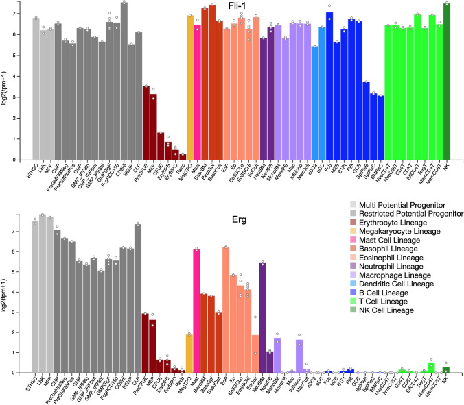
Hematopoietic stem cells (HSCs) through a process of self-renewal, proliferation, and differentiation are responsible for the steady production of progenitors/mature blood cells, maintenance of the HSC pool, and generation of all mature blood cells [11]. Self-renewal and differentiation are governed by a set of critical regulatory genes and factors within the bone marrow niche [11]. Fli-1 is expressed in most hematopoietic multi-potential, restricted progenitors, and all types of mature blood cells (Fig. 1A) [12, 13]. Indeed, Fli-1 expression in hematopoietic cells is essential for maintenance of HSCs and for the differentiation of progenitor cells [14]. In this study, Badwe et al. [14], demonstrated that global ablation of Fli-1 leads to embryonic lethality due to complete peripheral blood failure and the production of aberrant vasculature. Fli-1 is recruited to the regulatory region of most essential hematopoietic genes. These results are consistent with the original study by Hart et al. [15], in which Fli-1 knock-out embryos die at embryonic day E11.5–12.5, mainly due to failure of hematopoiesis and vascular development. A recent study using human embryonic stem cells (hESCs) further revealed that FLI1 over-expression alone induces HSC expansion [16]. Moreover, when FLI1 is activated in conjunction with PKC, hESCs undergo differentiation to endothelial like cells [16]. Interestingly, PKC activation by phorbol esters leads to phosphorylation and activation of FLI-1, resulting in megakaryocytic differentiation [17]. This observation raises the interesting possibility that a phosphorylated form of FLI1 may control endothelial versus ESC development.
Fig. 1.
Expression of murine Fli-1 and Erg in different hematopoietic cells. Taken with permission from Haemosphere. Data show wide spread Fli-1 expression in various hematopoietic lineages when compared to Erg expression. Sites to visit (https://www.haemosphere.org/expression/show?geneId=ENSMUSG00000040732) and (https://www.haemosphere.org/expression/show?geneId=ENSMUSG00000016087) [12, 13]
Fli-1 and Erg are implicated in definitive and adult hematopoiesis [15, 18]. Both factors are derived from an ancestral ETS gene following genomic duplication and eventual chromosomal segregation. The requirement for both Fli-1 and Erg in hematopoiesis may be due to enormous evolutionary demand to preserve HSCs, as these genes appeared to have overlapped function [19]. In point of fact, double heterozygous mutations of both Fli-1 and Erg in mice result in a significant megakaryopoiesis deficiency that is much stronger than defects observed in each individual single-gene knock-out. Moreover, a loss of HSCs in double Fli-1/erg knock-out mice is accompanied by reduced number of committed hematopoietic progenitors compared with the single heterozygous loss in each mutant mouse [18]. Interestingly, heterozygous ErgMld2 mutant allele mice, with a point mutation in the ETS domain, exhibit haploin sufficiency and develop steady-state hematopoiesis; they display defects in stress hematopoiesis after bone marrow transplantation or during recovery from myelotoxic stress [20]. This study is also suggesting similar role for Fli-1 like Erg in HSC self-renewal during stress hematopoiesis.
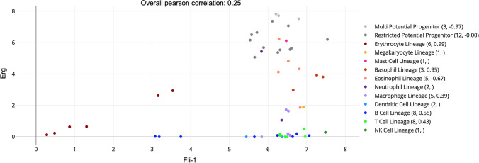
A comparison between expression of Fli-1 and Erg shows that while steady Fli-1 expression is observed in multi-potential and restricted potential progenitors and in most lineages; Erg expression is not detectable in every lineage (Fig. 1B) [12, 13]. These data are supported by an analysis of the Cancer Genome Atlas (TCGA), comparing both Erg and Fli-1 expression in various hematopoietic cells (Fig. 2) [12, 13]. This result suggests that Fli-1 and Erg may have overlapped as well as distinct target genes [18]. Although, as described above, the overlapping and compensatory roles of Fli-1 and Erg in hematopoiesis are well established (18–20), whether these ETS genes also control unique target genes is yet to be determined. ERG activation via translocations was exclusively detected in prostate cancer. Indeed, drug-mediated activation of FLI1 in prostate cancer inhibited tumor progression [21], further suggesting common as well as distinct target regulation by FLI1 versus ERG.
Fig. 2.
Relative expression of murine Erg and Fli-1 in various hematopoietic cells; 0.25 radio present the overall Pearson correlation between Erg and Fli-1. Data were taken with permission from Haemosphere (https://www.haemosphere.org/expression/show?geneId=ENSMUSG00000040732) [12, 13]
Fli-1 plays a critical role in megakaryopoiesis and platelet development
The earliest evidence linking Fli-1 to megakaryopoiesis originated from correlation studies in hematopoietic cells, which identified the promoters of the thrombopoietin receptor (MPL/TPOR), GATA1, and glycoprotein IX (GpIIb/CD42) as direct downstream targets [22–24]. This prediction was later confirmed in Fli-1 knock-out mice, which develop early lethality and thrombocytopenia [15]. A similar phenotype is also observed in patients with Jacobsen or Paris-Trousseau Syndrome, who exhibit thrombocytopenia and platelet deficiency. Indeed, hemizygous mutation within the FLI1 gene has been identified in these patients, signifying FLI1 deficiency as the cause of this disorder [25]. In addition to Jacobsen/Paris-Trousseau, a recent study identified hemizygous mutations in RUNX1 or FLI1 in 6 patients with excessive bleeding and platelet dense granule secretion defects [26]. Jacobsen's syndrome is also characterized by multiple congenital anomalies, cardiac defects, psychomotor retardation, and deletion of chromosome 11 at 11q23.3 [27]. While thrombocytopenia in Jacobson’s syndrome was attributed to FLI1 deficiency, loss of other genes surrounding this locus may also contribute to this or other abnormalities in these patients.
It is now well recognized that both erythroid cells and megakaryocytes originate from a common progenitor known as megakaryocyte erythroid progenitors (MEP) [28]. In the last decade, significant efforts were undertaken to uncover the genetic factors that instruct MEPs to differentiate into either erythroid or megakaryocyte cells. Fli-1 has become such a candidate factor due to the profound megakaryocytic phenotype in knock-out mice and mutations within this TF in diseases associated with platelet deficiency (as discussed earlier). Transfection of FLI-1 into the myelogenous leukemia cell line K562, which lacks this TF, induced megakaryocytic differentiation associated with the activation of specific downstream megakaryocytic target genes [17, 23, 24]. As the frequency of using therapeutic transfusion for various thrombotic disorders increases around the world, a simple technology has recently been developed to obtain platelets by forcing human bone marrow erythroid progenitors to transdifferentiate into megakaryocytic cells following infection with lentivirus vectors carrying the FLI1 and ERG genes [29]. In this study, synergy between FLI1 and ERG was critical to produce higher numbers of megakaryocytes. In another approach, large production of megakaryocytes and erythrocytes was achieved through co-expression of FLI1, GATA1, and TAL1/SCL in human pluripotent stem cells (hPSC) and stimulation with thrombopoietin (TPO) or erythropoietin (EPO) [30, 31]. In early culture (day 9), these three TFs contributed to biopotential, erythroid and megakaryocytic populations regardless of cytokine stimulations. However, in late stage (day 20), while loss of FLI1 expression for an unknown reason was seen in mature erythroid cells, megakaryocytic populations were mostly enriched for FLI1 and TAL1 expression [31]. This result confirms the previous observations [28], highlighting FLI1 as a critical player in bifurcation of MEP to erythroid or megakaryocytic lineages.
FLI1 also interacts with RUNX1 during megakaryocytic differentiation and this interaction appears to be necessary for the induction of megakaryocytic genes [32]. Moreover, mutations in both genes were identified in patients with platelet deficiency [26]. Binding of FLI1 to RUNX1 strictly depends on loss of phosphorylation of serine 10 on the FLI1 protein [32]. Interestingly, phorbol ester (TPA)-induced activation of FLI1 in K562 cells also coincided with reduced FLI1 phosphorylation during megakaryocytic differentiation [17], indicating requirement for both FLI1 and RUNX1 cooperation toward this maturation process.
In hematopoietic cells, it is now well established that lineage specific differentiation depends on combinational interactions between certain TFs. Accordingly, genome-wide binding site analysis identified complex interactions between FLI1, GATA1, GATA2, RUNX1, and SCL/TAL1 in primary human megakaryocytes [33]. As GATA1 is involved in both erythroid and megakaryocytic differentiation [17], the ratio of these five TFs in the complex may be critical to dictate the fate of multipotent MEPs, a notion that has recently been addressed using single-cell mass cytometry and absolute quantitation by mass spectrometry [34]. In this study, a single change in the level of one TF could alter the extent of differentiation of progenitors toward the megakaryocyte fate.
The LIM domain-binding protein 1 (LDB1) was previously reported to play a critical role in erythropoiesis [35, 36]. In a recent study, Giraud et al. [37], demonstrated interaction between Fli-1 and LDB1 on enhancer region of the megakaryocytic-specific genes. The Fli-1 and LDB1 complex binds preferentially to the enhancer regions containing TAL1:GATA1 motif. This binding was demonstrated to modulate the 3D chromatin organization by promoting chromatin looping between enhancers and promoters. LDB1 is likely another critical regulator of erythroid and megakaryocytic differentiation by interacting and modulating FLI1, GATA1, GATA2, RUNX1, and TAL1 complex.
Nfe2 is another Fli-1 interacting protein critical for megakaryocytic differentiation. Ablation of Nfe2 in mice causes embryonic lethality associated with bleeding and platelet deficiency [38]. In this study, A complex interaction between Nfe2, Fli-1, and Runx1 was detected by Chromatin Immunoprecipitation (ChIp) analysis at proximal sites within the promoter of megakaryocytic marker genes. These interactions may be involved in late-stage differentiation of megakaryocytes to platelets. Interestingly, while Nfe2 function is critical in mammals, its function in Zebrafish appeared dispensable for young but required for adult thrombocyte formation [39].
In a recent development, our group identified the Wiscott Aldrich syndrome (WAS) gene WASP and its associated protein WIPF as direct targets of FLI1. WAS is a rare X-linked recessive disease that affects both cellular and humoral immunodeficiency, eczema, high susceptibility to infections, microthrombocytopenia (low platelet count), increased risk of auto-immune disorders, and lymphomas [40, 41]. Knockdown of both WASP and WIPF in the MEP like cell line, HEL, blocked megakaryocytic differentiation, indicating the involvement of FLI1 in WAS. Interestingly, WASP and WIPF knock-down upregulated GATA1, which positively regulates FLI1 expression. These data further emphasize the critical role FLI1 plays in megakaryocyte differentiation, implicating this transcription factor in regulating microthrombocytopenia associated with Wiskott-Aldrich syndrome [41].
Fli-1 and erythroid development
As noted, Fli-1 was first discovered in erythroleukemia induced by F-MuLV [3, 4], suggesting a role for this TF in inhibiting erythropoiesis. Indeed, over-expression of Fli-1 in hematopoietic progenitors inhibits erythroid differentiation, emphasizing its critical role in erythropoiesis [42, 43]. Erythroid transformation is also seen in transgenic mice over-expressing the EWS–FLI1 fusion protein [44]. This erythroid differentiation suppression ability was later confirmed in several Fli-1-deficient animal models [15, 45–47]. Since EPO stimulation is required to promote erythroid differentiation, Fli-1 appeared to suppress EPO-induced differentiation in favor of EPO-induced proliferation of erythroid progenitors [47, 48].
As mentioned above, FLI1 operates in a multi-protein complex with at least five other TFs to induce or suppress gene expression and direct MEPs toward either erythroid or megakaryocytic differentiation [33]. The hematopoietic lineage-restricted gene GATA1 was the first to be implicated in erythroid differentiation, and its expression negatively correlates with FLI1 in erythroleukemia cell lines [49]. This study by Athanasiou et al. [49] demonstrates that FLI1 over-expression in erythroleukemic cells suppresses differentiation through downregulation of GATA1 and that the GATA1 promoter is negatively regulated by FLI1. In a yeast two-hybrid system with a cDNA library of a leukemic cell line, FLI1 was found to bind to GATA1, leading to increased transcriptional activity of megakaryocytic-specific genes [50, 51]. Interestingly, binding of another ETS-related gene, Spi1, to GATA1 causes inactivation of GATA1 and inhibition of erythroid differentiation [50]. Since GATA1 expression is only slightly downregulated during megakaryocytic differentiation [28, 51], it is possible that moderate expression following its interaction with FLI1 contributes to megakaryocytic differentiation, but overt expression is necessary for erythroid differentiation. Indeed, while ablation of gata1 in mouse embryos resulted in a complete lack of erythroid cell precursor development and early lethality [52], a conditional knock-down of gata1 in adults led to a block at the late state red cell generation, trombocytopenia, and an excessive proliferation of megakaryocytes in the spleen [53]. The combined aforementioned data reveal a critical role for GATA-1 and Fli-1 interaction in both erythropoiesis and megakaryopoiesis.
The hematopoiesis restricted Kruppel-like factor 1 (EKLF/KLF) represents another critical factor in erythropoiesis. Loss- and gain-of-function studies clearly demonstrated a role for EKLF in the commitment of bi-potential MEPs to erythroid differentiation at the expense of megakaryocytic differentiation [54]. Ablation of eklf in mice resulted in early lethality (E15.5) and was associated with severe aneia [54, 55]. In man, mutations within the promoter or coding sequence of EKLF resulted in the rare blood group In(Lu) phenotype [56]. In knock-out mice, ablation of eklf significantly increased the number of circulating platelets [57]. This result suggested that a lack of EKLF may block erythroid differentiation at the expense of megakaryocytic differentiation. These data further confirmed the presence of bi-potential—MEPs and the critical role these TFs in their commitment to lineage-restricted differentiation.
As EKLF is critical for erythroid differentiation, its ability to trans-activate the β-globin gene was repressed by FLI1. In this study, Starck et al. [58] showed that FLI1 represses transcription of EKLF in erythroleukemia cell lines. This repression required the ETS domain as well as the N- and C-terminus of FLI1, which bind EKLF. Conversely, EKLF also blocked the trans-activating ability of FLI1 on megakaryocytic-specific promoters. These results demonstrate a negative cross-antagonist relationship between FLI1 and EKLF. This conclusion was further confirmed using conditional shRNA studies in which EKLF depletion resulted in suppression of erythroid differentiation at the expense of megakaryocytic differentiation, mediated through FLI1 downregulation [59].
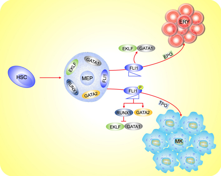
In totality, these results point to a critical role played by FLI1 in the determination of MEP fate toward either an erythroid or megakaryocyte cell lineage. Figure 3 depicts how FLI1 in coordination with other TFs and growth factors regulate this process in multipotent MEBs, originated from HSCs.
Fig. 3.
The role of TFs in derivation of erythroid and megakaryocytes lineages from the multipotent MEP cells. Differential expression of 5 TFs EKLF, GATA1, GATA2, FLI1, and RUNX1 determines the fate of HSC-derived MEP cells to become either erythroid or megakaryocytes. Lower FLI1 expression favors erythroid differentiation through negative regulation of GATA1 and EKLF, in coordination with erythropoietin (EPO). Higher and phosphorylated FLI1* promotes megakaryocytic differentiation through upregulation of RUNX, downregulation of EKLF and GATA1, in coordination with thrombopoietin (TPO)
Fli-1 and T-cell development
High expression level of Fli-1 is detected at various stages of T-cell development (Fig. 1A). In T lymphocytes, FLI1 transcription is upregulated by other ETS proteins including ETS1, ETS2, ELF1, and FLI1 itself, but suppressed by TEL [60]. While complete ablation of Fli-1 in mice resulted in early embryonic lethality [15], a subsequent engineered model by the same research group, in which N-terminal region is deleted (Fli-1ΔNT), resulted in a thymic hypo-cellularity phenotype [61]. Fli-1ΔNT mice are viable and express the truncated Fli-1 protein, indicating its role in T-cell development. In contrast, transgenic over-expression of Fli-1 driven by the H2K promoter, which expected to express in various hematopoietic cells, leads to a higher number of T and B cells [62]. Recently, over-expressing Fli-1 in hematopoietic progenitors by retrovirus transduction was shown to result in pronounced delay in the transition of T cells from a double-negative (DN) to a double-positive (DP) cell [63]. These progenitors also displayed inhibition of CD4 differentiation and enhanced CD8 development. Transplantation of these Fli-1 over-expressing progenitors into lethally irradiated mice eventually resulted in development of a pre-T-cell lymphoblastic leukemia/lymphoma, associated with increased expression of NOTCH1 in tumors. In a recent study by this group, retroviral transduction Fli-1 over-expressing OP9‐DL1 stroma-derived T cells delayed the transition of CD4(–)/CD8(–) DN to CD4( +)/CD8( +) DP cells by deregulating normal DN thymocyte development [64]. Overall, these studies suggest a critical role for Fli-1 in both the DN2 to DN3 transition and αβ/γδ lineage commitment. Finally, in a CRISPR screening platform to identify transcription factors that control the regulatory T (Treg) cells suppress effector T (Teff), Fli-1 was identified as the top candidate [65]. Genetic deletion of Fli-1 improved TEFF differentiation and enhanced protective immunity during viral and bacterial infection and cancer. Thus, Fli-1 is an essential regulator of T-cell development.
Fli-1 and B-cell development
In addition to T cells, high levels of Fli-1 are detected in various stages of B-cell development (Fig. 1A). A critical role of Fli-1 in B cells was first observed in an Fli-1 knock-out mouse model engineered to delete the carboxy-terminal activation (CTA) domain [designated Fli-1ΔCTA], leading to expression of a truncated protein [46]. Unlike knock-out fli-1 mice, homozygous fli-1ΔCTA mice are viable and have fewer splenic follicular B cells and higher transitional and marginal zone B cells. While the expression of genes implicated in B-cell development including CD79, PAX5, E2A, and EGR1 is reduced in fli-1ΔCTA mice, the level of ID1 and ID2 is elevated. Additionally, diminished responsiveness to mitogens is seen in naive B cells isolated from fli-1ΔCTA mice. In contrast to knock-out mice, transgenic over-expression of Fli-1 in the thymus and spleen increases B-cell number and activity [62]. This was associated with increased incidence of a progressive immunological renal disease and ultimately renal failure. These transgenic fli-1 mice also exhibited hyper-gammaglobulinemia, splenomegaly (enlargement of spleen), and B-cell hyperplasia accompanied with abnormal CD5+/B220+-B and CD3+/ B220+-T lymphoid cells in peripheral lymphoid tissues. These characteristics, in addition to the detection of various autoantibodies in the sera, implicate fli-1 in B-cell proliferation and survival.
As Fli-1 modulation of gene expression impacts B-lymphocyte function, it was shown that over-expression of this TF plays an important role in the auto-immune disease, systemic lupus erythematous (SLE) in mice [62]. Recent studies identified FLI1 as a critical regulator of inflammatory mediators including MCP-1, CCL5, IL-6, G-CSF, CXCL2, GM-CSF, and caspase-1 [66–72]. Remarkably, treatment of a mouse model of lupus with the anti-Fli-1 compounds camptothecin and topotecan significantly inhibited pathological signs of the disease, further implicating this TF in this auto-immune disorder [73]. However, as camptothecin and topotecan may act independently of Fli-1, whether more specific inhibitors of this TF or genetic depletion of Fli-1 in B cells can rescue SLE in mice and human awaits further analysis. These studies highlight the need to develop better and more specific anti-Fli-1 compounds for various diseases that may be driven by FLI1, as discussed below.
Fli-1 in the development of other blood cells
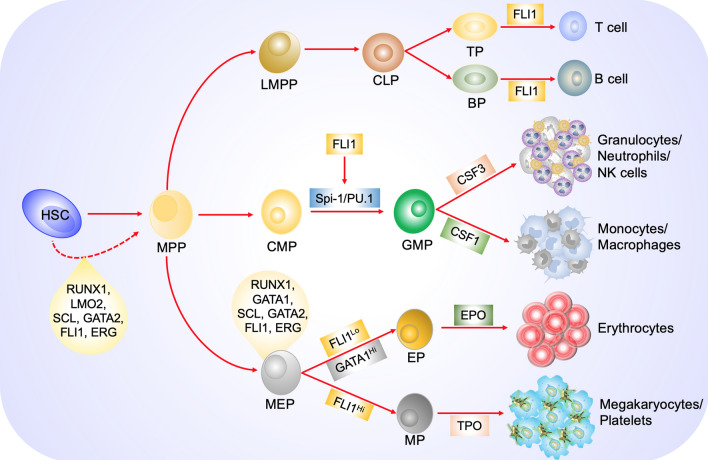
In addition to erythroid/megakaryocytes, Fli-1 expression affects the development of other myeloid cells. In fli-1ΔCTA mice, there is a significant decrease in the number of mature macrophages, monocytes, and dendritic cells [74]. Moreover, in Fli1–/–: Fli1 + / + chimeric mice generated through morula-stage aggregation, a significant reduction in Fli1–/– neutrophil granulocyte and monocyte counts as well as an increase in natural killer (NK) cells were observed [75]. Interestingly, Fli-1 regulates Spi-1/PU.1, which is a known regulator of monocytes and granulocytes [76]. Whether FLI1 expression in myeloid progenitors controls myeloid differentiation through Spi-1/PU.1 or through other mechanisms remained to be demonstrated. A summary of the role of FLI1 in various hematopoietic lineages is depicted in Fig. 4.
Fig. 4.
The role of FLI1 in hematopoiesis: FLI1, in combination with other transcription factors (TFs), maintains HSC survival, proliferation, and differentiation. In cooperation with these additional TFs, FLI1 expression level (LowLo or HighHi) defines the fate of MEPs to become erythroid or megakaryocytic cells, respectively. Regulation of the ETS gene Spi-1/PU.1 by FLI1 promotes CMP differentiation to monocyte/macrophages or granulocytes/neutrophils. Finally, FLI1 expression plays a critical role in differentiation of lymphoid progenitors toward mature T and B cells. HSC (hematopoietic stem cell), MPP (multi-potential progenitor), MEP (megakaryocyte erythrocyte progenitor), LMPP (lymphoid multi-potential progenitor), CMP (common myeloid progenitor), LMPP (lymphoid primed multi-potential progenitor), CLP (common lymphoid progenitor), GMP (granulocyte monocyte progenitor), EP (erythroid progenitor), MP (megakaryocyte progenitor), BP (B-cell progenitor), TP (T-cell progenitor)
Fli-1 and its role in the onset of hematopoiesis
During embryogenesis, blood cells arise from haemangioblasts, which give rise to both endothelial and hematopoietic cells [77, 78]. The commitment to become either blood or endothelial cells is controlled by sequence-specific DNA-binding proteins. The combinatorial expression of a relatively limited number of regulatory transcription factors is sufficient to promote cell fate identity through their action on the underlying gene regulatory networks (GRNs) [79]. Indeed, Fli-1 as well as Gata2, Runx1, Erg, Lmo2, Lyl1, and Tal1 were identified as critical regulators of cell fate decisions during hematopoietic stem/progenitor cell production [80]. Accordingly, mice lacking Fli-1 are embryonically lethal due to defects in blood vessel formation and multiple hematopoietic abnormalities [15, 45], indicating that this ETS gene is involved in the regulation of endothelial and hematopoietic cell fate determination. In a recent study, single-cell transcriptomic analysis demonstrated a new dynamical function of GRNs during embryonic hematopoiesis. This study revealed that while Erg/Fli-1 expression promotes endothelial cell fate, Runx1/Gata2 promotes hematopoietic fate [81]. However, when Fli-1 is co-expressed together with Runx1, it also promotes hematopoietic fate. These observations highlight the complex regulatory networks that govern blood/endothelial cell fate, and a need for further investigation. Fli-1 is also critically important for angiogenesis and endothelial function through regulation of other genes, as was previously reviewed by us [10].
Fli-1 and malignant transformation
In addition to its role in erythroleukemias, FLI1 is translocated in 75% of human Ewing sarcomas, generating the potent oncogenic fusion protein EWS-FLI1 [7]. In the past 2 decades, work from our group and others revealed that Fli-1 transcriptional activation affects several hallmarks of cancer including proliferation, survival, differentiation, angiogenesis, genomic instability, and immune surveillance [10]. Higher protein translation by Fli-1 could accelerate cancer progression. Moreover, FLI1 and CD13/ANPEP over-expression drive resistance to BRAF inhibitors in melanomas [82], suggesting a general role for this ETS TF in drug resistance. FLI1 is highly expressed in various human cancers including those of the, lung, melanoma, leukemia, and lymphoma. In breast cancer, FLI1 expression correlates with tumorigenesis, invasion, and metastasis [10]. In a small proportion of prostate cancer patients, a translocation between FLI1 and TMPRSS2 generates the fusion oncogene TMPRSS2-FLI1, which is associated with tumor initiation [83]. Table 1 summarizes tissue expression and the list of malignancies induced by FLI1 relative to other ETS genes [84–101].
Table 1.
The role of Fli-1 and other ETS gene subfamily in cancer progression. Tissue expression and mutation/translocation of all subfamily of the ETS genes was highlighted. Tissue expression data were obtained from human protein atlas (www.peoreinatlas.org)
| Sub family | ETS gene | Tissue expression | Cancer type mutation | Ref |
|---|---|---|---|---|
| ERG | ERG | All tissues | Leukemia, prostate cancer, sarcoma | [85, 91, 98] |
| FLI1 | All tissues | Leukemia, lymphoma, sarcoma | [7, 10, 89] | |
| FEV | Brain, intestine, pituitary gland, stomach | Mixed phenotype acute leukemia | [100] | |
| ELF | ELF1 | All tissues | Prostate cancer | [92, 96] |
| ELF2 (NERF) | All tissues | |||
| ELF4 (MEF) | All tissues | Various sites | [101] | |
| ERF | ERF (PE2) | All tissues | Prostate cancer | [95] |
| ETV3 (PE1) | All tissues | Breast cancer | [90] | |
| ELG | GABPα | All tissues | Many cancers | [99] |
| ESE | ELF3 (ESE1/ESX) | Many tissues | Epithelial cancers |
[96] [96] [96] |
| ELF5 (ESE2) | Many tissues | Epithelial cancers | ||
| ESE3 (EHF) | Many tissues | Epithelial cancers | ||
| PDEF | SPDEF (PDEF/PSE) | Many tissues | Epithelial cancers | |
| PEA3 | ETV4 (PEA3/E1AF) | Many tissues | Prostate cancer | [86] |
| ETV5 (ERM) | All tissues | Prostate cancer | [87] | |
| ETV1 (ER81) | Many tissues | Prostate cancer | [86] | |
| SPI | SPI1 (PU.1) | Many tissues | Leukemia | [94] |
| SPIB | Many tissues | |||
| SPIC | Lymphoid tissues | |||
| ER71 | ETV2 (ER71) | Many tissues | ||
| TCF | ELK1 | All tissues | ||
| ELK4 (SAP1) | All tissues | Prostate cancer | [88] | |
| ELK3 (NET/SAP2) | All tissues | |||
| TEL | ETV6 (TEL) | All tissues | Leukemia | [84, 93] |
| ETV7 (TEL2) | Many tissues |
FLI1 targeted therapy for the treatment of various diseases
Based on its role on diverse biological processes from cancer initiation and progression to auto-immune diseases including SLE and Systemic sclerosis/scleroderma, FLI1 has been proposed [102] to be an excellent target for drug development. In the past decade, several approaches have been undertaken to identify inhibitors for FLI1 or EWS-FLI1. These efforts led to the identification of small molecules/compounds targeting the DNA or RNA-binding activity of FLI1 [44, 103, 104]. Among these, trabectedin (ET-743) achieved approval by the US Food and Drug Administration (FDA) for the treatments of patients with ovarian cancer or soft-tissue sarcomas [103], and the YK-4–279 derivative TK-216 has recently shown clinical activity in Ewing sarcoma patients in phase I study [104]. Whether these compounds also affect FLI1 in hematological malignancies needs further investigation.
Using a different approach, our group used a luciferase-based expression assay to identify FDA-approved drugs that antagonize FLI1 transcriptional activation. These drugs exhibit strong anti-cancer activity in in vitro and in vivo models of leukemia [105]. Among these are drugs that alter topoisomerase I function (Camptothecin), chemotherapeutic agents (Etoposide), and calcium uptake inhibitors (A23187) that block protein kinase C-delta (PKCδ) activity. Mechanistically, these PKCδ inhibitors were shown to block phosphorylation of FLI1, a critical event that is necessary for its DNA-binding activity [17, 105]. Since these drugs interact with other target proteins in addition to PKCδ, there is a pressing need to isolate specific inhibitors of FLI1. Recently, we have identified two new classes of anti-FLI1 compounds with potent anti-leukemia activity [106, 107]. One class of these compounds was shown to inhibit FLI1 translation, thereby inhibiting leukemia [106]. The other class included two structurally related compounds (A661/A665) that specifically interact with the DNA-binding-motif of FLI1, causing inhibition of FLI1’s downstream targets [107]. Remarkably, we have shown that inhibition of FLI1 via these compounds induces erythroid-to-megakaryocytic differentiation and suppresses leukemogenesis both in vitro and in vivo. Mithramycin is another compound that similar to A661/A665 blocks FLI1 through DNA-binding interference [108]. In another screen for anti-Fli-1 drugs, Rajes et al. [109] showed that the anti-malarial Lumefantrine binds FLI1 and inhibits its activity, leading to growth suppression and apoptosis in culture and tumors in vivo. The aforementioned drugs could therefore be used in future studies for the treatment of cancers driven by abundant expression of FLI1.
Interestingly, in addition to inhibitors, activators of Fli-1 were recently identified and shown to exhibit strong anti-leukemia activity. Among these are the PKC agonist 12-O-tetradecanoylphorbol-13-acetate (TPA) and A75 [17]. Phosphorylation of PKCẟ by these compounds was shown to increase phosphorylation and activation of FLI1, leading to induction of megakaryocytic differentiation in erythroleukemia cell lines. The anti-leukemic effect of these FLI1 agonists significantly increased when combined with a proteosome inhibitor through inhibition of PKCẟ downregulation [110]. Fli-1 activation was also reported via stimulation of leukemic cells with G-CSF through increase protein stability [111]. A summary of drugs with anti-Fli-1 or agonist activity is shown in Table 2. Some of these compounds can be used for both research and treatment of disease associated with aberrant Fli-1 expression.
Table 2.
List of compounds/cytokine with Fli-1 enhanced or inhibitory activity and their mechanism of action
| Chemical name | Function | Fli-1 activity | Mechanism of Fli-1 inhibitory | Ref |
|---|---|---|---|---|
| Camptothecin | Topoisomerase inhibitor | Inhibition | Protein and m-RNA downregulation | [105] |
| A1544 and A1545 | Flavagline- like | Inhibition | Inhibition of MAPK and elongation | [106] |
| Mithramycin MTM | Antibiotic | Inhibition | DNA-binding Interference | [108] |
| Etoposide | Chemotherapeutic drug | Inhibition | Fli-1 downregulation | [105] |
| Trabectedin | ET-743 | Inhibition | Transcriptional downregulation | [103] |
| YK-4–279 | RNA helicase A (RHA) inhibitor | Inhibition | Transcriptional downregulation | [44] |
| Calcimycin (A23187) | Calcium ionophore | Inhibition | Inhibition of phosphorylation | [105] |
| A661 and A665 | Diterpenoid compounds | Inhibition | DNA-binding interference | [107] |
| Lumefantrine | Anti-malaria drug | Inhibition | Drug–protein interaction | [109] |
| TPA | Phorbol ester | Activation | Increase phosphorylation | [17, 110] |
| A75 | Phorbol ester | Activation | Increase phosphorylation | [17, 110] |
| G-CSF | Cytokine | Activation | Increase protein stability | [111] |
Conclusion and future perspectives
Data from over 2000 publications in the past 30 years have established FLI1 as a key factor in healthy development and malignant transformation. These studies have emphasized the essential role of FLI1 in hematopoietic stem cell maintenance and differentiation. In humans, abnormalities in FLI1 expression cause several diseases including auto-immune disorders such as systemic sclerosis and systemic lupus erythematosus. Aberrant expression of FLI1 in various forms of cancer also revealed a critical role in neoplastic transformation. FLI1 has therefore emerged as a novel therapeutic target for certain auto-immune diseases and cancers. The development of clinically approved drugs targeting FLI1 could profoundly impact the treatment of diseases and cancers driven by abnormal FLI1 expression, and bear the fruits of 30 years of extensive basic and translational research.
Acknowledgements
We would like to thank contribution of Chunlin Wang, Wuling Liu, and Analin Hu in preparation of this manuscript.
Abbreviations
- F-MuLV
Friend murine leukemia virus
- SFFV
Spleen focus forming virus
- FV
Friend virus
- Fli-1
Friend virus leukemia integration 1
- TF
Transcription factor
- ETS
E26 transformation-specific
- ATA
Amino-terminal transactivation
- CTA
Carboxy-terminal transactivation
- HSCs
Hematopoietic stem cells
- TME
Tumor microenvironment
- hESCs
Human embryonic stem cells
- TCGA
The cancer genome atlas
- MEP
Megakaryocyte erythroid progenitors
- TPO
Thrombopoietin
- EPO
Erythropoietin
- TPA
Tetradecanoylphorbol-13-acetate
- LDBI
LIM domain-binding protein 1
- ChIp
Chromatin immunoprecipitation
- WAS
Wiscott Aldrich syndrome
- KLF1
Kruppel-like factor 1
- DN
Double-negative
- DP
Double-positive (DP)
- Treg
Regulatory T
- Teff
Effector T
- CTA
Carboxy-terminal activation
- SLE
Systemic lupus erythematous
- NKs
Natural killer cells
- PKCδ
Protein kinase C-delta
Authors’ contributions
All authors contributed to writing this paper. All authors read and approved the final manuscript.
Funding
This work was supported by research grants from the Natural National Science Foundation of China (U1812403 and 21867009), the Science and Technology Department of Guizhou Province innovation and project grants (QKHPTRC[2019]5627), and the 100 Leading Talents of Guizhou Province to Y.B.-D.
Declarations
Ethics approval and consent to participate
Not applicable.
Consent for publication
Not applicable.
Code availability
Not applicable.
Consent to participate
Not applicable.
Availability of data and materials
Not applicable.
Competing interests
The authors declare that there are no conflicts of interest.
Footnotes
Publisher's Note
Springer Nature remains neutral with regard to jurisdictional claims in published maps and institutional affiliations.
References
- 1.Friend C. Cell-free transmission in adult Swiss mice of a disease having the character of a leukemia. J Exp Med. 1957;105:307–318. doi: 10.1084/jem.105.4.307. [DOI] [PMC free article] [PubMed] [Google Scholar]
- 2.Howard JC, Yousefi S, Cheong G, et al. Temporal order and functional analysis of mutations within the Fli-1 and p53 genes during the erythroleukemias induced by F-MuLV. Oncogene. 1993;8:2721–2729. [PubMed] [Google Scholar]
- 3.Ben-David Y, Giddens EB, Bernstein A. Identification and mapping of a common proviral integration site Fli-1 in erythroleukemia cells induced by Friend murine leukemia virus. Proc Natl Acad Sci USA. 1990;87:1332–1336. doi: 10.1073/pnas.87.4.1332. [DOI] [PMC free article] [PubMed] [Google Scholar]
- 4.Ben-David Y, Giddens EB, Letwin K, et al. Erythroleukemia induction by Friend murine leukemia virus: insertional activation of a new member of the ets gene family, Fli-1, closely linked to c-ets-1. Genes Dev. 1991;5:908–918. doi: 10.1101/gad.5.6.908. [DOI] [PubMed] [Google Scholar]
- 5.Truong AH, Ben-David Y. The role of Fli-1 in normal cell function and malignant transformation. Oncogene. 2000;19(55):6482–6489. doi: 10.1038/sj.onc.1204042. [DOI] [PubMed] [Google Scholar]
- 6.Ben-David Y, Prideaux VR, Chow V, et al. Inactivation of the p53 oncogene by internal deletion or retroviral integration in erythroleukemic cell lines induced by Friend leukemia virus. Oncogene. 1988;3:179–185. [PubMed] [Google Scholar]
- 7.Delattre O, Zucman J, Plougastel B, et al. Gene fusion with an ETS DNA-binding domain caused by chromosome translocation in human tumours. Nature. 1992;359:162–165. doi: 10.1038/359162a0. [DOI] [PubMed] [Google Scholar]
- 8.May WA, Gishizky ML, Lessnick SL, et al. Ewing sarcoma 11;22 translocation produces a chimeric transcription factor that requires the DNA-binding domain encoded by FLI1 for transformation. Proc Natl Acad Sci USA. 1993;90:5752–5756. doi: 10.1073/pnas.90.12.5752. [DOI] [PMC free article] [PubMed] [Google Scholar]
- 9.Im YH, Kim HT, Lee C, et al. EWS-FLI1, EWS-ERG, and EWS-ETV1 oncoproteins of Ewing tumor family all suppress transcription of transforming growth factor beta type II receptor gene. Can Res. 2000;60:1536–1540. [PubMed] [Google Scholar]
- 10.Li Y, Luo H, Liu T, et al. The ets transcription factor Fli-1 in development, cancer and disease. Oncogene. 2015;34:2022–2031. doi: 10.1038/onc.2014.162. [DOI] [PMC free article] [PubMed] [Google Scholar]
- 11.Orkin SH, Zon LI. Hematopoiesis: an evolving paradigm for stem cell biology. Cell. 2008;132:631–644. doi: 10.1016/j.cell.2008.01.025. [DOI] [PMC free article] [PubMed] [Google Scholar]
- 12.De Graaf CA, Choi J, Baldwin TM, et al. Haemopedia: an expression atlas of murine hematopoietic cells. Stem Cell Rep. 2016;7:571–582. doi: 10.1016/j.stemcr.2016.07.007. [DOI] [PMC free article] [PubMed] [Google Scholar]
- 13.Choi J, Baldwin TM, Wong M, et al. Haemopedia RNA-seq: a database of gene expression during haematopoiesis in mice and humans. Nucleic Acids Res. 2019;47:D780–D785. doi: 10.1093/nar/gky1020. [DOI] [PMC free article] [PubMed] [Google Scholar]
- 14.Badwe CR, Lis R, Barcia Durán JG, et al. Fli1 is essential for the maintenance of hematopoietic stem cell homeostasis and function. Blood. 2017;130:3769. doi: 10.1182/blood.V130.Suppl_1.3769.3769. [DOI] [Google Scholar]
- 15.Hart A, Melet F, Grossfeld P, et al. Fli-1 is required for murine vascular and megakaryocytic development and is hemizygously deleted in patients with thrombocytopenia. Immunity. 2000;13:167–177. doi: 10.1016/s1074-7613(00)00017-0. [DOI] [PubMed] [Google Scholar]
- 16.Zhao H, Zhao Y, Li Z, et al. FLI1 and PKC co-activation promote highly efficient differentiation of human embryonic stem cells into endothelial-like cells. Cell Death Dis. 2018;9:131. doi: 10.1038/s41419-017-0162-9. [DOI] [PMC free article] [PubMed] [Google Scholar]
- 17.Liu T, Yao Y, Zhang G, et al. A screen for Fli-1 transcriptional modulators identifies PKC agonists that induce erythroid to megakaryocytic differentiation and suppress leukemogenesis. Oncotarget. 2017;8:16728–16743. doi: 10.18632/oncotarget.14377. [DOI] [PMC free article] [PubMed] [Google Scholar]
- 18.Kruse EA, Loughran SJ, Baldwin TM, et al. Dual requirement for the ETS transcription factors Fli-1 and Erg in hematopoietic stem cells and the megakaryocyte lineage. Proc Natl Acad Sci USA. 2009;106:13814–13819. doi: 10.1073/pnas.0906556106. [DOI] [PMC free article] [PubMed] [Google Scholar]
- 19.Ben-David Y, Bernstein A. Friend virus-induced erythroleukemia and the multistage nature of cancer. Cell. 1991;66(5):831–834. doi: 10.1016/0092-8674(91)90428-2. [DOI] [PubMed] [Google Scholar]
- 20.Ng AP, Loughran SJ, Metcalf D, Hyland CD, de Graaf CA, Hu Y, et al. Erg is required for self-renewal of hematopoietic stem cells during stress hematopoiesis in mice. Blood. 2011;118(9):2454–2461. doi: 10.1182/blood-2011-03-344739. [DOI] [PubMed] [Google Scholar]
- 21.Ma Y, Xu B, Yu J, et al. Fli-1 activation through targeted promoter activity regulation using a novel 3′, 5′-diprenylated chalcone inhibits growth and metastasis of prostate cancer cells. Int J Mol Sci. 2020;21(6):2216. doi: 10.3390/ijms21062216. [DOI] [PMC free article] [PubMed] [Google Scholar]
- 22.Deveaux S, Filipe A, Lemarchandel V, et al. Analysis of the thrombopoietin receptor (MPL) promoter implicates GATA and Ets proteins in the coregulation of megakaryocyte-specific genes. Blood. 1996;87:4678–4685. doi: 10.1182/blood.V87.11.4678.bloodjournal87114678. [DOI] [PubMed] [Google Scholar]
- 23.Athanasiou M, Clausen PA, Mavrothalassitis GJ, et al. Increased expression of the ETS-related transcription factor FLI-1/ERGB correlates with and can induce the megakaryocytic phenotype. Cell Growth Differ. 1996;7:1525–1534. [PubMed] [Google Scholar]
- 24.Bastian LS, Kwiatkowski BA, Breininger J, et al. Regulation of the megakaryocytic glycoprotein IX promoter by the oncogenic Ets transcription factor Fli-1. Blood. 1999;93:2637–2644. doi: 10.1182/blood.V93.8.2637. [DOI] [PubMed] [Google Scholar]
- 25.Raslova H, Komura E, Le Couedic JP, et al. FLI1 monoallelic expression combined with its hemizygous loss underlies Paris-Trousseau/Jacobsen thrombopenia. J Clin Investig. 2004;114:77–84. doi: 10.1172/JCI21197. [DOI] [PMC free article] [PubMed] [Google Scholar]
- 26.Stockley J, Morgan NV, Bem D, et al. Enrichment of FLI1 and RUNX1 mutations in families with excessive bleeding and platelet dense granule secretion defects. Blood. 2013;122:4090–4093. doi: 10.1182/blood-2013-06-506873. [DOI] [PMC free article] [PubMed] [Google Scholar]
- 27.Krishnamurti L, Neglia JP, Nagarajan R, et al. Paris-Trousseau syndrome platelets in a child with Jacobsen's syndrome. Am J Hematol. 2001;66:295–299. doi: 10.1002/ajh.1061. [DOI] [PubMed] [Google Scholar]
- 28.Klimchenko O, Mori M, Distefano A, et al. A common bipotent progenitor generates the erythroid and megakaryocyte lineages in embryonic stem cell-derived primitive hematopoiesis. Blood. 2009;114:1506–1517. doi: 10.1182/blood-2008-09-178863. [DOI] [PubMed] [Google Scholar]
- 29.Siripin D, Kheolamai P, U-Pratya Y, et al. Transdifferentiation of erythroblasts to megakaryocytes using FLI1 and ERG transcription factors. Thromb Haemost. 2015;114:593–602. doi: 10.1160/TH14-12-1090. [DOI] [PubMed] [Google Scholar]
- 30.Moreau T, Evans AL, Vasquez L, et al. Corrigendum: Large-scale production of megakaryocytes from human pluripotent stem cells by chemically defined forward programming. Nat Commun. 2016;8:15076. doi: 10.1038/ncomms11208. [DOI] [PMC free article] [PubMed] [Google Scholar]
- 31.Dalby A, Ballester-Beltran J, Lincetto C, et al. Transcription factor levels after forward programming of human pluripotent stem cells with GATA1, FLI1, and TAL1 determine megakaryocyte versus erythroid cell fate decision. Stem cell reports. 2018;11:1462–1478. doi: 10.1016/j.stemcr.2018.11.001. [DOI] [PMC free article] [PubMed] [Google Scholar]
- 32.Huang H, Yu M, Akie TE, et al. Differentiation-dependent interactions between RUNX-1 and FLI-1 during megakaryocyte development. Mol Cell Biol. 2009;29:4103–4115. doi: 10.1128/MCB.00090-09. [DOI] [PMC free article] [PubMed] [Google Scholar]
- 33.Tijssen MR, Cvejic A, Joshi A, et al. Genome-wide analysis of simultaneous GATA1/2, RUNX1, FLI1, and SCL binding in megakaryocytes identifies hematopoietic regulators. Dev Cell. 2011;20:597–609. doi: 10.1016/j.devcel.2011.04.008. [DOI] [PMC free article] [PubMed] [Google Scholar]
- 34.Palii CG, Cheng Q, Gillespie MA, et al. Single-cell proteomics reveal that quantitative changes in co-expressed lineage-specific transcription factors determine cell fate. Cell Stem Cell. 2019;24:812–820.e5. doi: 10.1016/j.stem.2019.02.006. [DOI] [PMC free article] [PubMed] [Google Scholar]
- 35.Soler E, Andrieu-Soler C, de Boer E, et al. The genome-wide dynamics of the binding of Ldb1 complexes during erythroid differentiation. Genes Dev. 2010;24(3):277–289. doi: 10.1101/gad.551810. [DOI] [PMC free article] [PubMed] [Google Scholar]
- 36.Li L, Freudenberg J, Cui K, et al. Ldb1-nucleated transcription complexes function as primary mediators of global erythroid gene activation. Blood. 2013;121(22):4575–4585. doi: 10.1182/blood-2013-01-479451. [DOI] [PMC free article] [PubMed] [Google Scholar]
- 37.Giraud G, Kolovos P, Boltsis I, et al. Interplay between FLI-1 and the LDB1 complex in murine erythroleukemia cells and during megakaryopoiesis. iScience. 2021;24(3):102210. doi: 10.1016/j.isci.2021.102210. [DOI] [PMC free article] [PubMed] [Google Scholar]
- 38.Shivdasani RA, Rosenblatt MF, Zucker-Franklin D, et al. Transcription factor NF-E2 is required for platelet formation independent of the actions of thrombopoietin/MGDF in megakaryocyte development. Cell. 1995;81:695–704. doi: 10.1016/0092-8674(95)90531-6. [DOI] [PubMed] [Google Scholar]
- 39.Rost MS, Shestopalov I, Liu Y, et al. Nfe2 is dispensable for early but required for adult thrombocyte formation and function in zebrafish. Blood Adv. 2018;2:3418–3427. doi: 10.1182/bloodadvances.2018021865. [DOI] [PMC free article] [PubMed] [Google Scholar]
- 40.Wang C, Sample KM, Gajendran B, Kapranov P, Liu W, Hu A, et al. FLI1 induces megakaryopoiesis gene expression through WAS/WIP-dependent and independent mechanisms; implications for Wiskott-Aldrich syndrome. Front Immunol. 2021;12:607836. doi: 10.3389/fimmu.2021.607836. [DOI] [PMC free article] [PubMed] [Google Scholar]
- 41.Bosticardo M, Marangoni F, Aiuti A, Villa A, Grazia RM. Recent advances in understanding the pathophysiology of Wiskott-Aldrich syndrome. Blood. 2009;113(25):6288–6295. doi: 10.1182/blood-2008-12-115253. [DOI] [PubMed] [Google Scholar]
- 42.Pereira R, Quang CT, Lesault I, et al. FLI-1 inhibits differentiation and induces proliferation of primary erythroblasts. Oncogene. 1999;18:1597–1608. doi: 10.1038/sj.onc.1202534. [DOI] [PubMed] [Google Scholar]
- 43.Tamir A, Howard J, Higgins RR, et al. Fli-1, an Ets-related transcription factor, regulates erythropoietin-induced erythroid proliferation and differentiation: evidence for direct transcriptional repression of the Rb gene during differentiation. Mol Cell Biol. 1999;19:4452–4464. doi: 10.1128/MCB.19.6.4452. [DOI] [PMC free article] [PubMed] [Google Scholar]
- 44.Minas TZ, Han J, Javaheri T, Hong SH, Schlederer M, Saygideğer-Kont Y, et al. YK-4-279 effectively antagonizes EWS-FLI1 induced leukemia in a transgenic mouse model. Oncotarget. 2015;6(35):37678–37694. doi: 10.18632/oncotarget.5520. [DOI] [PMC free article] [PubMed] [Google Scholar]
- 45.Spyropoulos DD, Pharr PN, Lavenburg KR, et al. Hemorrhage, impaired hematopoiesis, and lethality in mouse embryos carrying a targeted disruption of the Fli1 transcription factor. Mol Cell Biol. 2000;20:5643–5652. doi: 10.1128/MCB.20.15.5643-5652.2000. [DOI] [PMC free article] [PubMed] [Google Scholar]
- 46.Zhang XK, Moussa O, LaRue A, et al. The transcription factor Fli-1 modulates marginal zone and follicular B cell development in mice. J Immunol. 2008;181:1644–1654. doi: 10.4049/jimmunol.181.3.1644. [DOI] [PMC free article] [PubMed] [Google Scholar]
- 47.Zochodne B, Truong AH, Stetler K, et al. Epo regulates erythroid proliferation and differentiation through distinct signaling pathways: implication for erythropoiesis and Friend virus-induced erythroleukemia. Oncogene. 2000;19:2296–2304. doi: 10.1038/sj.onc.1203590. [DOI] [PubMed] [Google Scholar]
- 48.Starck J, Weiss-Gayet M, Gonnet C, et al. Inducible Fli-1 gene deletion in adult mice modifies several myeloid lineage commitment decisions and accelerates proliferation arrest and terminal erythrocytic differentiation. Blood. 2010;116:4795–4805. doi: 10.1182/blood-2010-02-270405. [DOI] [PubMed] [Google Scholar]
- 49.Athanasiou M, Mavrothalassitis G, Sun-Hoffman L, et al. FLI-1 is a suppressor of erythroid differentiation in human hematopoietic cells. Leukemia. 2000;14:439–445. doi: 10.1038/sj.leu.2401689. [DOI] [PubMed] [Google Scholar]
- 50.Rekhtman N, Radparvar F, Evans T, et al. Direct interaction of hematopoietic transcription factors PU.1 and GATA-1: functional antagonism in erythroid cells. Genes Dev. 1999;13:1398–1411. doi: 10.1101/gad.13.11.1398. [DOI] [PMC free article] [PubMed] [Google Scholar]
- 51.Eisbacher M, Holmes ML, Newton A, et al. Protein-protein interaction between Fli-1 and GATA-1 mediates synergistic expression of megakaryocyte-specific genes through cooperative DNA binding. Mol Cell Biol. 2003;23:3427–3441. doi: 10.1128/MCB.23.10.3427-3441.2003. [DOI] [PMC free article] [PubMed] [Google Scholar]
- 52.Fujiwara Y, Browne CP, Cunniff K, et al. Arrested development of embryonic red cell precursors in mouse embryos lacking transcription factor GATA-1. Proc Natl Acad Sci USA. 1996;93:12355–12358. doi: 10.1073/pnas.93.22.12355. [DOI] [PMC free article] [PubMed] [Google Scholar]
- 53.Jagadeeswaran P, Lin S, Weinstein B, et al. Loss of GATA1 and gain of FLI1 expression during thrombocyte maturation. Blood Cells Mol Dis. 2010;44:175–180. doi: 10.1016/j.bcmd.2009.12.012. [DOI] [PMC free article] [PubMed] [Google Scholar]
- 54.Frontelo P, Manwani D, Galdass M, et al. Novel role for EKLF in megakaryocyte lineage commitment. Blood. 2007;110:3871–3880. doi: 10.1182/blood-2007-03-082065. [DOI] [PMC free article] [PubMed] [Google Scholar]
- 55.Perkins AC, Sharpe AH, Orkin SH. Lethal beta-thalassaemia in mice lacking the erythroid CACCC-transcription factor EKLF. Nature. 1995;375:318–322. doi: 10.1038/375318a0. [DOI] [PubMed] [Google Scholar]
- 56.Singleton BK, Burton NM, Green C, et al. Mutations in EKLF/KLF1 form the molecular basis of the rare blood group In(Lu) phenotype. Blood. 2008;112:2081–2088. doi: 10.1182/blood-2008-03-145672. [DOI] [PubMed] [Google Scholar]
- 57.Tallack MR, Perkins AC. Megakaryocyte-erythroid lineage promiscuity in EKLF null mouse blood. Haematologica. 2010;95:144–147. doi: 10.3324/haematol.2009.010017. [DOI] [PMC free article] [PubMed] [Google Scholar]
- 58.Starck J, Cohet N, Gonnet C, et al. Functional cross-antagonism between transcription factors FLI-1 and EKLF. Mol Cell Biol. 2003;23:1390–1402. doi: 10.1128/MCB.23.4.1390-1402.2003. [DOI] [PMC free article] [PubMed] [Google Scholar]
- 59.Bouilloux F, Juban G, Cohet N, et al. EKLF restricts megakaryocytic differentiation at the benefit of erythrocytic differentiation. Blood. 2008;112:576–584. doi: 10.1182/blood-2007-07-098996. [DOI] [PubMed] [Google Scholar]
- 60.Svenson JL, Chike-Harris K, Amria MY, et al. The mouse and human Fli1 genes are similarly regulated by Ets factors in T cells. Genes Immun. 2010;11(2):161–172. doi: 10.1038/gene.2009.73. [DOI] [PMC free article] [PubMed] [Google Scholar]
- 61.Melet F, Motro B, Rossi DJ, et al. Generation of a novel Fli-1 protein by gene targeting leads to a defect in thymus development and a delay in Friend virus-induced erythroleukemia. Mol Cell Biol. 1996;16:2708–2718. doi: 10.1128/MCB.16.6.2708. [DOI] [PMC free article] [PubMed] [Google Scholar]
- 62.Zhang L, Eddy A, Teng YT, et al. An immunological renal disease in transgenic mice that overexpress Fli-1, a member of the ets family of transcription factor genes. Mol Cell Biol. 1995;15:6961–6970. doi: 10.1128/MCB.15.12.6961. [DOI] [PMC free article] [PubMed] [Google Scholar]
- 63.Smeets MF, Chan AC, Dagger S, et al. Fli-1 overexpression in hematopoietic progenitors deregulates T cell development and induces pre-T cell lymphoblastic leukaemia/lymphoma. PLoS ONE. 2013;8:e62346. doi: 10.1371/journal.pone.0062346. [DOI] [PMC free article] [PubMed] [Google Scholar]
- 64.Smeets MF, Wiest DL, Izon DJ. Fli-1 regulates the DN2 to DN3 thymocyte transition and promotes gammadelta T-cell commitment by enhancing TCR signal strength. Eur J Immunol. 2014;44:2617–2624. doi: 10.1002/eji.201444442. [DOI] [PMC free article] [PubMed] [Google Scholar]
- 65.Chen Z, Arai E, Khan O, Zhang Z, Ngiow SF, He Y, et al. In vivo CD8+ T cell CRISPR screening reveals control by Fli1 in infection and cancer. Cell. 2021;184(5):1262–1280.e22. doi: 10.1016/j.cell.2021.02.019. [DOI] [PMC free article] [PubMed] [Google Scholar]
- 66.Hodge DR, Li D, Qi SM, Farrar WL. IL-6 induces expression of the Fli-1 proto-oncogene via STAT3. Biochem Biophys Res Commun. 2002;292(1):287–291. doi: 10.1006/bbrc.2002.6652. [DOI] [PubMed] [Google Scholar]
- 67.Lennard Richard ML, Nowling TK, Brandon D, Watson DK, Zhang XK. Fli-1 controls transcription from the MCP-1 gene promoter, which may provide a novel mechanism for chemokine and cytokine activation. Mol Immunol. 2015;63(2):566–573. doi: 10.1016/j.molimm.2014.07.013. [DOI] [PMC free article] [PubMed] [Google Scholar]
- 68.Lennard Richard ML, Sato S, Suzuki E, Williams S, Nowling TK, Zhang XK. The Fli-1 transcription factor regulates the expression of CCL5/RANTES. J Immunol. 2014;193(6):2661–2668. doi: 10.4049/jimmunol.1302779. [DOI] [PMC free article] [PubMed] [Google Scholar]
- 69.Lennard Richard ML, Brandon D, Lou N, Sato S, Caldwell T, Nowling TK, et al. Acetylation impacts Fli-1-driven regulation of granulocyte colony stimulating factor. Eur J Immunol. 2016;46(10):2322–2332. doi: 10.1002/eji.201646315. [DOI] [PMC free article] [PubMed] [Google Scholar]
- 70.Lou N, Lennard Richard ML, Yu J, Brandon M, Zhang XK. The Fli-1 transcription factor is a critical regulator for controlling the expression of chemokine C-X-C motif ligand 2 (CXCL2) Mol Immunol. 2017;81:59–66. doi: 10.1016/j.molimm.2016.11.007. [DOI] [PubMed] [Google Scholar]
- 71.Li P, Goodwin AJ, Cook JA, Halushka PV, Zhang XK, Fan H. Fli-1 transcription factor regulates the expression of caspase-1 in lung pericytes. Mol Immunol. 2019;108:1–7. doi: 10.1016/j.molimm.2019.02.003. [DOI] [PMC free article] [PubMed] [Google Scholar]
- 72.Wang X, Lennard Richard M, Li P, Henry B, Schutt S, Yu XZ, et al. Expression of GM-CSF is regulated by Fli-1 transcription factor, a potential drug target. J Immunol. 2021;206(1):59–66. doi: 10.4049/jimmunol.2000664. [DOI] [PMC free article] [PubMed] [Google Scholar]
- 73.Wang X, Oates JC, Helke KL, Gilkeson GS, Zhang XK. Camptothecin and topotecan, inhibitors of transcription factor Fli-1 and topoisomerase, markedly ameliorate lupus nephritis in NZBWF1 mice and reduce the production of inflammatory mediators in human renal cells. Arthritis Rheumatol. 2021;73(8):1478–1488. doi: 10.1002/art.41685. [DOI] [PMC free article] [PubMed] [Google Scholar]
- 74.Suzuki E, Williams S, Sato S, et al. The transcription factor Fli-1 regulates monocyte, macrophage and dendritic cell development in mice. Immunology. 2013;139:318–327. doi: 10.1111/imm.12070. [DOI] [PMC free article] [PubMed] [Google Scholar]
- 75.Masuya M, Moussa O, Abe T, et al. Dysregulation of granulocyte, erythrocyte, and NK cell lineages in Fli-1 gene-targeted mice. Blood. 2005;105:95–102. doi: 10.1182/blood-2003-12-4345. [DOI] [PubMed] [Google Scholar]
- 76.Starck J, Mouchiroud G, Gonnet C, et al. Unexpected and coordinated expression of Spi-1, Fli-1, and megakaryocytic genes in four Epo-dependent cell lines established from transgenic mice displaying erythroid-specific expression of a thermosensitive SV40 T antigen. Exp Hematol. 1999;27:630–641. doi: 10.1016/s0301-472x(99)00006-5. [DOI] [PubMed] [Google Scholar]
- 77.Kennedy M, Firpo M, Choi K, et al. A common precursor for primitive erythropoiesis and definitive haematopoiesis. Nature. 1997;386(6624):488–493. doi: 10.1038/386488a0. [DOI] [PubMed] [Google Scholar]
- 78.Huber TL, Kouskoff V, Fehling HJ, et al. Haemangioblast commitment is initiated in the primitive streak of the mouse embryo. Nature. 2004;432(7017):625–630. doi: 10.1038/nature03122. [DOI] [PubMed] [Google Scholar]
- 79.Iwafuchi-Doi M, Zaret KS. Cell fate control by pioneer transcription factors. Development. 2016;143(11):1833–1837. doi: 10.1242/dev.133900. [DOI] [PMC free article] [PubMed] [Google Scholar]
- 80.Wilson NK, Foster SD, Wang X, et al. Combinatorial transcriptional control in blood stem/progenitor cells: genome-wide analysis of ten major transcriptional regulators. Cell Stem Cell. 2010;7(4):532–544. doi: 10.1016/j.stem.2010.07.016. [DOI] [PubMed] [Google Scholar]
- 81.Bergiers I, Andrews T, Vargel Bölükbaşı Ö, et al. Single-cell transcriptomics reveals a new dynamical function of transcription factors during embryonic hematopoiesis. Elife. 2018;7:e29312. doi: 10.7554/eLife.29312. [DOI] [PMC free article] [PubMed] [Google Scholar]
- 82.Azimi A, Tuominen R, Costa Svedman F, Caramuta S, Pernemalm M, Frostvik Stolt M, et al. Silencing FLI or targeting CD13/ANPEP lead to dephosphorylation of EPHA2, a mediator of BRAF inhibitor resistance, and induce growth arrest or apoptosis in melanoma cells. Cell Death Dis. 2017;8(8):e3029. doi: 10.1038/cddis.2017.406. [DOI] [PMC free article] [PubMed] [Google Scholar]
- 83.Paulo P, Barros-Silva JD, Ribeiro FR, et al. FLI1 is a novel ETS transcription factor involved in gene fusions in prostate cancer. Genes Chromosom Cancer. 2012;51:240–249. doi: 10.1002/gcc.20948. [DOI] [PubMed] [Google Scholar]
- 84.Golub TR, Barker GF, Bohlander SK, Hiebert SW, Ward DC, Bray-Ward P, et al. Fusion of the TEL gene on 12p13 to the AML1 gene on 21q22 in acute lymphoblastic leukemia. Proc Natl Acad Sci USA. 1995;92(11):4917–4921. doi: 10.1073/pnas.92.11.4917. [DOI] [PMC free article] [PubMed] [Google Scholar]
- 85.Tomlins SA, Rhodes DR, Perner S, Dhanasekaran SM, Mehra R, Sun XW, et al. Recurrent fusion of TMPRSS2 and ETS transcription factor genes in prostate cancer. Science. 2005;310(5748):644–648. doi: 10.1126/science.1117679. [DOI] [PubMed] [Google Scholar]
- 86.Tomlins SA, Mehra R, Rhodes DR, Smith LR, Roulston D, Helgeson BE, et al. TMPRSS2:ETV4 gene fusions define a third molecular subtype of prostate cancer. Cancer Res. 2006;66(7):3396–3400. doi: 10.1158/0008-5472.CAN-06-0168. [DOI] [PubMed] [Google Scholar]
- 87.Helgeson BE, Tomlins SA, Shah N, Laxman B, Cao Q, Prensner JR, et al. Characterization of TMPRSS2:ETV5 and SLC45A3:ETV5 gene fusions in prostate cancer. Cancer Res. 2008;68(1):73–80. doi: 10.1158/0008-5472.CAN-07-5352. [DOI] [PubMed] [Google Scholar]
- 88.Rickman DS, Pflueger D, Moss B, VanDoren VE, Chen CX, de la Taille A, Kuefer R, Tewari AK, Setlur SR, Demichelis F, Rubin MA. SLC45A3-ELK4 is a novel and frequent erythroblast transformation-specific fusion transcript in prostate cancer. Cancer Res. 2009;69(7):2734–2738. doi: 10.1158/0008-5472.CAN-08-4926. [DOI] [PMC free article] [PubMed] [Google Scholar]
- 89.Bonetti P, Testoni M, Scandurra M, Ponzoni M, Piva R, Mensah AA, Rinaldi A, Kwee I, Tibiletti MG, Iqbal J, Greiner TC, Chan WC, et al. Deregulation of ETS1 and FLI1 contributes to the pathogenesis of diffuse large B-cell lymphoma. Blood. 2013;122(13):2233–2241. doi: 10.1182/blood-2013-01-475772. [DOI] [PubMed] [Google Scholar]
- 90.Mesquita B, Lopes P, Rodrigues A, Pereira D, Afonso M, Leal C, Henrique R, Lind GE, Jerónimo C, Lothe RA, Teixeira MR. Frequent copy number gains at 1q21 and 1q32 are associated with overexpression of the ETS transcription factors ETV3 and ELF3 in breast cancer irrespective of molecular subtypes. Breast Cancer Res Treat. 2013;138(1):37–45. doi: 10.1007/s10549-013-2408-2. [DOI] [PubMed] [Google Scholar]
- 91.Zhang J, McCastlain K, Yoshihara H, Xu B, Chang Y, Churchman ML, Wu G, Li Y, Wei L, Iacobucci I, et al. Deregulation of DUX4 and ERG in acute lymphoblastic leukemia. Nat Genet. 2016;48(12):1481–1489. doi: 10.1038/ng.3691. [DOI] [PMC free article] [PubMed] [Google Scholar]
- 92.Ando M, Kawazu M, Ueno T, Koinuma D, Ando K, Koya J, Kataoka K, Yasuda T, Yamaguchi H, Fukumura K, et al. Mutational landscape and antiproliferative functions of ELF transcription factors in human cancer. Cancer Res. 2016;76(7):1814–1824. doi: 10.1158/0008-5472.CAN-14-3816. [DOI] [PubMed] [Google Scholar]
- 93.Lilljebjörn H, Henningsson R, Hyrenius-Wittsten A, Olsson L, Orsmark-Pietras C, von Palffy S, Askmyr M, Rissler M, Schrappe M, Cario G, et al. Identification of ETV6-RUNX1-like and DUX4-rearranged subtypes in paediatric B-cell precursor acute lymphoblastic leukaemia. Nat Commun. 2016;7:11790. doi: 10.1038/ncomms11790. [DOI] [PMC free article] [PubMed] [Google Scholar]
- 94.Seki M, Kimura S, Isobe T, Yoshida K, Ueno H, Nakajima-Takagi Y, Wang C, Lin L, Kon A, Suzuki H, et al. Recurrent SPI1 (PU.1) fusions in high-risk pediatric T cell acute lymphoblastic leukemia. Nat Genet. 2017;49(8):1274–1281. doi: 10.1038/ng.3900. [DOI] [PubMed] [Google Scholar]
- 95.Bose R, Karthaus WR, Armenia J, Abida W, Iaquinta PJ, Zhang Z, Wongvipat J, Wasmuth EV, Shah N, Sullivan PS, et al. ERF mutations reveal a balance of ETS factors controlling prostate oncogenesis. Nature. 2017;546(7660):671–675. doi: 10.1038/nature22820. [DOI] [PMC free article] [PubMed] [Google Scholar]
- 96.Budka JA, Ferris MW, Capone MJ, Hollenhorst PC. Common ELF1 deletion in prostate cancer bolsters oncogenic ETS function, inhibits senescence and promotes docetaxel resistance. Genes Cancer. 2018;9(5–6):198–214. doi: 10.18632/genesandck;ancer.182. [DOI] [PMC free article] [PubMed] [Google Scholar]
- 97.Luk IY, Reehorst CM, Mariadason JM. ELF3, ELF5, EHF and SPDEF transcription factors in tissue homeostasis and cancer. Molecules. 2018;23(9):2191. doi: 10.3390/molecules23092191. [DOI] [PMC free article] [PubMed] [Google Scholar]
- 98.Zaliova M, Potuckova E, Hovorkova L, et al. ERG deletions in childhood acute lymphoblastic leukemia with DUX4 rearrangements are mostly polyclonal, prognostically relevant and their detection rate strongly depends on screening method sensitivity. Haematologica. 2019;104(7):1407–1416. doi: 10.3324/haematol.2018.204487. [DOI] [PMC free article] [PubMed] [Google Scholar]
- 99.Yuan X, Dai M, Xu D. TERT promoter mutations and GABP transcription factors in carcinogenesis: more foes than friends. Cancer Lett. 2020;493:1–9. doi: 10.1016/j.canlet.2020.07.003. [DOI] [PubMed] [Google Scholar]
- 100.Montgomery-Goecker C, Koduru P, Botten G, Xu J, Ghisoli M, Goldman SC, et al. Mixed phenotype acute leukemia, b/myeloid (bilineal and biphenotypic), with t(2;22)(q35;q12);EWSR1-FEV. J Pediatr Hematol Oncol. 2021;43(3):e388–e394. doi: 10.1097/MPH.0000000000001934. [DOI] [PubMed] [Google Scholar]
- 101.Suico MA, Shuto T, Kai H. Roles and regulations of the ETS transcription factor ELF4/MEF. J Mol Cell Biol. 2017;9(3):168–177. doi: 10.1093/jmcb/mjw051. [DOI] [PMC free article] [PubMed] [Google Scholar]
- 102.Erkizan HV, Kong Y, Merchant M, et al. A small molecule blocking oncogenic protein EWS-FLI1 interaction with RNA helicase A inhibits growth of Ewing's sarcoma. Nat Med. 2009;15:750–756. doi: 10.1038/nm.1983. [DOI] [PMC free article] [PubMed] [Google Scholar]
- 103.Grohar PJ, Griffin LB, Yeung C, et al. Ecteinascidin 743 interferes with the activity of EWS-FLI1 in Ewing sarcoma cells. Neoplasia (New York, N.Y.) 2011;13:145–153. doi: 10.1593/neo.101202. [DOI] [PMC free article] [PubMed] [Google Scholar]
- 104.Zöllner SK, Amatruda JF, Bauer S, et al. Ewing sarcoma-diagnosis, treatment, clinical challenges and future perspectives. J Clin Med. 2021;10(8):1685. doi: 10.3390/jcm10081685. [DOI] [PMC free article] [PubMed] [Google Scholar]
- 105.Li YJ, Zhao X, Vecchiarelli-Federico LM, et al. Drug-mediated inhibition of Fli-1 for the treatment of leukemia. Blood Cancer J. 2012;2:e54. doi: 10.1038/bcj.2011.52. [DOI] [PMC free article] [PubMed] [Google Scholar]
- 106.Song J, Yuan C, Yang J, et al. Novel flavagline-like compounds with potent Fli-1 inhibitory activity suppress diverse types of leukemia. FEBS J. 2018;285:4631–4645. doi: 10.1111/febs.14690. [DOI] [PubMed] [Google Scholar]
- 107.Liu T, Xia L, Yao Y, et al. Identification of diterpenoid compounds that interfere with Fli-1 DNA binding to suppress leukemogenesis. Cell Death Dis. 2019;10:117. doi: 10.1038/s41419-019-1363-1. [DOI] [PMC free article] [PubMed] [Google Scholar]
- 108.Hou C, Mandal A, Rohr J, Tsodikov OV. Allosteric interference in oncogenic FLI1 and ERG transactions by mithramycins. Structure. 2020;29(5):404–412.e4. doi: 10.1016/j.str.2020.11.012. [DOI] [PMC free article] [PubMed] [Google Scholar]
- 109.Rajesh Y, Biswas A, Kumar U, Banerjee I, Das S, Maji S, Das SK, Emdad L, Cavenee WK, Mandal M, Fisher PB. Lumefantrine, an antimalarial drug, reverses radiation and temozolomide resistance in glioblastoma. Proc Natl Acad Sci USA. 2020;117(22):12324–12331. doi: 10.1073/pnas.1921531117. [DOI] [PMC free article] [PubMed] [Google Scholar]
- 110.Yao Y, Liu W, Gajendran B, Wang C, Zacksenhaus E, Sample KM, Varier KM, Hao X, Ben-David Y. Ubash3b promotes TPA-mediated suppression of leukemogenesis through accelerated downregulation of PKCδ protein. Biochimie. 2021;184:8–17. doi: 10.1016/j.biochi.2021.02.001. [DOI] [PubMed] [Google Scholar]
- 111.Mora-Garcia P, Wei J, Sakamoto KM. G-CSF induces stabilization of ETS protein Fli-1 during myeloid cell development. Pediatr Res. 2005;57(1):63–66. doi: 10.1203/01.PDR.0000147729.55592.2C. [DOI] [PubMed] [Google Scholar]
Associated Data
This section collects any data citations, data availability statements, or supplementary materials included in this article.
Data Availability Statement
Not applicable.


