Abstract
The health risks that arise from environmental exposures vary widely within and across human populations, and these differences are largely determined by genetic variation and gene-by-environment (gene–environment) interactions. However, risk assessment in laboratory mice typically involves isogenic strains and therefore, does not account for these known genetic effects. In this context, genetically heterogenous cell lines from laboratory mice are promising tools for population-based screening because they provide a way to introduce genetic variation in risk assessment without increasing animal use. Cell lines from genetic reference populations of laboratory mice offer genetic diversity, power for genetic mapping, and potentially, predictive value for in vivo experimentation in genetically matched individuals. To explore this further, we derived a panel of fibroblast lines from a genetic reference population of laboratory mice (the Diversity Outbred, DO). We then used high-content imaging to capture hundreds of cell morphology traits in cells exposed to the oxidative stress-inducing arsenic metabolite monomethylarsonous acid (MMAIII). We employed dose-response modeling to capture latent parameters of response and we then used these parameters to identify several hundred cell morphology quantitative trait loci (cmQTL). Response cmQTL encompass genes with established associations with cellular responses to arsenic exposure, including Abcc4 and Txnrd1, as well as novel gene candidates like Xrcc2. Moreover, baseline trait cmQTL highlight the influence of natural variation on fundamental aspects of nuclear morphology. We show that the natural variants influencing response include both coding and non-coding variation, and that cmQTL haplotypes can be used to predict response in orthogonal cell lines. Our study sheds light on the major molecular initiating events of oxidative stress that are under genetic regulation, including the NRF2-mediated antioxidant response, cellular detoxification pathways, DNA damage repair response, and cell death trajectories.
Author summary
Exposure to environmental toxicants leads to adverse health outcomes. Natural genetic variation regulates the likelihood and severity of these outcomes, but studying the underlying genes and pathways in human populations is challenging. Population-based rodent models simulate the genetic variation of the human population offering numerous advantages as experimental genetic models for studying gene–environment interactions and in chemical risk asessment. These include exquisite environmental control and higher power for mapping the genes and pathways that influence sensitivity and resilience to environmental exposures. We leveraged a genetically diverse laboratory mouse population to investigate the genetic regulation of arsenic response. To minimize animal use, cells were derived from each individual animal through a minimally invasive tail biopsy. These cells provide a reusable genetic resource for chemical or drug screening through which predictions of risk can be made and tested in genetically matched laboratory mice. To evaluate the utility of this resource, we used high content imaging to quantify changes in cell morphology following exposure to the arsenic metabolite MMAIII. Using dose-response modeling, we identified the subset of morphological changes that are informative of arsenic response and that exhibit robust interindividual variation. We then used genetic association mapping to identify several hundred loci regulating individual differences in arsenic response. To nominate candidate driver genes, we integrated various lines of evidence from published arsenic studies and molecular data from the same mouse population. Our work demonstrates that genetic variation in the molecular pathways regulating arsenic transport, oxidative stress, and DNA damage contribute to variation in arsenic response. Additionally, we show that this genetic variation predicts cellular response in independent experiments. Our data establish a population-based approach for studying gene–environment interactions using inexpensive and high-throughput cell morphology traits in an experimental system that enables recursive in vitro and in vivo experimentation across genetically matched individuals.
Introduction
Recent advances in microscopy-based, high-content cellular screening (HCS) have made it cost-effective to analyze cellular phenotypes at scale [1–5]. Cellular morphology is a useful phenotype for understanding how genetic factors regulate the state of metazoan cells, ranging from yeast to human induced pluripotent stem cells (iPSCs) [6,7]. The established correlation of cell morphology traits to molecular -omics traits underscores the potential to quantitatively analyze morphological changes as an indicator of cell state changes in response to environmental perturbations [8–10]. Here, we use cell morphology traits acquired from HCS to identify genomic loci associated with variation in the cellular response to acute arsenical exposure.
Arsenic is a known carcinogen and a widespread contaminant of groundwater, exposing up to an estimated 220 million people worldwide [11]. At the cellular level, arsenic exposure induces oxidative stress, DNA damage, and cytotoxicity [12–16]. Ingested inorganic arsenic is metabolized by the liver through methylation and reducing reactions that generate circulating metabolites including monomethylarsonic acid (MMAV), monomethylarsonous acid (MMAIII), dimethylarsinic acid (DMAV), and dimethylarsinous acid (DMAIII) [17–19]. Genetic mapping studies have revealed genes and variants that regulate arsenic metabolism, as well as oxidative stress response and DNA damage repair [20–36]. The metabolite MMAIII is more toxic than ingested inorganic arsenic and causes DNA damage through oxidative stress, although not all cell types, such as fibroblasts, can methylate arsenic [16,37,38,39]. We sought to harness a population-based cellular model from laboratory mice and uncover gene–environment interactions for the metabolite MMAIII using high-content cell morphology traits.
Genetically diverse laboratory mouse populations are powerful experimental tools for genetic analysis, and they are well established in the study of gene–environment interactions in vivo [40,41]. Cell lines from these genetic reference populations offer potential as a ‘new approach methodology’ wherein genetic screens can be performed in vitro to identify haplotypes that confer sensitivity and resilience to toxicant exposure [42,43]. Where informative cellular or molecular phenotypes exist, approaches such as these have the potential to reduce the use of animals in assessing hazards of chemical exposure. Thus, we created a genetically diverse panel of primary fibroblast cell lines from the Diversity Outbred (DO) mouse population [44]. DO mice are outbred animals descended from eight inbred mouse strains: A/J (AJ), C57BL/6J (B6), 129S1/SvImJ (129), NOD/ShiLtJ (NOD), NZO/HILtJ (NZO), CAST/EiJ (CAST), PWK/PhJ (PWK), and WSB/EiJ (WSB). These inbred strains represent three sub-species of Mus musculus and thus possess far more genetic variation than traditional mouse crosses, capturing roughly 45 million segregating single nucleotide polymorphisms (SNPs) [44,45].
Using a HCS technique similar to Cell Painting where cells and organelles are stained with multiplexed fluorophores [1], we show that changes in cell morphology that occur during an acute oxidative stress response can be summarized through dose-response modeling. These cell state changes vary across genetically diverse fibroblast lines, revealing both sensitivity and resiliency to arsenic exposure. We used the inorganic arsenic metabolite MMAIII for our screen because MMAIII induces cancer in organs that are downstream of the liver, such as the kidney [46,47]. Using quantitative trait locus (QTL) mapping, we found 854 suggestive cell morphology QTLs (cmQTLs; LOD score > 7.5) that regulate the cellular response to MMAIII. Additionally, we show that the effects of the cmQTLs are both reproducible and predictive of a cell line’s MMAIII sensitivity. At the gene and pathway level, many cmQTLs recapitulate genetic associations that have been previously found in human population studies, demonstrating the translational utility of our population-based cellular model. We highlight the roles of Xrcc2 and Txnrd1 alleles that modulate MMAIII-induced cell death, and we provide new associations for a host of candidate genes that interact with MMAIII. This proof-of-concept study demonstrates that high throughput cell morphology traits provide robust phenotypes for population-based screening of gene–environment interactions in the context of a chemical exposure. Cell lines from laboratory mouse genetic reference populations provide an avenue to introduce genetic variation in risk assessment, where susceptible and resilient genetic backgrounds can be identified in vitro for targeted in vivo studies.
Results
Cellular morphology is influenced by genetic variation and environmental factors such as chemical exposures [7,48]. We sought to use morphological traits to quantify the key cellular events in response to MMAIII exposure and to identify the genetic determinants of MMAIII sensitivity in an unbiased screen. We established a population-based cellular model by deriving a panel of ‘tail tip’ fibroblast lines from each of 600 mice Diversity Outbred (DO) mice (Fig 1A and 1B). Tail tip fibroblast cell lines can be readily established through minimally invasive biopsies, the cells are adherent, and they can be easily maintained for many passages when collected from young donors. To observe effects of acute exposure to MMAIII, we treated 226 of these DO fibroblast lines with eight increasing concentrations of monomethylarsonous acid (MMAIII) across 76 randomized 96-well plates (Fig 1D; see Methods). Based on the genetic architecture of the DO population, we expected 226 individual cell lines would allow us to detect QTLs explaining >20% of the phenotypic variance with 90% power [49,50]. We employed multiplex fluorescent labeling to quantify changes in cell morphology traits associated with oxidative stress and genotoxicity. These included nuclei (Hoechst 33342), mitochondria (MitoTracker Deep Red), and to quantify DNA damage indirect immunolabeling with an antibody recognizing the DNA damage response marker, phosphorylated H2AX (γH2AX)[51,52] (Fig 1C). We captured 180,255 images and performed image analysis using Harmony 4.9 to extract 673 image-based, morphological traits from 2,721,560 cells (Fig 1B).
Fig 1. HCS of MMAIII-exposed DO Fibroblasts.
(A) 600+ primary fibroblasts were derived from Diversity Outbred (DO) mice aged 4–6 weeks. 226 of these DO fibroblast lines were exposed to 8 concentrations of MMAIII (0 μM, 0.01 μM, 0.1 μM, 0.75 μM, 1.0 μM, 1.25 μM, 2.0 μM, and 5.0 μM). Cell lines were randomly seeded into 96-well plates (4 columns spanning two plates, see Supplementals for more information). Image analysis was performed at the whole well level and summarized across concentrations using dose-response modeling. (B) Table with experimental summary. (C) Example images showing fibroblasts labeled with Hoechst 33342, an anti-gamma γH2AX antibody with an Alexafluor 488 donkey anti-rabbit secondary, MitoTracker Deep Red, and the merged image. Plates were imaged using an Operetta High Content Imager (PerkinElmer) at 20X. (D) Example merged images showing a fibroblast morphology across three representative doses of MMAIII (0 μM, 0.75 μM, 5.0 μM). (E) Dose-response modeling was performed to summarize the cellular morphological changes across all 8 concentrations of MMAIII. Model parameters describing the starting asymptote, shape (slope), sensitivity (EC5, EC10, EC25, EC50, EC75, EC90), and maximum asymptote were extracted from these models as quantitative traits. (F) Quantitative trait loci (QTL) mapping was used to associate variation in the dose-response model parameters (i.e., EC50) with genetic variation in the DO fibroblast population through whole genome QTL scans.
Sources of variation in cell morphology traits
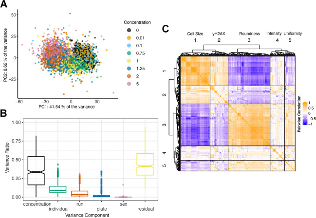
We assessed the main drivers of variation in these morphological traits by performing principal components analysis. The first principal component, accounting for 41.5% of the observed variation across all morphological traits were correlated with MMAIII concentration, and there was a clear dose-dependent effect (Fig 2A). Following Matthew et al. [7], we performed a decomposition of the sources of variation contributing to each trait by fitting a random effects linear model with terms for inter-plate effects (‘plate’), batch effects (12 samples per ‘run’), MMAIII concentration (‘concentration’), DO donor (‘individual’), and the sex of cell donor (‘sex’) (Fig 2B). Among these factors, arsenic ‘concentration’ explained the most variation, followed by ‘individual’ (i.e., donor genetic background). While we randomized DO cell lines by column and MMAIII concentrations by row within a plate, we observed that inter-plate and inter-run effects also influence variance in measured morphological traits (Fig 2B). Depending on the trait, ‘individual’ explained ~0–40% of the variance with an average of 10%, suggesting that a subset of these traits (those with >20%) would provide sufficient signal for genetic mapping based on the size and architecture of our DO cell population [49,50].
Fig 2. HCS Features are Influenced by MMAIII Concentration and Genetic Background.
(A) Principal Component Analysis (PCA) of the raw image analysis feature dataset colored by the concentration of MMAIII. Among known factors, MMAIII concentration was correlated with PC1 (41.54%). (B) Boxplot showing the aggregated results from variance component analysis (VCA) performed across all cellular features, including MMAIII concentration, DO cell lines (individual), each 96-well plate (plate), run (group batch), sex, and residual variation. (C) Heatmap showing the Pearson’s pairwise correlation structure of the raw cellular features. The heatmap and dendrogram were generated using the R package ComplexHeatmap’s Heatmap() function with column_split and row_split, each set to 5.
While HCS produces thousands of morphological traits, many of them are highly correlated (Fig 2C). The correlated groups could be loosely categorized as traits describing `cell size', `γH2AX foci', `cell roundness', `intensity', and `uniformity' (Fig 2C). While there are a variety of dimension reduction techniques that take advantage of correlation to summarize high dimensional data, we were most interested in traits exhibiting non-linear, dose-dependent responses.
Dose-response modeling and genetic mapping of cell morphology quantitative trait loci (cmQTL)
Dose-response models provide benchmark dose estimates of the concentrations at which an exposure to a given chemical could pose a health risk [53]. To focus on the subset of traits exhibiting dose-dependent responses, we performed dose-response modeling using the drc R package [54] for each cellular trait, individual, and replicate experiment (Fig 1E). These models provided quantitative dose-response parameters (DRPs) describing each donor individual’s cellular response including effective concentrations (EC’s), starting/maximum asymptotes, and rates of change (slopes) [55]. For example, an individual’s EC50 represents the concentration of MMAIII at which there is a 50% change in a given cellular feature relative to baseline. Following dose-response modeling, we applied an interplate batch correction and summarized across intraindividual replicates which resulted in 5,105 cmDRPs from 568 cellular traits (see Methods).
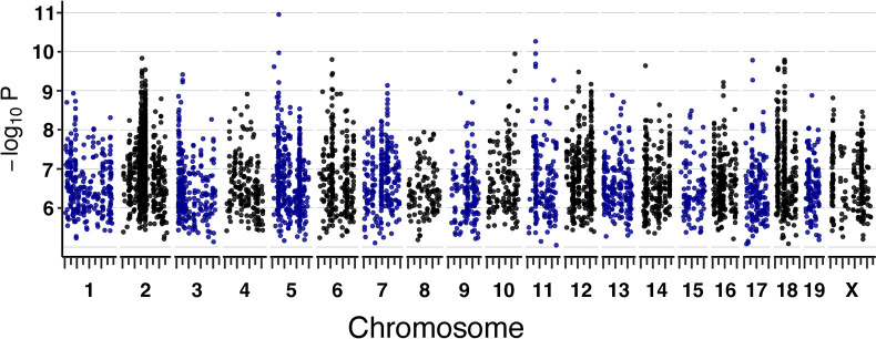
To reveal genetic loci that influence sensitivity to arsenic metabolite MMAIII, we performed quantitative trait loci (QTL) mapping, treating the 5105 cmDRPs as quantitative traits (see Methods) (Fig 1F). To account for the data’s complicated structure and redundancy in the context of multiple testing burden, we calculated an experiment-wide, genome-wide false discovery rate (FDR) significance threshold, which resulted in one maximum peak meeting significance (FDR ≤ 10%) (Fig 3). Given that this work represents a proof of principle and cmDRPs are potentially noisy as modeled quantities, we also used a lenient significance threshold of LOD score > 7.5, which corresponds to ~80% power for a moderately polygenic trait in the DO [50]. Of the 5105 cmDRPs, 854 possessed suggestive genetic loci associations, with the strongest LOD score being 10.95. Chromosomes 2, 3, 6, 12, 14, 18 contained cmQTL hotspots, which contain genetic associations for numerous traits, many of which proved to be correlated (Figs 2C and 3). cmQTLs with suggestive LOD scores included EC’s, slope, and maximum asymptotes, which we refer to as dose-response cmQTLs, in addition to baseline (starting asymptote) cmQTLs.
Fig 3. Dose-Response Modeled cmQTL in DO Fibroblasts Exposed to MMAIII.
Summary of cmQTL maximum peaks for 5100 cmDRPs. Each points represents the strength of the genetic association as a LOD score on the y-axis (-log10P) across the mouse genome (x-axis). On the x-axis, long tick marks represent the start of the chromosome and 50 Mbp intervals, while the short tick marks are at 25 Mbp intervals.
Candidate cmQTL genes identified using differential gene expression analysis, gene set enrichment, and data integration
To nominate candidate genes and variants within cmQTL, we used several approaches. We generated bulk RNA-seq data from 16 DO fibroblast lines across 0 and 0.75 μM exposures which we used for differential expression (DE) analysis. Then we used gene set enrichment analysis (GSEA) to identify individual genes, and groups of genes, that showed differential expression in response to MMAIII (S3 Table). We interrogated published gene–arsenic interactions through the Comparative Toxicogenomics Database (CTD) [56] and we quantified the number of interaction annotations in CTD across all curated studies involving MMAIII, MMAV, DMAIII, DMAV, sodium arsenite, sodium arsenate, arsenic, and arsenic trioxide. For any causal variants that exert their effects through gene expression, the contributing haplotypes and direction of their effects can be correlated across eQTL and cmQTL in datasets generated from the same genetic reference population (DO). Therefore, we also correlated the cmQTL haplotype effects with previous DO eQTL from liver, heart, kidney, striatum, pancreatic islet cells, and mESCs (see Methods). Finally, local variant association mapping within each cmQTL allowed us to identify the locations of SNPs and structural variants with the highest LOD scores in each interval in relation to gene positions.
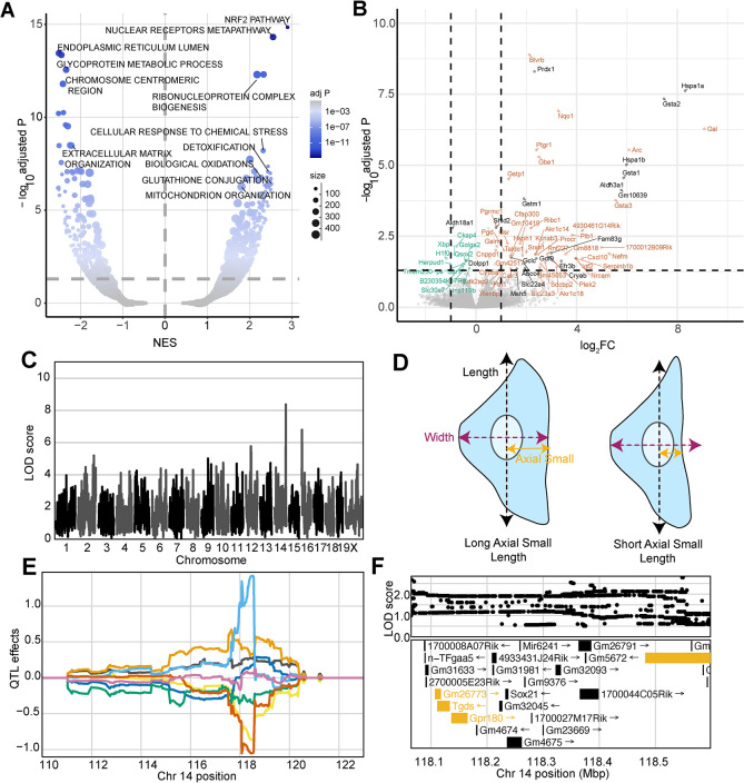
At the pathway level, the most upregulated gene set in dosed samples was ‘NRF2 activation (WP2884)’, which is a well-established response to oxidative stress following arsenical exposure [57–60] (Fig 4A). NRF2, also known as NFE2L2, is a transcription factor that is shuttled to the nucleus following dissociation from KEAP1 in response to the generation of ROS [61–63]. In the nucleus, NFE2L2 binds antioxidant response elements (AREs) upstream of many redox homeostasis and cellular defense genes to drive their transcription in response to stress, including arsenical exposure [57,58,64–67]. These data provided multiple lines of evidence supporting Nfe2l2 (Nrf2) as a candidate gene for the cmQTL hotspot that we found on chromosome 2 (Fig 3). Our gene expression analysis also revealed five candidate genes for other response cmQTL with LOD scores > 8 (Fig 4B). Three of the five genes were present within the same CI, including Hspa1b, Hspa1a, and Msh5, and the former two differentially expressed genes (DEGs) have over 80 previously defined interactions with arsenic metabolites in CTD. We also found that among the other DEGs 73 (89%) have not previously been associated with MMAIII, though many have been associated with arsenic or other arsenic metabolites.
Fig 4. Differential Expression and cmQTL Together Support MMAIII Glutathione Conjugation and its Export via ABCC4.
(A) Volcano plot showing the normalized effect sizes (NES) and -log10 Benjamini-Hochberg adjusted p values (adj. P) of the score-based gene set enrichment (GSEA) results from differential expression (DE) analysis across the 0 and 0.75 μM MMAIII exposed DO fibroblasts groups (n = 32, 16 individuals). Genes were considered “not expressed” if expression levels were TPM < .5 or if at least half of the points were below this cutoff. Each point represents a gene set from `GO:Component',`REACTOME', `KEGG', `WikiPathways', `GO:Tissue', `GO:Molecular Function' and `GO:Biological Process'. The size of each points represents the number of genes within the gene set and the color represents the -log10 adj. P (y-axis). Horizontal dashed line indicates the adj. P significance threshold (adj. P = 0.05). (B) Volcano plot showing the log2-fold change (log2FC) and adjusted p values (-log10 adj. P) for single genes. The horizontal indicates the adj. P significance threshold (adj. P = 0.05) and the vertical lines represent the ± 1 log2fold change for reference. Points labeled with gene names are significantly differentially expressed (adj. P < .05) with effect sizes > 0.75 log2FC or < -0.25 log2FC. Colors represent genes withing cmQTL confidence intervals (black), upregulated (orange) and downregulated (green) DE. (C) QTL scan for the EC05 cell MitoTracker size (‘EC05_nonborder_mitosmooth_axial_small_length_mean_per_well’) cmQTL with the maximum peak at chromosome 14:118483436 bp (GRCm38) and a LOD score of 8.36. (D) Cartoon fibroblast cells depicting the two measurements of cell length (black), width (purple), and axial small width (yellow). Fibroblast on the left has a longer axial small length compared to the fibroblast on the right. (E) Haplotype effects plot showing the eight DO founders (colors, see Methods) for the EC05 cell MitoTracker size (‘EC05_nonborder_mitosmooth_axial_small_length_mean_per_well’) cmQTL across the surrounding region on chromosome 14 (Mbp). Colors indicate founder mouse strains: A/J (yellow), C57BL/6J (gray), 129S1/SvImJ (orange), NOD/ShiLtJ (dark blue), NZO/HILtJ (light blue), CAST/EiJ (green), PWK/PhJ (red), and WSB/EiJ (purple). (F) Variant association mapping within the CI the cmQTL EC05 cell MitoTracker size (‘EC05_nonborder_mitosmooth_axial_small_length_mean_per_well’). Top panel shows the LOD scores of the known, segregating variants in the 8 DO founders (GRCm38). Bottom panel shows the gene models within the respective CI. Each point represents a variant. Colors indicate whether a gene is expressed > 0.5 TPM (gold) or < 0.5 TPM (black). The arrow indicates the direction of transcription.
Natural variation in cellular detoxification pathways partially explains arsenic sensitivity
The other two DEGs within dose-response cmQTL were Cryab and Abcc4, each with ≥ 19 published arsenical interactions (Fig 4B). SNPs in Abcc4 have been previously associated with sensitivity to arsenic [68]. Abcc4 encodes the protein ABCC4/MRP4, which has been shown to export glutathionylated MMAIII from cells [69,70]. Glutathione transferases like Gstm1, Gsta1, and Gstp1 were also significantly upregulated in our expression dataset. These genes are members of the glutathione conjugation pathway which is a detoxification pathway that leads to glutathionylation of MMAIII (MMADGIII) (Fig 4A and 4B) [69,71]. We found multiple cmQTLs at the Abcc4 locus and they were all for traits related to changes in cell size (i.e., length, compactness) (Fig 4C). For example, one of these response cmQTL was EC5 of the change in axial small length or the dose at which 5% of the cell population exhibited measurable differences in cell size (defined by the smoothed MitoTracker labeling which captures the cytoplasmic area occupied by mitochondria) (Fig 4D). Variant association mapping revealed that the highest scoring SNPs in these cmQTLs were within the Abcc4 gene, and the haplotype effects indicated that changes in cell size (‘shrinkage’) occur at lower doses in individuals with PWK haplotypes compared to those with NZO haplotypes (Fig 4E and 4F). Taken together, these data support a model where sensitivity to arsenic exposure in the DO population is partly regulated by natural variation in the efficiency of MMAIII detoxification and export.
Xrcc2 haplotypes modulate and predict of cellular responses
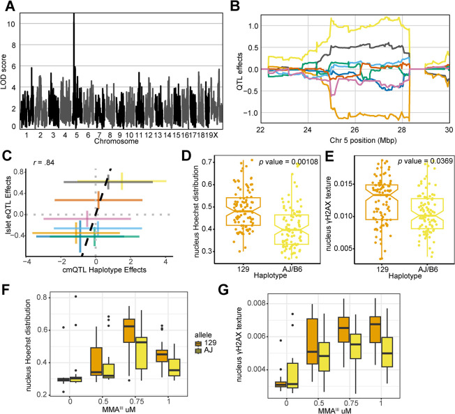
The cmQTL with the highest LOD score was on chromosome 5 at 27,327,254 bp (GRCm38) for the response cmQTL EC90 nucleus Hoechst distribution texture hole (EC90 Nonborder Nucleus Symmetry 02 SER Hole (Hoechst) Mean Per Well) (Fig 5A–5D). Hoechst nuclear fluorescence in cells with the 129 haplotype resembled apoptotic nuclei [72] and were brighter and more uniform than those found in cells with AJ/B6 haplotypes (Figs 5B and S1A). The highest associated SNPs for this cmQTL were located in two genes: Actr3b and Xrcc2 (S1B Fig), however several key points suggest Xrcc2 as the more likely candidate. First, Xrcc2’s paralogs, Xrcc1 [73,74] and Xrcc3 [75,76] have both been associated with genetic susceptibility to arsenical exposure. Second, knockdowns of Xrcc2 were previously shown to increase both γH2AX intensity and chromosomal abnormalities [77], and Xrcc2 is a member of the Biological Fibroblast Apoptosis (GO:0044346) and DNA Damage Repair pathways (R-MMU-5693532). Lastly, the cmQTL haplotype effects are highly correlated with an Xrcc2 eQTL in pancreatic islets cells from the same mouse population (Fig 5C). Taken together, these results suggested that genetic variation at this locus may be mediating DNA damage-induced apoptosis through Xrcc2 expression.
Fig 5. Xrcc2 haplotype modulates chromosomal organization and DNA damage during acute MMAIII exposure.
(A) QTL scan for the EC90 nucleus Hoechst distribution texture hole (`EC90 Hoechst Nucleus Symmetry (02) Hole Mean per Well') cmQTL with the maximum peak at chromosome 5: 27,327,254 bp (GRCm38) and a LOD score of 10.95. (B) Haplotype effects plot showing the eight DO founders (colors, see Methods) for the EC90 nucleus Hoechst distribution texture hole (`EC90 Hoechst Nucleus Symmetry (02) Hole Mean per Well') cmQTL across the surrounding region on chromosome 5 (Mbp). Colors indicate founder mouse strains: A/J (yellow), C57BL/6J (gray), 129S1/SvImJ (orange), NOD/ShiLtJ (dark blue), NZO/HILtJ (light blue), CAST/EiJ (green), PWK/PhJ (red), and WSB/EiJ (purple). (C) Pairwise correlation between the haplotype effects of Xrcc2 expression in pancreatic islet cells at chromosome 5:27,327,254 bp (GRCm38) and the haplotype effects of the EC90 nucleus Hoechst distribution texture hole (`EC90 Hoechst Nucleus Symmetry (02) Hole Mean per Well') cellular feature (Pearson correlation coefficient (r) = .84). The colors are the same as in panel B. (D) Boxplot showing the nucleus Hoechst distribution texture hole cellular feature at 1 μM MMAIII for the top 129 (n = 24, 4 replicates each; orange) and AJ/B6 (n = 24, 4 replicates each; yellow) haplotypes and technical replicates in the DO fibroblasts. (E) Boxplot showing the γH2AX fluorescence texture bright cellular feature at 1 μM MMAIII for the top 129 (n = 24, 4 replicates each; orange) and AJ/B6 (n = 24, 4 replicates each; yellow) haplotypes and technical replicates in the DO fibroblasts. (F) Boxplot showing the nucleus Hoechst distribution texture hole cellular phenotype in a follow-up experiment where DO fibroblasts with 129 (n = 5; orange) and AJ (n = 5; yellow) haplotypes exposed to increasing MMAIII concentrations. (G) Boxplot showing the nucleus γH2AX texture bright cellular phenotype in a follow-up experiment where DO fibroblasts with 129 (n = 5) and AJ (n = 5) haplotypes exposed to increasing MMAIII concentrations. Colors indicate the DO founder strains (see Methods).
Because of the role in Xrcc2 in DNA damage and apoptosis, we reasoned that γH2AX fluorescence might also be higher in cells with the more sensitive 129 haplotype compared to cells with the more resistant AJ/B6 haplotypes at the higher MMAIII concentrations. As expected, we observed a significant difference for fibroblasts with 129 haplotypes (n = 24, 4 replicates each) compared to individuals with AJ/B6 haplotypes (n = 24, 4 replicates each) for the nucleus Hoechst distribution texture hole phenotype at the 1.0 μM concentration based on a t-test accounting for the replicate structure with a random effect (p value = 0.0011)[78] (Fig 5D). Using this same technique, we also observed that ‘nucleus γH2AX texture bright’ feature (a proxy for γH2AX fluorescence) was significantly higher in the fibroblasts with the 129 haplotype compared to the AJ/B6 haplotypes (p value = 0.0369) (Figs 5E and S1A). Indeed, the ‘nucleus γH2AX texture bright’ feature was significantly higher in the fibroblasts with the 129 haplotype compared to the AJ/B6 haplotypes (Figs 5E and S1A). We sought to assess the reproducibility of these effects, both for the original phenotype and the increase in γH2AX. Taking advantage of our full panel of 600 cell lines, we selected an orthogonal group of lines based on their haplotype at this locus (n = 5 for each allele). Not only were we able to recreate the original nuclear symmetry difference between genetic backgrounds (Fig 5F), but we also observed the same γH2AX fluorescence effects that were found in the original screen (Fig 5G). This example shows that genetic variation in near Xrcc2 influences sensitivity and that the haplotype effects of cmQTL have predictive value for identifying sensitive cell lines.
Non-coding genetic variation influences TXNRD1 cell fate during induced oxidative stress
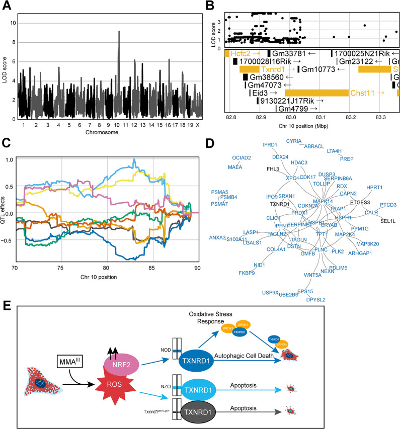
To further investigate how these data could be used for the discovery of gene–environment interactions, cmQTL mapping was performed in a subset of cells lacking accumulated DNA damage. Linear classification was performed to separate cells into γH2AX-positive and γH2AX-negative populations prior to feature extraction. To do this, we took advantage of PHENOLogic machine learning algorithms of the Harmony 4.9 software and gated the imaged cells into γH2AX-positive and γH2AX-negative populations prior to feature extraction, dose-response modeling, and mapping. We detected a cmQTL for the rate of MitoTracker area change in γH2AX-negative cells with a LOD score of 9.16 on chromosome 10 (Fig 6A). This locus was also detected in our original dataset with similar haplotype effects, but with a lower cmQTL LOD score of 7.71 (S2A, S2B and S2C Fig). Upon variant association mapping the highest LOD scoring variants were in the 3’ UTR of the Txnrd1 gene (Fig 6C), a gene that is highly expressed in fibroblasts and has been previously shown to respond to arsenical exposure via changes in NRF2-mediated expression. Moreover, the reducing capacity of TXNRD1 protein is directly inhibited by MMAIII binding [79,80]. As a selenoprotein, the 3’ UTR of Txnrd1 plays a crucial role in recoding a UGA stop codon into a selenocysteine amino acid which is required for function of the TXNRD1 protein as a reducing agent [81–83].
Fig 6. Noncoding Variation in Txnrd1 Modulates MMAIII-Induced Cell Death.
(A) QTL scan for the `γH2AX-negative cells slope Cell Area μm2 mean per well' cmQTL with the maximum peak at Chromosome 10: 82,906,780 bp (GRCm38) and a LOD score of 9.16. (B) Variant association mapping within the CI the cmQTL `γH2AX-negative cells slope Cell Area μm2 mean per well'. Top panel shows the LOD scores of the known, segregating variants in the 8 DO founders (GRCm38). Bottom panel shows the gene models within the respective CI. Each point represents a variant. Colors indicate whether a gene is expressed > 0.5 TPM (gold) or < 0.5 TPM (black). The arrow indicates the direction of transcription. (C) Haplotype effects plot showing the eight DO founders (colors, see Methods) for the `γH2AX-negative cells slope Cell Area μm2 mean per well' cmQTL across the surrounding region on chromosome 10 (Mbp). (D) String-db functional enrichment network of the significantly increased protein interactors detected using immunoprecipitation mass spectrometry (IP-MS) in DO fibroblasts with NOD alleles (n = 6) at the maximum locus for the `γH2AX-negative cells slope Cell Area μm2 mean per well' cmQTL exposed to 0 and 0.75 μM MMAIII concentrations. Colors indicate whether a protein, or node, was shared with a similar experiment in DO fibroblasts with the NZO allele (n = 5). Black represents shared TXNRD1 interactors, and blue represents unique NOD-TXNRD1 interactors. (E) Mechanistic summary of allele specific Txnrd1 responses across the NOD haplotype (blue), NZO haplotype (light blue), and heterozygous SECIS knockout model (Txnrd1em1Lgr/+). Our data suggest DO fibroblasts with the NOD allele have a more robust oxidative stress response upon MMAIII exposure, ultimately succumbing to autophagic cell death represented by increased cell size at medium MMAIII concentrations. In comparison, DO fibroblasts with the NZO allele the Txnrd1em1Lgr/+ alleles exhibit morphology consistent with apoptosis as shown by brighter Hoechst 33342 labeling and smaller cells.
To interrogate the plausibility of Txnrd1 as the candidate for these two cmQTL, we performed score-based GSEA using gene expression data from the bulk RNA-seq data based on their sensitive (NZO, n = 6) and resistant (NOD, n = 6) haplotypes at this locus. We found upregulation of DNA damage and replicative stress gene sets in cells with NZO haplotypes and upregulation of oxidative stress response, p38/MAPK signaling, TGF signaling, RAS signaling, lysosome, and autophagy-related pathways in cells with NOD haplotypes (S4 Table). Among these pathways was nanoparticle triggered autophagic cell death, which can be induced by the treatment of gold, the active component of the TXNRD1 inhibitor auranophin [84]. We did not observe signficant differential gene expression in Txnrd1 based on the haplotype at this locus (S5 Table). However, there were differences in TXNRD1 protein levels in both unexposed cells (p value = 0.0935) and exposed cells (p value = 0.0414) between NOD and NZO haplotypes when accounting for sex and replicate using a t-test [78](S2D Fig). To assess a functional difference in TXNRD1 between these haplotypes, we performed immunoprecipitation followed by tandem mass spectrometry (IP-MS) to quantify protein-protein interactions. Following subtraction of a non-specific binding partner control, we found that compared to healthy, unexposed controls, 0.75 μM MMAIII exposed NOD haplotype cells (n = 6) had a larger number (106) of significant, positive interactors compared to NZO (n = 5) TXNRD1 interactors (33). We visualized these interactions using ‘string-d’ to generate a functional enrichment map of the PPIs for MMAIII exposed NOD and NZO TXNRD1 where we observed that there were very few genes overlapping between alleles, and that MAPK14 was a hub for NOD interactors which included proteins involved in oxidative stress (i.e., PRDX1, SRXN1) and autophagy/p38 (i.e., MAPK14, TOLLIP) (Fig 6D and S6 Table). Considering the gene expression and IP-MS data together, it was evident that in exposed DO fibroblasts, NOD TXNRD1 was involved in autophagy. Previous studies of Txnrd1 deficiency have shown disruption of lysosomal-autophagy in favor of apoptotic cell death [85,86], implying an apoptotic phenotype of cells with NZO haplotypes (NZO-TXNRD1) is akin to that seen with TXNRD1 deficiency. During apoptotic cell death, cell structure and cytoskeleton are quickly degraded, but during autophagy the cytoskeleton is maintained [87–89]; providing a basis for our ability to distinguish between these two pathways and to interrogate their genetic regulation using cmQTL. Taken together, these data support a model whereby natural variation in Txnrd1 influences the trajectory of cell death pathways following MMAIII exposure in the DO population (Fig 6E).
While we did not find coding variants unique to the NZO or NOD Txnrd1 alleles, we found that two SNPs private to the NZO haplotype (rs227869362 and rs257393906) in the 3’ UTR were adjacent to the selenocysteine insertion element (SECIS), which is essential for recoding the UGA stop codon to selenocysteine during translation [82]. We also searched publicly available data for structural variants and INDELs in the 3’ UTR but did not find any that were unique to the NZO haplotype [90]. To determine the functional consequences of variation impacting the SECIS element of Txnrd1, we used CRISPR/cas9 to delete the SECIS in C57BL/6J mice (Txnrd1em1Lgr). While heterozygous mice carrying this deletion were viable and fertile, homozygous mice could not be recovered. Since a full protein knockout of Txnrd1 causes recessive embryonic lethality [91], we concluded that deletion of the SECIS element alone is the functional equivalent of a null allele (see Methods). We then isolated tail tip fibroblasts from heterozygous mice and found that the cell area of arsenic-exposed Txnrd1em1Lgr/+ fibroblasts more closely resembled fibroblasts with the NZO haplotype than their WT controls following MMAIII exposure (S2E and S2F Fig). Similarly, nuclear Hoechst 33342 labeling was brighter and more uniform in the Txnrd1em1Lgr/+ nuclei with increasing MMAIII concentration. Taken together, these data highlight the functional importance of non-coding variation in the 3’ UTR of a key selenoprotein in the context of sensitivity to arsenic induced oxidative stress. Detailed molecular and functional studies are needed to determine the impact of single nucleotide variants on Sec recoding in Txnrd1. However, there is at least one study demonstrating that naturally occurring and engineered single nucleotide variants in the 3’ UTR of the human selenoprotein, SEP15, influence UGA readthrough and dampen the cellular response to selenium stimulation [92].
Natural genetic variation influences fibroblast morphology
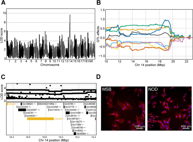
While our primary focus was on population variation in arsenic response, we unexpectedly observed variation in fibroblast morphology in unexposed cells and our genetic analysis revealed multiple loci contributing to this baseline morphological variation (i.e. starting asymptote cmQTL). The highest scoring of these baseline cmQTL (LOD 9.64) was on proximal chromosome 14 (Fig 7A–7B). Several of the top LOD scoring variants were in Ube2e2, which was one of only three protein coding genes expressed in fibroblasts within the confidence interval (Fig 7C). This cmQTL is for a trait that describes the brightness of Hoechst labeling (i.e., texture feature bright 1 pixel mean per well) which is directly related to the distribution and amount of chromatin in the nucleus (Fig 7D) [93]. The ubiquitin conjugating enzyme E2 (UBE2E2) functions in the nucleus to post-translationally modify proteins that regulate the G1/S phase transition together with Trim28 [94], which could explain the difference in Hoechst labeling as mitotic cells accumulate more Hoechst due to their DNA content. This example highlights the role of genetic variation in the regulation of morphology, potentially through variation in basic cellular functions (i.e. cell cycle) providing an exciting avenue for further study.
Fig 7. Genetic variation influences fibroblast morphology at baseline.
(A) QTL scan for the baseline nucleus Hoechst texture bright (`d_nonborder_nucleus_hoechst_33342_ser_bright_1_px_mean_per_well') cmQTL with the maximum peak at chromosome 14:19401644 bp (GRCm38) and a LOD score of 9.64. (B) Haplotype effects plot showing the eight DO founders (colors, see Methods) for the baseline nucleus Hoechst texture bright (`d_nonborder_nucleus_hoechst_33342_ser_bright_1_px_mean_per_well') cmQTL across the surrounding region on chromosome 14 (Mbp). Colors indicate founder mouse strains: A/J (yellow), C57BL/6J (gray), 129S1/SvImJ (orange), NOD/ShiLtJ (dark blue), NZO/HILtJ (light blue), CAST/EiJ (green), PWK/PhJ (red), and WSB/EiJ (purple). (C) Variant association mapping within the CI the cmQTL baseline nucleus Hoechst texture bright (`d_nonborder_nucleus_hoechst_33342_ser_bright_1_px_mean_per_well'). Top panel shows the LOD scores of the known, segregating variants in the 8 DO founders (GRCm38). Bottom panel shows the gene models within the respective CI. Each point represents a variant. Colors indicate whether a gene is expressed > 0.5 TPM (gold) or < 0.5 TPM (black). The arrow indicates the direction of transcription. (D) Representative images for the two fibroblast lines showing higher Hoechst 33342 texture bright in the sample with the NOD allele at the chromosome 14 locus compared to the WSB. Nuclei are shown in blue by Hoechst 33342 labeling and mitochondria are shown in red by MitoTracker Deep Red. Scale bar indicates 100 μm.
Discussion
We created a new model for in vitro analysis of gene–environment interactions using cell lines from a laboratory mouse genetic reference population and performed HCS to quantify changes in cell morphology in response to acute MMAIII exposure. We used dose-response modeling to summarize cellular morphology changes across increasing MMAIII concentrations, from which we estimated individual genotype-level dose-response parameters for cmQTL mapping. We used orthogonal gene expression datasets from previous DO studies [95], pathway information, and gene–chemical interaction data from arsenicals through CTD (https://ctdbase.org) to refine our cmQTL and to identify candidate genes. A summary of these methods, and the major experimental findings from our work, can be found in S3 Fig. We found 70 novel gene expression signatures for MMAIII exposure. Among them were five significantly differentially expressed genes that were also located in our higher LOD score (> 8) cmQTL CIs, including Abcc4. The Abcc4 gene is an intriguing candidate for the EC5 of the change in axial small length cmQTL based on functional activity and gene expression. Variants in the 3’ UTR of Abcc4 can regulate its expression through impacting miRNA binding [96]. We speculate that unique variants in NZO (rs240728821) and PWK (rs245333533) may be acting similarly.
While some cmQTL result from variants that exert their effects through gene expression, others can result from variants that exert their effects post-transcriptionally, to impact protein abundance or function. Therefore, we also considered candidate genes that did not exhibit differential expression in response to MMAIII. Taking advantage of published gene-arsenic interaction data in CTD, we identified 88 genes among our cmQTL that were previously associated susceptibility to arsenic. Only six of these genes (Abcc4, Nfe2l2, Cbs, Gclc, Gstm1, and Xpc) contained non-synonymous coding SNPs that affect the response to arsenicals in previous studies (https://ctdbase.org/). Among the remaining candidate genes there are likely to be many genes with unrecognized roles in the genetic regulation of arsenic response and these warrant further study. Our findings highlight the role of natural variation in MMAIII detoxification, export, and the role of variation affecting the trajectory of cell death pathways that are induced in response to ROS and DNA damage.
Our QTL analysis of machine-learning (ML) derived features (specifically slope γH2AX-negative cell area μm2) identified Txnrd1, which has multiple gene–arsenic associations annotated in CTD. Our variant association analysis revealed high LOD-scoring SNPs adjacent to the SECIS element in the 3’ UTR of Txnrd1 that influence cell size during acute MMAIII exposure. We validated this finding by constructing a CRISPR-deleted SECIS element in the 3’ UTR, which recapitulated the apoptotic, shrunken cellular phenotype we observed in MMAIII exposed fibroblasts with NZO haplotypes. The essentiality of the SECIS element for Sec recoding, relicensing a stop codon into selenocysteine, has been previously demonstrated [83]. However, our study is the first to show that this element is also required for normal embryonic development, similar to the phenotype reported for a Txnrd1 null allele [91]. Taken together, these results show that the deletion of the SECIS element is equivalent to complete loss of TXNRD1 function, lending support to the idea that single nucleotide variants in and around the SECIS element are likely to be detrimental to protein function. When we compared global gene expression differences between the NOD and NZO haplotypes at this locus, we found enrichment for pro-cancer signaling including RAS, TGF, and p38/MAPK signaling in the NOD haplotype compared to the NZO haplotype. This was further supported by our protein interaction data where we found increased NOD TXNRD1 affinity for MAPK14 and oxidative stress-related proteins compared to NZO, which may explain the resistance to MMAIII-induced morphology changes. Together these data support our hypothesis that 3’ UTR variation in Txnrd1 has functional consequences on TXNRD1 function, though direct biochemical analyses are needed to fully understand the molecular consequences of these variants.
Xrcc2’s involvement in the DNA damage pathway may also indicate a cancer-related outcome for the highest cmQTL related to Hoechst labeling distribution. This cmQTL region shares conserved synteny with a region significantly associated with susceptibility to arsenic-induced skin lesions in a Bangladeshi population [97]. Variant association mapping detected a non-synonymous variant in mouse Xrcc2 (rs3156627) unique to the 129, sensitive haplotype that overlaps a human XRCC2 variant (rs2098040934), although neither are predicted to be deleterious [98]. It is also possible that non-coding SNPs lead to differential regulation of Xrcc2 transcript levels in AJ and 129 haplotypes, which could also explain the nuclear morphological changes following MMAIII exposure. If these non-coding variants are influencing gene expression, we would expect to find eQTL with matching haplotype effects. Lacking contemporaneous eQTL data from our screen, we looked to eQTL datasets generated from other DO studies and found matching haplotype effects in at least one other tissue where Xrcc2 is expressed. This provides evidence that the variants underlying Xrcc2 cmQTL may exert their effects through gene expression in MMAIII exposed fibroblasts. Integrative analyses such as these are a key advantage to working with a genetic reference population where there are a growing number of publicly available -omics and QTL datasets available to power candidate gene and pathway discovery [42,99,100].
The variants driving cmQTL could be coding or non-coding and our candidate gene / variant analyses provide several examples of this as described above. This is a key advantage of cmQTL over molecular QTL, like eQTL or protein (pQTL) which are each limited to variants that exert their effects through transcript or protein abundance, respectively. Cell morphology is one of the many cellular-level phenotypes that result from the complex interplay of gene regulatory networks, making them an inexpensive and informative phenotype to use for QTL mapping. Despite our success in identifying cmQTL and, in some case, the genes and variants affecting cellular response to MMAIII, our study had several shortcomings that could be addressed in future work. Quantitative variation in cell morphology traits can be attributed to genetic background and dose-response, but we also found substantial unexplained residual variation in some traits. Previous studies of cell morphology in genetically diverse yeast cell populations have shown that some traits are prone to high experimental variability, especially for features that have high cell-to-cell variability [6]. Since our features are whole-well summaries, cell-to-cell variability is likely a major contributor to our observed residual variation. We also found that the γH2AX features had high residual variation that is likely due to the indirect immunolabeling method used for γH2AX detection, which is a multistep staining method that relies on two antibodies and is known to have more experimental variability than direct labeling with a fluorophore-conjugated primary antibody. Other studies mapping cmQTL were limited by lack of genetic diversity [9,48]. While our study successfully addressed these issues [50,95,101], we found that it was underpowered for detecting QTL effects explaining less than 20% of trait variation and future studies would benefit from technological advances that reduce residual variation.
To induce cell morphology changes that were sufficiently extreme to fit the asymptotes of a sigmoidal dose-response curve, we used concentrations of MMAIII that are unlikely to be encountered through environmental or occupational exposures. Other studies have shown that cell morphology was impacted following lower concentration and longer exposures of arsenic [102], so an important future direction is to explore the differences between acute and chronic exposures. Another statistical challenge arose because existing dose-response modeling software does not allow for direct inclusion of batch effects, and thus, our study design required a post-hoc adjustment of estimated dose-response parameters. This problem may be addressed in future releases of R/drc [54]. We further acknowledge that dose-response modeling results can vary based on the choice of software, the form of the model, and as we observed, the genetic background of the samples [103]. Despite these challenges, we identified hundreds of loci where natural genetic variation in the DO founder strains influences the fibroblast responses to MMAIII and baseline fibroblast morphology.
Our in vitro system used primary fibroblasts, which are a highly abundant cell type that contribute to a diverse range of cellular and tissue functions [104,105]. However, the genetic effects in fibroblasts may not always recapitulate the same molecular mechanisms of sensitivity and resistance as those found in other specialized cell types. Primary fibroblasts are also a limited resource because they will undergo senescence, and they are more difficult to genetically manipulate than transformed cell lines or pluripotent cells. For these reasons, we have generated induced pluripotent stem cell (iPSCs; n = 284) from this panel for future work. iPSCs also enable differentiation into other cell types, 3-dimensional cell models, organoids, or scaffolded arrays, which can be screened across a variety of environmental conditions, including other toxicants, drugs, or other culture conditions.
In conclusion, our study demonstrates that dynamic changes in cell morphology occurring in response to acute exposure to MMAIII across a population of genetically diverse cells exhibit predictable dose-response relationships. These relationships display inter-individual variation and genetic mapping of individual-level dose-response parameters can identify genetic variation that regulates the molecular initiating events that occur during an acute exposure. Our findings indicate that these genetic loci and their associated effects have predictive value for identifying sensitive and resilient individuals in vitro. While further work is needed to explore the applicability of these predictions to in vivo responses, cell lines derived from mouse genetic reference populations present an exciting opportunity for iterative in vitro screening and precise in vivo testing of genetically encoded susceptibility and resistance to the effects of toxic exposure.
Materials and methods
Ethics statement
All procedures involving laboratory mice were approved by The Institutional Animal Care and Use Committee of The Jackson Laboratory (under Animal Use Summary #20030).
Fibroblast derivation
We modified a previously described protocol for deriving fibroblasts by biopsying 2–3 mm tail tips from adult male and female DO (RRID:IMSR_JAX:009376) mice, aged approximately 4–6 weeks, using a procedure approved by The Jackson Laboratory’s Institutional Animal Care and Use Committee [106]. Samples were initially collected into Advanced RPMI 1640 cell culture media (Gibco) supplemented with 1.0% penicillin/streptomycin (P/S) (Gibco), 1.0% GlutaMAX-I (GlutaMAX)(Gibco), 1.0% MEM Non-Essential Amino Acids (NEAAs)(Gibco), and 0.0005% 2-mercaptoethanol (BME)(Gibco). Tail tissue was minced using razor blades and digested in media containing collagenase D (Gibco) at a concentration of 2.5 mg/ml on an orbital shaker at 37°C. The digested samples were further fragmented using micropipettes ranging from p1000 to p200 and dissociated in RPMI 1640 media containing 1.0% P/S, 1% Glutamax, 1.0% non-essential amino acids, 0.0005% BME, and 10% fetal bovine serum (FBS)(Gibco), hereinafter referred to as ‘fibroblast media’, for approximately 3–5 days (passage number 0; P0). All passaging was done using a phosphate-buffered saline pH 7.2 (1X, PBS) wash and 0.05% Trypsin-EDTA (Trypsin) (Gibco). Individual DO fibroblast samples were expanded to P5 with reserves frozen at approximate densities of 3.5 x 10⁵ cells/ml at passage numbers P2, P3, and P5 in freezing media containing RPMI 1640 with 10% dimethyl sulfoxide (DMSO) and 10% FBS. All DO fibroblast samples were transferred to liquid nitrogen holding tanks for long-term storage after controlled freeze (-1°C/min) 24–48 hours at -80°C.
Genotyping
DNA was collected from the spleen tissue of each DO mouse and genotyped using the Giga Mouse Universal Genotyping Array (GigaMUGA; [107]). The haplotypes were reconstructed using a hidden Markov model to estimate genotype probabilities at each locus for the population, as described previously [108].
Exposure of Fibroblasts to MMAIII and sample preparation
Frozen aliquots of P5 fibroblast lines were thawed and grown in fibroblast media in 60-mm tissue culture-treated plates for 48 hours. Trypsinized cells were resuspended and viable cell density was estimated using Trypan Blue and a Nexcelom Cellometer Auto T4 Plus Cell Counter. 100 μl of each fibroblast line was seeded into four columns (four technical replicates) distributed across two CellCarrier Ultra 96-well black, clear bottom, tissue culture treated microplates using the Integra Assist Plus at a density of ~2500 viable cells/well after randomization across columns. After 24 hours, the fibroblast media was replaced with 100 μL of fibroblast media containing monomethylarsonous acid (MMAIII; Toronto Research Chemicals) at the concentrations 0 μM, 0.01 μM, 0.1 μM, 0.75 μM, 1.0 μM, 1.25 μM, 2.0 μM, and 5.0 μM across plates and rows.
After the cell lines were exposed to MMAIII for 24 hours, the media was replaced with media containing MitoTracker Deep Red (200 nM; Invitrogen), and the cells were incubated at 37°C for 20 minutes in 96-well plates. The cells were then fixed in ice-cold 100% methanol on ice for 10 minutes. After the cells were washed three times with PBS, they were bathed in a 1.0% bovine serum albumin (Fraction V) (BSA), 0.1% Tween solution overnight at 4°C on a shaker. Blocking solution was then replaced with anti-γH2AX (phosphorylated S139) antibody (Abcam, ab11174, 1:2000) in the blocking solution and incubated at room temperature for 2 hours on a shaker. After washing the cells three times with PBS, Alexafluor 488 donkey anti-rabbit secondary antibody (1:2000; Abcam) was added for 1 hour at RT on a shaker in blocking solution. Next, Hoechst 33342 (1:8000; Abcam) was added to the cells and incubated for 10 minutes at RT on a shaker. The plates were subsequently washed, and 100 μL of PBS was left in each well for storage at 4°C and imaging.
Automated image acquisition
96-well plates were imaged using an Operetta CLS for the MMAIII screen and Xrcc2 follow-up experiments equipped with a 20x/1.0 water immersion objective and binning 2. A single z-plane was acquired from 25 contiguous fields per well. Exposure times, focal heights, and excitation power settings for the Operetta CLS screen were: Hoechst 33342 (time: 100 ms, power: 100, height: -5), Alexa 488 (time: 200 ms, power: 100, height: -5), MitoTracker Deep Red (time: 500 ms, power: 100, height: -5). Exposure times, focal heights, and excitation power settings for the Xrcc2 follow-up experiments were: Hoechst 33342 (time: 300 ms, power: 100, height: -6), Alexa 488 (time: 80 ms, power: 100, height: -6), MitoTracker Deep Red (time: 200 ms, power: 100, height: -6). Lastly, Txnrd1em1Lgr/+ and control fibroblast samples were imaged using the Opera Phenix High-Content Imaging System with a 20x/1.0 water immersion objective, binning 2, and a single z-plane with 25 contiguous fields per well. Exposure times, focal heights, and excitation power settings were as follows: Hoechst 33342 (time: 100 ms, power: 80, height: -10) and MitoTracker Deep Red (time: 40 ms, power: 50, height: -10).
Image analysis / cellular segmentation
Flatfield corrected images were analyzed and processed using Harmony 4.9 software with PhenoLOGIC (PerkinElmer). Gaussian smoothed images were used for image segmentation, with a focus on two main regions of interest (ROIs), including using Hoechst 33342 to define the nucleus and MitoTracker Deep Red to define the cytoplasm surrounding each nuclear ROI. Fluorescence patterning (i.e., texture) and intensity were measured in the nuclear and cytoplasmic regions using the Hoechst 33342, γH2AX/Alexa-488, and MitoTracker Deep Red/MitoTracker Deep Red Gaussian smoothed channels. Features including nuclear area, Hoechst 33342 intensity, and nucleus edge texture were extracted and represented as mean +/- SD per well. Additionally, a spot analysis was performed using the γH2AX/Alexa-488 labeling in the nucleus. The second image analysis approach used the PhenoLOGIC machine learning (PerkinElmer) algorithms in the Harmony 4.9 software to define sub-populations of cells based on γH2AX/Alexa-488 (γH2AX-positive and γH2AX-negative) and MitoTracker Deep Red (stressed and unstressed) fluorescence prior to feature extraction to generate features including ‘MitoTracker Cell Area in γH2AX negative cells’. For more information about the specific Harmony HCS features, an Image Analysis Guide may be requested from PerkinElmer. More background information for image-based profiling and HCS features can be found through the CellProfiler documentation [109].
Feature variance and relatedness
Principal components analysis was performed on the cellular features across all concentrations, individuals, and plates using the `pca' function from the R pcaMethods with the option `scale = “uv”'. Variance component analysis was performed using the ‘lmer’ function from the R package lme4. The sources of variation included in the model were sex, DO generation (‘generation’), DO donor (‘individual’), 96-well plate (‘plate’), and run (See Eq 1). Variance components were extracted from the model using the function ‘VarCorr’ for each of the random effects (generation, sex, individual, and plate). Residual variance was extracted as the sigma from the model summaries. Ratios of the variance components were determined by dividing each variance component by the sum of all the variance components and the residual variance.
| Eq 1 |
Lastly, the pairwise correlation structure of these data was calculated using the `cor' function in the WGCNA R package with the option `use = "pairwise.complete.obs"'. The heatmap was created using the ComplexHeatmap R package, and the dendrogram added using the `column_split' and `row_split' options each set to 5. We added terms to the heatmap clusters based on a qualitative examination of the clustered trait names.
Cellular feature dose-response modeling
The drc R package [54] was used to perform dose-response modeling for the 673 cellular features. For each of the 226 individuals and cellular features, four dose-response models for each technical replicate column were fit to the four-parameter log-logistic dose-response model (see Eq 2) using the ‘drm’ function with the ‘fct’ set to ‘LL.4’ with log-normalized cellular features using the ‘bcVal = 0’ option. Based on Eq 2 [54] where x represents concentration, b represents the slope, d represents the upper asymptote, c represents the lower asymptote, and e represents the EC50, these model parameters were extracted from the summary of the model fits. Additionally, the ‘ED’ function was used to estimate the EC5, EC10, EC25, EC75, and EC90 for each model fit ‘relative’ to the asymptotes. Four replicates for each model fit parameter summary were estimated for each DO individual and cellular feature.
| Eq 2 |
LMM / BLUP estimation
The dose-response parameter replicates for each DO individual’s concentration response parameters were summarized using Eq 3 using a linear mixed effects model (LMM) to adjust for batch effects using LMM. The LMM was fit using the ‘lmer’ function from the R package lme4. Each cellular feature was modeled where yi is the dose-response parameter estimate for a given cellular feature and where each DO individual (i) was modeled with varying intercepts through random effects for mouse/individual and 96-well plate. The random error term εi is assumed to εi ~ N(0, σ2), and σ2 is the error variance. Data without the effect of plate were extracted as the best linear unbiased predictors (BLUPs) of the random effect for DO individual and used for QTL mapping analysis.
| Eq 3 |
Cellular feature QTL mapping
All data were converted to the normal quantiles calculated from the ranked data, i.e., the rank-based inverse normal transformation (rankZ) to force a Gaussian distribution for mapping. QTL mapping was performed using the qtl2 R package. Briefly, a genetic relationship matrix (i.e., kinship matrix) was calculated from the genotype probabilities using the ‘calc_kinship’ function with the ‘leave one chromosome out’ (loco) option for genetic mapping and the “overall” option for heritability (h2) estimation. Sex and DO generation were included as covariates that were assigned binary values in the LMM for QTL mapping.
For QTL mapping, whole genome scans were performed by first testing individual loci spanning the genome for association with each cellular feature (using qtl2’s ‘scan1’ function). The haplotype effects were then estimated at detected QTL as BLUPs (using the ‘scan1blups’ function) to identify the parental haplotypes driving each QTL and their respective directionality. SNP-association mapping was performed using the ‘scan1snps’ function and the known variants across the eight founder strains of the DO (https://doi.org/10.6084/m9.figshare.5280229.v3). Genome-wide FDR = 0.10 was calculated using the permutations (n = 100,000) for the ‘EC50 number of nuclei’ trait as simulated permutations for all 5105 cmDRPs mapped.
RNA-seq of fibroblast samples
32 fibroblast cell lines, including those with NOD (n = 6), NZO (n = 6), and NOD/NZO heterozygous (n = 4) haplotypes at Chr10:82.89 Mbp (GRCm38) were thawed into 60-mm cell culture-treated plates and grown to confluency (≥ 0.8 x 10⁶ cells/ml) in fibroblast media. Each cell line was then passaged equally into two 60-mm cell culture dishes and grown to 75% confluency, upon which one 60-mm dish received 0.75 μM MMAIII containing fibroblast media and one 60-mm dish received standard fibroblast media. Following 24-hr exposure, both treated and untreated samples were independently collected as cell pellets and snap frozen on dry ice for 15 minutes. Samples were stored at -80°C prior to RNA isolation. RNA was extracted using a NucleoMag RNA Kit (Macherey Nagel) and purified with a KingFisher Flex system (ThermoFisher). Library preparation was enriched for polyA containing mRNA using the KAPA mRNA HyperPrep Kit (Rocher Sequencing and Life Science). Paired-end sequencing was performed with a read-length of 150 bp on an Illumina NovaSeq 6000. Genes were tagged as “not expressed” and filtered out if the the median TPM was < .05 for half or more of the samples and the remaining expressed genes were highlighted in “gold” in the variant association mapping plots.
Transcriptomic profiling
Genotypes for each sample were reconstructed using the genotype by RNA-seq pipeline (GBRS) and aligned to the 8 founder allele-specific genome using GBRS RNA-seq pipeline to quantify read counts for each gene [110] (available through GitHub at TheJacksonLaboratory/gbrs_nextflow). These expected counts were the input for differential expression between the 0 and 0.75 μM exposures using the R package DEseq2 [111]. The fgsea R package was then used to perform a score-based gene set enrichment analysis [112]. The input for GSEA was the exposure-based log2 fold-change for each gene normalized by its standard error. Gene Ontology (GO), REACTOME, WikiPathways, and Biocarta genesets for Mus musculus were obtained via the R package msigdb [113]. Additionally, the R package ClusterProfiler was used to assess enrichment of the significant differentially expressed gene set based on the outlying alleles for the cmQTL on chromosome 10 (GRCm38) [114].
CTD database mining
The Comparative Toxicogenomics database (CTD) was used to identify gene–arsenic interactions previously defined for candidate genes within cmQTL CIs. The gene–arsenic interactions were downloaded for these arsenicals: monomethylarsonic acid (MMAV), monomethylarsonous acid (MMAIII), dimethylarsinic acid (DMAV), dimethylarsinous acid (DMAIII), arsenic trioxide (ATO), sodium arsenite, sodium arsenate, and elemental arsenic (As). NCBI gene ID’s were then merged to Ensembl IDs and their mouse orthologs obtained through Ensembl’s BioMart tool [115]. The number of `Interactions' were aggregated for each gene across the arsenicals to get an `Interaction Count' for the genes within cmQTL CIs.
Comparing haplotype effects to eQTL across tissues
To determine whether cmQTLs colocalized with eQTLs from other DO tissues, Pearson correlation coefficients and Spearman correlation coefficients were calculated between the haplotype effects of eQTLs across liver, kidney, heart, bone, mESCs, striatum, and pancreatic islet cells which are all publicly available, published datasets that can be accessed through DODB (https://dodb.jax.org/) and all genes within the cmQTL CIs (< 8 Mb wide) that were shown to be expressed in fibroblasts [95]. cmQTL and eQTL haplotype effects were calculated using the coefficients from the ‘fit1’ function in the qtl2 R package.
TXNRD1 relative abundance
DO fibroblasts were selected based on their genotypes at the Txnrd1 locus, representing 6 NOD, 5 NZO, and 4 NOD/NZO haplotypes balanced for both male and female lines. Each line was split into two 60 mm dishes where one 60 mm plate received 0 μM MMAIII containing media (unexposed) while the other contained 0.75 μM MMAIII containing media. After 24 hours, cell pellets split into two vials and snap frozen on dry ice for further processing and liquid chromatography tandem MS (LC-MS/MS) analyses. Protein pellets were resuspended in 150 uL of 50 mM HEPES, pH 7.4, and lysed by passing through a syringe with 28 gauge needle (10 passes), vortexing for 30 seconds, and waterbath sonicating for 5 minutes (30 seconds on, 30 seconds off). Lysates were then clarified via centrifugation at 21,000 x g for 10 minutes at 4°C. Clarified lysates were quantified using a microBCA assay and 20 μg samples were diluted to 50 uL for digestion in 50 mM HEPES, pH 8.2. Samples were then reduced with 10 mM DTT at 37°C for 30 minutes, alkylated with 15 mM IAA at room temperature in the dark for 20 minutes, and trypsin digested overnight at 37°C (trypsin:protein ratio of 1:50). Samples were then cleaned-up using Millipore P10 zip-tips, dried in a vacuum centrifuge, reconstituted in 20 μL of 98% water/2% ACN with 0.1% formic acid, and transferred to mass spec vials. Each sample was analyzed using Thermo Eclipse Tribrid Orbitrap Mass Spectrometer coupled to a nano-flow UltiMate 3000 chromatography system on a Thermo 50 cm EasySpray C18 column as described previously with the exception that the gradient was scaled down to a 90 minute gradient [116]. TXNRD1 abundance was determined based on the target peptide: IEQIEAGTPGR. Raw peak data was processed using Skyline (version 22.2.1.278) and further analyzed in R. All mass spectrometry analysis was performed in the in The Jackson Laboraory (JAX) Mass Spectrometry and Protein Chemistry Service.
Immunoprecipitation mass spectrometry (IP-MS)
Immunoprecipitation mass spectrometry (IP-MS) was performed using a rabbit antibody derived against the mouse TXNRD1 protein gifted from Dr. Edward Schmidt (Montana State University) to determine TXNRD1 binding partners using the samples and instrumentation described in the ‘TXNRD1 Relative Abundance’ section. M-280 Sheep Anti-Rabbit IgG Dynabeads (Invitrogen, 11203D) were prepared and coupled to the rabbit anti-mouse TXNRD1 antibody according to manufacturer protocol; additional IgG control beads with no TXNRD1 were also prepared as a non-specific binding partner control for the beads. A ratio of 5 ug of antibody to 5 x 10⁷ beads was used. All Dynabeads were then blocked with 5 mg/mL BSA overnight at 4°C during the antibody coupling step. Coupled and control IgG Dynabeads were then bound to 250 μg of protein lysate at room temperature with rotation for one hour. Heterozygous samples were pooled and used as IgG subtractive controls to assess non-specific binding for the beads. All bound bead fractions were clarified with a magnet, then washed three times with Wash Buffer A (10 mM HEPES at pH7.4, 10 mM KCl, 50 mM NaCl, 1 mM MgCl2, NP-40 (0.05% w/v)), followed by two washes with Wash Buffer B (10 mM HEPES at pH7.4, 10 mM KCl, NP-40 (0.05% w/v)). Washed beads were then digested on-bead as described for the relative abundance section above with the exception of 500 ng of trypsin being used. Samples were then purified using a Millipore P10 Zip-tip and prepped for tandem mass spectrometry analysis, both as described above in the relative abundance section. Raw data was analyzed using the Thermo Proteomic Discoverer software as described previously in the JAX Mass Spectrometry and Protein Chemistry Service using standard operating protocols [116].
PPI and functional enrichment
The string_db R package was used to assess the functional enrichment of proteins binding TXNRD1 to generate protein-protein interaction (PPI) networks for the allele-specific IP-MS results [117]. A score threshold of ‘400’ was used to identify functional interactions between TXNRD1-interacting proteins (nodes) across NOD and NZO haplotypes at the Chromosome 10 locus, which were indicated as edges in the igraph R package visualization. The PPI was colored based on shared (black) and unique (blue) proteins across alleles.
Deletion of Txnrd1 SECIS element
To delete a 200-bp region containing the 75-bp SECIS translational regulatory element of Txnrd1 (MGI:1354175, NCBI Gene: 50493, ENSMUSG00000020250) as well as the flanking regions where 3’ UTR variants are found in NZO haplotypes, C57BL/6J (The Jackson Laboratory stock #000664, RRID:IMSR_JAX:000664RRID:JAX000664) embryos were engineered using CRISPR/Cas9. The murine SECIS element of Txnrd1 is located at 1967–2042 bp in the Txnrd1 mRNA NM_001042513.1. Two sets of gRNAs were used (gRNA up 1:GGAGGCTGCAGCATCGCACT, gRNA down 1: GGGTTAATGATACTAGAGAT, gRNA up 2: GAGGCTGCAGCATCGCACTG, gRNA down 2: GGTTAATGATACTAGAGATA) with no repair template. Off-target effects were assessed using the Benchling algorithm (https://benchling.org) and for all gRNAs, potential off-target sites were scored <2.0. Two F0 founders (male 5007 and female 5016) carrying the expected 220-bp deletion at chr10:82,896,230–82,896,450 (GRCm38) were identified by PCR. PCR genotyping primers were designed to amplify a 565-bp WT product and a 365-bp deletion product (SECIS_500_FWD 5’ CCTTCCTCTTT CTGCAGATATT 3’, SECIS_500_REV 5’ ACC CAC TTCCACACAGTAAAG 3’). Male founder 5007 was backcrossed to C57BL/6J females and PCR genotyping (primers) was used to identify N1 heterozygous offspring. After two more backcrosses N3 animals were intercrossed to generate N3F1 and N3F2 animals for phenotyping and tail tip fibroblast biopsy. The heterozygous crosses resulted in 320 animals, 211 animals were heterozygous (66%), 109 were wildtype (34%) and 0 were homozygous for the deletion allele. This 2:1 Mendelian ratio (het:WT) was consistent with recessive embryonic lethality of the deletion allele. Targeted nanopore sequencing (Oxford Nanopore Technologies) of the flanking genomic region was used to confirm the expected SECIS deletion and to confirm the lack of closely linked off-target mutations the coding and intronic regions of the Txnrd1 gene. The resulting strain C57BL/6J-Txnrd1em1Lgr/Lgr was assigned The Jackson Laboratory stock #37668. All experiments using mice were approved by The Jackson Laboratory’s Institutional Animal Care and Use Committees.
Supporting information
(A) Representative images for the two fibroblast lines at a 1.0 μM MMAIII concentration with nuclei labeled by Hoechst 33342 (blue) and γH2AX (Alexa-488 secondary; green) for primary fibroblasts with a 129 allele (orange; n = 3) versus an AJ/B6 allele (yellow; n = 3) at the maximum position for the EC90 nucleus Hoechst distribution texture hole (`EC90 Hoechst Nucleus Symmetry (02) Hole Mean per Well') cmQTL. (B) Variant association mapping within the CI the cmQTL EC90 nucleus Hoechst distribution texture hole (`EC90 Hoechst Nucleus Symmetry (02) Hole Mean per Well'). Top panel shows the LOD scores of the known, segregating variants in the 8 DO founders (GRCm38). Bottom panel shows the gene models within the respective CI. Each point represents a variant. Colors indicate whether a gene is expressed > 0.5 TPM (gold) or < 0.5 TPM (black).
(TIF)
(A) QTL scan for the EC90 cell MitoTracker distribution (EC90_nonborder_mitosmooth_symmetry) cmQTL with the maximum peak at chromosome 10:82,967,807 bp (GRCm38) and a LOD score of 7.64. (B) Haplotype effects plot showing the eight DO founders (colors, see Methods) for the EC90 cell MitoTracker distribution (EC90_nonborder_mitosmooth_symmetry) cmQTL across the surrounding region on chromosome 10 (Mbp). (C) Variant association mapping within the CI the cmQTL `γH2AX-negative cells slope Cell Area μm2 mean per well'. Top panel shows the LOD scores of the known, segregating variants in the 8 DO founders (GRCm38). Bottom panel shows the gene models within the respective CI. Each point represents a variant. Colors indicate whether a gene is expressed > 0.5 TPM (gold) or < 0.5 TPM (black). The arrow indicates the direction of transcription. (D) Relative abundance of TXNRD1 compared between DO fibroblast lines with NOD (n = 6), NZO (n = 5), and NOD/NZO (n = 4) alleles at the chromosome 10 locus. (E) MitoTracker Deep Red Cell Area across increasing MMAIII concentration for Txnrd1em1Lgr/+ (n = 3) compared to B6 control (n = 3) primary fibroblasts. Colors indicate wild-type (black) compared to Txnrd1em1Lgr/+ (gray) primary fibroblast lines. (F) `Hoechst 33342 intensity' across increasing MMAIII concentration for Txnrd1em1Lgr/+ (n = 3) compared to B6 control (n = 3) primary fibroblasts. Colors indicate wild-type (black) compared to Txnrd1em1Lgr/+ (gray) primary fibroblast lines.
(TIF)
Primary fibroblasts were derived from Diversity Outbred (DO) mice and were exposed to 8 increasing concentrations of MMAIII. They were labeled with Hoechst 33342, MitoTracker Deep Red, and γH2AX/488, imaged using the Operetta (PerkinElmer) at 20X, and images were analyzed using Harmony 4.9 to yield 673 HCS cellular features. These cellular features were then fit to a log-logistic dose-response model where parameters were extracted including the starting asymptote, slope, EC5, EC10, EC25, EC50, EC75, EC90, and maximum asymptote to yield. Following linear mixed modeling summarization of replicates and inter-plate batch correction, 5105 cellular traits remained for cmQTL mapping. Whole genome scans were performed across all 5105 cellular features to identify cellular morphology quantitative trait loci (QTL) influencing fibroblast sensitivity to MMAIII where 1 cmQTL reached significance (FDR ≤ .1) and 854 cmQTLs had suggestive LOD scores > 7.5. Differential expression, gene set enrichment, previous gene-arsenical interactions curated from the Comparative Toxicogenomics Database (CTD), and correlated haplotype effects between cmQTL and DO eQTL data across many tissues were used to nominate candidate genes and variants.
(TIF)
(CSV)
(CSV)
(CSV)
(CSV)
(CSV)
(XLSX)
(CSV)
Acknowledgments
We thank Drs. Edward Schmidt and Dr. Justin Prigge, Montana State University for providing the TXNRD1 antibody. We also acknowledge the support of The Jackson Laboratory Mass Spectrometry and Protein Chemistry Service, Protein Sciences, The Jackson Laboratory Genome Technologies Core, and Jackson Laboratory Computational Sciences for their expert assistance. We also thank Dr. Belinda Cornes and Robert Sellers for their computational support during the early stages of this project. Lastly, we thank Dr. Stephen Straub at PerkinElmer for his support including reviewing this manuscript.
Data Availability
All statistical analyses were performed using the R statistical programming language (v4.1.3). The data, supplemental tables, and analysis pipelines used to process, analyze, report, and visualize these findings are publicly available (https://figshare.com/s/9fef96d398460d5c6dde). The raw and processed RNA-seq data are available from Gene Expression Omnibus (GEO) (GSE247877). All images are available through a publicly accessible Omero webclient and server (https://images.jax.org/webclient/?show=screen-101).
Funding Statement
This work was funded by the National Institutes of Health, National Institute of Environmental Health Sciences, R01ES029916 (L.G.R., G.C., and R.K.) and by the NIH Office of Research Infrastructure Programs, Division of Comparative Medicine, P40 OD011102 and U42 OD010921, Mouse Mutant Resource and Research Center at The Jackson Laboratory (L.G.R.). The mass spectrometry-based proteomics was performed at The Jackson Laboratory utilizing a Thermo Eclipse Tribrid Orbitrap mass spectrometer obtained through NIH S10 award (S10 OD026816). The Scientific Services at The Jackson Laboratory are supported by The Jackson Laboratory Cancer Center grant NIH/NCI P30CA034196.
References
- 1.Bray M-A, Singh S, Han H, Davis CT, Borgeson B, Hartland C, et al. Cell Painting, a high-content image-based assay for morphological profiling using multiplexed fluorescent dyes. Nature Protocols. 2016;11(9):1757–74. doi: 10.1038/nprot.2016.105 [DOI] [PMC free article] [PubMed] [Google Scholar]
- 2.Taylor DL, Giuliano KA. Multiplexed high content screening assays create a systems cell biology approach to drug discovery. Drug Discov Today Technol. 2005;2(2):149–54. Epub 2005/07/01. doi: 10.1016/j.ddtec.2005.05.023 . [DOI] [PubMed] [Google Scholar]
- 3.Abraham VC, Taylor DL, Haskins JR. High content screening applied to large-scale cell biology. Trends Biotechnol. 2004;22(1):15–22. Epub 2003/12/24. doi: 10.1016/j.tibtech.2003.10.012 . [DOI] [PubMed] [Google Scholar]
- 4.Zhou X, Cao X, Perlman Z, Wong ST. A computerized cellular imaging system for high content analysis in Monastrol suppressor screens. J Biomed Inform. 2006;39(2):115–25. Epub 2005/07/14. doi: 10.1016/j.jbi.2005.05.008 . [DOI] [PubMed] [Google Scholar]
- 5.Carpenter AE, Jones TR, Lamprecht MR, Clarke C, Kang IH, Friman O, et al. CellProfiler: image analysis software for identifying and quantifying cell phenotypes. Genome Biology. 2006;7(10):R100. doi: 10.1186/gb-2006-7-10-r100 [DOI] [PMC free article] [PubMed] [Google Scholar]
- 6.Nogami S, Ohya Y, Yvert G. Genetic complexity and quantitative trait loci mapping of yeast morphological traits. PLoS Genet. 2007;3(2):e31. Epub 2007/02/27. doi: 10.1371/journal.pgen.0030031 ; PubMed Central PMCID: PMC1802830. [DOI] [PMC free article] [PubMed] [Google Scholar]
- 7.Matthew T, Jatin A, Samira A, Beth AC, Emily P, Dhara L, et al. High-dimensional phenotyping to define the genetic basis of cellular morphology. bioRxiv. 2023:2023.01.09.522731. doi: 10.1101/2023.01.09.522731 [DOI] [Google Scholar]
- 8.Haghighi M, Caicedo JC, Cimini BA, Carpenter AE, Singh S. High-dimensional gene expression and morphology profiles of cells across 28,000 genetic and chemical perturbations. Nature Methods. 2022;19(12):1550–7. doi: 10.1038/s41592-022-01667-0 [DOI] [PMC free article] [PubMed] [Google Scholar]
- 9.Tegtmeyer M, Arora J, Asgari S, Cimini BA, Nadig A, Peirent E, et al. High-dimensional phenotyping to define the genetic basis of cellular morphology. Nature Communications. 2024;15(1):347. doi: 10.1038/s41467-023-44045-w [DOI] [PMC free article] [PubMed] [Google Scholar]
- 10.Rohban MH, Singh S, Wu X, Berthet JB, Bray M-A, Shrestha Y, et al. Systematic morphological profiling of human gene and allele function via Cell Painting. eLife. 2017;6:e24060. doi: 10.7554/eLife.24060 [DOI] [PMC free article] [PubMed] [Google Scholar]
- 11.Podgorski J, Berg M. Global threat of arsenic in groundwater. Science. 2020;368(6493):845–50. doi: 10.1126/science.aba1510 [DOI] [PubMed] [Google Scholar]
- 12.Nesnow S, Roop BC, Lambert G, Kadiiska M, Mason RP, Cullen WR, et al. DNA Damage Induced by Methylated Trivalent Arsenicals Is Mediated by Reactive Oxygen Species. Chemical Research in Toxicology. 2002;15(12):1627–34. doi: 10.1021/tx025598y [DOI] [PubMed] [Google Scholar]
- 13.Jha AN, Noditi M, Nilsson R, Natarajan AT. Genotoxic effects of sodium arsenite on human cells. Mutat Res. 1992;284(2):215–21. Epub 1992/12/16. doi: 10.1016/0027-5107(92)90005-m . [DOI] [PubMed] [Google Scholar]
- 14.Hei TK, Liu SX, Waldren C. Mutagenicity of arsenic in mammalian cells: role of reactive oxygen species. Proc Natl Acad Sci U S A. 1998;95(14):8103–7. Epub 1998/07/08. doi: 10.1073/pnas.95.14.8103 ; PubMed Central PMCID: PMC20936. [DOI] [PMC free article] [PubMed] [Google Scholar]
- 15.Matsui M, Nishigori C, Imamura S, Miyachi Y, Toyokuni S, Takada J, et al. The Role of Oxidative DNA Damage in Human Arsenic Carcinogenesis: Detection of 8-Hydroxy-2′-Deoxyguanosine in Arsenic-Related Bowen’s Disease. Journal of Investigative Dermatology. 1999;113(1):26–31. doi: 10.1046/j.1523-1747.1999.00630.x [DOI] [PubMed] [Google Scholar]
- 16.Mass MJ, Tennant A, Roop BC, Cullen WR, Styblo M, Thomas DJ, et al. Methylated trivalent arsenic species are genotoxic. Chem Res Toxicol. 2001;14(4):355–61. Epub 2001/04/17. doi: 10.1021/tx000251l . [DOI] [PubMed] [Google Scholar]
- 17.Rehman K, Naranmandura H. Arsenic metabolism and thioarsenicals. Metallomics. 2012;4(9):881–92. doi: 10.1039/c2mt00181k [DOI] [PubMed] [Google Scholar]
- 18.Challenger F. Biological methylation. Chemical Reviews. 1945;36(3):315–61. [Google Scholar]
- 19.Cullen WR. Chemical mechanism of arsenic biomethylation. Chemical research in toxicology. 2014;27(4):457–61. doi: 10.1021/tx400441h [DOI] [PubMed] [Google Scholar]
- 20.Pierce BL, Tong L, Argos M, Gao J, Jasmine F, Roy S, et al. Arsenic metabolism efficiency has a causal role in arsenic toxicity: Mendelian randomization and gene-environment interaction. International Journal of Epidemiology. 2014;42(6):1862–72. doi: 10.1093/ije/dyt182 [DOI] [PMC free article] [PubMed] [Google Scholar]
- 21.Tamayo LI, Kumarasinghe Y, Tong L, Balac O, Ahsan H, Gamble M, et al. Inherited genetic effects on arsenic metabolism: A comparison of effects on arsenic species measured in urine and in blood. Environmental Epidemiology. 2022;6(6):e230. doi: 10.1097/EE9.0000000000000230 -202212000-00004. [DOI] [PMC free article] [PubMed] [Google Scholar]
- 22.Ahsan H, Chen Y, Kibriya MG, Slavkovich V, Parvez F, Jasmine F, et al. Arsenic Metabolism, Genetic Susceptibility, and Risk of Premalignant Skin Lesions in Bangladesh. Cancer Epidemiology, Biomarkers & Prevention. 2007;16(6):1270–8. doi: 10.1158/1055-9965.EPI-06-0676 [DOI] [PubMed] [Google Scholar]
- 23.Rodrigues EG, Kile M, Hoffman E, Quamruzzaman Q, Rahman M, Mahiuddin G, et al. GSTO and AS3MT genetic polymorphisms and differences in urinary arsenic concentrations among residents in Bangladesh. Biomarkers. 2012;17(3):240–7. doi: 10.3109/1354750X.2012.658863 [DOI] [PMC free article] [PubMed] [Google Scholar]
- 24.Hernández A, Marcos R. Genetic variations associated with interindividual sensitivity in the response to arsenic exposure. Pharmacogenomics. 2008;9(8):1113–32. doi: 10.2217/14622416.9.8.1113 . [DOI] [PubMed] [Google Scholar]
- 25.Faita F, Cori L, Bianchi F, Andreassi MG. Arsenic-Induced Genotoxicity and Genetic Susceptibility to Arsenic-Related Pathologies. International Journal of Environmental Research and Public Health. 2013;10(4):1527–46. doi: 10.3390/ijerph10041527 [DOI] [PMC free article] [PubMed] [Google Scholar]
- 26.Jansen RJ, Argos M, Tong L, Li J, Rakibuz-Zaman M, Islam MT, et al. Determinants and Consequences of Arsenic Metabolism Efficiency among 4,794 Individuals: Demographics, Lifestyle, Genetics, and Toxicity. Cancer Epidemiol Biomarkers Prev. 2016;25(2):381–90. Epub 2015/12/18. doi: 10.1158/1055-9965.EPI-15-0718 ; PubMed Central PMCID: PMC4767610. [DOI] [PMC free article] [PubMed] [Google Scholar]
- 27.Saint-Jacques N, Parker L, Brown P, Dummer TJ. Arsenic in drinking water and urinary tract cancers: a systematic review of 30 years of epidemiological evidence. Environ Health. 2014;13:44. Epub 2014/06/04. doi: 10.1186/1476-069x-13-44 ; PubMed Central PMCID: PMC4088919. [DOI] [PMC free article] [PubMed] [Google Scholar]
- 28.Concha G, Vogler G, Nermell B, Vahter M. Intra-individual variation in the metabolism of inorganic arsenic. International archives of occupational and environmental health. 2002;75(8):576–80. doi: 10.1007/s00420-002-0361-1 [DOI] [PubMed] [Google Scholar]
- 29.Lovreglio P D’Errico MN, Gilberti ME, Drago I, Basso A Apostoli P, et al. The influence of diet on intra and inter-individual variability of urinary excretion of arsenic species in Italian healthy individuals. Chemosphere. 2012;86(9):898–905. doi: 10.1016/j.chemosphere.2011.10.050 [DOI] [PubMed] [Google Scholar]
- 30.Gelmann ER, Gurzau E, Gurzau A, Goessler W, Kunrath J, Yeckel CW, et al. A pilot study: The importance of inter-individual differences in inorganic arsenic metabolism for birth weight outcome. Environmental Toxicology and Pharmacology. 2013;36(3):1266–75. doi: 10.1016/j.etap.2013.10.006 [DOI] [PMC free article] [PubMed] [Google Scholar]
- 31.Hernández A, Xamena N, Surrallés J, Sekaran C, Tokunaga H, Quinteros D, et al. Role of the Met287Thr polymorphism in the AS3MT gene on the metabolic arsenic profile. Mutation Research/Fundamental and Molecular Mechanisms of Mutagenesis. 2008;637(1–2):80–92. doi: 10.1016/j.mrfmmm.2007.07.004 [DOI] [PubMed] [Google Scholar]
- 32.Vahter M. Genetic polymorphism in the biotransformation of inorganic arsenic and its role in toxicity. Toxicology Letters. 2000;112–113:209–17. doi: 10.1016/s0378-4274(99)00271-4 [DOI] [PubMed] [Google Scholar]
- 33.Steinmaus C, Yuan Y, Kalman D, Rey OA, Skibola CF, Dauphine D, et al. Individual differences in arsenic metabolism and lung cancer in a case-control study in Cordoba, Argentina. Toxicology and applied pharmacology. 2010;247(2):138–45. doi: 10.1016/j.taap.2010.06.006 [DOI] [PMC free article] [PubMed] [Google Scholar]
- 34.Pierce BL, Kibriya MG, Tong L, Jasmine F, Argos M, Roy S, et al. Genome-wide association study identifies chromosome 10q24. 32 variants associated with arsenic metabolism and toxicity phenotypes in Bangladesh. PLoS genetics. 2012;8(2):e1002522. doi: 10.1371/journal.pgen.1002522 [DOI] [PMC free article] [PubMed] [Google Scholar]
- 35.Karagas MR, Gossai A, Pierce B, Ahsan H. Drinking water arsenic contamination, skin lesions, and malignancies: a systematic review of the global evidence. Current environmental health reports. 2015;2(1):52–68. doi: 10.1007/s40572-014-0040-x [DOI] [PMC free article] [PubMed] [Google Scholar]
- 36.Pierce BL, Tong L, Argos M, Gao J, Jasmine F, Roy S, et al. Arsenic metabolism efficiency has a causal role in arsenic toxicity: Mendelian randomization and gene-environment interaction. International journal of epidemiology. 2013;42(6):1862–72. doi: 10.1093/ije/dyt182 [DOI] [PMC free article] [PubMed] [Google Scholar]
- 37.Tokar EJ, Kojima C, Waalkes MP. Methylarsonous acid causes oxidative DNA damage in cells independent of the ability to biomethylate inorganic arsenic. Arch Toxicol. 2014;88(2):249–61. Epub 2013/10/05. doi: 10.1007/s00204-013-1141-2 ; PubMed Central PMCID: PMC3946729. [DOI] [PMC free article] [PubMed] [Google Scholar]
- 38.Ahmad S, Kitchin KT, Cullen WR. Plasmid DNA damage caused by methylated arsenicals, ascorbic acid and human liver ferritin. Toxicol Lett. 2002;133(1):47–57. Epub 2002/06/22. doi: 10.1016/s0378-4274(02)00079-6 . [DOI] [PubMed] [Google Scholar]
- 39.Dopp E, Hartmann LM, Florea AM, von Recklinghausen U, Pieper R, Shokouhi B, et al. Uptake of inorganic and organic derivatives of arsenic associated with induced cytotoxic and genotoxic effects in Chinese hamster ovary (CHO) cells. Toxicology and Applied Pharmacology. 2004;201(2):156–65. doi: 10.1016/j.taap.2004.05.017 [DOI] [PubMed] [Google Scholar]
- 40.French JE, Gatti DM, Morgan DL, Kissling GE, Shockley KR, Knudsen GA, et al. Diversity Outbred Mice Identify Population-Based Exposure Thresholds and Genetic Factors that Influence Benzene-Induced Genotoxicity. Environ Health Perspect. 2015;123(3):237–45. Epub 2014/11/07. doi: 10.1289/ehp.1408202 ; PubMed Central PMCID: PMC4348743 distributes Diversity Outbred mice. H.C.P. is a scientist at Alion, an NIH contractor. K.G.S. is a scientist at Integrated Laboratory Systems Inc., an NIH contractor. The other authors declare they have no actual or potential competing financial interests. [DOI] [PMC free article] [PubMed] [Google Scholar]
- 41.Dickson PE, Ndukum J, Wilcox T, Clark J, Roy B, Zhang L, et al. Association of novelty-related behaviors and intravenous cocaine self-administration in Diversity Outbred mice. Psychopharmacology (Berl). 2015;232(6):1011–24. Epub 2014/09/23. doi: 10.1007/s00213-014-3737-5 ; PubMed Central PMCID: PMC4774545. [DOI] [PMC free article] [PubMed] [Google Scholar]
- 42.Swanzey E O’Connor C Reinholdt LG. Mouse genetic reference populations: cellular platforms for integrative systems genetics. Trends in Genetics. 2021;37(3):251–65. doi: 10.1016/j.tig.2020.09.007 [DOI] [PMC free article] [PubMed] [Google Scholar]
- 43.Schmeisser S, Miccoli A, von Bergen M, Berggren E, Braeuning A, Busch W, et al. New approach methodologies in human regulatory toxicology—Not if, but how and when! Environ Int. 2023;178:108082. Epub 2023/07/10. doi: 10.1016/j.envint.2023.108082 ; PubMed Central PMCID: PMC10858683. [DOI] [PMC free article] [PubMed] [Google Scholar]
- 44.Churchill GA, Gatti DM, Munger SC, Svenson KL. The diversity outbred mouse population. Mammalian genome. 2012;23(9):713–8. doi: 10.1007/s00335-012-9414-2 [DOI] [PMC free article] [PubMed] [Google Scholar]
- 45.Yang H, Wang JR, Didion JP, Buus RJ, Bell TA, Welsh CE, et al. Subspecific origin and haplotype diversity in the laboratory mouse. Nature genetics. 2011;43(7):648–55. doi: 10.1038/ng.847 [DOI] [PMC free article] [PubMed] [Google Scholar]
- 46.Dopp E, von Recklinghausen U, Diaz-Bone R, Hirner AV, Rettenmeier AW. Cellular uptake, subcellular distribution and toxicity of arsenic compounds in methylating and non-methylating cells. Environmental Research. 2010;110(5):435–42. doi: 10.1016/j.envres.2009.08.012 [DOI] [PubMed] [Google Scholar]
- 47.Krishnamohan M, Ng J. Monomethylarsonous Acid (MMAIII) is Carnogenic in Mice. The Toxicologist, Supplement to Toxicological Sciences. 2006;90(1):2086. [Google Scholar]
- 48.Vigilante A, Laddach A, Moens N, Meleckyte R, Leha A, Ghahramani A, et al. Identifying Extrinsic versus Intrinsic Drivers of Variation in Cell Behavior in Human iPSC Lines from Healthy Donors. Cell Rep. 2019;26(8):2078–87.e3. Epub 2019/02/21. doi: 10.1016/j.celrep.2019.01.094 ; PubMed Central PMCID: PMC6381787. [DOI] [PMC free article] [PubMed] [Google Scholar]
- 49.Gatti DM, Svenson KL, Shabalin A, Wu LY, Valdar W, Simecek P, et al. Quantitative trait locus mapping methods for diversity outbred mice. G3 (Bethesda). 2014;4(9):1623–33. Epub 2014/09/23. doi: 10.1534/g3.114.013748 ; PubMed Central PMCID: PMC4169154. [DOI] [PMC free article] [PubMed] [Google Scholar]
- 50.Keele GR. Which mouse multiparental population is right for your study? The Collaborative Cross inbred strains, their F1 hybrids, or the Diversity Outbred population. G3: Genes, Genomes, Genetics. 2023;13(4):jkad027. [DOI] [PMC free article] [PubMed] [Google Scholar]
- 51.Sedelnikova OA, Rogakou EP, Panyutin IG, Bonner WM. Quantitative detection of (125)IdU-induced DNA double-strand breaks with gamma-H2AX antibody. Radiat Res. 2002;158(4):486–92. Epub 2002/09/19. doi: 10.1667/0033-7587(2002)158[0486:qdoiid]2.0.co;2 . [DOI] [PubMed] [Google Scholar]
- 52.Mah LJ, El-Osta A, Karagiannis TC. gammaH2AX: a sensitive molecular marker of DNA damage and repair. Leukemia. 2010;24(4):679–86. Epub 2010/02/05. doi: 10.1038/leu.2010.6 . [DOI] [PubMed] [Google Scholar]
- 53.Slob W. Dose-response modeling of continuous endpoints. Toxicol Sci. 2002;66(2):298–312. Epub 2002/03/16. doi: 10.1093/toxsci/66.2.298 . [DOI] [PubMed] [Google Scholar]
- 54.Ritz C, Baty F, Streibig JC, Gerhard D. Dose-Response Analysis Using R. PLOS ONE. 2016;10(12):e0146021. doi: 10.1371/journal.pone.0146021 [DOI] [PMC free article] [PubMed] [Google Scholar]
- 55.Ritz C, Jensen S.M., Gerhard D., & Streibig J.C. Dose-Response Analysis Using R. Chapman and Hall/CRC 2019. [Google Scholar]
- 56.Davis AP, Wiegers TC, Johnson RJ, Sciaky D, Wiegers J, Mattingly CJ. Comparative Toxicogenomics Database (CTD): update 2023. Nucleic Acids Res. 2023;51(D1):D1257–d62. Epub 2022/09/29. doi: 10.1093/nar/gkac833 ; PubMed Central PMCID: PMC9825590. [DOI] [PMC free article] [PubMed] [Google Scholar]
- 57.Aono J, Yanagawa T, Itoh K, Li B, Yoshida H, Kumagai Y, et al. Activation of Nrf2 and accumulation of ubiquitinated A170 by arsenic in osteoblasts. Biochemical and Biophysical Research Communications. 2003;305(2):271–7. doi: 10.1016/s0006-291x(03)00728-9 [DOI] [PubMed] [Google Scholar]
- 58.Pi J, Qu W, Reece JM, Kumagai Y, Waalkes MP. Transcription factor Nrf2 activation by inorganic arsenic in cultured keratinocytes: involvement of hydrogen peroxide. Experimental Cell Research. 2003;290(2):234–45. doi: 10.1016/s0014-4827(03)00341-0 [DOI] [PubMed] [Google Scholar]
- 59.Lau A, Whitman SA, Jaramillo MC, Zhang DD. Arsenic-mediated activation of the Nrf2-Keap1 antioxidant pathway. J Biochem Mol Toxicol. 2013;27(2):99–105. Epub 2012/11/29. doi: 10.1002/jbt.21463 ; PubMed Central PMCID: PMC3725327. [DOI] [PMC free article] [PubMed] [Google Scholar]
- 60.Janasik B, Reszka E, Stanislawska M, Jablonska E, Kuras R, Wieczorek E, et al. Effect of Arsenic Exposure on NRF2-KEAP1 Pathway and Epigenetic Modification. Biol Trace Elem Res. 2018;185(1):11–9. Epub 2017/12/17. doi: 10.1007/s12011-017-1219-4 ; PubMed Central PMCID: PMC6097044. [DOI] [PMC free article] [PubMed] [Google Scholar]
- 61.Itoh K, Wakabayashi N, Katoh Y, Ishii T, Igarashi K, Engel JD, et al. Keap1 represses nuclear activation of antioxidant responsive elements by Nrf2 through binding to the amino-terminal Neh2 domain. Genes & development. 1999;13(1):76–86. doi: 10.1101/gad.13.1.76 [DOI] [PMC free article] [PubMed] [Google Scholar]
- 62.Dinkova-Kostova AT, Holtzclaw WD, Cole RN, Itoh K, Wakabayashi N, Katoh Y, et al. Direct evidence that sulfhydryl groups of Keap1 are the sensors regulating induction of phase 2 enzymes that protect against carcinogens and oxidants. Proceedings of the National Academy of Sciences. 2002;99(18):11908–13. doi: 10.1073/pnas.172398899 [DOI] [PMC free article] [PubMed] [Google Scholar]
- 63.Zhang DD, Hannink M. Distinct Cysteine Residues in Keap1 Are Required for Keap1-Dependent Ubiquitination of Nrf2 and for Stabilization of Nrf2 by Chemopreventive Agents and Oxidative Stress. Molecular and Cellular Biology. 2003;23(22):8137–51. doi: 10.1128/MCB.23.22.8137-8151.2003 [DOI] [PMC free article] [PubMed] [Google Scholar]
- 64.Nguyen T, Huang HC, Pickett CB. Transcriptional regulation of the antioxidant response element. Activation by Nrf2 and repression by MafK. The Journal of biological chemistry. 2000;275(20):15466–73. doi: 10.1074/jbc.M000361200 [DOI] [PubMed] [Google Scholar]
- 65.Banning A, Deubel S, Kluth D, Zhou Z, Brigelius-Flohé R. The GI-GPx Gene Is a Target for Nrf2. Molecular and Cellular Biology. 2005;25(12):4914–23. doi: 10.1128/MCB.25.12.4914-4923.2005 [DOI] [PMC free article] [PubMed] [Google Scholar]
- 66.Kim YC, Masutani H, Yamaguchi Y, Itoh K, Yamamoto M, Yodoi J. Hemin-induced activation of the thioredoxin gene by Nrf2. A differential regulation of the antioxidant responsive element by a switch of its binding factors. The Journal of biological chemistry. 2001;276(21):18399–406. doi: 10.1074/jbc.M100103200 [DOI] [PubMed] [Google Scholar]
- 67.Hayashi A, Suzuki H, Itoh K, Yamamoto M, Sugiyama Y. Transcription factor Nrf2 is required for the constitutive and inducible expression of multidrug resistance-associated protein1 in mouse embryo fibroblasts. Biochemical and Biophysical Research Communications. 2003;310(3):824–9. doi: 10.1016/j.bbrc.2003.09.086 [DOI] [PubMed] [Google Scholar]
- 68.Banerjee M, Marensi V, Conseil G, Le XC, Cole SP, Leslie EM. Polymorphic variants of MRP4/ABCC4 differentially modulate the transport of methylated arsenic metabolites and physiological organic anions. Biochem Pharmacol. 2016;120:72–82. Epub 2016/10/26. doi: 10.1016/j.bcp.2016.09.016 . [DOI] [PubMed] [Google Scholar]
- 69.Kala SV, Kala G, Prater CI, Sartorelli AC, Lieberman MW. Formation and Urinary Excretion of Arsenic Triglutathione and Methylarsenic Diglutathione. Chemical Research in Toxicology. 2004;17(2):243–9. doi: 10.1021/tx0342060 [DOI] [PubMed] [Google Scholar]
- 70.Banerjee M, Carew MW, Roggenbeck BA, Whitlock BD, Naranmandura H, Le XC, et al. A novel pathway for arsenic elimination: human multidrug resistance protein 4 (MRP4/ABCC4) mediates cellular export of dimethylarsinic acid (DMAV) and the diglutathione conjugate of monomethylarsonous acid (MMAIII). Molecular pharmacology. 2014;86(2):168–79. doi: 10.1124/mol.113.091314 [DOI] [PubMed] [Google Scholar]
- 71.Suzuki KT, Tomita T, Ogra Y, Ohmichi M. Glutathione-conjugated Arsenics in the Potential Hepato-enteric Circulation in Rats. Chemical Research in Toxicology. 2001;14(12):1604–11. doi: 10.1021/tx0155496 [DOI] [PubMed] [Google Scholar]
- 72.Jia Y, Liu D, Xiao D, Ma X, Han S, Zheng Y, et al. Expression of AFP and STAT3 Is Involved in Arsenic Trioxide-Induced Apoptosis and Inhibition of Proliferation in AFP-Producing Gastric Cancer Cells. PLOS ONE. 2013;8(1):e54774. doi: 10.1371/journal.pone.0054774 [DOI] [PMC free article] [PubMed] [Google Scholar]
- 73.Breton CV, Zhou W, Kile ML, Houseman EA, Quamruzzaman Q, Rahman M, et al. Susceptibility to arsenic-induced skin lesions from polymorphisms in base excision repair genes. Carcinogenesis. 2007;28(7):1520–5. Epub 2007/03/22. doi: 10.1093/carcin/bgm063 . [DOI] [PubMed] [Google Scholar]
- 74.Chiang CI, Huang YL, Chen WJ, Shiue HS, Huang CY, Pu YS, et al. XRCC1 Arg194Trp and Arg399Gln polymorphisms and arsenic methylation capacity are associated with urothelial carcinoma. Toxicol Appl Pharmacol. 2014;279(3):373–9. Epub 2014/07/16. doi: 10.1016/j.taap.2014.06.027 . [DOI] [PubMed] [Google Scholar]
- 75.Andrew AS, Mason RA, Kelsey KT, Schned AR, Marsit CJ, Nelson HH, et al. DNA repair genotype interacts with arsenic exposure to increase bladder cancer risk. Toxicol Lett. 2009;187(1):10–4. Epub 2009/05/12. doi: 10.1016/j.toxlet.2009.01.013 ; PubMed Central PMCID: PMC2680739. [DOI] [PMC free article] [PubMed] [Google Scholar]
- 76.Kundu M, Ghosh P, Mitra S, Das JK, Sau TJ, Banerjee S, et al. Precancerous and non-cancer disease endpoints of chronic arsenic exposure: the level of chromosomal damage and XRCC3 T241M polymorphism. Mutat Res. 2011;706(1–2):7–12. Epub 2010/11/03. doi: 10.1016/j.mrfmmm.2010.10.004 ; PubMed Central PMCID: PMC3457014. [DOI] [PMC free article] [PubMed] [Google Scholar]
- 77.Saxena S, Somyajit K, Nagaraju G. XRCC2 Regulates Replication Fork Progression during dNTP Alterations. Cell reports (Cambridge). 2018;25(12):3273–82.e6. doi: 10.1016/j.celrep.2018.11.085 [DOI] [PubMed] [Google Scholar]
- 78.Kuznetsova A, Brockhoff PB, Christensen RHB. lmerTest Package: Tests in Linear Mixed Effects Models. Journal of Statistical Software. 2017;82(13):1–26. doi: 10.18637/jss.v082.i13 [DOI] [Google Scholar]
- 79.Meno SR, Nelson R, Hintze KJ, Self WT. Exposure to monomethylarsonous acid (MMA(III)) leads to altered selenoprotein synthesis in a primary human lung cell model. Toxicol Appl Pharmacol. 2009;239(2):130–6. Epub 2008/12/20. doi: 10.1016/j.taap.2008.11.011 ; PubMed Central PMCID: PMC2758422. [DOI] [PMC free article] [PubMed] [Google Scholar]
- 80.Ganyc D, Talbot S, Konate F, Jackson S, Schanen B, Cullen W, et al. Impact of trivalent arsenicals on selenoprotein synthesis. Environ Health Perspect. 2007;115(3):346–53. Epub 2007/04/14. doi: 10.1289/ehp.9440 ; PubMed Central PMCID: PMC1849912. [DOI] [PMC free article] [PubMed] [Google Scholar]
- 81.Shen Q, Chu FF, Newburger PE. Sequences in the 3’-untranslated region of the human cellular glutathione peroxidase gene are necessary and sufficient for selenocysteine incorporation at the UGA codon. J Biol Chem. 1993;268(15):11463–9. Epub 1993/05/25. . [PubMed] [Google Scholar]
- 82.Berry MJ, Banu L, Chen YY, Mandel SJ, Kieffer JD, Harney JW, et al. Recognition of UGA as a selenocysteine codon in type I deiodinase requires sequences in the 3’ untranslated region. Nature. 1991;353(6341):273–6. Epub 1991/09/19. doi: 10.1038/353273a0 . [DOI] [PubMed] [Google Scholar]
- 83.Berry MJ, Banu L, Harney JW, Larsen PR. Functional characterization of the eukaryotic SECIS elements which direct selenocysteine insertion at UGA codons. Embo j. 1993;12(8):3315–22. Epub 1993/08/01. doi: 10.1002/j.1460-2075.1993.tb06001.x ; PubMed Central PMCID: PMC413599. [DOI] [PMC free article] [PubMed] [Google Scholar]
- 84.Cox AG, Brown KK, Arner ESJ, Hampton MB. The thioredoxin reductase inhibitor auranofin triggers apoptosis through a Bax/Bak-dependent process that involves peroxiredoxin 3 oxidation. Biochemical pharmacology. 2008;76(9):1097–109. doi: 10.1016/j.bcp.2008.08.021 [DOI] [PubMed] [Google Scholar]
- 85.Nagakannan P, Iqbal MA, Yeung A, Thliveris JA, Rastegar M, Ghavami S, et al. Perturbation of redox balance after thioredoxin reductase deficiency interrupts autophagy-lysosomal degradation pathway and enhances cell death in nutritionally stressed SH-SY5Y cells. Free Radical Biology and Medicine. 2016;101:53–70. doi: 10.1016/j.freeradbiomed.2016.09.026 [DOI] [PubMed] [Google Scholar]
- 86.Lin Y-X, Gao Y-J, Wang Y, Qiao Z-Y, Fan G, Qiao S-L, et al. pH-Sensitive Polymeric Nanoparticles with Gold(I) Compound Payloads Synergistically Induce Cancer Cell Death through Modulation of Autophagy. Molecular pharmaceutics. 2015;12(8):2869–78. doi: 10.1021/acs.molpharmaceut.5b00060 [DOI] [PubMed] [Google Scholar]
- 87.Thorburn A. Apoptosis and autophagy: regulatory connections between two supposedly different processes. Apoptosis. 2008;13(1):1–9. Epub 2007/11/09. doi: 10.1007/s10495-007-0154-9 ; PubMed Central PMCID: PMC2601595. [DOI] [PMC free article] [PubMed] [Google Scholar]
- 88.Galluzzi L, Maiuri MC, Vitale I, Zischka H, Castedo M, Zitvogel L, et al. Cell death modalities: classification and pathophysiological implications. Cell Death Differ. 2007;14(7):1237–43. Epub 2007/04/14. doi: 10.1038/sj.cdd.4402148 . [DOI] [PubMed] [Google Scholar]
- 89.Lüthi AU, Martin SJ. The CASBAH: a searchable database of caspase substrates. Cell Death Differ. 2007;14(4):641–50. Epub 2007/02/03. doi: 10.1038/sj.cdd.4402103 . [DOI] [PubMed] [Google Scholar]
- 90.Ferraj A, Audano PA, Balachandran P, Czechanski A, Flores JI, Radecki AA, et al. Resolution of structural variation in diverse mouse genomes reveals chromatin remodeling due to transposable elements. Cell Genom. 2023;3(5):100291. Epub 2023/05/25. doi: 10.1016/j.xgen.2023.100291 ; PubMed Central PMCID: PMC10203049. [DOI] [PMC free article] [PubMed] [Google Scholar]
- 91.Bondareva AA, Capecchi MR, Iverson SV, Li Y, Lopez NI, Lucas O, et al. Effects of thioredoxin reductase-1 deletion on embryogenesis and transcriptome. Free Radic Biol Med. 2007;43(6):911–23. Epub 2007/08/19. doi: 10.1016/j.freeradbiomed.2007.05.026 ; PubMed Central PMCID: PMC2099259. [DOI] [PMC free article] [PubMed] [Google Scholar]
- 92.Hu YJ, Korotkov KV, Mehta R, Hatfield DL, Rotimi CN, Luke A, et al. Distribution and functional consequences of nucleotide polymorphisms in the 3’-untranslated region of the human Sep15 gene. Cancer Res. 2001;61(5):2307–10. Epub 2001/03/31. . [PubMed] [Google Scholar]
- 93.Gregoire M, Hernandez-Verdun D, Bouteille M. Visualization of chromatin distribution in living PTO cells by Hoechst 33342 fluorescent staining. Exp Cell Res. 1984;152(1):38–46. Epub 1984/05/01. doi: 10.1016/0014-4827(84)90228-3 . [DOI] [PubMed] [Google Scholar]
- 94.Zhang R-Y, Liu Z-K, Wei D, Yong Y-L, Lin P, Li H, et al. UBE2S interacting with TRIM28 in the nucleus accelerates cell cycle by ubiquitination of p27 to promote hepatocellular carcinoma development. Signal Transduction and Targeted Therapy. 2021;6(1):64. doi: 10.1038/s41392-020-00432-z [DOI] [PMC free article] [PubMed] [Google Scholar]
- 95.Vincent M, Gerdes Gyuricza I, Keele GR, Gatti DM, Keller MP, Broman KW, et al. QTLViewer: an interactive webtool for genetic analysis in the Collaborative Cross and Diversity Outbred mouse populations. G3 Genes|Genomes|Genetics. 2022;12(8). doi: 10.1093/g3journal/jkac146 [DOI] [PMC free article] [PubMed] [Google Scholar]
- 96.Chen Q, Meng F, Wang L, Mao Y, Zhou H, Hua D, et al. A polymorphism in ABCC4 is related to efficacy of 5-FU/capecitabine-based chemotherapy in colorectal cancer patients. Scientific reports. 2017;7(1):7059. doi: 10.1038/s41598-017-07491-3 [DOI] [PMC free article] [PubMed] [Google Scholar]
- 97.Kibriya MG, Jasmine F, Parvez F, Argos M, Roy S, Paul-Brutus R, et al. Association between genome-wide copy number variation and arsenic-induced skin lesions: a prospective study. Environmental Health. 2017;16(1):75. doi: 10.1186/s12940-017-0283-8 [DOI] [PMC free article] [PubMed] [Google Scholar]
- 98.Vaser R, Adusumalli S, Leng SN, Sikic M, Ng PC. SIFT missense predictions for genomes. Nature Protocols. 2016;11(1):1–9. doi: 10.1038/nprot.2015.123 [DOI] [PubMed] [Google Scholar]
- 99.Saul MC, Philip VM, Reinholdt LG, Chesler EJ. High-Diversity Mouse Populations for Complex Traits. Trends Genet. 2019;35(7):501–14. Epub 2019/05/28. doi: 10.1016/j.tig.2019.04.003 ; PubMed Central PMCID: PMC6571031. [DOI] [PMC free article] [PubMed] [Google Scholar]
- 100.Li H, Auwerx J. Mouse Systems Genetics as a Prelude to Precision Medicine. Trends Genet. 2020;36(4):259–72. Epub 2020/02/11. doi: 10.1016/j.tig.2020.01.004 ; PubMed Central PMCID: PMC7106150. [DOI] [PMC free article] [PubMed] [Google Scholar]
- 101.Skelly DA, Czechanski A, Byers C, Aydin S, Spruce C, Olivier C, et al. Mapping the Effects of Genetic Variation on Chromatin State and Gene Expression Reveals Loci That Control Ground State Pluripotency. Cell Stem Cell. 2020;27(3):459–69.e8. Epub 2020/08/17. doi: 10.1016/j.stem.2020.07.005 ; PubMed Central PMCID: PMC7484384. [DOI] [PMC free article] [PubMed] [Google Scholar]
- 102.Riedmann C, Ma Y, Melikishvili M, Godfrey SG, Zhang Z, Chen KC, et al. Inorganic Arsenic-induced cellular transformation is coupled with genome wide changes in chromatin structure, transcriptome and splicing patterns. BMC Genomics. 2015;16(1):212. doi: 10.1186/s12864-015-1295-9 [DOI] [PMC free article] [PubMed] [Google Scholar]
- 103.Breznau N, Rinke EM, Wuttke A, Nguyen HHV, Adem M, Adriaans J, et al. Observing many researchers using the same data and hypothesis reveals a hidden universe of uncertainty. Proc Natl Acad Sci U S A. 2022;119(44):e2203150119. Epub 2022/10/29. doi: 10.1073/pnas.2203150119 ; PubMed Central PMCID: PMC9636921. [DOI] [PMC free article] [PubMed] [Google Scholar]
- 104.Hatton IA, Galbraith ED, Merleau NSC, Miettinen TP, Smith BM, Shander JA. The human cell count and size distribution. Proceedings of the National Academy of Sciences. 2023;120(39):e2303077120. doi: 10.1073/pnas.2303077120 [DOI] [PMC free article] [PubMed] [Google Scholar]
- 105.Plikus MV, Wang X, Sinha S, Forte E, Thompson SM, Herzog EL, et al. Fibroblasts: Origins, definitions, and functions in health and disease. Cell. 2021;184(15):3852–72. Epub 2021/07/24. doi: 10.1016/j.cell.2021.06.024 ; PubMed Central PMCID: PMC8566693. [DOI] [PMC free article] [PubMed] [Google Scholar]
- 106.Hansen S, Girirajan S, Canfield T, Eichler E. Establishment and Propagation of Adult Mouse Fibroblast Cultures. Elsevier; 2011. [Google Scholar]
- 107.Morgan AP, Fu C-P, Kao C-Y, Welsh CE, Didion JP, Yadgary L, et al. The mouse universal genotyping array: from substrains to subspecies. G3: Genes, Genomes, Genetics. 2016;6(2):263–79. [DOI] [PMC free article] [PubMed] [Google Scholar]
- 108.Broman KW, Gatti DM, Svenson KL, Sen Ś, Churchill GA. Cleaning Genotype Data from Diversity Outbred Mice. G3 Genes|Genomes|Genetics. 2019;9(5):1571–9. doi: 10.1534/g3.119.400165 [DOI] [PMC free article] [PubMed] [Google Scholar]
- 109.Stirling DR, Swain-Bowden MJ, Lucas AM, Carpenter AE, Cimini BA, Goodman A. CellProfiler 4: improvements in speed, utility and usability. BMC Bioinformatics. 2021;22(1):433. doi: 10.1186/s12859-021-04344-9 [DOI] [PMC free article] [PubMed] [Google Scholar]
- 110.Kwangbom C, Hao H, Daniel MG, Vivek MP, Narayanan R, Isabela Gerdes G, et al. Genotype-free individual genome reconstruction of Multiparental Population Models by RNA sequencing data. bioRxiv. 2020:2020.10.11.335323. doi: 10.1101/2020.10.11.335323 [DOI] [Google Scholar]
- 111.Love MI, Huber W, Anders S. Moderated estimation of fold change and dispersion for RNA-seq data with DESeq2. Genome Biology. 2014;15(12):550. doi: 10.1186/s13059-014-0550-8 [DOI] [PMC free article] [PubMed] [Google Scholar]
- 112.Alexey AS. An algorithm for fast preranked gene set enrichment analysis using cumulative statistic calculation. bioRxiv. 2016:060012. doi: 10.1101/060012 [DOI] [Google Scholar]
- 113.Liberzon A, Subramanian A, Pinchback R, Thorvaldsdóttir H, Tamayo P, Mesirov JP. Molecular signatures database (MSigDB) 3.0. Bioinformatics. 2011;27(12):1739–40. Epub 2011/05/07. doi: 10.1093/bioinformatics/btr260 ; PubMed Central PMCID: PMC3106198. [DOI] [PMC free article] [PubMed] [Google Scholar]
- 114.Carlson M. org. Mm. eg. db: Genome wide annotation for Mouse. R package version 3.14.0. 2021. [Google Scholar]
- 115.Cunningham F, Allen JE, Allen J, Alvarez-Jarreta J, Amode MR, Armean IM, et al. Ensembl 2022. Nucleic Acids Res. 2022;50(D1):D988–d95. Epub 2021/11/19. doi: 10.1093/nar/gkab1049 ; PubMed Central PMCID: PMC8728283. [DOI] [PMC free article] [PubMed] [Google Scholar]
- 116.Thatcher K, Mattern CR, Chaparro D, Goveas V, McDermott MR, Fulton J, et al. Temporal Progression of Aortic Valve Pathogenesis in a Mouse Model of Osteogenesis Imperfecta. J Cardiovasc Dev Dis. 2023;10(8). Epub 2023/08/25. doi: 10.3390/jcdd10080355 ; PubMed Central PMCID: PMC10455328. [DOI] [PMC free article] [PubMed] [Google Scholar]
- 117.Szklarczyk D, Franceschini A, Wyder S, Forslund K, Heller D, Huerta-Cepas J, et al. STRING v10: protein-protein interaction networks, integrated over the tree of life. Nucleic Acids Res. 2015;43(Database issue):D447–52. Epub 2014/10/30. doi: 10.1093/nar/gku1003 ; PubMed Central PMCID: PMC4383874. [DOI] [PMC free article] [PubMed] [Google Scholar]
Associated Data
This section collects any data citations, data availability statements, or supplementary materials included in this article.
Supplementary Materials
(A) Representative images for the two fibroblast lines at a 1.0 μM MMAIII concentration with nuclei labeled by Hoechst 33342 (blue) and γH2AX (Alexa-488 secondary; green) for primary fibroblasts with a 129 allele (orange; n = 3) versus an AJ/B6 allele (yellow; n = 3) at the maximum position for the EC90 nucleus Hoechst distribution texture hole (`EC90 Hoechst Nucleus Symmetry (02) Hole Mean per Well') cmQTL. (B) Variant association mapping within the CI the cmQTL EC90 nucleus Hoechst distribution texture hole (`EC90 Hoechst Nucleus Symmetry (02) Hole Mean per Well'). Top panel shows the LOD scores of the known, segregating variants in the 8 DO founders (GRCm38). Bottom panel shows the gene models within the respective CI. Each point represents a variant. Colors indicate whether a gene is expressed > 0.5 TPM (gold) or < 0.5 TPM (black).
(TIF)
(A) QTL scan for the EC90 cell MitoTracker distribution (EC90_nonborder_mitosmooth_symmetry) cmQTL with the maximum peak at chromosome 10:82,967,807 bp (GRCm38) and a LOD score of 7.64. (B) Haplotype effects plot showing the eight DO founders (colors, see Methods) for the EC90 cell MitoTracker distribution (EC90_nonborder_mitosmooth_symmetry) cmQTL across the surrounding region on chromosome 10 (Mbp). (C) Variant association mapping within the CI the cmQTL `γH2AX-negative cells slope Cell Area μm2 mean per well'. Top panel shows the LOD scores of the known, segregating variants in the 8 DO founders (GRCm38). Bottom panel shows the gene models within the respective CI. Each point represents a variant. Colors indicate whether a gene is expressed > 0.5 TPM (gold) or < 0.5 TPM (black). The arrow indicates the direction of transcription. (D) Relative abundance of TXNRD1 compared between DO fibroblast lines with NOD (n = 6), NZO (n = 5), and NOD/NZO (n = 4) alleles at the chromosome 10 locus. (E) MitoTracker Deep Red Cell Area across increasing MMAIII concentration for Txnrd1em1Lgr/+ (n = 3) compared to B6 control (n = 3) primary fibroblasts. Colors indicate wild-type (black) compared to Txnrd1em1Lgr/+ (gray) primary fibroblast lines. (F) `Hoechst 33342 intensity' across increasing MMAIII concentration for Txnrd1em1Lgr/+ (n = 3) compared to B6 control (n = 3) primary fibroblasts. Colors indicate wild-type (black) compared to Txnrd1em1Lgr/+ (gray) primary fibroblast lines.
(TIF)
Primary fibroblasts were derived from Diversity Outbred (DO) mice and were exposed to 8 increasing concentrations of MMAIII. They were labeled with Hoechst 33342, MitoTracker Deep Red, and γH2AX/488, imaged using the Operetta (PerkinElmer) at 20X, and images were analyzed using Harmony 4.9 to yield 673 HCS cellular features. These cellular features were then fit to a log-logistic dose-response model where parameters were extracted including the starting asymptote, slope, EC5, EC10, EC25, EC50, EC75, EC90, and maximum asymptote to yield. Following linear mixed modeling summarization of replicates and inter-plate batch correction, 5105 cellular traits remained for cmQTL mapping. Whole genome scans were performed across all 5105 cellular features to identify cellular morphology quantitative trait loci (QTL) influencing fibroblast sensitivity to MMAIII where 1 cmQTL reached significance (FDR ≤ .1) and 854 cmQTLs had suggestive LOD scores > 7.5. Differential expression, gene set enrichment, previous gene-arsenical interactions curated from the Comparative Toxicogenomics Database (CTD), and correlated haplotype effects between cmQTL and DO eQTL data across many tissues were used to nominate candidate genes and variants.
(TIF)
(CSV)
(CSV)
(CSV)
(CSV)
(CSV)
(XLSX)
(CSV)
Data Availability Statement
All statistical analyses were performed using the R statistical programming language (v4.1.3). The data, supplemental tables, and analysis pipelines used to process, analyze, report, and visualize these findings are publicly available (https://figshare.com/s/9fef96d398460d5c6dde). The raw and processed RNA-seq data are available from Gene Expression Omnibus (GEO) (GSE247877). All images are available through a publicly accessible Omero webclient and server (https://images.jax.org/webclient/?show=screen-101).


