Abstract
Heat-treated Lactiplantibacillus plantarum nF1 (HT-nF1) increases immune cell activation and the production of various immunomodulators (e.g., interleukin (IL)-12) as well as immunoglobulin (Ig) G, which plays an important role in humoral immunity, and IgA, which activates mucosal immunity. To determine the effect of HT-nF1 intake on improving immune function, a randomized, double-blind, placebo-controlled study was conducted on 100 subjects with normal white blood cell counts. The HT-nF1 group was administered capsules containing 5 × 10¹¹ cells of HT-nF1 once a day for 8 weeks. After 8 weeks of HT-nF1 intake, significant changes in IL-12 were observed in the HT-nF1 group (p = 0.045). In particular, the change in natural killer (NK) cell activity significantly increased in subjects with low secretory (s) IgA (≤49.61 μg/mL) and low NK activity (E:T = 10:1) (≤3.59%). These results suggest that HT-nF1 has no safety issues and improves the innate immune function by regulating T helper (Th)1-related immune factors. Therefore, we confirmed that HT-nF1 not only has a positive effect on regulating the body’s immunity, but it is also a safe material for the human body, which confirms its potential as a functional health food ingredient.
Keywords: immune function, IL-12, NK cell activity, heat-treated Lactiplantibacillus plantarum nF1, postbiotics
1. Introduction
The immune response is a defense system that maintains homeostasis and plays a very important role in identifying, resisting, and eliminating external pathogens as well as deformed and malignant cells that occur naturally in the body [1]. Natural killer (NK) cells, macrophages, and dendritic cells are involved in this process [2,3]. In particular, NK cells are key factors in innate immunity [4]. NK cells are activated by various cytokines such as interleukin (IL)-2 and IL-12 [5]. They play an important role in increasing the innate immune response to pathogens by secreting interferon (IFN)-γ, as well as activating and maturing monocytes and dendritic cells [6]. In addition, they are known to play an important role during the early stages of the T helper (Th)1 immune response [7]. In particular, IL-12 plays an important role in the immune response by increasing the production of IFN-γ in NK cells and T cells and inducing the proliferation of natural killer cells and the Th1 response [8].
Recently, the importance of health and immunity has been emphasized because of the spread of infectious diseases such as COVID-19 [9]. Several studies [10,11] have prompted the immune-enhancing effect of probiotics to prevent or ameliorate viral infections. Interestingly, heat-treated, dead lactic acid bacteria were shown to have a significant antiviral effect [10]. Jung et al. reported that heat-treated Lactobacillus casei increased the survival rate of mice infected with influenza virus strain H3N2 and reduced the amount of virus in the lungs [12]. A study by Kobayasi et al. also demonstrated that mice administered heat-treated Lactobacillus strain b240 orally exhibited increased immunoglobulin expression and enhanced T-cell activity, resulting in increased resistance to the influenza virus [13]. In addition, heat-killed L. plantarum L-137 [14] and L. casei Shirota [15] were reported to have immunomodulatory activity through Th1 response.
Heat-treated Lactiplantibacillus plantarum nF1 (HT-nF1) is an inactivated form of L. plantarum nF1 isolated from Korean kimchi. Previous in vitro and in vivo studies revealed that activated immune cells induced the production of immunomodulatory substances, such as cytokines [16]. In addition, we confirmed that it has an immune-enhancing effect that induces the activity of NK cells and macrophages, resulting in an immunological defense function to protect the body from antigens such as pathogenic microorganisms [17]. Moreover, nF1 was effective at protecting mice from influenza virus attacks by activating anti-immune responses against three types of influenza viruses (A/H1N1, A/H3N2, and B/Yamagata) [18]. Food (yogurt), in which nF1 was added, had a protective effect against influenza virus infection by attenuating the innate immune system, evidenced by increased NK cell activity and cytokine production [19].
To confirm that these research results were similar in clinical study, we determined the effect of intake of HT-nF1 on immune function and safety in adult men and women with normal white blood cell (WBC) counts.
2. Materials and Methods
2.1. Study Subjects
Subjects were recruited from the Global Medical Research Center (Seoul, Korea), and 100 healthy adult volunteers were enrolled. All subjects were aged 19–75 years and the white blood cell (WBC) count in the peripheral blood of them was 3–8 × 10³ cells/μL, which is slightly lower than the normal number of WBCs in the blood. The exclusion criteria were as follows: (1) presence or history of disease (diabetes; cardiovascular system, including myocardial infarction or cerebrovascular disease within 6 months; immune system; respiratory system; liver biliary; kidney and urinary tract; nervous system; musculoskeletal disorders; psychic; infection and blood neoplasia; gastrointestinal symptoms, including heartburn or indigestion); (2) continuous consumption of antibiotics within the preceding 2 months of the initial visit; (3) presence or history of neurologically or psychologically important medial disease; (4) uncontrolled high blood pressure; (5) AST and ALT levels greater than three times the standard upper limit; (6) serum creatinine levels greater than two times the standard upper limit; (7) vaccination within one month prior to the screening test; (8) alcohol overuse and excessive smoking; (9) continuous consumption of probiotics (e.g., lactic acid bacteria), postbiotics (e.g., heat-killed probiotics), and prebiotics (e.g., dietary fiber, fructo-oligosaccharide) within the preceding month of the initial visit; (10) continuous consumption of dietary supplements within the preceding month of the initial visit; (11) participation in another clinical trial within the preceding month; and (12) pregnancy or lactation.
All subjects gave their informed consent for inclusion before they participated in the study. The study protocol was reviewed and approved by the Institutional Review Board (IRB) of the Global Medical Research Center, and the study was conducted after approval in October 2022 (IRB Number: GIRB-22O19-KQ) following the Declaration of Helsinki. The study protocol was registered in the International Clinical Trials Registry Platform of the WHO on 22 November 2022, with the following identification number: KCT0007927.
2.2. Study Design
The study was designed as a randomized, placebo-controlled, double-blind trial. After conducting a screening test and obtaining written informed consent, a total of 100 subjects were randomly assigned to a placebo or HT-nF1 group for 8 weeks of treatment. Randomization was carried out using a computer-generated random list. The block randomization method was used, in which the control group and test group were assigned 1:1, and the allocation ratio of men and women in each group was nearly equal. Because this study was conducted in a double-blind fashion, the assigned group of subjects was not disclosed to the researchers or subjects until the end of the study. Subjects were advised to take one capsule daily with plenty of water. For 8 weeks, the placebo group (n = 50) took a placebo, and the HT-nF1 (n = 50) group took capsules containing 5 × 10¹¹ cells nF1. Food intake, Pittsburgh Sleep Quality Index, Global Physical Activity Questionnaire, Global Assessment of Recent Stress Scale, and Fatigue Severity Scale were recorded at baseline, 4 weeks, and 8 weeks.
2.3. Anthropometric Measurements and Blood Collection
Weight and body mass index (BMI) were measured. BMI was calculated as kilograms per square meter (kg/m2). The subjects were kept in a stable state for more than 10 min and their vital signs (pulse, blood pressure, and body temperature) were measured. After the subjects fasted for 12 h, venous blood samples were collected into regular tubes and EDTA-treated tubes. Blood samples were centrifuged to obtain serum and plasma and stored at −70 °C until analysis.
2.4. Isolation of Plasma and Peripheral Blood Mononuclear Cells (PBMCs)
PBMCs were separated from whole blood using density gradient centrifugation. Whole blood drawn from each participant was centrifuged at 1500× g for 10 min to obtain plasma and the buffy coat. After centrifugation, the plasma was transferred to fresh tubes. The buffy coat was mixed with phosphate-buffered saline (PBS) (Biowest, Nuaillé, France). The mixture of buffy coat and PBS was transferred to the top of Histopaque-1077 (Sigma Aldrich, St. Louis, MO, USA) and centrifuged at 1500× g for 15 min. Subsequently, the mononuclear cell layer was mixed with PBS and centrifuged at 2500× g for 15 min. The supernatant was removed, and the remaining cells were suspended in Roswell Park Memorial Institute 1640 medium (RPMI) (Biowest, Nuaillé, France), supplemented with 45% fetal bovine serum (Biowest, Nuaillé, France) and 10% dimethyl sulfoxide (Sigma Aldrich, St. Louis, MO, USA). The cells were then aliquoted, frozen, and stored in liquid nitrogen.
2.5. Cytotoxicity of NK Cells
NK cell-mediated cytotoxicity against K562 cells (Korean Cell Line Bank, Seoul, Korea) was assessed using the CytoTox 96 non-radioactive cytotoxicity assay kit (Promega Inc., Madison, WI, USA). Effector cells (PBMC) and target cells (2 × 10⁴ cells/well, K562 cells) were co-cultured in a 96-well plate at effector-to-target (E:T) cell ratios of 25:1, 10:1, and 5:1 and incubated for 4 h at 37 °C. After incubation, the plate was centrifuged at 250× g for 4 min. The supernatant was transferred to a fresh 96-well plate, and the CytoTox 96® Reagent was added. Subsequently, they were incubated for 30 min at room temperature and protected from light. Finally, a stop solution was added and cytotoxicity was measured using a microplate reader (BioTek Instruments, Inc., Winooski, VT, USA) at 490 nm [20]. The cytotoxicity (%) was calculated using the following formula:
2.6. Safety Parameter
Safety indicator tests were conducted as follows under a 12 h study period at baseline and week 8. Safety index evaluation was performed using hematological tests (RBC, Hb, Hct, MCV, MCH, MCHC, and PLT), differential cell counts (neutrophils, lymphocytes, monocytes, eosinophils, and basophils), blood chemical tests (AST, ALT, BUN, albumin, creatinine, eGFR, and TSH), and urine tests (specific gravity, pH, protein, glucose, ketone, bilirubin, urobilinogen, nitrite, erythrocyte, and leukocyte).
2.7. Statistical Analysis
The results were analyzed according to the intention-to-treat (ITT) principle. A normality test was performed on data distribution, and data with a non-normal distribution were converted to a normal distribution and analyzed. Statistical analysis was performed using the Statistical Analysis Systems package version 9.4 (SAS Institute, Cary, NC, USA), and statistical significance was defined as p < 0.05. In the case of continuous variables, the mean and standard error were presented, and in the case of functional evaluation, an estimate (β, change in the placebo group compared with the change in the test group) was also presented. In addition, the number of subjects was presented for categorical variables.
An intergroup comparison of subject characteristics at baseline was analyzed using a Student’s t-test for continuous variables and a Chi-square test or Fisher’s exact test for categorical variables. Intergroup comparison of compliance was analyzed using a Student’s t-test. Dietary intake and activity rate were analyzed using a linear mixed-effect model by considering group, time (week), and interaction between group and time (group × week) as random and fixed effects to analyze differences between groups according to intake period and comparison before and after within groups.
Functional evaluation variables were analyzed. To analyze differences between groups according to the intake period and comparison before and after within groups, a linear mixed effects model was used to analyze group, time (week), and the interaction between group and time (group × week). The WBC count at baseline and fat intake for 8 weeks, which had significant differences between groups, were corrected for analysis. In addition, values exceeding three times the interquartile range (less than Q1 − 3xIQR or greater than Q3 + 3xIQR) were considered outliers and excluded from the analysis. To confirm the effect of HT-nF1, the qualitative interaction trees (QUINT) package of R statistical software version 4.1.2, based on a machine learning algorithm, was used. The basic characteristics of the subjects (i.e., effect modifiers) that distinguish between subjects with a positive effect of HT-nF1 intake (responders) and those with no effect (non-responders) were selected. Based on this, the significance of the functional evaluation variables for each response and the non-response group was evaluated using a linear mixed-effects model.
Continuous variables among vital signs and clinical pathology tests were analyzed for differences between groups according to intake period and for comparison before and after within groups. Group, time (week), and the interaction between group and time (group × week) were considered as random and fixed effects and analyzed using a linear mixed-effect model. In addition, comparisons between groups of categorical variables during clinical pathological examination were analyzed using Fisher’s exact test, and comparisons before and after within groups were analyzed using McNemar’s test. Also, the occurrence, type, symptom severity, and causal relationship with the test food are described for adverse reactions, and comparisons between groups were performed using Fisher’s exact test.
3. Results
3.1. Subject Characteristics
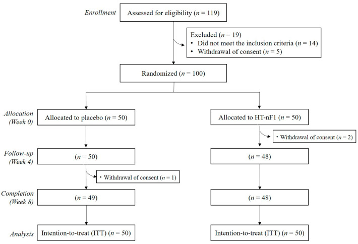
The progress of this study is shown in Figure 1. A total of 119 people were screened, and 100 people who met the inclusion/exclusion criteria were randomly assigned to a control group (n = 50) and a test group (n = 50). Of 100 subjects, 2 (1 in the control group and 1 in the test group) were excluded because they stopped participating. Baseline characteristics are listed in Table 1. The average age was 38.5 ± 1.7 years for the placebo group and 40.2 ± 1.7 years for the HT-nF1 group, whereas the gender ratio was the same for both groups. There was a significant difference between the groups in WBC count, which was 5.1 ± 0.1 × 10³ cells/μL in the placebo group and 5.7 ± 0.2 × 10³ cells/μL in the HT-nF1 group (p = 0.010); however, differences in WBC may not be clinically significant because of changes within the normal range (4.0–10.0 × 10³ cells/μL). There was no significant difference between the groups for all other indicators. Compliance was above 100% on average for both groups, and there was no significant difference between groups.
Figure 1.
CONSORT diagram for the flow of subjects through the study. HT-nF1, heat-treated Lactiplantibacillus plantarum nF1.
Table 1.
Baseline characteristics of intention-to-treat (ITT) subjects who participated in this study (1).
| Variables | Placebo (n = 50) | HT-nF1 (n = 50) | p-Value (2) |
|---|---|---|---|
| Age (year) | 38.5 ± 1.7 | 40.2 ± 1.7 | 0.492 |
| Gender (male/female) | 16/34 | 16/34 | 1.000 |
| Menstruation (Y/N/NA) | 29/5/16 | 26/8/16 | 0.652 |
| Alcohol drinker (Y/N) | 29/21 | 22/28 | 0.161 |
| Alcohol amount (SD/week) | 0.04 ± 0.03 | 0.29 ± 0.19 | 0.191 |
| Smoker (Y/N) | 2/48 | 4/46 | 0.678 |
| Smoking amount (cigarettes/d) | 0.1 ± 0.1 | 0.3 ± 0.2 | 0.383 |
| BMI (kg/m3) | 23.4 ± 0.4 | 23.2 ± 0.4 | 0.698 |
| Natural killer cell activity (%) | |||
| E:T = 5:1 | 11.7 ± 1.1 | 13.3 ± 1.7 | 0.430 |
| E:T = 10:1 | 17.3 ± 2.0 | 18.2 ± 2.6 | 0.761 |
| E:T = 25:1 | 20.9 ± 2.3 | 26.4 ± 3.1 | 0.155 |
| WBC (103/μL) | 5.1 ± 0.1 | 5.7 ± 0.2 | 0.010 |
(1) Mean ± SE (all such values). HT-nF1, heat-treated Lactiplantibacillus plantarum nF1; Y, yes; N, no; NA, not applicable; SD, standard drink; BMI, body mass index; E, effector cell; T, target cell; WBC, white blood cell. (2) Student’s t-test for continuous variables, and Chi-square test or Fisher’s exact test for categorical variables were used to compare differences between the groups.
3.2. Effects of HT-nF1 on Immune Markers
NK cell activity was measured based on E:T ratios of 25:1, 10:1, and 5:1 (Table 2). As shown in Table 2, there was no significant difference in NK cell activity measured at baseline between the placebo and HT-nF1 groups. Compared with the baseline, serum NK activity decreased in both groups at all E:T ratios at week 8. There was no significant difference in the effect on NK cell activity between the HT-nF1 group and the placebo group. Cytokine and immunoglobulin levels are listed in Table 2. After 8 weeks of intake, a change in IL-12 was significantly increased in the HT-nF1 group compared with the change in the placebo group (β = 0.6, p = 0.045). Compared with the baseline, serum IL-12 levels were significantly decreased in the placebo group at week 8 (p = 0.007). Other indicators (IL-10, TNF-α, and slgA) were not significantly different between the HT-nF1 and placebo groups.
Table 2.
Natural killer cell activity, serum cytokine, and immunoglobulin levels at the baseline and endpoint in the placebo and HT-nF1 groups (1).
| Variables | Placebo (n = 50) | HT-nF1 (n = 50) | Estimate (2) | p-Value (2) | ||
|---|---|---|---|---|---|---|
| Baseline | Week 8 | Baseline | Week 8 | |||
| NK cell activity (%) | ||||||
| E:T = 5:1 | 11.7 ± 1.1 | 6.7 ± 0.8 ** | 13.3 ± 1.7 | 7.1 ± 0.9 *** | −1.0 | 0.652 |
| E:T = 10:1 | 17.3 ± 2.0 | 9.0 ± 1.3 ** | 18.2 ± 2.6 | 10.3 ± 1.6 ** | 1.2 | 0.732 |
| E:T = 25:1 | 20.9 ± 2.3 | 15.2 ± 2.5 * | 26.4 ± 3.1 | 18.2 ± 2.8 ** | −2.0 | 0.649 |
| IL-10 (pg/mL) | 2.4 ± 0.2 | 2.4 ± 0.2 | 2.9 ± 0.4 | 3.1 ± 0.4 | 0.0 | 0.979 |
| IL-12 (pg/mL) | 1.6 ± 0.3 | 1.0 ± 0.1 ** | 1.0 ± 0.1 | 1.0 ± 0.1 | 0.6 | 0.045 |
| TNF-α (pg/mL) | 3.4 ± 0.4 | 3.9 ± 0.5 | 4.3 ± 0.6 | 4.2 ± 0.6 | −0.5 | 0.523 |
| slgA (μg/mL) | 74.7 ± 4.5 | 71.4 ± 4.5 | 81.3 ± 5.3 | 78.4 ± 5.3 | −0.9 | 0.888 |
(1) Mean ± SE (all such values). HT-nF1, heat-treated Lactiplantibacillus plantarum nF1; E, effector cell; T, target cell; IL, interleukin; TNF, tumor necrosis factor; sIgA, secretory immunoglobulin A. (2) Estimates and p-values were from a linear mixed-effect model to compare the changes for 8 weeks between the groups. WBC at baseline and dietary fat intake for 8 weeks were adjusted. * p < 0.05, ** p < 0.05, *** p < 0.05 different from baseline.
3.3. Effects of HT-nF1 on Responders and Non-Responders
We confirmed the effects of HT-nF1 using the QUINT package based on a machine-learning algorithm. To analyze the responders and non-responders following intake of HT-nF1, two criteria at baseline were selected as effect modifiers: secretory immunoglobulin A (sIgA) and NK cell activity (E:T = 10:1). Based on these factors, the effect in the HT-nF1 group compared with the placebo was confirmed.
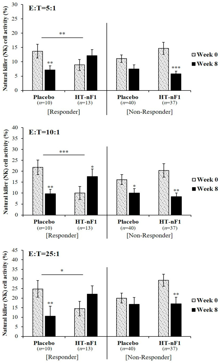
A stratified analysis was performed based on sIgA (Figure 2). In subjects with sIgA levels ≤49.61 μg/mL (responder) (placebo, n = 10; HT-nF1, n = 13), NK cell activity was reduced in the placebo group after 8 weeks from baseline. However, NK cell activity increased in the group that consumed HT-nF1 for 8 weeks. At a concentration ratio of E:T = 5:1, when HT-nF1 was ingested for 8 weeks, it increased by approximately 1.3-fold to 12.2 ± 2.1% compared with before ingestion. In the case of E:T = 10:1, it increased significantly to 17.6 ± 3.4%. Even at a concentration of E:T = 25:1, ingestion of HT-nF1 increased NK cell activity by approximately 1.5-fold to 22.1 ± 4.3%. Furthermore, we compared the change in NK cell activity when placebo and HT-nF1 were each consumed for 8 weeks. The results indicated a change in NK cell activity in the HT-nF1 group at concentrations of E:T = 5:1, 10:1, and 25:1, which were significantly increased compared with the change in NK cell activity in the placebo (E:T = 5:1, β = 9.2, p = 0.004; E:T = 10:1, β = 20.5, p < 0.001; E:T = 25:1, β = 18.4, p = 0.011). For cases in which sIgA was high (>49.61 μg/mL), there was no significant change in NK cell activity.
Figure 2.
NK cell activity through stratified analysis according to sIgA in responders (≤49.61 μg/mL) and non-responders (>49.61 μg/mL) to HT-nF1. NK cell, natural killer cell; HT-nF1, heat-treated Lactiplantibacillus plantarum nF1; sIgA, secretory immunoglobulin A; E, effector cell; T, target cell. The estimate and p-value were from a linear mixed-effect model to compare the changes for 8 weeks between the groups. WBC at baseline and dietary fat intake for 8 weeks were adjusted. * p < 0.05; ** p < 0.01; *** p < 0.001.
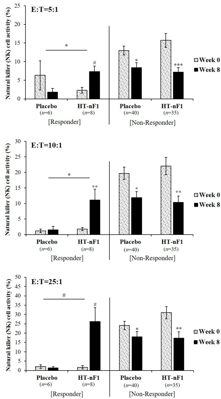
The results based on NK cell activity (E:T = 10:1) (Figure 3) also indicated that in subjects with low NK cell activity (≤3.59%, responder) (placebo, n = 6; HT-nF1, n = 8), it has increased when HT-nF1 was consumed for 8 weeks. At E:T = 5:1 and 10:1 concentration ratios, the NK cell activity of the HT-nF1 group was 7.3 ± 1.5% and 11.1 ± 3.5%, respectively, which significantly increased by approximately 3.1- and 6.1-fold (E:T = 5:1, p = 0.083; E:T = 10:1, p = 0.003) after 8 weeks of treatment. At a ratio of E:T = 25:1, the NK cell activity of the placebo group decreased, but when HT-nF1 was consumed for 8 weeks, it increased approximately 15-fold to 26.2% ± 7.5% (p = 0.071). The change in NK cell activity when HT-nF1 was ingested for 8 weeks compared with the change in NK cell activity when the placebo was ingested for 8 weeks, a significant increase was observed for HT-nF1 at all ratios (E:T = 5:1, β = 12.2, p = 0.041; E:T = 10:1, β = 9.3, p = 0.037; E:T = 25:1, β = 17.9, p = 0.086). Consistently, subjects with high NK cell activity (>3.59%, non-responder) showed no significant change.
Figure 3.
NK cell activity through stratified analysis according to NK cell activity (E:T = 10:1) in responders (≤3.59%) and non-responders (>3.59%) to HT-nF1. HT-nF1, heat-treated Lactiplantibacillus plantarum nF1; E, effector cell; T, target cell. The estimate and p-value were from a linear mixed-effect model to compare the changes for 8 weeks between the groups. WBC at baseline and dietary fat intake for 8 weeks were adjusted. Not detected was excluded; placebo (n = 4), HT-nF1 (n = 7). * p < 0.05; ** p < 0.01; *** p < 0.001; # p < 0.1.
3.4. Safety Analysis
To analyze the safety of HT-nF1, hematological and blood biochemical analyses were performed at baseline and week 8 (Table 3). There were significant differences in some indicators when comparing before and after intake between groups and within groups, but all changes were within the normal range and there were no clinically significant changes. No serious adverse reactions related to the test food were observed, and there were no significant differences between groups in terms of occurrence, type, symptom severity, or causal relationship with the test food.
Table 3.
Hematological and blood chemistry tests at the baseline and endpoint in the placebo and HT-nF1 groups (1).
| Variables | Placebo (n = 50) | HT-nF1 (n = 50) | p-Value (2) | ||||
|---|---|---|---|---|---|---|---|
| Baseline | Week 8 | Baseline | Week 8 | Group | Week | Group × Week | |
| Hematological test | |||||||
| RBC (106/μL) | 4.6 ± 0.1 | 4.6 ± 0.1 | 4.6 ± 0.1 | 4.5 ± 0.1 ** | 0.391 | 0.061 | 0.043 |
| Hemoglobin (g/dL) | 13.8 ± 0.2 | 13.7 ± 0.2 | 13.8 ± 0.2 | 13.5 ± 0.2 ** | 0.814 | 0.020 | 0.064 |
| Hematocrit (%) | 41.6 ± 0.6 | 41.4 ± 0.6 | 41.8 ± 0.5 | 40.6 ± 0.5 ** | 0.680 | 0.006 | 0.045 |
| Platelet (103/μL) | 287.8 ± 9.9 | 287.7 ± 9.8 | 275.3 ± 8.8 | 279.9 ± 7.7 | 0.379 | 0.727 | 0.661 |
| Neutrophils (%) | 55.8 ± 1.2 | 54.6 ± 1.3 | 55.9 ± 1.3 | 52.5 ± 1.2 ** | 0.525 | 0.006 | 0.187 |
| Lymphocytes (%) | 33.3 ± 1.0 | 34.7 ± 1.2 | 34.5 ± 1.3 | 37.2 ± 1.2 * | 0.219 | 0.005 | 0.402 |
| Monocytes (%) | 6.7 ± 0.3 | 6.8 ± 0.2 | 6.7 ± 0.2 | 7.1 ± 0.2 | 0.735 | 0.052 | 0.435 |
| Eosinophils (%) | 3.5 ± 0.5 | 3.1 ± 0.3 * | 2.3 ± 0.3 | 2.5 ± 0.2 | 0.055 | 0.473 | 0.038 |
| Basophils (%) | 0.8 ± 0.1 | 0.8 ± 0.1 | 0.7 ± 0.1 | 0.7 ± 0.0 | 0.318 | 0.531 | 0.409 |
| Blood chemistry test | |||||||
| AST (U/L) | 20.9 ± 0.8 | 21.3 ± 1.0 | 22.6 ± 0.9 | 23.2 ± 1.3 | 0.129 | 0.493 | 0.883 |
| ALT (U/L) | 18.5 ± 1.6 | 19.4 ± 1.8 | 20.1 ± 1.8 | 22.1 ± 2.3 | 0.339 | 0.242 | 0.609 |
| BUN (mg/dL) | 12.1 ± 0.6 | 12.2 ± 0.5 | 12.5 ± 0.5 | 12.6 ± 0.5 | 0.624 | 0.871 | 0.837 |
| Albumin (g/dL) | 4.5 ± 0.0 | 4.4 ± 0.0 | 4.5 ± 0.0 | 4.5 ± 0.0 * | 0.483 | 0.007 | 0.829 |
| Creatinine (mg/dL) | 0.71 ± 0.02 | 0.72 ± 0.02 | 0.71 ± 0.02 | 0.71 ± 0.02 | 0.908 | 0.292 | 0.257 |
(1) Mean ± SE (all such values). HT-nF1, heat-treated Lactiplantibacillus plantarum nF1; RBC, red blood cell; AST, aspartate aminotransferase; ALT, alanine aminotransferase; BUN, blood urea nitrogen. (2) A linear mixed-effect model to compare the changes after 8 weeks between the group, week, and group × week. * p < 0.05; ** p < 0.01 different from baseline.
4. Discussion
Immune regulation in the human body occurs through the processes that are promoted and suppressed, and if immune regulation fails, diseases or other problems can occur [21]. In addition, interest in improving human immunity has increased because of various factors, such as the prevalence of infectious diseases and the aging population. The demand for immune-enhancing functional materials is also steadily increasing [22,23,24]. Recent studies have indicated that probiotics are associated with immunomodulatory effects [25,26,27]. Chong et al. [25] evaluated the effect of L. plantarum DR7 on upper respiratory tract infection and confirmed that NK cell activity was enhanced in subjects who consumed L. plantarum DR7 compared with the control group. L. plantarum YU regulates immune function through activation of the Th1 immune response and lgA production [26]. In addition, probiotics, such as L. casei Shirota, L. acidophilus X37, and B. bifidum MF 20/5, increase innate immunity by increasing NK cell activity [27]. Postbiotics are inactivated bacterial cells that provide health benefits to humans, which include non-living bacteria, cell walls, and metabolites. They have similar effects as probiotics but are used in patients with weakened immune systems, which has been reported recently [28,29,30,31]. Postbiotics are associated with immunomodulation and strengthening the innate and adaptive immune systems [32,33]. Heat-treated L. sakei KU15041 and L. curvatus KU15003 regulate immunity by increasing the expression of TNF-α, IL-1β, and IL-6 in RAW 264.7 cells [34]. Heat-killed L. casei IMAU60214 improved immune function by regulating macrophages in malnourished children [35]. When heat-treated L. plantarum LM1004 was consumed for 8 weeks, it stimulated innate immunity and significantly increased NK cells and IL-12 levels [36]. Hirose et al. [37] suggested that the daily consumption of heat-killed L. plantarum L-137 increases Th1 immunity and confirmed that it was safe, even when consumed at high doses and for long periods [38]. In addition, in many studies [39,40,41], Lactobacillus increased the levels of Th1 cytokines (e.g., IL-12, IFN-r). However, since it did not affect the production of Th2 cytokines (e.g., IL-4, IL-5), it is thought to enhance immunity through the Th1 mechanism.
This study was performed to evaluate the effect of 8 weeks of consumption of heat-treated L. plantarum nF1 (HT-nF1) on improving immunity. NK cells play an important role in innate immune responses [42]. They also regulate immune responses by producing cytokines, which can activate various cell types involved in the adaptive and innate immune systems [6,43,44]. Depending on the type of cytokine, Th1 cells and Th2 cells are converted into lymphocytes. Th1, which induces a normal immune response, is an inflammatory response that involves TNF-α, IL-2, IL-12, and IFN-γ. Th2 induces an autoimmune response and activates humoral immunity by producing IL-4, IL-5, IL-6, and IL-10 [45,46]. Increased IL-12 production is associated with increased cellular immunity and phagocytic function [47]. IL-12 is also important for inducing Th1 immunity [48] and directly activates CD56+ NK cell-mediated cytotoxicity [49]. IL-12 affects NK cell regulation [50]. The results of the present study confirmed that a change in IL-12 was significantly increased in the test group compared with the control group in all subjects (β = 0.6, p = 0.045). NK activity may also be increased through a significant increase in IL-12; however, intake of HT-nF1 did not affect NK activity in all subjects (Table 2). Because this study was conducted from November to March, most of the subjects may have experienced cold stress [51,52,53,54] due to the winter weather and sudden temperature changes [55]. In addition, in the case of female subjects, immune-related indicators may be affected by physiological and general environmental conditions, such as the menstrual cycle [37,56,57]. However, we confirmed that the intake of HT-nF1 had a positive effect on improving immunity in subjects with low slgA and NK activity, that is, with a relatively weak immune system (Figure 2 and Figure 3).
There are various types of immunoglobulins that play an important role in immunity, such as IgG, IgM, IgA, and IgE [58]. Of these, IgA has a primary defense role against pathogens and activates mucosal immunity [59,60]. Individuals with low slgA levels are readily exposed to various diseases, such as respiratory infections [61,62]. Probiotics and postbiotics can regulate immunity by increasing the secretion of slgA [63,64,65,66,67]. The consumption of milk containing L. rhamnosus HN001 or Bif. lactis HN019 increased NK activity in elderly people over 70 years of age [68]. NK activity also increases when healthy elderly people take probiotic supplements [69,70,71]. In addition, in a double-blind, placebo-controlled clinical study, heat-killed L. gasseri TMC0356 regulated immunity by significantly increasing the number of CD8 + T cells [72]. Moreover, an increase in NK cell activity was observed in the splenocytes of mice orally administered a postbiotic complex containing L. plantarum during immunosuppression [33]. Therefore, our results suggest that nF1 intake maintains the immune status by activating NK cells in subjects with relatively weak immune systems.
Previous studies showed that nF1 induced spleen cell proliferation and promoted a Th1 response and cytokine (IL-12, TNF-α, and IFN-γ) production [16,41]. nF1 increased the production of TNF-α, IL-2, and IL-6 in RAW 264.7 macrophages, which resulted in the activation of NF-κB and phosphorylation of IκBα [17]. In addition, nF1 intake improves immune function by increasing the levels of total IgG, TNF-α, IL-2, and IL-12 in the serum of cyclophosphamide (CPP) immunosuppressed mice [16,17]. Lee et al. [73] demonstrated that immunity was regulated by increasing the level of total IgA in mice with colon cancer induced by azoxymethane (AOM)/dextran sodium sulfate (DSS) exposure. In humans, nF1 regulates immune function [46,74]. In IBS patients, subjects who consumed kimchi containing nF1 showed regulated immune function with modulated levels of cytokines (TNF-α, IL-12) in the serum [74]. Moreover, when healthy older adults consumed yogurt containing nF1 (heat-treated L. plantarum nF1, L. paracasei, and B. lactis) for 12 weeks, NK cell activity and the concentration of IFN-γ and IgG1 increased, thus improving immune function [46]. In addition, the effect of regulating immune function was confirmed when nF1 was added to food [19,46,75]. When yogurt containing nF1 was consumed by mice infected with influenza A virus (IAV), NK cell activity increased [19]. NK cell activity was also increased in mice that consumed kimchi containing nF1 [75]. Therefore, it is thought that the nF1 immune modulation is effective based on the Th1 response. In the present study, NK cell activity significantly increased in subjects with low slgA and NK cell activity (E:T = 10:1). These results suggest that nF1 strengthens and regulates the immune system and restores normal immunity in people with weak immune systems (e.g., children [16,76], elderly [16,46], and patients [74]).
In the safety evaluation, the frequency of adverse events and blood levels were considered negligible as they occurred within the normal or reference range. Therefore, heat-treated Lactiplantibacillus plantarum nF1 not only has a positive effect on regulating immunity but is also a safe material for humans, thus confirming its potential as a raw material that can help improve immunity.
5. Conclusions
In this study, we examined the effect of nF1 for 8 weeks on immune function in a clinical study. A change in IL-12 was significantly increased in the HT-nF1 group compared with the placebo group, and the change in NK cell activity was significantly increased in subjects with low slgA and subjects with low NK cell activity, which indicates that nF1 intake was more effective. Therefore, intake of nF1 may be an effective strategy to improve immune function.
Acknowledgments
We thank the research volunteers who participated in the studies described in this manuscript. We appreciate the BiofoodCRO for supporting the clinical trial.
Author Contributions
Conceptualization, I.A.K., J.Y.K. and K.-Y.P.; methodology, I.A.K., J.S., C.B., J.Y.K., S.L. and K.J.K.; validation, I.A.K., J.S. and C.B.; formal analysis, J.Y.K., S.L. and K.J.K.; investigation, I.A.K., J.S. and C.B.; data curation, J.Y.K., S.L., and K.J.K.; visualization, S.-Y.L. and G.-H.H.; writing—original draft preparation, G.-H.H. and S.-Y.L.; supervision, C.B. and K.-Y.P.; funding acquisition, K.T.K. and M.G.K.; project administration, K.-Y.P. All authors have read and provided critical feedback and helped the research, analysis, and manuscript. All authors have read and agreed to the published version of the manuscript.
Institutional Review Board Statement
The study was conducted in accordance with the Declaration of Helsinki and approved by the Institutional Review Board of Global Medical Research Center (No. GIRB-22O19-KQ and 28 October 2022).
Informed Consent Statement
All subjects gave their informed consent for inclusion before they participated in the study.
Data Availability Statement
Data are included in the article.
Conflicts of Interest
Authors Geun-Hye Hong, So-Young Lee, Ki Tae Kim, Min Gee Kim and Kun-Young Park were employed by the company IMMUNOBIOTECH Corp. The remaining authors declare that the research was conducted in the absence of any commercial or financial relationships that could be construed as a potential conflict of interest.
Funding Statement
This work was supported by the “Food Functionally Evaluation program (G0220300-01)” under the Ministry of Agriculture, Food and Rural Affairs and partly the Korea Food Research Institute.
Footnotes
Disclaimer/Publisher’s Note: The statements, opinions and data contained in all publications are solely those of the individual author(s) and contributor(s) and not of MDPI and/or the editor(s). MDPI and/or the editor(s) disclaim responsibility for any injury to people or property resulting from any ideas, methods, instructions or products referred to in the content.
References
- 1.Cannon J.G. Inflammatory cytokines in nonpathological States. News Physiol. Sci. 2000;15:298–303. doi: 10.1152/physiologyonline.2000.15.6.298. [DOI] [PubMed] [Google Scholar]
- 2.Medzhitov R., Janeway C., Jr. Innate immunity. N. Engl. J. Med. 2000;343:338–344. doi: 10.1056/NEJM200008033430506. [DOI] [PubMed] [Google Scholar]
- 3.Nicholson L.B. The immune system. Essays Biochem. 2016;60:275–301. doi: 10.1042/EBC20160017. [DOI] [PMC free article] [PubMed] [Google Scholar]
- 4.Mace E.M. Human natural killer cells: Form, function, and development. J. Allergy Clin. Immunol. 2023;151:371–385. doi: 10.1016/j.jaci.2022.09.022. [DOI] [PMC free article] [PubMed] [Google Scholar]
- 5.Waldmann T.A. The biology of interleukin-2 and interleukin-15: Implications for cancer therapy and vaccine design. Nat. Rev. Immunol. 2006;6:595–601. doi: 10.1038/nri1901. [DOI] [PubMed] [Google Scholar]
- 6.Vivier E., Tomasello E., Baratin M., Walzer T., Ugolini S. Functions of natural killer cells. Nat. Immunol. 2008;9:503–510. doi: 10.1038/ni1582. [DOI] [PubMed] [Google Scholar]
- 7.Scharton T.M., Scott P. Natural killer cells are a source of interferon gamma that drives differentiation of CD4+ T cell subsets and induces early resistance to Leishmania major in mice. J. Exp. Med. 1993;178:567–577. doi: 10.1084/jem.178.2.567. [DOI] [PMC free article] [PubMed] [Google Scholar]
- 8.Konjevic G.M., Vuletic A.M., Martinovic K.M.M., Larsen A.K., Jurisic V.B. The role of cytokines in the regulation of NK cells in the tumor environment. Cytokine. 2019;117:30–40. doi: 10.1016/j.cyto.2019.02.001. [DOI] [PubMed] [Google Scholar]
- 9.Sievers B.L., Cheng M.T.K., Csiba K., Meng B., Gupta R.K. SARS-CoV-2 and innate immunity: The good, the bad, and the “goldilocks”. Cell. Mol. Immunol. 2024;21:171–183. doi: 10.1038/s41423-023-01104-y. [DOI] [PMC free article] [PubMed] [Google Scholar]
- 10.Singh K., Rao A. Probiotics: A potential immunomodulator in COVID-19 infection management. Nutr. Res. 2021;87:1–12. doi: 10.1016/j.nutres.2020.12.014. [DOI] [PMC free article] [PubMed] [Google Scholar]
- 11.Wang Y.H., Limaye A., Liu J.R., Wu T.N. Potential probiotics for regulation of the gut-lung axis to prevent or alleviate influenza in vulnerable populations. J. Tradit. Complement. Med. 2022;13:161–169. doi: 10.1016/j.jtcme.2022.08.004. [DOI] [PMC free article] [PubMed] [Google Scholar]
- 12.Jung Y.J., Lee Y.T., Ngo V.L., Cho Y.H., Ko E.J., Hong S.M., Kim K.H., Jang J.H., Oh J.S., Park M.K., et al. Heat-killed Lactobacillus casei confers broad protection against influenza a virus primary infection and develops heterosubtypic immunity against future secondary infection. Sci. Rep. 2017;7:17360. doi: 10.1038/s41598-017-17487-8. [DOI] [PMC free article] [PubMed] [Google Scholar]
- 13.Kobayashi N., Saito T., Uematsu T., Kishi K., Toba M., Kohda N., Suzuki T. Oral administration of heat-killed Lactobacillus pentosus strain b240 augments protection against influenza virus infection in mice. Int. Immunopharmacol. 2011;11:199–203. doi: 10.1016/j.intimp.2010.11.019. [DOI] [PubMed] [Google Scholar]
- 14.Murosaki S., Yamamoto Y., Ito K., Inokuchi T., Kusaka H., Ikeda H., Yoshikai Y. Heat-killed Lactobacillus plantarum L-137 suppresses naturally fed antigen-specific IgE production by stimulation of IL-12 production in mice. J. Allergy Clin. Immunol. 1998;102:57–64. doi: 10.1016/S0091-6749(98)70055-7. [DOI] [PubMed] [Google Scholar]
- 15.Matsuzaki T., Chin J. Modulating immune responses with probiotic bacteria. Immunol. Cell Biol. 2000;78:67–73. doi: 10.1046/j.1440-1711.2000.00887.x. [DOI] [PubMed] [Google Scholar]
- 16.Choi D.W., Jung S.Y., Kang J., Nam Y.D., Lim S.I., Kim K.T., Shin H.S. Immune-enhancing effect of nanometric Lactobacillus plantarum nF1 (nLp-nF1) in a mouse model of cyclophosphamide-induced immunosuppression. J. Microbiol. Biotechnol. 2018;28:218–226. doi: 10.4014/jmb.1709.09024. [DOI] [PubMed] [Google Scholar]
- 17.Moon P.D., Lee J.S., Kim H.Y., Han N.R., Kang I., Kim H.M., Jeong H.J. Heat-treated Lactobacillus plantarum increases the immune responses through activation of natural killer cells and macrophages on in vivo and in vitro models. J. Med. Microbiol. 2019;68:467–474. doi: 10.1099/jmm.0.000938. [DOI] [PubMed] [Google Scholar]
- 18.Park S., Kim J.I., Bae J.Y., Yoo K., Kim H., Kim I.H., Park M.S., Lee I. Effects of heat-killed Lactobacillus plantarum against influenza viruses in mice. J. Microbiol. 2018;56:145–149. doi: 10.1007/s12275-018-7411-1. [DOI] [PubMed] [Google Scholar]
- 19.Kim D.H., Chung W.C., Chun S.H., Han J.H., Song M.J., Lee K.W. Enhancing the natural killer cell activity and anti-influenza effect of heat-treated Lactobacillus plantarum nF1-fortified yogurt in mice. J. Dairy Sci. 2018;101:10675–10684. doi: 10.3168/jds.2018-15137. [DOI] [PubMed] [Google Scholar]
- 20.Park S.Y., Kim K.J., Jo S.M., Jeon J.Y., Kim B.R., Hwang J.E., Kim J.Y. Euglena gracilis (Euglena) powder supplementation enhanced immune function through natural killer cell activity in apparently healthy participants: A randomized, double-blind, placebo-controlled trial. Nutr. Res. 2023;119:90–97. doi: 10.1016/j.nutres.2023.09.004. [DOI] [PubMed] [Google Scholar]
- 21.Albers R., Bourdet-Sicard R., Braun D., Calder P.C., Herz U., Lambert C., Lenoir-Wijnkoop I., Méheust A., Ouwehand A., Phothirath P., et al. Monitoring immune modulation by nutrition in the general population: Identifying and substantiating effects on human health. Br. J. Nutr. 2013;110:S1–S30. doi: 10.1017/S0007114513001505. [DOI] [PubMed] [Google Scholar]
- 22.Kaminogawa S., Nanno M. Modulation of immune functions by foods. Evid. Based. Complement. Alternat. Med. 2004;1:241–250. doi: 10.1093/ecam/neh042. [DOI] [PMC free article] [PubMed] [Google Scholar]
- 23.Cho Y.J., Son H.J., Kim K.S. 14-week randomized, placebo-controlled, double-blind clinical trial to evaluate the efficacy and safety of ginseng polysaccharide (Y-75) J. Transl. Med. 2014;12:283. doi: 10.1186/s12967-014-0283-1. [DOI] [PMC free article] [PubMed] [Google Scholar]
- 24.Kwak J.H., Baek S.H., Woo Y., Han J.K., Kim B.G., Kim O.Y., Lee J.H. Beneficial immunostimulatory effect of short-term Chlorella supplementation: Enhancement of natural killer cell activity and early inflammatory response (randomized, double-blinded, placebo-controlled trial) Nutr. J. 2012;11:53. doi: 10.1186/1475-2891-11-53. [DOI] [PMC free article] [PubMed] [Google Scholar]
- 25.Chong H.X., Yusoff N.A.A., Hor Y.Y., Lew L.C., Jaafar M.H., Choi S.B., Yusoff M.S.B., Wahid N., Abdullah M.F., Zakaria N., et al. Lactobacillus plantarum DR7 improved upper respiratory tract infections via enhancing immune and inflammatory parameters: A randomized, double-blind, placebo-controlled study. J. Dairy Sci. 2019;102:4783–4797. doi: 10.3168/jds.2018-16103. [DOI] [PubMed] [Google Scholar]
- 26.Kawashima T., Hayashi K., Kosaka A., Kawashima M., Igarashi T., Tsutsui H., Tsuji N.M., Nishimura I., Hayashi T., Obata A. Lactobacillus plantarum strain YU from fermented foods activates Th1 and protective immune responses. Int. Immunopharmacol. 2011;11:2017–2024. doi: 10.1016/j.intimp.2011.08.013. [DOI] [PubMed] [Google Scholar]
- 27.Takagi A., Matsuzaki T., Sato M., Nomoto K., Morotomi M., Yokokura T. Enhancement of natural killer cytotoxicity delayed murine carcinogenesis by a probiotic microorganism. Carcinogenesis. 2001;22:599–605. doi: 10.1093/carcin/22.4.599. [DOI] [PubMed] [Google Scholar]
- 28.Salminen S., Collado M.C., Endo A., Hill C., Lebeer S., Quigley E.M., Sanders M.E., Shamir R., Swann J.R., Szajewska H., et al. The International Scientific Association of Probiotics and Prebiotics (ISAPP) consensus statement on the definition and scope of postbiotics. Nat. Rev. Gastroenterol. Hepatol. 2021;18:649–667. doi: 10.1038/s41575-021-00440-6. [DOI] [PMC free article] [PubMed] [Google Scholar]
- 29.Siciliano R.A., Reale A., Mazzeo M.F., Morandi S., Silvetti T., Brasca M. Paraprobiotics: A new perspective for functional foods and nutraceuticals. Nutrients. 2021;13:1225. doi: 10.3390/nu13041225. [DOI] [PMC free article] [PubMed] [Google Scholar]
- 30.Deshpande G., Athalye-Jape G., Patole S. Para-probiotics for preterm neonates—The next frontier. Nutrients. 2018;10:871. doi: 10.3390/nu10070871. [DOI] [PMC free article] [PubMed] [Google Scholar]
- 31.Kim B.Y., Park S.S. The concepts and applications of postbiotics for the development of health functional food product. Curr. Top. Lact. Acid Bact. Probiotics. 2021;7:14–22. doi: 10.35732/ctlabp.2021.7.1.14. [DOI] [Google Scholar]
- 32.De Marco S., Sichetti M., Muradyan D., Piccioni M., Traina G., Pagiotti R., Pietrella D. Probiotic cell-free supernatants exhibited anti-inflammatory and antioxidant activity on human gut epithelial cells and macrophages stimulated with LPS. Evid. Based Complement. Alternat. Med. 2018;2018:1756308. doi: 10.1155/2018/1756308. [DOI] [PMC free article] [PubMed] [Google Scholar]
- 33.Jung Y.J., Kim H.S., Jaygal G., Cho H.R., Lee K.B., Song I.B., Kim J.H., Kwak M.S., Han K.H., Bae M.J., et al. Postbiotics enhance NK cell activation in stress-induced mice through gut microbiome regulation. J. Microbiol. Biotechnol. 2022;32:612–620. doi: 10.4014/jmb.2111.11027. [DOI] [PMC free article] [PubMed] [Google Scholar]
- 34.Hyun J.H., Woo I.K., Kim K.T., Park Y.S., Kang D.K., Lee N.K., Paik H.D. Heat-Treated Paraprobiotic Latilactobacillus sakei KU15041 and Latilactobacillus curvatus KU15003 show an antioxidant and immunostimulatory effect. J. Microbiol. Biotechnol. 2024;34:358–366. doi: 10.4014/jmb.2309.09007. [DOI] [PMC free article] [PubMed] [Google Scholar]
- 35.Rocha-Ramírez L.M., Hernández-Ochoa B., Gómez-Manzo S., Marcial-Quino J., Cárdenas-Rodríguez N., Centeno-Leija S., García-Garibay M. Impact of heat-killed Lactobacillus casei strain IMAU60214 on the immune function of macrophages in malnourished children. Nutrients. 2020;12:2303. doi: 10.3390/nu12082303. [DOI] [PMC free article] [PubMed] [Google Scholar]
- 36.Bae W.Y., Min H., Shin S.L., Kim T.R., Lee H., Sohn M., Park K.S. Effect of orally administered heat-treated Lactobacillus plantarum LM1004 on the innate immune system: A randomized, placebo-controlled, double-blind study. J. Funct. Foods. 2022;98:105293. doi: 10.1016/j.jff.2022.105293. [DOI] [Google Scholar]
- 37.Hirose Y., Murosaki S., Yamamoto Y., Yoshikai Y., Tsuru T. Daily intake of heat-killed Lactobacillus plantarum L-137 augments acquired immunity in healthy adults. J. Nutr. 2006;136:3069–3073. doi: 10.1093/jn/136.12.3069. [DOI] [PubMed] [Google Scholar]
- 38.Nakai H., Murosaki S., Yamamoto Y., Furutani M., Matsuoka R., Hirose Y. Safety and efficacy of using heat-killed Lactobacillus plantarum L-137: High-dose and long-term use effects on immune-related safety and intestinal bacterial flora. J. Immunotoxicol. 2021;18:127–135. doi: 10.1080/1547691X.2021.1979698. [DOI] [PubMed] [Google Scholar]
- 39.Kato I., Tanaka K., Yokokura T. Lactic acid bacterium potently induces the production of interleukin-12 and interferon-γ by mouse splenocytes. Int. J. Immunopharmacol. 1999;21:121–131. doi: 10.1016/S0192-0561(98)00072-1. [DOI] [PubMed] [Google Scholar]
- 40.Gill H.S., Rutherfurd K.J., Prasad J., Gopal P.K. Enhancement of natural and acquired immunity by Lactobacillus rhamnosus (HN001), Lactobacillus acidophilus (HN017) and Bifidobacterium lactis (HN019) Br. J. Nutr. 2000;83:167–176. doi: 10.1017/S0007114500000210. [DOI] [PubMed] [Google Scholar]
- 41.Lee H.A., Kim H., Lee K.W., Park K.Y. Dead Lactobacillus plantarum stimulates and skews immune responses toward T helper 1 and 17 polarizations in RAW 264.7 cells and mouse splenocytes. J. Microbiol. Biotechnol. 2016;26:469–476. doi: 10.4014/jmb.1511.11001. [DOI] [PubMed] [Google Scholar]
- 42.Gui Q., Wang A., Zhao X., Huang S., Tan Z., Xiao C., Yang Y. Effects of probiotic supplementation on natural killer cell function in healthy elderly individuals: A meta-analysis of randomized controlled trials. Eur. J. Clin. Nutr. 2020;74:1630–1637. doi: 10.1038/s41430-020-0670-z. [DOI] [PMC free article] [PubMed] [Google Scholar]
- 43.Camous X., Pera A., Solana R., Larbi A. NK cells in healthy aging and age-associated diseases. BioMed Res. Int. 2012;2012:195956. doi: 10.1155/2012/195956. [DOI] [PMC free article] [PubMed] [Google Scholar]
- 44.Gayoso I., Sanchez-Correa B., Campos C., Alonso C., Pera A., Casado J.G., Morgado S., Tarazona R., Solana R. Immunosenescence of human natural killer cells. J. Innate Immun. 2011;3:337–343. doi: 10.1159/000328005. [DOI] [PubMed] [Google Scholar]
- 45.Mosmann T.R., Cherwinski H., Bond M.W., Giedlin M.A., Coffman R.L. Two types of murine helper T cell clone. I. Definition according to profiles of lymphokine activities and secreted proteins. J. Immunol. 1986;136:2348–2357. doi: 10.4049/jimmunol.136.7.2348. [DOI] [PubMed] [Google Scholar]
- 46.Lee A., Lee Y.J., Yoo H.J., Kim M., Chang Y., Lee D.S., Lee J.H. Consumption of dairy yogurt containing Lactobacillus paracasei ssp. paracasei, Bifidobacterium animalis ssp. lactis and heat-treated Lactobacillus plantarum improves immune function including natural killer cell activity. Nutrients. 2017;9:558. doi: 10.3390/nu9060558. [DOI] [PMC free article] [PubMed] [Google Scholar]
- 47.Ertel W., Keel M., Neidhardt R., Steckholzer U., Kremer J.P., Ungethuem U., Trentz O. Inhibition of the defense system stimulating interleukin-12 interferon-γ pathway during critical illness. Blood. 1997;89:1612–1620. doi: 10.1182/blood.V89.5.1612. [DOI] [PubMed] [Google Scholar]
- 48.Trinchieri G. Proinflammatory and immunoregulatory functions of interleukin-12. Int. Rev. Immunol. 1998;16:365–396. doi: 10.3109/08830189809043002. [DOI] [PubMed] [Google Scholar]
- 49.Klein-Franke A., Anderer F.A. IL-12-mediated activation of MHC-unrestricted cytotoxicity of human PBMC subpopulations: Synergic action of a plant rhamnogalacturonan. Anticancer Res. 1995;15:2511–2516. [PubMed] [Google Scholar]
- 50.Steinberg C., Eisenächer K., Gross O., Reindl W., Schmitz F., Ruland J., Krug A. The IFN regulatory factor 7-dependent type I IFN response is not essential for early resistance against murine cytomegalovirus infection. Eur. J. Immunol. 2009;39:1007–1018. doi: 10.1002/eji.200838814. [DOI] [PubMed] [Google Scholar]
- 51.Haseda A., Nishimura M., Sugawara M., Kudo M., Nakagawa R., Nishihira J. Effect of daily intake of heat-killed Lactobacillus plantarum HOKKAIDO on immunocompetence: A randomized, double-blind, placebo-controlled, parallel-group study. Bioact. Compd. Health Dis. 2020;3:32–54. [Google Scholar]
- 52.Hu G.Z., Yang S.J., Hu W.X., Wen Z., He D., Zeng L.F., Xiang Q., Wu X.M., Zhou W.Y., Zhu Q.X. Effect of cold stress on immunity in rats. Exp. Ther. Med. 2016;11:33–42. doi: 10.3892/etm.2015.2854. [DOI] [PMC free article] [PubMed] [Google Scholar]
- 53.Jiang X.H., Guo S.Y., Xu S. Cold stress induces the suppression of splenic NK cell activity and the c-fos expression in rat brain. Chin. J. Appl. Physiol. 2002;18:313–316. [PubMed] [Google Scholar]
- 54.Makino T., Kato K., Mizukami H. Processed aconite root prevents cold-stress-induced hypothermia and immuno-suppression in mice. Biol. Pharm. Bull. 2009;32:1741–1748. doi: 10.1248/bpb.32.1741. [DOI] [PubMed] [Google Scholar]
- 55.Brenner I.K.M., Castellani J.W., Gabaree C., Young A.J., Zamecnik J., Shephard R.J., Shek P.N. Immune changes in humans during cold exposure: Effects of prior heating and exercise. J. Appl. Physiol. 1999;87:699–710. doi: 10.1152/jappl.1999.87.2.699. [DOI] [PubMed] [Google Scholar]
- 56.Souza S.S.D., Castro F.A.D., Mendonca H.C.D., Palma P.V.B., Morais F.R., Ferriani R.A., Voltarelli J.C. Influence of menstrual cycle on NK activity. J. Reprod. Immunol. 2001;50:151–159. doi: 10.1016/S0165-0378(00)00091-7. [DOI] [PubMed] [Google Scholar]
- 57.Cho J.M., Chae J., Jeong S.R., Moon M.J., Shin D.Y., Lee J.H. Immune activation of Bio-Germanium in a randomized, double-blind, placebo-controlled clinical trial with 130 human subjects: Therapeutic opportunities from new insights. PLoS ONE. 2020;15:e0240358. doi: 10.1371/journal.pone.0240358. [DOI] [PMC free article] [PubMed] [Google Scholar]
- 58.Castro-Quintas Á., Palma-Gudiel H., San Martín-González N., Caso J.R., Leza J.C., Fañanás L. Salivary secretory immunoglobulin A as a potential biomarker of psychosocial stress response during the first stages of life: A systematic review. Front. Neuroendocrinol. 2023;71:101083. doi: 10.1016/j.yfrne.2023.101083. [DOI] [PubMed] [Google Scholar]
- 59.Carpenter G.H. Salivary factors that maintain the normal oral commensal microflora. J. Dent. Res. 2020;99:644–649. doi: 10.1177/0022034520915486. [DOI] [PMC free article] [PubMed] [Google Scholar]
- 60.Woof J.M., Kerr M.A. The function of immunoglobulin A in immunity. J. Pathol. 2006;208:270–282. doi: 10.1002/path.1877. [DOI] [PubMed] [Google Scholar]
- 61.Welch T.R. 50 years ago in the journal of pediatrics: Comment on IgA deficiency and susceptibility to infection. J. Pediatr. 2018;192:104. doi: 10.1016/j.jpeds.2017.07.029. [DOI] [PubMed] [Google Scholar]
- 62.da Silva Campos M.J., Alves C.C.S., Raposo N.R.B., Ferreira A.P., Vitral R.W.F. Influence of salivary secretory immunoglobulin A level on the pain experienced by orthodontic patients. Med. Sci. Monit. 2010;16:CR405–CR409. [PubMed] [Google Scholar]
- 63.Perdigón G., Valdez J.C., Rachid M. Antitumour activity of yogurt: Study of possible immune mechanisms. J. Dairy Res. 1998;65:129–138. doi: 10.1017/S0022029997002604. [DOI] [PubMed] [Google Scholar]
- 64.Park J.H., Um J.I., Lee B.J., Goh J.S., Park S.Y., Kim W.S., Kim P.H. Encapsulated Bifidobacterium bifidum potentiates intestinal IgA production. Cell. Immunol. 2002;219:22–27. doi: 10.1016/S0008-8749(02)00579-8. [DOI] [PubMed] [Google Scholar]
- 65.Hosono A., Ozawa A., Kato R., Ohnishi Y., Nakanishi Y., Kimura T., Nakamura R. Dietary fructooligosaccharides induce immunoregulation of intestinal IgA secretion by murine Peyer’s patch cells. Biosci. Biotechnol. Biochem. 2003;67:758–764. doi: 10.1271/bbb.67.758. [DOI] [PubMed] [Google Scholar]
- 66.Lin W.Y., Kuo Y.W., Chen C.W., Huang Y.F., Hsu C.H., Lin J.H., Liu C.R., Chen J.F., Hsia K.C., Ho H.H. Viable and heat-killed probiotic strains improve oral immunity by elevating the IgA concentration in the oral mucosa. Curr. Microbiol. 2021;78:3541–3549. doi: 10.1007/s00284-021-02569-8. [DOI] [PMC free article] [PubMed] [Google Scholar]
- 67.Lin C.W., Chen Y.T., Ho H.H., Kuo Y.W., Lin W.Y., Chen J.F., Lin J.H., Liu C.R., Lin C.H., Yeh Y.T., et al. Impact of the food grade heat-killed probiotic and postbiotic oral lozenges in oral hygiene. Aging. 2022;14:2221–2238. doi: 10.18632/aging.203923. [DOI] [PMC free article] [PubMed] [Google Scholar]
- 68.Gill H.S., Rutherfurd K.J., Cross M.L. Dietary probiotic supplementation enhances natural killer cell activity in the elderly: An investigation of age-related immunological changes. J. Clin. Immunol. 2001;21:264–271. doi: 10.1023/A:1010979225018. [DOI] [PubMed] [Google Scholar]
- 69.Bruins M.J., Van Dael P., Eggersdorfer M. The role of nutrients in reducing the risk for noncommunicable diseases during aging. Nutrients. 2019;11:85. doi: 10.3390/nu11010085. [DOI] [PMC free article] [PubMed] [Google Scholar]
- 70.Miller L.E., Lehtoranta L., Lehtinen M.J. Short-term probiotic supplementation enhances cellular immune function in healthy elderly: Systematic review and meta-analysis of controlled studies. Nutr. Res. 2019;64:1–8. doi: 10.1016/j.nutres.2018.12.011. [DOI] [PubMed] [Google Scholar]
- 71.Miller L.E., Lehtoranta L., Lehtinen M.J. The effect of Bifidobacterium animalis ssp. lactis HN019 on cellular immune function in healthy elderly subjects: Systematic review and meta-analysis. Nutrients. 2017;9:191. doi: 10.3390/nu9030191. [DOI] [PMC free article] [PubMed] [Google Scholar]
- 72.Miyazawa K., Kawase M., Kubota A., Yoda K., Harata G., Hosoda M., He F. Heat-killed Lactobacillus gasseri can enhance immunity in the elderly in a double-blind, placebo-controlled clinical study. Benefic. Microbes. 2015;6:441–449. doi: 10.3920/BM2014.0108. [DOI] [PubMed] [Google Scholar]
- 73.Lee H.A., Kim H., Lee K.W., Park K.Y. Dead nano-sized Lactobacillus plantarum inhibits azoxymethane/dextran sulfate sodium-induced colon cancer in Balb/c mice. J. Med. Food. 2015;18:1400–1405. doi: 10.1089/jmf.2015.3577. [DOI] [PubMed] [Google Scholar]
- 74.Kim H.Y., Park E.S., Choi Y.S., Park S.J., Kim J.H., Chang H.K., Park K.Y. Kimchi improves irritable bowel syndrome: Results of a randomized, double-blind placebo-controlled study. Food Nutr. Res. 2022;23:66. doi: 10.29219/fnr.v66.8268. [DOI] [PMC free article] [PubMed] [Google Scholar]
- 75.Lee H.A., Kim H., Lee K.W., Park K.Y. Dietary Nanosized Lactobacillus plantarum enhances the anticancer effect of kimchi on azoxymethane and dextran sulfate sodium− induced colon cancer in C57BL/6J mice. J. Environ. Pathol. Toxicol Oncol. 2016;35:147–159. doi: 10.1615/JEnvironPatholToxicolOncol.2016015633. [DOI] [PubMed] [Google Scholar]
- 76.Lee Y.M., Cho Y.S., Jeong S.J. Effect of probiotics on stool characteristic of bottle fed infants. Funct. Foods Health Dis. 2019;9:157–165. doi: 10.31989/ffhd.v9i3.579. [DOI] [Google Scholar]
Associated Data
This section collects any data citations, data availability statements, or supplementary materials included in this article.
Data Availability Statement
Data are included in the article.


