Abstract
The Notch and Wnt pathways are two of only a handful of highly conserved signalling pathways that control cell-fate decisions during animal development (Pires-daSilva and Sommer in Nat Rev Genet 4: 39–49, 2003). These two pathways are required together to regulate many aspects of metazoan development, ranging from germ layer patterning in sea urchins (Peter and Davidson in Nature 474: 635–639, 2011) to the formation and patterning of the fly wing (Axelrod et al in Science 271:1826–1832, 1996; Micchelli et al in Development 124:1485–1495, 1997; Rulifson et al in Nature 384:72–74, 1996), the spacing of the ciliated cells in the epidermis of frog embryos (Collu et al in Development 139:4405–4415, 2012) and the maintenance and turnover of the skin, gut lining and mammary gland in mammals (Clayton et al in Nature 446:185–189, 2007; Clevers in Cell 154:274–284, 2013; Doupe et al in Dev Cell 18:317–323, 2010; Lim et al in Science 342:1226–1230, 2013; Lowell et al in Curr Biol 10:491–500, 2000; van et al in Nature 435:959–963, 2005; Yin et al in Nat Methods 11:106–112, 2013). In addition, many diseases, including several cancers, are caused by aberrant signalling through the two pathways (Bolós et al in Endocr Rev 28: 339–363, 2007; Clevers in Cell 127: 469–480, 2006). In this review, we will outline the two signalling pathways, describe the different points of interaction between them, and cover how these interactions influence development and disease.
Keywords: Notch, Wnt, Signalling crosstalk, Development, Disease
Notch signalling
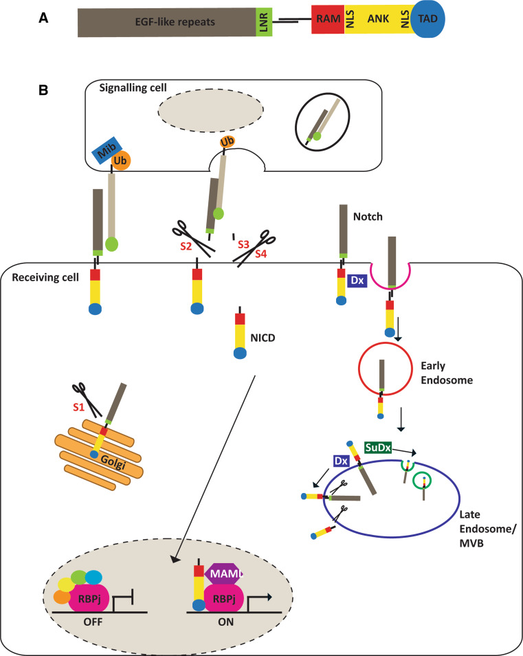
The Notch gene encodes a single-pass transmembrane receptor protein that functions as a membrane-bound transcription factor. The extracellular domain contains EGF-like repeats, which are responsible for ligand binding, and the LIN12-Notch repeats that prevent premature receptor activation [16]. The intracellular region comprises the RAM23 domain that is required for the interaction with members of the RBPj family of transcription factors [17], seven cdc10/ankyrin repeats that bind the Mastermind family of co-activators [18], nuclear localisation sequences, a transcriptional activation domain, and a PEST domain that is involved in protein degradation (Fig. 1a). Following binding of the Delta/Serrate/Jagged family of ligands, the Notch protein undergoes sequential cleavage by the ADAM10/Kuzbanian metalloprotease and γ-secretase enzymes (Fig. 1b). The second of these cleavages occurs within the transmembrane domain and releases the intracellular domain (NICD) [19, 20]. NICD then translocates to the nucleus, where it binds the RBPj transcription factor and the Mastermind co-activator to activate transcription of target genes. The RBPj transcription factors are also known as RBPjκ or the CSL family, whose name is derived from the human, Drosophila and C. elegans homologues CBF-1, Suppressor of Hairless (Su(H)), and LAG1, respectively. We shall use the term RBPj to denote this family of transcription factors. The most well characterised Notch/RBPj target genes are the basic helix–loop–helix transcription factors: Hairy and Enhancer of Split (Hes) and Hes related with YRPW motif (Hey) [21, 22], although genes regulating the cell cycle or apoptosis are also directly expressed in response to Notch signalling [23–29]. It is important to note that in the absence of Notch signalling, RBPj functions as a transcriptional repressor by binding co-repressors, such as Hairless in Drosophila, and MINT, KyoT2, HDAC (histone-deacetylase), and SMRT (silencing mediator of retinoid and thyroid receptors) in vertebrates (Fig. 1b; reviewed in [30]). Lastly, there is good evidence that NICD can regulate gene expression through its interaction with other transcription factors, including SMADs and LEF1 (reviewed in [31–35]).
Fig. 1.
Notch signalling pathway. a. Schematic of mammalian Notch proteins. The extracellular domain contains the EGF-like repeats that bind ligand and the LIN12-Notch repeats (LNR) that prevent premature receptor activation. The main regions of the intracellular domain are the RAM23 (RAM) domain that is required for the interaction with members of the RBPj family of transcription factors, the ankyrin repeats (ANK) that bind the Mastermind (MAML) family of co-activators, two nuclear localisation sequences (NLS), and a transcriptional activation domain (TAD). b. The mammalian Notch receptors are synthesised in the ER as a co-linear precursor which is cleaved by a Furin-like convertase at site 1 (S1) within the Golgi. Cleavage results in two non-covalently associated subunits expressed at the cell surface. Notch signalling is triggered by ligand binding to the EGF-like repeats (grey), exposing a second cleavage site (S2) processed by ADAM metalloproteases, generating a Notch intermediate. Two more cleavages occur at site 3 and 4 (S3 and S4) of the transmembrane domain by γ-secretase, releasing the Notch intracellular domain (NICD). NICD migrates to the nucleus and binds to RBPj transcription factors displacing co-repressors and recruiting activators, such as Mastermind. Endocytic trafficking of Notch ligands, promoted by E3 Ubiquitin (Ub) ligases Mindbomb (Mib) and Neuralized (not shown) regulate productive ligand–receptor interactions. Ligand-independent Notch activation can also occur by Deltex (Dx) promoting the endocytosis and trafficking of Notch through the early endosome and its cleavage on the outer membrane of the multivesicular body (MVB). Suppressor of Deltex [Su(Dx)] counters Dx activity and promotes lysosomal degradation of NICD
Notch activity is also tightly regulated by ubiquitination and endocytosis which can lead to signalling in the absence of a Delta/Serrate/Jagged ligand (Fig. 1b). Notch receptors present on the surface of the cell are constantly endocytosed and recycled back to the membrane [36] or degraded in the lysosome [37]. The trafficking of Notch through the endocytic system is regulated by the balance between the E3 ubiquitin ligases, Deltex and Suppressor of Deltex. Suppressor of Deltex favours the trafficking to the lysosome and hence Notch degradation. On the other hand, Deltex promotes the retention of Notch in the limiting membrane of the multivesicular body, which can lead to the release of NICD by γ-secretase-mediated cleavage and ligand-independent signalling once the extracellular domain of Notch has been degraded within the multivesicular body [38].
Wnt signalling
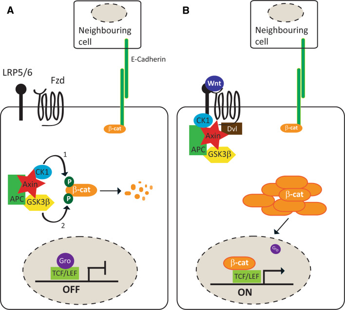
A conserved Wnt signalling pathway has been identified in vertebrate and invertebrate model systems, which regulates the cytosolic and nuclear levels of β-catenin (Armadillo in Drosophila). This is termed the Wnt/β-catenin signalling pathway and is depicted in Fig. 2.
Fig. 2.
Wnt/β-catenin signalling pathway. a. In the absence of Wnt ligand, β-catenin is recruited by the Axin destruction complex, sequentially phosphorylated by CK1 and GSK and targeted for degradation. Cytosolic levels of β-catenin (β-cat) are maintained at a low level, and β-catenin is mainly found at adherens junction. b. Extracellular Wnt binds to Frizzled (Fzd) and LRP5/6 co-receptors at the cell membrane and activates signalling. Subsequently, Dishevelled (Dvl) inactivates the destruction complex and β-cat accumulates in the cytosol. This allows translocation of β-cat to the nucleus where it activates transcription of target genes upon binding to LEF/TCF transcription factors
In the absence of Wnt ligands, the β-catenin molecules that are not found with the adherens junctions, but are instead present in the cytosol, are bound and processed by a destruction complex. This destruction complex is formed by the scaffolding proteins Axin and APC [39, 40], and the kinases GSK3β (Shaggy in Drosophila) [41] and Casein kinase 1 (CK1) [42–44]. The limiting component within this destruction complex is Axin, as it is the least abundant member, but interacts with all the other components. Once β-catenin is bound to the destruction complex, it is initially phosphorylated by CK1α generating a binding site for GSK3β, which subsequently phosphorylates three further Ser/Thr residues. Phosphorylated β-catenin interacts with the E3 ubiquitin ligase β-TrCP (β-transducin repeat containing protein), which targets it for proteosomal degradation [45, 46]. In this way, the cytoplasmic concentration of β-catenin is maintained at low levels. Therefore, in unstimulated cells, most of the endogenous β-catenin is found at the membrane, bound to E-cadherin, α-catenin and the cytoskeleton, regulating cell–cell adhesion [47, 48]. In the nucleus, in the absence of Wnt/β-catenin signalling, the TCF/LEF family of transcription factors interact with Groucho proteins and together act as transcriptional repressors [49, 50].
In the presence of Wnt ligands, a receptor complex containing Frizzled and LRP5/6 proteins is formed at the plasma membrane. This induces the phosphorylation of LRP5/6 by GSK3β, priming a second phosphorylation by CK1α. Subsequently, both Dishevelled (Dvl, Dsh in Drosophila) and Axin are recruited to the membrane, with Dvl interacting with the C-terminal tail of the Frizzled protein and Axin with the hyperphosphorylated LRP5/6, forming an intracellular bridging complex (reviewed in [51]). This sequesters Axin away from the destruction complex, allowing cytoplasmic β-catenin/Armadillo to accumulate quickly. Upon stabilisation, β-catenin translocates to the nucleus and accumulates [52, 53], whereupon it binds with LEF/TCF family of transcription factors [54, 55]. This interaction physically displaces Groucho [56] and recruits transcriptional co-activators, including Pygopus and Legless [57–59], leading to the expression of specific target genes, such as Axin2 and c-Myc [60, 61].
Wnt proteins also activate several other downstream signalling pathways: the most well characterised of these non-canonical Wnt pathways are: (I) the planar cell polarity pathway, which was identified in Drosophila and is required to establish the polarity within the plane of an epithelium [62, 63]; and (II) the Wnt/calcium pathway, first described in vertebrates [64].
Mechanisms underpinning the crosstalk between the Notch and Wnt pathways
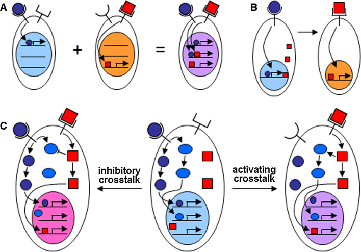
The molecular mechanisms underpinning the interactions between signalling pathways can be placed roughly into three classes: co-operative regulation of transcriptional targets; transcriptional targets of one pathway affecting another, resulting in temporally or spatially separated activity; and direct molecular crosstalk between the signal transduction machineries (Fig. 3). The described interactions between Notch and Wnt signalling fall into each of these categories.
Fig. 3.
Interactions between the Notch and Wnt signalling pathways. Notch and Wnt signalling pathways interact in three main ways. a Co-operative regulation of transcriptional targets. When both pathways are active at the same time, additional targets that require input from both pathways can be transcribed. b Transcriptional targets of one pathway affect the other. In this case, one pathway activates expression of the ligand for the second pathway, resulting in sequential signalling. c Direct molecular crosstalk. Crosstalk can be inhibitory (left) or activating (right). If the pathways are linear, then crosstalk can alter relative output levels (quantitative—not shown). If more than one branch is activated downstream of a ligand (blue), then crosstalk can affect the type of response (qualitative—compare left and right with centre)
Co-operative regulation of transcriptional targets
One of the first pieces of direct evidence of an interaction between the Notch and Wnt signalling pathways was the discovery that Notch and Wingless together regulate vestigial expression at the boundary formed between the developing dorsal and ventral surfaces of the fly wing; Wingless was the first Wnt ligand identified in Drosophila. The enhancer element that regulates expression of vestigial at the dorso-ventral boundary contains both dTCF and Suppressor of Hairless (the Drosophila homologue of RBPj) binding sites. Co-activation of both pathways leads to synergistic activation of the enhancer element in the developing wing [65]. Co-operative regulation of gene expression has also been observed in vertebrates, where NICD, β-catenin and RBPj form a complex that activates transcription [66, 67]. For example, Yamamizu and colleagues detected a complex of RBPj, NICD and β-catenin that binds to RBPj binding sites within the enhancer/promoter elements of several arterial genes. The complex forms in the endothelial cells that line both embryonic and adult arteries but not in the cells lining veins, suggesting that the complex plays an important role in distinguishing arterial and venous endothelial cells. In keeping with this, dual activation of β-catenin and NICD is required to induce arterial endothelial cell fate in Flk1+ ES-derived cells, a fate that neither protein alone was able to induce, and both proteins enhanced arterial gene expression during in vivo angiogenesis.
Transcription-dependent interaction
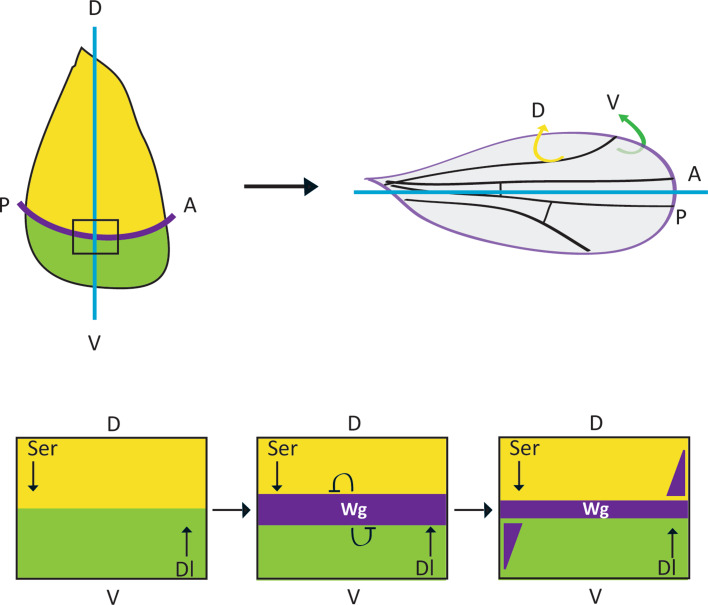
A common motif of Notch–Wnt interactions is the expression of one pathway’s ligand in response to signalling through the other. This is used repeatedly throughout development to generate either temporal or spatial separation of Notch and Wnt pathway activity. For example, the oscillations in gene expression that drive somitogenesis are due in part to the LEF1-mediated regulation of Delta-like1 ligand in vertebrates [68]. This forms part of the interlinked but out-of-phase oscillations of Wnt, Notch and FGF activity that pattern the segmented vertebrate body plan [69]. Alternatively, spatial separation of Notch and Wnt signalling is required during formation of boundaries between developing tissues. This occurs during the segmentation of the vertebrate hindbrain into rhombomeres [70, 71] and the specification of the dorsal (D) and ventral (V) compartments in the developing Drosophila wing (Fig. 4) [72–75]. In both cases, Notch signalling is activated in the cells that make up the boundary between adjacent compartments. These boundary cells then produce long-range signalling molecules, such as Wnts, that orchestrate the growth and patterning of the neighbouring compartments. Within the developing Drosophila wing, Notch signalling is initially activated in a broad stripe at the dorso-ventral boundary, or future wing margin. This induces the expression of the Wnt protein Wingless which refines this stripe of Notch activity in two ways. Firstly, Wingless signalling maintains Notch ligand expression in the cells that flank the margin so that they can signal back to the margin cells and maintain Notch activity and wingless expression [4, 73]. Secondly, Wingless also inhibits Notch signalling in the cells outside of the margin through direct crosstalk (see below). In addition to patterning the body plan by forming boundaries between defined territories, transcription-dependent Wnt–Notch interactions are also required to specify the size of particular domains. For example, Wnt signalling positively regulates the expression of the Notch ligand Jag1 during the development of the otic placode in the mouse to specify the size of the placode [76].
Fig. 4.
Wingless–Notch interactions shape the developing fly wing. The adult fly wing comprises two overlaid epithelial sheets: the dorsal side (D-yellow) and the ventral side (V-green) with wing margin tissue at the D-V border (purple) (top panel). The adult wing tissue is derived from the larval imaginal disc (left), which is patterned by Wingless–Notch interactions (outlined below). Within the developing wing, Notch signalling is initially activated in a broad stripe at the D-V boundary, by expression of the ligands Serrate and Delta in the D and V compartments (left). Notch activates the expression of Wg (purple), which then refines this stripe of Notch activity in two ways: Wingless signalling maintains Notch ligand expression in the cells that flank the margin so that they can signal back to the margin cells and maintain Notch activity, and Wingless also inhibits Notch signalling in the cells outside of the margin through direct crosstalk. These interactions result in a gradient of Wingless activity with high threshold targets expressed close to the D-V boundary and lower threshold targets expressed further away
Not all transcriptional interactions are at the level of Notch/Wnt ligands. For example, Notch/RBPj-dependent activation of Frizzled receptor expression is required for optimal differentiation of dendritic cells from haematopoietic stem cells (HSCs). Here the surrounding stroma expresses Notch ligands, activating Notch signalling in HSCs and upregulating Frizzled expression [77]. In the developing Drosophila eye, PCP signalling defines the fate of two photoreceptors: R3 and R4. Frizzled/Dsh signalling is active in R3 and activates expression of Neuralized, which promotes Delta function in R3 and therefore Notch activity in R4 [78]. Conversely, in mammary stem cells (MaSCs) Wnt signalling inhibits Notch activity through a β-catenin/Pygopus2-dependent remodelling of the chromatin at the Notch3 locus that prevents expression of the Notch3 gene [79]. This promotes the self-renewal of MaSC as Notch3 signalling is required for the differentiation of these cells.
Direct molecular crosstalk between signal transduction machinery
There are many lines of evidence that suggest that Notch and Wnt signal transduction machineries can interact with each other and directly affect signalling output from the other pathway. Below we will summarise the major findings.
Dishevelled inhibits Notch
Molecularly, Dishevelled has been shown to physically interact with Notch in vivo and in yeast-2-hybrid studies [3, 80–82]. Furthermore, the two proteins co-localise when expressed in Drosophila S2 cells [3]. Functionally, the Dishevelled interaction inhibits Notch signalling and has been shown to disrupt the lateral inhibition signal mediated by Notch that limits the specification of sensory organ precursor (SOP) cells [3]. Lateral inhibition signals ensure that those SOPs that do develop are separated from one another. Consequently, many more SOPs form when Dishevelled is overexpressed and neighbouring cells can also develop as SOPs. Interestingly, there is a marked difference in the Dishevelled and Armadillo/β-catenin overexpression phenotypes [83]. Although many more SOPs develop in both cases, the SOPs are evenly spaced when Armadillo/β-catenin is overexpressed. Thus, Dishevelled has the dual function of activating Armadillo and simultaneously inhibiting Notch activity. However, it is not entirely understood how Dishevelled inhibits Notch signalling, although experiments suggest one mechanism may involve Wingless/Dishevelled promoting Notch endocytosis [81]. Lastly, similar inhibition of Notch signalling by Dishevelled has been shown during the establishment of planar polarity in the Drosophila eye and leg epithelium, linking Dishevelled’s role in Wnt/PCP signalling to the positioning of Notch activity [84, 85].
Dishevelled inhibits RBPj
We recently identified a novel point of crosstalk, whereby Dishevelled limits Notch signalling in mammalian cells [6]. In reporter gene assays, Dishevelled inhibits Notch pathway activity induced by treatment with Notch ligand, or overexpression of active forms of the Notch and RBPj proteins. Dishevelled does so by binding and sequestering RBPj proteins away from the nuclear fraction that contains active transcription factors. To assess the importance of this crosstalk in vivo, we examined whether Dishevelled influenced the spacing of ciliated cells in the epidermis of the Xenopus embryo. The spacing of these cells is regulated by Notch-dependent lateral inhibition [86]. We found that the level of Notch signalling, and thus the spacing of the ciliated cells, is regulated by the level of Dishevelled and its ability to bind RBPj. Furthermore, this mode of crosstalk is qualitatively different from other forms of crosstalk between developmental signalling pathways and Notch receptors described previously [31]: Dishevelled inhibits signalling by all four Notch paralogues as it targets the unique and common pathway component, RBPj, that is found downstream of all Notch proteins.
GSK-3β/Notch crosstalk
In mammalian cells, GSK-3β has been shown to physically bind and phosphorylate the intracellular domain of two Notch paralogues. However, the outcome of this interaction varies as N1 ICD is positively regulated, whilst the activity of N2 ICD is negatively regulated by GSK-3β. GSK-3β stabilises N1 ICD via phosphorylation, which promotes Notch signalling [87]. Consistent with this, the activity of a Notch reporter construct is reduced in GSK-3β null fibroblasts but it is not abolished. In contrast, GSK-3β does not affect the stability of N2 ICD, but instead reduces the ability of N2 ICD to signal. Negative regulation may occur through a decrease of the ability of Notch2 to bind co-factors due to the juxtaposition of the binding sites for GSK-3β and the co-activator CBP [88]. In the latter study, Wnt signalling, which reduces the activity of GSK-3β, was shown to increase the activation of the Hes-1 promoter by Notch2. This difference between Notch1 and Notch2 may reflect the specificity of individual Notch molecules, but it may also reflect the importance of cellular context in signalling outcomes as the experiments were conducted in different systems.
Notch inhibits Armadillo/β-catenin
Several groups have described complex genetic interactions between Notch and wingless mutations in Drosophila, which suggest that Notch can inhibit Wingless signalling [80, 89–93]. More recent reports provided a detailed mechanism for these genetic interactions, whereby Notch acts independently of ligand and RBPj-dependent transcription to reduce the amount of the active form of Armadillo in the cell. In the wing disc, membrane-bound Notch associates with Armadillo at the adherens junction and as Notch is endocytosed it causes ‘active’ Armadillo to be trafficked as well [94]. Consequently, Armadillo appears within endosomes and is ultimately degraded, reducing pathway activity. In vertebrates, a similar trafficking model exists for Notch/β-catenin antagonism in the differentiation of multipotent cardiac progenitor cells [95, 96]. Again, this work demonstrated that membrane-bound Notch can physically associate with dephosphorylated, ‘active’ β-catenin and promote its endosomal trafficking, leading to its degradation in the lysosome. Interestingly, the inhibition of β-catenin function does not require the function of γ-secretase, arguing that the inhibition of Wnt/β-catenin signalling by membrane-bound Notch does not require the release of NICD or the expression of downstream Notch targets [95, 97]. On the other hand, work from several groups has shown that NICD can also inhibit Wnt/β-catenin signalling [98], and that this inhibition may be mediated by the expression of downstream Hes/Hey proteins [99] or the recruitment of transcriptional co-repressors by NICD to β-catenin binding sites within Wnt target genes [100]. Recent work from our own lab suggests that NICD inhibits β-catenin activity directly by forming a complex that prevents β-catenin binding its target sites, and instead recruits β-catenin to NICD/RBPj targets sites ([6] Hidalgo-Sastre, Acar et al. unpublished), consistent with the observations in cell lines, neural precursor cells and arterial endothelial cells described above [66, 67, 101].
Effect of direct molecular crosstalk
One key property that direct inhibitory molecular crosstalk can confer is the rapid switch between Notch-ON/Wnt-OFF and Wnt-ON/Notch-OFF states without having to rely on transcriptional feedback loops. The activity of Dishevelled in promoting Wnt signalling and limiting signalling through the Notch pathway drives the Wnt-ON/Notch-OFF state. Conversely, Notch can both limit β-catenin activity and promote RBPj transcriptional activity, possibly also co-opting β-catenin to RBPj promoters, to generate a Notch-ON/Wnt-OFF state. In both cases, the binary switch between states can occur rapidly and independently of the transcription of pathway components. Given the many contexts in which Notch and Wnt have opposing effects (reviewed in [102]), the temporal transition from a state of Notch-ON/Wnt-OFF to Wnt-ON/Notch-OFF, or vice versa, is critical for robust and precise development. For example, during early embryonic myogenesis and in muscle repair in the adult, the requirement to switch between high Notch signalling and high Wnt signalling is evident [103, 104]. Here, an initial peak of Notch signalling is required to trigger the differentiation programme of progenitors and to expand the muscle progenitor pool prior to terminal differentiation. However, Notch activity must be silenced and Wnt signalling then activated for terminal differentiation to occur appropriately. If this temporal switch does not take place and the Notch signal is maintained, proper myogenesis fails to occur [104], even in the presence of active Wnt signalling [103]. The latter result suggests that direct inhibitory crosstalk may be a more common feature of the cell-fate antagonism between the Notch and Wnt pathways than has previously been appreciated.
Integrated Notch–Wnt activity and the control of gene regulatory networks
The Wnt and Notch signalling pathways are so intertwined during development that it has been suggested that they form an integrated signalling device termed ‘Wntch’ [102]. The role of Wntch is to limit variability in terms of sharpening boundaries and regulating stochastic cell-fate decisions at the population level. Comparison of tissue homeostasis in the fly and mammalian gut suggests that Wntch activity can also regulate the balance between stem cells, progenitors and differentiated cells within a tissue, as we shall outline below.
One classic example of sharpening boundaries is the patterning of the Drosophila wing margin, as outlined briefly above (Fig. 4). Notch activity positions the boundary between the dorsal and ventral compartments and induces wingless expression in the boundary cells. Wingless then signals to the surrounding cells to increase expression of the Notch ligands Delta and Serrate, which signal back to Notch receptors at the boundary to maintain wingless expression [4, 74]. However, this transcriptional feedback loop is also impacted by direct molecular crosstalk. The initial wingless expression gradient is broad and shallow, but as Wingless activates Dishevelled and Dishevelled inhibits Notch signalling, Notch activity and thus wingless expression is refined into a sharp stripe at the boundary [5]. This refinement is required for the proper patterning of the adult wing. There are many similarities between the patterning of the Drosophila wing and the patterning of the rhombomeres in the vertebrate hindbrain. Here again, Wnt proteins expressed at the rhombomere boundary promote Notch ligand expression in adjacent cells to maintain Notch activity. The boundary then also serves as a Wnt signalling source as Notch promotes Wnt expression [70, 71, 105]. Thus, the mechanisms that maintain the spatial separation of Notch and Wnt signalling to pattern tissues appear to be conserved from Drosophila to vertebrates.
The recent turn to computational modelling of biological phenomena has confirmed just how important the direct molecular crosstalk between Notch and Wnt pathways is in patterning tissues. In fact, mathematical modelling of Drosophila wing patterning has shown that it is only once the crosstalk between Notch and Wnt is factored into the analysis that the model can reproduce the dynamics seen in vivo [106]. This study also revealed a point of crosstalk that had previously been overlooked and is required to maintain Notch signalling at the dorso-ventral boundary, and thus generate a stable pattern of gene expression in the surrounding tissue. A refractoriness to Wingless signalling must be induced within the wingless-expressing cells of the boundary; otherwise, the boundary cells would also respond to Wingless and inhibit Notch activity through Dishevelled-mediated crosstalk, thus temporally limiting expression of wingless and its target genes. Similarly, modelling of the developing chick feather bud has revealed a requirement for Notch/Wnt crosstalk [107]. A “zone of polarizing activity” located in the posterior feather bud mediates the directional elongation of the feather bud primordium and ensures proper feather orientation. Transplantation experiments show that it is a dermal nuclear β-catenin positive zone (DBZ) that bears the polarizing activity. The DBZ is shaped by Wnt7a, which is secreted from the posterior epithelium and acts on dermal cells to promote stabilisation and nuclear translocation of β-catenin. This activation of Wnt/β-catenin signalling has two functional effects: non-muscle myosin IIB is activated, mediating directional elongation of the feather bud; and Jagged-1 is expressed activating Notch signalling in the surrounding area. Feedback from the Notch pathway is then required to refine the boundary of the DBZ. Currently, the molecular nature of Notch feedback onto Wnt/β-catenin signalling is not known, but it is required to translate a weak and noisy gradient of Wnt7a protein into a broad and relatively homogeneous response within a field of cells with a sharp boundary between responding and non-responding cells. When Notch signalling is disrupted by treating the chick embryo with a γ-secretase inhibitor, many fewer cells respond to the Wnt7a protein gradient and the sharp boundary of the response is lost. The requirement for Wnt/Notch feedback is recapitulated in a mathematical model in which crosstalk stimulates the change from a noisy, gradual Wnt gradient to a definitive threshold Wnt response. This also raises the idea that interactions between Notch and Wnt signalling not only allow the smooth transition from a Notch-ON/Wnt-OFF to a Notch-OFF/Wnt-ON state without transcription, but also limit noise within a system to provide a co-ordinated response to a Wnt signal [102, 108].
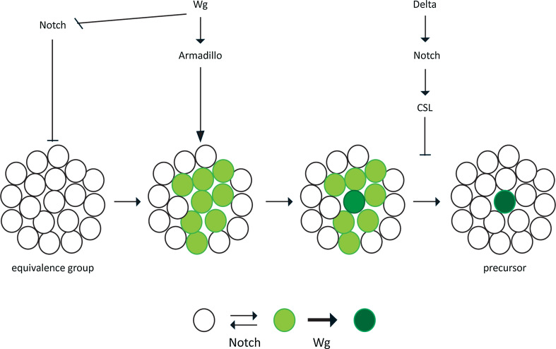
The role for Notch and Wnt in governing stochastic fate decisions at the population level is best studied in the equivalence groups that produce the sensory organ precursors (SOP) or the muscle precursors in Drosophila (Fig. 5), although there are many examples in both invertebrates and vertebrates. In both cases, a group of cells is established that all have the potential to become either the Achaete/Scute-expressing SOP or S59/Slouch-expressing muscle precursor but only one cell from the group will eventually adopt the fate and in doing so will force the others to adopt a secondary fate, though lateral inhibition [109–113]. The balance of Wingless and Notch signalling tightly controls the probability of cells adopting the precursor fate, but it does not determine which cell will adopt the fate. Initially, RBPj-independent Notch signalling maintains the cells within the equivalence group in a naïve state and Ac/Sc or S59/Slouch are not expressed. Wingless signalling then overcomes this Notch-mediated inhibition, promoting a subset of the group to express Ac/Sc or S59/Slouch in a ‘transition state’. RBPj-dependent Notch signalling then limits the number of cells that maintain gene expression and adopt the precursor fate through lateral inhibition: namely, the cell that stochastically expresses the highest level of Notch ligand signals to the surrounding cells to prevent them adopting the SOP or muscle precursor fate. Consequently, Notch ligand expression is reduced in the surrounding cells, reducing the Notch signal received by the cell that is adopting the SOP or muscle cell fate. This fall in Notch signalling promotes the further differentiation of the cell adopting the SOP or muscle cell fate and its increased Notch ligand expression. Thus, this positive feedback loop enforces a binary outcome with one cell per cluster adopting the SOP or muscle precursor fate.
Fig. 5.
Wntch signalling controls cell-fate decisions in equivalence groups RBPj-independent Notch signalling maintains the cells within the equivalence group in a naïve state and Achaete/Scute (Ac/Sc) or S59/Slouch are not expressed (white). Wg signalling then overcomes this Notch-mediated inhibition, promoting members of the group to express A/Sc or S59/Slouch in a ‘transition state’ (light green). RBPj-dependent Notch signalling then limits the number of cells that maintain gene expression and adopt the precursor fate through lateral inhibition: that is the cell that stochastically expresses the highest level of Notch ligand (dark green) signals to the surrounding cells to prevent them adopting the primary fate. A schematic is shown below
The many different scenarios in which interactions between Notch and Wnt signalling regulate stochastic cell-fate decisions raises the question of how the two pathways can directly regulate so many different cell-fate decisions; it is difficult to see how the RBPj and TCF transcription factors at the base of the two pathways can distinguish between promoters of many different cell type-specific genes. The answer most likely lies in the fact that both signalling pathways regulate cell-fate decisions in a permissive manner and are guided to the promoters of cell type-specific genes by pioneer transcription factors already present within the cells, rather than specifying fate directly [114, 115].
Gut homeostasis as a paradigm for understanding Notch–Wnt interactions in controlling the differentiation along a cell lineage
The maintenance of the gut brings together in one tissue many aspects of Wntch interactions that have been described thus far: evolutionary conservation of Notch–Wnt interactions; iterative use of Notch/Wnt antagonism to regulate fate decisions; Notch ligands being a transcriptional target of Wnt activity; and regulation of stochastic fate decisions at the population level. We will now outline the mechanisms governing homeostasis on the fly and the mammalian intestine (Fig. 6).
Fig. 6.
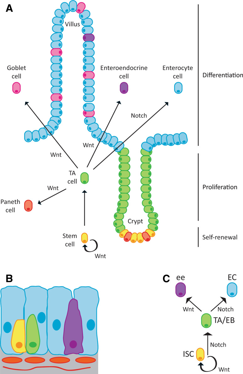
Wnt and Notch interactions control gut homeostasis. a Schematic representation of the mammalian intestine. Intestinal stem cells (ISC-yellow) reside at the bottom of the crypt along with Paneth cells. Stem cell self-renewal is balanced with the production of transit amplifying cells (TA-green), which move up into the proliferative zone. TA cells give rise to absorptive enterocytes (blue) and the secretory lineage that includes enteroendocrine cells (purple), goblet cells (pink) and Paneth cells (coral). b. Schematic of the fly midgut, which follows a similar but less elaborative pattern to the mammalian model. Upon division, stem cells can give rise to the intermediate non-amplifying precursor the enteroblast (EB-green), which then differentiates into either the absorptive lineage (enterocyte-EC) or the secretory lineage (enteroendocrine-ee). The cells are wrapped in muscle fibres (orange and grey), which are the source of the Wnt ligand. c. Diagram showing the opposing effects of Notch and Wnt signalling at the ISC to TA/EB step and the adoption of the secretory (purple) versus the absorptive (blue) lineage step. This model holds for both mammalian and fly models. (adapted from [116, 139])
The processes regulating the turnover of the fly gut and mammalian intestine are remarkably well conserved. In both the fly and mammalian intestine, the cell lineages comprise an intestinal stem cell (ISC) population that resides next to niche cells; the Paneth cells within the intestinal crypts in mammals and the escargot + nests in the fly intestine [8, 116–118]. At division, the ISCs undergo self-renewal and/or differentiation into the transit amplifying (TA) population, termed enteroblasts (EBs) in the fly. The TA population then differentiates into the mature cells found in the gut: the absorptive lineage [enterocytes (ECs)] and the secretory lineage including goblet cells, enteroendocrine cells and Paneth cells in mammals, or the enteroendocrine (ee) cells in the fly. The intestine is one of the most rapidly renewing tissues in the adult; in the mouse, each crypt can generate up to 200 cells per day, replacing the cells within the intestinal epithelial lining every 4–5 days [119]. Such a high turnover rate means that the self-renewal and differentiation along each lineage must be tightly controlled at the population level in order to maintain tissue integrity and prevent tumour formation. Notch and Wnt signals are a vital part of this regulation.
In both cases, Wnt/Wingless signalling promotes the self-renewal of ISCs [8, 117]. Whilst it is not clear how the transition from ISC to TA fate is controlled in the mammalian gut lining, Notch promotes the adoption of the EB fate in Drosophila, suggesting that antagonism between the two pathways may control the switch between these two fates [116, 118]. However, competition between the two pathways is clearly seen at the stage of TA/EB differentiation and lineage bifurcation with Wnt promoting the secretory lineage (goblet, Paneth and enteroendocrine cells in mammals, ee cells in Drosophila), whilst Notch promotes the adoption of the absorptive enterocyte cell fate [12, 13, 120, 121]. At both the self-renewal versus TA decision and the lineage split, there must be a clear Notch-ON/Wnt-OFF or Wnt-ON/Notch-OFF response in order to generate the stable, binary fate decisions. It is likely that direct crosstalk between the two pathways plays a significant role in maintaining the bistable outcome of these decisions. Moreover, the sequential Wnt–Notch activity is achieved in part through upregulation of Notch ligands in response to Wnt/Wingless signalling in both Drosophila and mammals (Jag1 in mice, Delta in flies [117, 118, 122]).
Recent work has shown that the ISCs in both tissues are maintained with neutral drift dynamics, meaning that the fate of the two daughter cells of a dividing ISC are specified by signalling between the two daughter cells and their local environment, rather than being specified through an invariant asymmetric cell division [116, 123, 124]. Consequently, when the ISC divides it is possible to obtain two ISCs, two TA/EB cells or one of each, although the division usually yields one ISC and one TA/EB cell. This also means that if an ISC is lost through differentiation, it can be replaced by the symmetric division of a neighbouring ISC. Given the importance of Wnt/Wingless and Notch signalling in ISCs and TA/EB cells, interactions between the two pathways may play a role in maintaining the balance between the ISC and TA/EB cell populations. Specifically, Wnt–Notch interactions may regulate the stochastic cell-fate specification of the two daughter cells from a dividing ISC to control the number of ISCs and TA/EB cells within the tissue as a whole.
Due to the genetic tractability, it has been possible to demonstrate that the two pathways control the balance between ISCs and EB cells in Drosophila. Reducing Notch dosage shifts the stable ISC:EB ratio to favour the ISC population, whereas Notch gain of function mutations favour the differentiation of EB cells increasing their proportion within the stable adult population [116]. The opposite phenotype is seen when Wingless signalling is increased [117]. In this scenario, the ISC population is increased and ISC-like tumours develop, which can be rescued by forced Notch activation. These results also suggest that Notch/Wnt interactions may regulate the balance of stem cells and differentiated cells in other tissues where stem cell number is regulated by neutral drift and the two pathways have opposing effects on differentiation, such as the skin [7, 9–11]. Given the opposing effects of Notch and Wnt signalling on mammary gland stem cells [125–128], it will be interesting to investigate how Notch/Wnt interactions affect mammary gland biology.
Clinical relevance of Notch–Wnt crosstalk
Given the importance of Notch and Wnt signalling during development, it is not surprising that their manipulation is key to the successful generation of differentiated cells from stem cells for therapeutic benefit, for instance to regulate the neural differentiation of ES cells [129]. Furthermore, the results of Trowbridge and colleagues are of direct relevance to patients needing bone marrow transplants. Using a mouse model, they found that GSK-3β inhibition following haematopoietic stem cell (HSC) transplantation augmented haematopoietic repopulation in recipient mice, improved neutrophil and megakaryocyte recovery, recipient survival and enhanced sustained long-term haematopoietic repopulation [130]. This was due to the increase in both Wnt and Notch signalling following treatment with the GSK-3β inhibitor, which improves HSC survival and self-renewal; however, it is not clear whether the increase in Notch signalling is due to altered phosphorylation of Notch by GSK-3β or increased Notch ligand expression downstream of β-catenin/TCF transcription. Therefore, GSK-3β inhibition might be a clinical means to improve the outcome of patients receiving transplanted HSCs.
Manipulating Notch and Wnt signalling may also significantly improve the treatment of liver disease. Following significant hepatic injury, damaged cells are replaced by the proliferation and differentiation of hepatic progenitor cells (HPCs). As with the intestinal lining, Notch and Wnt signalling have opposing effects on the differentiation of HPCs, with Notch signalling promoting cholangiocyte differentiation and Wnt signalling driving cells into the hepatocyte lineage [131]. Interactions are also seen between the two pathways, with Wnt signalling promoting hepatocyte differentiation, in part by inducing the expression of Numb, which inhibits Notch signalling and prevents cholangiocyte formation [131]. However, it has become clear recently that an imbalance between these pathways during liver regeneration can lead to liver disease. In acute necrotising hepatitis and the cirrhosis that follows hepatitis C virus infection, there is an excess of Wnt signalling promoting hepatocyte differentiation [131, 132]. In contrast, excessive Notch signalling occurs in primary biliary cirrhosis and primary sclerosing cholangitis driving cholangiocyte differentiation [131, 132]. Consequently, rebalancing the interactions between the two pathways is likely to significantly influence the treatment of these debilitating diseases.
Interactions between the pathways also play an important role in other human pathologies. In both breast and colorectal cancer, there is a recurrence of the common interaction seen during development whereby Wnt signalling activates Notch by inducing Notch ligand expression [122, 133]. Furthermore, the transformation of breast epithelial cells by Wnt signalling does not occur in the presence of Notch inhibitors, suggesting the requirement for Wnt/Notch interactions in disease progression [122, 133, 134]. However, the situation is more complicated in colorectal cancer. In this case, the activation of Notch signalling with Wnt causes many more adenomas to develop and at an earlier age [135]. On the other hand, the progression of these adenomas is limited, with the adenomas arising when both Notch and Wnt are active being of a lower grade [100]. This raises the interesting possibility that an intermediate level of Notch signalling rather than a high level will drive colorectal cancer, as it will enhance tumour initiation but not interfere with progression. The interactions between the pathways may also ensure that this normally happens, as the induction of Jagged1 by Wnt signalling will activate the Notch pathway [122], but the crosstalk between Dishevelled and Notch will limit its strength [6].
Lastly, the interactions between the pathways should influence how we target the pathways therapeutically. For example, in cases where disease initiation or progression are reliant on Wnt activation with the concomitant inhibition of Notch signalling mediated by crosstalk, treating with an inhibitor at the level of the Wnt ligand may be advantageous as Wnt–Notch crosstalk will be lost (leading to increased Notch signalling [6, 88] ) as well as the Wnt signal. In contrast, an inhibitor of the Wnt transcriptional response [136, 137] may not be as useful as there will only be a loss of Wnt-driven transcription and no effect on direct Notch crosstalk. Alternatively, drugs such as gamma-secretase inhibitors, which inhibit signalling by reducing Notch cleavage, may also result in the advantageous inhibition of β-catenin signalling by increasing the amount of membrane-bound Notch protein that is able to interact with and inhibit β-catenin [138]. Thus, given appropriate knowledge of the signalling context, the mimicking of inhibitory crosstalk opens new avenues for therapeutic drug development by offering the promise of specificity in targeting signalling pathways.
References
- 1.Pires-daSilva A, Sommer RJ. The evolution of signalling pathways in animal development. Nat Rev Genet. 2003;4:39–49. doi: 10.1038/nrg977. [DOI] [PubMed] [Google Scholar]
- 2.Peter IS, Davidson EH. A gene regulatory network controlling the embryonic specification of endoderm. Nature. 2011;474:635–639. doi: 10.1038/nature10100. [DOI] [PMC free article] [PubMed] [Google Scholar]
- 3.Axelrod JD, Matsuno K, Artavanis-Tsakonas S, Perrimon N. Interaction between Wingless and Notch signaling pathways mediated by dishevelled. Science. 1996;271:1826–1832. doi: 10.1126/science.271.5257.1826. [DOI] [PubMed] [Google Scholar]
- 4.Micchelli CA, Rulifson EJ, Blair SS. The function and regulation of cut expression on the wing margin of Drosophila: Notch, Wingless and a dominant negative role for Delta and Serrate. Development. 1997;124:1485–1495. doi: 10.1242/dev.124.8.1485. [DOI] [PubMed] [Google Scholar]
- 5.Rulifson EJ, Micchelli CA, Axelrod JD, Perrimon N, Blair SS. Wingless refines its own expression domain on the Drosophila wing margin. Nature. 1996;384:72–74. doi: 10.1038/384072a0. [DOI] [PubMed] [Google Scholar]
- 6.Collu GM, Hidalgo-Sastre A, Acar A, Bayston L, Gildea C, Leverentz MK, Mills CG, Owens TW, Meurette O, Dorey K, Brennan K. Dishevelled limits Notch signalling through inhibition of CSL. Development. 2012;139:4405–4415. doi: 10.1242/dev.081885. [DOI] [PMC free article] [PubMed] [Google Scholar]
- 7.Clayton E, Doupe DP, Klein AM, Winton DJ, Simons BD, Jones PH. A single type of progenitor cell maintains normal epidermis. Nature. 2007;446:185–189. doi: 10.1038/nature05574. [DOI] [PubMed] [Google Scholar]
- 8.Clevers H. The intestinal crypt, a prototype stem cell compartment. Cell. 2013;154:274–284. doi: 10.1016/j.cell.2013.07.004. [DOI] [PubMed] [Google Scholar]
- 9.Doupe DP, Klein AM, Simons BD, Jones PH. The ordered architecture of murine ear epidermis is maintained by progenitor cells with random fate. Dev Cell. 2010;18:317–323. doi: 10.1016/j.devcel.2009.12.016. [DOI] [PubMed] [Google Scholar]
- 10.Lim X, Tan SH, Koh WL, Chau RM, Yan KS, Kuo CJ, van Amerongen R, Klein AM, Nusse R. Interfollicular epidermal stem cells self-renew via autocrine Wnt signaling. Science. 2013;342:1226–1230. doi: 10.1126/science.1239730. [DOI] [PMC free article] [PubMed] [Google Scholar]
- 11.Lowell S, Jones P, Le Roux I, Dunne J, Watt FM. Stimulation of human epidermal differentiation by delta-notch signalling at the boundaries of stem-cell clusters. Curr Biol. 2000;10:491–500. doi: 10.1016/s0960-9822(00)00451-6. [DOI] [PubMed] [Google Scholar]
- 12.van Es JH, van Gijn ME, Riccio O, van den Born M, Vooijs M, Begthel H, Cozijnsen M, Robine S, Winton DJ, Radtke F, Clevers H. Notch/gamma-secretase inhibition turns proliferative cells in intestinal crypts and adenomas into goblet cells. Nature. 2005;435:959–963. doi: 10.1038/nature03659. [DOI] [PubMed] [Google Scholar]
- 13.Yin X, Farin HF, van Es JH, Clevers H, Langer R, Karp JM. Niche-independent high-purity cultures of Lgr5 intestinal stem cells and their progeny. Nat Methods. 2013;11:106–112. doi: 10.1038/nmeth.2737. [DOI] [PMC free article] [PubMed] [Google Scholar]
- 14.Bolós V, Grego-Bessa J, de la Pompa JL. Notch signaling in development and cancer. Endocr Rev. 2007;28:339–363. doi: 10.1210/er.2006-0046. [DOI] [PubMed] [Google Scholar]
- 15.Clevers H. Wnt/beta-catenin signaling in development and disease. Cell. 2006;127:469–480. doi: 10.1016/j.cell.2006.10.018. [DOI] [PubMed] [Google Scholar]
- 16.Sanchez-Irizarry C, Carpenter AC, Weng AP, Pear WS, Aster JC, Blacklow SC. Notch subunit heterodimerization and prevention of ligand-independent proteolytic activation depend, respectively, on a novel domain and the LNR repeats. Mol Cell Biol. 2004;24:9265–9273. doi: 10.1128/MCB.24.21.9265-9273.2004. [DOI] [PMC free article] [PubMed] [Google Scholar]
- 17.Tamura K, Taniguchi Y, Minoguchi S, Sakai T, Tun T, Furukawa T, Honjo T. Physical interaction between a novel domain of the receptor Notch and the transcription factor RBP-J kappa/Su(H) Curr Biol. 1995;5:1416–1423. doi: 10.1016/s0960-9822(95)00279-x. [DOI] [PubMed] [Google Scholar]
- 18.Ehebauer MT, Chirgadze DY, Hayward P, Martinez Arias A, Blundell TL. High-resolution crystal structure of the human Notch 1 ankyrin domain. Biochem J. 2005;392:13–20. doi: 10.1042/BJ20050515. [DOI] [PMC free article] [PubMed] [Google Scholar]
- 19.Rebay I, Fleming RJ, Fehon RG, Cherbas L, Cherbas P, Artavanis-Tsakonas S. Specific EGF repeats of Notch mediate interactions with Delta and Serrate: implications for Notch as a multifunctional receptor. Cell. 1991;67:687–699. doi: 10.1016/0092-8674(91)90064-6. [DOI] [PubMed] [Google Scholar]
- 20.Nichols JT, Miyamoto A, Olsen SL, D’Souza B, Yao C, Weinmaster G. DSL ligand endocytosis physically dissociates Notch1 heterodimers before activating proteolysis can occur. J Cell Biol. 2007;176:445–458. doi: 10.1083/jcb.200609014. [DOI] [PMC free article] [PubMed] [Google Scholar]
- 21.Iso T, Kedes L, Hamamori Y. HES and HERP families: multiple effectors of the Notch signaling pathway. J Cell Physiol. 2003;194:237–255. doi: 10.1002/jcp.10208. [DOI] [PubMed] [Google Scholar]
- 22.Fischer A, Gessler M. Delta-Notch and then? Protein interactions and proposed modes of repression by Hes and Hey bHLH factors. Nucleic Acids Res. 2007;35:4583–4596. doi: 10.1093/nar/gkm477. [DOI] [PMC free article] [PubMed] [Google Scholar]
- 23.Jeffries S, Robbins DJ, Capobianco AJ. Characterization of a high-molecular-weight Notch complex in the nucleus of Notch(ic)-transformed RKE cells and in a human T-cell leukemia cell line. Mol Cell Biol. 2002;22:3927–3941. doi: 10.1128/MCB.22.11.3927-3941.2002. [DOI] [PMC free article] [PubMed] [Google Scholar]
- 24.Joshi I, Minter LM, Telfer J, Demarest RM, Capobianco AJ, Aster JC, Sicinski P, Fauq A, Golde TE, Osborne BA. Notch signaling mediates G1/S cell-cycle progression in T cells via cyclin D3 and its dependent kinases. Blood. 2009;113:1689–1698. doi: 10.1182/blood-2008-03-147967. [DOI] [PMC free article] [PubMed] [Google Scholar]
- 25.Klinakis A, Szabolcs M, Politi K, Kiaris H, Artavanis-Tsakonas S, Efstratiadis A. Myc is a Notch1 transcriptional target and a requisite for Notch1-induced mammary tumorigenesis in mice. Proc Natl Acad Sci USA. 2006;103:9262–9267. doi: 10.1073/pnas.0603371103. [DOI] [PMC free article] [PubMed] [Google Scholar]
- 26.Palomero T, Lim WK, Odom DT, Sulis ML, Real PJ, Margolin A, Barnes KC, O’Neil J, Neuberg D, Weng AP, Aster JC, Sigaux F, Soulier J, Look AT, Young RA, Califano A, Ferrando AA. Notch1 directly regulates c-MYC and activates a feed-forward-loop transcriptional network promoting leukemic cell growth. Proc Natl Acad Sci USA. 2006;103:18261–18266. doi: 10.1073/pnas.0606108103. [DOI] [PMC free article] [PubMed] [Google Scholar]
- 27.Rangarajan A, Talora C, Okuyama R, Nicolas M, Mammucari C, Oh H, Aster JC, Krishna S, Metzger D, Chambon P, Miele L, Aguet M, Radtke F, Dotto GP. Notch signaling is a direct determinant of keratinocyte growth arrest and entry into differentiation. EMBO J. 2001;20:3427–3436. doi: 10.1093/emboj/20.13.3427. [DOI] [PMC free article] [PubMed] [Google Scholar]
- 28.Ronchini C, Capobianco AJ. Induction of cyclin D1 transcription and CDK2 activity by Notch(ic): implication for cell cycle disruption in transformation by Notch(ic) Mol Cell Biol. 2001;21:5925–5934. doi: 10.1128/MCB.21.17.5925-5934.2001. [DOI] [PMC free article] [PubMed] [Google Scholar]
- 29.Weng AP, Millholland JM, Yashiro-Ohtani Y, Arcangeli ML, Lau A, Wai C, Del Bianco C, Rodriguez CG, Sai H, Tobias J, Li Y, Wolfe MS, Shachaf C, Felsher D, Blacklow SC, Pear WS, Aster JC. c-Myc is an important direct target of Notch1 in T-cell acute lymphoblastic leukemia/lymphoma. Genes Dev. 2006;20:2096–2109. doi: 10.1101/gad.1450406. [DOI] [PMC free article] [PubMed] [Google Scholar]
- 30.Kovall RA, Blacklow SC. Mechanistic insights into Notch receptor signaling from structural and biochemical studies. Curr Top Dev Biol. 2010;92:31–71. doi: 10.1016/S0070-2153(10)92002-4. [DOI] [PubMed] [Google Scholar]
- 31.Hurlbut GD, Kankel MW, Lake RJ, Artavanis-Tsakonas S. Crossing paths with Notch in the hyper-network. Curr Opin Cell Biol. 2007;19:166–175. doi: 10.1016/j.ceb.2007.02.012. [DOI] [PubMed] [Google Scholar]
- 32.Blokzijl A, Dahlqvist C, Reissmann E, Falk A, Moliner A, Lendahl U, Ibanez CF. Cross-talk between the Notch and TGF-beta signaling pathways mediated by interaction of the Notch intracellular domain with Smad3. J Cell Biol. 2003;163:723–728. doi: 10.1083/jcb.200305112. [DOI] [PMC free article] [PubMed] [Google Scholar]
- 33.Dahlqvist C, Blokzijl A, Chapman G, Falk A, Dannaeus K, Ibanez CF, Lendahl U. Functional Notch signaling is required for BMP4-induced inhibition of myogenic differentiation. Development. 2003;130:6089–6099. doi: 10.1242/dev.00834. [DOI] [PubMed] [Google Scholar]
- 34.Ross DA, Kadesch T. The notch intracellular domain can function as a coactivator for LEF-1. Mol Cell Biol. 2001;21:7537–7544. doi: 10.1128/MCB.21.22.7537-7544.2001. [DOI] [PMC free article] [PubMed] [Google Scholar]
- 35.Wang J, Shelly L, Miele L, Boykins R, Norcross MA, Guan E. Human Notch-1 inhibits NF-kappa B activity in the nucleus through a direct interaction involving a novel domain. J Immunol. 2001;167:289–295. doi: 10.4049/jimmunol.167.1.289. [DOI] [PubMed] [Google Scholar]
- 36.McGill MA, Dho SE, Weinmaster G, McGlade CJ. Numb regulates post-endocytic trafficking and degradation of Notch1. J Biol Chem. 2009;284:26427–26438. doi: 10.1074/jbc.M109.014845. [DOI] [PMC free article] [PubMed] [Google Scholar]
- 37.Jehn BM, Dittert I, Beyer S, von der Mark K, Bielke W. c-Cbl binding and ubiquitin-dependent lysosomal degradation of membrane-associated Notch1. J Biol Chem. 2002;277:8033–8040. doi: 10.1074/jbc.M108552200. [DOI] [PubMed] [Google Scholar]
- 38.Pasternak SH, Bagshaw RD, Guiral M, Zhang S, Ackerley CA, Pak BJ, Callahan JW, Mahuran DJ. Presenilin-1, nicastrin, amyloid precursor protein, and gamma-secretase activity are co-localized in the lysosomal membrane. J Biol Chem. 2003;278:26687–26694. doi: 10.1074/jbc.m304009200. [DOI] [PubMed] [Google Scholar]
- 39.Kishida S, Yamamoto H, Ikeda S, Kishida M, Sakamoto I, Koyama S, Kikuchi A. Axin, a negative regulator of the Wnt signaling pathway, directly interacts with adenomatous polyposis coli and regulates the stabilization of beta-catenin. J Biol Chem. 1998;273:10823–10826. doi: 10.1074/jbc.273.18.10823. [DOI] [PubMed] [Google Scholar]
- 40.Hart MJ, de los Santos R, Albert IN, Rubinfeld B, Polakis P. Downregulation of beta-catenin by human Axin and its association with the APC tumor suppressor, beta-catenin and GSK3 beta. Curr Biol. 1998;8:573–581. doi: 10.1016/s0960-9822(98)70226-x. [DOI] [PubMed] [Google Scholar]
- 41.Yost C, Torres M, Miller JR, Huang E, Kimelman D, Moon RT. The axis-inducing activity, stability, and subcellular distribution of beta-catenin is regulated in Xenopus embryos by glycogen synthase kinase 3. Genes Dev. 1996;10:1443–1454. doi: 10.1101/gad.10.12.1443. [DOI] [PubMed] [Google Scholar]
- 42.Amit S, Hatzubai A, Birman Y, Andersen JS, Ben-Shushan E, Mann M, Ben-Neriah Y, Alkalay I. Axin-mediated CKI phosphorylation of beta-catenin at Ser 45: a molecular switch for the Wnt pathway. Genes Dev. 2002;16:1066–1076. doi: 10.1101/gad.230302. [DOI] [PMC free article] [PubMed] [Google Scholar]
- 43.Liu C, Li Y, Semenov M, Han C, Baeg GH, Tan Y, Zhang Z, Lin X, He X. Control of beta-catenin phosphorylation/degradation by a dual-kinase mechanism. Cell. 2002;108:837–847. doi: 10.1016/s0092-8674(02)00685-2. [DOI] [PubMed] [Google Scholar]
- 44.Yanagawa S, Matsuda Y, Lee JS, Matsubayashi H, Sese S, Kadowaki T, Ishimoto A. Casein kinase I phosphorylates the Armadillo protein and induces its degradation in Drosophila. EMBO J. 2002;21:1733–1742. doi: 10.1093/emboj/21.7.1733. [DOI] [PMC free article] [PubMed] [Google Scholar]
- 45.Aberle H, Bauer A, Stappert J, Kispert A, Kemler R. Beta-catenin is a target for the ubiquitin-proteasome pathway. EMBO J. 1997;16:3797–3804. doi: 10.1093/emboj/16.13.3797. [DOI] [PMC free article] [PubMed] [Google Scholar]
- 46.Latres E, Chiaur DS, Pagano M. The human F box protein beta-Trcp associates with the Cul1/Skp1 complex and regulates the stability of beta-catenin. Oncogene. 1999;18:849–854. doi: 10.1038/sj.onc.1202653. [DOI] [PubMed] [Google Scholar]
- 47.Peifer M, McCrea PD, Green KJ, Wieschaus E, Gumbiner BM. The vertebrate adhesive junction proteins beta-catenin and plakoglobin and the Drosophila segment polarity gene armadillo form a multigene family with similar properties. J Cell Biol. 1992;118:681–691. doi: 10.1083/jcb.118.3.681. [DOI] [PMC free article] [PubMed] [Google Scholar]
- 48.Heuberger J, Birchmeier W. Interplay of cadherin-mediated cell adhesion and canonical Wnt signaling. Cold Spring Harb Perspect Biol. 2010;2:a002915. doi: 10.1101/cshperspect.a002915. [DOI] [PMC free article] [PubMed] [Google Scholar]
- 49.Brannon M, Gomperts M, Sumoy L, Moon RT, Kimelman D. A beta-catenin/XTcf-3 complex binds to the siamois promoter to regulate dorsal axis specification in Xenopus. Genes Dev. 1997;11:2359–2370. doi: 10.1101/gad.11.18.2359. [DOI] [PMC free article] [PubMed] [Google Scholar]
- 50.Cavallo RA, Cox RT, Moline MM, Roose J, Polevoy GA, Clevers H, Peifer M, Bejsovec A. Drosophila Tcf and Groucho interact to repress Wingless signalling activity. Nature. 1998;395:604–608. doi: 10.1038/26982. [DOI] [PubMed] [Google Scholar]
- 51.van Amerongen R, Nusse R. Towards an integrated view of Wnt signaling in development. Development. 2009;136:3205–3214. doi: 10.1242/dev.033910. [DOI] [PubMed] [Google Scholar]
- 52.Tolwinski NS, Wieschaus E. Rethinking WNT signaling. Trends Genet. 2004;20:177–181. doi: 10.1016/j.tig.2004.02.003. [DOI] [PubMed] [Google Scholar]
- 53.Tolwinski NS, Wieschaus E. A nuclear function for armadillo/beta-catenin. PLoS Biol. 2004;2:E95. doi: 10.1371/journal.pbio.0020095. [DOI] [PMC free article] [PubMed] [Google Scholar]
- 54.Molenaar M, van de Wetering M, Oosterwegel M, Peterson-Maduro J, Godsave S, Korinek V, Roose J, Destree O, Clevers H. XTcf-3 transcription factor mediates beta-catenin-induced axis formation in Xenopus embryos. Cell. 1996;86:391–399. doi: 10.1016/s0092-8674(00)80112-9. [DOI] [PubMed] [Google Scholar]
- 55.van de Wetering M, Cavallo R, Dooijes D, van Beest M, van Es J, Loureiro J, Ypma A, Hursh D, Jones T, Bejsovec A, Peifer M, Mortin M, Clevers H. Armadillo coactivates transcription driven by the product of the Drosophila segment polarity gene dTCF. Cell. 1997;88:789–799. doi: 10.1016/s0092-8674(00)81925-x. [DOI] [PubMed] [Google Scholar]
- 56.Daniels DL, Weis WI. Beta-catenin directly displaces Groucho/TLE repressors from Tcf/Lef in Wnt-mediated transcription activation. Nat Struct Mol Biol. 2005;12:364–371. doi: 10.1038/nsmb912. [DOI] [PubMed] [Google Scholar]
- 57.Kramps T, Peter O, Brunner E, Nellen D, Froesch B, Chatterjee S, Murone M, Zullig S, Basler K. Wnt/wingless signaling requires BCL9/legless-mediated recruitment of pygopus to the nuclear beta-catenin-TCF complex. Cell. 2002;109:47–60. doi: 10.1016/s0092-8674(02)00679-7. [DOI] [PubMed] [Google Scholar]
- 58.Parker DS, Jemison J, Cadigan KM. Pygopus, a nuclear PHD-finger protein required for Wingless signaling in Drosophila. Development. 2002;129:2565–2576. doi: 10.1242/dev.129.11.2565. [DOI] [PubMed] [Google Scholar]
- 59.Thompson B, Townsley F, Rosin-Arbesfeld R, Musisi H, Bienz M. A new nuclear component of the Wnt signalling pathway. Nat Cell Biol. 2002;4:367–373. doi: 10.1038/ncb786. [DOI] [PubMed] [Google Scholar]
- 60.Jho EH, Zhang T, Domon C, Joo CK, Freund JN, Costantini F. Wnt/beta-catenin/Tcf signaling induces the transcription of Axin2, a negative regulator of the signaling pathway. Mol Cell Biol. 2002;22:1172–1183. doi: 10.1128/MCB.22.4.1172-1183.2002. [DOI] [PMC free article] [PubMed] [Google Scholar]
- 61.He TC, Sparks AB, Rago C, Hermeking H, Zawel L, da Costa LT, Morin PJ, Vogelstein B, Kinzler KW. Identification of c-MYC as a target of the APC pathway. Science. 1998;281:1509–1512. doi: 10.1126/science.281.5382.1509. [DOI] [PubMed] [Google Scholar]
- 62.Adler PN. The genetic control of tissue polarity in Drosophila. BioEssays. 1992;14:735–741. doi: 10.1002/bies.950141103. [DOI] [PubMed] [Google Scholar]
- 63.Vinson CR, Adler PN. Directional non-cell autonomy and the transmission of polarity information by the frizzled gene of Drosophila. Nature. 1987;329:549–551. doi: 10.1038/329549a0. [DOI] [PubMed] [Google Scholar]
- 64.Kühl M, Sheldahl LC, Park M, Miller JR, Moon RT. The Wnt/Ca2+pathway: a new vertebrate Wnt signaling pathway takes shape. Trends Genet. 2000;16:279–283. doi: 10.1016/s0168-9525(00)02028-x. [DOI] [PubMed] [Google Scholar]
- 65.Klein T, Arias AM. The vestigial gene product provides a molecular context for the interpretation of signals during the development of the wing in Drosophila. Development. 1999;126:913–925. doi: 10.1242/dev.126.5.913. [DOI] [PubMed] [Google Scholar]
- 66.Jin YH, Kim H, Ki H, Yang I, Yang N, Lee KY, Kim N, Park HS, Kim K. Beta-catenin modulates the level and transcriptional activity of Notch1/NICD through its direct interaction. Biochim Biophys Acta. 2009;1793:290–299. doi: 10.1016/j.bbamcr.2008.10.002. [DOI] [PubMed] [Google Scholar]
- 67.Yamamizu K, Matsunaga T, Uosaki H, Fukushima H, Katayama S, Hiraoka-Kanie M, Mitani K, Yamashita JK. Convergence of Notch and beta-catenin signaling induces arterial fate in vascular progenitors. J Cell Biol. 2010;189:325–338. doi: 10.1083/jcb.200904114. [DOI] [PMC free article] [PubMed] [Google Scholar]
- 68.Galceran J, Sustmann C, Hsu SC, Folberth S, Grosschedl R. LEF1-mediated regulation of Delta-like1 links Wnt and Notch signaling in somitogenesis. Genes Dev. 2004;18:2718–2723. doi: 10.1101/gad.1249504. [DOI] [PMC free article] [PubMed] [Google Scholar]
- 69.Dequeant ML, Glynn E, Gaudenz K, Wahl M, Chen J, Mushegian A, Pourquie O. A complex oscillating network of signaling genes underlies the mouse segmentation clock. Science. 2006;314:1595–1598. doi: 10.1126/science.1133141. [DOI] [PubMed] [Google Scholar]
- 70.Amoyel M, Cheng YC, Jiang YJ, Wilkinson DG. Wnt1 regulates neurogenesis and mediates lateral inhibition of boundary cell specification in the zebrafish hindbrain. Development. 2005;132:775–785. doi: 10.1242/dev.01616. [DOI] [PubMed] [Google Scholar]
- 71.Cheng YC, Amoyel M, Qiu X, Jiang YJ, Xu Q, Wilkinson DG. Notch activation regulates the segregation and differentiation of rhombomere boundary cells in the zebrafish hindbrain. Dev Cell. 2004;6:539–550. doi: 10.1016/s1534-5807(04)00097-8. [DOI] [PubMed] [Google Scholar]
- 72.de Celis JF, Garcia-Bellido A, Bray SJ. Activation and function of Notch at the dorsal-ventral boundary of the wing imaginal disc. Development. 1996;122:359–369. doi: 10.1242/dev.122.1.359. [DOI] [PubMed] [Google Scholar]
- 73.Micchelli CA, Blair SS. Dorsoventral lineage restriction in wing imaginal discs requires Notch. Nature. 1999;401:473–476. doi: 10.1038/46779. [DOI] [PubMed] [Google Scholar]
- 74.Rulifson EJ, Blair SS. Notch regulates wingless expression and is not required for reception of the paracrine wingless signal during wing margin neurogenesis in Drosophila. Development. 1995;121:2813–2824. doi: 10.1242/dev.121.9.2813. [DOI] [PubMed] [Google Scholar]
- 75.Klein T, Arias AM. Different spatial and temporal interactions between Notch, wingless, and vestigial specify proximal and distal pattern elements of the wing in Drosophila. Dev Biol. 1998;194:196–212. doi: 10.1006/dbio.1997.8829. [DOI] [PubMed] [Google Scholar]
- 76.Jayasena CS, Ohyama T, Segil N, Groves AK. Notch signaling augments the canonical Wnt pathway to specify the size of the otic placode. Development. 2008;135:2251–2261. doi: 10.1242/dev.017905. [DOI] [PMC free article] [PubMed] [Google Scholar]
- 77.Zhou J, Cheng P, Youn JI, Cotter MJ, Gabrilovich DI. Notch and wingless signaling cooperate in regulation of dendritic cell differentiation. Immunity. 2009;30:845–859. doi: 10.1016/j.immuni.2009.03.021. [DOI] [PMC free article] [PubMed] [Google Scholar]
- 78.del Alamo D, Mlodzik M. Frizzled/PCP-dependent asymmetric neuralized expression determines R3/R4 fates in the Drosophila eye. Dev Cell. 2006;11:887–894. doi: 10.1016/j.devcel.2006.09.016. [DOI] [PubMed] [Google Scholar]
- 79.Gu B, Watanabe K, Sun P, Fallahi M, Dai X. Chromatin effector Pygo2 mediates Wnt–notch crosstalk to suppress luminal/alveolar potential of mammary stem and basal cells. Cell Stem Cell. 2013;13:48–61. doi: 10.1016/j.stem.2013.04.012. [DOI] [PMC free article] [PubMed] [Google Scholar]
- 80.Hayward P, Brennan K, Sanders P, Balayo T, DasGupta R, Perrimon N, Martinez-Arias A. Notch modulates Wnt signalling by associating with Armadillo/β-catenin and regulating its transcriptional activity. Development. 2005;132:1819–1830. doi: 10.1242/dev.01724. [DOI] [PMC free article] [PubMed] [Google Scholar]
- 81.Munoz-Descalzo S, Sanders PG, Montagne C, Johnson RI, Balayo T, Arias AM. Wingless modulates the ligand independent traffic of Notch through Dishevelled. Fly (Austin) 2010;4:182–193. doi: 10.4161/fly.4.3.11998. [DOI] [PubMed] [Google Scholar]
- 82.Ramain P, Khechumian K, Seugnet L, Arbogast N, Ackermann C, Heitzler P. Novel Notch alleles reveal a Deltex-dependent pathway repressing neural fate. Current Biol. 2001;11:1729–1738. doi: 10.1016/s0960-9822(01)00562-0. [DOI] [PubMed] [Google Scholar]
- 83.Brennan K, Klein T, Wilder E, Arias AM. Wingless modulates the effects of dominant negative notch molecules in the developing wing of Drosophila. Dev Biol. 1999;216:210–229. doi: 10.1006/dbio.1999.9502. [DOI] [PubMed] [Google Scholar]
- 84.Capilla A, Johnson R, Daniels M, Benavente M, Bray SJ, Galindo MI. Planar cell polarity controls directional Notch signaling in the Drosophila leg. Development. 2012;139:2584–2593. doi: 10.1242/dev.077446. [DOI] [PMC free article] [PubMed] [Google Scholar]
- 85.Strutt D, Johnson R, Cooper K, Bray S. Asymmetric localization of frizzled and the determination of notch-dependent cell fate in the Drosophila eye. Curr Biol. 2002;12:813–824. doi: 10.1016/s0960-9822(02)00841-2. [DOI] [PubMed] [Google Scholar]
- 86.Deblandre GA, Wettstein DA, Koyano-Nakagawa N, Kintner C. A two-step mechanism generates the spacing pattern of the ciliated cells in the skin of Xenopus embryos. Development. 1999;126:4715–4728. doi: 10.1242/dev.126.21.4715. [DOI] [PubMed] [Google Scholar]
- 87.Foltz DR, Santiago MC, Berechid BE, Nye JS. Glycogen synthase kinase-3beta modulates notch signaling and stability. Curr Biol. 2002;12:1006–1011. doi: 10.1016/s0960-9822(02)00888-6. [DOI] [PubMed] [Google Scholar]
- 88.Espinosa L, Ingles-Esteve J, Aguilera C, Bigas A. Phosphorylation by glycogen synthase kinase-3 beta down-regulates Notch activity, a link for Notch and Wnt pathways. J Biol Chem. 2003;278:32227–32235. doi: 10.1074/jbc.M304001200. [DOI] [PubMed] [Google Scholar]
- 89.Brennan K, Tateson R, Lewis K, Arias AM. A functional analysis of Notch mutations in Drosophila. Genetics. 1997;147:177–188. doi: 10.1093/genetics/147.1.177. [DOI] [PMC free article] [PubMed] [Google Scholar]
- 90.Couso JP, Martinez Arias A. Notch is required for wingless signaling in the epidermis of Drosophila. Cell. 1994;79:259–272. doi: 10.1016/0092-8674(94)90195-3. [DOI] [PubMed] [Google Scholar]
- 91.Lawrence N, Langdon T, Brennan K, Arias AM. Notch signaling targets the Wingless responsiveness of a Ubx visceral mesoderm enhancer in Drosophila. Current Biol. 2001;11:375–385. doi: 10.1016/s0960-9822(01)00120-8. [DOI] [PubMed] [Google Scholar]
- 92.Wesley CS. Notch and wingless regulate expression of cuticle patterning genes. Mol Cell Biol. 1999;19:5743–5758. doi: 10.1128/mcb.19.8.5743. [DOI] [PMC free article] [PubMed] [Google Scholar]
- 93.Young MW, Wesley CS. Diverse roles for the Notch receptor in the development of D. melanogaster. Perspect Dev Neurobiol. 1997;4:345–355. [PubMed] [Google Scholar]
- 94.Sanders PG, Munoz-Descalzo S, Balayo T, Wirtz-Peitz F, Hayward P, Arias AM. Ligand-independent traffic of Notch buffers activated Armadillo in Drosophila. PLoS Biol. 2009;7:e1000169. doi: 10.1371/journal.pbio.1000169. [DOI] [PMC free article] [PubMed] [Google Scholar]
- 95.Kwon C, Cheng P, King IN, Andersen P, Shenje L, Nigam V, Srivastava D. Notch post-translationally regulates beta-catenin protein in stem and progenitor cells. Nat Cell Biol. 2011;13:1244–1251. doi: 10.1038/ncb2313. [DOI] [PMC free article] [PubMed] [Google Scholar]
- 96.Kwon C, Qian L, Cheng P, Nigam V, Arnold J, Srivastava D. A regulatory pathway involving Notch1/beta-catenin/Isl1 determines cardiac progenitor cell fate. Nat Cell Biol. 2009;11:951–957. doi: 10.1038/ncb1906. [DOI] [PMC free article] [PubMed] [Google Scholar]
- 97.Yeh JT, Binari R, Gocha T, Dasgupta R, Perrimon N. PAPTi: a peptide aptamer interference toolkit for perturbation of protein-protein interaction networks. Sci Rep. 2013;3:1156. doi: 10.1038/srep01156. [DOI] [PMC free article] [PubMed] [Google Scholar]
- 98.Acosta H, Lopez SL, Revinski DR, Carrasco AE. Notch destabilises maternal beta-catenin and restricts dorsal-anterior development in Xenopus. Development. 2011;138:2567–2579. doi: 10.1242/dev.061143. [DOI] [PubMed] [Google Scholar]
- 99.Deregowski V, Gazzerro E, Priest L, Rydziel S, Canalis E. Notch 1 overexpression inhibits osteoblastogenesis by suppressing Wnt/beta-catenin but not bone morphogenetic protein signaling. J Biol Chem. 2006;281:6203–6210. doi: 10.1074/jbc.M508370200. [DOI] [PubMed] [Google Scholar]
- 100.Kim HA, Koo BK, Cho JH, Kim YY, Seong J, Chang HJ, Oh YM, Stange DE, Park JG, Hwang D, Kong YY. Notch1 counteracts WNT/beta-catenin signaling through chromatin modification in colorectal cancer. J Clin Invest. 2012;122:3248–3259. doi: 10.1172/JCI61216. [DOI] [PMC free article] [PubMed] [Google Scholar]
- 101.Shimizu T, Kagawa T, Inoue T, Nonaka A, Takada S, Aburatani H, Taga T. Stabilized beta-catenin functions through TCF/LEF proteins and the Notch/RBP-Jkappa complex to promote proliferation and suppress differentiation of neural precursor cells. Mol Cell Biol. 2008;28:7427–7441. doi: 10.1128/MCB.01962-07. [DOI] [PMC free article] [PubMed] [Google Scholar]
- 102.Hayward P, Kalmar T, Arias AM. Wnt/Notch signalling and information processing during development. Development. 2008;135:411–424. doi: 10.1242/dev.000505. [DOI] [PubMed] [Google Scholar]
- 103.Brack AS, Conboy IM, Conboy MJ, Shen J, Rando TA. A temporal switch from notch to Wnt signaling in muscle stem cells is necessary for normal adult myogenesis. Cell Stem Cell. 2008;2:50–59. doi: 10.1016/j.stem.2007.10.006. [DOI] [PubMed] [Google Scholar]
- 104.Rios AC, Serralbo O, Salgado D, Marcelle C. Neural crest regulates myogenesis through the transient activation of NOTCH. Nature. 2011;473:532–535. doi: 10.1038/nature09970. [DOI] [PubMed] [Google Scholar]
- 105.Riley BB, Chiang MY, Storch EM, Heck R, Buckles GR, Lekven AC. Rhombomere boundaries are Wnt signaling centres that regulate metameric patterning in the zebrafish hindbrain. Dev Dyn. 2004;231:278–291. doi: 10.1002/dvdy.20133. [DOI] [PubMed] [Google Scholar]
- 106.Buceta J, Herranz H, Canela-Xandri O, Reigada R, Sagues F, Milan M. Robustness and stability of the gene regulatory network involved in DV boundary formation in the Drosophila wing. PLoS One. 2007;2:e602. doi: 10.1371/journal.pone.0000602. [DOI] [PMC free article] [PubMed] [Google Scholar]
- 107.Li A, Chen M, Jiang TX, Wu P, Nie Q, Widelitz R, Chuong CM. Shaping organs by a wingless-int/Notch/nonmuscle myosin module which orients feather bud elongation. Proc Natl Acad Sci USA. 2013;110:E1452–E1461. doi: 10.1073/pnas.1219813110. [DOI] [PMC free article] [PubMed] [Google Scholar]
- 108.Martinez-Arias A, Hayward P. Filtering transcriptional noise during development: concepts and mechanisms. Nat Rev Genet. 2006;7:34–44. doi: 10.1038/nrg1750. [DOI] [PubMed] [Google Scholar]
- 109.Baylies MK, Martinez Arias A, Bate M. Wingless is required for the formation of a subset of muscle founder cells during Drosophila embryogenesis. Development. 1995;121:3829–3837. doi: 10.1242/dev.121.11.3829. [DOI] [PubMed] [Google Scholar]
- 110.Cubas P, de Celis JF, Campuzano S, Modolell J. Proneural clusters of achaete-scute expression and the generation of sensory organs in the Drosophila imaginal wing disc. Genes Dev. 1991;5:996–1008. doi: 10.1101/gad.5.6.996. [DOI] [PubMed] [Google Scholar]
- 111.Heitzler P, Simpson P. The choice of cell fate in the epidermis of Drosophila. Cell. 1991;64:1083–1092. doi: 10.1016/0092-8674(91)90263-x. [DOI] [PubMed] [Google Scholar]
- 112.Parks AL, Muskavitch MA. Delta function is required for bristle organ determination and morphogenesis in Drosophila. Dev Biol. 1993;157:484–496. doi: 10.1006/dbio.1993.1151. [DOI] [PubMed] [Google Scholar]
- 113.Skeath JB, Carroll SB. Regulation of achaete-scute gene expression and sensory organ pattern formation in the Drosophila wing. Genes Dev. 1991;5:984–995. doi: 10.1101/gad.5.6.984. [DOI] [PubMed] [Google Scholar]
- 114.Bernard F, Krejci A, Housden B, Adryan B, Bray SJ. Specificity of Notch pathway activation: twist controls the transcriptional output in adult muscle progenitors. Development. 2010;137:2633–2642. doi: 10.1242/dev.053181. [DOI] [PMC free article] [PubMed] [Google Scholar]
- 115.Terriente-Felix A, Li J, Collins S, Mulligan A, Reekie I, Bernard F, Krejci A, Bray S. Notch cooperates with Lozenge/Runx to lock haemocytes into a differentiation programme. Development. 2013;140:926–937. doi: 10.1242/dev.086785. [DOI] [PMC free article] [PubMed] [Google Scholar]
- 116.de Navascues J, Perdigoto CN, Bian Y, Schneider MH, Bardin AJ, Martinez-Arias A, Simons BD. Drosophila midgut homeostasis involves neutral competition between symmetrically dividing intestinal stem cells. EMBO J. 2012;31:2473–2485. doi: 10.1038/emboj.2012.106. [DOI] [PMC free article] [PubMed] [Google Scholar]
- 117.Lin G, Xu N, Xi R. Paracrine Wingless signalling controls self-renewal of Drosophila intestinal stem cells. Nature. 2008;455:1119–1123. doi: 10.1038/nature07329. [DOI] [PubMed] [Google Scholar]
- 118.Ohlstein B, Spradling A. Multipotent Drosophila intestinal stem cells specify daughter cell fates by differential notch signaling. Science. 2007;315:988–992. doi: 10.1126/science.1136606. [DOI] [PubMed] [Google Scholar]
- 119.Reya T, Clevers H. Wnt signalling in stem cells and cancer. Nature. 2005;434:843–850. doi: 10.1038/nature03319. [DOI] [PubMed] [Google Scholar]
- 120.van Es JH, Jay P, Gregorieff A, van Gijn ME, Jonkheer S, Hatzis P, Thiele A, van den Born M, Begthel H, Brabletz T, Taketo MM, Clevers H. Wnt signalling induces maturation of Paneth cells in intestinal crypts. Nat Cell Biol. 2005;7:381–386. doi: 10.1038/ncb1240. [DOI] [PubMed] [Google Scholar]
- 121.van Es JH, Sato T, van de Wetering M, Lyubimova A, Nee AN, Gregorieff A, Sasaki N, Zeinstra L, van den Born M, Korving J, Martens AC, Barker N, van Oudenaarden A, Clevers H. Dll1 + secretory progenitor cells revert to stem cells upon crypt damage. Nat Cell Biol. 2012;14:1099–1104. doi: 10.1038/ncb2581. [DOI] [PMC free article] [PubMed] [Google Scholar]
- 122.Rodilla V, Villanueva A, Obrador-Hevia A, Robert-Moreno A, Fernandez-Majada V, Grilli A, Lopez-Bigas N, Bellora N, Alba MM, Torres F, Dunach M, Sanjuan X, Gonzalez S, Gridley T, Capella G, Bigas A, Espinosa L. Jagged1 is the pathological link between Wnt and Notch pathways in colorectal cancer. Proc Natl Acad Sci USA. 2009;106:6315–6320. doi: 10.1073/pnas.0813221106. [DOI] [PMC free article] [PubMed] [Google Scholar]
- 123.Lopez-Garcia C, Klein AM, Simons BD, Winton DJ. Intestinal stem cell replacement follows a pattern of neutral drift. Science. 2010;330:822–825. doi: 10.1126/science.1196236. [DOI] [PubMed] [Google Scholar]
- 124.Snippert HJ, van der Flier LG, Sato T, van Es JH, van den Born M, Kroon-Veenboer C, Barker N, Klein AM, van Rheenen J, Simons BD, Clevers H. Intestinal crypt homeostasis results from neutral competition between symmetrically dividing Lgr5 stem cells. Cell. 2010;143:134–144. doi: 10.1016/j.cell.2010.09.016. [DOI] [PubMed] [Google Scholar]
- 125.Bouras T, Pal B, Vaillant F, Harburg G, Asselin-Labat ML, Oakes SR, Lindeman GJ, Visvader JE. Notch signaling regulates mammary stem cell function and luminal cell-fate commitment. Cell Stem Cell. 2008;3:429–441. doi: 10.1016/j.stem.2008.08.001. [DOI] [PubMed] [Google Scholar]
- 126.van Amerongen R, Bowman AN, Nusse R. Developmental stage and time dictate the fate of Wnt/beta-catenin-responsive stem cells in the mammary gland. Cell Stem Cell. 2012;11:387–400. doi: 10.1016/j.stem.2012.05.023. [DOI] [PubMed] [Google Scholar]
- 127.Yalcin-Ozuysal O, Fiche M, Guitierrez M, Wagner KU, Raffoul W, Brisken C. Antagonistic roles of Notch and p63 in controlling mammary epithelial cell fates. Cell Death Differ. 2010;17:1600–1612. doi: 10.1038/cdd.2010.37. [DOI] [PubMed] [Google Scholar]
- 128.Zeng YA, Nusse R. Wnt proteins are self-renewal factors for mammary stem cells and promote their long-term expansion in culture. Cell Stem Cell. 2010;6:568–577. doi: 10.1016/j.stem.2010.03.020. [DOI] [PMC free article] [PubMed] [Google Scholar]
- 129.Lowell S, Benchoua A, Heavey B, Smith AG. Notch promotes neural lineage entry by pluripotent embryonic stem cells. PLoS Biol. 2006;4:e121. doi: 10.1371/journal.pbio.0040121. [DOI] [PMC free article] [PubMed] [Google Scholar]
- 130.Trowbridge JJ, Xenocostas A, Moon RT, Bhatia M. Glycogen synthase kinase-3 is an in vivo regulator of hematopoietic stem cell repopulation. Nat Med. 2006;12:89–98. doi: 10.1038/nm1339. [DOI] [PubMed] [Google Scholar]
- 131.Boulter L, Govaere O, Bird TG, Radulescu S, Ramachandran P, Pellicoro A, Ridgway RA, Seo SS, Spee B, Van Rooijen N, Sansom OJ, Iredale JP, Lowell S, Roskams T, Forbes SJ. Macrophage-derived Wnt opposes Notch signaling to specify hepatic progenitor cell fate in chronic liver disease. Nat Med. 2012;18:572–579. doi: 10.1038/nm.2667. [DOI] [PMC free article] [PubMed] [Google Scholar]
- 132.Spee B, Carpino G, Schotanus BA, Katoonizadeh A, Vander Borght S, Gaudio E, Roskams T. Characterisation of the liver progenitor cell niche in liver diseases: potential involvement of Wnt and Notch signalling. Gut. 2010;59:247–257. doi: 10.1136/gut.2009.188367. [DOI] [PubMed] [Google Scholar]
- 133.Ayyanan A, Civenni G, Ciarloni L, Morel C, Mueller N, Lefort K, Mandinova A, Raffoul W, Fiche M, Dotto GP, Brisken C. Increased Wnt signaling triggers oncogenic conversion of human breast epithelial cells by a Notch-dependent mechanism. Proc Natl Acad Sci USA. 2006;103:3799–3804. doi: 10.1073/pnas.0600065103. [DOI] [PMC free article] [PubMed] [Google Scholar]
- 134.Collu GM, Brennan K. Cooperation between Wnt and Notch signalling in human breast cancer. Breast Cancer Res. 2007;9:105. doi: 10.1186/bcr1671. [DOI] [PMC free article] [PubMed] [Google Scholar]
- 135.Fre S, Pallavi SK, Huyghe M, Lae M, Janssen KP, Robine S, Artavanis-Tsakonas S, Louvard D. Notch and Wnt signals cooperatively control cell proliferation and tumorigenesis in the intestine. Proc Natl Acad Sci USA. 2009;106:6309–6314. doi: 10.1073/pnas.0900427106. [DOI] [PMC free article] [PubMed] [Google Scholar]
- 136.Gonsalves FC, Klein K, Carson BB, Katz S, Ekas LA, Evans S, Nagourney R, Cardozo T, Brown AM, DasGupta R. An RNAi-based chemical genetic screen identifies three small-molecule inhibitors of the Wnt/wingless signaling pathway. Proc Natl Acad Sci USA. 2011;108:5954–5963. doi: 10.1073/pnas.1017496108. [DOI] [PMC free article] [PubMed] [Google Scholar]
- 137.Chen B, Dodge ME, Tang W, Lu J, Ma Z, Fan CW, Wei S, Hao W, Kilgore J, Williams NS, Roth MG, Amatruda JF, Chen C, Lum L. Small molecule-mediated disruption of Wnt-dependent signaling in tissue regeneration and cancer. Nat Chem Biol. 2009;5:100–107. doi: 10.1038/nchembio.137. [DOI] [PMC free article] [PubMed] [Google Scholar]
- 138.Arcaroli JJ, Quackenbush KS, Purkey A, Powell RW, Pitts TM, Bagby S, Tan AC, Cross B, McPhillips K, Song EK, Tai WM, Winn RA, Bikkavilli K, Vanscoyk M, Eckhardt SG, Messersmith WA. Tumours with elevated levels of the Notch and Wnt pathways exhibit efficacy to PF-03084014, a gamma-secretase inhibitor, in a preclinical colorectal explant model. Br J Cancer. 2013;109:667–675. doi: 10.1038/bjc.2013.361. [DOI] [PMC free article] [PubMed] [Google Scholar]
- 139.Heath JK. Transcriptional networks and signaling pathways that govern vertebrate intestinal development. Curr Top Dev Biol. 2010;90:159–192. doi: 10.1016/S0070-2153(10)90004-5. [DOI] [PubMed] [Google Scholar]


