Abstract
Objective
We aim to compare the effects of proton pump inhibitors (PPIs) and histamine-2 receptor antagonists (H2RAs) on the gut microbiota through longitudinal analysis.
Design
Healthy volunteers were randomly assigned to receive either PPI (n=23) or H2RA (n=26) daily for seven consecutive days. We collected oral (saliva) and faecal samples before and after the intervention for metagenomic next-generation sequencing. We analysed intervention-induced alterations in the oral and gut microbiome including microbial abundance and growth rates, oral-to-gut transmissions, and compared differences between the PPI and H2RA groups.
Results
Both interventions disrupted the gut microbiota, with PPIs demonstrating more pronounced effects. PPI usage led to a significantly higher extent of oral-to-gut transmission and promoted the growth of specific oral microbes in the gut. This led to a significant increase in both the number and total abundance of oral species present in the gut, including the identification of known disease-associated species like Fusobacterium nucleatum and Streptococcus anginosus. Overall, gut microbiome-based machine learning classifiers could accurately distinguish PPI from non-PPI users, achieving an area under the receiver operating characteristic curve (AUROC) of 0.924, in contrast to an AUROC of 0.509 for H2RA versus non-H2RA users.
Conclusion
Our study provides evidence that PPIs have a greater impact on the gut microbiome and oral-to-gut transmission than H2RAs, shedding light on the mechanism underlying the higher risk of certain diseases associated with prolonged PPI use.
Trial registration number
ChiCTR2300072310.
Keywords: PROTON PUMP INHIBITION, INTESTINAL MICROBIOLOGY
WHAT IS ALREADY KNOWN ON THIS TOPIC.
Long-term proton pump inhibitor (PPI) usage may have links to specific medical conditions and alterations in the microbiota composition. Nevertheless, a definitive causal relationship between the interplay of drugs, diseases and microbiota is yet to be fully understood.
Histamine-2 receptor antagonists (H2RAs) are commonly regarded as a safer alternative to PPIs; however, there has been limited research exploring the gut signature of H2RAs and its comparison with that of PPIs.
WHAT THIS STUDY ADDS
PPI had a greater impact on disrupting the gut microbiota compared with H2RA, by inducing a higher extent of oral-to-gut transmission and promoting the growth of certain oral species in the gut.
Importantly, PPI significantly increases the prevalence and abundance of known disease-associated species in the gut including Fusobacterium nucleatum and Streptococcus anginosus.
HOW THIS STUDY MIGHT AFFECT RESEARCH, PRACTICE OR POLICY
Our study provides insights into the mechanism underlying the higher risk of certain diseases associated with prolonged PPI use compared with H2RAs. This knowledge can help healthcare practitioners make more informed decisions when prescribing these medications to improve patient health outcomes, especially for long-term usage.
Introduction
Gastric acid suppressants, such as proton pump inhibitors (PPIs) and histamine-2 receptor antagonists (H2RAs), play a pivotal role in managing various GI disorders including dyspepsia, peptic ulceration and GORD in the world.1 2 They are commonly prescribed medications for patients with cancer, liver diseases or other serious medical conditions, as they help alleviate stomach discomfort that may arise as a result of treatment or complications associated with these diseases.3–7 While both medication classes have proven efficacy in treating acid-related conditions, there has been a notable trend of prolonged usage of PPIs being more associated with an increased risk or progression of bowel diseases like colorectal cancer (CRC) and IBD, as well as pneumonia and enteric infections like Clostridium difficile infection.5 6 8–10 On the other hand, H2RAs are widely considered to be a safer alternative.7 11
Recently, researchers have raised concerns that long-term PPI use can lead to changes in the gut microbiome, characterised by alterations in the abundance and diversity of various microbial species.12–15 The disruption of the gut microbiome composition has the potential to increase the risk of disease or further exacerbate existing health conditions.16 A study, based on sequencing of the 16S rRNA gene, took three independent cohorts from the Netherlands to investigate the influence of PPI use on the gut microbiome and thus found PPI use is associated with decreased bacterial richness and 20% of the identified bacteria in the gut showed significant deviation, including genera Enterococcus, Streptococcus, Staphylococcus and the potentially pathogenic species Escherichia coli. These findings align with known changes that predispose individuals to C. difficile infections and may potentially explain the increased risk of enteric infections in PPI users.13 Additionally, two large cohort studies that employed shotgun metagenomic analysis demonstrated the influence of PPIs on the gut microbiome. However, only two genera, Lactobacillus and Streptococcus, exhibited a consistent increasing trend in PPI users between the two studies.17 18 It is important to note that the majority of these studies are cross-sectional, and the study populations often consist of individuals with underlying diseases and polypharmacy. This introduces numerous confounding factors that may complicate the analysis, even though many of these studies employ special univariate or multivariate analysis to mitigate the effects. Nevertheless, distinguishing the true microbiome signatures of the drug remains challenging given these complexities. Moreover, there has been limited research investigating the microbiome signature of H2RAs and its comparison to that of PPIs.
To address these concerns, we conducted a longitudinal study involving 49 healthy participants to compare the microbiome alterations caused by PPIs and H2RAs. Understanding the impact of these medications on the human microbiome is of utmost significance, as it can lead to a deeper understanding of the intricate interplay between gastric acid suppressants and the human microbiota and its correlation with the increased risk of various diseases. These insights will serve as valuable guidance for clinical practices and health management by optimising treatment strategies, ensuring patients receive the best possible medical outcomes and overall well-being.
Methods
Patient and public involvement
Patients and/or the public were not involved in the design, conduct, reporting, or dissemination plans of this research.
Subject recruitment
This study strictly adhered to the Consolidated Standards of Reporting Trials and Strengthening The Organization and Reporting of Microbiome Studies (STORMS) guidelines to ensure transparency and consistency in the reporting of the methods and results.19 20 Subjects were recruited between May and June 2022 at the Huazhong University of Science and Technology in Wuhan, China.
The participants’ clinical data and lifestyle data, including health status, medical history, dietary habits and daily routines, were obtained through a self-reported questionnaire. All the enrolled healthy individuals had to meet the following criteria: (1) aged 18–45 years; (2) 19 kg/m2≤BMI≤24 kg/m2; (3) no use of antibiotics, probiotics or gastric acid inhibitors within the previous 3 months; (4) non-smoking; (5) no severe GI disorders such as ulcerative colitis, Crohn’s disease or acute diarrhoea; (6) no severe oral diseases, including periodontitis or gingivitis; (7) no history of severe, progressive, or uncontrolled cardiac, hepatic, renal, or mental diseases; (8) no history of cancer or antitumour treatments such as chemotherapy, radiotherapy or immunotherapy; and (9) no history of drug or alcohol abuse.
A total of 64 volunteers were initially recruited, and after evaluating the results of the questionnaire based on the aforementioned criteria, 8 individuals were excluded. The remaining 56 participants were randomly divided into two groups to ensure balance in key features such as gender, age and baseline condition: the PPI group (participants taking omeprazole, a PPI, n=28) and the H2RA group (participants taking famotidine, an H2RA, n=28). Seven volunteers dropped out of the study due to a 2-month summer vacation before the first sampling point, while 49 finished (PPI group, n=23; H2RA group, n=26). Randomisation was performed using a computer-generated random number table and be concealed until interventions were assigned. Refer online supplemental figure 1 for more details on the overview of the experiment. Relevant metadata is described in online supplemental table 1.
gutjnl-2023-330168supp004.pdf (61.5KB, pdf)
gutjnl-2023-330168supp003.xlsx (1.2MB, xlsx)
Differences in clinical data between the PPI and H2RA groups
Statistical analysis was employed to examine differences in all collected clinical data (online supplemental table 2). The Wilcoxon rank-sum test was employed for continuous variables, while the χ2 test was used for categorical variables.
Intervention procedures and sample collection
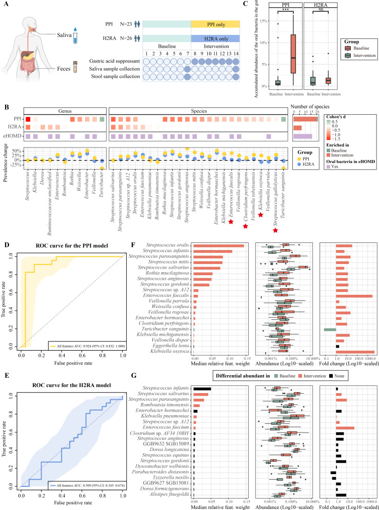
All enrolled participants provided baseline saliva and stool samples on day 0 (12 June 2022). The PPI group was then administered a 7-day course of omeprazole (20 mg daily) and provided another set of saliva and stool samples on day 7 (19 June 2022). Likewise, the H2RA group underwent similar procedures but received famotidine (20 mg daily). Refer figure 1A for the overall design of the experiments.
Figure 1.
Alterations of gut microbiota induced by PPI and H2RA in healthy volunteers. (A) Study flowchart illustrating the procedures of intervention and sample collection for the proton pump inhibitor (PPI; n=23) and histamine-2 receptor antagonist (H2RA; n=26) groups. (B) Heatmap showing the effect sizes (measured by Cohen’s d) of the significantly altered gut microbial taxa (ie, genera and species) before and after the PPI and H2RA intervention. Red tiles represent drug-enriched taxa, while green tiles represent drug-depleted ones. The purple tiles denote the oral taxa according to the expanded Human Oral Microbiome Database (eHOMD). The adjacent bar plot shows the total numbers of significantly altered taxa in the corresponding groups. The scatter plot below illustrates the prevalence changes of the taxa above. Red stars highlight the species previously associated with colorectal cancer.53 (C) Box plot comparing the accumulated abundance of the oral bacteria in the gut between the PPI group and the H2RA group at baseline and after the intervention. The ‘oral bacteria’ were defined as those presented in ≥10% of saliva samples (see Methods section). Wilcoxon rank-sum test was used to compare continuous variables between groups. NS, not significant; *p<0.05; **p<0.01; ***p<0.001; ****p<0.0001. (D) Performance of the machine learning classifier to distinguish the gut microbial profiles before and after the PPI intervention. The area under the receiver operating characteristic (AUROC) curve was used to measure the performance in fivefold three-times repeated cross-validation. (E) Performance of the H2RA classifier. (F) The top 20 most important features of the PPI classifier (left panel) and their abundance (mid panel), and abundance fold change (right panel) before and after the intervention. The bar or box colours represent the group in which the feature was enriched. Green indicates enrichment in the baseline samples, red indicates enrichment after the intervention and black indicates no enrichment. (G) Top 20 features of the H2RA classifier and their abundance, and abundance fold change before and after the intervention.
Saliva samples were collected by the participants at home in the early morning, before oral hygiene and breakfast. To prevent DNA degradation, a 2 mL room-temperature stabilising reagent kit (Cat.No. CY-98000A, Huachenyang, Shenzhen, China) was used to mix with the 2 mL saliva collected from each participant. For stool sample collection, the participants used the Faecal Sample Collection Kit (Cat.No. CY-98000PS-P1, Huachenyang, Shenzhen, China) on the same day as the saliva sample collection. The kit also contained 2 mL of room-temperature stabilising reagent. The collected saliva and stool samples were all transferred to freezers at −80°C within 12 hours of collection and stored until DNA extraction.
Furthermore, it is important to note that this study is a part of a larger randomised clinical trial (RCT). The complete experimental design encompasses an additional week during which participants were administered gastric acid suppressants while concurrently taking probiotics (from day 8 to day 14). Saliva and faecal samples were collected on day 14 and 7 days after discontinuation of the drugs (day 21). Additionally, there was a control group that only took probiotics for a week. All ethical procedures were executed in accordance with the comprehensive experimental design. For a detailed understanding of the entire design and protocol, refer to online supplemental file 1.
gutjnl-2023-330168supp001.pdf (445.4KB, pdf)
DNA extraction, library construction and shotgun metagenomic sequencing
The DNA in the faecal and salivary samples was extracted using a MagPure Stool DNA KF kit B (Magen, China), following the manufacturer’s instructions. The concentration and purity of genomic DNA in each sample were measured using a Qubit dsDNA BR Assay kit (Invitrogen, USA). To assess DNA integrity, 1% agarose gel electrophoresis was performed.
Subsequently, 1 µg genomic DNA was randomly fragmented by Covaris LE220 (Covaris, Inc, USA) according to the manufacturer’s instructions. The fragmented DNA was selected by magnetic beads to an average size of 200–500 bp. The selected fragments underwent end-repair, 3′adenylated, adapters-ligation, PCR amplifying and the products were purified by the magnetic beads. The double-stranded PCR products were heat denatured and circularised using the splint oligo sequence. The single-strand circle DNA was formatted as the final library and qualified by QC. Whole genome sequencing was performed on the MGISEQ-2000 platform with paired-end read-length of 150 bp (PE150) at BGI (Wuhan, China). We obtained 76.4±6.0 and 38.5±2.7 million pairs of raw reads (mean±SD) for each salivary and faecal sample, respectively (online supplemental table 3).
Raw metagenome data processing
Raw data with adapter sequences or low-quality sequences was filtered by SOAPnuke21 with parameters ‘-n 0.01 -l 20 -q 0.4 --adaMis 3 --outQualSys 1 –minReadLen 150’. The quality of the filtered read pairs was evaluated by FastQC (V.0.11.9). Bowtie2 (V.2.5.0)22 was then used to remove the host DNA contamination against the human genome version GRCh38/hg38. After removing low-quality bases, adapter sequences, short reads and human contaminations, a total of 25.3±16.2 and 38.3±2.8 million pairs of reads per salivary and faecal sample were obtained, respectively (online supplemental table 3). The resulting data, referred to as the ‘clean data’, were deposited in the Genome Sequence Archive (GSA), which is affiliated with China National Center for Bioinformation—National Genomics Data Center, under the BioProject ID: PRJCA016454. These clean data were used for subsequent analyses in this study. Refer to online supplemental table 4 for detailed descriptions of each sample along with their corresponding GSA accession IDs.
Taxonomic profiling of metagenome data
Species-level profiling was performed on all the samples with MetaPhlAn4 (V.4.0.3)23 with default parameters using the marker gene database of mpa_vJan21_CHOCOPhlAnSGB_202103. Only species with a maximum abundance greater than 0.001 and an average abundance greater than 0.0001 across all samples were retained.
Differential abundance analysis of species (biomarkers) preintervention and postintervention
Differential abundant species were identified by comparing longitudinal samples from the same individuals before and after each intervention using the paired Wilcoxon rank-sum test and linear discriminant analysis (LDA) effect size (LEfSe),24 which were performed using the ‘wilcox.test’ function in the Stats R package (V.4.2.0) and ‘run_lefse’ function in microbiomeMarker R package (V.1.2.2),25 respectively. Species with p values less than 0.05 in the Wilcoxon rank-sum test and an LDA score greater than 2 in the LEfSe analysis were selected. The final differential abundant species list was determined by the intersection of the two results. Cohen’s d for paired samples was calculated using the ‘cohen.d’ function in effsize R package (V.0.8.1).26 This effect size index allows for comparisons across different groups or studies by standardising the results. R V.4.2.0 was used throughout the study.
Oral bacteria used in figure 1C
Oral bacteria used in figure 1C was defined as the bacteria observed in ≥10% of saliva samples, a threshold suggested by a previous study.27 A total of 287 species from the saliva samples were considered oral bacteria, but only 19 oral species were found in the gut samples in the PPI group and H2RA group, which were used to calculate the accumulated abundance of the oral bacteria in the gut. See online supplemental table 5 for the detailed list of the oral bacteria and their relative abundances in the gut.
Machine learning classifiers for discriminating preintervention and postintervention samples
The modelling and evaluation were performed using the SIAMCAT R package (V.2.2.0).28 The features, which represented the relative abundances of all annotated species in each sample, were normalised using z-score standardisation with log10 transformation. To avoid infinite values from the logarithm, a pseudo-count of 1e-06 was added to all values. Additionally, the minimum quantile of the SD for all features was set to 0.1 to prevent underestimation. Preintervention and postintervention samples of each drug were labelled and randomly split into test and training sets in a fivefold three-times repeated cross-validation. The remaining folds were used as training data to develop a RandomForest model for each test fold. No feature selection was performed throughout the analysis, which was in line with the recommendations provided by SIAMCAT28 and recent studies.29
The RandomForest models are available on Figshare (https://figshare.com/articles/dataset/RandomForest_classifiers/23912331).
Analysis of bacterial strains and oral-to-gut transmission
Strain-level profiling was performed with StrainPhlAn 430 using the custom species-level genome bins (SGB) marker database, with parameters ‘--marker_in_n_samples 1 --sample_with_n_markers 10 --phylophlan_mode accurate --mutation_rates’. All the SGBs detected by MetaPhlAn 4 in all the oral samples were included to detect the occurrences of oral-to-gut transmission. Oral-to-gut transmission events are then defined as pairs of saliva and gut samples from the same individual collected at the same time point with phylogenetic distance below the strain identity threshold for a certain SGB (online supplemental table 6). We finally selected 0.03 as the strain identity threshold recommended by StrainPhlAn, but a more stringent strain identity threshold (eg, 0.01) was also examined and the findings in our study remained robust (online supplemental figure 2).
gutjnl-2023-330168supp005.pdf (117.6KB, pdf)
Calculation of bacterial growth rates
To accurately quantify microbial growth dynamics (peak-to-trough ratio, PTR),31 we employed DEMIC, a multi-sample algorithm that uses contigs and coverage values to estimate the relative distances of contigs from the replication origin and compare bacterial growth rates between metagenomic samples.32 To ensure comprehensive coverage of species, we combined all the genomes of SGBs profiled by MetaPhlAn4 in both oral and faecal sites (fetched from http://segatalab.cibio.unitn.it/data/Pasolli_et_al.html), as well as the metagenome-assembled genomes (MAGs) generated in our study. We further dereplicated this combined set of genomes using dRep (V.3.4.2) with default parameters,33 resulting in a unique set of 1186 species-level genomes that served as the input for DEMIC. Finally, we conducted DEMIC analysis following the instructions provided in the manual.
The MAGs were assembled as follows: each individual’s saliva and faecal samples were independently subjected to de novo metagenomic assembly using metaSPAdes (V.3.15.5) with default parameters,34 followed by assembly refinement by metaMIC (V.1.0)35 and binning using metaWRAP (V.1.3.2).36 After undergoing refinement using the ‘bin_refinement’ module in metaWRAP with parameters (-c 50 -x 10), we obtained a total of 14 044 and 8496 genomic bins from the faecal and salivary samples, respectively.
The MAG sequence datasets generated in this study are available on Figshare (https://figshare.com/projects/Gut_and_Oral_Microbiome_alterations_of_healthy_volunteers_with_proton_pump_inhibitor_and_histamine-2_receptor_antagonist/174960).
Enrichment analysis of selected species in the gut microbiome across multiple diseases
We searched in GMrepo (V.2) for the species detected with oral-to-gut transmission to gather information on their enrichment in diseases.37 GMrepo is a database of curated and consistently annotated human gut metagenomes, in which the enrichment of bacterial/archaeal species (ie, markers) has been precalculated for 83 cohorts, corresponding to 47 diseases. The markers were identified within each cohort using LEfSe analysis with an LDA cut-off of 2.37 Out of 42 species that were detected in the oral-to-gut transmission analysis, 22 were identified as disease markers in GMrepo. We counted the number of cohorts in which these 22 species were found to be enriched in the disease samples compared with the corresponding controls of the same cohort (online supplemental table 7). This count provides insight into the strength and robustness of the association between these species and diseases.
Comparisons of the gut microbial signatures of PPI between this study and the literature
The gut microbiome signatures (microbial biomarkers) of PPIs in Forslund et al’s study were accessed from their publication’s online supplemental table 6 for data comparison.17 38 In our study, we quantified the differences between the PPI signatures at the genus and species levels using Cliff’s delta. Calculation of Cliff’s delta was conducted using the ‘cliff.delta’ function from the effsize R package (V.0.8.1).
Results
PPI disrupts gut microbiome significantly more than H2RA
We initially assessed variations of all the lifestyle indices and participants’ metadata between the PPI and H2RA groups and did not find any statistically significant differences (online supplemental table 2). Thus, we directly examined the effects of drug administration on the gut microbiome. A total of 23 species (markers) were observed to exhibit significant enrichment in the gut following the administration of the two drugs, many of them also showed increased prevalence in the gut samples after drug interventions (figure 1B). Additionally, 16 out of the 23 markers (~70%) were typically found in the oral microbiome, as documented in the expanded Human Oral Microbiome Database (eHOMD; figure 1B).39
Among these markers, PPI and H2RA induced 20 and 7 species, respectively, with four shared by both drugs. The shared markers all belonged to the Streptococcus genus (S. salivarius, S. parasanguinis, S. sp. A12, S. oralis), which is part of the normal oral microbiota.40 41 Furthermore, all the four markers except S. parasanguinis exhibited significantly higher effect sizes (Cohen’s d) in the PPI group than in the H2RA group (online supplemental table 8). Besides, we observed a significant increase in the total abundance of oral bacteria in the gut induced by both drugs (figure 1C; online supplemental table 5). However, the increase was significantly higher in the PPI group (with a median of ~5.29%; p=1.67e-09) than in the H2RA group (0.39%; p=0.13; figure 1C).
To further quantify the extent of gut microbiome alteration caused by the two drugs, we built a RandomForest classifier for each group to classify samples collected before or after drug administration. We used all microbial abundances rather than the features picked by feature selection as input to train the model to maximise the retention of information in the data and avoid potential omission of important features that may not be individually significant but play an important role in combination with other features.29 In our analyses, the PPI group exhibited an excellent accuracy of 0.924 in the area under the receiver operating characteristic curve (AUROC) during fivefold three-times repeated cross-validations, indicating high discriminatory power of the classifier (figure 1D). In contrast, the H2RA group showed a much lower AUROC of 0.509, suggesting that the gut microbiome alterations induced by H2RA treatment were not substantial enough for accurate differentiation between samples collected before and after intervention (figure 1E).
Furthermore, we identified the top 20 most important features in each classifier based on their median relative feature weights. Notably, in the PPI model, 19 of the top features overlapped with the PPI markers identified above, implying that these marker species played a crucial role in distinguishing pre-PPI and post-PPI intervention samples (figure 1F). However, only six of the top features were observed overlapped with the H2RA markers in the H2RA classifier. Additionally, the abundance fold change of the H2RA top features were two orders of magnitude lower than that in the PPI group (figure 1G; online supplemental table 9). These findings suggest that the gut microbiome perturbation caused by H2RA treatment was relatively minor, leading to reduced discriminatory power of the H2RA classifier in distinguishing between pre-H2RA and post-H2RA intervention samples.
In conclusion, our findings demonstrate that PPI has a significantly higher impact on disrupting the overall gut microbiota compared with H2RA.
PPI and H2RA induce few disruptions to oral microbiome
It remains to be determined whether the drug-induced gut microbiome alterations could be in part attributed to alterations in the oral microbiome. We thus investigated the impact of drug administration on the oral microbiome. Notably, PPI intervention did not significantly change the abundances of any oral taxa (species and higher taxonomic ranks; online supplemental figure 3A). Additionally, none of the four H2RA-enriched oral species could be detected in the gut (online supplemental figure 3A). These results suggest that the drug-induced oral microbiome alterations were not the source of the drug-induced gut microbiome changes. This prompts us to explore the oral-to-gut transmission in our subsequent analysis.
gutjnl-2023-330168supp006.pdf (130.5KB, pdf)
PPI induces significantly higher oral-to-gut transmission than H2RA
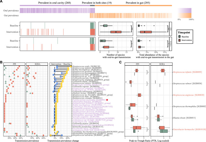
To quantify the oral-to-gut transmission before and after the drug intervention, we adopted the recommended pipeline by Valles-Colomer et al and used StrainPhlAn 4 to identify potentially transmissible species at the strain level among the 582 species identified by MetaPhlAn 4 in this study (see Methods section; online supplemental table 6).30 42 At baseline, we identified a total of 21 transmissible species and 17 (80.95%) of them were prevalent in both body sites (ie, with >10% prevalence in both the oral and faecal species at baseline; figure 2A; online supplemental table 10), suggesting a frequent oral-to-gut transmission in the healthy participants. There were no significant differences in both the numbers and total abundances of transmissible species in the baseline samples between the two drug groups (p>0.05; online supplemental figure 4A and table 11).
Figure 2.
PPI induces higher oral-to-gut transmission than H2RA and promotes the growth of transmitted species in the gut. (A) Prevalent species in the oral cavity, gut or both and their oral-to-gut transmission before and after the drug intervention. The top heatmap depicting the prevalence of species sorted from left to right in descending order of abundance (species prevalent in both sites were sorted based on their gut abundance). Species were divided into four categories using criteria defined by a previous study, including ‘faecal’ species (n=295, 50.69% of total) that were observed in >10% of faecal samples and <10% of saliva samples, ‘oral’ species (n=268, 46.05%) that were only prevalent in oral samples, and ‘both’ species (n=19, 3.26%) that were prevalent in >10% of saliva and stool samples. The heatmap in the bottom left displays all the species detected with oral-to-gut transmission at two timepoints in the PPI and H2RA group. The boxplot, sorted from left to right, compares the number and total abundance of species with oral-to-gut transmission before and after drug administration in the two groups. NS, not significant; *p<0.05; **p<0.01; ***p<0.001; ****p<0.0001. Wilcoxon rank-sum test. (B) Scatter plot showing the oral-to-gut transmission prevalence of the bacteria before and after the administration of PPI and H2RA drugs. The green triangle in the left two panels represents the oral-to-gut transmission prevalence of the bacteria at baseline, while the red square represents the transmission prevalence after intervention. The right panel summarises the change of the oral-to-gut transmission prevalence before and after the intervention in two groups. The grey labels indicate the ‘both’ species while purple labels indicate the ‘oral’ species defined in (A). Red star highlights Fusobacterium nucleatum, a well-studied marker of colorectal cancer according to previous studies.44 (C) Boxplot showing the species with significantly different bacterial growth rates (measured by peak-to-trough ratio) in the PPI group. The red labels indicate the PPI markers (ie, bacteria were significantly more abundant in the gut after the PPI usage compared with baseline). Wilcoxon rank-sum test was used to compare continuous variables between groups. NS, not significant; *p<0.05; **p<0.01; ***p<0.001; ****p<0.0001. H2RA, histamine-2 receptor antagonist; PPI, proton pump inhibitor.
gutjnl-2023-330168supp007.pdf (93.8KB, pdf)
After PPI usage, we observed a significant increase in the number of transmissible species (with a median of 9) compared with baseline (figure 2A; p=1.02e-04; Wilcoxon rank-sum test for paired samples). Additionally, the overall abundance of these species was also significantly increased after PPI usage (figure 2A; p=1.5e-04). Similar patterns were observed after H2RA usage, but the extent of the increase was notably smaller compared with the PPI group (online supplemental figure 4B).
Furthermore, we compared the changes in the prevalence of transmissible species between the PPI and H2RA groups. Out of the 42 SGBs that showed oral-to-gut transmission in at least one sample in either of the groups, 37 exhibited increased prevalence after PPI usage, compared with only 15 after the H2RA usage. Fourteen SGBs demonstrated an increase in transmission prevalence in both groups, but most of them (12 SGBs) showed higher prevalence increases in the PPI group than in the H2RA group (figure 2B; online supplemental table 12).
The significantly higher oral-to-gut transmission induced by PPI could be further supported by the Bray-Curtis dissimilarity (BCD) analysis between the oral and gut microbiome compositions of the same participants. At baseline, a BCD of nearly 1 was observed, highlighting the distinct compositions of the oral and gut microbiomes (online supplemental figure 3B). However, following two drug administrations, a significant shift in the microbial composition between the oral cavity and the gut within the same individual. Notably, the PPI group exhibited a more pronounced decrease in BCD after drug use compared with the H2RA group (online supplemental figure 3B; Wilcoxon rank-sum test for paired samples; p=4.77e-06 in the PPI group; p=0.045 in the H2RA group).
Strikingly, we found that Fusobacterium nucleatum, a well-studied marker of CRC, was identified as a transmissible species in ~9% of participants after PPI usage but none after H2RA usage. F. nucleatum is prevalent in the oral cavity and has been found to promote inflammation and immune evasion when transmitted to the gut, creating a favourable environment for tumour growth. It also interacts with immune cells and other bacteria in the gut, facilitating an immunosuppressive environment that promotes tumour growth and metastasis.43 44
These findings demonstrate that PPI induces significantly more species to be transmitted from the oral to the gut than H2RA, resulting in a more prevalent distribution of these species in the gut, including a known CRC-associated marker.
PPI promotes the growth of transmitted species in the gut
To assess whether drug usage could potentially influence the abundance of gut species by promoting or inhibiting their growth, we employed an index named PTR to quantify bacterial growth dynamics. Specifically, we used the DEMIC tool to estimate the relative distances of contigs from the replication origin and thereby accurately assess bacterial growth rates across different samples.31 32 Despite the high sequencing depth demand of this method, we were able to quantify the growth rates for 355 out of the 1186 species (online supplemental table 13).
After PPI usage, a total of five species exhibited significantly increased growth rates (PTR values). Among them, three (60% out of the total) overlapped with the PPI-enriched markers, indicating that PPI further enhanced the growth of the transmitted species in the gut (figure 2C). Notably, PPI also promotes the growth of non-maker species, such as Streptococcus thermophilus and Streptococcus rubneri. Additionally, we observed that the growth of Blautia obeum was suppressed by PPI usage, in line with a recent study by Maier et al. 45 In contrast, H2RA usage did not significantly affect the growth of these species (figure 2C).
It is important to note that these observed effects of PPI usage may be attributed to direct drug-microbiota interactions or within-microbiota interactions, which require experimental validation in the future.
PPI-induced gut microbial markers are associated with risks of multiple diseases
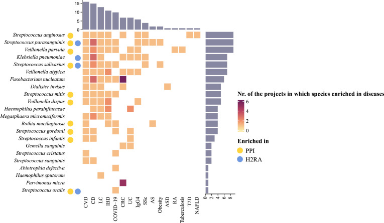
To access the disease risks associated with the PPI-induced and H2RA-induced oral-to-gut transmissible species, we searched the GMrepo V.2 database,37 a repository of gut microbiome-derived disease-marker relationships (see Methods section). Out of the 42 transmissible species induced by either PPI or H2RA, 22 showed significant enrichment in the disease population collected in 26 cohorts, corresponding to 16 diseases, including cardiovascular diseases (CVDs), Crohn’s disease, liver cirrhosis, IBDs, COVID-19, CRC and others (figure 3; online supplemental table 7). Of the 22 species, 10 were PPI-induced and associated with 15 diseases, while 4 were H2RA-induced and associated with 11 diseases. Notably, some of these species have been validated to play a role in the occurrence and progression of the corresponding diseases. For example, S. anginosus, identified as being enriched in eight distinct diseases within our study, along with Streptococcus constellatus (which was among the 42 transmissible species and exhibited an elevated transmission prevalence following PPI usage) and Streptococcus intermedius, constitute the Streptococcus anginosus group (SAG). This group of bacteria can lead to both pyogenic infections and the development of malignant tumours.46 47 They can move from the intestines to other organs, causing widespread infections such as liver abscess and endocarditis. Some studies have also found that SAG could be the pathogenic bacteria involved in gastric cancer.48 Furthermore, a cross-sectional study involving 8973 participants revealed that S. anginosus and S. oralis have strong associations with coronary artery calcium score, indicating a high risk of CVD.49 The study also identified other related species, including S. parasanguinis and Streptococcus gordonii, which were detected as PPI markers and related to several other diseases in our research.
Figure 3.
Association between oral-to-gut transmitted species and disease risks according to the GMrepo database. Species that showed oral-to-gut transmission in at least one participant were shown. The heatmap shows the number of projects in which the corresponding species were enriched in the disease group according to the GMrepo database (see Methods section). The bar plot above the heatmap displays the total number of species enriched in each disease. The bar plot on the right displays the total number of diseases the corresponding species was enriched in. Yellow dots represent the species were PPI markers, and blue dots were H2RA markers. AS, ankylosing spondylitis; ASD, autism spectrum disorder; CD, Crohn’s disease; CRC, colorectal cancer; CVD, cardiovascular disease; H2RA, histamine-2 receptor antagonist; IBD, inflammatory bowel diseases; IgG4, immunoglobulin G4-related disease; LC, liver cirrhosis; NAFLD, non-alcohol fatty liver disease; PPI, proton pump inhibitor; RA, rheumatoid arthritis; SSc, systemic sclerosis; T2D, type 2 diabetes mellitus; UC, ulcerative colitis.
These results suggest that the oral-to-gut transmission of specific species induced by PPI use may contribute to a broader spectrum of disease risks compared with H2RA. The identified associations between these transmissible species and various diseases underscore the potential significance of gut microbiome alterations induced by PPI in shaping disease susceptibilities.
Discussion
Principal findings
The present study aimed to investigate and compare the effects of two widely used gastric acid suppressants, PPIs and H2RAs, on the gut microbiome and oral-to-gut transmission. By employing a longitudinal approach with a healthy cohort, we were able to mitigate potential confounding factors, such as disease status and individual variability, and provide robust insights into the impact of these drugs on the gut microbial community.
Our findings reveal that PPI usage has a more pronounced effect on disrupting the gut microbiota compared with H2RA. PPIs induced a higher extent of oral-to-gut transmission, promoting the presence of oral species in the gut. Furthermore, we observed an increase in the growth rate of specific transmitted and native gut microbes, potentially influenced by the drug. Notably, several of these transmitted species have been implicated in various diseases, indicating a possible link between PPI-induced gut microbiome alterations and disease susceptibilities. For instance, the detection of F. nucleatum, a known biomarker of CRC, only in the PPI group after oral-to-gut transmission raises concerns about its potential role in increased disease risks. Additionally, PPI-induced markers are associated with more disease than H2RA.
Comparison with previous studies
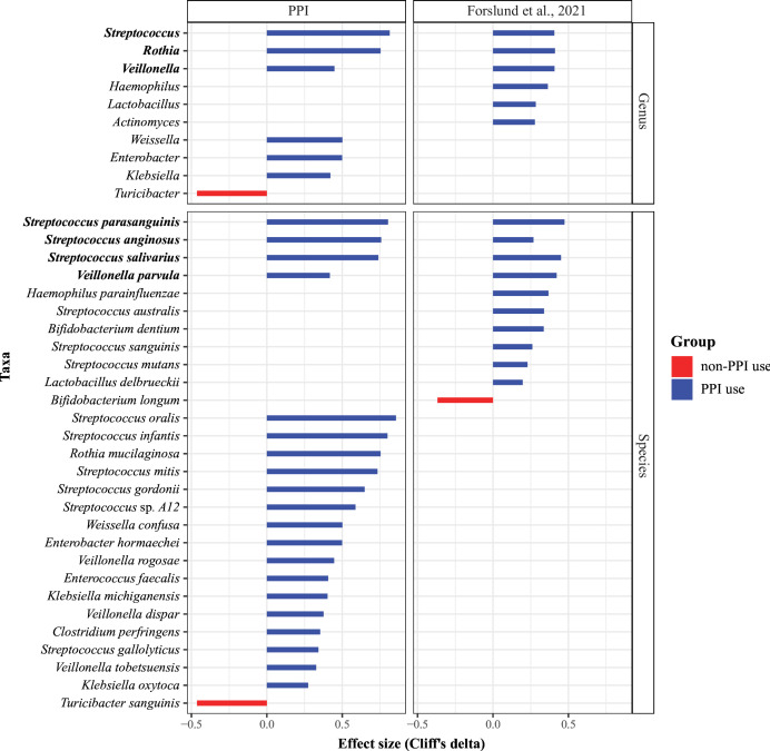
The gut microbiome signatures (microbial biomarkers) of PPIs had been previously reported in cross-sectional cohorts such as the MetaCardis cohort studied by Forslund et al.17 Their study integrated multi-omics analyses of 2173 European residents from the MetaCardis cohort. To address potential confounders, they adopted a post-hoc testing approach for deconfounding univariate biomarker analysis, considering multiple medications and risk factors. The researchers compared the gut microbial biomarkers associated with PPIs identified through their methodology with previously published data from two distinct patient cohorts investigated by Vich Vila et al and achieved a high level of congruence.14 In this study, we compared the results provided by Forslund et al with our findings (see Methods section). Our analysis revealed both overlaps and differences in the PPI markers between the MetaCardis and our study (figure 4; online supplemental table 14).
Figure 4.
Comparison of proton pump inhibitor (PPI) signatures in the gut identified in this study to those of the MetaCardis cohort. Bar plots show the magnitude and direction of effect size (Cliff’s delta) of PPI intervention on microbiome features. These effects are compared with the previously published data studied by Forslund et al.17 Taxon names in bold represent the enrichment direction of these taxa is consistent in both studies.
Specifically, 4 out of 21 species-level markers (20 markers mentioned in the previous section and 1 marker named Turicibacter sanguinis depleted after PPI usage) overlapped with those in the MetaCardis cohort. The overlapped markers included S. parasanguinis, S. anginosus, S. salivarius and Veillonella parvula, belonging to the Streptococcus and Veillonella genera, commonly present in both the oral cavity and the gut. The Rothia genus also showed consistent enrichment in the PPI intervention group in both studies.
However, some genera, such as Haemophilus, Lactobacillus and Actinomyces, were only differentially present in the Metacardis cohort, while others like Weissella, Enterobacter and Klebsiella were only differentially present in our PPI group. Furthermore, in the MetaCardis cohort, the species enriched in non-PPI users was Bifidobacterium longum, whereas in our study, the species enriched at baseline in the PPI group was T. sanguinis. These differences likely reflected the different designs of the studies, such as longitudinal versus cross-sectional analyses and disease versus healthy cohorts.
Strengths and limitations of this study
This study possesses several strengths. First, we employed a cohort of healthy volunteers and conducted a longitudinal study to mitigate the influence of confounding factors, including diseases and individual variations. This allowed us to identify real PPI microbiome markers, raising concerns for other researchers to consider excluding PPI markers when identifying disease-related markers in patient cohorts. Second, many studies have mainly concentrated on the microbiome of either the oral cavity or the gut, often providing descriptions of their presence but ignoring the underlying causes for the enrichment of oral bacteria in the gut. In contrast, our investigation comprehensively addressed both ends of the GI tract, shedding light on how gastric acid suppressants contribute to the process of oral-to-gut transmission. Third, there is a scarcity of research investigating the effects of H2RA on human oral and gut microbiomes. In this study, we assessed the disruptions caused by H2RA to these microbiomes and demonstrated the viability of choosing H2RA over PPI in clinical practice when the symptoms are mild.7 11 Finally, due to the substantial host DNA contamination in saliva samples, the majority of studies have employed 16S rRNA gene sequencing instead of shotgun sequencing to profile the oral microbiome, which made it challenging to determine whether the bacteria enriched in the gut truly originated from the oral cavity at species and strain level. In our study, we conducted shotgun sequencing on all saliva samples, generating ample data for in-depth strain-level analysis.
This study also has some limitations. First, we opted for a short-term drug intervention with standard doses to prioritise participant well-being, ensuring that any potential disruptions to the gut microbiota caused by the drugs could be reversible.50 However, it is important to acknowledge that prolonged or excessive use of PPIs and H2RAs is common in clinical practice, leading to potential long-term risks. For the exploration of prolonged drug effects, it might be more appropriate to consider observational or interventional studies involving patients who must take PPIs over extended periods rather than conducting long-term RCTs in the absence of clear indications, based on substantial disruptions to the gut microbiome observed in this and previous studies with PPIs.6 7 We encourage further research to delve into the consequences of prolonged PPI usage, carefully balancing treatment benefits with potential risks. In our study, a 1 week use of gastric acid suppressants may not fully capture dose-dependent microbial effects, and the varying gastric acid suppression efficiencies of the standard doses of PPI and H2RA may contribute to the observed differences.51 52 Nonetheless, from a clinical perspective, comparing the recommended dosages of these two drugs is more meaningful than evaluating them solely based on their respective gastric acid suppression levels when assessing their effects on the microbiota. Second, in order to minimise potential confounding factors between the two groups, we limited our study’s age range to 18–45, which may restrict the generalisability of our findings to elderly populations. Finally, we did not collect blood samples, perform transcriptome or metabolomic analyses, nor did we assess the physical and chemical characteristics of the samples. The integration of these multi-omics data has the potential to provide further substantiation of our findings. It is imperative that these aspects be incorporated in future research to expand and fortify our insights.
In conclusion, our study provides evidence that PPIs have a greater impact on the gut microbiome and oral-to-gut transmission than H2RAs, uncovering a potential mechanism underlying the higher risk of certain diseases associated with PPIs. Our results underscore the need for caution in PPI usage and highlight the relevance of exploring alternative treatments such as H2RAs. However, further research is required to elucidate the effects of long-term H2RA use on the gut microbiome and its implications for human health, as related investigations are still limited.
gutjnl-2023-330168supp002.pdf (33.5KB, pdf)
Acknowledgments
We would like to thank the clinical doctor Desheng Hu from the Union Hospital, Tongji Medical College, Huazhong University of Science and Technology for his kind suggestions. We thank all the generous participants of this study for their supports. We thank Junan Chen, Na L Gao, Mingyu Wang, Yingjian Wu, Yun Y Li, Huarui Wang for their assistance during the sample collection.
Footnotes
Contributors: Study concept and design: W-HC. Subjects recruitment: JZ, CS. Random allocation sequence generation: ML. Participants assignment: GH. Samples collection: JZ, CS, ML, GH. Data acquisition: JZ. Analysis and interpretation of data: W-HC, JZ. Drafting of the manuscript: JZ. Revising of the manuscript: W-HC, X-MZ. All the authors approved the final version of the manuscript. W-HC supervised the study and is the guarantor.
Funding: This work was supported by the National Key R&D Programme of China (2019YFA0905600, 2020YFA0712403), NNSF-VR Sino-Swedish Joint Research Programme (82161138017), National Natural Science Foundation of China (T2225015, 61932008), Shanghai Municipal Science and Technology Major Project (2018SHZDZX01) and Greater Bay Area Institute of Precision Medicine (Guangzhou) (Grant No. IPM21C008).
Competing interests: None declared.
Patient and public involvement: Patients and/or the public were not involved in the design, or conduct, or reporting, or dissemination plans of this research.
Provenance and peer review: Not commissioned; externally peer reviewed.
Supplemental material: This content has been supplied by the author(s). It has not been vetted by BMJ Publishing Group Limited (BMJ) and may not have been peer-reviewed. Any opinions or recommendations discussed are solely those of the author(s) and are not endorsed by BMJ. BMJ disclaims all liability and responsibility arising from any reliance placed on the content. Where the content includes any translated material, BMJ does not warrant the accuracy and reliability of the translations (including but not limited to local regulations, clinical guidelines, terminology, drug names and drug dosages), and is not responsible for any error and/or omissions arising from translation and adaptation or otherwise.
Data availability statement
Data are available in a public, open access repository. All data relevant to the study are included in the article or uploaded as supplementary information. The sequencing data reported in this study have been made available in the China National Center for Bioinformation (CNCB)—National Genomics Data Center (NGDC) under BioProject accession number PRJCA016454. All other data are available in the manuscript including its supplementary files, or from the corresponding authors upon reasonable request. The source codes describing all of the analysis performed in this paper are available online (https://github.com/whchenlab/PPI-vs.-H2RA-effects-on-gut-microbiome).
Ethics statements
Patient consent for publication
Not applicable.
Ethics approval
This study involves human participants and this study was approved by the Clinical Trial Ethics Committee of Huazhong University of Science and Technology (decision number: 2021-S305-1). After a full explanation of all aspects of the study and the available alternative interventions, a written informed consent form was obtained from all participants.
References
- 1. Gyawali CP, Fass R. Management of gastroesophageal reflux disease. Gastroenterology 2018;154:302–18. 10.1053/j.gastro.2017.07.049 [DOI] [PubMed] [Google Scholar]
- 2. Maret-Ouda J, Markar SR, Lagergren J. Gastroesophageal reflux disease: A review. JAMA 2020;324:2536. 10.1001/jama.2020.21360 [DOI] [PubMed] [Google Scholar]
- 3. Chalabi M, Cardona A, Nagarkar DR, et al. Efficacy of chemotherapy and Atezolizumab in patients with non-small-cell lung cancer receiving antibiotics and proton pump inhibitors: pooled post hoc analyses of the OAK and POPLAR trials. Ann Oncol 2020;31:525–31. 10.1016/j.annonc.2020.01.006 [DOI] [PubMed] [Google Scholar]
- 4. Llorente C, Jepsen P, Inamine T, et al. Publisher correction: gastric acid suppression promotes alcoholic liver disease by inducing overgrowth of intestinal Enterococcus. Nat Commun 2017;8:2137. 10.1038/s41467-017-01779-8 [DOI] [PMC free article] [PubMed] [Google Scholar]
- 5. Xia B, Yang M, Nguyen LH, et al. Regular use of proton pump inhibitor and the risk of inflammatory bowel disease: pooled analysis of 3 prospective cohorts. Gastroenterology 2021;161:1842–52. 10.1053/j.gastro.2021.08.005 [DOI] [PubMed] [Google Scholar]
- 6. Abrahami D, Pradhan R, Yin H, et al. Proton pump inhibitors and the risk of inflammatory bowel disease: population-based cohort study. Gut 2023;72:1288–95. 10.1136/gutjnl-2022-328866 [DOI] [PubMed] [Google Scholar]
- 7. Abrahami D, McDonald EG, Schnitzer ME, et al. Proton pump inhibitors and risk of colorectal cancer. Gut 2022;71:111–8. 10.1136/gutjnl-2021-325096 [DOI] [PubMed] [Google Scholar]
- 8. Kichenadasse G, Miners JO, Mangoni AA, et al. Proton pump inhibitors and survival in patients with colorectal cancer receiving Fluoropyrimidine-based chemotherapy. J Natl Compr Canc Netw 2021;19:1037–44. 10.6004/jnccn.2020.7670 [DOI] [PubMed] [Google Scholar]
- 9. Seto CT, Jeraldo P, Orenstein R, et al. Prolonged use of a proton pump inhibitor reduces microbial diversity: implications for Clostridium difficile susceptibility. Microbiome 2014;2:42. 10.1186/2049-2618-2-42 [DOI] [PMC free article] [PubMed] [Google Scholar]
- 10. Inghammar M, Svanström H, Voldstedlund M, et al. Proton-pump inhibitor use and the risk of community-associated Clostridium difficile infection. Clin Infect Dis 2021;72:e1084–9. 10.1093/cid/ciaa1857 [DOI] [PMC free article] [PubMed] [Google Scholar]
- 11. Park J-H, Lee J, Yu S-Y, et al. Comparing proton pump inhibitors with Histamin-2 receptor blockers regarding the risk of Osteoporotic fractures: a nested case-control study of more than 350,000 Korean patients with GERD and peptic ulcer disease. BMC Geriatr 2020;20:407. 10.1186/s12877-020-01794-3 [DOI] [PMC free article] [PubMed] [Google Scholar]
- 12. Yadlapati R, Kahrilas PJ. “The "dangers" of chronic proton pump inhibitor use”. J Allergy Clin Immunol 2018;141:79–81. 10.1016/j.jaci.2017.06.017 [DOI] [PubMed] [Google Scholar]
- 13. Imhann F, Bonder MJ, Vich Vila A, et al. Proton pump inhibitors affect the gut Microbiome. Gut 2016;65:740–8. 10.1136/gutjnl-2015-310376 [DOI] [PMC free article] [PubMed] [Google Scholar]
- 14. Vich Vila A, Collij V, Sanna S, et al. Impact of commonly used drugs on the composition and metabolic function of the gut Microbiota. Nat Commun 2020;11:362. 10.1038/s41467-019-14177-z [DOI] [PMC free article] [PubMed] [Google Scholar]
- 15. Freedberg DE, Toussaint NC, Chen SP, et al. Proton pump inhibitors alter specific Taxa in the human gastrointestinal Microbiome: A crossover trial. Gastroenterology 2015;149:883–5. 10.1053/j.gastro.2015.06.043 [DOI] [PMC free article] [PubMed] [Google Scholar]
- 16. de Vos WM, Tilg H, Van Hul M, et al. Gut Microbiome and health: mechanistic insights. Gut 2022;71:1020–32. 10.1136/gutjnl-2021-326789 [DOI] [PMC free article] [PubMed] [Google Scholar]
- 17. Forslund SK, Chakaroun R, Zimmermann-Kogadeeva M, et al. Combinatorial, additive and dose-dependent drug-Microbiome associations. Nature 2021;600:500–5. 10.1038/s41586-021-04177-9 [DOI] [PubMed] [Google Scholar]
- 18. Nagata N, Nishijima S, Miyoshi-Akiyama T, et al. Population-level Metagenomics Uncovers distinct effects of multiple medications on the human gut Microbiome. Gastroenterology 2022;163:1038–52. 10.1053/j.gastro.2022.06.070 [DOI] [PubMed] [Google Scholar]
- 19. Moher D, Hopewell S, Schulz KF, et al. CONSORT 2010 statement: updated guidelines for reporting parallel group randomised trials. BMJ 2010;340:c869. 10.1136/bmj.c869 [DOI] [PMC free article] [PubMed] [Google Scholar]
- 20. Mirzayi C, Renson A, Genomic Standards Consortium, et al. Reporting guidelines for human Microbiome research: the STORMS checklist. Nat Med 2021;27:1885–92. 10.1038/s41591-021-01552-x [DOI] [PMC free article] [PubMed] [Google Scholar]
- 21. Chen Y, Chen Y, Shi C, et al. Soapnuke: a Mapreduce acceleration-supported software for integrated quality control and Preprocessing of high-throughput sequencing data. Gigascience 2018;7:1–6. 10.1093/gigascience/gix120 [DOI] [PMC free article] [PubMed] [Google Scholar]
- 22. Langmead B, Salzberg SL. Fast Gapped-read alignment with Bowtie 2. Nat Methods 2012;9:357–9. 10.1038/nmeth.1923 [DOI] [PMC free article] [PubMed] [Google Scholar]
- 23. Blanco-Míguez A, Beghini F, Cumbo F, et al. Extending and improving Metagenomic Taxonomic profiling with Uncharacterized species using Metaphlan 4. Nat Biotechnol February 23, 2023. 10.1038/s41587-023-01688-w [DOI] [PMC free article] [PubMed] [Google Scholar]
- 24. Segata N, Izard J, Waldron L, et al. Metagenomic biomarker discovery and explanation. Genome Biol 2011;12:R60. 10.1186/gb-2011-12-6-r60 [DOI] [PMC free article] [PubMed] [Google Scholar]
- 25. Cao Y, Dong Q, Wang D, et al. microbiomeMarker: an R/Bioconductor package for Microbiome marker identification and visualization. Bioinformatics 2022;38:4027–9. 10.1093/bioinformatics/btac438 [DOI] [PubMed] [Google Scholar]
- 26. Torchiano M. Effsize—a package for efficient effect size computation; 2016. 10.5281/ZENODO.1480624 [DOI]
- 27. Schmidt TS, Hayward MR, Coelho LP, et al. Extensive transmission of Microbes along the gastrointestinal tract. Elife 2019;8:e42693. 10.7554/eLife.42693 [DOI] [PMC free article] [PubMed] [Google Scholar]
- 28. Wirbel J, Zych K, Essex M, et al. Microbiome meta-analysis and cross-disease comparison enabled by the SIAMCAT machine learning Toolbox. Genome Biol 2021;22:93. 10.1186/s13059-021-02306-1 [DOI] [PMC free article] [PubMed] [Google Scholar]
- 29. Li M, Liu J, Zhu J, et al. Performance of gut Microbiome as an independent diagnostic tool for 20 diseases: cross-cohort validation of machine-learning classifiers. Gut Microbes 2023;15:2205386. 10.1080/19490976.2023.2205386 [DOI] [PMC free article] [PubMed] [Google Scholar]
- 30. Truong DT, Tett A, Pasolli E, et al. Microbial strain-level population structure and genetic diversity from Metagenomes. Genome Res 2017;27:626–38. 10.1101/gr.216242.116 [DOI] [PMC free article] [PubMed] [Google Scholar]
- 31. Korem T, Zeevi D, Suez J, et al. Growth Dynamics of gut Microbiota in health and disease inferred from single Metagenomic samples. Science 2015;349:1101–6. 10.1126/science.aac4812 [DOI] [PMC free article] [PubMed] [Google Scholar]
- 32. Gao Y, Li H. Quantifying and comparing bacterial growth Dynamics in multiple Metagenomic samples. Nat Methods 2018;15:1041–4. 10.1038/s41592-018-0182-0 [DOI] [PMC free article] [PubMed] [Google Scholar]
- 33. Olm MR, Brown CT, Brooks B, et al. dRep: a tool for fast and accurate Genomic comparisons that enables improved genome recovery from Metagenomes through de-replication. ISME J 2017;11:2864–8. 10.1038/ismej.2017.126 [DOI] [PMC free article] [PubMed] [Google Scholar]
- 34. Nurk S, Meleshko D, Korobeynikov A, et al. metaSPAdes: a new versatile Metagenomic assembler. Genome Res 2017;27:824–34. 10.1101/gr.213959.116 [DOI] [PMC free article] [PubMed] [Google Scholar]
- 35. Lai S, Pan S, Sun C, et al. metaMIC: reference-free Misassembly identification and correction of de novo Metagenomic assemblies. Genome Biol 2022;23:242. 10.1186/s13059-022-02810-y [DOI] [PMC free article] [PubMed] [Google Scholar]
- 36. Uritskiy GV, DiRuggiero J, Taylor J. Metawrap-a flexible pipeline for genome-resolved Metagenomic data analysis. Microbiome 2018;6:158. 10.1186/s40168-018-0541-1 [DOI] [PMC free article] [PubMed] [Google Scholar]
- 37. Dai D, Zhu J, Sun C, et al. Gmrepo V2: a Curated human gut Microbiome database with special focus on disease markers and cross-Dataset comparison. Nucleic Acids Res 2022;50:D777–84. 10.1093/nar/gkab1019 [DOI] [PMC free article] [PubMed] [Google Scholar]
- 38. Forslund SK, Chakaroun R, Zimmermann-Kogadeeva M, et al. Data from: Combinatorial, additive and dose-dependent drug-Microbiome associations. Nature 2021;600:500–5. 10.1038/s41586-021-04177-9 [DOI] [PubMed] [Google Scholar]
- 39. Escapa IF, Chen T, Huang Y, et al. New insights into human nostril Microbiome from the expanded human oral Microbiome database (eHOMD): a resource for the Microbiome of the human Aerodigestive tract. mSystems 2018;3:e00187-18. 10.1128/mSystems.00187-18 [DOI] [PMC free article] [PubMed] [Google Scholar]
- 40. Belstrøm D, Constancias F, Markvart M, et al. Transcriptional activity of predominant Streptococcus species at multiple oral sites associate with Periodontal status. Front Cell Infect Microbiol 2021;11:752664. 10.3389/fcimb.2021.752664 [DOI] [PMC free article] [PubMed] [Google Scholar]
- 41. Lee K, Walker AR, Chakraborty B, et al. Novel Probiotic mechanisms of the oral bacterium Streptococcus SP. A12 as explored with functional Genomics. Appl Environ Microbiol 2019;85:85. 10.1128/AEM.01335-19 [DOI] [PMC free article] [PubMed] [Google Scholar]
- 42. Valles-Colomer M, Blanco-Míguez A, Manghi P, et al. The person-to-person transmission landscape of the gut and oral Microbiomes. Nature 2023;614:125–35. 10.1038/s41586-022-05620-1 [DOI] [PMC free article] [PubMed] [Google Scholar]
- 43. Ranjbar M, Salehi R, Haghjooy Javanmard S, et al. The Dysbiosis signature of Fusobacterium Nucleatum in colorectal cancer-cause or consequences? A systematic review. Cancer Cell Int 2021;21:194. 10.1186/s12935-021-01886-z [DOI] [PMC free article] [PubMed] [Google Scholar]
- 44. Zhang Y, Zhang L, Zheng S, et al. Fusobacterium Nucleatum promotes colorectal cancer cells adhesion to endothelial cells and facilitates Extravasation and metastasis by inducing Alpk1/NF-kappaB/Icam1 axis. Gut Microbes 2022;14:2038852. 10.1080/19490976.2022.2038852 [DOI] [PMC free article] [PubMed] [Google Scholar]
- 45. Maier L, Pruteanu M, Kuhn M, et al. Extensive impact of non-antibiotic drugs on human gut bacteria. Nature 2018;555:623–8. 10.1038/nature25979 [DOI] [PMC free article] [PubMed] [Google Scholar]
- 46. Pilarczyk-Zurek M, Sitkiewicz I, Koziel J. The clinical view on Streptococcus Anginosus group - opportunistic pathogens coming out of hiding. Front Microbiol 2022;13:956677. 10.3389/fmicb.2022.956677 [DOI] [PMC free article] [PubMed] [Google Scholar]
- 47. Jiang S, Li M, Fu T, et al. Clinical characteristics of infections caused by Streptococcus Anginosus group. Sci Rep 2020;10:9032. 10.1038/s41598-020-65977-z [DOI] [PMC free article] [PubMed] [Google Scholar]
- 48. Zi M, Zhang Y, Hu C, et al. A literature review on the potential clinical implications of Streptococci in gastric cancer. Front Microbiol 2022;13:1010465. 10.3389/fmicb.2022.1010465 [DOI] [PMC free article] [PubMed] [Google Scholar]
- 49. Sayols-Baixeras S, Dekkers KF, Baldanzi G, et al. Streptococcus species abundance in the gut is linked to Subclinical coronary Atherosclerosis in 8973 participants from the SCAPIS cohort. Circulation 2023;148:459–72. 10.1161/CIRCULATIONAHA.123.063914 [DOI] [PMC free article] [PubMed] [Google Scholar]
- 50. Koo SH, Deng J, Ang DSW, et al. Effects of proton pump inhibitor on the human gut Microbiome profile in multi-ethnic groups in Singapore. Singapore Med J 2019;60:512–21. 10.11622/smedj.2018152 [DOI] [PMC free article] [PubMed] [Google Scholar]
- 51. Miner PB, Allgood LD, Grender JM. Comparison of gastric pH with Omeprazole magnesium 20.6 mg (Prilosec OTC) O.M. Famotidine 10 mg (Pepcid AC) B.D. and Famotidine 20 mg B.D. over 14 days of treatment. Aliment Pharmacol Ther 2007;25:103–9. 10.1111/j.1365-2036.2006.03129.x [DOI] [PubMed] [Google Scholar]
- 52. Abe Y, Inamori M, Togawa J-I, et al. The comparative effects of single intravenous doses of Omeprazole and Famotidine on Intragastric pH. J Gastroenterol 2004;39:21–5. 10.1007/s00535-003-1240-6 [DOI] [PubMed] [Google Scholar]
- 53. Kwong TNY, Wang X, Nakatsu G, et al. Association between bacteremia from specific Microbes and subsequent diagnosis of colorectal cancer. Gastroenterology 2018;155:383–90. 10.1053/j.gastro.2018.04.028 [DOI] [PubMed] [Google Scholar]
Associated Data
This section collects any data citations, data availability statements, or supplementary materials included in this article.
Supplementary Materials
gutjnl-2023-330168supp004.pdf (61.5KB, pdf)
gutjnl-2023-330168supp003.xlsx (1.2MB, xlsx)
gutjnl-2023-330168supp001.pdf (445.4KB, pdf)
gutjnl-2023-330168supp005.pdf (117.6KB, pdf)
gutjnl-2023-330168supp006.pdf (130.5KB, pdf)
gutjnl-2023-330168supp007.pdf (93.8KB, pdf)
gutjnl-2023-330168supp002.pdf (33.5KB, pdf)
Data Availability Statement
Data are available in a public, open access repository. All data relevant to the study are included in the article or uploaded as supplementary information. The sequencing data reported in this study have been made available in the China National Center for Bioinformation (CNCB)—National Genomics Data Center (NGDC) under BioProject accession number PRJCA016454. All other data are available in the manuscript including its supplementary files, or from the corresponding authors upon reasonable request. The source codes describing all of the analysis performed in this paper are available online (https://github.com/whchenlab/PPI-vs.-H2RA-effects-on-gut-microbiome).


