Abstract
Venezuelan equine encephalitis (VEE) virus antigenic subtypes and varieties are considered either epidemic/epizootic or enzootic. In addition to epidemiological differences between the epidemic and enzootic viruses, several in vitro and in vivo laboratory markers distinguishing the viruses have been identified, including differential plaque size, sensitivity to interferon (IFN), and virulence for guinea pigs. These observations have been shown to be useful predictors of natural, equine virulence and epizootic potential. Chimeric viruses containing variety IAB (epizootic) nonstructural genes with variety IE (enzootic) structural genes (VE/IAB-IE) or IE nonstructural genes and IAB structural genes (IE/IAB) were constructed to systematically analyze and map viral phenotype and virulence determinants. Plaque size analysis showed that both chimeric viruses produced a mean plaque diameter that was intermediate between those of the parental strains. Additionally, both chimeric viruses showed intermediate levels of virus replication and virulence for guinea pigs compared to the parental strains. However, IE/IAB produced a slightly higher viremia and an average survival time 2 days shorter than the VE/IAB-IE virus. Finally, IFN sensitivity assays revealed that only one chimera, VE/IAB-IE, was intermediate between the two parental types. The second chimera, containing the IE nonstructural genes, was at least five times more sensitive to IFN than the IE parental virus and greater than 50 times more sensitive than the IAB parent. These results implicate viral components in both the structural and nonstructural portions of the genome in contributing to the epizootic phenotype and indicate the potential for epidemic emergence from the IE enzootic VEE viruses.
Venezuelan equine encephalitis (VEE) has been an important human and equine disease for much of this century, and recent epidemics (26, 33) clearly indicate that VEE viruses still pose a serious public health threat. VEE viruses are serologically classified into six distinct antigenic subtypes (31, 35). Historically, only viruses in subtype I, varieties AB and C, are associated with major epidemics and epizootics. These antigenic varieties have been isolated only during VEE outbreaks in human and equine populations, with one possible exception (24). Equine mortality due to these viruses can reach 83%; in humans, while the mortality rate is low (<1%), neurologic disease, including disorientation, ataxia, mental depression, and convulsions, can occur in up to 14% of those infected (13, 33). In contrast, viruses classified in the remaining subtypes and varieties (II to VI and ID to IF) of the VEE antigenic complex are considered enzootic. These viruses are usually not associated with human or equine disease, and they circulate continually in sylvatic or swamp habitats (31, 32).
In addition to the epidemiological differences between the epidemic and enzootic viruses, several in vitro and in vivo laboratory markers that distinguish these viruses have been identified. Differential plaque size was one of the first markers believed to be useful in distinguishing epizootic and enzootic VEE virus strains (6). Epidemic IAB viruses generally produce small plaques, while plaques of enzootic IE strains are significantly larger (19). A second marker was identified in 1974 when Calisher and Maness noted that an enzootic IE strain of VEE virus did not kill guinea pigs when inoculated intraperitoneally (2). Specifically, the IAB strain Trinidad donkey (TRD) killed 10 of 10 adult guinea pigs, while the IE strain 68U201 killed 0 of 10 guinea pigs when the animals were inoculated subcutaneously (11). Subsequent studies confirmed this finding and revealed a general trend that IE strains of VEE virus are not typically lethal in guinea pigs and produce lower viremias, while IAB strains are highly virulent and lethal for guinea pigs (28, 29). Additionally, the differences in guinea pig lethality correlated with equine virulence; inbred strain 13 and English short-hair guinea pigs survived infections with VEE viruses that were known to be benign for equines, while equine-virulent VEE viruses were lethal for guinea pigs (28). A third marker, sensitivity or resistance to interferon (IFN), was recently described as useful for distinguishing epizootic and enzootic VEE viruses (30). These observations have been shown to be useful predictors of natural, equine virulence. However, no studies have been performed to examine the molecular determinants involved in natural equine virulence, epizootic potential, or markers of these traits. The work presented here describes an approach, using chimeric infectious clones, to systematically analyze and map viral phenotypic and virulence determinants.
MATERIALS AND METHODS
Viruses.
TRD virus (subtype IAB) was used in the construction of pVE/IC-109, which has been described elsewhere (14). The enzootic IE sequences were derived from strain 68U201, originally isolated from a sentinel hamster near La Avellana, Guatemala, in 1968, and subsequently passaged once in suckling mice and twice in baby hamster kidney (BHK-21) cells. Both genomes have been completely sequenced, and comparison of the two sequences has shown approximately 25% nucleotide sequence divergence (16, 20).
Generation of chimeric infectious clones.
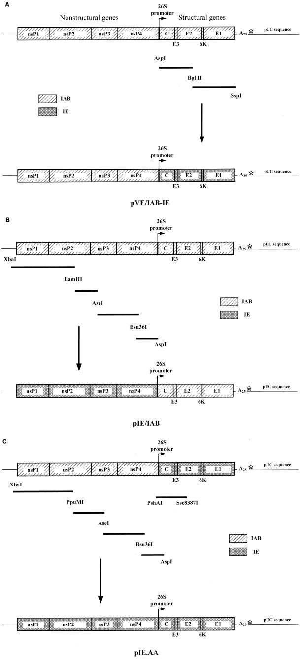
To construct the first chimeric infectious clone, pVE/IAB-IE, two overlapping cDNA fragments of 2.25 and 2.36 kb, covering the entire structural gene region of the IE virus 68U201, were generated by reverse transcription-PCR (RT-PCR). Superscript II (Gibco BRL, Gaithersburg, Md.) was used to generate cDNA, and Pfu Turbo polymerase (Stratagene, La Jolla, Calif.) was used for cDNA amplification (see Table 1 for primers used). These two PCR products were cloned into a pBluescript II SK(+) (Stratagene) shuttle vector to produce a complete 4.4-kb subgenomic cDNA that was subsequently transferred into the IAB infectious clone, pVE/IC-109 (Fig. 1A).
TABLE 1.
Primers used in the construction of chimeric and parental infectious clones
| Primer | Orientationa | Positionb | Sequence (5′→3′) |
|---|---|---|---|
| V-1(+)-T7P/Xba | F | 1–14 | AAGCTTCTAGATTCTAATACGACTCACTATAATGGGCGGCGCATG |
| V-2908(+) | F | 2908–2929 | TGTGTGGAAGACTCTTGCAGGG |
| V-3141(−) | R | 3117–3141 | CACTGCTCTGTGGTCAAATCTATCC |
| V-4252(+) | F | 4252–4276 | CAAAGTGTCTGAAGTGGAAGGTGAC |
| V-4447(−) | R | 4426–4447 | CACATCTGCGTCTGTTGTGTCC |
| V-6509(+) | F | 6509–6530 | GCTGCCCTGTATGCAAAGACTC |
| V-6707(−) | R | 6684–6707 | GCACCAATTCTCTATGAATCCCAC |
| V-7037(+) | F | 7037–7064 | ATCAATATGGTCATAGCTAGCAGAGTTC |
| V-7606(−) | R | 7582–7606 | GCATTGGGTACATTGGTTGATAAGG |
| V-9101(+) | F | 9101–9128 | GTACTTTACTGTCCATGAGCGGTAGTTC |
| V-9284(−) | R | 9257–9284 | ACCCATTTGTCATTCTGTGTACGGTATG |
| V-11464(−) | R | 11436–11464 | GAAATATTAAAAACAAAATCCAATTATGG |
F, forward; R, reverse.
Position listed corresponds to nucleotides in strain 68U201 to which the primer anneals.
FIG. 1.
Construction of IE and IAB VEE virus infectious clones. Boxes containing diagonal lines represent sequences derived from TRD, while hatched boxes contain 68U201 IE sequences. The asterisk is the site of the MluI restriction enzyme recognition sequence used for runoff transcription. PCR-amplified fragments (dark bars) were inserted using the restriction enzymes indicated. (See text for full details.) (A) Construction of pVE/IAB-IE by insertion of two IE PCR fragments into the existing pVE/IC-109 clone. BglII and SspI cuts were partial digests. (B) pIE/IAB was produced by incorporation of four IE fragments into pVE/IC-109. (C) The IE parental infectious clone was derived from pVE/IAB-IE by substitution of 68U201 sequences from the existing IAB sequences. An additional fragment (PshAI-Sse8387I) was required to correct for a deletion incurred during cloning.
A second chimeric virus, consisting of the IE nonstructural genes and the IAB structural genes (pIE/IAB), was constructed by RT-PCR amplification of the IE nonstructural genes in four overlapping fragments. These fragments were combined by producing successive subclones using restriction enzyme sites conserved between the TRD and 68U201 genomes, followed by substitution of this entire fragment for the nonstructural genes in the existing IAB infectious clone (Fig. 1B).
The parental IE infectious clone, pIE.AA, was constructed by cloning the four IE nonstructural cDNAs into the previously constructed chimera pVE/IAB-IE (Fig. 1C). An additional 1.5-kb PshAI/Sse8387I fragment was inserted from a subclone containing the IE 26S sequences to correct for a cloning deletion. All of the final infectious clones were sequenced to ensure that no aberrant or lethal mutations had been introduced during the cloning process. The final sequence of the 68U201 sequence of pIE.AA differed from the published sequence of 68U201 at three nucleotide positions: 5406 (T to A), 5675 (T to G), and 6171 (G to A). The first change results in an amino acid change from Val to Asp at nsP3-459, the second changes Ser to Ala (nsP3-549), and the third changes Gly to Asp (nsP4-152). These same changes were also present in the PCR product (sequenced directly) used to produce the clone, so the differences are likely due to variation in passage history.
In vitro transcription and rescue of recombinant viruses.
The parental and chimeric infectious clones were linearized with restriction endonuclease MluI to produce cDNA templates for RNA synthesis. In vitro transcription was performed as previously described (25) from the T7 RNA polymerase promoter, using an m7G(5′)ppp(5′)A cap analogue (New England Biolabs, Beverly, Mass.). After in vitro transcription, the RNAs were transfected into BHK-21 cells by electroporation (23). Virus was harvested after approximately 48 h when cytopathic effects (CPE) were evident in greater than 75% of the cells. Virus titers were determined by plaque assay on Vero 76 (V76) cells and reported as PFU per milliliter.
Immunofluorescence.
Each rescued virus was characterized by an immunofluorescence assay using variety-specific monoclonal antibodies (MAbs) (27) to confirm the antigenic origin of the structural genes. MAb 1A1B-9 reacts with variety IE viruses but not with IAB viruses; MAb 1A3A-5 is positive for IAB but not IE viruses. These antibodies were diluted 1:400 in phosphate-buffered saline (PBS) and incubated with acetone-fixed monolayers of V76 cells that had been infected with the parental or chimeric viruses for 24 h at a multiplicity of infection of 0.1. Detection was performed with a secondary goat anti-mouse antibody conjugated to fluorescein isothiocyanate (Sigma, St. Louis, Mo.).
Plaque size analysis.
The plaque phenotypes were compared essentially as described by Martin et al. (19). V76 cells were seeded into six-well tissue culture plates and allowed to grow to confluency. Tenfold dilutions of the virus were adsorbed to the monolayers for 1 h at 37°C. A 4-ml overlay consisting of minimal essential medium with 0.4% agar (Sigma) was added, and the cells were incubated at 37°C for 72 h. Agar plugs were removed, and the cells were stained with 0.25% crystal violet in 20% methanol. Approximately 15 to 30 well-isolated plaques were measured for each virus, and the means were compared by analysis of variance (ANOVA) using Bonferroni comparisons.
IFN assays.
One assay to determine the IFN sensitivity of the parental and chimeric VEE viruses was performed, in three replicate experiments, essentially as previously described (30). Briefly, monolayers of L929 mouse fibroblast cells in 96-well plates were primed with twofold dilutions of mouse IFN-α/β (Sigma), ranging from 2,000 to 0.1 U/ml, for 24 h. After priming, 50 μl of virus suspension (10,000 PFU/ml) was added to quadruplicate wells, and the virus was allowed to adsorb for 1 h at 37°C. Additional minimal essential medium supplemented with penicillin-streptomycin and 5% fetal bovine serum was added (100 μl/well). Cells were monitored daily for signs of CPE. Controls included unprimed L929 cells infected with each virus and uninfected cells primed with IFN. Final determination of the concentration of IFN inhibiting 50% of the CPE was made on day 5 postinfection. Additionally, to corroborate the results obtained using the IFN sensitivity assay described in the literature (30), assays were performed to determine the titer of virus being produced at selected IFN concentrations. Duplicate 25-cm2 flasks of L929 cells were induced with 0, 0.1, or 10 U of IFN-α/β per ml 24 h prior to infection with 2 × 10⁵ PFU of one of the four parental or chimeric viruses. At 1 and 3 days, 0.5-ml aliquots of supernatant were removed and analyzed by plaque assay to determine viral titer. Both titers and IFN resistance endpoints were statistically compared by ANOVA.
Guinea pig virulence.
Stocks of parental IE and IAB viruses as well as chimeric IAB/IE and IE/IAB viruses were inoculated into 3- to 5-week-old (300- to 500-g) English short-hair guinea pigs. A 0.2-ml aliquot of each virus (1,000 to 100,000 PFU), diluted in PBS, was injected subcutaneously. Serum (20 μl) was collected daily by the saphenous vein method (9) for 5 days, and the titer of virus in the blood determined by plaque assay. Animals were observed twice daily for signs of infection, and mean day of death was documented. Average survival time and virus titer (which was affected by variable group size due to mortality during the course of the study) were statistically compared using a one-way ANOVA and a two-way ANOVA or general linear model, respectively.
RESULTS
Generation and rescue of recombinant viruses.
Previous studies have proposed that viral genetic elements contributing to vector specificity or conferring subtype characteristics may be expressed from both the nonstructural and structural gene regions. In an attempt to broadly define putative virulence genes and identify possible multigenic determinants, chimeric IE and IAB variety viruses were engineered to contain reciprocal combinations of the entire structural gene region of one variety of virus with the nonstructural genes of the other variety (Fig. 1).
Chimera pVE/IAB-IE was constructed to contain the IAB nonstructural genes with the IE structural genes, while the counterpart to this first construct, pIE/IAB, contained the IE nonstructural genes and the IAB structural genes. Both chimeric viruses reacted as expected antigenically; fluorescence was detected in VE/IAB-IE virus-infected BHK-21 cells using the IE virus-specific MAb 1A1B-9, while a MAb specific for variety IAB, IC, and subtype II viruses (MAb 1A3A-5) showed no immunofluorescence. Conversely, the IE/IAB virus-infected cells demonstrated only 1A3A-5 MAb-specific immunofluorescence.
Plaque size analysis.
Size determination plaque assays were performed to compare the recombinant chimeric viruses with both parental strains. Although individual viruses typically produced a mixed-plaque size phenotype, the parental epizootic strain produced significantly smaller plaques than the enzootic IE parent. The average plaque sizes (± standard deviation) were as follows: VE/IC-109 (IAB parental), 3.3 ± 1.1 mm; IE.AA (IE parental), 5.5 ± 0.4 mm; VE/IAB-IE (chimera), 4.6 ± 0.7 mm; and IE/IAB (chimera), 4.5 ± 0.6 mm (Table 2). The mean plaque diameters of both chimeric viruses were intermediate between those of the parental strains, and comparison of the means indicated that both chimeras produced an average plaque size that was significantly different from that of the IAB and IE parental strains (all P values < 0.005).
TABLE 2.
In vitro and in vivo markers of VEE virus virulence
| Virus
|
IFN analyses
|
Guinea pig virulence
|
||||||
|---|---|---|---|---|---|---|---|---|
| Name | Gene structure | Mean plaque diam (mm) ± SD | Sensitivity (U/ml) | Titera (log10 PFU/ml) at IFN concn of:
|
No. dead/no. infected | Mean day of death ± SD | ||
| 0 U/ml | 0.1 U/ml | 10 U/ml | ||||||
| VE/IC-109 | IAB | 3.3 ± 1.1 | 5.6 | 8.2 ± 0.2 | 7.9 ± 0.4 | 7.3 ± 0.4 | 4/4 | 3.8 ± 0.4 |
| IE.AA | IE | 5.5 ± 0.4 | 0.4 | 7.3 ± 0.4 | 5.6 ± 0.1 | 5.4 ± 0.3 | 0/4 | NAb |
| VE/IAB-IE | IAB nonstructural genes, IE structural genes | 4.6 ± 0.7 | 2.3 | 8.4 ± 0.2 | 8.2 ± 0.1 | 7.6 ± 0.1 | 3/4 | 6.0 ± 1.2 |
| IE/IAB | IE nonstructural genes, IAB structural genes | 4.5 ± 0.6 | <0.1 | 6.8 ± 0.4 | 5.2 ± 0.3 | 4.7 ± 0.2 | 4/4 | 4.3 ± 0.4 |
Titers shown are for 24 h postinfection.
NA, not applicable.
IFN sensitivity.
A correlation between virulence of a given VEE virus strain and its sensitivity or resistance to IFN has been well established (10, 30). This same approach was used with the viruses in this study; the parental IAB strain (VE/IC-109) was the most resistant to IFN-α/β (50% protective dose = 5.6 U), while the IE parental virus (IE.AA) was extremely sensitive to induction with IFN-α/β (50% protective dose = 0.4 U) (Table 2). One chimera, VE/IAB-IE, was intermediate between the two parental types but still quite resistant to IFN (50% protective dose = 2.3 U). Interestingly, the chimera containing the IE nonstructural genes was at least as sensitive to IFN-α/β as the IE parental virus (50% protective dose < 0.1 U). Statistical analysis of variance indicated that the VE/IC-109 parent was significantly different from the IE/IAB chimera (P < 0.03) and that the IE.AA parent was significantly different from the VE/IAB-IE chimera (P < 0.05). Additional VEE virus isolates that had been previously characterized by the IFN sensitivity technique were assayed here to confirm the reproducibility of the test. Strains Fe3-7c (Everglades, subtype II) and 93-42124 (subtype IE) were categorized as sensitive and resistant, respectively, with 50% inhibition endpoints at <0.01 U/ml (Fe3-7c) and 26 U/ml (93-42124). The IFN sensitivity classifications assigned to these two virus strains corresponded to those previously determined (30), although all VEE viruses were more sensitive to IFN-α/β in this report.
Additionally, the plaque titers of all four viruses were determined at 5 days postinfection, using the supernatant from the well identified as the 50% inhibition endpoint, and at days 1 and 3 from cells induced with 0, 0.1, or 10 U of IFN-α/β per ml. At the 50% endpoint doses, none of the viruses were significantly different, with titers ranging from 3.0 to 5.7 log10 50% tissue culture infective doses/ml. Determination of viral titers at the defined IFN-α/β doses of 10 and 0.1 U/ml confirmed the results of the IFN sensitivity assay; each parental virus was significantly different (P <0.05) from the chimeric virus containing the structural genes of that parent (Table 2).
Virulence for guinea pigs.
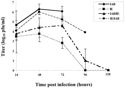
Outbred guinea pigs were infected with each of the viruses by subcutaneous inoculation, and daily serum samples were used to determine the viremia throughout the course of infection (Fig. 2). The clone-derived IAB virus was by far the most virulent in guinea pigs (Table 2 and Fig. 2). It produced a very high titered (4.6 to 6.3 log10 PFU/ml), rapid viremia that consistently killed the animals in only 3 to 4 days. In contrast, the IE parental virus was avirulent; animals infected with this virus exhibited few clinical signs of illness, produced a low-titered viremia (2.8 to 3.5 log10 PFU/ml) that was cleared within 3 days, and subsequently recovered from the infection. Guinea pigs receiving either VE/IAB-IE or IE/IAB virus showed intermediate levels of virus replication and virulence, both being statistically different from the IE parent in average survival time (P < 0.01) and, at later time points, guinea pig viremia (P < 0.02 at 96 h). Neither chimera differed significantly from the IAB parent for guinea pig viremia (P = 0.06 to 0.36); however, the VE/IAB-IE chimera had an average survival time statistically different from that of VE/IC-109 (P = 0.03). The IE/IAB chimeric virus appeared to be more virulent than the reciprocal chimera (3.9 to 6.0 log10 and 3.8 to 4.6 log10 PFU/ml, respectively). Both chimeras were lethal for guinea pigs; however, the IE/IAB virus produced a slightly higher viremia and resulted in an average survival time that was 2 days shorter than that of the VE/IAB-IE virus.
FIG. 2.
Viremia of parental and chimeric VEE viruses in outbred English short-haired guinea pigs.
DISCUSSION
Determination of the molecular basis of reemergence of VEE epidemics and epizootics, as well as the differential virulence of the epizootic and enzootic viruses, are significant issues in arbovirology. Previous studies have shown that laboratory mutations in the VEE virus genome can lead to altered ability to efficiently infect and disseminate in mosquitoes (34) and mice (4), change cell type specificity (20), and generate viral phenotypic changes (5, 7, 12). However, determinants of the viral genome directly involved in natural virulence have not yet been elucidated. Using a molecular genetic approach exploiting infectious clone technology, we determined that viral genetic determinants in both the structural and nonstructural gene regions are involved in natural enzootic versus epizootic virulence.
Most studies concerning the role of VEE viral genetics in virulence have focused on the structural proteins, in particular, the surface glycoproteins. A single mutation in the E2 glycoprotein of the TRD strain delayed replication of the mutant in mice by almost 2 days and significantly reduced the pathogenicity of the virus (4). Interestingly, because the envelope proteins are in close association with each other as a dimeric spike structure in the mature virion (1, 22), Davis and colleagues (3) showed that attenuating mutations in E2 glycoprotein could be compensated for by changes in the E1 protein.
The E2 protein is also important in infection of the insect vector. One MAb-resistant mutant, containing a single mutation in E2, was restricted in its ability to disseminate from the mosquito midgut following oral infection. Dissemination of this mutant was identical to that of the wild-type virus when inoculated into mosquitoes, suggesting that the level of inhibition was the viral entry into midgut epithelial cells (34).
More recently, mutations leading to attenuation of the VEE virus TRD strain to generate the TC-83 vaccine strain were identified (14). A major determinant of attenuation occurred at amino acid position E-120 in the E2 glycoprotein gene. Incorporation of an additional mutation at genome nucleotide position 3 in the 5′ noncoding region (NCR) significantly enhanced the attenuated character of the vaccine strain. The results of our work contrasting enzootic IE and epizootic IAB strains corroborated this finding. The chimeric IE/IAB virus, containing the virulent IAB structural genes, demonstrated an intermediate mean plaque size, an increased resistance to IFN, and an increase in guinea pig virulence compared to the wild-type IE parent. These findings support the hypothesis that a major virulence determinant is present in the structural gene region, perhaps being enhanced by components of the nonstructural or 5′ noncoding regions of the genome. However, Kinney et al. (14) found that incorporation of the TRD nucleotide in the 5′ NCR into the TC-83 infectious clone by itself was insufficient to generate the virulent phenotype; the mutation in the E2 gene was required in concert to show any alteration in virulence. The phenotype of the VE/IAB-IE virus seems to contradict this finding. This virus does not maintain the IE-like characteristics as would be expected if mutations in the E2 gene were absolutely essential for virulence. The VE/IAB-IE virus was markedly different from the IE parent in ability to replicate in the guinea pig model (Table 1 and Fig. 2). In fact, this virus exhibited a mortality pattern in guinea pigs that was more similar to that of the virulent IAB parent virus than the avirulent IE parent, suggesting that a component present in the 5′ NCR or the nonstructural genes is an additional major determinant of viral pathogenesis. It is important to note that the information derived from the TRD parent–TC-83 derivative comparison, a pair containing only 11 differences in the entire genome (15), is merely a point to begin comparisons between the wild-type virulence determinants of IAB versus IE viruses, which involve many more genetic differences that may have profound effects on viral phenotype. Because protein products from both the alphaviral structural and nonstructural regions interact with sequence domains in the other region, the 25% sequence divergence between the IE and IAB viruses may result in less competent chimeras, thus limiting the utility of the large-gene-region chimeric viruses. However, chimeric alphaviruses have previously been shown to be useful for studies of viral replication and recombination (8, 17). In this study, because both VEE virus chimeras demonstrated increased competence and virulence compared to the IE parent, our results reveal that chimeric infectious clones can provide valuable insight into the potential for a given gene region to contain virulence determinants.
One potential candidate for a nonstructural protein involved in VEE virulence is nsP3, which contains an extremely hypervariable region in its carboxy-terminal region (20). The nsP3 gene has been shown to tolerate numerous mutations, including large deletions, and still produce viable virus particles in vertebrate cells. Interestingly, some nsP3 mutations in another alphavirus, Sindbis virus, while doing little to impair replication in mammalian cells, significantly reduced the replicative ability of the virus in mosquito cells (18). Presumably, if the nsP3 gene is involved in virulence, it could require additional mutations to completely alter the virulence phenotype since no difference in virulence in mice was noted for a TC-83 vaccine strain derivative that contained the wild-type TRD nsP3 sequences (14). However, this would not be unexpected based on our results demonstrating that intermediate virulence and pathogenicities were observed for both the VE/IAB-IE and the IE/IAB viruses.
Finally, it is important to note that the three different virulence markers used in this study, differential plaque size, sensitivity to IFN, and virulence for guinea pigs, may assess very different aspects of viral virulence. These markers were chosen because they have previously been shown to distinguish between epizootic and enzootic VEE virus strains, but they are unlikely to separate them based on the same mechanisms. Plaque size, for example, is likely to be a reflection of the surface charge of the glycoproteins as the viral particles move through an unpurified agar overlay containing polyanions that may bind virus, thus affecting mobility but not binding or entry into the cell (13, 19). Assays such as IFN sensitivity and guinea pig virulence are more likely to distinguish the epizootic and enzootic strains based on biological or epidemiological differences. Interestingly, the IE/IAB results presented here indicate that even these two methods of phenotype characterization may employ different mechanisms. The IE/IAB virus, containing the virulent IAB structural genes, was more virulent in guinea pigs than its counterpart but was just as sensitive to IFN-α/β (producing the same titers in the presence of IFN) as the IE parental strain. This could imply that while IFN-α/β may modulate VEE virus pathogenesis, it may not be the dominant mechanism of protection from infection in guinea pigs and possibly equines.
Most importantly, it is clear that the enzootic IE VEE viruses, which had previously been considered to have no equine virulence, may indeed possess some epidemic potential in both the structural and nonstructural portions of the genome. This is a significant finding considering that in 1993 and 1996, IE VEE viruses caused outbreaks of equine encephalitis in southern Mexico, with over 160 cases documented (21). Utilizing multiple approaches to characterize genotypic markers associated with virulence, chimeric viruses should be a useful approach for elucidating genetic determinants involved in pathogenesis, transmission, and evolution of the multiple subtypes and varieties of the VEE antigenic complex. Construction of additional chimeras with this system, as well as using more closely related VEE viruses, to investigate progressively more defined viral genetic elements will eventually provide a better understanding of the mechanisms of VEE virus virulence.
ACKNOWLEDGMENTS
We thank Jonathan Smith and Bruce Crise for helpful discussions during the course of these studies and Daniel Freeman for assistance with statistical analyses.
This research was supported in part by grant AI-10984 from the National Institutes of Health and by the John D. and Catherine T. MacArthur Foundation. A. M. Powers was supported by the James W. McLaughlin Fellowship Fund. A. C. Brault was supported by NIH emerging tropical diseases T32 grant AI107526.
REFERENCES
- 1.Anthony R P, Brown D T. Protein-protein interactions in an alphavirus membrane. J Virol. 1991;65:1187–1194. doi: 10.1128/jvi.65.3.1187-1194.1991. [DOI] [PMC free article] [PubMed] [Google Scholar]
- 2.Calisher C H, Maness K C. Virulence of Venezuelan equine encephalomyelitis virus subtypes for various laboratory hosts. Appl Microbiol. 1974;28:881–884. doi: 10.1128/am.28.5.881-884.1974. [DOI] [PMC free article] [PubMed] [Google Scholar]
- 3.Davis N L, Brown K W, Greenwald G F, Zajac A J, Zacny V L, Smith J F, Johnston R E. Attenuated mutants of Venezuelan equine encephalitis virus containing lethal mutations in the PE2 cleavage signal combined with a second-site suppressor mutation in E1. Virology. 1995;212:102–110. doi: 10.1006/viro.1995.1458. [DOI] [PubMed] [Google Scholar]
- 4.Davis N L, Grieder F B, Smith J F, Greenwald G F, Valenski M L, Sellon D C, Charles P C, Johnston R E. A molecular genetic approach to the study of Venezuelan equine encephalitis virus pathogenesis. Arch Virol. 1994;9:99–109. doi: 10.1007/978-3-7091-9326-6_11. [DOI] [PubMed] [Google Scholar]
- 5.Davis N L, Powell P, Greenwald G F, Willis L V, Johnson B J B, Smith J F, Johnston R E. Attenuating mutations in the E2 glycoprotein gene of Venezuelan equine encephalitis virus: construction of single and multiple mutants in a full-length clone. Virology. 1991;183:20–31. doi: 10.1016/0042-6822(91)90114-q. [DOI] [PubMed] [Google Scholar]
- 6.Franck P T, Johnson K M. An outbreak of Venezuelan equine encephalomeylitis in Central America. Evidence for exogenous source of a virulent virus subtype. Am J Epidemiol. 1971;94:487–495. doi: 10.1093/oxfordjournals.aje.a121346. [DOI] [PubMed] [Google Scholar]
- 7.Grieder F B, Davis N L, Aronson J F, Charles P C, Sellon D C, Suzuki K, Johnston R E. Specific restrictions in the progression of Venezuelan equine encephalitis virus-induced disease resulting from single amino acid changes in the glycoproteins. Virology. 1995;206:994–1006. doi: 10.1006/viro.1995.1022. [DOI] [PubMed] [Google Scholar]
- 8.Hahn C S, Lustig S, Strauss E G, Strauss J H. Western equine encephalitis virus is a recombinant virus. Proc Natl Acad Sci USA. 1988;85:5997–6001. doi: 10.1073/pnas.85.16.5997. [DOI] [PMC free article] [PubMed] [Google Scholar]
- 9.Hem A, Smith A J, Solberg P. Saphenous vein puncture for blood sampling of the mouse, rat, hamster, gerbil, guinea pig, ferret and mink. Lab Anim. 1998;32:364–368. doi: 10.1258/002367798780599866. [DOI] [PubMed] [Google Scholar]
- 10.Jahrling P B, Dendy E, Eddy G A. Correlates to increased lethality of attenuated Venezuelan encephalitis virus vaccine for immunosuppressed hamsters. Infect Immun. 1974;9:924–930. doi: 10.1128/iai.9.5.924-930.1974. [DOI] [PMC free article] [PubMed] [Google Scholar]
- 11.Jahrling P B, Eddy G A. Comparisons among members of the Venezuelan encephalitis virus complex using hydroxylapatite column chromatography. Am J Epidemiol. 1977;106:408–417. doi: 10.1093/oxfordjournals.aje.a112483. [DOI] [PubMed] [Google Scholar]
- 12.Johnson B J, Kinney R M, Kost C L, Trent D W. Molecular determinants of alphavirus neurovirulence: nucleotide and deduced protein sequence changes during attenuation of Venezuelan equine encephalitis virus. J Gen Virol. 1986;67:1951–1960. doi: 10.1099/0022-1317-67-9-1951. [DOI] [PubMed] [Google Scholar]
- 13.Johnson K M, Martin D H. Venezuelan equine encephalitis. Adv Vet Sci Comp Med. 1974;18:79–116. [PubMed] [Google Scholar]
- 14.Kinney R M, Chang G J, Tsuchiya K R, Sneider J M, Roehrig J T, Woodward T M, Trent D W. Attenuation of Venezuelan equine encephalitis virus strain TC-83 is encoded by the 5′-noncoding region and the E2 envelope glycoprotein. J Virol. 1993;67:1269–1277. doi: 10.1128/jvi.67.3.1269-1277.1993. [DOI] [PMC free article] [PubMed] [Google Scholar]
- 15.Kinney R M, Johnson B J, Welch J B, Tsuchiya K R, Trent D W. The full-length nucleotide sequences of the virulent Trinidad donkey strain of Venezuelan equine encephalitis virus and its attenuated vaccine derivative, strain TC-83. Virology. 1989;170:19–30. doi: 10.1016/0042-6822(89)90347-4. [DOI] [PubMed] [Google Scholar]
- 16.Kinney R M, Tsuchiya K R, Sneider J M, Trent D W. Molecular evidence for the origin of the widespread Venezuelan equine encephalitis epizootic of 1969 to 1972. J Gen Virol. 1992;73:3301–3305. doi: 10.1099/0022-1317-73-12-3301. [DOI] [PubMed] [Google Scholar]
- 17.Kuhn R J, Griffin D E, Owen K E, Niesters H G, Strauss J H. Chimeric Sindbis-Ross River viruses to study interactions between alphavirus nonstructural and structural regions. J Virol. 1996;70:7900–7909. doi: 10.1128/jvi.70.11.7900-7909.1996. [DOI] [PMC free article] [PubMed] [Google Scholar]
- 18.Lastarza M W, Grakoui A, Rice C M. Deletion and duplication mutations in the C-terminal nonconserved region of Sindbis virus nsP3: effects on phosphorylation and on virus replication in vertebrate and invertebrate cells. Virology. 1994;202:224–232. doi: 10.1006/viro.1994.1338. [DOI] [PubMed] [Google Scholar]
- 19.Martin D H, Dietz W H, Alvaerez O, Jr, Johnson K M. Epidemiological significance of Venezuelan equine encephalomyelitis virus in vitro markers. Am J Trop Med Hyg. 1982;31:561–568. doi: 10.4269/ajtmh.1982.31.561. [DOI] [PubMed] [Google Scholar]
- 20.Oberste M S, Parker M D, Smith J F. Complete sequence of Venezuelan equine encephalitis virus subtype IE reveals conserved and hypervariable domains within the C terminus of nsP3. Virology. 1996;219:314–320. doi: 10.1006/viro.1996.0254. [DOI] [PubMed] [Google Scholar]
- 21.Oberste M S, Schmura S M, Weaver S C, Smith J F. Geographic distribution of Venezuelan equine encephalitis virus subtype IE genotypes in Central America and Mexico. Am J Trop Med Hyg. 1999;60:630–634. doi: 10.4269/ajtmh.1999.60.630. [DOI] [PubMed] [Google Scholar]
- 22.Paredes A M, Brown D T, Rothnagel R, Chiu W, Schoepp R J, Johnston R E, Prasad B V. Three-dimensional structure of a membrane-containing virus. Proc Natl Acad Sci USA. 1993;90:9095–9099. doi: 10.1073/pnas.90.19.9095. [DOI] [PMC free article] [PubMed] [Google Scholar]
- 23.Powers A M, Kamrud K I, Olson K E, Higgs S, Carlson J O, Beaty B J. Molecularly engineered resistance to California serogroup virus replication in mosquito cells and mosquitoes. Proc Natl Acad Sci USA. 1996;93:4187–4191. doi: 10.1073/pnas.93.9.4187. [DOI] [PMC free article] [PubMed] [Google Scholar]
- 24.Powers A M, Oberste M S, Brault A C, Rico-Hesse R, Schmura S M, Smith J F, Kang W, Sweeney W P, Weaver S C. Repeated emergence of epidemic/epizootic Venezuelan equine encephalitis from a single genotype of enzootic subtype ID virus. J Virol. 1997;71:6697–6705. doi: 10.1128/jvi.71.9.6697-6705.1997. [DOI] [PMC free article] [PubMed] [Google Scholar]
- 25.Powers A M, Olson K E, Higgs S, Carlson J O, Beaty B J. Intracellular immunization of mosquito cells to LaCrosse virus using a recombinant Sindbis virus vector. Virus Res. 1994;32:57–67. doi: 10.1016/0168-1702(94)90061-2. [DOI] [PubMed] [Google Scholar]
- 26.Rico-Hesse R, Weaver S C, de Siger J, Medina G, Salas R A. Emergence of a new epidemic/epizootic Venezuelan equine encephalitis virus in South America. Proc Natl Acad Sci USA. 1995;92:5278–5281. doi: 10.1073/pnas.92.12.5278. [DOI] [PMC free article] [PubMed] [Google Scholar]
- 27.Roehrig J T, Bolin R A. Monoclonal antibodies capable of distinguishing epizootic from enzootic varieties of subtype I Venezuelan equine encephalitis viruses in a rapid indirect immunofluorescence assay. J Clin Microbiol. 1997;35:1887–1890. doi: 10.1128/jcm.35.7.1887-1890.1997. [DOI] [PMC free article] [PubMed] [Google Scholar]
- 28.Scherer W F, Chin J. Responses of guinea pigs to infections with strains of Venezuelan encephalitis virus, and correlations with equine virulence. Am J Trop Med Hyg. 1977;26:307–312. doi: 10.4269/ajtmh.1977.26.307. [DOI] [PubMed] [Google Scholar]
- 29.Scherer W F, Chin J, Ordonez J V. Further observations on infections of guinea pigs with Venezuelan encephalitis virus strains. Am J Trop Med Hyg. 1979;28:725–728. [PubMed] [Google Scholar]
- 30.Spotts D R, Reich R M, Kalkhan M A, Kinney R M, Roehrig J T. Resistance to alpha/beta interferons correlates with the epizootic and virulence potential of Venezuelan equine encephalitis viruses and is determined by the 5′ noncoding region and glycoproteins. J Virol. 1998;72:10286–10291. doi: 10.1128/jvi.72.12.10286-10291.1998. [DOI] [PMC free article] [PubMed] [Google Scholar]
- 31.Walton T E, Grayson M A. Venezuelan equine encephalomyelitis. In: Monath T P, editor. The arboviruses: epidemiology and ecology. IV. Boca Raton, Fla: CRC Press; 1988. pp. 203–233. [Google Scholar]
- 32.Weaver S C, Bellew L A, Rico-Hesse R. Phylogenetic analysis of alphaviruses in the Venezuelan equine encephalitis complex and identification of the source of epizootic viruses. Virology. 1992;191:282–290. doi: 10.1016/0042-6822(92)90190-z. [DOI] [PubMed] [Google Scholar]
- 33.Weaver S C, Salas R, Rico-Hesse R, Ludwig G V, Oberste M S, Boshell J, Tesh R B. Re-emergence of epidemic Venezuelan equine encephalomyelitis in South America. VEE Study Group. Lancet. 1996;348:436–440. doi: 10.1016/s0140-6736(96)02275-1. [DOI] [PubMed] [Google Scholar]
- 34.Woodward T M, Miller B R, Beaty B J, Trent D W, Roehrig J T. A single amino acid change in the E2 glycoprotein of Venezuelan equine encephalitis virus affects replication and dissemination in Aedes aegypti mosquitoes. J Gen Virol. 1991;72:2431–2435. doi: 10.1099/0022-1317-72-10-2431. [DOI] [PubMed] [Google Scholar]
- 35.Young N A, Johnson K M. Antigenic variants of Venezuelan equine encephalitis virus: their geographic distribution and epidemiologic significance. Am J Epidemiol. 1969;89:286–307. doi: 10.1093/oxfordjournals.aje.a120942. [DOI] [PubMed] [Google Scholar]


