Abstract
Colorectal cancer (CRC) is a common type of gastrointestinal tract (GIT) cancer and poses an enormous threat to human health. Current strategies for metastatic colorectal cancer (mCRC) therapy primarily focus on chemotherapy, targeted therapy, immunotherapy, and radiotherapy; however, their adverse reactions and drug resistance limit their clinical application. Advances in nanotechnology have rendered lipid nanoparticles (LNPs) a promising nanomaterial-based drug delivery system for CRC therapy. LNPs can adapt to the biological characteristics of CRC by modifying their formulation, enabling the selective delivery of drugs to cancer tissues. They overcome the limitations of traditional therapies, such as poor water solubility, nonspecific biodistribution, and limited bioavailability. Herein, we review the composition and targeting strategies of LNPs for CRC therapy. Subsequently, the applications of these nanoparticles in CRC treatment including drug delivery, thermal therapy, and nucleic acid-based gene therapy are summarized with examples provided. The last section provides a glimpse into the advantages, current limitations, and prospects of LNPs in the treatment of CRC.
Keywords: lipid nanoparticles, colorectal cancer, tumor targeting, drug delivery, nanotechnology
Introduction
Globally, colorectal cancer (CRC) is the third most frequently diagnosed malignancy and the second leading cause of cancer death.1–3 Most CRC cases arise sporadically, while approximately 35% of cases are attributed to heritable factors.4 It is believed that the vast majority of CRCs follow the adenoma-carcinoma sequence and serrated polyp-carcinoma sequence, largely due to genetic aberrations at the cellular level.5–7 Currently, the principal therapeutic approaches for CRC include surgical intervention, radiotherapy, chemotherapy, targeted therapy, and immunotherapy.8–10 However, the chemotherapeutic and targeted agents currently in use are associated with substantial treatment-related toxicities.11 These adverse effects significantly impair patients’ quality of life, constrain dosage limits, and may even cause the termination of treatment.12–14 Consequently, there is an urgent need to devise treatment strategies that minimize the non-specific distribution of drugs and mitigate toxic side effects, thereby improving both the quality of life and prognosis of CRC patients.
In response to these challenges, colon-specific drug delivery systems (CDDS), particularly those employing nanotechnology for colon targeting, have been tailored to meet the biological characteristics and clinical demands of CRC.15–17 Among the various existing drug carriers, nanoparticles (NPs) can enhance drug stability and solubility, facilitate transmembrane transport, and extend circulation time, thereby improving both safety and efficacy.18 Drugs absorbed in the colon are taken up by the intestinal mucosa and subsequently enter systemic circulation via the venous or lymphatic systems.19 Based on the pathophysiological characteristics of the microenvironment surrounding the disease site, scientists have developed several advanced CDDSs to optimize drug delivery to the colon. These systems employ various mechanisms to control and target the release of drugs, including pH-sensitive systems (eg, pH-responsive polycarboxybetaine-coated LNPs), enzyme-triggered systems (eg, the enzyme-responsive drug delivery system “EUG/CAS–MSNs–COOH”), and magnetically driven systems (eg, magnetically driven spmNP and EMHV DDSs).20–22 Selective surface receptor-mediated drug delivery systems (eg, folate receptor and chemokine-targeted systems) are also employed to specifically deliver drugs to cancerous cells, bypass toxic side effects, and enhance the therapeutic index.23
With the advent of nanotechnology, therapeutic lipid nanoparticles (LNPs) have been widely applied in drug delivery systems (DDSs) due to their efficiency and versatility.18 LNPs are lipid-based nanocarriers prepared through various methods such as lipid vesicle extrusion, rehydration, nanoprecipitation, and microfluidic mixing.24,25 One of their key components is ionizable lipids, whose structure and pKa are closely related to cytoplasmic release.26 However, the structure and delivery characteristics of LNPs depend on the combination of different lipids. The roles of other lipid components are also indispensable, as they influence the morphology, stability, and distribution of LNPs. For example, PEG lipids on the surface of the particles can prevent aggregation and extend circulation time in vivo.27 LNPs exhibit lower immunogenicity and cytotoxicity compared to polymeric and inorganic nanoparticles and can be engineered for targeted modifications.28,29 Owing to these properties, they can effectively cross physiological barriers and deliver drugs precisely to lesion sites. In fact, in intestinal disease, LNP-based DDSs have been extensively studied for their ability to target lesion sites.30,31 They minimize drug exposure to normal tissues while maintaining therapeutic concentrations at the lesion site, effectively inhibiting tumor growth.32 Additionally, the composition, size, and surface charge of these NPs are crucial factors that influence their accumulation in and clearance from the intestinal mucosa.33
Since several well-summarized reviews on nanoparticle-based therapy for cancer already exist, we specifically focus on the advancements of LNP-based anticancer therapy in CRC treatment. Firstly, we provide a brief overview of the formulation of LNPs to demonstrate how each component contributes to their overall functionality. Subsequently, we concentrate on the tumor-targeting strategies and colorectal-specific designs of LNPs, aimed at optimizing their effectiveness in treating colorectal diseases. Lastly, we summarize recent advancements in employing LNPs for colorectal cancer therapy, particularly in nucleic acid drug treatments. We also address the current challenges in this field, offering insights into future design strategies and applications.
Lipid Nanoparticle Delivery Systems: An Overview and Composition Analysis
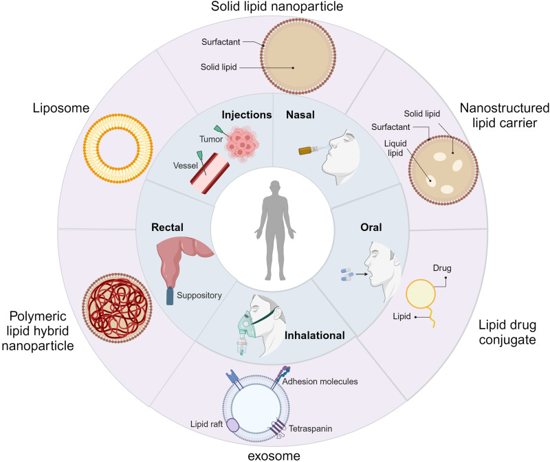
Liposomes discovered in the 1960s are considered the earliest version of LNPs.34 Since then, numerous liposome-based drugs have been extensively applied in medical practice.35 With the development of nanotechnology, the term “lipid nanoparticles” emerged in the early 1990s. To overcome the limitations of liposomes, solid lipid nanoparticles (SLNs) and nanostructured lipid carriers (NLCs) were developed.36,37 Both SLNs and NLCs, comprising lipids and stabilizers, offer enhanced physical stability.38 Extracellular vesicles are naturally occurring LNPs with a size range of 30–150 nm, which are produced by both tumor and non-tumor host cells. These vesicles, characterized by a phospholipid bilayer structure, play a pivotal role in intercellular communication.39 They can alter the biological state of recipient cells by transmitting proteins, nucleic acids, and other biomolecules, thereby affecting cancer recurrence, metastasis, and immune response.40,41 LNPs can be categorized into six types based on variations in structure and drug-loading mechanisms: liposomes, solid lipid nanoparticles (SLNs), nanostructured lipid carriers (NLCs), polymeric lipid hybrid nanoparticles (PLNs), lipid drug conjugates (LDCs), and exosomes (Figure 1). Each category represents a unique facet of this versatile drug delivery system, highlighting the evolution and diversification of LNP technology in biomedical research.42–45
Figure 1.
Classification and Administration Routes of LNPs. Created with BioRender.com.
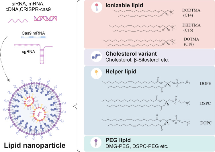
LNPs are multi-component drug delivery systems generally comprising cationic lipids (CLs) or ionizable lipids (ILs), helper lipids, cholesterol, and polyethylene glycol (PEG) lipids (Figure 2).46
Figure 2.
The structure and composition of LNPs. Created with BioRender.com.
Cationic/Ionizable Lipids: Key Components Shaping LNP Structure and Function
Cationic/ionizable lipids are fundamental components of LNPs, facilitating their self-assembly through electrostatic interactions.47 Historically, cationic lipids were developed for LNP assembly to interact with negatively charged nucleic acids.48 However, their application has been limited due to significant toxicity and diminished in vivo efficacy.49 To address these issues, ionizable lipids have been developed. These lipids remain neutral under physiological conditions, but acquire a net positive charge in acidic intracellular environments due to their tertiary amine components.50 These characteristics largely resolve toxicity and efficacy issues associated with cationic lipids.
Currently, widely used ionizable lipids are mainly divided into five categories: unsaturated, multi-tail, polymeric, biodegradable, and branched-tail lipids.51 These amphiphilic small molecules consist of three primary functional domains: hydrophilic head groups, linker groups, and hydrophobic tails.52 The size and charge density of the head group significantly influence processes such as nucleic acid encapsulation, LNP stability, biodegradation, cellular membrane interaction, and facilitation of endosomal escape.53 Linker groups, which bridge the head and tail groups, play a crucial role in modulating cytotoxicity, stability, biodegradability, and the transfection efficiency of LNPs.54 Meanwhile, the hydrophobic tails primarily contribute to particle formation and potency, impacting aspects like ionization (critical pKa) and lipophilicity (critical LogP).55 Collectively, these diverse functional elements of cationic and ionizable lipids are instrumental in defining the biological characteristics of LNPs.
Helper Lipids, Cholesterol, and Polyethylene Glycol Lipids
In addition to cationic/ionizable lipids, LNP formulations include helper lipids, cholesterol, and PEG lipids, each playing a vital role in preserving LNP properties and functionalities. Helper lipids are mostly phospholipids, accounting for approximately 10–20% of the total lipids in the formulations.25 Despite having received less research attention compared to other components, phospholipids significantly enhance LNP stability and facilitate encapsulation and delivery.56 Commonly used phospholipids in clinical practice include 1,2-distearoyl-sn-glycerol-3-phosphate choline (DSPC), 1,2-dioleoyl-sn-glycerol-3-phosphate ethanolamine (DOPE), and 1,2-dioleoyl-sn-glycerol-3-phosphate choline (DOPC).57 DSPC consists of a phosphatidylcholine head and two saturated 18-carbon tails. It undergoes a phase transition from an ordered gel phase to a disordered fluid crystalline phase when the temperature exceeds its phase-transition temperature.58,59 This shift aids in the formation of a tightly packed lipid bilayer structure. The choice of helper lipids can be adapted to meet different nucleic acid loading requirements. For example, DOPE, with its unsaturated tail and net neutral charge, forms a more fluid lipid layer, promoting the fusion of lipid membranes and endosomes, and ultimately improving RNA transfection efficiency.25,52,60 Consequently, DOPE is often used for mRNA delivery due to its higher RNA transfection efficiency compared to DSPC.61 DOPE also exhibits strong interaction with liver-synthesized apolipoprotein E (ApoE). Zhang et al reported that after intravenous administration, C12-200 LNPs containing DOPE primarily accumulated in the liver, whereas C12-200 LNPs containing DSPC accumulated in the spleen, highlighting the effect of auxiliary lipids on LNP organ distribution.57 Selecting phospholipids based on cell line type can enhance transfection efficiency, as demonstrated by Gretskaya et al, who found that liposomal complexes with DOPC had significantly higher transfection efficiency than those with DOPE in SW620 cells.62 These studies indicate the crucial role of phospholipids in the delivery efficiency of LNPs, emphasizing the importance of selecting the appropriate type of LNPs.
Cholesterol, an amphiphilic natural cell membrane constituent, serves as a helper lipid in LNPs.56 It is involved in the degradation of LNPs within the systemic circulation and assists in their subcellular transport.63,64 Recently, researchers have begun to focus on optimizing the formulation of LNPs. For example, Patel et al have investigated substituting cholesterol with hydroxycholesterol to enhance mRNA delivery to T cells, thereby promoting the endosomal escape of LNPs and advancing their applications in immunotherapy.65 The molecular structure of cholesterol derivatives augments cellular uptake and extends LNP half-life in circulation.63,66
Polyethylene glycol (PEG) lipids provide a polymeric shell for LNPs, with lipid domains deeply embedded within the particles and PEG domains extending to the surface of the particles.27,67 PEG lipids create a hydrophilic steric barrier via PEG chains on LNP surfaces, inhibiting aggregation under manufacturing conditions such as low pH and the presence of ethanol, and facilitating self-assembly.68,69 However, this characteristic leads to the “PEG dilemma”, necessitating careful consideration of the type and proportion of PEG lipids to balance LNP stability and drug release efficiency.70,71 Despite their minor proportion, PEG lipids significantly influence LNPs by (1) affecting particle size, which is crucial for transfection efficiency, biological distribution, and pharmacokinetics;56 (2) ensuring particle stability by preventing aggregation through steric hindrance effects;72 and (3) modulating LNP-cell interactions to avoid rapid LNP clearance and improve the circulation lifetimes.73–76 These effects are regulated by the molar ratio of PEG lipids, as well as the structure and length of the PEG chains and their lipid tails (alkyl/dialkyl chains).
Design of Colorectal-Targeted Lipid Nanoparticles
Carrier Design for Colorectal Targeting
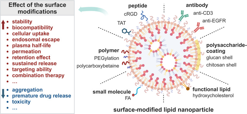
The treatment of colorectal cancer (CRC) has advanced significantly with the use of chemotherapy, radiotherapy, and biological agents, enhancing cancer therapeutic effectiveness. However, these treatments are associated with significant drawbacks, such as the development of drug resistance and toxicity to non-cancerous cells, which can hinder therapeutic success.77,78 The use of delivery vectors has emerged as a crucial strategy to address these challenges. An optimal delivery system should effectively encapsulate drugs, protect them from degradation, and specifically target lesion sites.79 The development of non-biodegradable NPs such as polymeric NPs, LNPs, micelles, gold NPs (AuNPs), and magnetic NPs marked a significant advancement in the targeted formulation and delivery of therapeutic agents to the affected sites in the colon and rectum.31,80–84 Researchers have used NPs as drug delivery vehicles in clinical trials for CRC treatment (Table 1). In particular, LNPs stand out as a highly promising nanomedicine delivery system owing to their excellent biodegradability, biocompatibility, straightforward structural design, and the ability to tailor their functionality to specific needs.85 Various surface modifications greatly enhance cellular uptake and targeting ability of LNPs (Figure 3). Nevertheless, the accumulation and retention of LNPs in the liver following systemic or local administration impede their application in extrahepatic organs. The heightened liver affinity of LNPs is mainly attributed to three pivotal factors. First, the distinctive anatomical and physiological features of the liver, such as discrete blood vessels and sluggish blood flow, facilitate LNP extravasation and reinforce its interaction with hepatic tissues.86 Yang et al leveraged these features to design hepatocyte nuclear factor 4 alpha (HNF4A)-mRNA LNPs for targeting hepatocytes through intravenous administration, holding promise for attenuating liver fibrosis.87 Second, endogenously synthesized ApoE from the liver adheres to the surface of LNPs, forming a protein crown known as the “Corona”. ApoE then combines with low-density lipoprotein receptor (LDLR) to facilitate endocytosis in liver cells.88,89 Lastly, the LNP-Corona complex is enriched in high-density lipoprotein (HDL), steering the preferential delivery of LNPs to the liver. Therefore, to advance the application of LNPs in CRC treatment, urgent strategies must be devised to redirect LNP delivery to extrahepatic organs, including the large intestine.88 With advancements in relevant technologies, organ, tissue, or cell-specific drug delivery using LNPs can be achieved through local administration or systemic intravenous delivery. Currently, implemented targeted local administration routes of LNPs include oral, intranasal, inhalation, rectal, and local injection routes (such as intramuscular and intratumoral injections) (Figure 1).90–92 An appropriate administration route can improve the targeting efficiency of LNPs. For example, LNPs can be readily directed to the liver due to their efficient circulation in the bloodstream, making intravenous injection an appropriate administration route for liver-targeted LNPs.
Table 1.
Representative NPs in Clinical Trials for Treatment of CRC
| Name | Indication | Intervention/Treatment | Stage | NCT Number |
|---|---|---|---|---|
| Cetuximab nanoparticles (p.o). | Colon Cancer Colo-rectal Cancer |
Drug: Cetuximab nanoparticles Drug: Oral approved anticancer drug |
Phase 1 (Unknown) | NCT03774680 |
| Nanoparticle Paclitaxel (i.p). | Peritoneal Neoplasms | Drug: nanoparticulate paclitaxel | Phase 1 (Completed) | NCT00666991 |
| Aguix Gadolinium-Based Nanoparticles (radiotherapy) | Brain Metastases | Radiation: Stereotactic Radiation Drug: AGuIX gadolinium-based nanoparticles Other: Placebo |
Phase 2 (Recruiting) | NCT04899908 |
| AZD4635 (p.o). | Advanced Solid Malignancies | Drug: AZD4635 Drug: Durvalumab Drug: Abiraterone Acetate |
Phase 1 (Completed) | NCT02740985 |
| CALAA-01 (i.v).93–96 | Cancer Solid Tumor |
Drug: CALAA-01 | Phase 1 (Terminated) | NCT00689065 |
| TKM 080301 (hepatic intra-arterial administration)97–99 | Primary or Secondary Liver Cancer | Drug: TKM-080301 | Phase 1 (Completed) | NCT01437007 |
| 9-ING-41 (i.v). | Advanced Cancers | Drug: 9-ING-41 Drug: Gemcitabine - 21 day cycle Drug: Doxorubicin. Drug: Lomustine Drug: Carboplatin. Drug: Nab paclitaxel. Drug: Paclitaxel. Drug: Gemcitabine - 28 day cycle Drug: Irinotecan |
Phase 2 (Recruiting) | NCT03678883 |
| Nal-IRI(i.v). | Metastatic Pancreatic, Colorectal, Gastroesophageal, or Biliary Cancer | Drug: Fluorouracil Drug: Irinotecan Sucrosofate Other: Laboratory Biomarker Analysis Drug: Leucovorin Calcium Drug: Rucaparib |
Phase ½ (Active, not recruiting) | NCT03337087 |
| Nanoparticle Paclitaxel (i.p). | Peritoneal Neoplasms | Drug: nanoparticulate paclitaxel | Phase 1 (Completed) | NCT00666991 |
Notes: Updated on 01/24/2024. All clinical trials listed in this table are based on ClinicalTrials.gov.
Abbreviations: P.O., oral; i.p., intraperitoneal; i.v., intravenous; nal-IRI, liposomal irinotecan.
Figure 3.
Representative Surface Modification Strategies for LNPs. Created with BioRender.com.
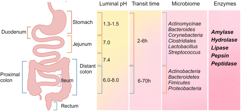
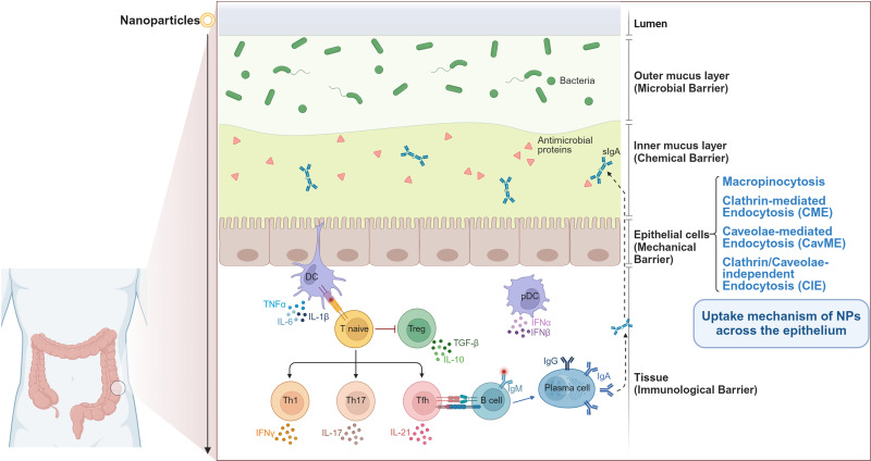
The oral colon-specific drug delivery system (OCDDS) aims to convey drugs orally directly to the colon, preventing premature drug release in the stomach, duodenum, jejunum, and ileum. Such targeted delivery mechanisms enable precise treatment of diseases like CRC and inflammatory bowel disease (IBD). The development of OCDDS must account for the unique anatomical and physiological properties of the gastrointestinal tract’s various segments, such as pH levels, enzyme activity, and microbiota composition (Figure 4).100–102 The large intestine, forming the lower segment of the human digestive tract, consists of the cecum, appendix, colon (ascending, transverse, descending, and sigmoid colons), rectum, and anal canal. It plays a crucial role in absorbing water and electrolytes from food residues, forming and temporarily storing feces, and facilitating controlled excretion.103 Drugs absorbed in the colorectum are taken up by the intestinal mucosa and then enter the systemic circulation via the venous/lymphatic system. In OCDDS, nanoparticles are formulated to withstand the harsh gastrointestinal environment, thereby protecting encapsulated drugs from extreme pH and enzymatic degradation.104 For example, Bajracharya et al used poly (methacrylic acid-co-methyl methacrylate) (1:2) as a surface coating to prepare E/AC-Au/MTX nanocomplexes.31 These nanocomplexes exhibited a remarkable entrapment efficiency of over 80% while demonstrating notable pH-response characteristics. Musa et al loaded NPs in the form of soft agglomerates, which reduced premature drug release.105 The in vitro release experiment showed that the 5-FU-loaded NPs sustained drug release via their response to the intracapsular sodium alginate coat, indicating their potential to achieve colon-specific targeting by oral intake. The epithelium of the large intestine is covered by a bilayered mucus structure comprising water, electrolytes, lipids, and glycoproteins. Numerous studies indicate that NPs, varying in size, shape, composition, and surface modifications, can penetrate this mucous layer and access the intestinal epithelium through different mechanisms (Figure 5).106,107 Oral administration of OCDDS offers several benefits, including enhanced patient compliance, ease and convenience of use, and a reduced likelihood of acute drug reactions.108,109 However, the patient-to-patient variability and the dynamic changes in the gastrointestinal environment under physiological and pathological conditions pose challenges to the clinical translation of OCDDS. Further research is needed to uncover the impact of these factors on NPs.
Figure 4.
Key physiological factors within the gastrointestinal (GI) tract that markedly influence drug absorption. The influence of luminal pH, gastrointestinal transit time, microbiome composition, and enzymatic activity is essential in the modulation of drug absorption processes. Created with BioRender.com.
Figure 5.
Structure of the intestinal mucosal barrier comprising microbial, chemical, mechanical, and immune barriers, along with the mechanism of NPs across the mechanical barrier.110 Current uptake mechanisms of NPs across intestinal epithelium include macropinocytosis, clathrin-mediated endocytosis (CME), caveolae-mediated endocytosis (CavME), and clathrin/caveolae-independent endocytosis (CIE). Created with BioRender.com.
Targeting Strategies of LNPs in Colorectal Cancer
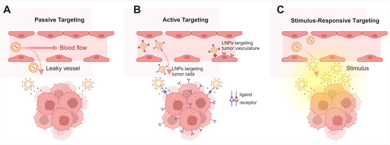
Current nanocarrier-based targeted delivery strategies for CRC can be broadly categorized into passive targeting, active targeting, and stimulus-responsive targeting (Figure 6). These approaches ingeniously exploit the biological characteristics of tumors and the tumor microenvironment (TME) to enable precise and effective drug delivery through distinct mechanisms. This section explores the principles and applications of these strategies in CRC treatment, highlighting their role in enhancing therapeutic efficacy.
Figure 6.
A diagram of major targeting strategies of LNPs for targeting CRC. (A) LNPs passively accumulate in tumors through enhanced permeability and retention effect; (B) Ligand-decorated LNPs actively target cancer cells and tumor vasculature; (C) LNPs respond to both endogenous and exogenous stimuli and release drugs rapidly at the lesion site. Created with BioRender.com.
Passive Targeting
The unique pathological and physiological features of tumors and TME make passive targeting strategies widely applicable in common nanocarrier systems. Tumor tissue is characterized by enlarged gaps between vascular endothelial cells, a dense extracellular matrix (ECM), and poor internal lymphatic drainage. This scenario leads to the enhanced permeability and retention (EPR) effect, facilitating the passive accumulation of nanomedicines at tumor sites (Figure 6A).111 This phenomenon alters the pharmacokinetics and pharmacodynamics of the encapsulated drugs and minimizes off-target toxicity, forming the cornerstone of passive targeting. In the mid-1990s, the first-generation lipid nanoparticles, liposomal doxorubicin (Doxil®) and liposomal daunorubicin (DaunoXome®), received FDA approval.35 Liposomal doxorubicin is a liposomal carrier of the anthracycline chemotherapeutic agent doxorubicin (DOX). The addition of PEG-lipid conjugates extends DOX’s plasma half-life to 45 hours in humans.112 Similarly, liposomal daunorubicin, which carries daunorubicin (DNX), exhibits altered metabolism and distribution, leading to increased tumor accumulation and reduced systemic toxicity.113 The PEGylation of LNPs is a common surface modification. PEG lipids create a hydrophilic barrier that resists binding to plasma proteins, prolonging circulation and maximizing EPR effect-mediated tumor accumulation.114,115
Glucan and chitosan, two naturally occurring polysaccharides, have been incorporated into various drug delivery systems due to their unique structural attributes. Incorporating a chitosan shell is a prevalent strategy to enhance the colorectal targeting of nanomedicines. This shell serves a dual purpose: it protects nanomedicines from degradation in the stomach and small intestine, and upon arrival in the colon, specific glucose hydrolases degrade the shell’s surface glucan.79 This degradation reveals folate residues on the nanoparticles, which then target tumor cells that overexpress folate receptors.116 Moreover, chitosan can improve adhesion and facilitate targeted, sustained drug release in the colon through hydrophilic and electrostatic interactions with mucin.117,118
The rapid clearance by the reticuloendothelial system (RES) and high interstitial fluid pressure (IFP) are key challenges to the efficiency of passive targeting in nanomedicines. The former reduces the half-life of LNPs, hindering accumulation at the target site, while the latter limits drug penetration into deep tumor tissues. Thus, optimizing the formulation, size, and surface charge of LNPs is crucial to enhancing targeting efficacy. Current research focuses on developing “second-generation” nanoparticles to further refine the pharmacokinetic and pharmacodynamic characteristics of drugs, aiming to bolster the treatment of solid tumors.119,120 Selective organ targeting (SORT) represents a significant innovation in this field. Cheng et al introduced a fifth type of lipid, the SORT molecule, into LNPs, altering their internal charge to achieve targeted delivery to extrahepatic organs.121
A recent study by Wang et al revealed a critical but previously underestimated barrier in NP delivery – the tumor vascular basement membrane (BM).122 BM is a dense, cross-linked, extracellular matrix layer beneath the endothelium, enveloping the endothelial and parietal cells of tumor blood vessels. It forms a formidable mechanical barrier with endothelial cells, which traps NPs in the subendothelial space and effectively blocks their entry into the tumor. The study revealed that local hyperthermia induces platelet aggregation and inflammation, attracting neutrophils to the NP pool. These neutrophils then move through the BM barrier and release NPs, facilitating increased NP penetration into deeper tumors. This finding underlines the need for further research and engineering strategies to overcome the BM barrier in NP-mediated drug delivery.
Active Targeting
Active targeting in nanomedicine primarily involves specific ligand modifications on nanoparticle surfaces. The ligands on the surfaces of NPs bind selectively to receptors on target cells, facilitating the delivery of drugs to specific cell types (Figure 6B).123,124 Notably, these receptors are minimally or even not expressed on normal cells but are highly or specifically present on the surfaces of cancer cells.125 Ligand-receptor interactions can trigger receptor-mediated endocytosis, promoting the uptake of LNPs by cancer cells.126 Various ligands, categorized into small molecules (folate, sugars) and macromolecules (antibodies, peptides, proteins, ligands, oligonucleotides), have been employed for LNP surface modifications.127 Several colorectal-specific biomarkers have been utilized in targeted ligand design.128 For instance, folate receptor-α (FR-α), overexpressed in many cancer types, binds naturally with folate (FA), which is stable, low in immunogenicity, and exhibits high affinity to FR-α.129 This renders FA a popular choice for nanomedicine targeting. Folate-bound Poly (lactic-co-glycolic acid) (PLGA) NPs loaded with kaempferitrin demonstrated enhanced cytotoxicity against colorectal cancer cells.130 A recent study analyzing four biomarkers in colorectal cancer tissues from 280 patients via immunohistochemistry revealed increased expression of FR-α (37.1%) compared to normal tissues, along with elevated levels of carcinoembryonic antigen (CEA) (98.8%), tumor-associated glycoprotein-72 (79%), and epidermal growth factor receptor (EGFR) (32.8%) in CRC, highlighting CEA’s potential as a future LNP drug target for CRC.128 CD44 is a common marker of cancer stem cells (CSCs) in colon cancer and is also highly expressed. Chondroitin sulfate (CS) is a highly sulfated glycosaminoglycan. It exhibits a high affinity for CD44, making CS-modified NPs ideal for tumor targeting.131
Peptides are another common targeting ligand for LNPs, favored for their strong binding to various cell targets, cost-effectiveness, high fidelity, and ability to attach to LNPs without hindering their binding ability. Tumor homing peptides (THPs) are a class of peptides that have homing effects on tumor tissue or blood vessels. They can recognize and bind to specific receptors or markers on the surface of tumor tissue or blood vessels. Peptides PIVO-8 (sequence: SNPFSKPYGLTV) and PIVO-24 (sequence: YPHYSLPGSSTL) functionalized liposomes inhibit tumor angiogenesis and increase apoptosis in colon HCT116 tumors in mice.132 Fluorescence images revealed that these PIVO-targeting liposomes significantly increase drug uptake by tumor vasculature endothelial cells via receptor-mediated endocytosis. These results suggest that LNPs have the potential to improve the therapeutic effect of colon cancer by recognizing the tumor vascular system through THPs. Additionally, the liposomes modified with BiP targeting peptides (WIFPWIQL) and dual-targeting liposomes modified with the NRG (GNGRG) and APRPG peptides inhibited colon tumor growth through the same mechanism.133,134 Cellular penetrating peptides (CPPs) are a class of peptides capable of transporting large molecules and small particles across the cell membrane and into the cytoplasm. The transactivator of transcription (TAT) peptide is the first peptide discovered to possess such ability.135 Kuai et al developed a specialized TAT peptide liposome.136 It possessed a thiol-cleavable (like L-Cysteine) long PEG brush layer and a short, non-cleavable PEG layer with TAT peptide attached to it. The extended PEG brush layer functions as a powerful spatial barrier that reduces the opsonization and non-specific cellular interactions of TAT liposomes during passive accumulation. When TAT peptide liposomes passively accumulate inside the tumor, L-Cysteine is injected to cleave the long PEG layer and expose the cell-penetrating TAT peptide, promoting the absorption of liposomes by tumor cells. This design enables specific drug release in the TME and has been proven effective in a mouse subcutaneous C26 colon cancer model.136 Integrins are vital members of the cell adhesion molecule family. Functioning as transmembrane glycoproteins, they play a crucial role in the adhesion and signal transduction between cells and between cells and the ECM. They also regulate cellular functions including adhesion, migration, proliferation, and apoptosis.137 Integrin expression is notably upregulated in a range of solid tumors and their associated blood vessels, highlighting its crucial role in cancer progression and invasion.138 The tripeptide sequence RGD, which is widely present in ECM proteins, can specifically bind to various integrins, making RGD peptides widely used as ligands targeting tumor cells. Liu et al constructed a cRGD peptide (Arg-Gly-Asp-d-Phe-Cys [RGDfC])-modified liposome that encapsulates matrine.139 In HT-29 colon cancer cell lines, this approach demonstrated an enhanced anti-proliferative effect, approximately two-fold greater, compared to free drugs. The PR_b-peptide (KSSPHSRNSGSGSGSGSGRGDSP) designed by the Kokkoli team binds the RGD motif with a synergistic PHSRN sequence, forming a fibronectin mimetic peptide specifically targets α5β1 integrins.140 PR_ B-peptide liposomes can effectively target colon cancer cells and serve as carriers for various drugs, including DOX, 5-FU, and tumor necrosis factor-α.141–144
While active targeting markedly enhances the specificity and therapeutic efficacy of nanomedicines, challenges such as ligand selection and rapid immune clearance in vivo need further investigation.
Stimulus-Responsive Targeting
Stimulus-responsive targeting is a sophisticated delivery strategy that utilizes NPs’ sensitivity to specific physical, chemical, and biological factors for precise drug release at the target site (Figure 6C). This approach involves constructing a Stimuli Response System (SRS) that rapidly and accurately responds to these stimuli by altering the composition and structure of nanocarriers. It addresses the current challenges such as slow drug release, low bioavailability, and suboptimal targeting.145,146 The triggers employed are broadly classified into endogenous and exogenous categories. Endogenous factors mainly refer to the characteristics of the TME, like low pH, tissue hypoxia, enzymatic activity, and redox status. Exogenous factors mainly include physical stimuli such as temperature, ultrasound, magnetic fields, and light, with pH-sensitive liposomes (PSL) and thermosensitive liposomes (TSL) being prime examples.
The tumor microenvironment (TME) in colorectal cancer is characterized by tissue hypoxia and low pH due to lactic acid production from tumor cell glycolysis. These characteristics contribute to cancer progression and chemoresistance through signaling pathways like hypoxia-inducible factor (HIF), presenting significant challenges for developing effective chemotherapy for colorectal cancer.147–151 The physical and chemical properties of pH-sensitive polymers, such as solubility, chain conformation, and surface activity, vary markedly with environmental pH.152 Drug delivery systems utilizing these polymers maintain stability in physiological environments but release their payload in acidic tumor settings, thus achieving tumor targeting. For instance, Juang et al developed pH-sensitive and peptide-modified LNPs to encapsulate the chemotherapy drug irinotecan and miR-200 which inhibits cancer cell metastasis.153 These NPs demonstrated pH-responsive release and enhanced cellular uptake driven by clathrin- and adsorptive-mediated endocytosis. They showed effective internalization and intracellular distribution in the acidic environment of the human colon cancer cell line HCT116, with their therapeutic effectiveness further confirmed in mouse models.
Thermosensitive liposomes are frequently employed as carriers for chemotherapy drugs, used in conjunction with local hyperthermia to trigger drug release and produce tumoricidal effects. Stimulus-responsive targeting strategies can be combined with active targeting modifications on the surface to further enhance drug targeting efficiency and increase cellular toxicity.
Applications of Lipid Nanoparticles (LNPs) in the Treatment of Colorectal Cancer
Colorectal cancer is a highly heterogeneous malignant tumor, making its effective treatment a significant challenge. LNPs have emerged as a versatile tool in this domain. They can encapsulate a diverse array of anti-tumor agents, including small molecules, peptides, proteins, and nucleic acids, offering selective targeting of cancer cells while sparing normal cells. Currently, researchers are actively exploring LNPs for targeted drug delivery, thermal therapy, and gene therapy specifically for colorectal cancer, showing promising potential in the field.
Lipid Nanoparticles (LNPs) for Traditional and Novel Drug Delivery in Colorectal Cancer Therapy
As research progresses, the efficacy of LNPs as carriers for various anti-tumor agents, including first-line drugs, biologics, and naturally derived anticancer compounds, has been increasingly validated in CRC therapy. Delivering drugs to the tumor sites using LNPs offers two major advantages: 1) LNPs can achieve comparable or superior therapeutic effects at lower drug doses compared to traditional formulations such as tablets, capsules, and liquids;154 2) LNPs can enhance drug pharmacokinetics by minimizing systemic distribution, reducing toxicity, and decreasing the frequency of administration.
5-Fluorouracil (5-Fu), a uracil derivative classified as an antimetabolic antitumor drug, is among the most frequently utilized chemotherapeutics for CRC. However, its clinical application is constrained by poor selectivity, substantial toxic side effects, and an exceedingly brief plasma half-life. In response to these challenges, Patel et al encapsulated 5-FU in solid lipid nanoparticles, observing a concentration-dependent reduction in cell viability within Caco-2 human colorectal adenocarcinoma cell lines.155 Doxorubicin (DOX), another common chemotherapy drug with strong anti-cancer activity, faces limitations due to its cardiotoxicity and lack of tumor specificity. To address this, Zhang et al developed ginger-derived nanovectors (GDNVs), a nanocarrier constructed from ginger lipids, capable of efficiently loading and targeting DOX delivery.156 Fluorescence imaging showed that GDNVs are internalized by tumor cells via the phagocytosis pathway with high efficiency. Udofot et al encapsulated 5-FU (pHLNps-5-FU) within PSL nanoparticles, which were further modified with anti-EGFR antibodies.157 Their evaluation in a subcutaneous tumor mouse model using the colon cancer cell line HCT-116 indicated a significant increase in tumor accumulation of pHLNps-5-FU, along with an extended plasma half-life. Irinotecan (IRI, CPT-11) is a second-line chemotherapeutic agent for advanced CRC that inhibits topoisomerase-1 to elicit anti-tumor activity. Bhaskaran et al prepared orally delivered IRI-loaded SLNs using cetyl palmitate via emulsification solvent evaporation and further modified them with chitosan.79 The surface-modified SLNs protect IRI from gastric acid, releasing only 3.33% in an acidic environment within 2 hours. Furthermore, these drugs encapsulated in LNPs demonstrated enhanced anti-cancer activity compared to their free-form counterparts.
Beyond standard anti-tumor drugs, LNPs have demonstrated significant potential as an effective vehicle for the targeted delivery of diverse innovative therapeutics in CRC, offering unique advantages. Al-Asmari et al reported that liposomes containing scorpion toxin were more effective in combating cancer than the free form of the toxin in the human colorectal cancer cell line HCT-8.158 The in vitro release at pH 7.5 showed an initial rapid release of venom within the first 2 hours, followed by a plateau. Increased efficacy was evidenced by a lower survival rate in treated cells, a rise in reactive oxygen species (ROS) production, and a greater number of apoptotic cells. Furthermore, cell cycle analysis suggested a halt in the G0/G1 phase among these cells.
The evolving fields of metagenomics and metabolomics have illuminated the critical role of gut microbiota in the onset and progression of CRC, thereby attracting substantial research interest.159 Numerous studies have focused on developing LNP-based drugs targeting gut microbiota for CRC treatment. Omega-3 polyunsaturated fatty acids (PUFAs) show a significant association with gut microbiota and bile acid levels, with Increased intake believed to reduce inflammation and strengthen anti-tumor immunity.160 The encapsulation of resveratrol in SLNs significantly enhanced the incorporation efficiency of ω-3 PUFAs in human HT-29 CRC cells and reduced tumor cell proliferation.161 Wu et al developed liposomes loaded with matairesinol, a compound exhibiting differential expression between healthy individuals and CRC patients, and found that they markedly improved CRC chemosensitivity by altering lipid metabolism.162 In both chemosensitive and drug-resistant CDX and PDX mouse models, matairesinol-liposomes notably increased the anti-cancer activity of 5-FU/calcium folinate combined with oxaliplatin (FOLFOX). Emerging strategies targeting gut microbiota and metabolic reprogramming through LNP technology offer promising directions and possibilities for the treatment of CRC.
Enhanced Thermal Therapy Efficacy Using Lipid Nanoparticles in Colorectal Cancer
LNPs exhibit robust capabilities in energy conversion and utilization, enabling them to produce thermal effects in response to various stimuli, including pH changes, ultrasound, magnetic fields, and light. This characteristic significantly enhances the efficacy of physical therapies. Superparamagnetic iron oxide nanoparticles (SPIONs), distinguished by their superparamagnetism, are extensively employed in medical applications, notably in magnetic resonance imaging (MRI) and magnetic hyperthermia.163 Shen et al developed folate-modified solid lipid nanoparticles (DFSLNs) encapsulating DOX and superparamagnetic iron oxide particles in pectin for colon-targeted delivery.116 This design combines chemotherapy and magnetic thermal ablation therapy. In addition to reducing cellular penetration through brush border membranes facilitated by proton-coupled FA transporters in the small intestine, DFSLNs also prolong retention time in the colon. Targeted LNPs boost the effectiveness of thermal therapy in cancer, especially when thermal therapy is aligned with other treatments.
Nucleic Acid-Based Gene Therapy in Colorectal Cancer Utilizing Lipid Nanoparticles
Gene therapy represents a promising avenue in cancer treatment, targeting pathogenic genes in a sequence-specific manner.164 This approach facilitates more precise and personalized anti-tumor therapy, underscoring its potential in oncological interventions. Gene therapy can be principally categorized into four types based on its potential mechanisms of action: (1) Gene addition or replacement, exemplified by mRNA encoding genes encapsulated in LNPs; (2) Regulation of gene expression, involving agents like miRNA, short-stranded small interfering RNA (siRNA), and long non-coding RNA (lncRNA);165 (3) Gene editing, utilizing tools such as Cas9 mRNA and single-guided RNA (sgRNA); and (4) DNA or RNA-based vaccines.166 For gene therapy to be efficacious in vivo, it necessitates a delivery platform that is safe, effective, and stable. This platform must shield nucleic acids from degradation while facilitating cellular uptake and subsequent release of these acids. LNPs optimally satisfy the aforementioned requirements.
LNPs loaded with siRNA, mRNA, or DNA can modulate the expression of cancer-related genes, either by upregulating or downregulating them, thereby achieving therapeutic effects in the treatment of CRC (Table 2).
Table 2.
Lipid Nanoparticles for Delivery of Nucleic Acid-Based Therapeutics in CRC
| Payload | Gene Target/Product | LNP Category | Administration Route | Model | Reference |
|---|---|---|---|---|---|
| Gene silencing | |||||
| siRNA | Indoleamine 2,3-dioxygenase-1 (IDO1) | Cationic lipid-assisted nanoparticles (CLANs) | Intravenous injection | Subcutaneous colorectal tumor model | [167] |
| siRNA | Hypoxia inducible factor 1α (HIF-1α) | RGD-targeted multifunctional lipid ECO/siHIF-1α nanoparticles | Intravenous injection | Mouse HT29 colon cancer model | [168] |
| siRNA | A proliferation-inducing ligand (APRIL) | Negative lipidoid nanoparticles (NLNs) | Enema delivery | CRC animal models | [169] |
| siRNA | Inhibitor of DNA-bind-2 (Id2) | Neutral liposome 1,2-dioleoyl-sn-glycero-3-phosphatidylcholine | Intraperitoneal administration | CRC animal models (CT-26) | [170] |
| siRNA | Survivin | Nanoliposomes | Transfection | LoVo cells | [171] |
| siRNA | Multidrug resistance gene (MDR1) | A carrier composed of a cationic oligomer (PEI(1200)), a hydrophilic polymer (polyethylene glycol) and a biodegradable lipid-based crosslinking moiety | Transfection | Human colon CSCs (CD133+ enriched cell population) | [172] |
| siRNA | E2F1 | Nanoliposomes | Transfection | Cultured colon carcinoma cells and cultured human biopsy of colonic mucosa | [173] |
| siRNA | PCTAIRE1 | Lipid nanoparticles | Intratumor injection | Mouse HCT116 subcutaneous tumor models | [174] |
| siRNA | RelA | An amphiphilic cationic cyclodextrin (CD) nanoparticle modified with PEGylated folate | Intravenous injection | Mouse CT26 subcutaneous tumor models | [175] |
| siRNA | Integrin-β1 | Dimethyldioctadecylammonium bromide (DDAB)-methoxy poly (ethylene glycol) (mPEG)-poly-ε-caprolactone (PCL) hybrid nanoparticles (HNPs) | Transfection | Regorafenib-resistant human colon cancer cell line (SW-48) | [176] |
| Gene expression | |||||
| mRNA | ALKBH5 | Exosome-liposome hybrid nanoparticles | Intratumor injection | Mouse preclinical tumor models | [177] |
| cDNA | FAS | Cationic lipid nanoparticle DOTAP-Cholesterol | Intravenous injection | Mouse CT26 subcutaneous tumor models | [178] |
| Gene editing | |||||
| sgRNA | KRAS | Nanoliposomal (NL) particle | Intravenous injection | Mice with KRAS-mutated CRC | [179] |
| mRNA | Cas9 | ||||
In CRC therapy, most LNP-based nucleic acid drugs focus on regulatory factors associated with cell proliferation, cell cycle, metastasis, and apoptosis, consequently inhibiting tumor growth. A noteworthy example involves the LNP-encapsulated siRNA targeting APRIL (A Proliferation-Inducing Ligand), a crucial regulator of cell proliferation. This ligand is characteristically overexpressed in colorectal cancer tissues, where it stimulates the growth of tumor cells. Silencing APRIL has been demonstrated to effectively control tumor progression.180 Analogously, the siRNA-mediated knockdown of DNA-bind-2 inhibitors (Id2) or cDNA overexpression of FAS has been observed to curb tumor cell proliferation and reduce tumor burden in mice.170,178 Additionally, cell cycle-related targets include the E2F1 transcription factor and PCTAIRE1 (also known as PCTK1 or Cyclin Dependent Kinase 16 [Cdk16]).173,174
To enhance anti-tumor efficacy, recent studies have extensively employed LNP siRNA to target and reshape the tumor microenvironment. Indoleamine 2,3-dioxygenase 1 (IDO1) is a tryptophan-degrading metabolic enzyme that is overexpressed in tumor-draining lymph nodes (TDLNs) and tumor tissues.181 This enzyme catalyzes the degradation of the essential amino acid tryptophan (TRP) into kynurenine (KYN), a process that directly activates regulatory T cells (Tregs) while simultaneously inducing the inactivation of cytotoxic T lymphocytes (CTL).182–184 This activity is crucial in establishing an immunosuppressive tumor microenvironment (ITM). Targeting IDO1 with siRNA has shown promise in improving immunotherapy outcomes. In a nude mouse subcutaneous tumor model using human colorectal cancer cells, the combined administration of oxaliplatin (OXA) and CLANsiIDO1 enhanced dendritic cell maturation, increased tumor-infiltrating T lymphocytes, and decreased regulatory T cells, thereby reversing IDO1-mediated immunosuppression.167 Moreover, there is a close interaction between the NF-κB signaling pathway and the tumor microenvironment.185 Zou et al found that using CD DTX.siRelA.PEG-FA nanoparticles to downregulate the NF-κB subunit RelA enhanced docetaxel’s apoptotic effects and inhibited tumor growth in mice.175 Specifically, the co-formulation exhibited pH-triggered release, with higher release in acidic environments. Upon entering the endosome (pH ≈ 5.5 to 6.0), it enabled the simultaneous release of DTX and siRNA into the cytoplasm, leading to a synergistic apoptotic effect. Additionally, factors like COX-2 and abnormal fatty acid metabolism, pivotal in the tumor microenvironment, have been targeted.186,187 Xu et al introduced EpCAM aptamers into 3WJ pRNA nanoparticles, enabling targeted delivery of Delta-5 desaturase (D5D) siRNA to human colon cancer HCA-7 cells.188 In mice bearing HCA-7 tumors, the administered nanoparticles facilitated a synergistic effect with γ-linolenic acid (DGLA). This combination promoted COX-2-catalyzed peroxidation of DGLA and the formation of 8-HOA, leading to the inhibition of histone deacetylases (HDAC) activity. This process effectively regulated the acetylation state of histones, induced apoptosis in tumor cells, and exhibited significant anti-tumor effects.
Finally, LNP siRNA delivery has also been employed to overcome the challenge of cancer drug resistance and restore tumor sensitivity to anticancer agents. A key player in chemotherapy sensitivity is the multidrug resistance gene 1 (MDR1), which regulates drug efflux through its encoded P-glycoprotein. This protein actively transports drugs out of cells, reducing intracellular drug concentrations and consequently leading to resistance. Research has shown that silencing MDR1 can significantly enhance the efficacy of paclitaxel, outperforming monotherapy approaches.172 Likewise, Zhiani et al demonstrated that the concurrent application of integrin-β1 siRNA/HNP and Regorafenib/HNP effectively downregulated integrin-β1 gene expression.176 This downregulation triggered apoptosis in drug-resistant cell lines and reinstated tumor cell sensitivity to the receptor tyrosine kinase inhibitor (RTKI) Regorafenib.189
mRNA therapy directly exerts anti-tumor effects through the delivery of mRNA-encoded functional proteins. Wu et al encapsulated ALKBH5 mRNA within folate-modified exosome liposome hybrid nanoparticles for application in patient-derived xenograft (PDX) model mice.177 Consequently, the ALKBH5 mRNA nanotherapeutic markedly suppressed colorectal tumor development in treated mice, which was attributed to the modulation of the ALKBH5/JMJD8/PKM2 axis and the inhibition of glycolysis. Golubovskaya et al employed LNP to deliver EpCAM-CD3 bispecific antibodies, encapsulating EpCAM-CD3-hFc mRNA-LNP.190 EpCAM (Epithelial Cell Adhesion Molecule) is a prevalent antigen on the surface of epithelial malignant tumor cells. The mRNA-encoded dual antibody specifically targets the EpCAM antigen on one end, while concurrently bridging T cells via the CD3 antibody on the other, thereby activating T cells to eradicate EpCAM-positive Lovo cells. This approach not only selectively eliminates tumor cells but also escalates IFN-γ secretion from T cells in a dose-dependent manner. Recently, da Silva et al developed an LNP platform via microfluidic mixing to deliver tumor necrosis factor-related apoptosis-inducing ligand (TRAIL) mRNA to the tumor microenvironment (TME). Combined with TME normalization, this platform effectively induced apoptosis in colon cancer cells. This work highlights the promising potential of LNP-mRNA therapeutics in solid tumor immunotherapy.191
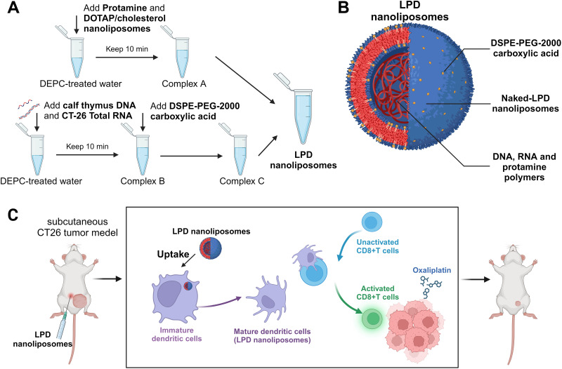
RNA vaccines function by introducing tumor antigen RNA into the body, thereby eliciting an immune response aimed at combating cancer. A critical challenge for this strategy is the safe and efficient delivery of RNA to the target site while minimizing RNA degradation. In response to this challenge, numerous researchers have turned to LNPs as a solution. To effectively induce anti-tumor immunity, Dai et al refined the synthesis of tumor RNA liposome-polycationic DNA complex (LPD) nanoliposomes vaccines, enhancing the total RNA encapsulation capacity for use in a CT-26 colorectal cancer mouse model (Figure 7A and B).192 Their results demonstrated that these nanoparticles could activate dendritic cells (DCs) and T cells, significantly impede tumor growth, and exhibit minimal toxicity to normal organs (Figure 7C). Additionally, Pam2Cys, a synthetic neutral fatty acid, known for its ability to activate the Toll-like receptor (TLR) 2/6 pathway, has been recognized for its potential to trigger both humoral and cellular adaptive immune responses.193 Gu et al developed an innovative antigen mRNA-LNP vaccine incorporating Pam2Cys.194 This vaccine delayed tumor progression and markedly improved survival rates. In the CT26 colon cancer mouse model, it was observed that this novel vaccine eradicated existing tumors in 10% of the subjects. Therefore, LNPs hold significant potential in gene therapy for CRC, particularly in terms of targeting tumors and enhancing immune responses.
Figure 7.
Schematic illustration of RNA-based LPD nanoliposome vaccines.192 (A) The stepwise preparation of the nanoparticles. (B) The components of LPD nanoliposomes. (C) The schematic diagram of LPD nanoliposomes’ effect on DC maturation and T cell activation. In combination with oxaliplatin, the vaccines induce activation of CD8+ T cells and exert anti-tumor effects. Created with BioRender.com.
Conclusion and Perspectives
Lipid-based nanoparticles, particularly SLNs, NLCs, and PLNs, are emerging as a promising platform for CRC therapy. Their attributes include biodegradability, biocompatibility, reduced toxicity, and customizable functionality. LNPs designed for CRC treatment can be administered through various routes, including oral, rectal, intravenous, intratumoral injection, hepatic artery infusion, and intraperitoneal administration. Optimized carrier formulations, appropriate targeting strategies, and suitable administration routes improve the pharmacokinetic and pharmacodynamic properties of LNPs, thereby enhancing their targeted anti-tumor effects. Noteworthy progress has been made with some of these platforms, indicating a bright future for this technology. This article reviews recent published examples of LNPs as nanocarriers for the treatment of colorectal cancer. A wide range of drugs for CRC treatment, including traditional chemotherapy agents, novel anti-cancer drugs, magnetic hyperthermia particles, and nucleic acid medications, have been successfully incorporated into LNPs. These strategies have shown promising results in tumor targeting and anti-tumor efficacy in colorectal cancer models, both in vivo and in vitro. This smart nanoplatform also allows for the loading of various antitumor agents, particularly the combination of novel and traditional drugs, to trigger a potent antitumor response in patients resistant to conventional therapeutic regimens.
Despite rapid advancements in LNPs for cancer therapy, their clinical application still faces obstacles: (1) Industrial scale-up. The complexity of LNP formulation and preparation, characterized by multi-step reactions, leads to inconsistent repeatability, posing a significant challenge for industrial-scale manufacturing. (2) Tumor microenvironment complexity: Current in vitro and in vivo models inadequately mimic the human tumor microenvironment, resulting in less effective clinical outcomes than anticipated. Optimizing LNP formulations and gaining a deeper understanding of the factors influencing their biological distribution are crucial for enhancing treatment efficacy. For instance, adjuvant lipids in mRNA-LNP vaccines have been adopted to improve adaptive immune responses.195 A thorough mechanistic exploration of the structure-activity relationship between various ionizable lipids and LNP distribution in specific organs or cells is also needed. (3) Biocompatibility. Concerns regarding the immunogenicity and toxicity of LNPs still exist. The immune response can accelerate the clearance of LNPs and even lead to serious complications such as hemolysis and thrombosis.196 Lipid components affect the immunogenicity of LNPs, as evidenced by the upregulation differences of various cytokines in LNPs with different ionizable lipids (eg, SM-102 and ALC-0315).197 PEG lipids may trigger PEG antibodies that cause severe hypersensitivity reactions.198,199 Replacing PEG lipids with poly sarcosine lipids (pSar) may address these issues.197 Currently, the toxicity assessment of nanoparticles (NPs) is not fully developed. Unstable NPs may form micrometer-sized aggregates, block capillary beds, and lead to serious complications.200 Injection of LNPs may cause liver or spleen damage and interfere with fatty acid and lipid metabolism.201 Future research should focus on addressing these challenges to fully realize the potential of LNP-based therapies for CRC. This includes developing scalable production methods, understanding and manipulating the tumor microenvironment, improving biocompatibility, and conducting comprehensive long-term safety studies.
In summary, despite some limitations, further research is imperative to refine therapeutic LNPs for CRC treatment, considering the increasing global burden of CRC.202 Progress in this area is vital for the clinical translation of LNPs and for advancing colorectal cancer therapy.
Acknowledgments
We would like to thank the Zhejiang Provincial Natural Science Foundation of China for funding this research work with the project number “LHDMZ23H160003”.
Disclosure
The authors report no conflicts of interest in this work.
References
- 1.Bray F, Ferlay J, Soerjomataram I, Siegel RL, Torre LA, Jemal A. Global cancer statistics 2018: GLOBOCAN estimates of incidence and mortality worldwide for 36 cancers in 185 countries. CA Cancer J Clin. 2018;68(6):394–424. doi: 10.3322/caac.21492 [DOI] [PubMed] [Google Scholar]
- 2.Morgan E, Arnold M, Gini A, et al. Global burden of colorectal cancer in 2020 and 2040: incidence and mortality estimates from GLOBOCAN. Gut. 2023;72(2):338–344. doi: 10.1136/gutjnl-2022-327736 [DOI] [PubMed] [Google Scholar]
- 3.Siegel RL, Wagle NS, Cercek A, Smith RA, Jemal A. Colorectal cancer statistics, 2023. CA Cancer J Clin. 2023;73(3):233–254. doi: 10.3322/caac.21772 [DOI] [PubMed] [Google Scholar]
- 4.Monahan KJ, Bradshaw N, Dolwani S, et al. Guidelines for the management of hereditary colorectal cancer from the British Society of Gastroenterology (BSG)/Association of Coloproctology of Great Britain and Ireland (ACPGBI)/United Kingdom Cancer genetics group (UKCGG). Gut. 2020;69(3):411–444. doi: 10.1136/gutjnl-2019-319915 [DOI] [PMC free article] [PubMed] [Google Scholar]
- 5.Tang J, Lam GT, Brooks RD, et al. Exploring the role of sporadic BRAF and KRAS mutations during colorectal cancer pathogenesis: a spotlight on the contribution of the endosome-lysosome system. Cancer Lett. 2024;585:216639. doi: 10.1016/j.canlet.2024.216639 [DOI] [PubMed] [Google Scholar]
- 6.Smit WL, Spaan CN, Johannes de Boer R, et al. Driver mutations of the adenoma-carcinoma sequence govern the intestinal epithelial global translational capacity. Proc Natl Acad Sci. 2020;117(41):25560–25570. [DOI] [PMC free article] [PubMed] [Google Scholar]
- 7.Rutter MD, Bretthauer M, Hassan C, et al. Principles for evaluation of surveillance after removal of colorectal polyps: recommendations from the World Endoscopy Organization. Gastroenterology. 2020;158(6):1529–1533.e1524. doi: 10.1053/j.gastro.2019.12.052 [DOI] [PubMed] [Google Scholar]
- 8.Punt CJ, Koopman M, Vermeulen L. From tumour heterogeneity to advances in precision treatment of colorectal cancer. Nat Rev Clin Oncol. 2017;14(4):235–246. doi: 10.1038/nrclinonc.2016.171 [DOI] [PubMed] [Google Scholar]
- 9.Cervantes A, Adam R, Roselló S, et al. Metastatic colorectal cancer: ESMO Clinical Practice Guideline for diagnosis, treatment and follow-up☆. Ann Oncol. 2023;34(1):10–32. doi: 10.1016/j.annonc.2022.10.003 [DOI] [PubMed] [Google Scholar]
- 10.Weng J, Li S, Zhu Z, et al. Exploring immunotherapy in colorectal cancer. J Hematol Oncol. 2022;15(1):95. doi: 10.1186/s13045-022-01294-4 [DOI] [PMC free article] [PubMed] [Google Scholar]
- 11.Kroschinsky F, Stölzel F, von Bonin S, et al. New drugs, new toxicities: severe side effects of modern targeted and immunotherapy of cancer and their management. Crit Care. 2017;21(1):89. doi: 10.1186/s13054-017-1678-1 [DOI] [PMC free article] [PubMed] [Google Scholar]
- 12.Xie Y-H, Chen Y-X, Fang J-Y. Comprehensive review of targeted therapy for colorectal cancer. Sig Transd Target Ther. 2020;5(1):22. doi: 10.1038/s41392-020-0116-z [DOI] [PMC free article] [PubMed] [Google Scholar]
- 13.Lund CM, Vistisen KK, Olsen AP, et al. The effect of geriatric intervention in frail older patients receiving chemotherapy for colorectal cancer: a randomised trial (GERICO). Br J Cancer. 2021;124(12):1949–1958. doi: 10.1038/s41416-021-01367-0 [DOI] [PMC free article] [PubMed] [Google Scholar]
- 14.Diaz LA Jr, Shiu KK, Kim TW, et al. Pembrolizumab versus chemotherapy for microsatellite instability-high or mismatch repair-deficient metastatic colorectal cancer (KEYNOTE-177): final analysis of a randomised, open-label, Phase 3 study. Lancet Oncol. 2022;23(5):659–670. doi: 10.1016/S1470-2045(22)00197-8 [DOI] [PMC free article] [PubMed] [Google Scholar]
- 15.McCoubrey LE, Favaron A, Awad A, Orlu M, Gaisford S, Basit AW. Colonic drug delivery: formulating the next generation of colon-targeted therapeutics. J Control Release. 2023;353:1107–1126. doi: 10.1016/j.jconrel.2022.12.029 [DOI] [PubMed] [Google Scholar]
- 16.Bhaskaran NA, Kumar L. Treating colon cancers with a non-conventional yet strategic approach: an overview of various nanoparticulate systems. J Control Release. 2021;336:16–39. doi: 10.1016/j.jconrel.2021.06.008 [DOI] [PubMed] [Google Scholar]
- 17.Dos Santos AM, Carvalho SG, Meneguin AB, Sábio RM, Gremião MPD, Chorilli M. Oral delivery of micro/nanoparticulate systems based on natural polysaccharides for intestinal diseases therapy: challenges, advances and future perspectives. J Control Release. 2021;334:353–366. [DOI] [PubMed] [Google Scholar]
- 18.Mitchell MJ, Billingsley MM, Haley RM, Wechsler ME, Peppas NA, Langer R. Engineering precision nanoparticles for drug delivery. Nat Rev Drug Discov. 2021;20(2):101–124. doi: 10.1038/s41573-020-0090-8 [DOI] [PMC free article] [PubMed] [Google Scholar]
- 19.Zhang Z, Lu Y, Qi J, Wu W. An update on oral drug delivery via intestinal lymphatic transport. Acta Pharmaceutica Sinica B. 2021;11(8):2449–2468. doi: 10.1016/j.apsb.2020.12.022 [DOI] [PMC free article] [PubMed] [Google Scholar]
- 20.Sung YJ, Guo H, Ghasemizadeh A, et al. Cancerous pH-responsive polycarboxybetaine-coated lipid nanoparticle for smart delivery of siRNA against subcutaneous tumor model in mice. Cancer Sci. 2022;113(12):4339–4349. doi: 10.1111/cas.15554 [DOI] [PMC free article] [PubMed] [Google Scholar]
- 21.Wijewantha N, Sane S, Eikanger M, et al. Enhancing anti-tumorigenic efficacy of eugenol in human colon cancer cells using enzyme-responsive nanoparticles. Cancers. 2023;15(4):1145. doi: 10.3390/cancers15041145 [DOI] [PMC free article] [PubMed] [Google Scholar]
- 22.Grifantini R, Taranta M, Gherardini L, et al. Magnetically driven drug delivery systems improving targeted immunotherapy for colon-rectal cancer. J Control Release. 2018;280:76–86. doi: 10.1016/j.jconrel.2018.04.052 [DOI] [PubMed] [Google Scholar]
- 23.Kunjiappan S, Pavadai P, Vellaichamy S, et al. Surface receptor‐mediated targeted drug delivery systems for enhanced cancer treatment: a state-of-the-art review. Drug Dev Res. 2021;82(3):309–340. doi: 10.1002/ddr.21758 [DOI] [PubMed] [Google Scholar]
- 24.Shepherd SJ, Warzecha CC, Yadavali S, et al. Scalable mRNA and siRNA lipid nanoparticle production using a parallelized microfluidic device. Nano Lett. 2021;21(13):5671–5680. doi: 10.1021/acs.nanolett.1c01353 [DOI] [PMC free article] [PubMed] [Google Scholar]
- 25.Eygeris Y, Gupta M, Kim J, Sahay G. Chemistry of Lipid Nanoparticles for RNA Delivery. Acc Chem Res. 2022;55(1):2–12. doi: 10.1021/acs.accounts.1c00544 [DOI] [PubMed] [Google Scholar]
- 26.Schlich M, Palomba R, Costabile G, et al. Cytosolic delivery of nucleic acids: the case of ionizable lipid nanoparticles. Bioeng Transl Med. 2021;6(2):e10213. doi: 10.1002/btm2.10213 [DOI] [PMC free article] [PubMed] [Google Scholar]
- 27.Albertsen CH, Kulkarni JA, Witzigmann D, Lind M, Petersson K, Simonsen JB. The role of lipid components in lipid nanoparticles for vaccines and gene therapy. Adv Drug Delivery Rev. 2022;188:114416. doi: 10.1016/j.addr.2022.114416 [DOI] [PMC free article] [PubMed] [Google Scholar]
- 28.Kiaie SH, Majidi Zolbanin N, Ahmadi A, et al. Recent advances in mRNA-LNP therapeutics: immunological and pharmacological aspects. J Nanobiotechnol. 2022;20(1):276. [DOI] [PMC free article] [PubMed] [Google Scholar]
- 29.Zhang R, Shao S, Piao Y, et al. Esterase-labile quaternium lipidoid enabling improved mRNA-LNP stability and spleen-selective mRNA transfection. Adv Mater. 2023;35(46):2303614. doi: 10.1002/adma.202303614 [DOI] [PubMed] [Google Scholar]
- 30.Han W, Xie B, Li Y, et al. Orally deliverable nanotherapeutics for the synergistic treatment of colitis-associated colorectal cancer. Theranostics. 2019;9(24):7458–7473. doi: 10.7150/thno.38081 [DOI] [PMC free article] [PubMed] [Google Scholar]
- 31.Bajracharya R, Baral KC, Lee SH, Song JG, Han H-K. Organometallic phyllosilicate-gold nanocomplex: an effective oral delivery system of methotrexate for enhanced in vivo efficacy against colorectal cancer. Int j Nanomed. 2023;18:7257–7266. doi: 10.2147/IJN.S437860 [DOI] [PMC free article] [PubMed] [Google Scholar]
- 32.Manzari MT, Shamay Y, Kiguchi H, Rosen N, Scaltriti M, Heller DA. Targeted drug delivery strategies for precision medicines. Nature Rev Mater. 2021;6(4):351–370. doi: 10.1038/s41578-020-00269-6 [DOI] [PMC free article] [PubMed] [Google Scholar]
- 33.Ejazi SA, Louisthelmy R, Maisel K. Mechanisms of nanoparticle transport across intestinal tissue: an oral delivery perspective. ACS nano. 2023;17(14):13044–13061. doi: 10.1021/acsnano.3c02403 [DOI] [PubMed] [Google Scholar]
- 34.Marx JL. Liposomes: research applications grow. Science. 1978;199(4333):1056–1128. doi: 10.1126/science.199.4333.1056 [DOI] [PubMed] [Google Scholar]
- 35.Fulton MD, Najahi-Missaoui W. Liposomes in cancer therapy: how did we start and where are we now. Int J Mol Sci. 2023;24(7):6615. doi: 10.3390/ijms24076615 [DOI] [PMC free article] [PubMed] [Google Scholar]
- 36.Mirchandani Y, Patravale VB, Brijesh S. Solid lipid nanoparticles for hydrophilic drugs. J Control Release. 2021;335:457–464. doi: 10.1016/j.jconrel.2021.05.032 [DOI] [PubMed] [Google Scholar]
- 37.Truong TH, Alcantara KP, Bulatao BPI, et al. Chitosan-coated nanostructured lipid carriers for transdermal delivery of tetrahydrocurcumin for breast cancer therapy. Carbohydr Polym. 2022;288:119401. doi: 10.1016/j.carbpol.2022.119401 [DOI] [PubMed] [Google Scholar]
- 38.Beloqui A, Solinís M, Rodríguez-Gascón A, Almeida AJ, Préat V. Nanostructured lipid carriers: promising drug delivery systems for future clinics. Nanomedicine. 2016;12(1):143–161. doi: 10.1016/j.nano.2015.09.004 [DOI] [PubMed] [Google Scholar]
- 39.Tkach M, Théry C. Communication by extracellular vesicles: where we are and where we need to go. Cell. 2016;164(6):1226–1232. doi: 10.1016/j.cell.2016.01.043 [DOI] [PubMed] [Google Scholar]
- 40.Wagner J, Gößl D, Ustyanovska N, et al. Mesoporous silica nanoparticles as pH-responsive carrier for the immune-activating drug resiquimod enhance the local immune response in mice. ACS nano. 2021;15(3):4450–4466. doi: 10.1021/acsnano.0c08384 [DOI] [PubMed] [Google Scholar]
- 41.Xu R, Rai A, Chen M, Suwakulsiri W, Greening DW, Simpson RJ. Extracellular vesicles in cancer—implications for future improvements in cancer care. Nat Rev Clin Oncol. 2018;15(10):617–638. doi: 10.1038/s41571-018-0036-9 [DOI] [PubMed] [Google Scholar]
- 42.Tenchov R, Bird R, Curtze AE, Zhou Q. Lipid nanoparticles─from liposomes to mRNA vaccine delivery, a landscape of research diversity and advancement. ACS nano. 2021;15(11):16982–17015. doi: 10.1021/acsnano.1c04996 [DOI] [PubMed] [Google Scholar]
- 43.Tenchov R, Sasso JM, Wang X, Liaw W-S, Chen C-A, Zhou QA. Exosomes─ nature’s lipid nanoparticles, a rising star in drug delivery and diagnostics. ACS nano. 2022;16(11):17802–17846. doi: 10.1021/acsnano.2c08774 [DOI] [PMC free article] [PubMed] [Google Scholar]
- 44.Sivadasan D, Sultan MH, Madkhali O, Almoshari Y, Thangavel N. Polymeric Lipid Hybrid Nanoparticles (PLNs) as emerging drug delivery platform-a comprehensive review of their properties, preparation methods, and therapeutic applications. Pharmaceutics. 2021;13(8):1291. doi: 10.3390/pharmaceutics13081291 [DOI] [PMC free article] [PubMed] [Google Scholar]
- 45.Shrivastava P, Gautam L, Jain A, Vishwakarma N, Vyas S, Vyas SP. Lipid drug conjugates for improved therapeutic benefits. Curr Pharm Des. 2020;26(27):3187–3202. doi: 10.2174/1381612826666200311124003 [DOI] [PubMed] [Google Scholar]
- 46.Cullis PR, Hope MJ. Lipid nanoparticle systems for enabling gene therapies. Mol Ther. 2017;25(7):1467–1475. doi: 10.1016/j.ymthe.2017.03.013 [DOI] [PMC free article] [PubMed] [Google Scholar]
- 47.Evers MJ, Kulkarni JA, van der Meel R, Cullis PR, Vader P, Schiffelers RM. State‐of‐the‐art design and rapid‐mixing production techniques of lipid nanoparticles for nucleic acid delivery. Small Methods. 2018;2(9):1700375. doi: 10.1002/smtd.201700375 [DOI] [Google Scholar]
- 48.Dutta K, Das R, Medeiros J, Kanjilal P, Thayumanavan S. Charge‐conversion strategies for nucleic acid delivery. Adv Funct Mater. 2021;31(24):2011103. doi: 10.1002/adfm.202011103 [DOI] [PMC free article] [PubMed] [Google Scholar]
- 49.Kara G, Calin GA, Ozpolat B. RNAi-based therapeutics and tumor targeted delivery in cancer. Adv Drug Delivery Rev. 2022;182:114113. doi: 10.1016/j.addr.2022.114113 [DOI] [PubMed] [Google Scholar]
- 50.Kulkarni JA, Witzigmann D, Thomson SB, et al. The current landscape of nucleic acid therapeutics. Nature Nanotechnol. 2021;16(6):630–643. doi: 10.1038/s41565-021-00898-0 [DOI] [PubMed] [Google Scholar]
- 51.Han X, Zhang H, Butowska K, et al. An ionizable lipid toolbox for RNA delivery. Nat Commun. 2021;12(1):7233. doi: 10.1038/s41467-021-27493-0 [DOI] [PMC free article] [PubMed] [Google Scholar]
- 52.Zhang Y, Sun C, Wang C, Jankovic KE, Dong Y. Lipids and lipid derivatives for RNA delivery. Chem Rev. 2021;121(20):12181–12277. doi: 10.1021/acs.chemrev.1c00244 [DOI] [PMC free article] [PubMed] [Google Scholar]
- 53.Ding F, Zhang H, Cui J, Li Q, Yang C. Boosting ionizable lipid nanoparticle-mediated in vivo mRNA delivery through optimization of lipid amine-head groups. Biomater Sci. 2021;9(22):7534–7546. doi: 10.1039/D1BM00866H [DOI] [PubMed] [Google Scholar]
- 54.Zhi D, Bai Y, Yang J, et al. A review on cationic lipids with different linkers for gene delivery. Adv Colloid Interface Sci. 2018;253:117–140. doi: 10.1016/j.cis.2017.12.006 [DOI] [PubMed] [Google Scholar]
- 55.Yu H, Dyett B, Kirby N, et al. pH‐Dependent Lyotropic Liquid Crystalline Mesophase and Ionization Behavior of Phytantriol‐Based Ionizable Lipid Nanoparticles. Small. 2024;20(20):2309200. doi: 10.1002/smll.202309200 [DOI] [PubMed] [Google Scholar]
- 56.Kulkarni JA, Witzigmann D, Leung J, Tam YYC, Cullis PR. On the role of helper lipids in lipid nanoparticle formulations of siRNA. Nanoscale. 2019;11(45):21733–21739. doi: 10.1039/C9NR09347H [DOI] [PubMed] [Google Scholar]
- 57.Zhang R, El-Mayta R, Murdoch TJ, et al. Helper lipid structure influences protein adsorption and delivery of lipid nanoparticles to spleen and liver. Biomater Sci. 2021;9(4):1449–1463. doi: 10.1039/D0BM01609H [DOI] [PMC free article] [PubMed] [Google Scholar]
- 58.Chen W, Duša F, Witos J, Ruokonen SK, Wiedmer SK. Determination of the main phase transition temperature of phospholipids by nanoplasmonic sensing. Sci Rep. 2018;8(1):14815. doi: 10.1038/s41598-018-33107-5 [DOI] [PMC free article] [PubMed] [Google Scholar]
- 59.Manni LS, Davies C, Wood K, Assenza S, Atkin R, Warr GG. Unusual phosphatidylcholine lipid phase behavior in the ionic liquid ethylammonium nitrate. J Colloid Interface Sci. 2023;643:276–281. doi: 10.1016/j.jcis.2023.03.161 [DOI] [PubMed] [Google Scholar]
- 60.Cheng Q, Wei T, Jia Y, et al. Dendrimer-based lipid nanoparticles deliver therapeutic FAH mRNA to normalize liver function and extend survival in a mouse model of hepatorenal tyrosinemia type I. Adv Mater. 2018;30(52):e1805308. [DOI] [PubMed] [Google Scholar]
- 61.Geng L, Kato N, Kodama Y, Mukai H, Kawakami S. Influence of lipid composition of messenger RNA-loaded lipid nanoparticles on the protein expression via intratracheal administration in mice. Int J Pharm. 2023;637:122896. doi: 10.1016/j.ijpharm.2023.122896 [DOI] [PubMed] [Google Scholar]
- 62.Gretskaya N, Akimov M, Andreev D, et al. Multicomponent lipid nanoparticles for RNA transfection. Pharmaceutics. 2023;15(4):1289. doi: 10.3390/pharmaceutics15041289 [DOI] [PMC free article] [PubMed] [Google Scholar]
- 63.Patel S, Ashwanikumar N, Robinson E, et al. Naturally-occurring cholesterol analogues in lipid nanoparticles induce polymorphic shape and enhance intracellular delivery of mRNA. Nat Commun. 2020;11(1):983. doi: 10.1038/s41467-020-14527-2 [DOI] [PMC free article] [PubMed] [Google Scholar]
- 64.Kawaguchi M, Noda M, Ono A, et al. Effect of cholesterol content of lipid composition in mRNA-LNPs on the protein expression in the injected site and liver after local administration in mice. J Pharmaceut Sci. 2023;112(5):1401–1410. doi: 10.1016/j.xphs.2022.12.026 [DOI] [PMC free article] [PubMed] [Google Scholar]
- 65.Patel SK, Billingsley MM, Frazee C, et al. Hydroxycholesterol substitution in ionizable lipid nanoparticles for mRNA delivery to T cells. J Control Release. 2022;347:521–532. doi: 10.1016/j.jconrel.2022.05.020 [DOI] [PMC free article] [PubMed] [Google Scholar]
- 66.Paunovska K, Gil CJ, Lokugamage MP, et al. Analyzing 2000 in vivo drug delivery data points reveals cholesterol structure impacts nanoparticle delivery. ACS nano. 2018;12(8):8341–8349. doi: 10.1021/acsnano.8b03640 [DOI] [PMC free article] [PubMed] [Google Scholar]
- 67.Xiao Y, Shi J. Lipids and the emerging RNA medicines. Chem Rev. 2021;121(20):12109–12111. doi: 10.1021/acs.chemrev.1c00778 [DOI] [PubMed] [Google Scholar]
- 68.Oziri OJ, Wang Y, Watanabe T, et al. PEGylation of silver nanoparticles by physisorption of cyclic poly (ethylene glycol) for enhanced dispersion stability, antimicrobial activity, and cytotoxicity. Nanoscale Adv. 2022;4(2):532–545. doi: 10.1039/d1na00720c [DOI] [PMC free article] [PubMed] [Google Scholar]
- 69.Wang H, Zhao P, Liang X, et al. Folate-PEG coated cationic modified chitosan--cholesterol liposomes for tumor-targeted drug delivery. Biomaterials. 2010;31(14):4129–4138. doi: 10.1016/j.biomaterials.2010.01.089 [DOI] [PubMed] [Google Scholar]
- 70.Tenchov R, Sasso JM, Zhou QA. PEGylated lipid nanoparticle formulations: immunological safety and efficiency perspective. Bioconjugate Chem. 2023;34(6):941–960. doi: 10.1021/acs.bioconjchem.3c00174 [DOI] [PMC free article] [PubMed] [Google Scholar]
- 71.Fang Y, Xue J, Gao S, et al. Cleavable PEGylation: a strategy for overcoming the “PEG dilemma” in efficient drug delivery. Drug Delivery. 2017;24(sup1):22–32. doi: 10.1080/10717544.2017.1388451 [DOI] [PMC free article] [PubMed] [Google Scholar]
- 72.Pannuzzo M, Esposito S, L-P W, et al. Overcoming nanoparticle-mediated complement activation by surface PEG pairing. Nano Lett. 2020;20(6):4312–4321. doi: 10.1021/acs.nanolett.0c01011 [DOI] [PubMed] [Google Scholar]
- 73.LoPresti ST, Arral ML, Chaudhary N, Whitehead KA. The replacement of helper lipids with charged alternatives in lipid nanoparticles facilitates targeted mRNA delivery to the spleen and lungs. J Control Release. 2022;345:819–831. doi: 10.1016/j.jconrel.2022.03.046 [DOI] [PMC free article] [PubMed] [Google Scholar]
- 74.Lankveld DP, Rayavarapu RG, Krystek P, et al. Blood clearance and tissue distribution of PEGylated and non-PEGylated gold nanorods after intravenous administration in rats. Nanomedicine. 2011;6(2):339–349. doi: 10.2217/nnm.10.122 [DOI] [PubMed] [Google Scholar]
- 75.Escalona-Rayo O, Papadopoulou P, Slütter B, Kros A. Biological recognition and cellular trafficking of targeted RNA-lipid nanoparticles. Curr Opin Biotechnol. 2024;85:103041. doi: 10.1016/j.copbio.2023.103041 [DOI] [PubMed] [Google Scholar]
- 76.Boehnke N, Straehla JP, Safford HC, et al. Massively parallel pooled screening reveals genomic determinants of nanoparticle delivery. Science. 2022;377(6604):eabm5551. doi: 10.1126/science.abm5551 [DOI] [PMC free article] [PubMed] [Google Scholar]
- 77.Nussinov R, Tsai C-J, Jang H. Anticancer drug resistance: an update and perspective. Drug Resist Updates. 2021;59:100796. doi: 10.1016/j.drup.2021.100796 [DOI] [PMC free article] [PubMed] [Google Scholar]
- 78.Sun C, Wang Z, Yang K, et al. Polyamine-responsive morphological transformation of a supramolecular peptide for specific drug accumulation and retention in cancer cells. Small. 2021;17(43):2101139. [DOI] [PubMed] [Google Scholar]
- 79.Bhaskaran NA, Jitta SR, Cheruku S, Kumar N, Kumar L. Orally delivered solid lipid nanoparticles of irinotecan coupled with chitosan surface modification to treat colon cancer: preparation, in-vitro and in-vivo evaluations. Int J Biol Macromol. 2022;211:301–315. doi: 10.1016/j.ijbiomac.2022.05.060 [DOI] [PubMed] [Google Scholar]
- 80.Bose S, Panda AK, Mukherjee S, Sa G. Curcumin and tumor immune-editing: resurrecting the immune system. Cell Division. 2015;10(1):1–13. doi: 10.1186/s13008-015-0012-z [DOI] [PMC free article] [PubMed] [Google Scholar]
- 81.Cisterna BA, Kamaly N, Choi WI, Tavakkoli A, Farokhzad OC, Vilos C. Targeted nanoparticles for colorectal cancer. Nanomedicine. 2016;11(18):2443–2456. doi: 10.2217/nnm-2016-0194 [DOI] [PMC free article] [PubMed] [Google Scholar]
- 82.Wong KE, Ngai SC, Chan K-G, Lee L-H, Goh B-H, Chuah L-H. Curcumin nanoformulations for colorectal cancer: a review. Front Pharmacol. 2019;10:152. [DOI] [PMC free article] [PubMed] [Google Scholar]
- 83.Pavitra E, Dariya B, Srivani G, et al. Engineered nanoparticles for imaging and drug delivery in colorectal cancer. Paper presented at: Seminars in cancer Biology; 2021. [DOI] [PubMed] [Google Scholar]
- 84.Wang X, Almoallim HS, Cui Q, Alharbi SA, Yang H. In situ decorated Au NPs on chitosan-encapsulated Fe3O4-NH2 NPs as magnetic nanocomposite: investigation of its anti-colon carcinoma, anti-gastric cancer and anti-pancreatic cancer. Int J Biol Macromol. 2021;171:198–207. [DOI] [PubMed] [Google Scholar]
- 85.Tang W-L, Tang W-H, S-D L. Cancer theranostic applications of lipid-based nanoparticles. Drug Discovery Today. 2018;23(5):1159–1166. doi: 10.1016/j.drudis.2018.04.007 [DOI] [PubMed] [Google Scholar]
- 86.Nakamura T, Sato Y, Yamada Y, et al. Extrahepatic targeting of lipid nanoparticles in vivo with intracellular targeting for future nanomedicines. Adv Drug Delivery Rev. 2022;188:114417. [DOI] [PubMed] [Google Scholar]
- 87.Yang T, Poenisch M, Khanal R, et al. Therapeutic HNF4A mRNA attenuates liver fibrosis in a preclinical model. J Hepatol. 2021;75(6):1420–1433. doi: 10.1016/j.jhep.2021.08.011 [DOI] [PubMed] [Google Scholar]
- 88.Liu K, Nilsson R, Lázaro-Ibáñez E, et al. Multiomics analysis of naturally efficacious lipid nanoparticle coronas reveals high-density lipoprotein is necessary for their function. Nat Commun. 2023;14(1):4007. doi: 10.1038/s41467-023-39768-9 [DOI] [PMC free article] [PubMed] [Google Scholar]
- 89.Paunovska K, Da Silva Sanchez AJ, Lokugamage MP, et al. The extent to which lipid nanoparticles require apolipoprotein e and low-density lipoprotein receptor for delivery changes with ionizable lipid structure. Nano Lett. 2022;22(24):10025–10033. doi: 10.1021/acs.nanolett.2c03741 [DOI] [PubMed] [Google Scholar]
- 90.Lokugamage MP, Vanover D, Beyersdorf J, et al. Optimization of lipid nanoparticles for the delivery of nebulized therapeutic mRNA to the lungs. Nat Biomed Eng. 2021;5(9):1059–1068. doi: 10.1038/s41551-021-00786-x [DOI] [PMC free article] [PubMed] [Google Scholar]
- 91.Din FU, Mustapha O, Kim DW, et al. Novel dual-reverse thermosensitive solid lipid nanoparticle-loaded hydrogel for rectal administration of flurbiprofen with improved bioavailability and reduced initial burst effect. Eur J Pharm Biopharm. 2015;94:64–72. doi: 10.1016/j.ejpb.2015.04.019 [DOI] [PubMed] [Google Scholar]
- 92.Kim J, Song S, Gwak M, et al. Micro-syringe chip-guided intratumoral administration of lipid nanoparticles for targeted anticancer therapy. Biomater Res. 2023;27(1):102. doi: 10.1186/s40824-023-00440-4 [DOI] [PMC free article] [PubMed] [Google Scholar]
- 93.Heidel JD, Yu Z, Liu JY, et al. Administration in non-human primates of escalating intravenous doses of targeted nanoparticles containing ribonucleotide reductase subunit M2 siRNA. Proc Natl Acad Sci U S A. 2007;104(14):5715–5721. doi: 10.1073/pnas.0701458104 [DOI] [PMC free article] [PubMed] [Google Scholar]
- 94.Heidel JD, Liu JY, Yen Y, et al. Potent siRNA inhibitors of ribonucleotide reductase subunit RRM2 reduce cell proliferation in vitro and in vivo. Clin Cancer Res. 2007;13(7):2207–2215. doi: 10.1158/1078-0432.CCR-06-2218 [DOI] [PubMed] [Google Scholar]
- 95.Bartlett DW, Davis ME. Impact of tumor-specific targeting and dosing schedule on tumor growth inhibition after intravenous administration of siRNA-containing nanoparticles. Biotechnol Bioeng. 2008;99(4):975–985. doi: 10.1002/bit.21668 [DOI] [PubMed] [Google Scholar]
- 96.Heidel JD. Linear cyclodextrin-containing polymers and their use as delivery agents. Expert Opin Drug Delivery. 2006;3(5):641–646. doi: 10.1517/17425247.3.5.641 [DOI] [PubMed] [Google Scholar]
- 97.Steegmaier M, Hoffmann M, Baum A, et al. BI 2536, a potent and selective inhibitor of polo-like kinase 1, inhibits tumor growth in vivo. Curr Biol. 2007;17(4):316–322. doi: 10.1016/j.cub.2006.12.037 [DOI] [PubMed] [Google Scholar]
- 98.Strebhardt K, Ullrich A. Targeting polo-like kinase 1 for cancer therapy. Nat Rev Cancer. 2006;6(4):321–330. doi: 10.1038/nrc1841 [DOI] [PubMed] [Google Scholar]
- 99.Barr FA, Silljé HH, Nigg EA. Polo-like kinases and the orchestration of cell division. Nat Rev Mol Cell Biol. 2004;5(6):429–440. doi: 10.1038/nrm1401 [DOI] [PubMed] [Google Scholar]
- 100.Prasad K, Badarinath A, Anilkumar P, et al. Colon targeted drug delivery systems: a review. J Global Trends Pharm Sci. 2011;2(4):459–475. [Google Scholar]
- 101.Hua S. Advances in oral drug delivery for regional targeting in the gastrointestinal tract - influence of physiological, pathophysiological and pharmaceutical factors. Front Pharmacol. 2020;11:524. doi: 10.3389/fphar.2020.00524 [DOI] [PMC free article] [PubMed] [Google Scholar]
- 102.Martinez MN, Amidon GL. A mechanistic approach to understanding the factors affecting drug absorption: a review of fundamentals. J Clin Pharmacol. 2002;42(6):620–643. doi: 10.1177/00970002042006005 [DOI] [PubMed] [Google Scholar]
- 103.Reinus J, Simon D. Gastrointestinal Anatomy and Physiology. Wiley Online Library; 2014. [Google Scholar]
- 104.Naeem M, Oshi MA, Kim J, et al. pH-triggered surface charge-reversal nanoparticles alleviate experimental murine colitis via selective accumulation in inflamed colon regions. Nanomedicine. 2018;14(3):823–834. doi: 10.1016/j.nano.2018.01.003 [DOI] [PubMed] [Google Scholar]
- 105.Musa N, Wong TW. Design of polysaccharidic nano-in-micro soft agglomerates as primary oral drug delivery vehicle for colon-specific targeting. Carbohydr Polym. 2020;247:116673. doi: 10.1016/j.carbpol.2020.116673 [DOI] [PubMed] [Google Scholar]
- 106.Ying K, Bai B, Gao X, Xu Y, Wang H, Xie B. Orally administrable therapeutic nanoparticles for the treatment of colorectal cancer. Front Bioeng Biotechnol. 2021;9:670124. doi: 10.3389/fbioe.2021.670124 [DOI] [PMC free article] [PubMed] [Google Scholar]
- 107.Wang S-Y, Meng Y-J, Li J, J-p L, Liu Z-Q, Li D-Q. A novel and simple oral colon-specific drug delivery system based on the pectin/modified nano-carbon sphere nanocomposite gel films. Int J Biol Macromol. 2020;157:170–176. doi: 10.1016/j.ijbiomac.2020.04.197 [DOI] [PubMed] [Google Scholar]
- 108.Griffin BT, Guo J, Presas E, Donovan MD, Alonso MJ, O’Driscoll CM. Pharmacokinetic, pharmacodynamic and biodistribution following oral administration of nanocarriers containing peptide and protein drugs. Adv Drug Delivery Rev. 2016;106:367–380. doi: 10.1016/j.addr.2016.06.006 [DOI] [PubMed] [Google Scholar]
- 109.Jain AK, Jain S. Advances in oral delivery of anti-cancer prodrugs. Expert Opin Drug Delivery. 2016;13(12):1759–1775. doi: 10.1080/17425247.2016.1200554 [DOI] [PubMed] [Google Scholar]
- 110.He Y, Cheng M, Yang R, et al. Research progress on the mechanism of nanoparticles crossing the intestinal epithelial cell membrane. Pharmaceutics. 2023;15(7):1816. doi: 10.3390/pharmaceutics15071816 [DOI] [PMC free article] [PubMed] [Google Scholar]
- 111.Takakura Y, Takahashi Y. Strategies for persistent retention of macromolecules and nanoparticles in the blood circulation. J Control Release. 2022;350:486–493. doi: 10.1016/j.jconrel.2022.05.063 [DOI] [PubMed] [Google Scholar]
- 112.Montanha MC, Howarth A, Mohamed DA, et al. A physiologically based pharmacokinetic model to predict pegylated liposomal doxorubicin disposition in rats and human. Drug Delivery Transl Res. 2022;12(9):2178–2186. doi: 10.1007/s13346-022-01175-w [DOI] [PubMed] [Google Scholar]
- 113.Crommelin DJ, van Hoogevest P, Storm G. The role of liposomes in clinical nanomedicine development. What now? Now what? J Control Release. 2020;318:256–263. doi: 10.1016/j.jconrel.2019.12.023 [DOI] [PubMed] [Google Scholar]
- 114.Jaradat E, Meziane A, Lamprou DA. Conventional vs PEGylated loaded liposomal formulations by microfluidics for delivering hydrophilic chemotherapy. Int J Pharm. 2024;655:124077. doi: 10.1016/j.ijpharm.2024.124077 [DOI] [PubMed] [Google Scholar]
- 115.Waggoner LE, Miyasaki KF, Kwon EJ. Analysis of PEG-lipid anchor length on lipid nanoparticle pharmacokinetics and activity in a mouse model of traumatic brain injury. Biomater Sci. 2023;11(12):4238–4253. doi: 10.1039/d2bm01846b [DOI] [PMC free article] [PubMed] [Google Scholar]
- 116.Shen MY, Liu TI, Yu TW, et al. Hierarchically targetable polysaccharide-coated solid lipid nanoparticles as an oral chemo/thermotherapy delivery system for local treatment of colon cancer. Biomaterials. 2019;197:86–100. doi: 10.1016/j.biomaterials.2019.01.019 [DOI] [PubMed] [Google Scholar]
- 117.Szymańska E, Winnicka K. Stability of chitosan-a challenge for pharmaceutical and biomedical applications. Mar Drugs. 2015;13(4):1819–1846. doi: 10.3390/md13041819 [DOI] [PMC free article] [PubMed] [Google Scholar]
- 118.Leonard TE, Liko AF, Gustiananda M, Putra ABN, Juanssilfero AB, Hartrianti P. Thiolated pectin-chitosan composites: potential mucoadhesive drug delivery system with selective cytotoxicity towards colorectal cancer. Int J Biol Macromol. 2023;225:1–12. doi: 10.1016/j.ijbiomac.2022.12.012 [DOI] [PubMed] [Google Scholar]
- 119.Wang J, Li Y, Nie G. Multifunctional biomolecule nanostructures for cancer therapy. Nature Rev Mater. 2021;6(9):766–783. doi: 10.1038/s41578-021-00315-x [DOI] [PMC free article] [PubMed] [Google Scholar]
- 120.Mundekkad D, Cho WC. Nanoparticles in clinical translation for cancer therapy. Int J Mol Sci. 2022;23(3):1685. doi: 10.3390/ijms23031685 [DOI] [PMC free article] [PubMed] [Google Scholar]
- 121.Cheng Q, Wei T, Farbiak L, Johnson LT, Dilliard SA, Siegwart DJ. Selective organ targeting (SORT) nanoparticles for tissue-specific mRNA delivery and CRISPR-Cas gene editing. Nature Nanotechnol. 2020;15(4):313–320. doi: 10.1038/s41565-020-0669-6 [DOI] [PMC free article] [PubMed] [Google Scholar]
- 122.Wang Q, Liang Q, Dou J, et al. Breaking through the basement membrane barrier to improve nanotherapeutic delivery to tumours. Nature Nanotechnol. 2023;19(1):95–105. doi: 10.1038/s41565-023-01498-w [DOI] [PubMed] [Google Scholar]
- 123.Marques A, Costa P, Velho S, Amaral M. Functionalizing nanoparticles with cancer-targeting antibodies: a comparison of strategies. J Control Release. 2020;320:180–200. doi: 10.1016/j.jconrel.2020.01.035 [DOI] [PubMed] [Google Scholar]
- 124.Jiang Z, Guan J, Qian J, Zhan C. Peptide ligand-mediated targeted drug delivery of nanomedicines. Biomater Sci. 2019;7(2):461–471. doi: 10.1039/C8BM01340C [DOI] [PubMed] [Google Scholar]
- 125.Stefanick JF, Omstead DT, Kiziltepe T, Bilgicer B. Dual-receptor targeted strategy in nanoparticle design achieves tumor cell selectivity through cooperativity. Nanoscale. 2019;11(10):4414–4427. doi: 10.1039/C8NR09431D [DOI] [PubMed] [Google Scholar]
- 126.Wang X, Qiu Y, Wang M, et al. Endocytosis and organelle targeting of nanomedicines in cancer therapy. Int j Nanomed. 2020;15:9447–9467. doi: 10.2147/IJN.S274289 [DOI] [PMC free article] [PubMed] [Google Scholar]
- 127.Moulahoum H, Ghorbanizamani F, Zihnioglu F, Timur S. Surface biomodification of liposomes and polymersomes for efficient targeted drug delivery. Bioconjugate Chem. 2021;32(8):1491–1502. doi: 10.1021/acs.bioconjchem.1c00285 [DOI] [PubMed] [Google Scholar]
- 128.Tiernan JP, Perry SL, Verghese ET, et al. Carcinoembryonic antigen is the preferred biomarker for in vivo colorectal cancer targeting. Br J Cancer. 2013;108(3):662–667. doi: 10.1038/bjc.2012.605 [DOI] [PMC free article] [PubMed] [Google Scholar]
- 129.Shia J, Klimstra DS, Nitzkorski JR, et al. Immunohistochemical expression of folate receptor alpha in colorectal carcinoma: patterns and biological significance. Human Pathol. 2008;39(4):498–505. doi: 10.1016/j.humpath.2007.09.013 [DOI] [PubMed] [Google Scholar]
- 130.Govindarasu M, Abirami P, Alharthi SS, Thiruvengadam M, Rajakumar G, Vaiyapuri M. Synthesis, physicochemical characterization, and in vitro evaluation of biodegradable PLGA nanoparticles entrapped to folic acid for targeted delivery of kaempferitrin. Biotechnol. Appl. Biochem. 2022;69(6):2387–2398. doi: 10.1002/bab.2290 [DOI] [PubMed] [Google Scholar]
- 131.Zu M, Ma L, Zhang X, Xie D, Kang Y, Xiao B. Chondroitin sulfate-functionalized polymeric nanoparticles for colon cancer-targeted chemotherapy. Colloids Surf B. 2019;177:399–406. doi: 10.1016/j.colsurfb.2019.02.031 [DOI] [PubMed] [Google Scholar]
- 132.Chang DK, Chiu CY, Kuo SY, et al. Antiangiogenic targeting liposomes increase therapeutic efficacy for solid tumors. J Biol Chem. 2009;284(19):12905–12916. doi: 10.1074/jbc.M900280200 [DOI] [PMC free article] [PubMed] [Google Scholar]
- 133.Katanasaka Y, Ishii T, Asai T, et al. Cancer antineovascular therapy with liposome drug delivery systems targeted to BiP/GRP78. Int J Cancer. 2010;127(11):2685–2698. doi: 10.1002/ijc.25276 [DOI] [PubMed] [Google Scholar]
- 134.Murase Y, Asai T, Katanasaka Y, et al. A novel DDS strategy, ”dual-targeting”, and its application for antineovascular therapy. Cancer Lett. 2010;287(2):165–171. doi: 10.1016/j.canlet.2009.06.008 [DOI] [PubMed] [Google Scholar]
- 135.Zorko M, Jones S, Langel Ü. Cell-penetrating peptides in protein mimicry and cancer therapeutics. Adv Drug Delivery Rev. 2022;180:114044. doi: 10.1016/j.addr.2021.114044 [DOI] [PubMed] [Google Scholar]
- 136.Kuai R, Yuan W, Li W, et al. Targeted delivery of cargoes into a murine solid tumor by a cell-penetrating peptide and cleavable poly(ethylene glycol) comodified liposomal delivery system via systemic administration. Mol Pharmaceut. 2011;8(6):2151–2161. doi: 10.1021/mp200100f [DOI] [PubMed] [Google Scholar]
- 137.Ludwig BS, Kessler H, Kossatz S, Reuning U. RGD-binding integrins revisited: how recently discovered functions and novel synthetic Ligands (Re-)Shape an ever-evolving field. Cancers. 2021;13(7):1711. doi: 10.3390/cancers13071711 [DOI] [PMC free article] [PubMed] [Google Scholar]
- 138.Hamidi H, Ivaska J. Every step of the way: integrins in cancer progression and metastasis. Nat Rev Cancer. 2018;18(9):533–548. doi: 10.1038/s41568-018-0038-z [DOI] [PMC free article] [PubMed] [Google Scholar]
- 139.Liu XY, Ruan LM, Mao WW, Wang JQ, Shen YQ, Sui MH. Preparation of RGD-modified long circulating liposome loading matrine, and its in vitro anti-cancer effects. Int J Med Sci. 2010;7(4):197–208. doi: 10.7150/ijms.7.197 [DOI] [PMC free article] [PubMed] [Google Scholar]
- 140.Kokkoli E, Mardilovich A, Wedekind A, Rexeisen EL, Garg A, Craig JA. Self-assembly and applications of biomimetic and bioactive peptide-amphiphiles. Soft Matter. 2006;2(12):1015–1024. doi: 10.1039/b608929a [DOI] [PubMed] [Google Scholar]
- 141.Pangburn TO, Bates FS, Kokkoli E. Polymersomes functionalized via “click” chemistry with the fibronectin mimetic peptides PR_b and GRGDSP for targeted delivery to cells with different levels of α5β1 expression. Soft Matter. 2012;8(16):4449–4461. doi: 10.1039/c2sm06922a [DOI] [Google Scholar]
- 142.Garg A, Kokkoli E. pH-Sensitive PEGylated liposomes functionalized with a fibronectin-mimetic peptide show enhanced intracellular delivery to colon cancer cell. Current Pharm Biotechnol. 2011;12(8):1135–1143. doi: 10.2174/138920111796117328 [DOI] [PubMed] [Google Scholar]
- 143.Garg A, Tisdale AW, Haidari E, Kokkoli E. Targeting colon cancer cells using PEGylated liposomes modified with a fibronectin-mimetic peptide. Int J Pharm. 2009;366(1–2):201–210. doi: 10.1016/j.ijpharm.2008.09.016 [DOI] [PMC free article] [PubMed] [Google Scholar]
- 144.Demirgöz D, Garg A, Kokkoli E. PR_b-targeted PEGylated liposomes for prostate cancer therapy. Langmuir. 2008;24(23):13518–13524. doi: 10.1021/la801961r [DOI] [PubMed] [Google Scholar]
- 145.Mi P. Stimuli-responsive nanocarriers for drug delivery, tumor imaging, therapy and theranostics. Theranostics. 2020;10(10):4557–4588. doi: 10.7150/thno.38069 [DOI] [PMC free article] [PubMed] [Google Scholar]
- 146.Ahn H, Park JH. Liposomal delivery systems for intestinal lymphatic drug transport. Biomater Res. 2016;20(1):36. doi: 10.1186/s40824-016-0083-1 [DOI] [PMC free article] [PubMed] [Google Scholar]
- 147.Liu C, Zhang W, Wang J, Si T, Xing W. Tumor-associated macrophage-derived transforming growth factor-β promotes colorectal cancer progression through HIF1-TRIB3 signaling. Cancer Sci. 2021;112(10):4198–4207. doi: 10.1111/cas.15101 [DOI] [PMC free article] [PubMed] [Google Scholar]
- 148.Tang Y, Weng X, Liu C, Li X, Chen C, Cantara S. Hypoxia enhances activity and malignant behaviors of colorectal cancer cells through the STAT3/MicroRNA-19a/PTEN/PI3K/AKT axis. Anal Cell Pathol. 2021;2021:4132488. doi: 10.1155/2021/4132488 [DOI] [PMC free article] [PubMed] [Google Scholar]
- 149.Mu M, Zhang Q, Li J, et al. USP51 facilitates colorectal cancer stemness and chemoresistance by forming a positive feed-forward loop with HIF1A. Cell Death Differ. 2023;30(11):2393–2407. doi: 10.1038/s41418-023-01228-8 [DOI] [PMC free article] [PubMed] [Google Scholar]
- 150.Li J, Yang P, Chen F, et al. Hypoxic colorectal cancer-derived extracellular vesicles deliver microRNA-361-3p to facilitate cell proliferation by targeting TRAF3 via the noncanonical NF-κB pathways. Clin Transl Med. 2021;11(3):e349. [DOI] [PMC free article] [PubMed] [Google Scholar]
- 151.Wang G, Yu Y, Wang YZ, et al. Role of SCFAs in gut microbiome and glycolysis for colorectal cancer therapy. J Cell Physiol. 2019;234(10):17023–17049. doi: 10.1002/jcp.28436 [DOI] [PubMed] [Google Scholar]
- 152.Ding H, Tan P, Fu S, et al. Preparation and application of pH-responsive drug delivery systems. J Control Release. 2022;348:206–238. doi: 10.1016/j.jconrel.2022.05.056 [DOI] [PubMed] [Google Scholar]
- 153.Juang V, Chang CH, Wang CS, Wang HE, Lo YL. pH-responsive PEG-shedding and targeting peptide-modified nanoparticles for dual-delivery of irinotecan and microRNA to enhance tumor-specific therapy. Small. 2019;15(49):e1903296. doi: 10.1002/smll.201903296 [DOI] [PubMed] [Google Scholar]
- 154.Thi TTH, Suys EJ, Lee JS, Nguyen DH, Park KD, Truong NP. Lipid-based nanoparticles in the clinic and clinical trials: from cancer nanomedicine to COVID-19 vaccines. Vaccines. 2021;9(4):359. doi: 10.3390/vaccines9040359 [DOI] [PMC free article] [PubMed] [Google Scholar]
- 155.Patel MN, Lakkadwala S, Majrad MS, et al. Characterization and evaluation of 5-fluorouracil-loaded solid lipid nanoparticles prepared via a temperature-modulated solidification technique. AAPS Pharm Sci Tech. 2014;15(6):1498–1508. doi: 10.1208/s12249-014-0168-x [DOI] [PMC free article] [PubMed] [Google Scholar]
- 156.Zhang M, Xiao B, Wang H, et al. Edible ginger-derived nano-lipids loaded with doxorubicin as a novel drug-delivery approach for colon cancer therapy. Mol Ther. 2016;24(10):1783–1796. doi: 10.1038/mt.2016.159 [DOI] [PMC free article] [PubMed] [Google Scholar]
- 157.Udofot O, Affram K, Smith T, et al. Pharmacokinetic, biodistribution and therapeutic efficacy of 5-fluorouracil-loaded pH-sensitive PEGylated liposomal nanoparticles in HCT-116 tumor bearing mouse. J Nat Sci. 2016;2(1):e171. [PMC free article] [PubMed] [Google Scholar]
- 158.Al-Asmari AK, Ullah Z, Al Balowi A, Islam M. In vitro determination of the efficacy of scorpion venoms as anti-cancer agents against colorectal cancer cells: a nano-liposomal delivery approach. Int j Nanomed. 2017;12:559–574. doi: 10.2147/IJN.S123514 [DOI] [PMC free article] [PubMed] [Google Scholar]
- 159.Wong SH, Yu J. Gut microbiota in colorectal cancer: mechanisms of action and clinical applications. Nat Rev Gastroenterol Hepatol. 2019;16(11):690–704. doi: 10.1038/s41575-019-0209-8 [DOI] [PubMed] [Google Scholar]
- 160.Aldoori J, Cockbain AJ, Toogood GJ, Hull MA. Omega-3 polyunsaturated fatty acids: moving towards precision use for prevention and treatment of colorectal cancer. Gut. 2022;71(4):822–837. doi: 10.1136/gutjnl-2021-326362 [DOI] [PubMed] [Google Scholar]
- 161.Serini S, Cassano R, Corsetto PA, Rizzo AM, Calviello G, Trombino S. Omega-3 PUFA loaded in resveratrol-based solid lipid nanoparticles: physicochemical properties and antineoplastic activities in human colorectal cancer cells in vitro. Int J Mol Sci. 2018;19(2):586. doi: 10.3390/ijms19020586 [DOI] [PMC free article] [PubMed] [Google Scholar]
- 162.Wu S, Wang J, Fu Z, et al. Matairesinol nanoparticles restore chemosensitivity and suppress colorectal cancer progression in preclinical models: role of lipid metabolism reprogramming. Nano Lett. 2023;23(5):1970–1980. doi: 10.1021/acs.nanolett.3c00035 [DOI] [PubMed] [Google Scholar]
- 163.Waddington DEJ, Boele T, Maschmeyer R, Kuncic Z, Rosen MS. High-sensitivity in vivo contrast for ultra-low field magnetic resonance imaging using superparamagnetic iron oxide nanoparticles. Sci Adv. 2020;6(29):eabb0998. doi: 10.1126/sciadv.abb0998 [DOI] [PMC free article] [PubMed] [Google Scholar]
- 164.Anguela XM, High KA. Entering the modern era of gene therapy. Ann Rev Med. 2019;70(1):273–288. doi: 10.1146/annurev-med-012017-043332 [DOI] [PubMed] [Google Scholar]
- 165.Hu B, Zhong L, Weng Y, et al. Therapeutic siRNA: state of the art. Signal Transduct Target Ther. 2020;5(1):101. doi: 10.1038/s41392-020-0207-x [DOI] [PMC free article] [PubMed] [Google Scholar]
- 166.Hou X, Zaks T, Langer R, Dong Y. Lipid nanoparticles for mRNA delivery. Nature Rev Mater. 2021;6(12):1078–1094. doi: 10.1038/s41578-021-00358-0 [DOI] [PMC free article] [PubMed] [Google Scholar]
- 167.Huang H, Jiang CT, Shen S, et al. Nanoenabled reversal of IDO1-mediated immunosuppression synergizes with immunogenic chemotherapy for improved cancer therapy. Nano Lett. 2019;19(8):5356–5365. doi: 10.1021/acs.nanolett.9b01807 [DOI] [PubMed] [Google Scholar]
- 168.Malamas AS, Jin E, Gujrati M, Lu ZR. Dynamic contrast enhanced MRI assessing the antiangiogenic effect of silencing HIF-1α with targeted multifunctional ECO/siRNA nanoparticles. Mol Pharmaceut. 2016;13(7):2497–2506. doi: 10.1021/acs.molpharmaceut.6b00227 [DOI] [PubMed] [Google Scholar]
- 169.Ding W, Wang G, Shao K, et al. Amelioration of colorectal cancer using negative lipidoid nanoparticles to encapsulate siRNA against April by enema delivery mode. Pathol Oncol Res. 2014;20(4):953–964. doi: 10.1007/s12253-014-9779-5 [DOI] [PubMed] [Google Scholar]
- 170.Gray MJ, Dallas NA, Van Buren G, et al. Therapeutic targeting of Id2 reduces growth of human colorectal carcinoma in the murine liver. Oncogene. 2008;27(57):7192–7200. doi: 10.1038/onc.2008.356 [DOI] [PMC free article] [PubMed] [Google Scholar]
- 171.Wang T, Liu Z, Zhang Z, et al. Evaluation of antitumor activity of survivin short interfering RNA delivered by lipid nanoparticles in colon cancer in vitro and in vivo. Oncol Lett. 2017;14(2):2001–2008. doi: 10.3892/ol.2017.6404 [DOI] [PMC free article] [PubMed] [Google Scholar]
- 172.Liu C, Zhao G, Liu J, et al. Novel biodegradable lipid nano complex for siRNA delivery significantly improving the chemosensitivity of human colon cancer stem cells to paclitaxel. J Control Release. 2009;140(3):277–283. doi: 10.1016/j.jconrel.2009.08.013 [DOI] [PubMed] [Google Scholar]
- 173.Bochicchio S, Dapas B, Russo I, et al. In vitro and ex vivo delivery of tailored siRNA-nanoliposomes for E2F1 silencing as a potential therapy for colorectal cancer. Int J Pharm. 2017;525(2):377–387. doi: 10.1016/j.ijpharm.2017.02.020 [DOI] [PubMed] [Google Scholar]
- 174.Yanagi T, Tachikawa K, Wilkie-Grantham R, et al. Lipid nanoparticle-mediated siRNA transfer against PCTAIRE1/PCTK1/Cdk16 inhibits in vivo cancer growth. Mol Ther Nucleic Acids. 2016;5(6):e327. doi: 10.1038/mtna.2016.40 [DOI] [PMC free article] [PubMed] [Google Scholar]
- 175.Zou Y, Xiao F, Song L, et al. A folate-targeted PEGylated cyclodextrin-based nanoformulation achieves co-delivery of docetaxel and siRNA for colorectal cancer. Int J Pharm. 2021;606:120888. doi: 10.1016/j.ijpharm.2021.120888 [DOI] [PubMed] [Google Scholar]
- 176.Zhiani M, Mousavi MA, Rostamizadeh K, et al. Apoptosis induction by siRNA targeting integrin-β1 and regorafenib/DDAB-mPEG-PCL hybrid nanoparticles in regorafenib-resistant colon cancer cells. Am J Cancer Res. 2021;11(4):1170–1184. [PMC free article] [PubMed] [Google Scholar]
- 177.Wu S, Yun J, Tang W, et al. Therapeutic m(6)A eraser ALKBH5 mRNA-loaded exosome-liposome hybrid nanoparticles inhibit progression of colorectal cancer in preclinical tumor models. ACS nano. 2023;17(12):11838–11854. doi: 10.1021/acsnano.3c03050 [DOI] [PubMed] [Google Scholar]
- 178.Merting AD, Poschel DB, Lu C, et al. Restoring FAS expression via lipid-encapsulated FAS DNA nanoparticle delivery is sufficient to suppress colon tumor growth in vivo. Cancers. 2022;14(2):361. doi: 10.3390/cancers14020361 [DOI] [PMC free article] [PubMed] [Google Scholar]
- 179.Ryu J-Y, Choi YJ, Won E-J, et al. Gene editing particle system as a therapeutic approach for drug-resistant colorectal cancer. Nano Res. 2020;13(6):1576–1585. doi: 10.1007/s12274-020-2773-1 [DOI] [Google Scholar]
- 180.Nowacka KH, Jabłońska E. Role of the April molecule in solid tumors. Cytokine Growth Factor Rev. 2021;61:38–44. doi: 10.1016/j.cytogfr.2021.08.001 [DOI] [PubMed] [Google Scholar]
- 181.Lemos H, Huang L, Prendergast GC, Mellor AL. Immune control by amino acid catabolism during tumorigenesis and therapy. Nat Rev Cancer. 2019;19(3):162–175. doi: 10.1038/s41568-019-0106-z [DOI] [PubMed] [Google Scholar]
- 182.Patel CH, Leone RD, Horton MR, Powell JD. Targeting metabolism to regulate immune responses in autoimmunity and cancer. Nat Rev Drug Discov. 2019;18(9):669–688. doi: 10.1038/s41573-019-0032-5 [DOI] [PubMed] [Google Scholar]
- 183.Wang Y, Gao D, Liu Y, et al. Immunogenic-cell-killing and immunosuppression-inhibiting nanomedicine. Bioact Mater. 2021;6(6):1513–1527. doi: 10.1016/j.bioactmat.2020.11.016 [DOI] [PMC free article] [PubMed] [Google Scholar]
- 184.Zhou Q, Han H, Jb L, et al. Up‐regulation of indoleamine 2, 3‐dioxygenase 1 (IDO1) expression and catalytic activity is associated with immunosuppression and poor prognosis in penile squamous cell carcinoma patients. Cancer Commun. 2020;40(1):3–15. doi: 10.1002/cac2.12001 [DOI] [PMC free article] [PubMed] [Google Scholar]
- 185.He R, He Y, Du R, et al. Revisiting of TAMs in tumor immune microenvironment: insight from NF-κB signaling pathway. Biomed Pharmacoth. 2023;165:115090. doi: 10.1016/j.biopha.2023.115090 [DOI] [PubMed] [Google Scholar]
- 186.Markosyan N, Li J, Sun YH, et al. Tumor cell-intrinsic EPHA2 suppresses anti-tumor immunity by regulating PTGS2 (COX-2). J Clin Invest. 2019;129(9):3594–3609. doi: 10.1172/JCI127755 [DOI] [PMC free article] [PubMed] [Google Scholar]
- 187.Manzo T, Prentice BM, Anderson KG, et al. Accumulation of long-chain fatty acids in the tumor microenvironment drives dysfunction in intrapancreatic CD8+ T cells. J Exp Med. 2020;217(8). doi: 10.1084/jem.20191920 [DOI] [PMC free article] [PubMed] [Google Scholar]
- 188.Xu Y, Pang L, Wang H, et al. Specific delivery of delta-5-desaturase siRNA via RNA nanoparticles supplemented with dihomo-γ-linolenic acid for colon cancer suppression. Redox Biol. 2019;21:101085. doi: 10.1016/j.redox.2018.101085 [DOI] [PMC free article] [PubMed] [Google Scholar]
- 189.Sasayama Y, Hasegawa M, Taguchi E, et al. In vivo activation of PEGylated long circulating lipid nanoparticle to achieve efficient siRNA delivery and target gene knock down in solid tumors. J Control Release. 2019;311–312:245–256. doi: 10.1016/j.jconrel.2019.09.004 [DOI] [PubMed] [Google Scholar]
- 190.Golubovskaya V, Sienkiewicz J, Sun J, et al. mRNA-Lipid Nanoparticle (LNP) delivery of humanized EpCAM-CD3 bispecific antibody significantly blocks colorectal cancer tumor growth. Cancers. 2023;15(10):2860. [DOI] [PMC free article] [PubMed] [Google Scholar]
- 191.da Silva WN, Carvalho Costa PA, Scalzo Júnior SRA, et al. Ionizable lipid nanoparticle-mediated TRAIL mRNA delivery in the tumor microenvironment to inhibit colon cancer progression. Int j Nanomed. 2024;19:2655–2673. doi: 10.2147/IJN.S452896 [DOI] [PMC free article] [PubMed] [Google Scholar]
- 192.Dai D, Yin Y, Hu Y, et al. Tumor RNA-loaded nanoliposomes increases the anti-tumor immune response in colorectal cancer. Drug Delivery. 2021;28(1):1548–1561. doi: 10.1080/10717544.2021.1954727 [DOI] [PMC free article] [PubMed] [Google Scholar]
- 193.Ashhurst AS, Johansen MD, Maxwell JWC, et al. Mucosal TLR2-activating protein-based vaccination induces potent pulmonary immunity and protection against SARS-CoV-2 in mice. Nat Commun. 2022;13(1):6972. doi: 10.1038/s41467-022-34297-3 [DOI] [PMC free article] [PubMed] [Google Scholar]
- 194.Gu Y, Yang J, He C, et al. Incorporation of a Toll-like receptor 2/6 agonist potentiates mRNA vaccines against cancer and infectious diseases. Signal Transduct Target Ther. 2023;8(1):273. doi: 10.1038/s41392-023-01479-4 [DOI] [PMC free article] [PubMed] [Google Scholar]
- 195.Han X, Alameh MG, Butowska K, et al. Adjuvant lipidoid-substituted lipid nanoparticles augment the immunogenicity of SARS-CoV-2 mRNA vaccines. Nature Nanotechnol. 2023;18(9):1105–1114. doi: 10.1038/s41565-023-01404-4 [DOI] [PubMed] [Google Scholar]
- 196.Yang M, Li J, Gu P, Fan X. The application of nanoparticles in cancer immunotherapy: targeting tumor microenvironment. Bioact Mater. 2021;6(7):1973–1987. doi: 10.1016/j.bioactmat.2020.12.010 [DOI] [PMC free article] [PubMed] [Google Scholar]
- 197.Kang DD, Hou X, Wang L, et al. Engineering LNPs with polysarcosine lipids for mRNA delivery. Bioact Mater. 2024;37:86–93. doi: 10.1016/j.bioactmat.2024.03.017 [DOI] [PMC free article] [PubMed] [Google Scholar]
- 198.Ju Y, Carreño JM, Simon V, Dawson K, Krammer F, Kent SJ. Impact of anti-PEG antibodies induced by SARS-CoV-2 mRNA vaccines. Nat Rev Immunol. 2023;23(3):135–136. doi: 10.1038/s41577-022-00825-x [DOI] [PMC free article] [PubMed] [Google Scholar]
- 199.Kozma GT, Mészáros T, Berényi P, et al. Role of anti-polyethylene glycol (PEG) antibodies in the allergic reactions to PEG-containing Covid-19 vaccines: evidence for immunogenicity of PEG. Vaccine. 2023;41(31):4561–4570. doi: 10.1016/j.vaccine.2023.06.009 [DOI] [PMC free article] [PubMed] [Google Scholar]
- 200.Li Y, Ayala-Orozco C, Rauta PR, Krishnan S. The application of nanotechnology in enhancing immunotherapy for cancer treatment: current effects and perspective. Nanoscale. 2019;11(37):17157–17178. doi: 10.1039/C9NR05371A [DOI] [PMC free article] [PubMed] [Google Scholar]
- 201.Bitounis D, Jacquinet E, Rogers MA, Amiji MM. Strategies to reduce the risks of mRNA drug and vaccine toxicity. Nat Rev Drug Discov. 2024;23(1):1–20. doi: 10.1038/d41573-023-00189-4 [DOI] [PubMed] [Google Scholar]
- 202.Sharma R, Abbasi-Kangevari M, Abd-Rabu R; Global. regional, and national burden of colorectal cancer and its risk factors, 1990–2019: a systematic analysis for the Global Burden of Disease Study 2019. Lancet Gastroenterol Hepatol. 2022;7(7):627–647. doi: 10.1016/S2468-1253(22)00044-9 [DOI] [PMC free article] [PubMed] [Google Scholar]


