Abstract
Advancing age is associated with several age-related diseases (ARDs), with musculoskeletal conditions impacting millions of elderly people worldwide. With orthopedic conditions contributing towards considerable number of patients, a deeper understanding of bone aging is the need of the hour. One of the underlying factors of bone aging is cellular senescence and its associated senescence associated secretory phenotype (SASP). SASP comprises of pro-inflammatory markers, cytokines and chemokines that arrest cell growth and development. The accumulation of SASP over several years leads to chronic low-grade inflammation with advancing age, also known as inflammaging. The pathways and molecular mechanisms focused on bone senescence and inflammaging are currently limited but are increasingly being explored. Most of the genes, pathways and mechanisms involved in senescence and inflammaging coincide with those associated with cancer and other ARDs like osteoarthritis (OA). Thus, exploring these pathways using techniques like sequencing, identifying these factors and combatting them with the most suitable approach are crucial for healthy aging and the early detection of ARDs. Several approaches can be used to aid regeneration and reduce senescence in the bone. These may be pharmacological, non-pharmacological and lifestyle interventions. With increasing evidence towards the intricate relationship between aging, senescence, inflammation and ARDs, these approaches may also be used as anti-aging strategies for the aging bone marrow (BM).
Keywords: cellular senescence, bone, aging, inflammaging, seno-therapeutics, anti-aging strategies, bone regeneration, therapeutic interventions
1. Introduction
Advancing age and age-related changes present major health challenges globally. It has been closely linked with several conditions, age-related diseases (ARDs) and is an established factor that contributes to reduced quality of life (QOL) in the elderly. In fact, as per the information shared by the World Health Organization (WHO), in 2020 the number of people over the age of 60, exceeded the number of children below the age of 5 worldwide [1]. While it must be noted that not everyone over the age of 60 lives with a diseased condition, advancing age has been identified as one of the major contributing factors to several diseases, including cancer [2,3,4], neurodegenerative [5,6,7] and musculoskeletal diseases [8,9,10].
All of these diseases are denoted by physical challenges in the elderly by increased frailty and reduced ability for repair and day-to-day functioning. At the cellular level, the common denominator apart from advancing age, can be pinned down to cellular senescence. Cellular senescence can be defined as an irreversible loss of a cell’s proliferative capacity [11] and often results from or into one of the hallmarks of aging including DNA damage, mitochondrial dysfunction and stem cell exhaustion [12,13]. Specifically in terms of the aging bone, the elderly are faced with reduced mobility, stiffness and pain in joints and susceptibility to diseases like osteoarthritis (OA), osteoporosis (OP) and rheumatoid arthritis (RA), with increased vulnerability towards to fracture impacting millions worldwide [14,15,16,17,18].
The bone by itself is an extremely dynamic organ, with a hard exterior and a tremendously active bone marrow (BM) that houses multiple types of immune cells, two types of progenitor or stem cells, various cytokines and growth factors [19]. Alongside housing these cells, the BM is the seat of stem cell formation that gives rise to the different cell types and provides the right environment for cellular communication necessary for its functioning [20]. However, with advancing age the functionality of bone has been known to deteriorate and cellular senescence within the bone reduces its ability to regenerate and repair [21,22,23]. Briefly, the number of stem cells and progenitor cells decline with advancing age and there is a decline in bone strength and bone density, also known as age-related bone loss [19,24]. The BM becomes more adipogenic while compensating for bone formation and thus has reduced osteogenic capacity [25]. Additionally, there is myeloid skewing wherein an increased number of hematopoietic cells shift toward the myeloid lineage with advancing age. This has also been linked to age-related malignancies [26]. We have previously discussed these in great detail, including the aging BM and its immune compartments in 2021 [27].
Another key concept that underpins these ARDs at the cellular level and has come to light more recently is inflammaging. It is defined as the ‘chronic low grade inflammation that occurs with advancing age’ [28,29,30] and has also been identified as one of the most potent risk factors that contribute to morbidity and several ARDs [31,32,33]. Due to their role in several diseases, both cellular senescence and inflammaging are increasingly being investigated at the genetic level and have been hypothesized as potential targets for anti-aging and regenerative strategies [34]. Some of these include senolytic drugs that target the apoptosis of identified senescent cells and their lysis [35,36], senomorphic drugs that aim to reduce the effect of pathways and factors associated with senescence, without killing these cells [37,38]. Additionally, other biomaterials and rejuvenation procedures include the use of biologics, such as platelet-rich plasma (PRP) [39,40] or methods like parabiosis that involves the surgical union of two living organisms such that it resembles a single physiological system [41,42]. Additionally, a natural step towards a healthy lifestyle that includes a balanced diet and regular exercise has always shown benefits for bone health [43].
In this review, we dissect the recent literature on bone aging and senescence, focusing on the effect of inflammation and inflammaging in association with bone-related ARDs. We then outline the pathways that have been established to be involved in cellular senescence and inflammaging in the bone and related orthopedic diseases like OA, OP and RA followed by their genetic landscape. We then combine this knowledge with therapeutic approaches by outlining different ways of combatting senescence using seno-therapeutics, other potential anti-aging strategies targeting cellular senescence and changes in lifestyle. Finally, we discuss the limitations, challenges and future perspectives of this dynamic field of bone aging and senescence.
2. Cellular Senescence and Inflammaging in the Bone
The term cellular senescence was first used by Hayflick et al. in 1961 [11] after which it has increasingly been used and explored in relation to advancing age, cancer and other age-related bone diseases [2,22,44,45]. Biologically, cellular senescence is an essential phenomenon for processes like tumor suppression, wound healing and tissue fibrosis; however, increasing evidence indicates the harmful effects of accumulated senescence and its characteristic senescence associated secretory phenotype (SASP) [46]. Characteristic features of senescent cells include enlarged cells, reduced proliferation and differentiation abilities, telomere shortening, chromatin remodeling, increase in cell cycle regulators like p16, p21 and p53 and an increase in senescence-associated beta-galactosidase (SA-β-gal) and SASP. SASP includes several proteins, pro-inflammatory cytokines and chemokines like interleukin 6 (IL6), interleukin 8 (IL8), interleukin 1 beta (IL1β), tumor necrosis factor alpha (TNFα), C-C motif chemokine ligands 2, 3, 5, 8 and many others. Overall, these senescence and SASP markers across different tissues have recently been further outlined and categorized thoroughly by Suryadevara and colleagues [47]. These have been shown to negatively impact the proliferation, growth and functional capacity of cells [48].
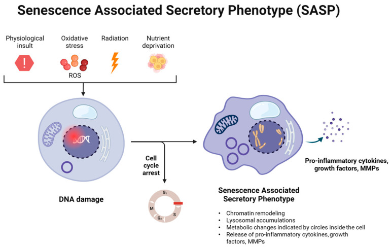
Cellular senescence may originate as a result of physiological insults, oxidative stress due to reactive oxygen species (ROS), radiation and nutrient deprivation. All of these factors can cause DNA damage that may lead to cell cycle arrest, rendering a cell to become senescent. These senescent cells then release SASP, which creates a microenvironment unsuitable for cell proliferation, growth, survival and eventually leads to cell damage and death. This has been indicated below in Figure 1. Interestingly, SASP has also been associated with being the fuel for chronic and systemic low-grade inflammation during aging–now known as ‘inflammaging’ [31]; both of which contribute to ARDs and related mortality.
Figure 1.
Mechanism of senescence-associated secretory phenotype (SASP) formation.
With respect to senescence in the bone, several in vitro and ex vivo studies have demonstrated increased senescence and age-related decline in the number and function within the stem cell compartments in the BM [24,49,50]. Radiation-induced senescent osteocytes were found to degenerate the differentiation potential of BM MSCs in vivo via the paracrine pathway [51]. In a doxorubicin induced senescence model, hypoxia inducible factor (HIF)-2α and p21 were found to be elevated and the osteoblast differentiation was inhibited. The same study along with in vivo results found that HIF-2α acts as an intrinsic factor for age-related bone loss [52].
Similarly, several articles have attempted to connect the links between aging, senescence and inflammaging, considering inflammaging is a relatively new concept [21,31,32]. However, to date, experimental evidence that is solely focused on bone senescence and inflammaging remains limited. Interestingly, reports of both cellular senescence and inflammaging within the bone have been addressed more frequently in conditions like OA [53,54], OP [55,56] and sometimes RA [57,58]. In the following sections, we discuss the pathways, genetics and anti-aging strategies taking inspiration from evidence presented in these age-related orthopedic conditions including OA, OP and RA.
3. Pathways Involved in Senescence and Inflammaging in the Bone
Numerous pathways and cycles are involved in the normal functioning of a body, which undergoes age-related changes that impact different tissues. Specifically, for pathways involved in senescence across tissues, Wang et al. have recently discussed them in a recent review article [59]. For this review, we will focus on the pathways that have demonstrated their involvement in cellular senescence, specifically in the bone and associated ARDs. In particular, cellular senescence has been observed and reported in bone progenitor cells known as mesenchymal stem cells (MSCs) [60], osteoblasts [56] and differentiated osteoblasts known as osteocytes [61]. These largely include pathways involved in cell cycle, growth promotion and DNA damage due to oxidative stress via the STING pathway. MSCs, osteoblasts and osteocytes have all been indicated to potentially be impacted by the pathways outlined below.
3.1. Cell Cycle Arrest: p16, p21, p53
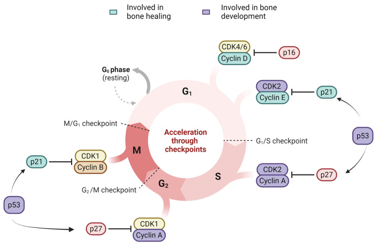
p16, p21 and p53 are key markers involved in cell cycle and DNA damage response (DDR), acting as cell cycle checkpoints [62]. Together, these three markers are the most commonly compromised ones in conditions like cancer, making them essential for programmed senescence, genomic stability and repair responses [62]. Senescent cells have been shown to have upregulated levels of cyclin-dependent kinase inhibitors (CKIs) like p16 and p21, indicated by upregulated mRNA levels of p16INK4a and p21 in elderly humans compared to young women [63]. p53 followed by p16, has been indicated to play a stronger role in senescence response to telomere dysfunction than p21 [64]. Interestingly, Chandra et al. found that targeting p21 but not p16-positive senescent BM cells in vivo prevented OP in mice [65]. BM MSCs from myelodysplastic syndrome (MDS) were found to demonstrate senescence phenotypes (enlarged cells, reduced proliferation, increased SA-β-gal) via activation of p21/p53. Interestingly, they too did not find any changes in the levels of p16 or pRb in MSCs from MDS patients [66]. Similarly, MSCs from patients with systemic lupus erythematosus (SLE) were also found to be senescent via p21/p53 activation [67]. The overall role of p16, p53 and p21 in cell cycle in relation to bone function is indicated in Figure 2.
Figure 2.
The role of p16, p53 and p21 in bone development and healing in cell cycle.
3.1.1. p16-Rb Pathway
p16, also known as INK4a, is a protein encoded by the gene cyclin-dependent kinase inhibitor 2A (CDK2NA), which is an essential inhibitor of cyclin-dependent kinase activity and a tumor suppressor gene. It has several functions in different stages of the cell cycle and is necessary for regulating uncontrolled cell proliferation in majority of the cell types [68]. It is essential for the cell cycle’s transition from G1 toS phase. In particular, p16 binds to CDK4/6 and prevents the phosphorylation of the Rb protein. It stops the formation of the cyclin D–CDK4/6 complex. In turn, hypo-phosphorylated Rb binds to E2F transcription factors, preventing cell cycle progression, thus leading to G1 cell cycle arrest, further preventing the transition into the S phase [69].
OA is a well-acknowledged age-related degenerative disease where senescence plays a significant role in its progression [53,54,70,71]. Philipot et al. demonstrated the accumulation of p16INK4a positive cells in an in vitro experiment using mature chondrocytes for the progression of OA [72]. In response to pro-inflammatory cytokines like IL6, IL8, and IL1β, chondrocytes produce matrix-remodeling regulatory matrix metalloproteases (MMPs) like MMP1 and MMP13, exhibiting the SASP characteristics. Together with these SASP factors, multiple pathways lead to the activation of p16, which in turn pushes them towards senescence. In vitro studies by Farr et al. indicated that growing osteoblasts in media conditioned with cellular senescence, impaired osteoblast mineralization, leading to increased osteoclast genesis. The same study with in vivo experiment in mice showed that the clearance of p16INK4a positive cells attenuated age-related bone loss [73].
3.1.2. p53-p21 Pathway
p21, also known as CIP1 and WAF1 is a protein encoded by the gene CDKN1A. It can induce cellular senescence in p53 dependent and p53 independent pathways. p53 is a tumor suppressor gene, which has the main implication in the protection of the DNA integrity of the cell [74]. In p53-dependent pathway, ATM-Chk2 or ATRChk1 pathways activate p53, thereby upregulating its downstream target p21 expression [75], where p21 binds to CDK-2, 4 and 6 and inhibits the cyclin–CDK complex formation, resulting in RB–E2F complex formation,. Consequently, this prevents cell cycle progression by inducing G1arrest and G2/M [74,76,77]. In the p53-independent pathway, p21 independent of p53 inhibits cyclin-CDK complex which further prevents cell cycle progression via Rb-E2F complex.
Additionally, p21 binds to and inhibits the activity of proliferating cell nuclear antigen (PCNA), a subunit of DNA polymerase, which results in the termination of DNA replication [68,78]. Englund et al. demonstrated the role of p21 in inducing cellular senescence via DDR and inflammatory signaling pathways in transgenic mice, with p21 overexpression in skeletal muscles. p21 overexpression in senescent cells corroborated with tissue fibrosis, low levels of skeletal muscle mass and reductions in physical function [79]. Loss of p53 in mice BM MSCs has been reported to alter bone remodeling and potentially impact cancer related bone modeling via negative regulation of osteoprotegerin (OPG) [80].
3.2. Growth Promotion Pathways—mTOR and, SIRT-1
3.2.1. mTOR
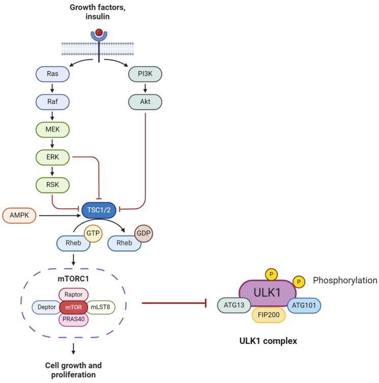
mTOR (mammalian target of rapamycin) is a critical negative regulator of autophagy. It is activated downstream of PI3K kinase and Akt kinase to inhibit autophagy. It forms two catalytic subunit protein complexes following a number of steps: mTORC1, which regulates cell growth and metabolism and mTORC2, which controls proliferation and survival [81]. mTORC1 actively inhibits autophagy by phosphorylating ULK1, thereby preventing its activation by AMPK (Figure 3). Senescent cells show failure in autophagy due to mitochondrial dysfunction, another hallmark of senescent cells [82].
Figure 3.
mTOR pathway and formation of subunit mTORC1 and inhibition of autophagy via ULK1 phosphorylation.
Zhang et al. showed a correlation between the overexpression of mTOR and disease progression using an in vivo OA mouse model [83]. Its ablation at the genetic level alleviated the OA pathogenesis via the upregulation of autophagy [84,85]. Pan et al. showed the attenuation of cartilage degradation in the OA mice model by rectifying the autophagy inhibition of chondrocytes via PI3K/AKT/mTOR signaling [86]. Cheng et al. demonstrated inhibition of autophagy improved OA-related degeneration in vivo in femoral condyles of rabbits and in vitro in human chondrosarcoma cells [84].
3.2.2. SIRTs
SIRTs (Sirtuins) are a family of nicotinamide adenine dinucleotide (NAD+)-dependent protein deacetylases, which regulate a comprehensive range of cellular processes ranging from cell metabolism, development and cellular senescence [87,88]. It comprises of seven members from SIRT 1–SIRT 7.
Among them, SIRT-1 is critical in maintaining cartilage health by promoting chondrocyte survival and ECM stabilization. It has been connected to longevity and extended lifespan [89]. Additionally, SIRT-1 activation has demonstrated a protective effect in OA by enhancing the trabecular and subchondral bone and by inhibiting chondrocyte apoptosis [90]. SIRT-1 is involved in multiple pathways, such as NF-κB, AMPK, mTOR, p53, PGC1α and FoxOs have been implicated in inflammation, mitochondria biogenesis, autophagy, energy metabolism, oxidative stress and cellular senescence [88]. In senescent cells, SIRT-1 is identified as a nuclear autophagy substrate degraded by being transported to cytoplasmic autophagosomes through LC3 recognition during senescence [91]. Batshon et al. showed cleavage of SIRT-1 into N-Terminal (NT) and C-Terminal (CT) by cathepsin B. Increased serum NT/CT SIRT-1 ratio reflected the early stage of OA and cellular senescence in chondrocytes [92]. Zhou et al. found that SIRT-1 regulated osteoblast senescence upon exposure to cadmium. Interestingly, the exposure simultaneously decreased SIRT-1, increased osteoblast senescence as well as increased p53, p16 and p21, which triggered DDR [93].
3.3. Reactive Oxygen Species (ROS)-Induced DNA Damage (Type 1 IFN and the STING Pathway)
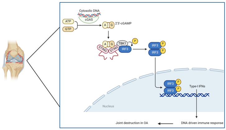
During aging, chronic low-grade systemic inflammation produces increased ROS levels, which lead to oxidative stress [94]. Oxidative stress induces DNA damage found at higher levels in aged cells and has been associated with diseases in almost every organ [95,96]. The release of mitochondrial or nuclear DNA from these damaged cells activates the innate immune system. One part of the innate immune activity is the cyclic GMP–AMP synthase (cGAS) stimulator of interferon genes (STING) signaling pathway. cGAS enzyme triggered by the detection of leaked DNA further catalyzes the production of cyclic GMP-AMP (cGAMP). Subsequently, cGAMP interacts with STING, activating the STING pathway via its various downstream targets, such as TANK-binding kinase 1 (TBK1), interferon regulatory factor 3 (IRF-3), and IkappaB kinase (IKK). Consequently, the induction of type 1 interferons commences. Additionally, the STING pathway regulates nuclear factor-kappa-light chain enhancer of B cells (NF-κB) signaling in chondrocytes implicated in SASP and senescence. DDR-induced type 1 interferon has been indicated to promote senescence and inhibit stem cell function from both hematopoietic and stromal compartments in the BM [97,98]. The cGAS–cGAMP-STING axis has now been suggested as the missing link between DNA damage, inflammation, cellular senescence and cancer [99]. Figure 4 below demonstrates this pathway in an OA joint.
Figure 4.
Role of cGAS-STING pathway in OA joint.
STING pathway induces ECM degradation via upregulation of matrix-degrading enzymes (ADAMTS5 and MMP13) as a part of SASP [100]. STING pathway activation is associated with cellular senescence and apoptosis [101]. Guo et al. demonstrated that the knockdown of STING using lentivirus in an in vivo OA mouse model improved senescence, apoptosis and ECM imbalance in chondrocytes [100]. Owing to their prominent role in inflammation and SASP factors elevation, they are becoming an attractive target for treating cellular senescence.
4. Genetics of Cellular Senescence in the Bone
During cell development, cellular impairments such as DNA damage, telomere shortening or dysfunction, oncogene activation or loss of tumor suppressor capabilities, epigenetic modifications and organelle destruction may occur. All the aforementioned processes contribute to cellular senescence, but in particular towards DDR [102]. Given that the bone tissues produce 95% of the body’s cells, numerous studies have utilized bone biopsies to investigate which, if any, genes play a crucial role in cellular senescence and what effect this has in the context of pathophysiology [103].
SASP, as described in Section 2 above comprises of a distinct secretory pattern, causing cell cycle termination. The diverse and tissue specific SASP factors typically consist of adhesion molecules, chemokines, cytokines, growth factors and lipid components [102]. These factors can have both local and systemic effects that can result in a variety of ARDs. In view of the molecular mechanisms and underlying pathways outlined above, the question then arises as to which genetic aberrations or abnormalities are contributive or causative of such phenomena.
4.1. Key Genes Associated with SASP
SASP research has indicated that age-related inflammatory responses, wound healing and cancer progression can all be mediated by the secretion of pro-inflammatory agents, growth factors, chemokines and proteases by aged cells [104,105]. Several conventional SASP proteins have been observed to be increased in aged bone matrix extracellular vehicles, including TGF-β2, OPG, MMP9, tissue inhibitor of metalloproteinase 1 (TIMP1), macrophage migration inhibitory factor 1 (MIF1), peroxiredoxins (PRDXs) and insulin like growth factor binding protein 3 (IGFBP3). Among these, TGF-β2 and OPG play a key role as mediators of bone metabolism; serum levels of both have been found to positively correlate with indicators of bone turnover [106]. Osteocytes and osteoblasts both express TIMP1, a tissue inhibitor of MMPs that controls MMP levels to maintain the equilibrium of bone matrix breakdown [107]. Additionally, TIMP/MMPs are able to modify the extracellular matrix to accommodate the immunological influx [108]. MIF1 is known to activate a number of transcription factors and stress kinases, such as adenosine monophosphate or AMP- activated protein kinase (AMPK) and B NF-κB, which can mediate inflammatory and tumorigenic signaling [109]. The way that aged bone cells influence the environment of nearby and distant cells and systems may therefore be explained by the elevated production of these SASP proteins [110].
Seven particular gene sets have been published specifically associated with senescence, the most comprehensive one being SenMayo, surpassed the others in identifying senescent cells with aging across tissues and species and in exhibiting responses to senescent cell clearance (based on normalised enrichment scores (NES) and p values) [102].
4.2. NGS Studies on Bone Aging
We have previously described key NGS studies targeting the aging BM [27]. Specifically for studies that investigated parameters for bone senescence, we list five key papers in Table 1 below.
Table 1.
List of studies aimed at bone senescence.
| Study Summary | NGS Methods | Key Findings | Key Genes with Elevated Expression | Reference |
|---|---|---|---|---|
| RNA profile expression in eight experimental models of cellular senescence commonly studied. WI-38 and IMR-90 fibroblasts, HUVECs and HAECs were cultured and utilized for downstream sequencing. | RNA seq, Illumina Hiseq 2500 with 2 × 150-bp strategy | 50 elevated and 18 reduced transcripts and the identification of subsets of transcripts (both coding and non-coding) display shared expression patterns across a range of senescent cell models. | SRPX, PURPL (p53 regulator) | [111] |
| RNA expression profiling of fibroblasts and their senescence induced by 5-aza identified 3 epigenetically silenced pathways. | Illumina HiSeq 2000; strategy not specified | 5-aza-induced senescence has been closely linked to alterations in the interferon/innate immunity pathway’s gene expression, and during immortalization, important regulators of this pathway are muted. | IL-1α and IL-1β | [112] |
| Use of multiple whole-transcriptome datasets, created by the authors or made publicly available, to characterise the heterogeneity of the programmed senescence. | Illumina HiSeq 2000 with 2× 150 bp | Demonstrates that ollowing senescence induction, the senescent phenotype identified for 55 genes at the core of the senescence-associated transcriptome is dynamic, changing at different intervals. | Upregulation of genes associated with G1 DNA damage checkpoint (PLK3 and CCND1) and upregulation of BCL2L2 (negative regulator of apoptosis) | [113] |
| Comprehensive analysis of the transcriptome and senolytic responses in a panel of 13 cancer cell lines rendered senescent by two distinct compounds. | HiSeq 2500; single-end 65 bp | Cell lines that were made senescent by two different substances showed that the senescence trigger has less of an impact on the composition of the SASP. The SENCAN gene expression classifier to detect senescence using machine learning was developed. | IL6 and CXCL8 | [114] |
| Gene set generation (SenMayo) consisting of 125 previously identified senescence/SASP-associated factors. | HiSeq 2000; strategy not specified | Provided a unique gene set (SenMayo) that can be utilized in bulk and scRNA-seq investigations to detect cells expressing high amounts of senescence/SASP genes. SenMayo rises with aging across tissues and species and is responsive to senescent cell clearance. | CDKN1A/P21Cip1 and several SASP markers such as CCL2 and IL6 showed consistent upregulation with aging | [102] |
A review of the literature pertaining to the sequencing of genes within the BM d-rived cells exhibits that the most impactful study in view of bone senescence was carried out by Saul et al. in 2022 [102]. This study took into account the lack of a defined ‘senescence gene set’ within the field, thus creating the 125 gene panel called ’SenMayo’, consisting of previously discovered senescence and SASP-associated genes. The authors aimed to identify commonly regulated genes in different age-related datasets (n = 15), using a transcriptome-wide approach that included whole-transcriptome as well as single cell RNA sequencing (scRNA-seq). In order to determine if senescence and SASP associated pathways were enriched with human aging and utilizing whole-bone biopsies, Saul et al. identified in particular, that genes regulating inflammatory mediators, such as NFKB1, RELA, and STAT3, were enriched. Additionally, CDKN1A/p21Cip1, CCL2, and IL6 were upregulated in both elderly cohorts, as the authors initially predicted.
4.3. Potential Pathways to Target in Bone Aging
Identification of upregulated and downregulated genes within bone senescence leads to the identification of key pathways within this phenomenon. Whilst it is challenging to eliminate pathways solely based on genetic influences, key molecular targets can be highlighted and potentially targeted in bone aging.
Saul et al.’s genetic panel consisted of several SASP factors (n = 83); transmembrane (n = 20) and intracellular (n = 22) proteins [102]. The key regulatory elements of the SenMayo genes, i.e., NFKB1 motif and BCL3, the latter of which is a key transcriptional coactivator for NF-κB, represented the leading transcription factor for a majority of SASP genes. This strongly suggested that targeting NF-κB signaling, a master regulator of gene transcription, is one of the cell’s most efficient ways to influence important and varied biological processes, including senescence [115].
This aging-related upregulation of inflammatory proteins (i.e., inflammaging), has been suggested to be the root cause of SASP, which releases inflammatory factors into the environment and causes neighboring cells to undergo bystander senescence or potentially change into pre-cancerous cells. The SASP phenotype activation occurs in conjunction with the overexpression of inflammatory markers, which is also present in senescence but is not related to terminal replication. The cause of this is unknown. That inflammation, which can cause tumorigenesis, is elevated during senescence—a process that suppresses tumor growth—seems incongruous. This could be an illustration of antagonistic pleiotropy; senescence prevents the development of cancer in its early stages but, as the organism ages, the secretory phenotype associated with senescence becomes detrimental [112]. Thus, it is becoming increasingly important to have regeneration and anti-aging strategies to combat cellular senescence in the aging bone.
5. Combatting Senescence in the Bone
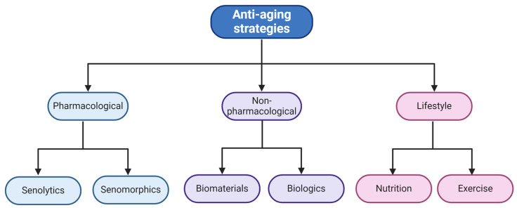
Approaches for regeneration of senescent cells in the bone may be classified as three types (Figure 5); 1. pharmacological with seno-therapeutics; 2. non-pharmacological with biologics and biomaterials; and 3. based on lifestyle. Pharmacological approaches use various types of drugs or conditions that reduce or eliminate senescence [37,116]. Non-pharmacological approaches use biomaterials like shell nacre and biologics like platelet-rich plasma (PRP) that have demonstrated anti-aging and anti-senescent properties in aged cells and in ARDs [117,118]. Finally, the lifestyle approach includes changes in diet, such as intermittent fasting or a diet high in antioxidants, to combat ROS-mediated senescence and regular exercises to naturally ameliorate cellular senescence [119,120].
Figure 5.
Regeneration interventions and anti-aging strategies for bone senescence.
5.1. Pharmacological Approaches
Seno-therapeutics are pharmacological interventions targeting senescent cells or SASP. It consists of three classes of therapeutics namely senolytics, senomorphics that are more established and seno-inflammation blockers that are currently being tested in vitro.
5.1.1. Senolytics
This is the class of therapeutics that may potentially eliminate senescent cells to prevent or alleviate several age-related conditions such as senile OP [121]. Research indicates that the selective elimination of senescent cells delays disease progression in OA and other ARDs [122]. One of the earliest studies published by Zhu et al. analyzed the differential gene expression in senescent versus non-senescent human pre-adipocytes and noticed the upregulation of genes involved in senescent cell anti-apoptotic pathways [123]. The authors identified over 46 potentially senolytic compounds that silenced key pro-survival anti-apoptotic genes such as EFNB1/3, PI3Kδ, p21 and BCL-xL (individually and in combination). The most effective compounds were Dasatinib (SRC/tyrosine kinase inhibitor) and Quercetin (natural flavonoids that interact with BCL-2 and P13K isoforms). Dasatinib, an FDA-approved cancer drug proved to be effective against senescent human preadipocytes and quercetin was effective against senescent human endothelial cells. They also showed that the combined therapy of Dasatinib and Quercetin proved more effective in eliminating senescent mice mesenchymal embryonic fibroblasts [123]. Since then, several compounds have been screened and tested in clinical trials as summarised by Chaib et al. [116].
Several classes of senolytics have been reported since then and have significantly shown to increase lifespan and mitigate ARDs [124]. Highly targeted treatments such as ABT-263, also known as the chemotherapeutic drug Navitoclax, is a BCL-2 pathway inhibitor that has been shown to cause thrombocytopenia and neutropenia under small doses [125]. Interestingly, while Chang and colleagues demonstrated rejuvenated BM HSCs in vivo by using ABT-263 to clear senescent cells [126], another study, which evaluated the effect of navitoclax on mice, showed trabecular bone loss and musculoskeletal dysfunction [127]. These studies thus warrant a closer look at the strategies adopted for targeting different components of the senescent cells anti-apoptotic pathways (SCAPs). Higher doses targeting single points in a pathway might have more off-target, detrimental effects and therefore a combination of different inhibitors in low-dosage can prove to be safer and more efficacious [128].
5.1.2. Senomorphics
These aim to target the pathways and factors associated with senescence without killing the senescent cells. Also known as SASP inhibitors, these agents try to make senescent cells mimic young cells by modulating their signal transduction mechanisms. The most popular senomorphics are rapamycin, resveratrol, NF-κB, JAK/STAT and p38MAPK inhibitors [37]. Rapamycin, also known as sirolimus, is an inhibitor of pro-senescent mTOR signaling. It acts by reducing the phosphorylation of S6K and 4E-BP downstream of TORC1 (discussed in Section 3.2.1). TORC1 forms a multiprotein complex with TORC2 and is regulated by mTOR [129]. Although effective in in vivo studies, side effects of rapamycin such as nuclear factor erythroid 2-related factor 2 (Nrf2) pathway activation and NF-κB suppression have been reported. Even though off-target, these effects supplement the senomorphic effect of rapamycin [130]. Resveratrol is a plant antitoxin and antioxidant that activates the SIRT-1 pathway, discussed previously in Section 3.2.2. SIRT proteins are key regulators of transcriptional processes and pathways involved in senescence and aging. However, with increasing age, their expression and functional activity decreases [91].
Studies show that regulated doses of resveratrol can suppress SASPs by activating P13K-aKT signaling in endothelial progenitor cells and BM stromal cells [131,132]. However, higher concentrations can induce senescence and apoptotic death. This has led to researchers investigating other novel SIRT activators such as SRT1720, STAC-5/9/10, SCIC2 and SCIC2.1 [133,134,135]. These anti-aging compounds are more stable and have demonstrated better bioavailability. However, it should be noted that the diet of the organisms is vital to its efficacy. Resveratrol performs better when supplemented with a high-fat diet, whereas the second-generation SIRT activators have high efficacy with a standard diet.
Other senolytic compounds currently being investigated are p53 activators (UBX0101) and PPARα agonist fenofibrate [136,137]. Another attractive target is p38MAPK. Mitogen-activated protein kinase (MAPK) modules regulate cell proliferation, differentiation and apoptosis processes. p38MAPK is a sub-family of this pathway and is activated in response to stress. Its ability to trigger cell-cycle arrest and induce senescence has been harnessed to create inhibitor compounds such as SB203580, UR-13756 and BIRB-796. They have been shown to block SASP secretion in senescent cells. SB2023580 suppresses p16 signaling which mediates cell cycle arrest and SA-β-gal activity [20]. R-13756 and BIRB-796 are second-generation p38 inhibitors with higher specificity as shown in vivo using human fibroblasts [138]. Interestingly, p38 MAPK inhibitors (MK2i) have indicated the potential to have anti-inflammatory effects in RA, which is an autoimmune and degenerative disease of joints. However, further studies are needed to confirm the same [139,140]. Another popular anti-aging compound is JAK/STAT inhibitor ruxolitinib, an FDA-approved drug used in myeloproliferative diseases. It has been shown to improve age-related bone loss in mice attributed to its ability to inhibit SASP secretions [73]. Nevertheless, with the current black box warning against JAK inhibition, alternative approaches for targeting senescence are needed [141].
5.1.3. Seno-Inflammation
This term refers to a dysregulated immune response that leads to chronic inflammation. It was proposed by Chung et al. in 2019 to describe the steady-state age-related senescent inflammation that exacerbates chronic diseases, associating with the concept of inflammaging [142]. On a molecular level NF-κB, signaling plays a major role in inflammation. It upregulates chemokines, interleukins (IL2, IL6 and IL-1β), adhesion molecules and c-reactive protein (CRP) triggering immune system activation [143]. On a cellular level, the dysregulated macrophage activity has been associated with aging in humans. Toll-like receptor (TLR) signaling, interferon-gamma (IFN)-γ activity, and NF-κB signaling changes are downstream markers of macrophage activity in senescent cells. Proteoglycan-4 (PRG-4/lubricin) is a modulator of TLR signaling and can regulate inflammatory response in vitro and in in vivo rat models of OA [144]. PRG-4 binds to and suppresses TLR2 and TLR4 activity in human OA synovial fluid, making it a potential anti-inflammatory therapeutic target [145]. Senescence-associated secretomes and growth factors are also being explored as therapeutics for reducing seno-inflammation. Platas et al. used a conditioned medium from MSC secretomes and cultured OA chondrocytes. The cells reduced inflammatory stress, assessed by markers IL-1β and SA-β-gal [146]. Growth hormones such as pegvisomant inhibit growth hormone/insulin-like growth factor-1 (IGF1) axis and modulate inflammation, making it an attractive therapeutic agent [147].
A few examples of pharmacological approaches with senolytic and senomorphic compounds that have been investigated are outlined in Table 2 below.
5.2. Non-Pharmacological Approaches
5.2.1. Materials for Bone Regeneration via Senescence Reduction
Regenerative medicine plays a crucial role in promoting cell neogenesis in the context of reducing senescence and promoting bone health. While seno-therapeutics have shown to increase the regenerative capacity of the bone; tissue engineering scaffolds, bioactive and biocompatible biomaterials can also provide the necessary microenvironment for tissue regeneration, growth and repair. They do so by improving the drug delivery and the temporary effect of the drug in vivo [117,148,149,150]. Hydrogels have been tested to provide an effective microenvironment for tissue growth and cell differentiation [151,152,153]. Quercetin-loaded hydrogels caused clearance of senescent cells in aged rats for a prolonged period thereby improving the in-vivo lifespan and delivery of the drug [154]. Microspheres loaded with drugs or growth factors serve as carriers for time-dependent and location-specific release. Thus, these platforms provide a major improvement in ameliorating side effects from seno-therapeutic drugs [155]. He et al. tested the efficacy of PEGylated (amalgamated with polyethylene glycol) hydrogels loaded with rapamycin on senescent MSCs. The oxidative function of this system was able to delay senescence by scavenging intracellular ROS [153]. New investigation on novel materials like shell nacre 3D scaffolds have demonstrated enhanced proliferation of MSCs from older donors that was comparable to MSCs from younger donors, in spite of previously observing higher number of SA-β-gal cells in the older MSCs in 2D cell culture [117].
Another exciting anti-aging compound suitable for bone regeneration is collagen. Collagen is the structural protein in the ECMand is biocompatible, water-soluble and easily metabolized. It can be extracted from abundant marine organisms, making it a suitable biomaterial for bone regeneration [156]. Collagen has been shown to increase mineral bone density and high osteoblastic activity, promoting self-renewal abilityand enhancing osteogenic capacity of the bone in vitro using rat BM MSCs and osteoblastic cells [157,158]. It is known that there is a high accumulation of ROS and associated inflammation in aging models. In 2019, Zhou et al. functionalized a titanium mesh-based porous scaffold using polypyrrole. Polypyrrole is an electroactive conductive polymer that can induce physical and chemical changes in response to electrical signals. They used this conductive polymer to scavenge ROS by providing free electrons. To increase the osteoconductivity of the biomaterial, they added hydroxyapatite nanoparticles to the scaffold. To increase the hydrophilicity and drug-loading efficacy of polypyrrole, they supplemented this system with polydopamine nanoparticles. Together when the polypyrrole polydopamine hydroxyapatite film was coated on a titanium mesh-based scaffold and tested on RAW264.7 macrophages, the scavenging activity of ROS was significantly reduced [159].
5.2.2. Anti-Aging Strategies Using Biologics
The use of biological adjuvants to study bone healing has yielded several treatments that induce signaling and anti-inflammatory effects. Stem cell therapy using MSC extracted from the BM has proven to deliver significant clinical advantages in tissue regeneration. MSCs secrete proteomes that regulate the immune system and demonstrate anti-apoptosis, anti-oxidation, cell homing and differentiation abilities [36]. This has been used to activate repair mechanisms in cartilage, muscle and bone and thus their potential to treat ARDs is highly probable [160,161]. MSCs have been an area of great interest within the scientific as well as the medical community for use in bone regeneration [162,163,164,165,166].
Blood and blood-related factors are also contributing towards reversing age-related molecular and cellular changes in blood, muscle, bone and nervous system. Parabiosis in the context of aging is the technique of surgically joining the circulatory system of a young organism with an aged organism to study the reverse effects of aging [167]. Heterochronic parabiosis (HP) performed on mice showed that youthful circulation increased the capacity of bone healing in aged mice with tibial fractures. This was marked by the increase in type 1 collagen production expressed in osteoblasts and modulation of the β-catenin pathway [168]. Thus, HP offers a unique but promising paradigm to tackle ARDs.
PRP and other platelet-derived biologics are currently gaining popularity as effective treatments for OA, fractures and enhancing wound healing. Due to its ease of harvesting and preparation, PRP treatments have been touted as an accessible form of treatment harboring anti-inflammatory effects [39]. It has often been used for enhancing cellular functionalities in OA [164,169,170,171] and has also been compared with collagen or hyaluronic acid in the treatment for degenerative diseases like OA with similar or better results [171,172,173]. PRP has been reported to reduce SASP factors like IL6, IL1β, TNFα [173] along with reducing inflammation in inflamed joints potentially targeting inflammaging [174]. Especially with respect to OA, PRP has been observed to reduce inflammation, alleviate pain and reduce pro-inflammatory cytokines in patients [175,176]. However, the lack of consistent results has deterred the progress of PRPs as clinical interventions. A systemic review done in 2023 on the comparison of PRP treatment versus approved treatments for knee OA stated that the current data from trials were inconsistent, biased and hence of low quality [177]. This warrants better methodological approach in the execution, reporting, and analysis of clinical trials, along with the need for standardized protocols for the application of PRPs.
A few examples of non-pharmacological approaches with biomaterials and biologics investigated have been outlined in Table 2 below.
5.3. Lifestyle Approaches as Regeneration and Anti-Aging Strategies
Healthy diet approaches along with exercise have been a sought after approach for managing several health conditions and contribute towards healthy aging [119,178]. The concepts of calorie restriction, intermittent fasting, fiber and protein rich diet along with exercises is possibly the most long standing and effective way to ensure that the negative effects of senescence are reduced in the elderly and encourages healthy aging. Consumption of naturally occurring compounds including anti-oxidants, polyunsaturated fatty acids (PUFA), minerals, vitamins and essential amino acids are key to longevity [179]. The combination of a healthy diet with regular exercises like walking and strength training in ‘blue zones’ has shown us that people can live up to a century and more [180].
A recent study investigated the effect of intermittent fasting for 30 days on n = 25 young healthy males by examining their mRNA levels from the blood samples of these donors. They found that at later time points (a week after 30 days), the level of inflammatory cytokines was reduced, there was induced autophagy and a reduction in the expression of senescent markers discussed in Section 3.1 of this article [181]. Fielding et al. studied the blood from n = 1377 older subjects (aged 70–89 years old) and found that lower functionalities like grip strength, gait, walking etcetera were all associated with high SASP and related markers. They were successfully able to link the SASP phenotype with physical activity in sedentary elderly population [120]. Another study by Maria Fastame indicated that a nutritious diet, strong physical health and well-being along with a sense of community was essential to the Sardinian ‘blue zone’ with several reported centenarians [182].
Martel et al. re-iterate the importance of diet and lifestyle changes and remind us that nutritious food, exercises and adequate sleep can produce anti-senescent effects [119]. They also outline that these findings are not surprising but have not been studied as thoroughly with respect to molecular mechanisms and pathways involved. Thus it is no surprise that an isolated life with excess alcohol consumption and smoking can offset the beneficial effects of any of the anti-senescent strategies [119]. Similarly, sleep deprivation for one night was associated with DDR and activated SASP in n = 29 older adults aged between 61-86 years old [183]. In vivo studies in mice revealed prevention of osteoblast senescence in older female mice when fed a blueberry based diet in their early days [184]. A few of the lifestyle approaches with nutrition and exercise routines investigated have been outlined in Table 2 below.
Table 2.
Examples of strategies for combatting bone senescence.
| Approach | Strategy | Model | Target | Reference |
|---|---|---|---|---|
| Pharmacological, senolytic | Dasatinib (D) and/or quercetin (Q) | in vitro and in vivo | D- senescent fat progenitors, Q- human endothelial cells and mouse BM MSCs, D + Q—mouse embryonic fibroblasts | [123] |
| Pharmacological, senolytic | Dasatinib + Quercetin | in vitro and in vivo | Trabecular and cortical bone in mice | [73] |
| Pharmacological, senolytic | ABT263 | In vivo | Senescent bone marrow hematopoietic stem cells in mice | [126] |
| Pharmacological, senolytic/senomorphic | Zeldronic acid | In vitro and in vivo | Bone | [185] |
| Pharmacological, senomorphic | Ruxolitinib | In vivo | metabolism | [186] |
| Non-pharmacological, biomaterial | Shell nacre | In vitro | BM MSCs | [117] |
| Non-pharmacological, biomaterial | PEGylated hydrogel with Rapamycin nanomicelles | In vitro and in vivo | BM MSCs | [153] |
| Non-pharmacological, biologics | HA v/s PRP in RCT | In vivo | Knee OA | [173] |
| Non-pharmacological, biologics | Collagen | In vitro | BM MSCs | [157] |
| Non-pharmacological, biologics | PRP | In vitro and in vivo | Injured tendons | [174] |
| Lifestyle | Intermittent fasting | In vivo, n = 25 young males | Senescence markers linked with diet and lifestyle | [181] |
| Lifestyle | Physical functioning | In vivo, n = 1377 older adults | Senescence biomarkers linked with physical functioning in elderly | [120] |
| Lifestyle | Diet + exercise | In vivo, n = 12 young males | Senescent cells in skeletal muscle linked with diet and exercise | [187] |
| Lifestyle | Diet + exercise + community | Mixed methods, n = 57 older adults | Nutritional habits and active lifestyle linked with longevity | [182] |
| Lifestyle | Sleep deprivation | In vivo, n = 29 older adults | Sleep deprivation linked with senescent markers | [183] |
6. Conclusions, Limitations and Future Directions
While cellular senescence, ARDs and inflammaging are increasingly contributing to the aging bone and BM, several interventions and anti-aging strategies may be used to combat this effect and aid the regeneration of the bone. We are mindful as authors that aging is a natural progression and that fit and healthy elderly people exist. With this review article, we hope to bring to light that the natural progression of advancing age does not necessarily have to be with diseased conditions. Understanding the molecular mechanisms, genetics and pathways is essential to our knowledge of bone aging. Anti-aging strategies exist and must be optimised for the needs of the individuals.
It is worth outlining that in our quest to discover the connections between bone senescence and ARDs, we found the majority of research evidence in literature in relation to OA, OP and RA. These three ARDs have been reported to be impacted by all of the pathways discussed in Section 3 above. They have also been evidenced with cellular senescence, (progenitor and bone cells for OA and OP as outlined above and T-cells for RA [188]), associated with inflammation and inflammaging and often require surgeries and life-long medication for treatment. This indicated a potentially common root cause for all of these three ARDs, that may be developed into biomarkers and possibly therapeutic targets with further research.
Considering pharmacological approaches for anti-aging strategies, variability and low efficacy of some agents have been observed which can be attributed to the fact that senescent cells have different transcriptional signatures based on their spatial and temporal status. Further screening (chemical, pharmacological and transcriptional) will be warranted to study the precise effects of these molecules in a context-dependent manner. In terms of lifestyle approaches, the aforementioned study was limited by investigating the effect of intermittent fasting only in males, thus their results are not applicable to females and similar studies focused on women are needed [181].
Future investigations focused on human samples from healthy donors across young and older adults, as well as those suffering from ARDs are critical. Additionally, age and gender matched studies of those with lifestyle disorders with a potential to develop ARDs will be of extreme significance. Genetic mapping of these samples and dissecting their pathways will help in early detection of these diseases. This is in turn will be useful in predicting the therapeutic approach (pharmacological or non-pharmacological) best suited for an individual impacted by an ARD. Nevertheless, time and again the benefits of healthy lifestyle have been shown to us and an increasing body of evidence indicates the anti-senescent effect of the same. Thus, irrespective of the presence or absence of disease, nutritious food and healthy lifestyle must be encouraged across age, gender and cultures to combat senescence in the most cost-effective manner.
Author Contributions
Conceptualization, P.G.; software, M.L., A.G., S.P. and P.G.; validation, M.L., A.G., S.P. and P.G.; writing—original draft preparation, M.L., A.G., S.P. and P.G.; writing—review and editing, M.L., A.G., S.P. and P.G.; supervision, P.G.; project administration, P.G.; funding acquisition, P.G. All authors have read and agreed to the published version of the manuscript.
Institutional Review Board Statement
Not applicable.
Informed Consent Statement
Not applicable.
Data Availability Statement
Not applicable.
Conflicts of Interest
Mr. Abhishek Goyal was employed in RAS Life Science Solutions. The authors declare that this research was conducted in the absence of any commercial or financial relationships that could be construed as a potential conflict of interest.
Funding Statement
This work received no external funding.
Footnotes
Disclaimer/Publisher’s Note: The statements, opinions and data contained in all publications are solely those of the individual author(s) and contributor(s) and not of MDPI and/or the editor(s). MDPI and/or the editor(s) disclaim responsibility for any injury to people or property resulting from any ideas, methods, instructions or products referred to in the content.
References
- 1.World Health Organization: Ageing and Health. [(accessed on 4 March 2024)]. Available online: https://www.who.int/news-room/fact-sheets/detail/ageing-and-health.
- 2.Campisi J. Aging, Cellular Senescence, and Cancer. Annu. Rev. Physiol. 2013;75:685–705. doi: 10.1146/annurev-physiol-030212-183653. [DOI] [PMC free article] [PubMed] [Google Scholar]
- 3.Hoeijmakers J.H. DNA damage, aging, and cancer. N. Engl. J. Med. 2009;361:1475–1485. doi: 10.1056/NEJMra0804615. [DOI] [PubMed] [Google Scholar]
- 4.Anisimov V.N. Biology of Aging and Cancer. Cancer Control. 2007;14:23–31. doi: 10.1177/107327480701400104. [DOI] [PubMed] [Google Scholar]
- 5.Heavener K.S., Bradshaw E.M. The aging immune system in Alzheimer’s and Parkinson’s diseases. Semin. Immunopathol. 2022;44:649–657. doi: 10.1007/s00281-022-00944-6. [DOI] [PMC free article] [PubMed] [Google Scholar]
- 6.Levy G. The Relationship of Parkinson Disease with Aging. Arch. Neurol. 2007;64:1242–1246. doi: 10.1001/archneur.64.9.1242. [DOI] [PubMed] [Google Scholar]
- 7.Calabrese V., Santoro A., Monti D., Crupi R., di Paola R., Latteri S., Cuzzocrea S., Zappia M., Giordano J., Calabrese E.J., et al. Aging and Parkinson’s Disease: Inflammaging, neuroinflammation and biological remodeling as key factors in pathogenesis. Free Radic. Biol. Med. 2018;115:80–91. doi: 10.1016/j.freeradbiomed.2017.10.379. [DOI] [PubMed] [Google Scholar]
- 8.Manolagas S.C., Parfitt A.M. What old means to bone. Trends Endocrinol. Metab. 2010;21:369–374. doi: 10.1016/j.tem.2010.01.010. [DOI] [PMC free article] [PubMed] [Google Scholar]
- 9.Scanzello C.R. Role of low-grade inflammation in osteoarthritis. Curr. Opin. Rheumatol. 2017;29:79–85. doi: 10.1097/BOR.0000000000000353. [DOI] [PMC free article] [PubMed] [Google Scholar]
- 10.Ganguly P., El-Jawhari J.J., Giannoudis P.V., Burska A.N., Ponchel F., Jones E.A. Age-related Changes in Bone Marrow Mesenchymal Stromal Cells. Cell Transplant. 2017;26:1520–1529. doi: 10.1177/0963689717721201. [DOI] [PMC free article] [PubMed] [Google Scholar]
- 11.Hayflick L., Moorhead P.S. The serial cultivation of human diploid cell strains. Exp. Cell Res. 1961;25:585–621. doi: 10.1016/0014-4827(61)90192-6. [DOI] [PubMed] [Google Scholar]
- 12.López-Otín C., Blasco M.A., Partridge L., Serrano M., Kroemer G. The hallmarks of aging. Cell. 2013;153:1194–1217. doi: 10.1016/j.cell.2013.05.039. [DOI] [PMC free article] [PubMed] [Google Scholar]
- 13.Schmauck-Medina T., Molière A., Lautrup S., Zhang J., Chlopicki S., Madsen H.B., Cao S., Soendenbroe C., Mansell E., Vestergaard M.B., et al. New hallmarks of ageing: A 2022 Copenhagen ageing meeting summary. Aging. 2022;14:6829–6839. doi: 10.18632/aging.204248. [DOI] [PMC free article] [PubMed] [Google Scholar]
- 14.Cross M., Smith E., Hoy D., Nolte S., Ackerman I., Fransen M., Bridgett L., Williams S., Guillemin F., Hill C.L., et al. The global burden of hip and knee osteoarthritis: Estimates from the Global Burden of Disease 2010 study. Ann. Rheum. Dis. 2014;73:1323–1330. doi: 10.1136/annrheumdis-2013-204763. [DOI] [PubMed] [Google Scholar]
- 15.Cui A., Li H., Wang D., Zhong J., Chen Y., Lu H. Global, regional prevalence, incidence and risk factors of knee osteoarthritis in population-based studies. eClinicalMedicine. 2020;29–30:100587. doi: 10.1016/j.eclinm.2020.100587. [DOI] [PMC free article] [PubMed] [Google Scholar]
- 16.Almutairi K., Nossent J., Preen D., Keen H., Inderjeeth C. The global prevalence of rheumatoid arthritis: A meta-analysis based on a systematic review. Rheumatol. Int. 2020;41:863–877. doi: 10.1007/s00296-020-04731-0. [DOI] [PubMed] [Google Scholar]
- 17.Black R.J., Cross M., Haile L.M., Culbreth G.T., Steinmetz J.D., Hagins H., Kopec J.A., Brooks P.M., Woolf A.D., Ong K.L., et al. Global, regional, and national burden of rheumatoid arthritis, 1990–2020, and projections to 2050: A systematic analysis of the Global Burden of Disease Study 2021. Lancet Rheumatol. 2023;5:e594–e610. doi: 10.1016/S2665-9913(23)00211-4. [DOI] [PMC free article] [PubMed] [Google Scholar]
- 18.Wu A.-M., Bisignano C., James S.L., Abady G.G., Abedi A., Abu-Gharbieh E., Alhassan R.K., Alipour V., Arabloo J., Asaad M., et al. Global, regional, and national burden of bone fractures in 204 countries and territories, 1990–2019: A systematic analysis from the Global Burden of Disease Study 2019. Lancet Healthy Longev. 2021;2:e580–e592. doi: 10.1016/S2666-7568(21)00172-0. [DOI] [PMC free article] [PubMed] [Google Scholar]
- 19.Morrison S.J., Scadden D.T. The bone marrow niche for haematopoietic stem cells. Nature. 2014;505:327–334. doi: 10.1038/nature12984. [DOI] [PMC free article] [PubMed] [Google Scholar]
- 20.Dimitriou R., Jones E., McGonagle D., Giannoudis P.V. Bone regeneration: Current concepts and future directions. BMC Med. 2011;9:66. doi: 10.1186/1741-7015-9-66. [DOI] [PMC free article] [PubMed] [Google Scholar]
- 21.Marie P.J. Bone Cell Senescence: Mechanisms and Perspectives. J. Bone Miner. Res. 2014;29:1311–1321. doi: 10.1002/jbmr.2190. [DOI] [PubMed] [Google Scholar]
- 22.Farr J.N., Khosla S. Cellular senescence in bone. Bone. 2019;121:121–133. doi: 10.1016/j.bone.2019.01.015. [DOI] [PMC free article] [PubMed] [Google Scholar]
- 23.Wan M., Gray-Gaillard E.F., Elisseeff J.H. Cellular senescence in musculoskeletal homeostasis, diseases, and regeneration. Bone Res. 2021;9:41. doi: 10.1038/s41413-021-00164-y. [DOI] [PMC free article] [PubMed] [Google Scholar]
- 24.Massaro F., Corrillon F., Stamatopoulos B., Dubois N., Ruer A., Meuleman N., Bron D., Lagneaux L. Age-related changes in human bone marrow mesenchymal stromal cells: Morphology, gene expression profile, immunomodulatory activity and miRNA expression. Front. Immunol. 2023;14:1267550. doi: 10.3389/fimmu.2023.1267550. [DOI] [PMC free article] [PubMed] [Google Scholar]
- 25.Kim M., Kim C., Choi Y.S., Kim M., Park C., Suh Y. Age-related alterations in mesenchymal stem cells related to shift in differentiation from osteogenic to adipogenic potential: Implication to age-associated bone diseases and defects. Mech. Ageing Dev. 2012;133:215–225. doi: 10.1016/j.mad.2012.03.014. [DOI] [PubMed] [Google Scholar]
- 26.Kovtonyuk L.V., Fritsch K., Feng X., Manz M.G., Takizawa H. Inflamm-Aging of Hematopoiesis, Hematopoietic Stem Cells, and the Bone Marrow Microenvironment. Front. Immunol. 2016;7:502. doi: 10.3389/fimmu.2016.00502. [DOI] [PMC free article] [PubMed] [Google Scholar]
- 27.Ganguly P., Toghill B., Pathak S. Aging, Bone Marrow and Next-Generation Sequencing (NGS): Recent Advances and Future Perspectives. Int. J. Mol. Sci. 2021;22:12225. doi: 10.3390/ijms222212225. [DOI] [PMC free article] [PubMed] [Google Scholar]
- 28.Fulop T., Witkowski J.M., Olivieri F., Larbi A. The integration of inflammaging in age-related diseases. Semin. Immunol. 2018;40:17–35. doi: 10.1016/j.smim.2018.09.003. [DOI] [PubMed] [Google Scholar]
- 29.Rezuș E., Cardoneanu A., Burlui A., Luca A., Codreanu C., Tamba B.I., Stanciu G.-D., Dima N., Bădescu C., Rezuș C. The Link Between Inflammaging and Degenerative Joint Diseases. Int. J. Mol. Sci. 2019;20:614. doi: 10.3390/ijms20030614. [DOI] [PMC free article] [PubMed] [Google Scholar]
- 30.Baylis D., Bartlett D.B., Patel H.P., Roberts H.C. Understanding how we age: Insights into inflammaging. Longev. Health. 2013;2:8. doi: 10.1186/2046-2395-2-8. [DOI] [PMC free article] [PubMed] [Google Scholar]
- 31.Olivieri F., Prattichizzo F., Grillari J., Balistreri C.R. Cellular Senescence and Inflammaging in Age-Related Diseases. Mediat. Inflamm. 2018;2018:9076485. doi: 10.1155/2018/9076485. [DOI] [PMC free article] [PubMed] [Google Scholar]
- 32.Franceschi C., Campisi J. Chronic Inflammation (Inflammaging) and Its Potential Contribution to Age-Associated Diseases. J. Gerontol. A Ser. Biol. Sci. Med. Sci. 2014;69((Suppl. 1)):S4–S9. doi: 10.1093/gerona/glu057. [DOI] [PubMed] [Google Scholar]
- 33.Franceschi C., Garagnani P., Vitale G., Capri M., Salvioli S. Inflammaging and ‘Garb-aging’. Trends Endocrinol. Metab. 2017;28:199–212. doi: 10.1016/j.tem.2016.09.005. [DOI] [PubMed] [Google Scholar]
- 34.Li X., Li C., Zhang W., Wang Y., Qian P., Huang H. Inflammation and aging: Signaling pathways and intervention therapies. Signal Transduct. Target. Ther. 2023;8:239. doi: 10.1038/s41392-023-01502-8. [DOI] [PMC free article] [PubMed] [Google Scholar]
- 35.Kirkland J.L., Tchkonia T. Senolytic drugs: From discovery to translation. J. Intern. Med. 2020;288:518–536. doi: 10.1111/joim.13141. [DOI] [PMC free article] [PubMed] [Google Scholar]
- 36.van Deursen J.M. Senolytic therapies for healthy longevity. Science. 2019;364:636–637. doi: 10.1126/science.aaw1299. [DOI] [PMC free article] [PubMed] [Google Scholar]
- 37.Zhang L., Pitcher L.E., Prahalad V., Niedernhofer L.J., Robbins P.D. Targeting cellular senescence with senotherapeutics: Senolytics and senomorphics. FEBS J. 2022;290:1362–1383. doi: 10.1111/febs.16350. [DOI] [PubMed] [Google Scholar]
- 38.Myrianthopoulos V. The emerging field of senotherapeutic drugs. Futur. Med. Chem. 2018;10:2369–2372. doi: 10.4155/fmc-2018-0234. [DOI] [PubMed] [Google Scholar]
- 39.Vun J., Iqbal N., Jones E., Ganguly P. Anti-Aging Potential of Platelet Rich Plasma (PRP): Evidence from Osteoarthritis (OA) and Applications in Senescence and Inflammaging. Bioengineering. 2023;10:987. doi: 10.3390/bioengineering10080987. [DOI] [PMC free article] [PubMed] [Google Scholar]
- 40.Du R., Lei T. Effects of autologous platelet-rich plasma injections on facial skin rejuvenation. Exp. Ther. Med. 2020;19:3024–3030. doi: 10.3892/etm.2020.8531. [DOI] [PMC free article] [PubMed] [Google Scholar]
- 41.Karin O., Alon U. Senescent cell accumulation mechanisms inferred from parabiosis. GeroScience. 2020;43:329–341. doi: 10.1007/s11357-020-00286-x. [DOI] [PMC free article] [PubMed] [Google Scholar]
- 42.Ashapkin V.V., Kutueva L.I., Vanyushin B.F. The Effects of Parabiosis on Aging and Age-Related Diseases. Rev. New Drug Targets Age-Relat. Disorders. 2020;1260:107–122. doi: 10.1007/978-3-030-42667-5_5. [DOI] [PubMed] [Google Scholar]
- 43.Colleluori G., Villareal D.T. Aging, obesity, sarcopenia and the effect of diet and exercise intervention. Exp. Gerontol. 2021;155:111561. doi: 10.1016/j.exger.2021.111561. [DOI] [PMC free article] [PubMed] [Google Scholar]
- 44.Campisi J., d’Adda di Fagagna F. Cellular senescence: When bad things happen to good cells. Nat. Rev. Mol. Cell Biol. 2007;8:729–740. doi: 10.1038/nrm2233. [DOI] [PubMed] [Google Scholar]
- 45.Rodier F., Campisi J. Four faces of cellular senescence. J. Cell Biol. 2011;192:547–556. doi: 10.1083/jcb.201009094. [DOI] [PMC free article] [PubMed] [Google Scholar]
- 46.Regulski M.J. Cellular Senescence: What, Why, and How. Wounds A Compend. Clin. Res. Pract. 2017;29:168–174. [PubMed] [Google Scholar]
- 47.Suryadevara V., Hudgins A.D., Rajesh A., Pappalardo A., Karpova A., Dey A.K., Hertzel A., Agudelo A., Rocha A., Soygur B., et al. SenNet recommendations for detecting senescent cells in different tissues. Nat. Rev. Mol. Cell Biol. 2024:1–23. doi: 10.1038/s41580-024-00738-8. [DOI] [PMC free article] [PubMed] [Google Scholar]
- 48.Lopes-Paciencia S., Saint-Germain E., Rowell M.-C., Ruiz A.F., Kalegari P., Ferbeyre G. The senescence-associated secretory phenotype and its regulation. Cytokine. 2019;117:15–22. doi: 10.1016/j.cyto.2019.01.013. [DOI] [PubMed] [Google Scholar]
- 49.Ganguly P., El-Jawhari J.J., Burska A.N., Ponchel F., Giannoudis P.V., Jones E.A. The Analysis of In Vivo Aging in Human Bone Marrow Mesenchymal Stromal Cells Using Colony-Forming Unit-Fibroblast Assay and the CD45lowCD271+ Phenotype. Stem Cells Int. 2019;2019:5197983. doi: 10.1155/2019/5197983. [DOI] [PMC free article] [PubMed] [Google Scholar]
- 50.Kuranda K., Vargaftig J., de la Rochere P., Dosquet C., Charron D., Bardin F., Tonnelle C., Bonnet D., Goodhardt M. Age-related changes in human hematopoietic stem/progenitor cells. Aging Cell. 2011;10:542–546. doi: 10.1111/j.1474-9726.2011.00675.x. [DOI] [PubMed] [Google Scholar]
- 51.Xu L., Wang Y., Wang J., Zhai J., Ren L., Zhu G. Radiation-Induced Osteocyte Senescence Alters Bone Marrow Mesenchymal Stem Cell Differentiation Potential via Paracrine Signaling. Int. J. Mol. Sci. 2021;22:9323. doi: 10.3390/ijms22179323. [DOI] [PMC free article] [PubMed] [Google Scholar]
- 52.Lee S.Y., Park K.H., Lee G., Kim S.-J., Song W.-H., Kwon S.-H., Koh J.-T., Huh Y.H., Ryu J.-H. Hypoxia-inducible factor-2α mediates senescence-associated intrinsic mechanisms of age-related bone loss. Exp. Mol. Med. 2021;53:591–604. doi: 10.1038/s12276-021-00594-y. [DOI] [PMC free article] [PubMed] [Google Scholar]
- 53.Liu Y., Zhang Z., Li T., Xu H., Zhang H. Senescence in osteoarthritis: From mechanism to potential treatment. Arthritis Res. Ther. 2022;24:174. doi: 10.1186/s13075-022-02859-x. [DOI] [PMC free article] [PubMed] [Google Scholar]
- 54.Jeon O.H., David N., Campisi J., Elisseeff J.H. Senescent cells and osteoarthritis: A painful connection. J. Clin. Investig. 2018;128:1229–1237. doi: 10.1172/JCI95147. [DOI] [PMC free article] [PubMed] [Google Scholar]
- 55.Föger-Samwald U., Kerschan-Schindl K., Butylina M., Pietschmann P. Age Related Osteoporosis: Targeting Cellular Senescence. Int. J. Mol. Sci. 2022;23:2701. doi: 10.3390/ijms23052701. [DOI] [PMC free article] [PubMed] [Google Scholar]
- 56.Pignolo R.J., Law S.F., Chandra A. Bone Aging, Cellular Senescence, and Osteoporosis. JBMR Plus. 2021;5:e10488. doi: 10.1002/jbm4.10488. [DOI] [PMC free article] [PubMed] [Google Scholar]
- 57.Del Rey M.J., Valín Á., Usategui A., Ergueta S., Martín E., Municio C., Cañete J.D., Blanco F.J., Criado G., Pablos J.L. Senescent synovial fibroblasts accumulate prematurely in rheumatoid arthritis tissues and display an enhanced inflammatory phenotype. Immun. Ageing. 2019;16:29. doi: 10.1186/s12979-019-0169-4. [DOI] [PMC free article] [PubMed] [Google Scholar]
- 58.Chalan P., Berg A.v.D., Kroesen B.-J., Brouwer L., Boots A. Rheumatoid Arthritis, Immunosenescence and the Hallmarks of Aging. Curr. Aging Sci. 2015;8:131–146. doi: 10.2174/1874609808666150727110744. [DOI] [PMC free article] [PubMed] [Google Scholar]
- 59.Wang B., Han J., Elisseeff J.H., Demaria M. The senescence-associated secretory phenotype and its physiological and pathological implications. Nat. Rev. Mol. Cell Biol. 2024:1–21. doi: 10.1038/s41580-024-00727-x. [DOI] [PubMed] [Google Scholar]
- 60.Weng Z., Wang Y., Ouchi T., Liu H., Qiao X., Wu C., Zhao Z., Li L., Li B. Mesenchymal Stem/Stromal Cell Senescence: Hallmarks, Mechanisms, and Combating Strategies. STEM CELLS Transl. Med. 2022;11:356–371. doi: 10.1093/stcltm/szac004. [DOI] [PMC free article] [PubMed] [Google Scholar]
- 61.Farr J.N., Kaur J., Doolittle M.L., Khosla S. Osteocyte Cellular Senescence. Curr. Osteoporos. Rep. 2020;18:559–567. doi: 10.1007/s11914-020-00619-x. [DOI] [PMC free article] [PubMed] [Google Scholar]
- 62.Kulaberoglu Y., Hergovich A., Gómez V. Genome Stability. Volume 26. Academic Press; Cambridge, MA, USA: 2021. The Role of p53/p21/p16 in DNA-Damage Signaling and DNA Repair; pp. 257–274. [Google Scholar]
- 63.Khosla S., Farr J.N., Monroe D.G. Cellular senescence and the skeleton: Pathophysiology and therapeutic implications. J. Clin. Investig. 2022;132:e154888. doi: 10.1172/JCI154888. [DOI] [PMC free article] [PubMed] [Google Scholar]
- 64.Beauséjour C.M., Krtolica A., Galimi F., Narita M., Lowe S.W., Yaswen P., Campisi J. Reversal of human cellular senescence: Roles of the p53 and p16 pathways. EMBO J. 2003;22:4212–4222. doi: 10.1093/emboj/cdg417. [DOI] [PMC free article] [PubMed] [Google Scholar]
- 65.Chandra A., Lagnado A.B., Farr J.N., Doolittle M., Tchkonia T., Kirkland J.L., LeBrasseur N.K., Robbins P.D., Niedernhofer L.J., Ikeno Y., et al. Targeted clearance of p21- but not p16-positive senescent cells prevents radiation-induced osteoporosis and increased marrow adiposity. Aging Cell. 2022;21:e13602. doi: 10.1111/acel.13602. [DOI] [PMC free article] [PubMed] [Google Scholar]
- 66.Fei C., Zhao Y., Guo J., Gu S., Li X., Chang C. Senescence of bone marrow mesenchymal stromal cells is accompanied by activation of p53/p21 pathway in myelodysplastic syndromes. Eur. J. Haematol. 2014;93:476–486. doi: 10.1111/ejh.12385. [DOI] [PubMed] [Google Scholar]
- 67.Gu Z., Jiang J., Tan W., Xia Y., Cao H., Meng Y., Da Z., Liu H., Cheng C. p53/p21 Pathway Involved in Mediating Cellular Senescence of Bone Marrow-Derived Mesenchymal Stem Cells from Systemic Lupus Erythematosus Patients. J. Immunol. Res. 2013;2013:134243. doi: 10.1155/2013/134243. [DOI] [PMC free article] [PubMed] [Google Scholar]
- 68.Shaikh A., Wesner A.A., Abuhattab M., Kutty R.G., Premnath P. Cell cycle regulators and bone: Development and regeneration. Cell Biosci. 2023;13:35. doi: 10.1186/s13578-023-00988-7. [DOI] [PMC free article] [PubMed] [Google Scholar]
- 69.Buj R., Aird K.M. p16: Cycling off the beaten path. Mol. Cell. Oncol. 2019;6:e1677140. doi: 10.1080/23723556.2019.1677140. [DOI] [PMC free article] [PubMed] [Google Scholar]
- 70.Zhang X.-X., He S.-H., Liang X., Li W., Li T.-F., Li D.-F. Aging, Cell Senescence, the Pathogenesis and Targeted Therapies of Osteoarthritis. Front. Pharmacol. 2021;12:728100. doi: 10.3389/fphar.2021.728100. [DOI] [PMC free article] [PubMed] [Google Scholar]
- 71.Xie J., Wang Y., Lu L., Liu L., Yu X., Pei F. Cellular senescence in knee osteoarthritis: Molecular mechanisms and therapeutic implications. Ageing Res. Rev. 2021;70:101413. doi: 10.1016/j.arr.2021.101413. [DOI] [PubMed] [Google Scholar]
- 72.Philipot D., Guérit D., Platano D., Chuchana P., Olivotto E., Espinoza F., Dorandeu A., Pers Y.-M., Piette J., Borzi R.M., et al. p16INK4a and its regulator miR-24 link senescence and chondrocyte terminal differentiation-associated matrix remodeling in osteoarthritis. Arthritis Res. Ther. 2014;16:R58. doi: 10.1186/ar4494. [DOI] [PMC free article] [PubMed] [Google Scholar]
- 73.Farr J.N., Xu M., Weivoda M.M., Monroe D.G., Fraser D.G., Onken J.L., Negley B.A., Sfeir J.G., Ogrodnik M.B., Hachfeld C.M., et al. Targeting cellular senescence prevents age-related bone loss in mice. Nat. Med. 2017;23:1072–1079. doi: 10.1038/nm.4385. Erratum in Nat. Med. 2017, 23, 1384. [DOI] [PMC free article] [PubMed] [Google Scholar]
- 74.Borrero L.J.H., El-Deiry W.S. Tumor suppressor p53: Biology, signaling pathways, and therapeutic targeting. Biochim. Biophys. Acta (BBA)-Rev. Cancer. 2021;1876:188556. doi: 10.1016/j.bbcan.2021.188556. [DOI] [PMC free article] [PubMed] [Google Scholar]
- 75.Fang C.-L., Liu B., Wan M. “Bone-SASP” in Skeletal Aging. Calcif. Tissue Int. 2023;113:68–82. doi: 10.1007/s00223-023-01100-4. [DOI] [PMC free article] [PubMed] [Google Scholar]
- 76.Mijit M., Caracciolo V., Melillo A., Amicarelli F., Giordano A. Role of p53 in the Regulation of Cellular Senescence. Biomolecules. 2020;10:420. doi: 10.3390/biom10030420. [DOI] [PMC free article] [PubMed] [Google Scholar]
- 77.Engeland K. Cell cycle regulation: p53-p21-RB signaling. Cell Death Differ. 2022;29:946–960. doi: 10.1038/s41418-022-00988-z. [DOI] [PMC free article] [PubMed] [Google Scholar]
- 78.Shaikh A. Master’s Thesis. The University of Wisconsin-Milwaukee; Milwaukee, WI, USA: 2017. [(accessed on 4 March 2024)]. UC2288 Improves Osteogenic Capacity of Murine Mesenchymal Stem Cells. Available online: https://dc.uwm.edu/etd/2946/ [Google Scholar]
- 79.Englund D.A., Jolliffe A., Aversa Z., Zhang X., Sturmlechner I., Sakamoto A.E., Zeidler J.D., Warner G.M., McNinch C., White T.A., et al. p21 induces a senescence program and skeletal muscle dysfunction. Mol. Metab. 2022;67:101652. doi: 10.1016/j.molmet.2022.101652. [DOI] [PMC free article] [PubMed] [Google Scholar]
- 80.Velletri T., Huang Y., Wang Y., Li Q., Hu M., Xie N., Yang Q., Chen X., Chen Q., Shou P., et al. Loss of p53 in mesenchymal stem cells promotes alteration of bone remodeling through negative regulation of osteoprotegerin. Cell Death Differ. 2020;28:156–169. doi: 10.1038/s41418-020-0590-4. [DOI] [PMC free article] [PubMed] [Google Scholar]
- 81.Saxton R.A., Sabatini D.M. mTOR Signaling in Growth, Metabolism, and Disease. Cell. 2017;169:361–371. doi: 10.1016/j.cell.2017.03.035. [DOI] [PubMed] [Google Scholar]
- 82.Weichhart T. mTOR as Regulator of Lifespan, Aging, and Cellular Senescence: A Mini-Review. Gerontology. 2018;64:127–134. doi: 10.1159/000484629. [DOI] [PMC free article] [PubMed] [Google Scholar]
- 83.Zhang Y., Vasheghani F., Li Y.-H., Blati M., Simeone K., Fahmi H., Lussier B., Roughley P., Lagares D., Pelletier J.-P., et al. Cartilage-specific deletion of mTOR upregulates autophagy and protects mice from osteoarthritis. Ann. Rheum. Dis. 2014;74:1432–1440. doi: 10.1136/annrheumdis-2013-204599. [DOI] [PubMed] [Google Scholar]
- 84.Cheng N.-T., Meng H., Ma L.-F., Zhang L., Yu H.-M., Wang Z.-Z., Guo A. Role of autophagy in the progression of osteoarthritis: The autophagy inhibitor, 3-methyladenine, aggravates the severity of experimental osteoarthritis. Int. J. Mol. Med. 2017;39:1224–1232. doi: 10.3892/ijmm.2017.2934. [DOI] [PMC free article] [PubMed] [Google Scholar]
- 85.Wu C.-J., Liu R.-X., Huan S.-W., Tang W., Zeng Y.-K., Zhang J.-C., Yang J., Li Z.-Y., Zhou Y., Zha Z.-G., et al. Senescent skeletal cells cross-talk with synovial cells plays a key role in the pathogenesis of osteoarthritis. Arthritis Res. Ther. 2022;24:59. doi: 10.1186/s13075-022-02747-4. [DOI] [PMC free article] [PubMed] [Google Scholar]
- 86.Pan X., Shan H., Bai J., Gao T., Chen B., Shen Z., Zhou H., Lu H., Sheng L., Zhou X. Four-octyl itaconate improves osteoarthritis by enhancing autophagy in chondrocytes via PI3K/AKT/mTOR signalling pathway inhibition. Commun. Biol. 2022;5:641. doi: 10.1038/s42003-022-03592-6. [DOI] [PMC free article] [PubMed] [Google Scholar]
- 87.Li Q., Cheng J.C., Jiang Q., Lee W.Y. Role of sirtuins in bone biology: Potential implications for novel therapeutic strategies for osteoporosis. Aging Cell. 2021;20:e13301. doi: 10.1111/acel.13301. [DOI] [PMC free article] [PubMed] [Google Scholar]
- 88.Liu Y., Zhang Z., Liu C., Zhang H. Sirtuins in osteoarthritis: Current understanding. Front. Immunol. 2023;14:1140653. doi: 10.3389/fimmu.2023.1140653. [DOI] [PMC free article] [PubMed] [Google Scholar]
- 89.Chen C., Zhou M., Ge Y., Wang X. SIRT1 and aging related signaling pathways. Mech. Ageing Dev. 2020;187:111215. doi: 10.1016/j.mad.2020.111215. [DOI] [PubMed] [Google Scholar]
- 90.Zhou Z., Deng Z., Liu Y., Zheng Y., Yang S., Lu W., Xiao D., Zhu W. Protective Effect of SIRT1 Activator on the Knee with Osteoarthritis. Front. Physiol. 2021;12:661852. doi: 10.3389/fphys.2021.661852. [DOI] [PMC free article] [PubMed] [Google Scholar]
- 91.Xu C., Wang L., Fozouni P., Evjen G., Chandra V., Jiang J., Lu C., Nicastri M., Bretz C., Winkler J.D., et al. SIRT1 is downregulated by autophagy in senescence and ageing. Nat. Cell Biol. 2020;22:1170–1179. doi: 10.1038/s41556-020-00579-5. [DOI] [PMC free article] [PubMed] [Google Scholar]
- 92.Batshon G., Elayyan J., Qiq O., Reich E., Ben-Aderet L., Kandel L., Haze A., Steinmeyer J., Lefebvre V., Zhang H., et al. Serum NT/CT SIRT1 ratio reflects early osteoarthritis and chondrosenescence. Ann. Rheum. Dis. 2020;79:1370–1380. doi: 10.1136/annrheumdis-2020-217072. [DOI] [PMC free article] [PubMed] [Google Scholar]
- 93.Zhou D., Ran Y., Yu R., Liu G., Ran D., Liu Z. SIRT1 regulates osteoblast senescence through SOD2 acetylation and mitochondrial dysfunction in the progression of Osteoporosis caused by Cadmium exposure. Chem. Biol. Interact. 2023;382:110632. doi: 10.1016/j.cbi.2023.110632. [DOI] [PubMed] [Google Scholar]
- 94.Zuo L., Prather E.R., Stetskiv M., Garrison D.E., Meade J.R., Peace T.I., Zhou T. Inflammaging and oxidative stress in human diseases: From molecular mechanisms to novel treatments. Int. J. Mol. Sci. 2019;20:4472. doi: 10.3390/ijms20184472. [DOI] [PMC free article] [PubMed] [Google Scholar]
- 95.Reddy V.P. Oxidative Stress in Health and Disease. Biomedicines. 2023;11:2925. doi: 10.3390/biomedicines11112925. [DOI] [PMC free article] [PubMed] [Google Scholar]
- 96.Pizzino G., Irrera N., Cucinotta M., Pallio G., Mannino F., Arcoraci V., Squadrito F., Altavilla D., Bitto A. Oxidative Stress: Harms and Benefits for Human Health. Oxid. Med. Cell. Longev. 2017;2017:8416763. doi: 10.1155/2017/8416763. [DOI] [PMC free article] [PubMed] [Google Scholar]
- 97.Yu Q., Katlinskaya Y.V., Carbone C.J., Zhao B., Katlinski K.V., Zheng H., Guha M., Li N., Chen Q., Yang T., et al. DNA-Damage-Induced Type I Interferon Promotes Senescence and Inhibits Stem Cell Function. Cell Rep. 2015;11:785–797. doi: 10.1016/j.celrep.2015.03.069. [DOI] [PMC free article] [PubMed] [Google Scholar]
- 98.Ganguly P., Burska A.N., Davis C.L., El-Jawhari J.J., Giannoudis P.V., Jones E.A. Intrinsic Type 1 Interferon (IFN1) Profile of Uncultured Human Bone Marrow CD45lowCD271+ Multipotential Stromal Cells (BM-MSCs): The Impact of Donor Age, Culture Expansion and IFNα and IFNβ Stimulation. Biomedicines. 2020;8:214. doi: 10.3390/biomedicines8070214. [DOI] [PMC free article] [PubMed] [Google Scholar]
- 99.Li T., Chen Z.J. The cGAS–cGAMP–STING pathway connects DNA damage to inflammation, senescence, and cancer. J. Exp. Med. 2018;215:1287–1299. doi: 10.1084/jem.20180139. [DOI] [PMC free article] [PubMed] [Google Scholar]
- 100.Guo Q., Chen X., Chen J., Zheng G., Xie C., Wu H., Miao Z., Lin Y., Wang X., Gao W., et al. STING promotes senescence, apoptosis, and extracellular matrix degradation in osteoarthritis via the NF-κB signaling pathway. Cell Death Dis. 2021;12:13. doi: 10.1038/s41419-020-03341-9. [DOI] [PMC free article] [PubMed] [Google Scholar]
- 101.Yang X., Zhao L., Pang Y. cGAS-STING pathway in pathogenesis and treatment of osteoarthritis and rheumatoid arthritis. Front. Immunol. 2024;15:1384372. doi: 10.3389/fimmu.2024.1384372. [DOI] [PMC free article] [PubMed] [Google Scholar]
- 102.Saul D., Kosinsky R.L., Atkinson E.J., Doolittle M.L., Zhang X., LeBrasseur N.K., Pignolo R.J., Robbins P.D., Niedernhofer L.J., Ikeno Y., et al. A new gene set identifies senescent cells and predicts senescence-associated pathways across tissues. Nat. Commun. 2022;13:4827. doi: 10.1038/s41467-022-32552-1. [DOI] [PMC free article] [PubMed] [Google Scholar]
- 103.He X., Hu W., Zhang Y., Chen M., Ding Y., Yang H., He F., Gu Q., Shi Q. Cellular senescence in skeletal disease: Mechanisms and treatment. Cell. Mol. Biol. Lett. 2023;28:88. doi: 10.1186/s11658-023-00501-5. [DOI] [PMC free article] [PubMed] [Google Scholar]
- 104.Coppé J.-P., Desprez P.-Y., Krtolica A., Campisi J. The Senescence-Associated Secretory Phenotype: The Dark Side of Tumor Suppression. Annu. Rev. Pathol. Mech. Dis. 2010;5:99–118. doi: 10.1146/annurev-pathol-121808-102144. [DOI] [PMC free article] [PubMed] [Google Scholar]
- 105.Freund A., Orjalo A.V., Desprez P.-Y., Campisi J. Inflammatory networks during cellular senescence: Causes and consequences. Trends Mol. Med. 2010;16:238–246. doi: 10.1016/j.molmed.2010.03.003. [DOI] [PMC free article] [PubMed] [Google Scholar]
- 106.Chen C., Liang M.-K., Zhang H., Peng Y.-Q., Wu X.-P., Wu X.-Y., Liao E.-Y. Relationships between age-related biochemical markers of bone turnover and OPG, TGF-β1 and TGF-β2 in native Chinese women. Endocr. Res. 2013;39:105–114. doi: 10.3109/07435800.2013.840654. [DOI] [PubMed] [Google Scholar]
- 107.Hatori K., Sasano Y., Takahashi I., Kamakura S., Kagayama M., Sasaki K. Osteoblasts and osteocytes express MMP2 and -8 and TIMP1, -2, and -3 along with extracellular matrix molecules during appositional bone formation. Anat. Rec. Part A Discov. Mol. Cell. Evol. Biol. 2004;277A:262–271. doi: 10.1002/ar.a.20007. [DOI] [PubMed] [Google Scholar]
- 108.Khokha R., Murthy A., Weiss A. Metalloproteinases and their natural inhibitors in inflammation and immunity. Nat. Rev. Immunol. 2013;13:649–665. doi: 10.1038/nri3499. [DOI] [PubMed] [Google Scholar]
- 109.Salminen A., Kaarniranta K. Control of p53 and NF-κB signaling by WIP1 and MIF: Role in cellular senescence and organismal aging. Cell. Signal. 2011;23:747–752. doi: 10.1016/j.cellsig.2010.10.012. [DOI] [PubMed] [Google Scholar]
- 110.Zhang C., Xu S., Zhang S., Liu M., Du H., Sun R., Jing B., Sun Y. Ageing characteristics of bone indicated by transcriptomic and exosomal proteomic analysis of cortical bone cells. J. Orthop. Surg. Res. 2019;14:129. doi: 10.1186/s13018-019-1163-4. [DOI] [PMC free article] [PubMed] [Google Scholar]
- 111.Casella G., Munk R., Kim K.M., Piao Y., De S., Abdelmohsen K., Gorospe M. Transcriptome signature of cellular senescence. Nucleic Acids Res. 2019;47:11476. doi: 10.1093/nar/gkz879. [DOI] [PMC free article] [PubMed] [Google Scholar]
- 112.Purcell M., Kruger A., Tainsky M.A. Gene expression profiling of replicative and induced senescence. Cell Cycle. 2014;13:3927–3937. doi: 10.4161/15384101.2014.973327. [DOI] [PMC free article] [PubMed] [Google Scholar]
- 113.Hernandez-Segura A., de Jong T.V., Melov S., Guryev V., Campisi J., DeMaria M. Unmasking Transcriptional Heterogeneity in Senescent Cells. Curr. Biol. 2017;27:2652–2660.e4. doi: 10.1016/j.cub.2017.07.033. [DOI] [PMC free article] [PubMed] [Google Scholar]
- 114.Jochems F., Thijssen B., De Conti G., Jansen R., Pogacar Z., Groot K., Wang L., Schepers A., Wang C., Jin H., et al. The Cancer SENESCopedia: A delineation of cancer cell senescence. Cell Rep. 2021;36:109441. doi: 10.1016/j.celrep.2021.109441. [DOI] [PMC free article] [PubMed] [Google Scholar]
- 115.Rothschild D.E., McDaniel D.K., Ringel-Scaia V.M., Allen I.C. Modulating inflammation through the negative regulation of NF-κB signaling. J. Leukoc. Biol. 2018;103:1131–1150. doi: 10.1002/JLB.3MIR0817-346RRR. [DOI] [PMC free article] [PubMed] [Google Scholar]
- 116.Chaib S., Tchkonia T., Kirkland J.L. Cellular senescence and senolytics: The path to the clinic. Nat. Med. 2022;28:1556–1568. doi: 10.1038/s41591-022-01923-y. [DOI] [PMC free article] [PubMed] [Google Scholar]
- 117.Wilson B.J., Owston H.E., Iqbal N., Giannoudis P.V., McGonagle D., Pandit H., Pampadykandathil L.P., Jones E., Ganguly P. In Vitro Osteogenesis Study of Shell Nacre Cement with Older and Young Donor Bone Marrow Mesenchymal Stem/Stromal Cells. Bioengineering. 2024;11:143. doi: 10.3390/bioengineering11020143. [DOI] [PMC free article] [PubMed] [Google Scholar]
- 118.Gentile P., Garcovich S. Systematic Review—The Potential Implications of Different Platelet-Rich Plasma (PRP) Concentrations in Regenerative Medicine for Tissue Repair. Int. J. Mol. Sci. 2020;21:5702. doi: 10.3390/ijms21165702. [DOI] [PMC free article] [PubMed] [Google Scholar]
- 119.Martel J., Ojcius D.M., Young J.D. Lifestyle interventions to delay senescence. Biomed. J. 2024;47:100676. doi: 10.1016/j.bj.2023.100676. [DOI] [PMC free article] [PubMed] [Google Scholar]
- 120.Fielding R.A., Atkinson E.J., Aversa Z., White T.A., Heeren A.A., Achenbach S.J., Mielke M.M., Cummings S.R., Pahor M., Leeuwenburgh C., et al. Associations between biomarkers of cellular senescence and physical function in humans: Observations from the lifestyle interventions for elders (LIFE) study. GeroScience. 2022;44:2757–2770. doi: 10.1007/s11357-022-00685-2. [DOI] [PMC free article] [PubMed] [Google Scholar]
- 121.Doolittle M.L., Monroe D.G., Farr J.N., Khosla S. The role of senolytics in osteoporosis and other skeletal pathologies. Mech. Ageing Dev. 2021;199:111565. doi: 10.1016/j.mad.2021.111565. [DOI] [PMC free article] [PubMed] [Google Scholar]
- 122.Hou A., Chen P., Tang H., Meng H., Cheng X., Wang Y., Zhang Y., Peng J. Cellular senescence in osteoarthritis and anti-aging strategies. Mech. Ageing Dev. 2018;175:83–87. doi: 10.1016/j.mad.2018.08.002. [DOI] [PubMed] [Google Scholar]
- 123.Zhu Y.I., Tchkonia T., Pirtskhalava T., Gower A.C., Ding H., Giorgadze N., Palmer A.K., Ikeno Y., Hubbard G.B., Lenburg M., et al. The Achilles’ heel of senescent cells: From transcriptome to senolytic drugs. Aging Cell. 2015;14:644–658. doi: 10.1111/acel.12344. [DOI] [PMC free article] [PubMed] [Google Scholar]
- 124.Kim E.-C., Kim J.-R. Senotherapeutics: Emerging strategy for healthy aging and age-related disease. BMB Rep. 2019;52:47–55. doi: 10.5483/BMBRep.2019.52.1.293. [DOI] [PMC free article] [PubMed] [Google Scholar]
- 125.Wilson W.H., O’Connor O.A., Czuczman M.S., LaCasce A.S., Gerecitano J.F., Leonard J.P., Tulpule A., Dunleavy K., Xiong H., Chiu Y.-L., et al. Navitoclax, a targeted high-affinity inhibitor of BCL-2, in lymphoid malignancies: A phase 1 dose-escalation study of safety, pharmacokinetics, pharmacodynamics, and antitumour activity. Lancet Oncol. 2010;11:1149–1159. doi: 10.1016/S1470-2045(10)70261-8. [DOI] [PMC free article] [PubMed] [Google Scholar]
- 126.Chang J., Wang Y., Shao L., Laberge R.-M., DeMaria M., Campisi J., Janakiraman K., Sharpless N.E., Ding S., Feng W., et al. Clearance of senescent cells by ABT263 rejuvenates aged hematopoietic stem cells in mice. Nat. Med. 2016;22:78–83. doi: 10.1038/nm.4010. [DOI] [PMC free article] [PubMed] [Google Scholar]
- 127.Sharma A.K., Roberts R.L., Benson R.D., Jr., Pierce J.L., Yu K., Hamrick M.W., McGee-Lawrence M.E. The Senolytic Drug Navitoclax (ABT-263) Causes Trabecular Bone Loss and Impaired Osteoprogenitor Function in Aged Mice. Front. Cell Dev. Biol. 2020;8:354. doi: 10.3389/fcell.2020.00354. [DOI] [PMC free article] [PubMed] [Google Scholar]
- 128.Xing X., Tang Q., Zou J., Huang H., Yang J., Gao X., Xu X., Ma S., Li M., Liang C., et al. Bone-targeted delivery of senolytics to eliminate senescent cells increases bone formation in senile osteoporosis. Acta Biomater. 2023;157:352–366. doi: 10.1016/j.actbio.2022.11.056. [DOI] [PubMed] [Google Scholar]
- 129.Lamming D.W., Ye L., Sabatini D.M., Baur J.A. Rapalogs and mTOR inhibitors as anti-aging therapeutics. J. Clin. Investig. 2013;123:980–989. doi: 10.1172/JCI64099. [DOI] [PMC free article] [PubMed] [Google Scholar]
- 130.Blagosklonny M.V. Cell senescence, rapamycin and hyperfunction theory of aging. Cell Cycle. 2022;21:1456–1467. doi: 10.1080/15384101.2022.2054636. [DOI] [PMC free article] [PubMed] [Google Scholar]
- 131.Xia L., Wang X.X., Hu X.S., Guo X.G., Shang Y.P., Chen H.J., Zeng C.L., Zhang F.R., Chen J.Z. Resveratrol reduces endothelial progenitor cells senescence through augmentation of telomerase activity by Akt-dependent mechanisms. Br. J. Pharmacol. 2008;155:387–394. doi: 10.1038/bjp.2008.272. [DOI] [PMC free article] [PubMed] [Google Scholar]
- 132.Ali D., Chen L., Kowal J.M., Okla M., Manikandan M., AlShehri M., AlMana Y., AlObaidan R., AlOtaibi N., Hamam R., et al. Resveratrol inhibits adipocyte differentiation and cellular senescence of human bone marrow stromal stem cells. Bone. 2020;133:115252. doi: 10.1016/j.bone.2020.115252. [DOI] [PubMed] [Google Scholar]
- 133.Palliyaguru D.L., Minor R.K., Mitchell S.J., Palacios H.H., Licata J.J., Ward T.M., Abulwerdi G., Elliott P., Westphal C., Ellis J.L., et al. Combining a High Dose of Metformin With the SIRT1 Activator, SRT1720, Reduces Life Span in Aged Mice Fed a High-Fat Diet. J. Gerontol. Ser. A. 2020;75:2037–2041. doi: 10.1093/gerona/glaa148. [DOI] [PMC free article] [PubMed] [Google Scholar]
- 134.Chen M., Tan J., Jin Z., Jiang T., Wu J., Yu X. Research progress on Sirtuins (SIRTs) family modulators. Biomed. Pharmacother. 2024;174:116481. doi: 10.1016/j.biopha.2024.116481. [DOI] [PubMed] [Google Scholar]
- 135.Scisciola L., Sarno F., Carafa V., Cosconati S., Di Maro S., Ciuffreda L., De Angelis A., Stiuso P., Feoli A., Sbardella G., et al. Two novel SIRT1 activators, SCIC2 and SCIC2.1, enhance SIRT1-mediated effects in stress response and senescence. Epigenetics. 2020;15:664–683. doi: 10.1080/15592294.2019.1704349. [DOI] [PMC free article] [PubMed] [Google Scholar]
- 136.Pawge G., Khatik G.L. p53 regulated senescence mechanism and role of its modulators in age-related disorders. Biochem. Pharmacol. 2021;190:114651. doi: 10.1016/j.bcp.2021.114651. [DOI] [PubMed] [Google Scholar]
- 137.Nogueira-Recalde U., Lorenzo-Gómez I., Blanco F.J., Loza M.I., Grassi D., Shirinsky V., Shirinsky I., Lotz M., Robbins P.D., Domínguez E., et al. Fibrates as drugs with senolytic and autophagic activity for osteoarthritis therapy. EBioMedicine. 2019;45:588–605. doi: 10.1016/j.ebiom.2019.06.049. [DOI] [PMC free article] [PubMed] [Google Scholar]
- 138.Alimbetov D., Davis T., Brook A.J.C., Cox L.S., Faragher R.G.A., Nurgozhin T., Zhumadilov Z., Kipling D. Suppression of the senescence-associated secretory phenotype (SASP) in human fibroblasts using small molecule inhibitors of p38 MAP kinase and MK2. Biogerontology. 2015;17:305–315. doi: 10.1007/s10522-015-9610-z. [DOI] [PMC free article] [PubMed] [Google Scholar]
- 139.Gaur R., Mensah K.A., Stricker J., Adams M., Parton A., Cedzik D., Connarn J., Thomas M., Horan G., Schafer P., et al. CC-99677, a novel, oral, selective covalent MK2 inhibitor, sustainably reduces pro-inflammatory cytokine production. Arthritis Res. Ther. 2022;24:199. doi: 10.1186/s13075-022-02850-6. [DOI] [PMC free article] [PubMed] [Google Scholar]
- 140.Ganguly P., Macleod T., Wong C., Harland M., McGonagle D. Revisiting p38 Mitogen-Activated Protein Kinases (MAPK) in Inflammatory Arthritis: A Narrative of the Emergence of MAPK-Activated Protein Kinase Inhibitors (MK2i) Pharmaceuticals. 2023;16:1286. doi: 10.3390/ph16091286. [DOI] [PMC free article] [PubMed] [Google Scholar]
- 141.Bray K. Black Box Warning for JAKis. [(accessed on 10 August 2023)]. Available online: http://www.medicalrepublic.com.au/black-box-warning-for-jakis/90848.
- 142.Chung H.Y., Kim D.H., Lee E.K., Chung K.W., Chung S., Lee B., Seo A.Y., Chung J.H., Jung Y.S., Im E., et al. Redefining Chronic Inflammation in Aging and Age-Related Diseases: Proposal of the Senoinflammation Concept. Aging Dis. 2019;10:367–382. doi: 10.14336/AD.2018.0324. [DOI] [PMC free article] [PubMed] [Google Scholar]
- 143.Liu T., Zhang L., Joo D., Sun S.-C. NF-κB signaling in inflammation. Signal Transduct. Target. Ther. 2017;2:17023. doi: 10.1038/sigtrans.2017.23. [DOI] [PMC free article] [PubMed] [Google Scholar]
- 144.Iqbal S.M., Leonard C., Regmi S.C., De Rantere D., Tailor P., Ren G., Ishida H., Hsu C., Abubacker S., Pang D.S., et al. Lubricin/Proteoglycan 4 binds to and regulates the activity of Toll-Like Receptors In Vitro. Sci. Rep. 2016;6:18910. doi: 10.1038/srep18910. [DOI] [PMC free article] [PubMed] [Google Scholar]
- 145.Alquraini A., Garguilo S., D’souza G., Zhang L.X., Schmidt T.A., Jay G.D., Elsaid K.A. The interaction of lubricin/proteoglycan 4 (PRG4) with toll-like receptors 2 and 4: An anti-inflammatory role of PRG4 in synovial fluid. Arthritis Res. Ther. 2015;17:353. doi: 10.1186/s13075-015-0877-x. [DOI] [PMC free article] [PubMed] [Google Scholar]
- 146.Platas J., Guillén M.I., del Caz M.D.P., Gomar F., Castejón M.A., Mirabet V., Alcaraz M.J. Paracrine effects of human adipose-derived mesenchymal stem cells in inflammatory stress-induced senescence features of osteoarthritic chondrocytes. Aging. 2016;8:1703–1717. doi: 10.18632/aging.101007. [DOI] [PMC free article] [PubMed] [Google Scholar]
- 147.Chesnokova V., Zonis S., Apaydin T., Barrett R., Melmed S. Non-pituitary growth hormone enables colon cell senescence evasion. Aging Cell. 2024:e14193. doi: 10.1111/acel.14193. [DOI] [PMC free article] [PubMed] [Google Scholar]
- 148.Hassan M., Sulaiman M., Yuvaraju P.D., Galiwango E., Rehman I.U., Al-Marzouqi A.H., Khaleel A., Mohsin S. Biomimetic PLGA/Strontium-Zinc Nano Hydroxyapatite Composite Scaffolds for Bone Regeneration. J. Funct. Biomater. 2022;13:13. doi: 10.3390/jfb13010013. [DOI] [PMC free article] [PubMed] [Google Scholar]
- 149.Yousefiasl S., Manoochehri H., Makvandi P., Afshar S., Salahinejad E., Khosraviyan P., Saidijam M., Asl S.S., Sharifi E. Chitosan/alginate bionanocomposites adorned with mesoporous silica nanoparticles for bone tissue engineering. J. Nanostructure Chem. 2022;13:389–403. doi: 10.1007/s40097-022-00507-z. [DOI] [Google Scholar]
- 150.Lee C.-S., Hsu G.C.-Y., Sono T., Lee M., James A.W. Development of a Biomaterial Scaffold Integrated with Osteoinductive Oxysterol Liposomes to Enhance Hedgehog Signaling and Bone Repair. Mol. Pharm. 2021;18:1677–1689. doi: 10.1021/acs.molpharmaceut.0c01136. [DOI] [PubMed] [Google Scholar]
- 151.Guillén-Carvajal K., Valdez-Salas B., Beltrán-Partida E., Salomón-Carlos J., Cheng N. Chitosan, Gelatin, and Collagen Hydrogels for Bone Regeneration. Polymers. 2023;15:2762. doi: 10.3390/polym15132762. [DOI] [PMC free article] [PubMed] [Google Scholar]
- 152.Gilarska A., Lewandowska-Łańcucka J., Horak W., Nowakowska M. Collagen/chitosan/hyaluronic acid-based injectable hydrogels for tissue engineering applications-design, physicochemical and biological characterization. Colloids Surfaces B Biointerfaces. 2018;170:152–162. doi: 10.1016/j.colsurfb.2018.06.004. [DOI] [PubMed] [Google Scholar]
- 153.He Z., Sun C., Ma Y., Chen X., Wang Y., Chen K., Xie F., Zhang Y., Yuan Y., Liu C. Rejuvenating Aged Bone Repair through Multihierarchy Reactive Oxygen Species-Regulated Hydrogel. Adv. Mater. 2023;36:e2306552. doi: 10.1002/adma.202306552. [DOI] [PubMed] [Google Scholar]
- 154.Xing X., Huang H., Gao X., Yang J., Tang Q., Xu X., Wu Y., Li M., Liang C., Tan L., et al. Local Elimination of Senescent Cells Promotes Bone Defect Repair during Aging. ACS Appl. Mater. Interfaces. 2022;14:3885–3899. doi: 10.1021/acsami.1c22138. [DOI] [PubMed] [Google Scholar]
- 155.Borges R., Genova L.A., Marchi J. Microspheres for Bone Regeneration. Technol. Appl. Role Drug Deliv. Syst. 2015:1–20. [Google Scholar]
- 156.Geahchan S., Baharlouei P., Rahman A. Marine Collagen: A Promising Biomaterial for Wound Healing, Skin Anti-Aging, and Bone Regeneration. Mar. Drugs. 2022;20:61. doi: 10.3390/md20010061. [DOI] [PMC free article] [PubMed] [Google Scholar]
- 157.Liu C., Sun J. Potential Application of Hydrolyzed Fish Collagen for Inducing the Multidirectional Differentiation of Rat Bone Marrow Mesenchymal Stem Cells. Biomacromolecules. 2014;15:436–443. doi: 10.1021/bm401780v. [DOI] [PubMed] [Google Scholar]
- 158.Yamada S., Nagaoka H., Terajima M., Tsuda N., Hayashi Y., Yamauchi M. Effects of fish collagen peptides on collagen post-translational modifications and mineralization in an osteoblastic cell culture system. Dent. Mater. J. 2013;32:88–95. doi: 10.4012/dmj.2012-220. [DOI] [PMC free article] [PubMed] [Google Scholar]
- 159.Zhou T., Yan L., Xie C., Li P., Jiang L., Fang J., Zhao C., Ren F., Wang K., Wang Y., et al. A Mussel-Inspired Persistent ROS-Scavenging, Electroactive, and Osteoinductive Scaffold Based on Electrochemical-Driven In Situ Nanoassembly. Small. 2019;15:e1805440. doi: 10.1002/smll.201805440. [DOI] [PubMed] [Google Scholar]
- 160.Lee W.Y.-W., Wang B. Cartilage repair by mesenchymal stem cells: Clinical trial update and perspectives. J. Orthop. Transl. 2017;9:76–88. doi: 10.1016/j.jot.2017.03.005. [DOI] [PMC free article] [PubMed] [Google Scholar]
- 161.Arjmand B., Sarvari M., Alavi-Moghadam S., Payab M., Goodarzi P., Gilany K., Mehrdad N., Larijani B. Prospect of Stem Cell Therapy and Regenerative Medicine in Osteoporosis. Front. Endocrinol. 2020;11:430. doi: 10.3389/fendo.2020.00430. [DOI] [PMC free article] [PubMed] [Google Scholar]
- 162.Tu J., Wang H., Li H., Dai K., Wang J., Zhang X. The in vivo bone formation by mesenchymal stem cells in zein scaffolds. Biomaterials. 2009;30:4369–4376. doi: 10.1016/j.biomaterials.2009.04.054. [DOI] [PubMed] [Google Scholar]
- 163.Bartold M., Gronthos S., Haynes D., Ivanovski S. Mesenchymal stem cells and biologic factors leading to bone formation. J. Clin. Periodontol. 2019;46:12–32. doi: 10.1111/jcpe.13053. [DOI] [PubMed] [Google Scholar]
- 164.Muiños-López E., Delgado D., Sánchez P., Paiva B., Anitua E., Fiz N., Aizpurua B., Guadilla J., Padilla S., Granero-Moltó F., et al. Modulation of Synovial Fluid-Derived Mesenchymal Stem Cells by Intra-Articular and Intraosseous Platelet Rich Plasma Administration. Stem Cells Int. 2016;2016:1247950. doi: 10.1155/2016/1247950. [DOI] [PMC free article] [PubMed] [Google Scholar]
- 165.Oryan A., Kamali A., Moshiri A., Eslaminejad M.B. Role of Mesenchymal Stem Cells in Bone Regenerative Medicine: What Is the Evidence? Cells Tissues Organs. 2017;204:59–83. doi: 10.1159/000469704. [DOI] [PubMed] [Google Scholar]
- 166.Rizzo M.G., Best T.M., Huard J., Philippon M., Hornicek F., Duan Z., Griswold A.J., Kaplan L.D., Hare J.M., Kouroupis D. Therapeutic Perspectives for Inflammation and Senescence in Osteoarthritis Using Mesenchymal Stem Cells, Mesenchymal Stem Cell-Derived Extracellular Vesicles and Senolytic Agents. Cells. 2023;12:1421. doi: 10.3390/cells12101421. [DOI] [PMC free article] [PubMed] [Google Scholar]
- 167.Zhang W., Qu J., Liu G.-H., Belmonte J.C.I. The ageing epigenome and its rejuvenation. Nat. Rev. Mol. Cell Biol. 2020;21:137–150. doi: 10.1038/s41580-019-0204-5. [DOI] [PubMed] [Google Scholar]
- 168.Baht G.S., Silkstone D., Vi L., Nadesan P., Amani Y., Whetstone H., Wei Q., Alman B.A. Erratum: Exposure to a youthful circulation rejuvenates bone repair through modulation of β-catenin. Nat. Commun. 2015;6:7761. doi: 10.1038/ncomms8761. [DOI] [PMC free article] [PubMed] [Google Scholar]
- 169.Ganguly P., Fiz N., Beitia M., Owston H.E., Delgado D., Jones E., Sánchez M. Effect of Combined Intraosseous and Intraarticular Infiltrations of Autologous Platelet-Rich Plasma on Subchondral Bone Marrow Mesenchymal Stromal Cells from Patients with Hip Osteoarthritis. J. Clin. Med. 2022;11:3891. doi: 10.3390/jcm11133891. [DOI] [PMC free article] [PubMed] [Google Scholar]
- 170.Szwedowski D., Szczepanek J., Paczesny Ł., Zabrzyński J., Gagat M., Mobasheri A., Jeka S. The Effect of Platelet-Rich Plasma on the Intra-Articular Microenvironment in Knee Osteoarthritis. Int. J. Mol. Sci. 2021;22:5492. doi: 10.3390/ijms22115492. [DOI] [PMC free article] [PubMed] [Google Scholar]
- 171.Philippart P., Meuleman N., Stamatopoulos B., Najar M., Pieters K., De Bruyn C., Bron D., Lagneaux L. In Vivo Production of Mesenchymal Stromal Cells After Injection of Autologous Platelet-Rich Plasma Activated by Recombinant Human Soluble Tissue Factor in the Bone Marrow of Healthy Volunteers. Tissue Eng. Part A. 2014;20:160–170. doi: 10.1089/ten.tea.2013.0244. [DOI] [PubMed] [Google Scholar]
- 172.Su K., Bai Y., Wang J., Zhang H., Liu H., Ma S. Comparison of hyaluronic acid and PRP intra-articular injection with combined intra-articular and intraosseous PRP injections to treat patients with knee osteoarthritis. Clin. Rheumatol. 2018;37:1341–1350. doi: 10.1007/s10067-018-3985-6. [DOI] [PubMed] [Google Scholar]
- 173.Cole B.J., Karas V., Hussey K., Merkow D.B., Pilz K., Fortier L.A. Hyaluronic Acid Versus Platelet-Rich Plasma: A Prospective, Double-Blind Randomized Controlled Trial Comparing Clinical Outcomes and Effects on Intra-articular Biology for the Treatment of Knee Osteoarthritis. Am. J. Sports Med. 2016;45:339–346. doi: 10.1177/0363546516665809. [DOI] [PubMed] [Google Scholar]
- 174.Zhang J., Middleton K.K., Fu F.H., Im H.-J., Wang J.H.-C. HGF Mediates the Anti-inflammatory Effects of PRP on Injured Tendons. PLoS ONE. 2013;8:e67303. doi: 10.1371/journal.pone.0067303. [DOI] [PMC free article] [PubMed] [Google Scholar]
- 175.Delgado D., Garate A., Sánchez P., Bilbao A.M., del Caño G.G., Salles J., Sánchez M. Biological and structural effects after intraosseous infiltrations of age-dependent platelet-rich plasma: An in vivo study. J. Orthop. Res. 2020;38:1931–1941. doi: 10.1002/jor.24646. [DOI] [PubMed] [Google Scholar]
- 176.Filardo G., Previtali D., Napoli F., Candrian C., Zaffagnini S., Grassi A. PRP Injections for the Treatment of Knee Osteoarthritis: A Meta-Analysis of Randomized Controlled Trials. Cartilage. 2020;13:364S–375S. doi: 10.1177/1947603520931170. [DOI] [PMC free article] [PubMed] [Google Scholar]
- 177.Costa L.A.V., Lenza M., Irrgang J.J., Fu F.H., Ferretti M. How Does Platelet-Rich Plasma Compare Clinically to Other Therapies in the Treatment of Knee Osteoarthritis? A Systematic Review and Meta-analysis. Am. J. Sports Med. 2022;51:1074–1086. doi: 10.1177/03635465211062243. [DOI] [PubMed] [Google Scholar]
- 178.Yeung S.S.Y., Kwan M., Woo J. Healthy Diet for Healthy Aging. Nutrients. 2021;13:4310. doi: 10.3390/nu13124310. [DOI] [PMC free article] [PubMed] [Google Scholar]
- 179.Bjørklund G., Shanaida M., Lysiuk R., Butnariu M., Peana M., Sarac I., Strus O., Smetanina K., Chirumbolo S. Natural Compounds and Products from an Anti-Aging Perspective. Molecules. 2022;27:7084. doi: 10.3390/molecules27207084. [DOI] [PMC free article] [PubMed] [Google Scholar]
- 180.Ungvari Z., Fazekas-Pongor V., Csiszar A., Kunutsor S.K. The multifaceted benefits of walking for healthy aging: From Blue Zones to molecular mechanisms. GeroScience. 2023;45:3211–3239. doi: 10.1007/s11357-023-00873-8. [DOI] [PMC free article] [PubMed] [Google Scholar]
- 181.Erlangga Z., Ghashang S.K., Hamdan I., Melk A., Gutenbrunner C., Nugraha B. The effect of prolonged intermittent fasting on autophagy, inflammasome and senescence genes expressions: An exploratory study in healthy young males. Hum. Nutr. Metab. 2023;32:200189. doi: 10.1016/j.hnm.2023.200189. [DOI] [Google Scholar]
- 182.Fastame M.C. Well-being, food habits, and lifestyle for longevity. Preliminary evidence from the sardinian centenarians and long-lived people of the Blue Zone. Psychol. Health Med. 2022;27:728–733. doi: 10.1080/13548506.2022.2038384. [DOI] [PubMed] [Google Scholar]
- 183.Carroll J.E., Cole S.W., Seeman T.E., Breen E.C., Witarama T., Arevalo J.M., Ma J., Irwin M.R. Partial sleep deprivation activates the DNA damage response (DDR) and the senescence-associated secretory phenotype (SASP) in aged adult humans. Brain, Behav. Immun. 2016;51:223–229. doi: 10.1016/j.bbi.2015.08.024. [DOI] [PMC free article] [PubMed] [Google Scholar]
- 184.Zhang J., Lazarenko O.P., Blackburn M.L., Shankar K., Badger T.M., Ronis M.J.J., Chen J.-R. Feeding Blueberry Diets in Early Life Prevent Senescence of Osteoblasts and Bone Loss in Ovariectomized Adult Female Rats. PLoS ONE. 2011;6:e24486. doi: 10.1371/journal.pone.0024486. [DOI] [PMC free article] [PubMed] [Google Scholar]
- 185.Samakkarnthai P., Saul D., Zhang L., Aversa Z., Doolittle M.L., Sfeir J.G., Kaur J., Atkinson E.J., Edwards J.R., Russell G.G., et al. In vitro and in vivo effects of zoledronic acid on senescence and senescence-associated secretory phenotype markers. Aging. 2023;15:3331–3355. doi: 10.18632/aging.204701. [DOI] [PMC free article] [PubMed] [Google Scholar]
- 186.Xu M., Palmer A.K., Ding H., Weivoda M.M., Pirtskhalava T., White T.A., Sepe A., O Johnson K., Stout M.B., Giorgadze N., et al. Targeting senescent cells enhances adipogenesis and metabolic function in old age. eLife. 2015;4:e12997. doi: 10.7554/eLife.12997. [DOI] [PMC free article] [PubMed] [Google Scholar]
- 187.Yang C., Jiao Y., Wei B., Yang Z., Wu J.-F., Jensen J., Jean W.-H., Huang C.-Y., Kuo C.-H. Aged cells in human skeletal muscle after resistance exercise. Aging. 2018;10:1356–1365. doi: 10.18632/aging.101472. [DOI] [PMC free article] [PubMed] [Google Scholar]
- 188.Gao Y., Cai W., Zhou Y., Li Y., Cheng J., Wei F. Immunosenescence of T cells: A key player in rheumatoid arthritis. Inflamm. Res. 2022;71:1449–1462. doi: 10.1007/s00011-022-01649-0. [DOI] [PubMed] [Google Scholar]
Associated Data
This section collects any data citations, data availability statements, or supplementary materials included in this article.
Data Availability Statement
Not applicable.


