Abstract
Background
Metabolic syndrome (MetS) is highly prevalent in individuals with schizophrenia (SZ), leading to negative consequences like premature mortality. Gut dysbiosis, which refers to an imbalance of the microbiota, and chronic inflammation are associated with both SZ and MetS. However, the relationship between gut dysbiosis, host immunological dysfunction, and SZ comorbid with MetS (SZ-MetS) remains unclear. This study aims to explore alterations in gut microbiota and their correlation with immune dysfunction in SZ-MetS, offering new insights into its pathogenesis.
Methods and results
We enrolled 114 Chinese patients with SZ-MetS and 111 age-matched healthy controls from Zhejiang, China, to investigate fecal microbiota using Illumina MiSeq sequencing targeting 16 S rRNA gene V3-V4 hypervariable regions. Host immune responses were assessed using the Bio-Plex Pro Human Cytokine 27-Plex Assay to examine cytokine profiles. In SZ-MetS, we observed decreased bacterial α-diversity and significant differences in β-diversity. LEfSe analysis identified enriched acetate-producing genera (Megamonas and Lactobacillus), and decreased butyrate-producing bacteria (Subdoligranulum, and Faecalibacterium) in SZ-MetS. These altered genera correlated with body mass index, the severity of symptoms (as measured by the Scale for Assessment of Positive Symptoms and Scale for Assessment of Negative Symptoms), and triglyceride levels. Altered bacterial metabolic pathways related to lipopolysaccharide biosynthesis, lipid metabolism, and various amino acid metabolism were also found. Additionally, SZ-MetS exhibited immunological dysfunction with increased pro-inflammatory cytokines, which correlated with the differential genera.
Conclusion
These findings suggested that gut microbiota dysbiosis and immune dysfunction play a vital role in SZ-MetS development, highlighting potential therapeutic approaches targeting the gut microbiota. While these therapies show promise, further mechanistic studies are needed to fully understand their efficacy and safety before clinical implementation.
Supplementary Information
The online version contains supplementary material available at 10.1186/s12967-024-05533-9.
Keywords: Schizophrenia, Metabolic syndrome, Gut dysbiosis, Gut-brain axis, Immunological dysfunction
Introduction
Schizophrenia (SZ) is a complex neurodevelopmental disorder marked by altered thinking processes and frequent deterioration of cognitive function, resulting in a 2-2.5 times higher risk of premature mortality and a reduced life expectancy of 15–20 years compared to the general population [1, 2]. SZ patients frequently suffer from metabolic syndromes (MetS) like abdominal obesity, hypertension, hyperglycemia/diabetes, and dyslipidemia, which significantly contribute to premature death [3, 4]. MetS affects 33.4% of SZ patients [5], occurring 2–3 times more frequently than in the general population. SZ-MetS lead to increased risks of cardiovascular disease, reduced quality of life, heightened psychological symptoms such as depression and anxiety, poorer physical health, and cognitive deficits [6, 7]. Research also suggests SZ-MetS influence disease progression, affecting negative symptoms, cognitive function, and brain white matter integrity [8, 9]. Thus, it is urgent to address the intertwined nature of SZ and MetS in current research and clinical practice to improve patient outcomes, as their coexistence exacerbates disease severity and complicates treatment efforts.
The underlying mechanisms contributing to the high prevalence of MetS in SZ patients remain unclear; however, several risk factors have been identified. These include the neurodevelopmental aspects of SZ itself, genetic predispositions, unhealthy dietary habits, physical inactivity, smoking, and particularly the side effects of antipsychotic medications that impair glucose tolerance, insulin sensitivity, lipid metabolism, and weight regulation [10]. Recent studies propose the potential involvement of the gut microbiota in the onset and development of both SZ and MetS, influencing metabolism, immunity, and brain function, yet the precise connection to SZ-MetS remains unclear. Emerging evidence has illustrated that changes in the composition and function of the gut microbiota, known as gut dysbiosis, are associated with both MetS and SZ [11–17]. Microbes enriched in both SZ and MetS/cardiovascular disease may contribute to the increased risk of these comorbidities in SZ patients. Notably, gut dysbiosis in MetS has been associated with cognitive deficits in SZ [9]. Our previous study identified fecal microbiota dysbiosis in elderly SZ patients, suggesting a role in immune system disturbances [13]. Immune dysfunction, particularly chronic low-grade inflammation (a persistent, mild level of inflammation occurring over a long period without noticeable symptoms), is a shared component of MetS and SZ, potentially triggered by gut dysbiosis [18]. Thus, the gut microbiota may serve as a crucial link between MetS and SZ, suggesting potential dual therapeutic benefits through microbiota manipulation. Identifying specific gut microbiota markers in SZ-MetS patients could pave the way for targeted treatments. However, there is a significant gap in understanding the specific interplay between gut dysbiosis and the co-occurrence of SZ and MetS, especially within the Chinese population, whose distinct dietary habits, lifestyle, and environmental exposures compared to Western countries can influence microbiota profiles.
In our current study, we enrolled hospitalized SZ-MetS patients and age- and gender-matched healthy controls from Quzhou, China. We used high-throughput sequencing to compare their fecal microbiota profiles and performed immunoassays to analyze peripheral immune responses. We also investigated correlations between SZ-MetS-related key bacteria and inflammatory cytokines. Our goal is to develop novel strategies for preventing, diagnosing, and treating SZ-MetS, potentially using microbiota-targeted therapies to address both conditions concurrently, with the understanding that further validation in different populations is necessary.
Materials and methods
Study participants
We recruited 114 SZ-MetS patients (aged 28 to 64) and 111 age- and gender-matched healthy controls (HCs) from Quzhou Third People’s Hospital, China, between June and November 2023. The study was approved by the Ethics Committee of Quzhou Third People’s Hospital (reference no. SY-2023-17). Prior to enrollment, written informed consent was obtained from all participants or their guardians if they had cognitive impairments or other disabilities. SZ was diagnosed based on Diagnostic and Statistical Manual for Psychiatric Disorders-Fifth Version (DSM-V) guidelines through structured clinical interviews by two experienced psychiatrists [13], while MetS diagnosis followed ATEP III criteria [19]. All patients had a minimum 5-year history of SZ and were in the maintenance phase of treatment. Psychiatric symptoms severity was assessed using the Scale for Assessment of Positive Symptoms (SAPS) and Scale for Assessment of Negative Symptoms (SANS). Higher SAPS and SANS scores indicated higher symptom severity. Detailed demographic data were collected using a series of questionnaires. The exclusion criteria for the study included individuals with a body mass index (BMI) > 28 kg/m2, a history of neurological illness, traumatic brain injury, substance-related disorders, first-degree relatives with a history of psychosis, any form of tumor, vegetarian diets, diarrhea or constipation within the preceding month, administration of antibiotics, prebiotics, probiotics, or synbiotics within the preceding month, treatment with antidepressant, mood stabilizers or other psychiatric drugs in last months, known active infections such as viral, bacterial, or fungal infections, and other diseases such as inflammatory bowel disease, irritable bowel syndrome or other autoimmune diseases [13]. Age- and gender-matched HCs were enrolled from the Department of Physical Examination at the same hospital. A trained physician conducted standardized physical examinations, psychiatric evaluations, reviewed medical histories, and documented current medications/supplements to identify eligible HCs.
Sample collection and processing
Approximately 2 g of fresh fecal sample was collected in a sterile plastic cup, and promptly stored at -80 °C within 15 min of preparation until further use. Fasting blood samples were obtained from participants in the early morning and stored at -80 °C for subsequent analysis.
Sequencing of the fecal microbiota and bioinformatic analysis
Bacterial genomic DNA extraction, amplicon library construction and sequencing have been previously described in details [13, 20–22]. The sequencing library was prepared at Hangzhou KaiTai Bio-lab, and sequencing was performed using the MiSeq system (Illumina). After sequencing, the raw data (> 100 bp) with error rate < 1% were processed and quality-controlled using QIIME2 (version 2020.11) [13, 20–25]. Before proceeding with data analysis, reads from each sample underwent normalization to ensure even sampling depths. Annotation used the SILVA database via RDP Classifier and UCLUST v1.2.22 in QIIME2. Bacterial α-diversity metrics, including Shannon, Simpson, observed species, ACE, and Chao1 estimator, were computed at a 97% similarity level. β-diversity was assessed using unweighted UniFrac, weighted UniFrac, Jaccard, and Bray-Curtis distances by QIIME2, visualized through principal coordinate analysis (PCoA) [26]. PERMANOVA tests evaluated the significance of β-diversity differences between groups. Statistical Analysis of Metagenomic Profiles (STAMP) software package v2.1.3 [27] and LEfSe [28] analyzed microbiota composition variations, focusing on taxa with an average relative abundance > 0.01% for biomarker discovery (LDA score > 3.5; p < 0.05). Correlation analysis used the sparse compositional correlation (SparCC) algorithm on the complete operational taxonomic unit (OTU) table at the genus level, with the network visualized in Cytoscape v3.6.1. SparCC was particularly suitable for our study as it helps uncover potential ecological interactions within the microbial community, such as mutualism or competition. Functional predictions from the closed OTU-table generated by QIIME were compared with the KEGG database using PiCRUSt v1.0.0, employing an ancestral-state reconstruction approach to infer microbial community functional potentials based on phylogenetic composition and categorizing them into KEGG pathways at levels 1–3 [29].
Serum cytokine analysis
To assess participants’ systemic immune function, we used a 27-plex human group I cytokine assay kit (Bio-Rad, CA, USA), following our previously described methodology [13, 20–22, 30]. 27 cytokines and chemokines were quantified using a 27-plex magnetic bead-based immunoassay kit according to the manufacturer’s instructions, which included 16 cytokines, 6 chemokines, and 5 growth factors. The Bio-Plex 200 system was utilized for the analysis of Bio-Rad 27-plex human group I cytokines and the Bio-Plex assay (Bio-Rad) was performed according to the manufacturer’s directions. The results expressed as picogram per milliliter (pg/mL) using standard curves integrated into the assay and Bio-Plex Manager v5.0 software with reproducible intra- and inter-assay CV values of 5–8%.
Statistical analysis
For continuous variables such as α-diversity indices, taxonomic abundance and cytokines, we used White’s nonparametric t-test, independent t-test, or Mann-Whitney U-test based on data distribution and assumptions. Categorical variables were compared using Pearson’s chi-square test or Fisher’s exact test. Spearman’s rank correlation test between microbial abundances and cytokines was employed for correlation analyses. Statistical analyses were performed with SPSS v24.0 (SPSS Inc., Chicago, IL) and STAMP v2.1.3, while graphical representations were created using R packages and GraphPad Prism v6.0. All significance tests were two-sided, and p-values were adjusted for multiple comparisons using the Benjamini-Hochberg method to control the False Discovery Rate (FDR). A threshold of FDR < 0.05 was considered statistically significant.
Accession number
The sequence data are available at GenBank Sequence Read Archive under accession number PRJNA1034806.
Results
Changed overall structure of the fecal microbiota in SZ-MetS patients
Participants’ demographics
In our study, we recorded participants’ characteristics, including age, gender, BMI, past medical history, and medication history, as summarized in Table 1. All SZ-MetS patients met DSM-V and ATEP III criteria and were in the maintenance phase of SZ. There were no significant differences in age, gender, smoking, or drinking between the SZ-MetS and control groups (p > 0.05).
Table 1.
Demographic and clinical characteristics of the subjects
| Characteristics | Control (n = 111) | SZ-Met (n = 114) | p valuea, b |
|---|---|---|---|
| Age (years, means ± SD) | 45.4 ± 11.4 | 47.5 ± 10.9 | > 0.05 |
| Gender (Female/Male), no | 32/79 | 40/75 | > 0.05 |
| BMI (kg/m2, means ± SD) | 23.1 ± 2.4 | 27.2 ± 3.6 | > 0.05 |
| Habitation (Urban/Rural), no | 98/13 | 96/19 | > 0.05 |
| Smoking, no | 15 | 18 | > 0.05 |
| Drinking, no | 28 | 28 | > 0.05 |
| Complications, no | |||
| Hypertension | 0 | 25 | 0.000 |
| Diabetes mellitus | 0 | 69 | 0.000 |
| Hyperlipidemia | 0 | 106 | 0.000 |
| SAPS (means ± SD) | - | 10.1 ± 9.3 | ND |
| SANS (means ± SD) | - | 46.5 ± 16.2 | ND |
*BMI: Body mass index; ND: no detected; no, number; SAPS, Scale for the Assessment of Positive Symptoms; SANS, Scale for the Assessment of Negative Symptoms; SD, standard deviation
a Mann Whitney test for continuous variables; b Pearson’s Chi square test for categorical variables
Bacterial diversity analyses
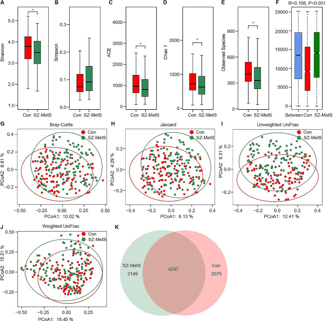
After sequencing, we obtained 13,459,449 high-quality sequence reads from 16,582,495 raw sequence reads, with an average of 59,819 reads per sample. Data normalization to 30,000 reads per sample was performed for microbiota analysis, identifying 8,521 bacterial OTUs across the cohort. Comparing α-diversity indices between SZ-MetS patients and HCs (Fig. 1A-E), we observed reduced bacterial diversity (Shannon index) and richness (observed species, ACE, and Chao1) in SZ-MetS patients (p < 0.05). β-diversity analysis indicated significant differences in fecal microbiota composition between SZ-MetS and HC groups (ANOSIM R = 0.106, p = 0.001; Fig. 1F). PCoA using multiple algorithms (unweighted UniFrac, weighted UniFrac, Bray-Curtis, and Jaccard) clustered SZ-MetS patients and HCs into distinct groups (ADONIS test: p < 0.01; Fig. 1G-J). Additionally, a Venn diagram revealed slightly more unique bacterial phylotypes in SZ-MetS patients (2,149 OTUs) compared to HCs (2,075 OTUs) (Fig. 1K). These results underscore a discernible divergence in the overall structure of the fecal microbiota between SZ-MetS patients and HCs.
Fig. 1.
Comparison of the overall structure of the fecal microbiota between SZ-MetS patients and healthy controls. (A-E) α-diversity indices (Shannon and Simpson) and richness indices (ACE, Chao1, and observed species) were utilized to assess the overall structure of fecal microbiota. Data are presented as mean ± standard deviation. Unpaired two-tailed t-tests were employed for inter-group comparisons. (F) Analysis of similarities (ANOSIM) demonstrated significant differences in fecal microbiota composition between SZ-MetS patients and healthy controls. (G-J) Principal coordinate analysis (PCoA) plots illustrate β-diversity of individual fecal microbiota based on Bray–Curtis, Jaccard, unweighted UniFrac, and weighted UniFrac distances. Each symbol represents a sample. (K) Venn diagram depicts overlap of Operational Taxonomic Units (OTUs) in microbiota associated with SZ-MetS and healthy controls
Fecal microbiota dysbiosis in SZ-MetS patients
Taxonomic composition
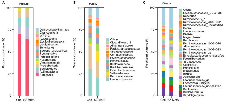
We compared the fecal microbiota of SZ-MetS patients and HCs at various taxonomic levels, categorizing sequencing reads into 15 phyla, 127 families, and 418 genera using the RDP classifier. Figure 2 displays differences in microbiota composition at the phylum, family, and genus levels. Firmicutes, Actinobacteria, Proteobacteria, Bacteroidetes, and Verrucomicrobia were predominant, comprising over 98.9% of the total sequences.
Fig. 2.
Taxonomic compositions of fecal microbiota in SZ-MetS patients and healthy controls. (A) Relative abundance of major phyla. (B) Relative abundance of major families. (C) Relative abundance of major genera
Specific bacterial taxa differences
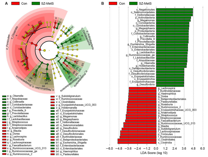
We employed the LEfSe method to identify key functional bacterial taxa with an average relative abundance exceeding 0.01%. Many key functional taxa at multiple taxonomic levels were identified (LDA score > 3.5, p < 0.05, Fig. 3). Figure 3A illustrates cladograms highlighting differentiating biomarkers from phylum to genus levels between SZ-MetS patients and HCs. Figure 3B highlights potential microbiota biomarkers at various taxonomic levels in SZ-MetS patients, with notable genera including Megamonas, Megasphaera, Collinsella, Prevotella_9, Escherichia_Shigella, Lactobacillus, Olsenella, Holdemanella, and Desulfovibrio.
Fig. 3.
Differential bacterial taxa in the fecal microbiota between the SZ-MetS patients and healthy controls. (A) LEfSe cladograms illustrating bacterial taxa significantly associated with SZ-MetS patients (green) or healthy controls (red). The size of each circle in the cladogram corresponds to the relative abundance of the bacterial taxon. Circles represent taxonomic levels from inner to outer: phylum, class, order, family, and genus. Statistical significance was determined using the Wilcoxon rank-sum test with p < 0.05. (B) Histogram depicting the distribution of Linear Discriminant Analysis (LDA) scores (> 3.5) for bacterial taxa that show the greatest differences in abundance between SZ-MetS patients and healthy controls (p < 0.05)
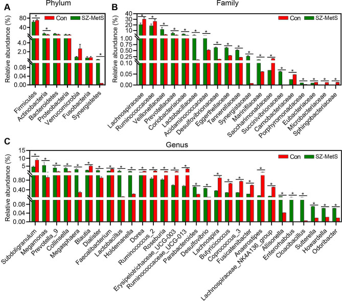
We also used the MetaStats 2.0 package to compare bacterial community profiles. Actinobacteria and Synergistetes were enriched in SZ-MetS patients, while Firmicutes were decreased at the phylum level (Fig. 4A). At the family level, SZ-MetS patients showed elevated levels of 12 families such as Veillonellaceae, Prevotellaceae, Coriobacteriaceae, Lactobacillaceae, Acidaminococcaceae, and Desulfovibrionaceae, with decreases observed in seven families like Lachnospiraceae and Ruminococcaceae (Fig. 4B). Consistent with LEfSe results, 29 genera exhibited differential expression between SZ-MetS patients and HCs (Fig. 4C). 14 genera known for butyrate production such as Subdoligranulum, Blautia, Faecalibacterium, Dorea, and Ruminococcus_2 were notably reduced in SZ-MetS patients, while 15 genera associated with acetate production like Megamonas, Prevotella_9, Collinsella, Megasphaera, Dialister, Lactobacillus, Holdemanella, and Desulfovibrio were enriched.
Fig. 4.
Comparisons of the relative abundance of abundant bacterial taxa in the fecal microbiota between SZ-MetS patients and healthy controls. (A) Key differential functional phyla. (B) Key differential functional families. (C) Key differential functional genera. The data are presented as the mean ± standard deviation. Data were analyzed using Mann–Whitney U-tests to assess differences between SZ-MetS patients (n = 114) and healthy controls (n = 111). * p < 0.05 compared with the control group
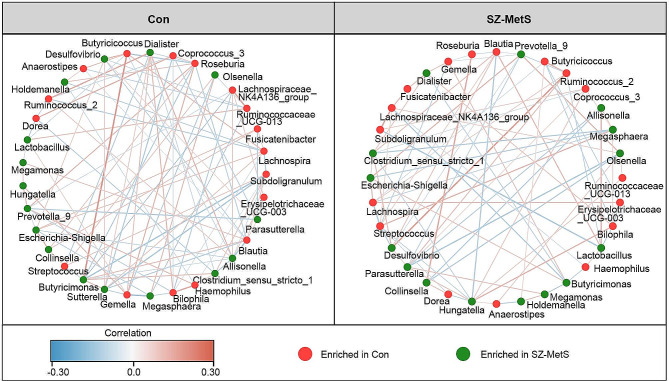
Additionally, we constructed a co-occurrence network among these key functional genera using SparCC on relative abundance data (Fig. 5), revealing a simpler co-occurrence pattern in SZ-MetS patients compared to HCs. These findings collectively indicate significant fecal microbiota dysbiosis in SZ-MetS patients.
Fig. 5.
Co-occurrence network of abundant fecal genera in SZ-MetS patients and controls. Co-occurrence network was constructed using the SparCC algorithm applied to relative abundance data at the genus level, illustrating potential ecological interactions within the microbial community. Cytoscape version 3.6.1 was used for network construction. The red and blue lines represent positive and negative correlations, respectively
Clinical relevance
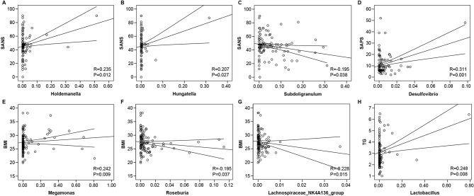
To assess the clinical relevance of fecal microbiota disturbances, we conducted Spearman rank correlations between key functional genera and clinical indicators in SZ-MetS. Our analysis revealed significant associations with indicators such as SANS, SAPS, BMI, and triglyceride (TG) levels (Fig. 6). Holdemanella and Hungatella, enriched in SZ-MetS, positively correlated with SANS (Fig. 6A, B), while the decreased genus Subdoligranulum showed a negative correlation (Fig. 6C). Desulfovibrio correlated positively with SAPS (Fig. 6D). Megamonas correlated positively with BMI (Fig. 6E), whereas Roseburia and Lachnospiraceae_NK4A136_group showed negative and positive correlations with BMI, respectively (Fig. 6F, G). Additionally, Lactobacillus exhibited a positive correlation with TG levels, suggesting a potential role in lipid metabolism (Fig. 6H). These findings highlight the significant links between altered fecal microbiota in SZ-MetS and clinical and metabolic characteristics.
Fig. 6.
Correlation analyses between SZ-MetS associated genera and clinical indicators. Spearman rank correlation analyses reveal significant associations between specific bacterial genera and clinical indicators in SZ-MetS patients, which underscore potential contributions of gut microbiota to metabolic and psychiatric health parameters in SZ-MetS patients. (A-C) Holdemanella, Hungatella, and Subdoligranulum show significant positive correlations with SANS (Scale for the Assessment of Negative Symptoms). (D) Desulfovibrio correlates positively with SAPS (Scale for the Assessment of Positive Symptoms). (E-G) Megamonas, Roseburia, and Lachnospiraceae_NK4A136_group exhibit positive correlations with BMI (Body Mass Index). (H) Lactobacillus correlates positively with TG (Triglycerides)
Differences in bacterial metabolic pathways in SZ-MetS patients versus HCs
Inferred microbiota function
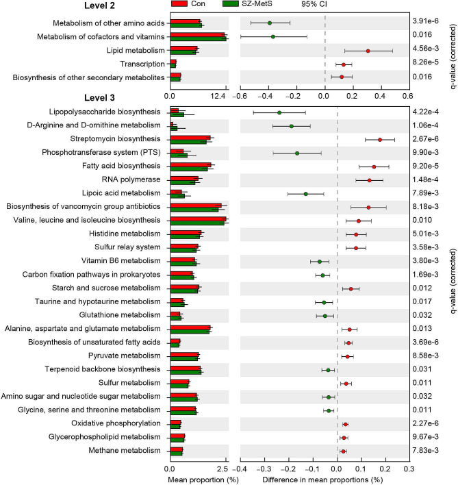
We used PiCRUSt to predict the functional profiles of microbial communities based on closed-reference OTUs (Fig. 7). Comparing 64 KEGG pathways at level 2 revealed five distinct categories between SZ-MetS patients and HCs. SZ-MetS patients showed enriched pathways in cofactor and vitamin metabolism, as well as metabolism of other amino acids, but reduced pathways in lipid metabolism, transcription, and biosynthesis of secondary metabolites. At KEGG level 3, 26 metabolic pathways differed significantly between two groups. (p < 0.05). SZ-MetS microbiota were enriched in 11 pathways, including LPS biosynthesis, PTS, lipoic acid metabolism, and vitamin B6 metabolism. Conversely, 15 pathways, such as fatty acid biosynthesis, histidine metabolism, starch and sucrose metabolism, pyruvate metabolism, biosynthesis of unsaturated fatty acids, alanine, aspartate, and glutamate metabolism, oxidative phosphorylation, and glycerophospholipid metabolism, were significantly reduced. These alterations in the functional profiles of fecal microbiota in SZ-MetS patients were linked to decreased metabolic activity and increased inflammation. MetS were characterized by low-grade inflammation and disrupted lipid and amino acid metabolism, features also seen in SZ [31]. These findings suggest that dysregulated microbial metabolic pathways might contribute to the pathogenesis and progression of both SZ and MetS.
Fig. 7.
PiCRUSt-based examination of the fecal microbiota of the SZ-MetS patients and controls. PiCRUSt analysis was employed to assess differences in bacterial functions between SZ-MetS patients and controls using a two-sided Welch’s t-test. Comparative analyses of KEGG functional categories (levels 2 and 3) are presented as percentages. Multiple testing correction based on the false discovery rate (FDR) was applied using the Benjamini–Hochberg method through STAMP
Correlation analysis between key functional taxa and inferred functions
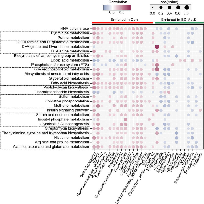
To link differential key functional genera with altered KEGG pathways at level 3 (Fig. 8), Pearson’s correlation analyses were conducted. Genera enriched in controls, like Subdoligranulum and Faecalibacterium, positively correlated with KEGG pathways such as carbohydrate metabolism, amino acid metabolism, lipid metabolism, energy metabolism, and nucleotide metabolism (p < 0.05), but negatively correlated with LPS biosynthesis, lipoic acid metabolism, and PTS (p < 0.05). In contrast, genera enriched in SZ-MetS patients, such as Lactobacillus, Escherichia-Shigella, and Dialister, exhibited predominantly negative correlations with dysregulated KEGG pathways, except for Megamonas. Notably, Megamonas positively correlated with glycerophospholipid metabolism, phenylalanine, tyrosine, and tryptophan biosynthesis, D-Arginine and D-ornithine metabolism, PTS, as well as the insulin signaling pathway (p < 0.05). These findings highlight a significant link between dysregulated metabolic pathways and altered fecal microbiota, suggesting that key functional bacteria in the microbiota may play a crucial role in the pathogenesis of SZ-MetS.
Fig. 8.
Correlation between key functional genera and KEGG metabolic pathways in the SZ-MetS patients. The figure depicts correlations between key functional bacterial genera and KEGG metabolic pathways in SZ-MetS patients. Colors indicate Pearson correlation coefficients: red denotes significant positive correlations (p < 0.05), blue indicates significant negative correlations (p < 0.05), and white signifies non-significant correlations (p > 0.05)
Immunological disturbance correlated with differential genera in SZ-MetS patients
Immunological findings
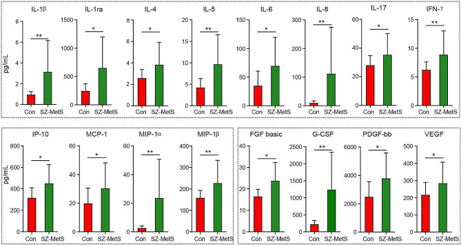
Using the Bio-Plex Pro Human Cytokine 27-Plex Assay on the Bio-Rad MAGPIX Multiplex Reader, we observed elevated levels of inflammatory cytokines such as IL-1β, IL-1ra, IL-4, IL-5, IL-6, IL-8, IL-17, and IFN-γ, alongside chemokines including IP-10, MCP-1, MIP-1α, and MIP-1β in SZ-MetS patients (see Fig. 9, p < 0.05 for each). This suggests the presence of chronic systemic low-grade inflammation in SZ-MetS.
Fig. 9.
Circulating immune dysfunction in SZ-MetS patients. Bio-Plex multiplexed immunoassays employ fluorescently colored magnetic beads to simultaneously quantify 27 inflammatory cytokines, chemokines, and growth factors in SZ-MetS patients compared to controls. Mean concentrations (pg/ml) with standard error of the mean (SEM) are provided, and statistical significance levels are indicated as follows: *p < 0.05, **p < 0.01
Microbiota-immune interactions
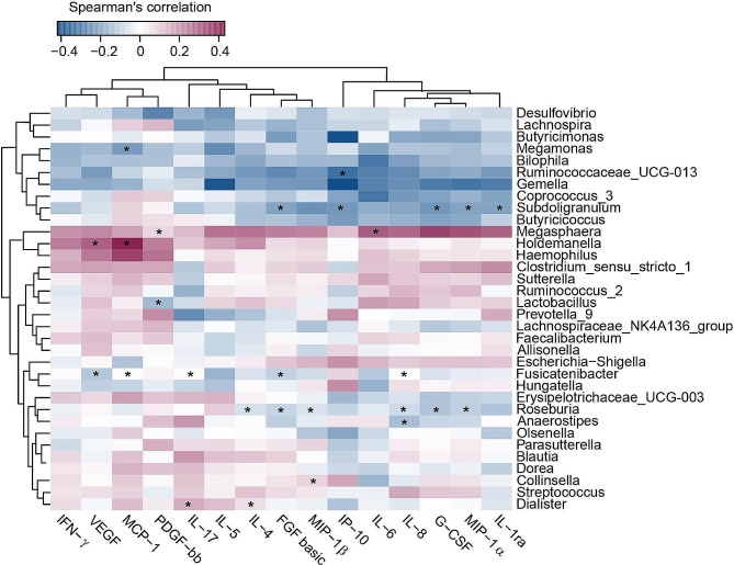
We conducted Spearman’s correlation analysis to explore the interaction between differential key functional genera in SZ-MetS patients and circulating inflammatory markers (Fig. 10). The decreased genera in SZ-MetS patients, such as Subdoligranulum, exhibited negative correlations with IL-1ra, MIP-1α, IP-10, FGF basic, and G-CSF levels. Additionally, Megamonas exhibited a negative correlation with MCP-1. On the other hand, the genera enriched in SZ-MetS patients, like Megasphaera, showed positive correlations with IL-6 and PDGF-bb. Holdemanella had a positive correlation with MCP-1 and VEGF, while Dialister exhibited a positive correlation with IL-4 and IL-17. These correlation analyses suggest that the differential bacteria associated with SZ-MetS may contribute to changes in the host immune response, potentially playing a direct role in the pathophysiology of SZ-MetS.
Fig. 10.
Spearman correlation heatmap between key functional differential genera and circulating inflammatory markers in SZ-MetS patients. The heatmap illustrates Spearman’s rank correlations (r) and associated probabilities (p-values) between key functional differential genera of gut microbiota and circulating inflammatory cytokines, chemokines, and growth factors in SZ-MetS patients. Significant correlations (*p < 0.05) are indicated
Discussion
The gut microbiota serves as a pivotal “metabolic organ”, essential for nutrient absorption, energy regulation, and metabolic homeostasis. A healthy gut microbiota is integral to maintaining host homeostasis [32], while a dysbiotic gut microbiota has been linked to a range of metabolic disorders, including MetS, obesity, and type 2 diabetes [33–36]. Furthermore, the gut microbiota exerts influence over neurochemical pathways via the gut-brain axis [37], thereby impacting brain development and behavior [38]. Due to its intricate and complex communication network between gut and brain, the gut microbiota is frequently termed the “second brain” [39], potentially playing a role in the onset of various psychiatric disorders, including SZ [40, 41]. Yuan et al. have outlined that gut dysbiosis might play a role in SZ by causing neuronal damage and disrupting brain development [42]. Emerging evidence suggests that the gut microbiota may serve as a potential bridge connecting between MetS and SZ [43–48]. People with SZ have a higher risk of developing MetS, which can lead to cardiovascular disease and premature mortality. Both MetS and SZ show gut microbiota changes, hinting at a potential link. However, the exact mechanisms behind gut dysbiosis and its connection to SZ-MetS are still being explored. One important proposed mechanism is the involvement of the gut microbiota in regulating inflammation and immune function. Chronic low-grade inflammation and immune dysregulation are common in both MetS and SZ, contributing to cognitive dysfunction related to metabolism in SZ patients [43]. Gut dysbiosis, including bacterial composition and metabolites, may drive this persistent low-grade inflammation [49]. Correcting immune dysfunction by targeting the gut microbiota holds promise for mitigating the progression of SZ-MetS. Consequently, unraveling the precise role of the gut microbiota in SZ-MetS could pave the way for novel therapeutic strategies, ultimately improving the quality of life for individuals affected by SZ-MetS.
A recent study has revealed a strong correlation between gut dysbiosis and the onset and progression of SZ [41]. However, the changes in gut microbiota among patients with SZ-MetS have received limited attention. In our current cross-sectional case-control study, significant alterations in the gut microbiota of individuals with SZ-MetS were observed compared to HCs. Unlike previous studies in SZ patients, decreased α-diversity and richness indices were observed in SZ-MetS cohort [13, 16], while increased α-diversity was found in Chinese male SZ-MetS patients when compared SZ patients without MetS [14]. Regarding β-diversity, the changes in SZ-MetS were relatively consistent with previous studies in SZ [13, 16]. PCoA revealed substantial differences in microbiota composition at a community level between the two groups. The observed alterations in both α-diversity (within-sample diversity) and β-diversity (between-sample diversity) indicate a more pronounced gut microbiota dysbiosis in SZ-MetS patients, indicating that the coexistence of SZ and MetS may exacerbate disruptions in the gut microbiota. In addition to changes in bacterial diversity, specific shifts in gut microbiota composition were identified in SZ-MetS patients. Notably, there was a decrease in genera with anti-inflammatory properties and butyrate production, such as Subdoligranulum, Blautia, Faecalibacterium, Dorea, and Ruminococcus_2. Conversely, 15 genera known for pro-inflammatory properties and acetate production, including Megamonas, Prevotella_9, Collinsella, Megasphaera, Dialister, Lactobacillus, Holdemanella, and Desulfovibrio, increased. These changes in gut microbiota in SZ-MetS patients align with previous research on SZ [8, 17, 50–52]. Interestingly, the enriched genera in SZ-MetS like Holdemanella, Hungatella, Desulfovibrio, Megamonas and Lactobacillus correlated positively with clinical markers such as SANS, SAPS, BMI, and TG. In contrast, decreased genera like Subdoligranulum, Roseburia and Lachnospiraceae_NK4A136 showed negative correlations with SZ-MetS-associated indicators, suggesting their potential as biomarkers for diagnosing SZ-MetS. Xing et al. constructed a diagnostic model using five microorganisms (Epulopiscium, p_75_a5, Dialister, Blautia, and Desulfovibrio) that effectively distinguished between SZ patients with and without MetS (AUC = 0.94) [14]. Li et al. found that altered genera such as Corynebacterium and Succinvibrio were linked to SZ symptom severity (PANSS scores) [51]. Schwarz et al. observed an increase in Lactobacillus in first-episode SZ, correlating with symptom severity [52]. Tasi et al. reported gender-dependent alterations in microbiota composition in SZ patients with central obesity (female BMI: 26.2 kg/m2; waist circumstance > 80 cm) [53]. Additionally, the functional prediction of gut microbiota has identified altered core metabolic pathways, suggesting that the changed SZ-MetS microbiota could directly regulate host metabolism, including lipid and amino acid metabolism, and promote the production of inflammatory inducers like LPS. These pathways are linked with inflammatory cytokines and coronary heart disease risk in SZ [54]. Obviously, the altered SZ-MetS microbiota, divided into two distinct clusters, were associated with increased cytokines, chemokines, and growth factors, indicating disrupted immune homeostasis. This immune imbalance in SZ and MetS correlated with altered key functional genera. Genera enriched in SZ-MetS microbiota positively correlated with inflammatory cytokines, whereas decreased genera were negatively correlated. The reduction in protective bacteria and increase in inflammatory mediators induced by SZ-MetS-associated microbiota might disturb metabolic homeostasis, activate the peripheral immune system, damage the blood-brain barrier (BBB), and trigger neuroinflammation, ultimately contributing to the onset of SZ-MetS. These findings suggest that gut microbiota dysbiosis in SZ-MetS exacerbates inflammation and metabolic disruptions, highlighting potential mechanisms underlying SZ-MetS. However, further research should prioritize more mechanistic studies to better understand these pathways.
Recent ongoing research has highlighted the role of gut microbiota metabolites as regulators of the gut-brain axis, particularly in SZ-MetS as mentioned above. Changes in the gut microbiota associated with SZ-MetS, particularly in bacteria producing short-chain fatty acids (SCFAs), have been observed. These SCFAs play critical roles in regulating host energy metabolism and immune homeostasis [55, 56]. A recent study has observed that changes in fecal SCFA levels correlate with subclinical inflammation and poorer cognitive performance in individuals with SZ [57]. Reductions in specific butyrate-producing bacteria within the SZ-MetS-associated gut microbiota, such as Subdoligranulum, Blautia, Faecalibacterium, Dorea, and Ruminococcus_2, may have a detrimental impact on SZ-MetS progression. Butyrate, a key metabolite in various physiological processes, can cross the BBB and regulate the microbiota-gut-brain axis, thereby influencing brain function [58, 59]. Butyrate produced by the gut microbiota has been shown to mitigate LPS-induced inflammation in various brain cells and attenuate the expression of pro-inflammatory cytokines such as IL-1β. However, there is still a lack of detailed mechanistic understanding regarding how butyrate affects the brain and behavior. Butyrate, produced by gut microbiota, has demonstrated anti-inflammatory effects by mitigating LPS-induced inflammation in microglia, hippocampal neurons, and co-cultures with microglia or astrocytes in rodents [60]. Matt et al. has demonstrated that butyrate can attenuate the expression of IL-1β in microglia in aged mice [61]. The anti-inflammatory effect induced by butyrate may directly or indirectly improve cognitive impairments in SZ, such as executive function, attention, memory, and language [59]. Research indicates that butyrate could help normalize behavioral and physiological abnormalities in drug-naïve first-episode SZ [62], and ameliorate cognitive impairment and neuronal spine loss induced by quinolinic acid in obesity models, along with promoting BDNF levels [63]. Moreover, butyrate shows promise in improving metabolic impairments like obesity, hypertension, dyslipidemia, hyperglycemia, and insulin resistance in animal and cell models [64, 65]. Exogenous butyrate supplementation has been shown to reduce weight gain by decreasing food intake and enhancing energy metabolism [66]. Given its beneficial effects on both SZ and MetS, enhancing butyrate-producing gut bacteria may be a valuable therapeutic approach. However, the SZ-MetS gut microbiota also shows an increase in pro-inflammatory bacteria and acetate-producing genera, consistent with previous SZ studies, highlighting acetate’s potential physiological effects and its impact on mental health [50, 52]. Acetate, another important SCFA, has been implicated in various physiological processes, including metabolic regulation and mental health impacts. Recently, Erny et al. identified acetate as a key microbiota-derived SCFA that drives microglia maturation and regulates their homeostatic metabolic state. They also demonstrated that acetate could influence microglial phagocytosis and affect disease progression during neurodegeneration [67]. Perry et al. also discovered that elevated acetate production in high-fat diet (HFD)-fed rodents mediated a microbiome-brain-β-cell axis, promoting MetS. This led to increased food intake (hyperphagia), elevated triglyceride levels (hypertriglyceridemia), ectopic lipid accumulation in the liver and skeletal muscle, as well as insulin resistance in both liver and muscle tissues [68]. Fecal microbiota transplantation studies further supported that the gut microbiota is the primary source of increased endogenous acetate production in HFD-fed rats. This heightened acetate production activates the parasympathetic nervous system, leading to increased glucose-stimulated insulin secretion, elevated ghrelin secretion, hyperphagia, and ultimately, obesity [68]. Given these findings, while butyrate shows promise for its anti-inflammatory and metabolic benefits, the role and mechanisms of acetate in SZ-MetS patients need further investigation. Based on current evidence, modulating the gut microbiota by supplementing with butyrate-producing bacteria and inhibiting acetate-producing bacteria may be considered a viable therapeutic approach for treating SZ-MetS. Further studies will be essential to understand the precise roles and interactions of these SCFAs and their impact on SZ-MetS.
Given the intimate link between gut microbiota and SZ-MetS, manipulating the gut microbiota could offer a dual therapeutic potential for both SZ and MetS. Schmitt et al. found that interventions targeting MetS, such as physical activity, psychosocial support, and dietary changes, can effectively alleviate SZ symptoms [69]. Recent evidence strongly suggests that gut microbiota could help treat antipsychotic-induced MetS and cognitive impairment in SZ [70]. Previous studies have identified that prolonged use of antipsychotics like olanzapine and clozapine is associated with high metabolic risks [71–73]. While olanzapine effectively treats SZ symptoms and reduces relapse chances, it often causes significant weight gain, increasing the risk of MetS and cardiovascular disease [74]. Studies showed that gut microbiota is essential for olanzapine-induced weight gain [74, 75]. Bahr et al. also reported that patients undergoing antipsychotic treatment exhibited a progressive elevation in the Firmicutes/Bacteroidetes ratio, correlating with higher BMI [72]. This suggested that modifying specific components of the gut microbiota through supplementation or removal could mitigate these metabolic side effects [76]. Huang et al. found that probiotics and dietary fiber can mitigate antipsychotic-induced weight gain in drug-naïve, first-episode SZ patients [77]. Prebiotics like galacto-oligosaccharides alongside olanzapine also decreased weight and metabolic disturbances in female rats by modulating the gut microbiota [78, 79]. Sevillano-Jiménez et al. found that an individualized nutritional education program incorporating a high content of prebiotics and probiotics (including dairy and fermented foods, green leafy vegetables, high-fiber fruits, and whole grains) effectively improves the cardio-metabolic profile in SZ [80], while another study confirmed the efficacy of dietary fiber and probiotics, alone and combined, in reducing metabolic side effects of atypical antipsychotic medications [81]. Ghaderi et al. showed that a 12-week supplementation with a probiotic mix (Bifidobacterium and Lactobacillus) and vitamin D significantly reduced metabolic abnormalities and improved psychiatric symptoms in SZ patients [82]. In another randomized, double-blind, placebo-controlled trial, the same group found that probiotics (comprising B. lactis, B. bifidum, B. longum, and L. acidophilus) combined with selenium improved general SZ symptoms and metabolic profiles [83]. Huang et al. observed that a subtype of Akkermansia muciniphila can improve olanzapine-related hyperglycemia and insulin resistance, restore intestinal barrier function, as well as alleviate systemic inflammation, thereby improving treatment adherence, quality of life, and functional outcomes of patients with SZ-MetS [84]. However, several studies also demonstrated that probiotic administration had no significant difference on the PANSS scale [85, 86], suggesting that the efficacy for SZ-MetS may depend on the specific probiotic strains used. Therefore, while the use of prebiotics and probiotics in treating SZ and its comorbidities like MetS shows promise, it remains in the early stages of research [70]. These findings suggest that gut microbiota modulation could serve as an adjunct treatment for SZ-MetS, potentially enhancing medication tolerance, improving patient quality of life, and reducing cardio-metabolic disorder risks.
However, there are several limitations in our study. Firstly, we did not gather complete clinical information for all SZ-MetS patients, including detailed parameters of MetS and PANSS scale scores. Additionally, while all SZ-MetS patients were maintained on stable antipsychotic treatment, the specific types of antipsychotics were not standardized, potentially influencing gut microbiota composition. Moreover, changes in dietary and living conditions during hospitalization could serve as confounding factors affecting gut microbiota. Secondly, despite the relatively large sample size of participants, the cross-sectional design with a discovery cohort can only be considered preliminary and cannot be further extrapolated. This limitation thereby restricts the ability to draw definitive conclusions regarding the role of gut microbiota in SZ-MetS subjects. Future studies using independent validation cohorts will be necessary to confirm and extend these findings, providing more robust evidence on the relationship between gut microbiota and SZ-MetS subjects. Thirdly, this study focused only on the associations among gut microbiota, immune disturbance and SZ-MetS, without investigating the causal relationship among them. To confirm the causality between gut microbiota dysbiosis and SZ-MetS development, larger, prospective longitudinal cohort studies or intervention studies are needed. Finally, our study only included Chinese participants, and the findings may not be generalizable to other ethnic groups, including Chinese ethnic minority groups, or populations from westernized countries, as the gut microbiota may be subjected to varying degrees of environmental, dietary, and genetic influences. Multicenter, multi-ethnic cohort studies will be essential to validate and extend our findings. Despite these limitations, our study laid the groundwork for further investigations into the roles and mechanisms of the key functional bacteria in SZ-MetS.
Conclusions
In summary, unlike previous studies explored gut dysbiosis in MetS or SZ separately, our study highlights the link between gut dysbiosis and immune dysfunction in SZ-MetS based on a larger cohort of Chinese SZ-MetS patients. The structural and functional dysbiosis of gut microbiota may influence peripheral inflammation in the host, actively contributing to the development of SZ-MetS. Specific changes in certain gut microbes, such as reduced butyrate-producing bacteria and enriched acetate-producing bacteria, were correlated with the clinical features and chronic low-grade inflammation of SZ-MetS, suggesting their pivotal roles in SZ-MetS pathogenesis. Our findings suggest that interventions targeting these specific microbiota changes, such as probiotics, prebiotics, or dietary modifications, could potentially mitigate the onset or progression of SZ-MetS. However, our study primarily focuses on Chinese SZ-MetS patients, limiting generalizability to other ethnic or geographic populations. Further large-scale, multicenter, multi-ethnic cohort studies, both longitudinal and interventional, are essential to offer comprehensive insights into the role of gut microbiota in SZ-MetS. Longitudinal studies are crucial as they can help establish causality and mechanisms by tracking changes in microbiota composition over time and correlating these changes with clinical outcomes in SZ-MetS patients. The randomized, double-blind, placebo-controlled interventional trials will be pivotal in exploring the therapeutic potential of modulating gut microbiota in SZ-MetS. Together, these findings collectively help to decipher the intricate links between gut microbiota, immune dysregulation, and disease pathogenesis in SZ-MetS, and pave the way for innovative, microbiota-based treatments aimed at preventing and treating SZ-MetS.
Electronic supplementary material
Below is the link to the electronic supplementary material.
Abbreviations
- ACE
Abundance-based Coverage Estimator
- ADONIS
Multivariate ANOVA based on similarity tests
- ANOSIM
Analysis of Similarities
- ATEP III
Adult Treatment Panel III
- BBB
Blood-brain barrier
- BMI
Body mass index
- CV
Coefficient of variation
- DSM-V
Diagnostic and Statistical Manual for Psychiatric Disorders-Fifth Version
- FDR
False Discovery Rate
- FGF basic
Fibroblast Growth Factor basic
- G-CSF
Granulocyte Colony-Stimulating Factor
- HCs
Healthy controls
- HFD
High-fat diet
- IFN-γ
Interferon-gamma
- IL
Interleukin
- IP-10
Interferon gamma-induced protein 10
- KEGG
Kyoto Encyclopedia of Genes and Genomes
- LDA
Linear discriminant analysis
- LEfSe
Linear discriminant analysis effect size
- LPS
Lipopolysaccharide
- MCP-1
Monocyte Chemoattractant Protein-1
- MetS
Metabolic syndrome
- MIP-1α
Macrophage Inflammatory Protein-1 alpha
- MIP-1β
Macrophage Inflammatory Protein-1 beta
- OTU
Operational Taxonomic Unit
- PANSS
Positive and Negative Syndrome Scale
- PCoA
Principal coordinate analysis
- PDGF-bb
Platelet-Derived Growth Factor bb
- PERMANOVA
Permutational Multivariate Analysis of Variance
- PiCRUSt
Phylogenetic investigation of communities by reconstruction of unobserved states
- QIIME2
Quantitative Insights into Microbial Ecology Version 2
- RDP
Ribosomal Database Project
- rRNA
Ribosomal RNA
- SANS
Scale for Assessment of Negative Symptoms
- SAPS
Scale for Assessment of Positive Symptoms
- SCFA
Short-chain fatty acid
- SparCC
Sparse compositional correlation
- STAMP
Statistical Analysis of Metagenomic Profiles
- SZ
Schizophrenia
- SZ-MetS
Schizophrenia comorbid with metabolic syndrome
- TG
Triglyceride
- VEGF
Vascular Endothelial Growth Factor
Author contributions
Z.X.L., Z.Y.L., and R.X.L. conceived and designed the experiments. Z.X.L., Z.Y.L.Y.W.C., X.L., Z.M.L., Y.Y., Y.W.W., L.S., Z.C.Z., J.G., W.H.L., W.W.D., and R.X.L. performed the experiments. Z.X.L., Y.W.C., X.L., and S.L. analyzed the data. Z.X.L., Z.Y.L., Y.W.C., X.L., and R.X.L. wrote the paper and edited the manuscript. All authors contributed to the article and approved the submitted version.
Funding
This present work was funded by the grants of the National S&T Major Project of China (2023YFC2308400), Key R&D Program of Quzhou (2023K187), Shandong Provincial Laboratory Project (SYS202202), the Fundamental Research Funds for the Central Universities (2022ZFJH003), the Research Project of Jinan Microecological Biomedicine Shandong Laboratory (JNL-2022033 C), the Taishan Scholar Foundation of Shandong Province (tsqn202103119), and the Foundation of China’s State Key Laboratory for Diagnosis and Treatment of Infectious Diseases (ZZ202316 and ZZ202319).
Data availability
The sequence data from this study have been deposited in the GenBank Sequence Read Archive (https://www.ncbi.nlm.nih.gov/sra) under the accession number PRJNA1034806.
Declarations
Competing interests
The authors declare that they have no known competing financial interests or personal relationships that could have appeared to influence the work reported in this paper.
Footnotes
Publisher’s Note
Springer Nature remains neutral with regard to jurisdictional claims in published maps and institutional affiliations.
Zongxin Ling, Zhiyong Lan, Yiwen Cheng and Xia Liu contributed equally to this work.
Contributor Information
Zongxin Ling, Email: lingzongxin@zju.edu.cn.
Rongxian Liao, Email: rongxian_liao@163.com.
References
- 1.Marder SR, Cannon TD, Schizophrenia. N Engl J Med. 2019;381(18):1753–61. 10.1056/NEJMra1808803 [DOI] [PubMed] [Google Scholar]
- 2.Solmi M, Seitidis G, Mavridis D, Correll CU, Dragioti E, Guimond S, Tuominen L, Dargél A, Carvalho AF, Fornaro M, et al. Incidence, prevalence, and global burden of schizophrenia - data, with critical appraisal, from the global burden of Disease (GBD) 2019. Mol Psychiatry. 2023;28(12):5319–27. 10.1038/s41380-023-02138-4 [DOI] [PubMed] [Google Scholar]
- 3.Plana-Ripoll O, Pedersen CB, Agerbo E, Holtz Y, Erlangsen A, Canudas-Romo V, Andersen PK, Charlson FJ, Christensen MK, Erskine HE, et al. A comprehensive analysis of mortality-related health metrics associated with mental disorders: a nationwide, register-based cohort study. Lancet. 2019;394(10211):1827–35. 10.1016/S0140-6736(19)32316-5 [DOI] [PubMed] [Google Scholar]
- 4.Erlangsen A, Andersen PK, Toender A, Laursen TM, Nordentoft M, Canudas-Romo V. Cause-specific life-years lost in people with mental disorders: a nationwide, register-based cohort study. Lancet Psychiatry. 2017;4(12):937–45. 10.1016/S2215-0366(17)30429-7 [DOI] [PubMed] [Google Scholar]
- 5.Vancampfort D, Stubbs B, Mitchell AJ, De Hert M, Wampers M, Ward PB, Rosenbaum S, Correll CU. Risk of metabolic syndrome and its components in people with schizophrenia and related psychotic disorders, bipolar disorder and major depressive disorder: a systematic review and meta-analysis. World Psychiatry. 2015;14(3):339–47. 10.1002/wps.20252 [DOI] [PMC free article] [PubMed] [Google Scholar]
- 6.Grover S, R P, Sahoo S, Gopal S, Nehra R, Ganesh A, Raghavan V, Sankaranarayan A. Relationship of metabolic syndrome and neurocognitive deficits in patients with schizophrenia. Psychiatry Res. 2019;278:56–64. 10.1016/j.psychres.2019.05.023 [DOI] [PubMed] [Google Scholar]
- 7.Perry BI, Upthegrove R, Thompson A, Marwaha S, Zammit S, Singh SP, Khandaker G. Dysglycaemia, inflammation and psychosis: findings from the UK ALSPAC Birth Cohort. Schizophr Bull. 2019;45(2):330–8. 10.1093/schbul/sby040 [DOI] [PMC free article] [PubMed] [Google Scholar]
- 8.Zhu F, Ju Y, Wang W, Wang Q, Guo R, Ma Q, Sun Q, Fan Y, Xie Y, Yang Z, et al. Metagenome-wide association of gut microbiome features for schizophrenia. Nat Commun. 2020;11(1):1612. 10.1038/s41467-020-15457-9 [DOI] [PMC free article] [PubMed] [Google Scholar]
- 9.Bora E, Akdede BB, Alptekin K. The relationship between cognitive impairment in schizophrenia and metabolic syndrome: a systematic review and meta-analysis. Psychol Med. 2017;47(6):1030–40. 10.1017/S0033291716003366 [DOI] [PubMed] [Google Scholar]
- 10.Arango C, Bobes J, Aranda P, Carmena R, Garcia-Garcia M, Rejas J. A comparison of schizophrenia outpatients treated with antipsychotics with and without metabolic syndrome: findings from the CLAMORS study. Schizophr Res. 2008;104(1–3):1–12. 10.1016/j.schres.2008.05.009 [DOI] [PubMed] [Google Scholar]
- 11.Dabke K, Hendrick G, Devkota S. The gut microbiome and metabolic syndrome. J Clin Invest. 2019;129(10):4050–7. 10.1172/JCI129194 [DOI] [PMC free article] [PubMed] [Google Scholar]
- 12.Jastroch M, Ussar S, Keipert S, Gut. Microbes Controlling Blood Sugar: No Fire Required! Cell Metab. 2020;31(3):443–4. [DOI] [PubMed] [Google Scholar]
- 13.Ling Z, Jin G, Yan X, Cheng Y, Shao L, Song Q, Liu X, Zhao L. Fecal dysbiosis and Immune Dysfunction in Chinese Elderly patients with Schizophrenia: an observational study. Front Cell Infect Microbiol. 2022;12:886872. 10.3389/fcimb.2022.886872 [DOI] [PMC free article] [PubMed] [Google Scholar]
- 14.Xing M, Gao H, Yao L, Wang L, Zhang C, Zhu L, Cui D. Profiles and diagnostic value of intestinal microbiota in schizophrenia patients with metabolic syndrome. Front Endocrinol (Lausanne). 2023;14:1190954. 10.3389/fendo.2023.1190954 [DOI] [PMC free article] [PubMed] [Google Scholar]
- 15.Xu R, Wu B, Liang J, He F, Gu W, Li K, Luo Y, Chen J, Gao Y, Wu Z, et al. Altered gut microbiota and mucosal immunity in patients with schizophrenia. Brain Behav Immun. 2020;85:120–7. 10.1016/j.bbi.2019.06.039 [DOI] [PubMed] [Google Scholar]
- 16.McGuinness AJ, Davis JA, Dawson SL, Loughman A, Collier F, O’Hely M, Simpson CA, Green J, Marx W, Hair C, et al. A systematic review of gut microbiota composition in observational studies of major depressive disorder, bipolar disorder and schizophrenia. Mol Psychiatry. 2022;27(4):1920–35. 10.1038/s41380-022-01456-3 [DOI] [PMC free article] [PubMed] [Google Scholar]
- 17.Murray N, Al Khalaf S, Bastiaanssen TFS, Kaulmann D, Lonergan E, Cryan JF, Clarke G, Khashan AS, O’Connor K. Compositional and functional alterations in intestinal microbiota in patients with psychosis or Schizophrenia: a systematic review and Meta-analysis. Schizophr Bull. 2023;49(5):1239–55. 10.1093/schbul/sbad049 [DOI] [PMC free article] [PubMed] [Google Scholar]
- 18.Arnoriaga-Rodríguez M, Fernández-Real JM. Microbiota impacts on chronic inflammation and metabolic syndrome - related cognitive dysfunction. Rev Endocr Metab Disord. 2019;20(4):473–80. 10.1007/s11154-019-09537-5 [DOI] [PubMed] [Google Scholar]
- 19.National Cholesterol Education Program (NCEP). Expert Panel on Detection E, and treatment of high blood cholesterol in adults (Adult Treatment Panel III). Third report of the National Cholesterol Education Program (NCEP) Expert Panel on detection, evaluation, and treatment of high blood cholesterol in adults (Adult Treatment Panel III) final report. Circulation. 2002;106(25):3143–421. 10.1161/circ.106.25.3143 [DOI] [PubMed] [Google Scholar]
- 20.Ling Z, Cheng Y, Yan X, Shao L, Liu X, Zhou D, Zhang L, Yu K, Zhao L. Alterations of the fecal microbiota in Chinese patients with multiple sclerosis. Front Immunol. 2020;11:590783. 10.3389/fimmu.2020.590783 [DOI] [PMC free article] [PubMed] [Google Scholar]
- 21.Ling Z, Zhu M, Yan X, Cheng Y, Shao L, Liu X, Jiang R, Wu S. Structural and functional dysbiosis of fecal microbiota in Chinese patients with Alzheimer’s Disease. Front Cell Dev Biol. 2020;8:634069. 10.3389/fcell.2020.634069 [DOI] [PMC free article] [PubMed] [Google Scholar]
- 22.Ling Z, Cheng Y, Liu X, Yan X, Wu L, Shao L, Gao J, Lei W, Song Q, Zhao L, et al. Altered oral microbiota and immune dysfunction in Chinese elderly patients with schizophrenia: a cross-sectional study. Transl Psychiatry. 2023;13(1):383. 10.1038/s41398-023-02682-1 [DOI] [PMC free article] [PubMed] [Google Scholar]
- 23.Ling Z, Shao L, Liu X, Cheng Y, Yan C, Mei Y, Ji F, Liu X. Regulatory T Cells and plasmacytoid dendritic cells within the Tumor Microenvironment in Gastric Cancer are correlated with gastric microbiota dysbiosis: a preliminary study. Front Immunol. 2019;10:533. 10.3389/fimmu.2019.00533 [DOI] [PMC free article] [PubMed] [Google Scholar]
- 24.Liu X, Shao L, Liu X, Ji F, Mei Y, Cheng Y, Liu F, Yan C, Li L, Ling Z. Alterations of gastric mucosal microbiota across different stomach microhabitats in a cohort of 276 patients with gastric cancer. EBioMedicine. 2019;40:336–48. 10.1016/j.ebiom.2018.12.034 [DOI] [PMC free article] [PubMed] [Google Scholar]
- 25.Caporaso JG, Kuczynski J, Stombaugh J, Bittinger K, Bushman FD, Costello EK, Fierer N, Pena AG, Goodrich JK, Gordon JI, et al. QIIME allows analysis of high-throughput community sequencing data. Nat Methods. 2010;7(5):335–6. 10.1038/nmeth.f.303 [DOI] [PMC free article] [PubMed] [Google Scholar]
- 26.Lozupone C, Knight R. UniFrac: a new phylogenetic method for comparing microbial communities. Appl Environ Microbiol. 2005;71(12):8228–35. 10.1128/AEM.71.12.8228-8235.2005 [DOI] [PMC free article] [PubMed] [Google Scholar]
- 27.Parks DH, Tyson GW, Hugenholtz P, Beiko RG. STAMP: statistical analysis of taxonomic and functional profiles. Bioinformatics. 2014;30(21):3123–4. 10.1093/bioinformatics/btu494 [DOI] [PMC free article] [PubMed] [Google Scholar]
- 28.Segata N, Izard J, Waldron L, Gevers D, Miropolsky L, Garrett WS, Huttenhower C. Metagenomic biomarker discovery and explanation. Genome Biol. 2011;12(6):R60. 10.1186/gb-2011-12-6-r60 [DOI] [PMC free article] [PubMed] [Google Scholar]
- 29.Langille MG, Zaneveld J, Caporaso JG, McDonald D, Knights D, Reyes JA, Clemente JC, Burkepile DE, Vega Thurber RL, Knight R, et al. Predictive functional profiling of microbial communities using 16S rRNA marker gene sequences. Nat Biotechnol. 2013;31(9):814–21. 10.1038/nbt.2676 [DOI] [PMC free article] [PubMed] [Google Scholar]
- 30.Ling Z, Zhu M, Liu X, Shao L, Cheng Y, Yan X, Jiang R, Wu S. Fecal fungal dysbiosis in Chinese patients with Alzheimer’s Disease. Front Cell Dev Biol. 2020;8:631460. 10.3389/fcell.2020.631460 [DOI] [PMC free article] [PubMed] [Google Scholar]
- 31.Chen X, Xu J, Tang J, Dai X, Huang H, Cao R, Hu J. Dysregulation of amino acids and lipids metabolism in schizophrenia with violence. BMC Psychiatry. 2020;20(1):97. 10.1186/s12888-020-02499-y [DOI] [PMC free article] [PubMed] [Google Scholar]
- 32.Cheng Y, Liu X, Chen F, Rolnik BM, Chleilat F, Ling Z, Snyder MP, Zhou X. The Roles and Mechanisms of Gut Microbiota in Food Allergy. Advanced Gut & Microbiome Research. 2023, 2023:9575410.
- 33.He Y, Wu W, Wu S, Zheng HM, Li P, Sheng HF, Chen MX, Chen ZH, Ji GY, Zheng ZD, et al. Linking gut microbiota, metabolic syndrome and economic status based on a population-level analysis. Microbiome. 2018;6(1):172. 10.1186/s40168-018-0557-6 [DOI] [PMC free article] [PubMed] [Google Scholar]
- 34.Vijay-Kumar M, Aitken JD, Carvalho FA, Cullender TC, Mwangi S, Srinivasan S, Sitaraman SV, Knight R, Ley RE, Gewirtz AT. Metabolic syndrome and altered gut microbiota in mice lacking toll-like receptor 5. Science. 2010;328(5975):228–31. 10.1126/science.1179721 [DOI] [PMC free article] [PubMed] [Google Scholar]
- 35.Zhao L, Zhang F, Ding X, Wu G, Lam YY, Wang X, Fu H, Xue X, Lu C, Ma J, et al. Gut bacteria selectively promoted by dietary fibers alleviate type 2 diabetes. Science. 2018;359(6380):1151–6. 10.1126/science.aao5774 [DOI] [PubMed] [Google Scholar]
- 36.Aron-Wisnewsky J, Vigliotti C, Witjes J, Le P, Holleboom AG, Verheij J, Nieuwdorp M, Clément K. Gut microbiota and human NAFLD: disentangling microbial signatures from metabolic disorders. Nat Rev Gastroenterol Hepatol. 2020;17(5):279–97. 10.1038/s41575-020-0269-9 [DOI] [PubMed] [Google Scholar]
- 37.Fung TC, Olson CA, Hsiao EY. Interactions between the microbiota, immune and nervous systems in health and disease. Nat Neurosci. 2017;20(2):145–55. 10.1038/nn.4476 [DOI] [PMC free article] [PubMed] [Google Scholar]
- 38.Diaz Heijtz R, Wang S, Anuar F, Qian Y, Björkholm B, Samuelsson A, Hibberd ML, Forssberg H, Pettersson S. Normal gut microbiota modulates brain development and behavior. Proc Natl Acad Sci U S A. 2011;108(7):3047–52. 10.1073/pnas.1010529108 [DOI] [PMC free article] [PubMed] [Google Scholar]
- 39.Ridaura V, Belkaid Y. Gut microbiota: the link to your second brain. Cell. 2015;161(2):193–4. 10.1016/j.cell.2015.03.033 [DOI] [PubMed] [Google Scholar]
- 40.Vasileva SS, Yang Y, Baker A, Siskind D, Gratten J, Eyles D. Associations of the gut Microbiome with Treatment Resistance in Schizophrenia. JAMA Psychiatry. 2024;81(3):292–302. 10.1001/jamapsychiatry.2023.5371 [DOI] [PMC free article] [PubMed] [Google Scholar]
- 41.Nikolova VL, Smith MRB, Hall LJ, Cleare AJ, Stone JM, Young AH. Perturbations in Gut Microbiota Composition in Psychiatric disorders: a review and Meta-analysis. JAMA Psychiatry. 2021;78(12):1343–54. 10.1001/jamapsychiatry.2021.2573 [DOI] [PMC free article] [PubMed] [Google Scholar]
- 42.Yuan X, Kang Y, Zhuo C, Huang XF, Song X. The gut microbiota promotes the pathogenesis of schizophrenia via multiple pathways. Biochem Biophys Res Commun. 2019;512(2):373–80. 10.1016/j.bbrc.2019.02.152 [DOI] [PubMed] [Google Scholar]
- 43.Zeng C, Yang P, Cao T, Gu Y, Li N, Zhang B, Xu P, Liu Y, Luo Z, Cai H. Gut microbiota: an intermediary between metabolic syndrome and cognitive deficits in schizophrenia. Prog Neuropsychopharmacol Biol Psychiatry. 2021;106:110097. 10.1016/j.pnpbp.2020.110097 [DOI] [PubMed] [Google Scholar]
- 44.Singh R, Stogios N, Smith E, Lee J, Maksyutynsk K, Au E, Wright DC, De Palma G, Graff-Guerrero A, Gerretsen P, et al. Gut microbiome in schizophrenia and antipsychotic-induced metabolic alterations: a scoping review. Ther Adv Psychopharmacol. 2022;12:20451253221096525. 10.1177/20451253221096525 [DOI] [PMC free article] [PubMed] [Google Scholar]
- 45.Vasileva SS, Tucker J, Siskind D, Eyles D. Does the gut microbiome mediate antipsychotic-induced metabolic side effects in schizophrenia? Expert Opin Drug Saf. 2022;21(5):625–39. 10.1080/14740338.2022.2042251 [DOI] [PubMed] [Google Scholar]
- 46.Ma J, Song XQ. Correlation between cognitive impairment and metabolic imbalance of gut microbiota in patients with schizophrenia. World J Psychiatry. 2023;13(10):724–31. 10.5498/wjp.v13.i10.724 [DOI] [PMC free article] [PubMed] [Google Scholar]
- 47.Wu H, Liu Y, Wang J, Chen S, Xie L, Wu X. Schizophrenia and obesity: may the gut microbiota serve as a link for the pathogenesis? iMeta. 2023;2(2):e99. 10.1002/imt2.99 [DOI] [PMC free article] [PubMed] [Google Scholar]
- 48.Amdanee N, Shao M, Hu X, Fang X, Zhou C, Chen J, Ridwan Chattun M, Wen L, Pan X, Zhang X, et al. Serum Metabolic Profile in Schizophrenia patients with antipsychotic-Induced Constipation and its relationship with gut microbiome. Schizophr Bull. 2023;49(3):646–58. 10.1093/schbul/sbac202 [DOI] [PMC free article] [PubMed] [Google Scholar]
- 49.Deng H, He L, Wang C, Zhang T, Guo H, Zhang H, Song Y, Chen B. Altered gut microbiota and its metabolites correlate with plasma cytokines in schizophrenia inpatients with aggression. BMC Psychiatry. 2022;22(1):629. 10.1186/s12888-022-04255-w [DOI] [PMC free article] [PubMed] [Google Scholar]
- 50.Shen Y, Xu J, Li Z, Huang Y, Yuan Y, Wang J, Zhang M, Hu S, Liang Y. Analysis of gut microbiota diversity and auxiliary diagnosis as a biomarker in patients with schizophrenia: a cross-sectional study. Schizophr Res. 2018;197:470–7. 10.1016/j.schres.2018.01.002 [DOI] [PubMed] [Google Scholar]
- 51.Li S, Zhuo M, Huang X, Huang Y, Zhou J, Xiong D, Li J, Liu Y, Pan Z, Li H, et al. Altered gut microbiota associated with symptom severity in schizophrenia. PeerJ. 2020;8:e9574. 10.7717/peerj.9574 [DOI] [PMC free article] [PubMed] [Google Scholar]
- 52.Schwarz E, Maukonen J, Hyytiäinen T, Kieseppä T, Orešič M, Sabunciyan S, Mantere O, Saarela M, Yolken R, Suvisaari J. Analysis of microbiota in first episode psychosis identifies preliminary associations with symptom severity and treatment response. Schizophr Res. 2018;192:398–403. 10.1016/j.schres.2017.04.017 [DOI] [PubMed] [Google Scholar]
- 53.Tsai YL, Liu YW, Wang PN, Lin CY, Lan TH. Gender differences in gut Microbiome Composition between Schizophrenia patients with normal body weight and central obesity. Front Psychiatry. 2022;13:836896. 10.3389/fpsyt.2022.836896 [DOI] [PMC free article] [PubMed] [Google Scholar]
- 54.Nguyen TT, Kosciolek T, Daly RE, Vázquez-Baeza Y, Swafford A, Knight R, Jeste DV. Gut microbiome in Schizophrenia: altered functional pathways related to immune modulation and atherosclerotic risk. Brain Behav Immun. 2021;91:245–56. 10.1016/j.bbi.2020.10.003 [DOI] [PMC free article] [PubMed] [Google Scholar]
- 55.Koh A, De Vadder F, Kovatcheva-Datchary P, Bäckhed F. From Dietary Fiber to host physiology: short-chain fatty acids as key bacterial metabolites. Cell. 2016;165(6):1332–45. 10.1016/j.cell.2016.05.041 [DOI] [PubMed] [Google Scholar]
- 56.Li Z, Qing Y, Cui G, Li M, Liu T, Zeng Y, Zhou C, Hu X, Jiang J, Wang D, et al. Shotgun metagenomics reveals abnormal short-chain fatty acid-producing bacteria and glucose and lipid metabolism of the gut microbiota in patients with schizophrenia. Schizophr Res. 2023;255:59–66. 10.1016/j.schres.2023.03.005 [DOI] [PubMed] [Google Scholar]
- 57.Kowalski K, Szponar B, Bochen P, Żebrowska-Różańska P, Łaczmański Ł, Samochowiec J, Misiak B. Altered levels of fecal short-chain fatty acids are associated with subclinical inflammation and worse cognitive performance in patients with schizophrenia. J Psychiatr Res. 2023;165:298–304. 10.1016/j.jpsychires.2023.07.042 [DOI] [PubMed] [Google Scholar]
- 58.O’Riordan KJ, Collins MK, Moloney GM, Knox EG, Aburto MR, Fülling C, Morley SJ, Clarke G, Schellekens H, Cryan JF. Short chain fatty acids: microbial metabolites for gut-brain axis signalling. Mol Cell Endocrinol. 2022;546:111572. 10.1016/j.mce.2022.111572 [DOI] [PubMed] [Google Scholar]
- 59.Dalile B, Van Oudenhove L, Vervliet B, Verbeke K. The role of short-chain fatty acids in microbiota-gut-brain communication. Nat Rev Gastroenterol Hepatol. 2019;16(8):461–78. 10.1038/s41575-019-0157-3 [DOI] [PubMed] [Google Scholar]
- 60.Huuskonen J, Suuronen T, Nuutinen T, Kyrylenko S, Salminen A. Regulation of microglial inflammatory response by sodium butyrate and short-chain fatty acids. Br J Pharmacol. 2004;141(5):874–80. 10.1038/sj.bjp.0705682 [DOI] [PMC free article] [PubMed] [Google Scholar]
- 61.Matt SM, Allen JM, Lawson MA, Mailing LJ, Woods JA, Johnson RW. Butyrate and Dietary Soluble Fiber improve Neuroinflammation Associated with Aging in mice. Front Immunol. 2018;9:1832. 10.3389/fimmu.2018.01832 [DOI] [PMC free article] [PubMed] [Google Scholar]
- 62.Li X, Fan X, Yuan X, Pang L, Hu S, Wang Y, Huang X, Song X. The role of Butyric Acid in Treatment Response in Drug-Naïve First Episode Schizophrenia. Front Psychiatry. 2021;12:724664. 10.3389/fpsyt.2021.724664 [DOI] [PMC free article] [PubMed] [Google Scholar]
- 63.Ge X, Zheng M, Hu M, Fang X, Geng D, Liu S, Wang L, Zhang J, Guan L, Zheng P, et al. Butyrate ameliorates quinolinic acid-induced cognitive decline in obesity models. J Clin Invest. 2023;133(4):e154612. 10.1172/JCI154612 [DOI] [PMC free article] [PubMed] [Google Scholar]
- 64.Bridgeman SC, Northrop W, Melton PE, Ellison GC, Newsholme P, Mamotte CDS. Butyrate generated by gut microbiota and its therapeutic role in metabolic syndrome. Pharmacol Res. 2020;160:105174. 10.1016/j.phrs.2020.105174 [DOI] [PubMed] [Google Scholar]
- 65.Li J, Zhao F, Wang Y, Chen J, Tao J, Tian G, Wu S, Liu W, Cui Q, Geng B, et al. Gut microbiota dysbiosis contributes to the development of hypertension. Microbiome. 2017;5(1):14. 10.1186/s40168-016-0222-x [DOI] [PMC free article] [PubMed] [Google Scholar]
- 66.Li Z, Yi CX, Katiraei S, Kooijman S, Zhou E, Chung CK, Gao Y, van den Heuvel JK, Meijer OC, Berbée JFP, et al. Butyrate reduces appetite and activates brown adipose tissue via the gut-brain neural circuit. Gut. 2018;67(7):1269–79. 10.1136/gutjnl-2017-314050 [DOI] [PubMed] [Google Scholar]
- 67.Erny D, Dokalis N, Mezö C, Castoldi A, Mossad O, Staszewski O, Frosch M, Villa M, Fuchs V, Mayer A, et al. Microbiota-derived acetate enables the metabolic fitness of the brain innate immune system during health and disease. Cell Metab. 2021;33(11):2260–e22762267. 10.1016/j.cmet.2021.10.010 [DOI] [PubMed] [Google Scholar]
- 68.Perry RJ, Peng L, Barry NA, Cline GW, Zhang D, Cardone RL, Petersen KF, Kibbey RG, Goodman AL, Shulman GI. Acetate mediates a microbiome-brain-β-cell axis to promote metabolic syndrome. Nature. 2016;534(7606):213–7. 10.1038/nature18309 [DOI] [PMC free article] [PubMed] [Google Scholar]
- 69.Schmitt A, Maurus I, Rossner MJ, Röh A, Lembeck M, von Wilmsdorff M, Takahashi S, Rauchmann B, Keeser D, Hasan A, et al. Effects of Aerobic Exercise on metabolic syndrome, Cardiorespiratory Fitness, and symptoms in Schizophrenia Include decreased mortality. Front Psychiatry. 2018;9:690. 10.3389/fpsyt.2018.00690 [DOI] [PMC free article] [PubMed] [Google Scholar]
- 70.Liu JCW, Gorbovskaya I, Hahn MK, Müller DJ. The gut microbiome in Schizophrenia and the potential benefits of prebiotic and probiotic treatment. Nutrients. 2021;13(4):1152. 10.3390/nu13041152 [DOI] [PMC free article] [PubMed] [Google Scholar]
- 71.Gorbovskaya I, Kanji S, Liu JCW, MacKenzie NE, Agarwal SM, Marshe VS, Sriretnakumar V, Verdu EF, Bercik P, De Palma G, et al. Investigation of the gut microbiome in patients with Schizophrenia and Clozapine-Induced Weight Gain: protocol and clinical characteristics of first patient cohorts. Neuropsychobiology. 2020;79(1):5–12. 10.1159/000494696 [DOI] [PubMed] [Google Scholar]
- 72.Bahr SM, Tyler BC, Wooldridge N, Butcher BD, Burns TL, Teesch LM, Oltman CL, Azcarate-Peril MA, Kirby JR, Calarge CA. Use of the second-generation antipsychotic, risperidone, and secondary weight gain are associated with an altered gut microbiota in children. Transl Psychiatry. 2015;5(10):e652. 10.1038/tp.2015.135 [DOI] [PMC free article] [PubMed] [Google Scholar]
- 73.Ye W, Xing J, Yu Z, Hu X, Zhao Y. Mechanism and treatments of antipsychotic-induced weight gain. Int J Obes (Lond). 2023;47(6):423–33. 10.1038/s41366-023-01291-8 [DOI] [PubMed] [Google Scholar]
- 74.Qian L, He X, Liu Y, Gao F, Lu W, Fan Y, Gao Y, Wang W, Zhu F, Wang Y, et al. Longitudinal gut microbiota dysbiosis underlies Olanzapine-Induced Weight Gain. Microbiol Spectr. 2023;11(4):e0005823. 10.1128/spectrum.00058-23 [DOI] [PMC free article] [PubMed] [Google Scholar]
- 75.Morgan AP, Crowley JJ, Nonneman RJ, Quackenbush CR, Miller CN, Ryan AK, Bogue MA, Paredes SH, Yourstone S, Carroll IM, et al. The antipsychotic olanzapine interacts with the gut microbiome to cause weight gain in mouse. PLoS ONE. 2014;9(12):e115225. 10.1371/journal.pone.0115225 [DOI] [PMC free article] [PubMed] [Google Scholar]
- 76.Mötteli S, Vetter S, Colla M, Hotzy F. Are probiotics effective in reducing the metabolic side effects of psychiatric medication? A scoping review of evidence from clinical studies. Transl Psychiatry. 2024;14(1):26. 10.1038/s41398-024-02735-z [DOI] [PMC free article] [PubMed] [Google Scholar]
- 77.Huang J, Kang D, Zhang F, Yang Y, Liu C, Xiao J, Long Y, Lang B, Peng X, Wang W, et al. Probiotics Plus Dietary Fiber supplements Attenuate Olanzapine-Induced Weight Gain in Drug-Naïve First-Episode Schizophrenia patients: two randomized clinical trials. Schizophr Bull. 2022;48(4):850–9. 10.1093/schbul/sbac044 [DOI] [PMC free article] [PubMed] [Google Scholar]
- 78.Kao AC, Spitzer S, Anthony DC, Lennox B, Burnet PWJ. Prebiotic attenuation of olanzapine-induced weight gain in rats: analysis of central and peripheral biomarkers and gut microbiota. Transl Psychiatry. 2018;8(1):66. 10.1038/s41398-018-0116-8 [DOI] [PMC free article] [PubMed] [Google Scholar]
- 79.Kao AC, Burnet PWJ, Lennox BR. Can prebiotics assist in the management of cognition and weight gain in schizophrenia? Psychoneuroendocrinology 2018, 95:179–85. [DOI] [PubMed]
- 80.Sevillano-Jiménez A, Romero-Saldaña M, García-Mellado JA, Carrascal-Laso L, García-Rodríguez M, Molina-Luque R, Molina-Recio G. Impact of high prebiotic and probiotic dietary education in the SARS-CoV-2 era: improved cardio-metabolic profile in schizophrenia spectrum disorders. BMC Psychiatry. 2022;22(1):781. 10.1186/s12888-022-04426-9 [DOI] [PMC free article] [PubMed] [Google Scholar]
- 81.Liu C, Kang D, Xiao J, Huang Y, Peng X, Wang W, Xie P, Yang Y, Zhao J, Wu R. Dietary fiber and probiotics for the treatment of atypical antipsychotic-induced metabolic side effects: study protocol for a randomized, double-blind, placebo-controlled trial. Trials. 2021;22(1):159. 10.1186/s13063-021-05123-w [DOI] [PMC free article] [PubMed] [Google Scholar]
- 82.Ghaderi A, Banafshe HR, Mirhosseini N, Moradi M, Karimi MA, Mehrzad F, Bahmani F, Asemi Z. Clinical and metabolic response to vitamin D plus probiotic in schizophrenia patients. BMC Psychiatry. 2019;19(1):77. 10.1186/s12888-019-2059-x [DOI] [PMC free article] [PubMed] [Google Scholar]
- 83.Jamilian H, Ghaderi A. The effects of Probiotic and Selenium Co-supplementation on Clinical and metabolic scales in Chronic Schizophrenia: a Randomized, Double-blind, placebo-controlled trial. Biol Trace Elem Res. 2021;199(12):4430–8. 10.1007/s12011-020-02572-3 [DOI] [PubMed] [Google Scholar]
- 84.Huang D, Gao J, Li C, Nong C, Huang W, Zheng X, Li S, Peng Y. A potential probiotic bacterium for antipsychotic-induced metabolic syndrome: mechanisms underpinning how Akkermansia muciniphila subtype improves olanzapine-induced glucose homeostasis in mice. Psychopharmacology. 2021;238(9):2543–53. 10.1007/s00213-021-05878-9 [DOI] [PubMed] [Google Scholar]
- 85.Dickerson FB, Stallings C, Origoni A, Katsafanas E, Savage CL, Schweinfurth LA, Goga J, Khushalani S, Yolken RH. Effect of probiotic supplementation on schizophrenia symptoms and association with gastrointestinal functioning: a randomized, placebo-controlled trial. Prim Care Companion CNS Disord. 2014;16(1):PCC13m01579. [DOI] [PMC free article] [PubMed] [Google Scholar]
- 86.Tomasik J, Yolken RH, Bahn S, Dickerson FB. Immunomodulatory effects of Probiotic supplementation in Schizophrenia patients: a Randomized, Placebo-Controlled Trial. Biomark Insights. 2015;10:47–54. 10.4137/BMI.S22007 [DOI] [PMC free article] [PubMed] [Google Scholar]
Associated Data
This section collects any data citations, data availability statements, or supplementary materials included in this article.
Supplementary Materials
Data Availability Statement
The sequence data from this study have been deposited in the GenBank Sequence Read Archive (https://www.ncbi.nlm.nih.gov/sra) under the accession number PRJNA1034806.


