Abstract
The fission yeast Mcs6–Mcs2–Pmh1 complex, homologous to metazoan Cdk7–cyclin H-Mat1, has dual functions in cell division and transcription: as a partially redundant cyclin-dependent kinase (CDK)-activating kinase (CAK) that phosphorylates the major cell cycle CDK, Cdc2, on Thr-167; and as the RNA polymerase (Pol) II carboxyl-terminal domain (CTD) kinase associated with transcription factor (TF) IIH. We analyzed conditional mutants of mcs6 and pmh1, which activate Cdc2 normally but cannot complete cell division at restrictive temperature and arrest with decreased CTD phosphorylation. Transcriptional profiling by microarray hybridization revealed only modest effects on global gene expression: a one-third reduction in a severe mcs6 mutant after prolonged incubation at 36°C. In contrast, a small subset of transcripts (∼5%) decreased by more than twofold after Mcs6 complex function was compromised. The signature of repressed genes overlapped significantly with those of cell separation mutants sep10 and sep15. Sep10, a component of the Pol II Mediator complex, becomes essential in mcs6 or pmh1 mutant backgrounds. Moreover, transcripts dependent on the forkhead transcription factor Sep1, which are expressed coordinately during mitosis, were repressed in Mcs6 complex mutants, and Mcs6 also interacts genetically with Sep1. Thus, the Mcs6 complex, a direct activator of Cdc2, also influences the cell cycle transcriptional program, possibly through its TFIIH-associated kinase function.
INTRODUCTION
Cyclin-dependent kinases (CDKs) play a central role in driving cell division in eukaryotic organisms and also perform essential, conserved functions in the transcription cycle of RNA polymerase (Pol) II (reviewed by Morgan, 1997). Whereas most CDKs can be classified as either cell cycle or transcriptional regulators based on a preeminent physiological function, the metazoan Cdk7 complex is essential in both cell division and gene expression, as the CDK-activating kinase (CAK) and as a component of the general transcription factor IIH (TFIIH) (reviewed by Harper and Elledge, 1998). To execute its dual functions in vivo, Cdk7 has evolved distinct substrate specificities for the activation segment (T-loop) of CDKs and the carboxy-terminal domain (CTD) of the Pol II large subunit, and mechanisms to allow their independent regulation (Garrett et al., 2001; Larochelle et al., 2001). Less clear is how (or whether) the Cdk7 complex serves to coordinate cell division with gene expression.
To address this question genetically, we turned to the fission yeast Schizosaccharomyces pombe, which also relies, in part, on a dual-function CDK complex to activate CDKs and phosphorylate Pol II (Buck et al., 1995; Damagnez et al., 1995; Hermand et al., 1998; Lee et al., 1999; Saiz and Fisher, 2002). The orthologue of Cdk7 in S. pombe is Mcs6, which associates with the cyclin Mcs2 and the RING-finger protein Pmh1. Both mcs6 and mcs2 were identified in genetic screens for positive regulators of Cdc2 (S. pombe Cdk1), the major cell cycle CDK (Molz et al., 1989; Molz and Beach, 1993), whereas pmh1 was identified through genome sequencing (Spåhr et al., 2003; our unpublished observations). A combination of biochemical and genetic evidence has established the Mcs6 complex as a CAK (Buck et al., 1995; Damagnez et al., 1995; Lee et al., 1999; Saiz and Fisher, 2002). Fission yeast also contains a second CAK, Csk1, which can maintain near-normal levels of Cdc2 activation in mcs6 mutant backgrounds (Lee et al., 1999; Saiz and Fisher, 2002). Consistent with redundancy of CAK function between Csk1 and Mcs6, csk1+ is not essential for viability of otherwise wild-type cells (Molz and Beach, 1993), but deletion of csk1 is synthetically lethal in combination with hypomorphic mcs6 mutations (Hermand et al., 1998; Lee et al., 1999; Saiz and Fisher, 2002).
In contrast to both fission yeast and metazoans, the budding yeast Saccharomyces cerevisiae has a single, non–cyclin-dependent CAK, Cak1, which is encoded by an essential gene (Espinoza et al., 1996; Kaldis et al., 1996; Thuret et al., 1996). The orthologue of Cdk7 in budding yeast is Kin28, which associates with TFIIH (Feaver et al., 1994), but is not a CAK and has a substrate specificity apparently restricted to components of the transcription machinery (Cismowski et al., 1995; Valay et al., 1995; Liu et al., 2004). Conditional kin28 mutants arrest growth with no discrete cell cycle timing but with diminished CTD phosphorylation and severely decreased levels of most Pol II transcripts (Cismowski et al., 1995; Valay et al., 1995; Holstege et al., 1998).
To investigate the role of the TFIIH-associated kinase in gene expression in an organism in which it also serves as a CAK, we created conditional alleles of two of the three subunits of the Mcs6 complex, all of which are encoded by essential genes. Temperature-sensitive mutants of mcs6 (Saiz and Fisher, 2002) and pmh1 (this report) arrest at restrictive temperature as multinucleate cells with uncleaved division septa and decreased CTD phosphorylation but with normal levels of CDK T-loop phosphorylation. This phenotype is morphologically and biochemically distinct from the classical cell cycle arrest due to CAK deficiency when both Mcs6 and Csk1 are inactivated by mutation (Lee et al., 1999; Saiz and Fisher, 2002), but it resembles the cell separation defects of mutants in sep10 and sep15 (Zilahi et al., 2000a; Szilagyi et al., 2002). Both Sep10 and Sep15 are components of the Pol II Mediator (Spåhr et al., 2001, 2003; Linder and Gustafsson, 2004), a large complex that transduces regulatory signals between transcriptional activator proteins bound at their cognate DNA-binding sites and the basal machinery engaged at the core promoter (reviewed by Rachez and Freedman, 2001).
Genome-wide expression profiling of Mcs6 complex mutants revealed modest reductions in general transcription but severe defects in transcribing a small subset of genes (∼5% of total). The signature of repressed genes includes a cluster of cell cycle-regulated transcripts involved in cytokinesis and cell separation (Rustici et al., 2004) and overlaps with genes down-regulated in mutants of sep10 and sep15. Moreover, transcripts dependent on the forkhead transcription factor Sep1 (Ribar et al., 1997) for periodic, cell cycle-regulated expression are repressed in mcs6 and pmh1 mutants. The Mcs6 complex also interacts genetically with both sep1 and sep10. These studies uncover a critical role of the core Pol II transcription machinery in coordinating the periodic transcription of genes important for cell division— and a second cell cycle function of the Mcs6 complex as part of TFIIH—and indicate that wild-type levels of Mcs6 activity are not generally required for mRNA synthesis in fission yeast.
MATERIALS AND METHODS
Yeast Methods
Strains used in these studies are listed in Table 1. Yeast cell culture and transformation were performed according to published methods (Moreno et al., 1991). To screen an S. pombe cDNA library in λZAPII (gift of J. Hurwitz, Memorial Sloan-Kettering Cancer Center), an ∼350-base pair probe was amplified by PCR with primers derived from the genomic sequence and labeled with the Multiprime system (Amersham Biosciences, Piscataway, NJ). Positive clones were completely sequenced on both strands.
Table 1.
Strains used in this study
| Strain | Genotype | Source |
|---|---|---|
| JS77 | leu1-32 ura4-D18 his3-D1 ade6-M216 h- | J. Hurwitz |
| JS78 | leu1-32 ura4-D18 his3-D1 ade6-M210 h+ | J. Hurwitz |
| KL3 | leu1-32/leu1-32 ura4-D18/ura4-D18 ade6-M216/ade6-M210 his3-D1/his3-D1 h-/h+ | This work |
| KL42 | pmh1::pmh1wtHA3/kanMX4 leu1-32 ura4-D18 his3-D1 ade6-M216 h- | This work |
| KL43 | pmh1::pmh1-26HA3/kanMX4 leu1-32 ura4-D18 his3-D1 ade6-M216 h- | This work |
| JS167 | mcs6::mcs6wtHA3/kanMX4 leu1-32 ura4-D18 his3-D1 ade6-M216 h- | J. Saiz |
| JS169 | mcs6::mcs6ts1HA3/kanMX4 leu1-32 ura4-D18 his3-D1 ade6-M216 h- | J. Saiz |
| JS182 | mcs6::mcs6ts2HA3/kanMX4 leu1-32 ura4-D18 his3-D1 ade6-M216 h- | J. Saiz |
| JS264 | mcs6::mcs6S165A/L238RHA3/kanMX4 leu1-32 ura4-D18 his3-D1 ade6-M216 h- | J. Saiz |
| 2-923 | sep10::ura4+ ura4-D18 lys1-131 h90 | M. Sipiczki |
| 2-871 | sep1-598 ura4-D18 h90 | M. Sipiczki |
To replace the pmh1+ gene with ura4+, the regions flanking the pmh1 coding sequence were amplified by PCR and cloned in pT7 (Stratagene, La Jolla, CA), and the ura4+ marker was cloned between the two flanking sequences. A linear fragment was then excised and used to transform a diploid strain stably maintained by ade– complementation on Edinburgh Minimal Medium (EMM)(–adenine) (Moreno et al., 1991). Transformants were selected in EMM(–uracil) and tested for correct replacement by PCR.
Generation, expression, and screening of mutant pmh1 alleles were carried out as described for the generation of mcs6ts1 and mcs6ts2 (Saiz and Fisher, 2002). Mutagenic PCR was catalyzed by AmpliTaq (Invitrogen, Carlsbad, CA) in reactions containing 1.8 mM MgCl2 + 0.5 mM MnCl2. Of ∼103 clones screened, 30 were selected for a growth defect at 36°C and pmh1-26 was chosen for further characterization. Sequencing revealed eight point mutations: Q17R, N23K, F47S, T56S, L63P, E70G, G95V, and K256R.
pmh1+ was replaced with wild-type and mutant versions fused at the carboxy terminus with three copies of the hemagglutinin (HA) epitope. The tagged pmh1 open reading frames (ORFs) (pmh1+ and pmh1-26) were subcloned into the pFA6a-kanMX4 plasmid. The 5′- and 3′-flanking sequences were then cloned to encompass the tagged pmh1 ORFs and the kanMX marker, in pBluescript SK(–) (Stratagene). The transformation and sporulation of the heterozygous diploid strains were performed as described above but with G418 (Geneticin) selection.
Double mutants with sep10 were constructed by protoplast fusion. The resulting diploid cells were sporulated and tetrads were dissected, resulting in complete and incomplete tetrads. Tests of viable spore clones for temperature sensitivity, kanamycin-resistance, and the presence of sep10 deletion by PCR revealed that the dead spores must have been sep10Δ mcs6ts or sep10Δ pmh1-26 double mutants. Images were captured with an Olympus BH-2 microscope fitted with a DP-70 digital camera and processed with its integrated software package.
Immunological and Biochemical Methods
Harvest and lysis of cells and processing of extracts for immunoblotting and biochemical analysis were performed as described previously (Saiz and Fisher, 2002). Antibodies used were 8WG16 (Covance, Princeton, NJ), specific for the unphosphorylated Pol II CTD; H14 (Covance), specific for the CTD phosphorylated at Ser-5; PSTAIRE (Santa Cruz Biotechnology, Santa Cruz, CA), which recognizes Cdc2; anti-phospho-Cdc2 (Cell Signaling Technology, Beverly, MA), specific for the phospho-Thr-167 form of Cdc2; anti-HA (16B12; Covance); and anti-FLAG (Sigma-Aldrich, St. Louis, MO). Immunoprecipitations were performed with 0.25–1.0 mg of extract incubated with ∼600 ng of antibody and 20 μl of a 1:1 slurry of protein A-Sepharose (Sigma-Aldrich) or protein G-agarose (Amersham Biosciences) for 2 h at 4°C. Cdc2 was precipitated with p13suc1 beads as described previously (Lee et al., 1999). For CTD kinase assays, the immunoprecipitates were washed three times with HBST (10 mM HEPES, pH 7.4, 150 mM NaCl, and 0.1% Triton X-100) and twice with HBSD (10 mM HEPES, pH 7.4, 150 mM NaCl, and 1 mM dithiothreitol [DTT]), and tested for kinase activity toward 5 μg of glutathione S-transferase (GST)-CTD in 30 μl of kinase buffer (HBSD + 10 mM MgCl2, 100 μM ATP, and 5 μCi of [γ-32P]ATP). Kinase reactions were carried out at 25°C for 15 min and stopped by adding sample buffer and heating at 95°C. Phosphorylated substrates were resolved by 10% SDS-PAGE and detected by autoradiography of dried gels.
For gel-exclusion chromatography, ∼1 mg of total extract protein was applied to a Superdex 200 column (Amersham Biosciences) equilibrated in 25 mM HEPES, pH 7.4, 150 mM NaCl, 1 mM EDTA, 1 mM DTT, and 10% glycerol. Fractions of 500 μl were collected; 5% of each was analyzed by immunoblotting.
Construction of Baculoviruses
To construct recombinant baculoviruses encoding wild-type or mutant, HA-tagged Mcs6, NcoI sites were introduced at the 5′ and 3′ ends of the Mcs6 ORF in pBluescript SK(–) to generate pBluescript-NcoI-Mcs6-NcoI, and mutants K40R/K41R (kinase-dead), T160A, S165A, and T160A/S165A were generated by oligonucleotide-directed mutagenesis (Kunkel, 1985) of pBluescript-NcoI-Mcs6-NcoI. The resulting NcoI fragments were subcloned in-frame with the HA epitope in the transfer vector pVLCDK2NHA for generation of baculoviruses (O'Reilly et al., 1993; Rosenblatt et al., 1993). FLAG-tagged Pmh1 baculovirus was generated with the Bac-to-Bac system (Invitrogen).
Microarray Hybridization
For both microarray and Northern blot hybridization, pmh1-26 and mcs6ts1 cells (∼1.5 × 10⁷) were harvested at OD ∼0.2 at various times after shift to 36°C. HA-tagged pmh1+ and mcs6+ strains were grown to OD 0.2 at 28°C and harvested (time 0). The sep10Δ or sep15ts cells were grown overnight at 33°C (semipermissive for sep15ts); the sep10 and sep15 data represent the averages of three biological repeats. Pellets were frozen in liquid N2 and kept at –80°C until further processing. RNA was isolated by phenol extraction and purified with RNeasy (QIAGEN, Valencia, CA). For Northern blot hybridization, gels were stained before transfer with ethidium bromide to ensure equal loading of rRNA, and membranes were hybridized with probes complementary to cdc2+ mRNA to ensure equal transfer (our unpublished data). For microarray hybridization, cDNA probes were prepared with Superscript (Invitrogen) and labeled with Cy3 or Cy5. Detailed methods are described in Lyne et al. (2003) and at: http://www.sanger.ac.uk/PostGenomics/S_pombe/. Microarrays were scanned with a Genepix 4000B scanner and analyzed with Genepix software (Axon Instruments, Union City, CA). Data filtering, normalization, and quality control were performed on all samples according to Lyne et al. (2003).
To quantify global effects on gene expression, we repeated microarray analysis of total RNA samples, from both the mcs6ts1 and pmh1-26 mutants at 0.5 and 2.0 h after shift to 36°C, spiked with external control RNAs (Supplemental Figure S2). The expression data were normalized using bacterial (Bacillus subtilis) and plant (Arabidopsis thaliana) spikes that were added in equal quantities to each of the RNA samples before labeling. Each microarray contains 1440 control elements for the spikes (720 each of bacterial and plant elements), which are spread across the complete grid of the array in a spatially even manner. The script to normalize the data was modified from the one reported previously (Lyne et al., 2003), which uses a sliding window of local spots to account for spatial differences in the Cy dye biases within the arrays. First, all elements were normalized using the experimental S. pombe elements. Using the S. pombe elements rather than the control elements allows for a more robust correction of local Cy biases, because there are many more of these elements throughout the array (van de Peppel et al., 2003). In a second step, a global mean ratio of control element signals, corrected locally in the first round, is applied to all other elements as a correction factor.
Clustering and visualization were done with GeneSpring (Silicon Genetics, Redwood City, CA). Gene annotations were taken from the S. pombe GeneDB database at The Wellcome Trust Sanger Institute (http://www.genedb.org/genedb/pombe/index.jsp). Data are available from ArrayExpress under accession numbers E-MEXP-191, E-MEXP-192, and E-MEXP-193 at http://www.ebi.ac.uk/arrayexpress/. All processed data are available at http://www.sanger.ac.uk/PostGenomics/Spombe/.
RESULTS
Pmh1, an Essential Homolog of Mat1 in Fission Yeast
The product of the pmh1+ gene (Wood et al., 2002; Spåhr et al., 2003) is ∼32% identical to mouse Mat1 and ∼35% identical to Tfb3, with highest homology residing in the amino-terminal region containing the RING-finger domain (Supplemental Figure S1A). We disrupted pmh1+ with ura4+ in a diploid, induced the resulting heterozygote to sporulate and dissected tetrads. We observed a 2:2 segregation of viability (Supplemental Figure S1B) with no recovery of uracil prototrophs, indicating that pmh1+ is essential. On selective germination in medium lacking uracil, pmh1Δ cells completed approximately six divisions before arresting with two DNA masses separated by a division septum (Supplemental Figure S1C); this is identical to mcs2Δ and mcs6Δ phenotypes (Molz and Beach, 1993; Buck et al., 1995; Damagnez et al., 1995). As expected, the lethality could be rescued by overexpression of wild-type Pmh1 but not by a truncated version lacking the RING domain (RING–) (our unpublished data).
Pmh1 Is a Component of the CAK Complex and TFIIH
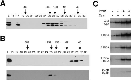
Pmh1 was shown to bind Mcs6, Mcs2, and components of core TFIIH when expressed in S. pombe (Spåhr et al., 2003). Gel-exclusion chromatography of extracts from cells expressing FLAG-tagged Pmh1 revealed two distinct Pmh1-containing complexes, with apparent sizes of ∼200 and ∼700 kDa (Figure 1A). This is reminiscent of the fractionation of Mat1 in mammalian extracts into two populations: a trimeric complex with Cdk7 and cyclin H, which migrates aberrantly with an apparent size of ∼240 kDa; and the >600-kDa TFIIH (Fisher et al., 1995). There was also a minor population at ∼50 kDa, roughly the expected size of a monomer. We next reconstituted the Mcs6–Mcs2–Pmh1 complex in baculovirus-infected insect cells (Figure 1B). Single infection with FLAG-Pmh1 virus yielded the presumed monomer at ∼50 kDa, which upon coexpression with Mcs6 and Mcs2 shifted to ∼200 kDa, approximately the size of the major Pmh1-containing complex in yeast.
Figure 1.
Pmh1 is a component of CAK and TFIIH. (A) Extracts from pmh1Δ yeast transformed with pREP41X-FLAG-pmh1+ and (B) insect cells expressing FLAG-Pmh1 alone (top) and Flag-Pmh1, Mcs6, and Mcs2 (bottom) were fractionated by Superdex 200 gel-filtration chromatography. Fractions were analyzed by immunoblotting with anti-FLAG antibody. We consistently observed two distinct bands of FLAG-Pmh1 in yeast extracts. (C) Activation of Mcs6 by Pmh1 independent of T-loop phosphorylation. Insect cells were coinfected to express Mcs6-HA (wild type, T160A, S165A, T160A, S165A, and the catalytically inactive K40R/K41R) and His-Mcs2 (present in all lanes). As indicated, cells also expressed Csk1 and/or FLAG-Pmh1. Mcs6 complexes were immunoprecipitated with anti-HA antibody and tested for CTD kinase activity.
Mat1 stabilizes active Cdk7–cyclin H complexes in the absence of phosphorylation (Fisher et al., 1995), although T-loop phosphorylation of the trimeric complex increased its CTD kinase activity ∼20-fold (Larochelle et al., 2001). We mutated the potential phosphoacceptor residues of the Mcs6 T-loop, Ser-165 and Thr-160, to alanine to test whether Pmh1 could likewise bypass the need for T-loop phosphorylation by Csk1. We infected insect cells with viruses encoding wild-type or mutant Mcs6 and Mcs2, together with Pmh1 and/or Csk1, and tested immunoprecipitates of HA-tagged Mcs6 protein for CTD kinase activity (Figure 1C). Activity of the wild-type and T160A proteins increased with Csk1 coexpression, consistent with activating phosphorylation at Ser-165 (Hermand et al., 1998; Lee et al., 1999). However, even the unphosphorylatable T160A/S165A mutant could be recovered in active form upon coexpression of Pmh1. Thus, as further evidence of conservation with metazoan Mat1, Pmh1 can facilitate Mcs6 activation in the absence of T-loop phosphorylation.
A pmh1 Mutant Is Defective in Cell Separation
To investigate the essential function(s) of the Mcs6 complex, we created temperature-sensitive mutants of pmh1. From a library of HA-tagged pmh1 point mutants screened for the ability to complement the deletion at 28°C but not at 36°C, we selected one allele, pmh1-26, which had the tightest temperature-sensitive phenotype, for integration and characterization. The temperature-sensitive lethality of the pmh1-26 strain could be suppressed by overexpression of wild-type, but not of RING–, Pmh1 (our unpublished data). The pmh1-26 mutant contains multiple point mutations (see Materials and Methods), two of which are predicted to change conserved, core hydrophobic residues: Phe-47-to-Ser (equivalent to Phe-39 in α-helix 2 of human Mat1) and Leu-63-to-Pro (Leu-53 in loop L2) (Gervais et al., 2001).
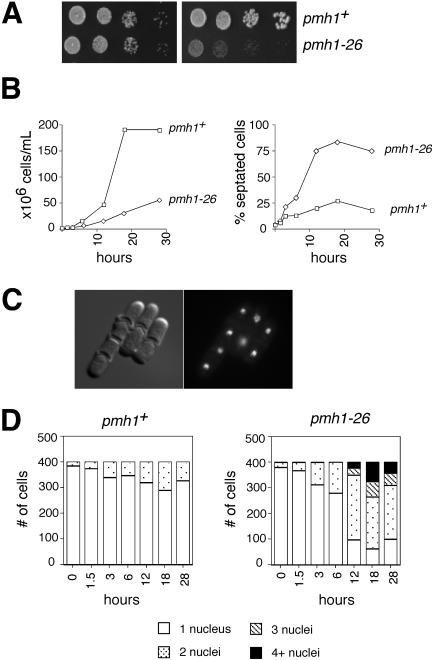
Haploid pmh1-26 cells grew normally at 28°C but stopped dividing at 36°C (Figure 2A) and accumulated with two or more nuclei and a septation index >70% (Figure 2, B and C). After 12 h at 36°C, most cells were binucleate with a single division septum, but there was a fraction with three or more nuclei and multiple septa (Figure 2D). Flow cytometry revealed that the mutant cells accumulated with DNA contents of 4C or more within 6–12 h of the shift to 36°C (our unpublished data).
Figure 2.
Temperature-sensitive lethality and cell separation defects in a pmh1-26 strain. (A) Serial dilutions of pmh1+ and pmh1-26 strains grown at 28 (left) and 36°C (right). (B) Cell number (measured by Coulter counter) and septation index during time course of incubation at 36°C of wild-type and pmh1-26 cells. (C) Micrographs of pmh1-26 cells fixed and stained with 4,6-diamidino-2-phenylindole and calcofluor after 12 h at 36°C. (D) Number of nuclei per cell as a function of time at 36°C in wild-type (left) and pmh1-26 (right) cells.
Thermal Inactivation of Pmh1 Causes Hypophosphorylation of Pol II CTD on Ser-5
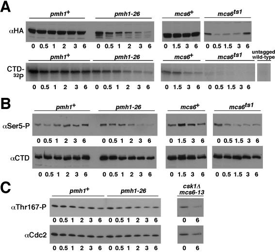
The pmh1-26 phenotype is similar to that of the temperature-sensitive mcs6ts1 mutant we described previously; unlike pmh1-26, however, mcs6ts1 exhibits delayed cell separation even at permissive temperature (Saiz and Fisher, 2002). Consistent with impaired function at 28°C, CTD kinase activity of the mutant Mcs6 protein was decreased, relative to wild-type, in unshifted cells (Figure 3A) and was too low to measure in immunoprecipitates from extracts of cells shifted to 36°C (Figure 3A; Saiz and Fisher, 2002). Nonetheless, Pol II phosphorylation on Ser-5 of the CTD heptad repeat was near normal at 28°C and reduced, relative to wild-type, only upon shift to 36°C (Figure 3B), suggesting some compensation for the diminished Mcs6 activity, perhaps by other kinases and/or by down-regulation of CTD phosphatase, at permissive temperature.
Figure 3.
CTD Ser-5 hypophosphorylation without CAK-deficiency in mcs6 and pmh1 mutants. In the indicated strains, incubated at 36°C for indicated times, we measured levels of wild-type and mutant, HA-tagged Pmh1, and Mcs6 proteins by immunoblotting with anti-HA antibody (A, top) and Pmh1- or Mcs6-associated CTD kinase in anti-HA immunoprecipitates (A, bottom); CTD Ser-5 phosphorylation (B, top) and total Pol II (B, bottom) by immunoblotting of total yeast extracts with antibodies H14 and 8WG16, respectively; and Cdc2 Thr-167 phosphorylation, by immunoblotting of p13Suc1 precipitates (C). As a control, Cdc2 Thr-167 phosphorylation in the CAK-deficient, mcs6-13 csk1Δ strain was analyzed at 28°C and at 6 h after shift to 36°C.
Recovery of total Pmh1 protein and associated CTD kinase activity diminished with increasing time at 36°C in the pmh1-26 but not in the pmh1+ strain, suggesting that the mutant protein is unstable (Figure 3A). Ser-5 phosphorylation decreased in apparent synchrony with Pmh1-associated kinase activity (Figure 3B). Loss of Ser-5 phosphorylation occurred to similar degrees but with different kinetics in mcs6ts1 and pmh1-26 cells, reaching a minimal baseline level after ∼1.5 and ∼3 h, respectively. The cell division arrest, likewise, occurred with different timing in the two mutants; whereas the pmh1-26 cells underwent at least one round of incomplete cell division at 36°C, the mcs6ts1 cells ceased dividing immediately upon shift to the restrictive temperature (Saiz and Fisher, 2002; data not shown).
In the pmh1-26 mutant after shift to restrictive temperature, the level of phosphorylation on Thr-167 of Cdc2 remained unchanged, in contrast to the decrease measured in CAK-deficient, csk1Δ mcs6-13 cells analyzed in parallel (Figure 3C). Thus, as was the case for mcs6 mutants (Saiz and Fisher, 2002), pmh1-26 cells were proficient at phosphorylating the CDK T-loop, possibly due to compensation by Csk1 for the loss of Mcs6-dependent CAK activity. Whether Mcs6 complex or csk1 mutants are defective in activating specific subpopulations of Cdc2 remains to be tested.
Impairment of Mcs6 Complex Function Disrupts Specific Programs of Gene Expression
The pmh1-26 and mcs6ts1 mutants had diminished Pol II CTD phosphorylation at restrictive temperature, consistent with transcriptional dysfunction, but the discrete defects in cell separation suggested that the derangements in gene expression might be selective, rather than general. To test this idea, we measured the changes in mRNA abundance due to Mcs6 complex impairment by hybridizations to fission yeast whole-genome microarrays (Lyne et al., 2003).
In either pmh1-26 or mcs6ts1, the shift to restrictive temperature caused only a modest decrease in global gene expression, which was quantified by normalization with external control RNA spikes (Lyne et al., 2003). In the pmh1-26 strain, there was no significant, global effect on gene expression at 0.5 h, and a 25% reduction in abundance of the median transcript after 2 h at 36°C (Supplemental Figure S2). In the mcs6ts1 mutant, transcription was generally decreased, but even after 2 h of cell division arrest at 36°C, when levels of Mcs6 protein and Ser-5 phosphorylation had decreased to minimal levels (Figure 3), the median transcript abundance was decreased by only 34% (Supplemental Figure S2). Thus, inactivation of the Mcs6 complex in the conditional mutants analyzed here had only a modest effect on global gene expression.
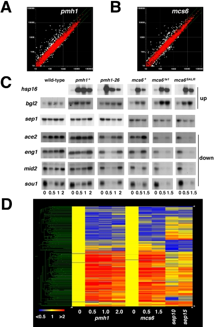
To identify transcripts that were especially sensitive to Mcs6 complex impairment, which might help to explain the cell division arrest, we also performed microarray experiments in which the data were normalized internally to the expression levels of all S. pombe genes (∼5000) at 28°C (Figure 4). By this normalization scheme, a small number of transcripts (∼500 or ∼10% of the total) changed in relative abundance by more than twofold upon shift to restrictive temperature, with roughly equal numbers of mRNAs increased and decreased in the mutants (Figure 4, A and B). Most of the increased transcripts are known to be induced by stress (Chen et al., 2003). Nonetheless, the results indicated that both mutants were capable of mounting a nearly normal response to heat shock. The two mutations, in two different subunits of the Mcs6 complex, produced defined alterations in gene expression that were remarkably similar, independent of the degree of global repression; the overlaps between genes repressed or induced in both mutants were highly significant (p < 10–50).
Figure 4.
Gene expression profiling in Mcs6-complex mutants. (A) Scatter plot of gene expression changes between pmh1-26 at 36°C and HA-tagged pmh1+ strain at 28°C; signals above the central green line are increased in the mutant relative to wild-type, whereas signals below the line are decreased. Top and bottom green lines indicate twofold cutoffs. (B) Comparison of gene expression changes between mcs6ts1 at 36°C and HA-tagged mcs6+ strain at 28°C as in A. (C) Northern blot hybridization to confirm up- and down-regulation of transcripts in mcs6 and pmh1 mutants. Blots were probed with labeled PCR fragments complementary to the indicated transcripts. The wild-type strain analyzed in the first column differs from the pmh1+ and mcs6+ reference strains in that it contained no tagged proteins; only one induced transcript, bgl2, was analyzed in this strain. We probed all blots for cdc2 mRNA, which, like sep1 mRNA, did not change in abundance in any of the samples (our unpublished data). (D) Clustering of microarray data from indicated time points in pmh1 (0, 0.5, 1, and 2 h) and mcs6 (0, 0.5, and 1.5 h) mutants reveals a transcriptional signature of TFIIH-associated kinase impairment, which overlaps with signatures of sep10 and sep15 mutants (both analyzed after overnight culture at semipermissive temperature). Repressed transcripts are in blue, induced transcripts are in red, and unchanged transcripts are in yellow, as indicated by color scale at lower left.
After prolonged periods at restrictive temperature (3, 6, and 12 h), mcs6ts1 and pmh1-26 strains had reduced expression of ribosomal protein genes, probably as a secondary effect of the prolonged arrest. Yields of RNA were comparable with those obtained from wild-type controls, however, and we obtained measurable fluorescence signals from 80 to 90% of all S. pombe genes, suggesting that transcription by Pol II still had not shut down (our unpublished data).
We confirmed changes in abundance of six transcripts (two increased and four decreased) in the mcs6 and pmh1 mutants by Northern blot hybridization (Figure 4C). For this analysis, we included mcs6S165A/L238R (Hermand et al., 2001; Saiz and Fisher, 2002), which showed similar derangement of gene expression in microarrays (our unpublished data). Two features of the mcs6 and pmh1 phenotypes became apparent. First, for the transcripts that decreased upon temperature shift (ace2, eng1, mid2, and sou1), expression seemed to recover at later time points, especially in the pmh1-26 mutant—an effect also manifest in the microarray data. Second, there was a cryptic defect in the reference strain in which a wild-type mcs6+ open reading frame was fused at the carboxy terminus with three HA-epitopes and integrated at its normal chromosomal locus. In this strain, the eng1, mid2, and sou1 transcripts decreased before recovering to near-normal levels. Although some transient decreases in mRNA abundance might be expected as a result of the temperature shift, the pmh1+ reference strain, which was constructed in the same way, displayed only a milder, transient dip in just one of the transcripts we analyzed (sou1), which also was observed in a true wild-type (untagged) strain. These effects suggest some impairment of function due to the epitope tagging of Mcs6, which affects the same transcripts as do the temperature-sensitive alleles.
Clustering of the data from multiple arrays revealed that the pmh1-26 and mcs6ts1 mutations had very similar effects on gene expression (Figure 4D), and thus defined a transcriptional signature of TFIIH-associated kinase dysfunction. The signature was similar to derangements of gene expression profiles in the cell-separation mutants sep10 and sep15 (Grallert et al., 1999) under semipermissive conditions. Figure 4D displays data for 671 genes that changed in abundance by twofold or more in at least two of nine conditions analyzed (see Materials and Methods). The overlaps between the Mcs6 complex and sep mutants were statistically highly significant, with p values of ∼1.8 × 10⁻¹¹ for the induced and ∼1.5 × 10⁻¹¹ for the repressed genes. Moreover, the data sets shared coregulated clusters of both induced and repressed genes, suggesting the existence of discrete gene expression modules coordinately controlled by the Mcs6 complex, Sep10, and Sep15.
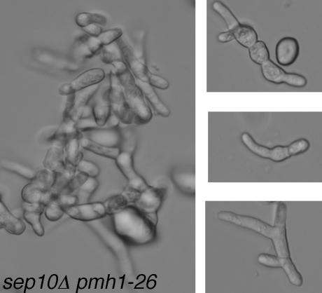
Sep15, the homolog of Med8, is an essential subunit of Pol II Mediator (Zilahi et al., 2000a; Spåhr et al., 2001). Sep10 (Szilagyi et al., 2002) is homologous to budding yeast Soh1 (now renamed Med31) and was recently shown to be a bona fide Mediator subunit (Linder and Gustafsson, 2004). Therefore, aside from phenotypic similarities and overlapping expression profiles, Sep10, Sep15, and the Mcs6 complex are all components of the core transcription apparatus. Although neither sep10+ nor its budding yeast homolog SOH1 is essential, combining either mcs6ts2 or pmh1-26 mutations with sep10Δ caused synthetic lethality; pmh1-26 sep10Δ spores germinated but arrested after a few divisions as microcolonies of misshapen, multiseptated, and branched cells (Figure 5).
Figure 5.
Synthetic lethality of sep10 and pmh1. The sep10Δ pmh1-26 spores formed microcolonies of 30–50 dead, misshapen cells. A microcolony is shown at left; individual, double-mutant cells are shown at right.
Mcs6 Interacts Genetically with the Forkhead Transcription Factor Sep1
The genes repressed in both Mcs6 complex and Mediator mutants included many implicated in cell separation or cytokinesis and assigned to clusters expressed periodically at the end of mitosis (Rustici et al., 2004). In particular, transcripts in cell cycle cluster 2 showed significant overlap with mcs6- and/or pmh1-repressed transcripts (p of ∼0.0016), and with sep10- and/or sep15-repressed transcripts (p of ∼0.0024).
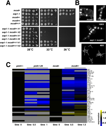
Another gene identified in the screen for cell-separation mutants, sep1+, encodes a forkhead protein required for periodic transcription of cell cycle-regulated gene clusters 1 and 2 (Ribar et al., 1997; Zilahi et al., 2000b; Rustici et al., 2004). The sep1 phenotype—long, occasionally branched chains of cells with multiple nuclei and division septa—resembles those of mcs6, pmh1, sep10, and sep15 mutants (Grallert et al., 1999; Ribar et al., 1999). We therefore looked for genetic interactions between sep1 and the Mcs6 complex. All combinations of sep1-1 with a panel of conditional mutants (mcs6ts1, mcs6ts2, mcs6S165A/L238R, and pmh1-26) were viable; certain mcs6 sep1-1 double mutants, however, had a lowered restrictive temperature (Figure 6A) and an exacerbation of the morphological defect. The mcs6ts1 sep1-1 strain formed long chains of highly branched and misshapen cells at permissive temperature (Figure 6B). Interestingly, the synthetic interaction was allele-specific; only combination with mcs6ts1 or mcs6ts2, not with mcs6S165A/L238R or pmh1-26, lowered the restrictive temperature (Figure 6A; our unpublished data).
Figure 6.
Genetic interaction between mcs6 and sep1. (A) Lowering of restrictive temperature in mcs6 sep1 double mutants. For each double mutant, two isolates were analyzed by growth on plates at 10-fold serial dilutions; these differed slightly in ability to grow at 33°C, but both sep1-1 mcs6ts1 and sep1-1 mcs6ts2 were reproducibly more temperature-sensitive than single-mutant parents. (B) Exacerbation of sep1 morphological phenotype in sep1 mcs6 double mutant at permissive temperature. (C) Overlap of Sep1-dependent with Mcs6-(left), and Pmh1-dependent (right) transcripts. Repressed transcripts are in blue, induced transcripts are in yellow, and unchanged transcripts are in black, as indicated by color scale at lower right. Transcripts for which no interpretable signal could be obtained are represented in light gray.
We also compared gene expression profiles in mcs6, pmh1, and sep1 mutants. Among 39 transcripts decreased in a sep1 mutant (Rustici et al., 2004), nearly all had decreased or absent signals in mcs6 and/or pmh1 mutants at 36°C (Figure 6C). The overlap set included several genes implicated in cytokinesis and cell division as direct and indirect targets of Sep1: mid2, an anillin homologue involved in cytokinesis (Tasto et al., 2003); cdc15, a medial ring component (Fankhauser et al., 1995; Zilahi et al., 2000b; Carnahan and Gould, 2003); ace2, a zinc-finger factor important for cell-cycle transcription (Rustici et al., 2004); eng1, a transcriptional target of Ace2, which encodes an endo-(1,3)-β-glucanase involved in cleaving the division septum (Martin-Cuadrado et al., 2003); and agn1 (which decreased in mcs6S165A/L238R; our unpublished data), a (1,3)-α-glucanase also implicated in cell separation (Dekker et al., 2004). The transcripts for which no signals were recorded at restrictive temperature, moreover, typically were detectable at time 0, suggesting that their disappearance from the arrays at later times was also due to decreased expression, not missing spots on the arrays. Thus, mcs6 and pmh1 mutants had statistically significant (p of ∼7.8 × 10⁻⁶) defects in transcribing the majority, if not the entirety of a cell cycle-regulated gene cluster.
Defects in transcribing Sep1-dependent genes in Mcs6 complex mutants are not due to decreased expression of the transcription factor; levels of the sep1+ transcript measured either by microarray or Northern blot hybridization (Figure 4C) did not change in either mcs6 or pmh1 mutants at the restrictive temperature. Together with the genetic interaction between mcs6 and sep1 (Figure 6, A and B), these data suggest that Sep1 and the Mcs6 complex act in concert to regulate downstream gene expression important for normal cell division.
DISCUSSION
Cell Cycle Gene Expression Is Sensitive to Mcs6 Complex Integrity
We selected conditional alleles of both mcs6 (Saiz and Fisher, 2002) and pmh1 (this report) in unbiased screens for loss of essential functions. In contrast, weaker alleles of mcs6 and mcs2 were previously isolated in screens for positive regulators of Cdc2, which by design were not likely to yield mutations that abrogated essential functions (Molz et al., 1989). The terminal phenotypes of mcs6ts1 and pmh1-26 are similar to each other and to those of mcs6, mcs2, and pmh1 null mutants and suggest a critical role for the Mcs6 complex late in cell division.
The failure to transcribe Sep1-dependent genes could explain the cell separation defect, but it cannot account for the lethality of mcs6 or pmh1 mutations; deletion of sep1 is not lethal (Ribar et al., 1999). The inviability of TFIIH and Mediator mutants at restrictive temperature is therefore likely to reflect a more extensive derangement of transcription. Cell death could result from simultaneous impairment of multiple gene expression programs (Sep1 dependent and independent) or from inactivation of a single (Sep1-independent) pathway. The sets of genes affected by mcs6 and pmh1 mutations overlap with those dysregulated in sep10 and sep15 strains, and both mcs6 sep10 and pmh1 sep10 double mutants are inviable. The synthetic lethality suggests some redundancy between Sep10 and the Mcs6 complex, which is intriguing because a mammalian Mediator complex containing the Soh1/Sep10 homolog, SMCC/TRAP, stimulated transcription in vitro specifically under conditions of limiting TFIIH activity (Gu et al., 1999).
Among the genes dependent on both the Mcs6 complex and Sep1 is ace2+, which encodes another transcription factor that controls periodic gene expression during the M/G1 interval of the cell cycle (Rustici et al., 2004); cell cycle cluster 2, which depends in part on Ace2 for expression, also overlaps significantly with the genes repressed in mcs6 and pmh1 mutants. Indeed, the transcripts repressed in Mcs6 complex mutants correlated better with those dependent on both Sep1 and Ace2 for expression than with those dependent only on Sep1, perhaps indicating an additional interaction of TFIIH with Ace2.
The allele-specific, synthetic interactions between mcs6 and sep1 might be evidence of a direct interaction, which has precedent in budding yeast. The forkhead proteins Fkh1 and Fkh2 of S. cerevisiae function redundantly to control transcription in G2/M (reviewed by Futcher, 2000), and fkh1 fkh2 mutants have a pseudohyphal morphology (Zhu et al., 2000) reminiscent of sep1. Fkh1 influences both Kin28 recruitment to actively transcribed loci and the phosphorylation patterns of elongating Pol II in vivo (Morillon et al., 2003); it will be important to address whether Sep1 interacts similarly with the Mcs6 complex.
Selective Functions for General Factors in Pol II Transcription
As surprising as the identification of specific gene expression clusters dependent on Mcs6 complex function is what we did not find: an allele of mcs6 or pmh1 that shut down Pol II-dependent gene expression. The expectation that Mcs6 complex impairment would repress transcription globally was based on studies of conditional kin28 mutants, which arrest in all phases of the division cycle and have severe defects in expressing a majority of Pol II-dependent genes (Cismowski et al., 1995; Valay et al., 1995; Holstege et al., 1998).
Whether Kin28 activity is actually required at a majority of class II promoters for transcription per se is uncertain. An alternative mechanism whereby defective CTD Ser-5 phosphorylation could lead to drops in steady-state abundance of many transcripts is through failure to recruit the mRNA-capping machinery and the consequent degradation of nascent transcripts (Rodriguez et al., 2000; Schroeder et al., 2000). Nonetheless, studies of transcription with defined systems in vitro—where effects on RNA stability can be discounted—showed stimulation by active Cdk7 at some mammalian promoters and by its presence, but not its activity, at others (Tirode et al., 1999). In Drosophila larval salivary glands, moreover, thermal inactivation of a temperature-sensitive Cdk7 decreased both the steady-state levels of transcripts from, and the density of Pol II molecules on, the heat shock genes, arguing for a direct effect on transcription in vivo (Schwartz et al., 2003).
Our analysis did not address when, during mRNA synthesis and maturation, Ser-5 phosphorylation of the CTD is required, but rather at which genes the normal function of the Mcs6 complex is needed. We have in effect divided the fission yeast transcriptome into two classes of genes: a surprisingly small class that is exquisitely sensitive to TFIIH-associated kinase impairment and a surprisingly large class that is relatively refractory. Such a distinction was not apparent in budding yeast (Holstege et al., 1998), irrespective of the mechanism by which Kin28 impairment affected gene expression.
Differential mRNA stability could by itself produce effects in microarray experiments that seem specific if, for example, the transcripts we have scored as being dependent on the Mcs6 complex for expression were simply those with the shortest half-lives and consequently were the first to be depleted after transcription by Pol II was globally repressed. A comparison with global data on mRNA turnover in vegetative cells, however, did not reveal any correlation between genes most strongly repressed in mcs6 mutants and transcript stability, and many of the genes repressed in the mcs6 mutants had half-lives >60 min (Marguerat and Bähler, unpublished observations).
There is precedent, on the other hand, for promoter-specific differences in the requirement for TFIIH-associated kinase in vivo. In Drosophila, transcription of histone genes, in contrast to that of heat shock genes, was refractory to a strong cdk7 mutation (Schwartz et al., 2003). Cells from Mat1–/– mouse embryos retain some transcriptional function (Rossi et al., 2001), as do specific cell lineages after conditional ablation of Mat1 (Korsisaari et al., 2002). Even in S. cerevisiae, both the copper-inducible CUP1 and heat shock-responsive SSA4 genes could still be induced after depletion of Kin28 (Lee and Lis, 1998; McNeil et al., 1998). A tfb3 mutant, moreover, had more limited depletion of transcripts encoding metabolic enzymes and ribosomal proteins (Jona et al., 2002).
Might gene expression independent of TFIIH-associated kinase—the exception in S. cerevisiae—be more the rule in S. pombe? We cannot exclude the possibility that conditional mcs6 and pmh1 alleles retain residual function at restrictive temperature, which suffices to support the bulk of mRNA synthesis but is inadequate to execute specific functions. (We attempted, unsuccessfully, to inactivate Mcs6 by promoter shutoff and with an analog-sensitive allele and so must rely exclusively on temperature-sensitive alleles.) Indeed, cell separation is likely to be especially sensitive to biosynthetic compromise, based on the sep-like morphology of cells treated with the protein synthesis inhibitor cycloheximide (Polanshek, 1977; Fantes, 1982). Although the phenotypic and biochemical abnormalities we observed could be threshold effects—the first transcripts lost and processes impeded when Mcs6 function is reduced but not eliminated—they nevertheless suggest a critical requirement for the TFIIH-associated kinase in supporting specific gene expression programs linked to cell cycle progression.
Consistent with that role, multiple mutations in two different subunits of the complex caused virtually identical changes in gene expression, which were independent of global effects on transcription. Moreover, measurements of CTD phosphorylation, and of Mcs6- and Pmh1-associated kinase activity indicate rapid, severe impairment of function in the mutants at restrictive temperature. In the mcs6ts1 strain, for example, CTD Ser-5 phosphorylation reached a nadir at 1.5 h (Figure 3B), and median gene expression was reduced by only one-third 30 min later (Figure S2). Finally, the conditional mcs6 and pmh1 phenotypes are similar to terminal mcs6Δ (Buck et al., 1995; Damagnez et al., 1995), mcs2Δ (Molz and Beach, 1993), and pmh1Δ (this report) phenotypes.
The view of CTD phosphorylation by a TFIIH-associated kinase as a near-universal requirement for mRNA synthesis rests heavily on the analysis of a single kin28 mutant (Holstege et al., 1998). A different paradigm may apply in fission yeast and, perhaps, in metazoans. We suggest that, whereas both holo-TFIIH and Mediator may be generally required for transcription, individual subunits or subcomplexes perform more specialized functions, analogous to the TATA-binding protein-associated factors, which exert activator-specific effects as subunits of TFIID (reviewed by Chang and Jaehning, 1997). Interestingly, a taf72 mutant in S. pombe also had a defect in cell separation (Yamamoto and Horikoshi, 1998).
A CDK Linking Gene Expression with Cell Cycle Control
Genetic analysis of the Mcs6 complex is complicated by its dual role in cell division and gene expression, but the unique organization of the fission yeast CAK–CDK network allowed us to analyze derangement of gene expression caused by inactivation of the Mcs6 complex, while bulk CDK activation was maintained by Csk1. Even within the transcriptional context, it now seems that Mcs6 serves a cell cycle function, possibly in close collaboration with transcription factors such as Sep1 and Ace2. Recent comparisons of gene expression in budding and fission yeast as they traverse the cell cycle revealed different circuitry, despite a small core group of conserved, periodically expressed genes. Whereas a cascade of interdependent transcriptional activators spans the cycle in S. cerevisiae, no such continuum exists in S. pombe, and in one case, two factors linked in series in budding yeast act in parallel in fission yeast (Simon et al., 2001; Rustici et al., 2004). We have now revealed a cell cycle-specific role for components of the Pol II core machinery in S. pombe that is not— or at least not yet—evident in S. cerevisiae. Moreover a CAK, a critical regulator of CDK activity, has emerged as a determinant of cell cycle-regulated transcription.
Supplementary Material
Acknowledgments
We thank members of the Fisher laboratory for advice and support; Stéphane Larochelle and Matthew Gamble for critical review of the manuscript; and Jerard Hurwitz, Scott Keeney, and Pamela Meluh for helpful discussions and constructive criticism. Samuel Marguerat communicated unpublished data on global mRNA stability in fission yeast, for which we are grateful. We also thank Julia Zhao and Agnes Viale of the Sloan-Kettering Institute Genomics Core Laboratory for invaluable assistance in analyzing microarray data; and J. Hurwitz, Kathleen Gould, and Dannel McCollum for providing reagents and strains. The work was supported by American Cancer Society Grant RSG-99-043-04-CCG (to R.P.F.), by Cancer Research UK Grant C9546/A5262 (to J. B.), and by a Wellcome Trust short-term travel grant (to I. M.).
This article was published online ahead of print in MBC in Press (http://www.molbiolcell.org/cgi/doi/10.1091/mbc.E04–11–0982) on April 13, 2005.
Note added in proof. While our manuscript was in review, Bamps et al. (2004) also reported that disruption of pmh1 was lethal.
The online version of this article contains supplemental material at MBC Online (http://www.molbiolcell.org).
References
- Bamps, S., Westerling, T., Pihlak, A., Tafforeau, L., Vandenhaute, J., Mäkelä, T. P., and Hermand, D. (2004). Mcs2 and a novel CAK subunit Pmh1 associate with Skp1 in fission yeast. Biochem. Biophys. Res. Commun. 325, 1424–1432. [DOI] [PubMed] [Google Scholar]
- Buck, V., Russell, P., and Millar, J.B.A. (1995). Identification of a cdk-activating kinase in fission yeast. EMBO J. 14, 6173–6183. [DOI] [PMC free article] [PubMed] [Google Scholar]
- Carnahan, R. H., and Gould, K. L. (2003). The PCH family protein, Cdc15p, recruits two F-actin nucleation pathways to coordinate cytokinetic actin ring formation in Schizosaccharomyces pombe. J. Cell Biol. 162, 851–862. [DOI] [PMC free article] [PubMed] [Google Scholar]
- Chang, M., and Jaehning, J. A. (1997). A multiplicity of mediators: alternative forms of transcription complexes communicate with transcriptional regulators. Nucleic Acids Res. 25, 4861–4865. [DOI] [PMC free article] [PubMed] [Google Scholar]
- Chen, D., Toone, W. M., Mata, J., Lyne, R., Burns, G., Kivinen, K., Brazma, A., Jones, N., and Bähler, J. (2003). Global transcriptional responses of fission yeast to environmental stress. Mol. Biol. Cell 14, 214–229. [DOI] [PMC free article] [PubMed] [Google Scholar]
- Cismowski, M. J., Laff, G. M., Solomon, M. J., and Reed, S. I. (1995). KIN28 encodes a C-terminal domain kinase that controls mRNA transcription in Saccharomyces cerevisiae but lacks cyclin-dependent kinase-activating kinase (CAK) activity. Mol. Cell. Biol. 15, 2983–2992. [DOI] [PMC free article] [PubMed] [Google Scholar]
- Damagnez, V., Mäkelä, T. P., and Cottarel, G. (1995). Schizosaccharomyces pombe Mop1-Mcs2 is related to mammalian CAK. EMBO J. 14, 6164–6172. [DOI] [PMC free article] [PubMed] [Google Scholar]
- Dekker, N., Speijer, D., Grun, C. H., Van Den Berg, M., De Haan, A., and Hochstenbach, F. (2004). Role of the α-glucanase Agn1p in fission-yeast cell separation. Mol. Biol. Cell 15, 3903–3914. [DOI] [PMC free article] [PubMed] [Google Scholar]
- Espinoza, F. H., Farrell, A., Erdjument-Bromage, H., Tempst, P., and Morgan, D. O. (1996). A cyclin-dependent kinase-activating kinase (CAK) in budding yeast unrelated to vertebrate CAK. Science 273, 1714–1717. [DOI] [PubMed] [Google Scholar]
- Fankhauser, C., Reymond, A., Cerutti, L., Utzig, S., Hofmann, K., and Simanis, V. (1995). The S. pombe cdc15 gene is a key element in the reorganization of F-actin at mitosis. Cell 82, 435–444. [DOI] [PubMed] [Google Scholar]
- Fantes, P. A. (1982). Dependency relations between events in mitosis in Schizosaccharomyces pombe. J. Cell Sci. 55, 383–402. [DOI] [PubMed] [Google Scholar]
- Feaver, W. J., Svejstrup, J. Q., Henry, N. L., and Kornberg, R. D. (1994). Relationship of CDK-activating kinase and RNA polymerase II CTD kinase TFIIH/TFIIK. Cell 79, 1103–1109. [DOI] [PubMed] [Google Scholar]
- Fisher, R. P., Jin, P., Chamberlin, H. M., and Morgan, D. O. (1995). Alternative mechanisms of CAK assembly require an assembly factor or an activating kinase. Cell 83, 47–57. [DOI] [PubMed] [Google Scholar]
- Futcher, B. (2000). Microarrays and cell cycle transcription in yeast. Curr. Opin. Cell Biol. 12, 710–715. [DOI] [PubMed] [Google Scholar]
- Garrett, S., Barton, W. A., Knights, R., Jin, P., Morgan, D. O., and Fisher, R. P. (2001). Reciprocal activation by cyclin-dependent kinases 2 and 7 is directed by substrate specificity determinants outside the T-loop. Mol. Cell. Biol. 21, 88–99. [DOI] [PMC free article] [PubMed] [Google Scholar]
- Gervais, V., Busso, D., Wasielewski, E., Poterszman, A., Egly, J.-M., Thierry, J.-C., and Kieffer, B. (2001). Solution structure of the N-terminal domain of the human TFIIH MAT1 subunit. J. Biol. Chem. 276, 7457–7464. [DOI] [PubMed] [Google Scholar]
- Grallert, A., Grallert, B., Zilahi, E., Szilagyi, Z., and Sipiczki, M. (1999). Eleven novel sep genes of Schizosaccharomyces pombe required for efficient cell separation and sexual differentiation. Yeast 15, 669–686. [DOI] [PubMed] [Google Scholar]
- Gu, W., Malik, S., Ito, M., Yuan, C. X., Fondell, J. D., Zhang, X., Martinez, E., Qin, J., and Roeder, R. G. (1999). A novel human SRB/MED-containing cofactor complex, SMCC, involved in transcription regulation. Mol. Cell 3, 97–108. [DOI] [PubMed] [Google Scholar]
- Harper, J. W., and Elledge, S. J. (1998). The role of Cdk7 in CAK function, a retro-retrospective. Genes Dev. 12, 285–289. [DOI] [PubMed] [Google Scholar]
- Hermand, D., Pihlak, A., Westerling, T., Damagnez, V., Vandenhaute, J., Cottarel, G., and Mäkelä, T. P. (1998). Fission yeast Csk1 is a CAK-activating kinase (CAKAK). EMBO J. 17, 7230–7238. [DOI] [PMC free article] [PubMed] [Google Scholar]
- Hermand, D., Westerling, T., Pihlak, A., Thuret, J.-Y., Vallenius, T., Tiainen, M., Vandenhaute, J., Cottarel, G., Mann, C., and Mäkelä, T. P. (2001). Specificity of Cdk activation in vivo by the two Caks Mcs6 and Csk1 in fission yeast. EMBO J. 20, 82–90. [DOI] [PMC free article] [PubMed] [Google Scholar]
- Holstege, F.C.P., Jennings, E. G., Wyrick, J. J., Lee, T. I., Hengartner, C. J., Green, M. J., Golub, T. R., Lander, E. S., and Young, R. A. (1998). Dissecting the regulatory circuitry of a eukaryotic genome. Cell 95, 717–728. [DOI] [PubMed] [Google Scholar]
- Jona, G., Livi, L. L., and Gileadi, O. (2002). Mutations in the RING domain of TFB3, a subunit of yeast transcription factor IIH, reveal a role in cell cycle progression. J. Biol. Chem. 277, 39409–39416. [DOI] [PubMed] [Google Scholar]
- Kaldis, P., Sutton, A., and Solomon, M. J. (1996). The Cdk-activating kinase (CAK) from budding yeast. Cell 86, 553–564. [DOI] [PubMed] [Google Scholar]
- Korsisaari, N., Rossi, D. J., Paetau, A., Charnay, P., Henkemeyer, M., and Mäkelä, T. P. (2002). Conditional ablation of the Mat1 subunit of TFIIH in Schwann cells provides evidence that Mat1 is not required for general transcription. J. Cell Sci. 115, 4275–4284. [DOI] [PubMed] [Google Scholar]
- Kunkel, T. A. (1985). Rapid and efficient site-directed mutagenesis without phenotypic selection. Proc. Natl. Acad. Sci. USA 82, 488–492. [DOI] [PMC free article] [PubMed] [Google Scholar]
- Larochelle, S., Chen, J., Knights, R., Pandur, J., Morcillo, P., Erdjument-Bromage, H., Tempst, P., Suter, B., and Fisher, R. P. (2001). T-loop phosphorylation stabilizes the CDK7-cyclin H-MAT1 complex in vivo and regulates its CTD kinase activity. EMBO J. 20, 3749–3759. [DOI] [PMC free article] [PubMed] [Google Scholar]
- Lee, D., and Lis, J. T. (1998). Transcriptional activation independent of TFIIH kinase and the RNA polymerase II mediator in vivo. Nature 393, 389–392. [DOI] [PubMed] [Google Scholar]
- Lee, K. M., Saiz, J. E., Barton, W. A., and Fisher, R. P. (1999). Cdc2 activation in fission yeast depends on Mcs6 and Csk1, two partially redundant Cdk-activating kinases (CAKs). Curr. Biol. 9, 441–444. [DOI] [PubMed] [Google Scholar]
- Linder, T., and Gustafsson, C. M. (2004). The Soh1/MED31 protein is an ancient component of Schizosaccharomyces pombe and Saccharomyces cerevisiae mediator. J. Biol. Chem. 279, 49455–49459. [DOI] [PubMed] [Google Scholar]
- Liu, Y., Kung, C., Fishburn, J., Ansari, A. Z., Shokat, K. M., and Hahn, S. (2004). Two cyclin-dependent kinases promote RNA polymerase II transcription and formation of the scaffold complex. Mol. Cell. Biol. 24, 1721–1735. [DOI] [PMC free article] [PubMed] [Google Scholar]
- Lyne, R., Burns, G., Mata, J., Penkett, C. J., Rustici, G., Chen, D., Langford, C., Vetrie, D., and Bähler, J. (2003). Whole-genome microarrays of fission yeast: characteristics, accuracy, reproducibility, and processing of array data. BMC Genomics 4, 27. [DOI] [PMC free article] [PubMed] [Google Scholar]
- Martin-Cuadrado, A. B., Duenas, E., Sipiczki, M., Vazquez de Aldana, C. R., and del Rey, F. (2003). The endo-beta-1,3-glucanase eng1p is required for dissolution of the primary septum during cell separation in Schizosaccharomyces pombe. J. Cell Sci. 116, 1689–1698. [DOI] [PubMed] [Google Scholar]
- McNeil, J. B., Agah, H., and Bentley, D. (1998). Activated transcription independent of the RNA polymerase II holoenzyme in budding yeast. Genes Dev. 12, 2510–2521. [DOI] [PMC free article] [PubMed] [Google Scholar]
- Molz, L., and Beach, D. (1993). Characterization of the fission yeast mcs2 cyclin and its associated protein kinase activity. EMBO J. 12, 1723–1732. [DOI] [PMC free article] [PubMed] [Google Scholar]
- Molz, L., Booher, R., Young, P., and Beach, D. (1989). cdc2 and the regulation of mitosis: six interacting mcs genes. Genetics 122, 773–782. [DOI] [PMC free article] [PubMed] [Google Scholar]
- Moreno, S., Klar, A., and Nurse, P. (1991). Molecular genetic analysis of fission yeast Schizosaccharomyces pombe. Methods Enzymol. 194, 795–823. [DOI] [PubMed] [Google Scholar]
- Morgan, D. O. (1997). Cyclin-dependent kinases: engines, clocks and microprocessors. Annu. Rev. Cell Dev. Biol. 13, 261–291. [DOI] [PubMed] [Google Scholar]
- Morillon, A., O'Sullivan, J., Azad, A., Proudfoot, N., and Mellor, J. (2003). Regulation of elongating RNA polymerase II by forkhead transcription factors in yeast. Science 300, 492–495. [DOI] [PubMed] [Google Scholar]
- O'Reilly, D. R., Miller, L. K., and Luckow, V. A. (1993). Baculovirus Expression Vectors: A Laboratory Manual, New York: W. H. Freeman.
- Polanshek, M. M. (1977). Effects of heat shock and cycloheximide on growth and division of the fission yeast, Schizosaccharomyces pombe. J. Cell Sci. 23, 1–23. [DOI] [PubMed] [Google Scholar]
- Rachez, C., and Freedman, L. P. (2001). Mediator complexes and transcription. Curr. Opin. Cell Biol. 13, 274–280. [DOI] [PubMed] [Google Scholar]
- Ribar, B., Banrevi, A., and Sipiczki, M. (1997). sep1+ encodes a transcription-factor homologue of the HNF-3/forkhead DNA-binding-domain family in Schizosaccharomyces pombe. Gene 202, 1–5. [DOI] [PubMed] [Google Scholar]
- Ribar, B., Grallert, A., and Olah, E. (1999). Deletion of the sep1(+) forkhead transcription factor homologue is not lethal but causes hyphal growth in Schizosaccharomyces pombe. Biochem. Biophys. Res. Commun. 263, 465–474. [DOI] [PubMed] [Google Scholar]
- Rodriguez, C. R., Cho, E. J., Keogh, M. C., Moore, C. L., Greenleaf, A. L., and Buratowski, S. (2000). Kin28, the TFIIH-associated carboxy-terminal domain kinase, facilitates the recruitment of mRNA processing machinery to RNA polymerase II. Mol. Cell. Biol. 20, 104–112. [DOI] [PMC free article] [PubMed] [Google Scholar]
- Rosenblatt, J., De Bondt, H., Jancarik, J., Morgan, D. O., and Kim, S.-H. (1993). Purification and crystallization of human cyclin-dependent kinase 2. J. Mol. Biol. 230, 1317–1319. [DOI] [PubMed] [Google Scholar]
- Rossi, D. J., Londesborough, A., Korsisaari, N., Pihlak, A., Lehtonen, E., Henkemeyer, M., and Mäkelä, T. P. (2001). Inability to enter S phase and defective RNA polymerase II CTD phosphorylation in mice lacking. Mat1. EMBO J. 20, 2844–2856. [DOI] [PMC free article] [PubMed] [Google Scholar]
- Rustici, G., Mata, J., Kivinen, K., Lio, P., Penkett, C. J., Burns, G., Hayles, J., Brazma, A., Nurse, P., and Bähler, J. (2004). Periodic gene expression program of the fission yeast cell cycle. Nat. Genet. 36, 809–817. [DOI] [PubMed] [Google Scholar]
- Saiz, J. E., and Fisher, R. P. (2002). A CDK-activating kinase network is required in cell cycle control and transcription in fission yeast. Curr. Biol. 12, 1100–1105. [DOI] [PubMed] [Google Scholar]
- Schroeder, S. C., Schwer, B., Shuman, S., and Bentley, D. (2000). Dynamic association of capping enzymes with transcribing RNA polymerase II. Genes Dev. 14, 2435–2440. [DOI] [PMC free article] [PubMed] [Google Scholar]
- Schwartz, B. E., Larochelle, S., Suter, B., and Lis, J. T. (2003). Cdk7 is required for full activation of Drosophila heat shock genes and RNA polymerase II phosphorylation in vivo. Mol. Cell. Biol. 23, 6876–6886. [DOI] [PMC free article] [PubMed] [Google Scholar]
- Simon, I., et al. (2001). Serial regulation of transcriptional regulators in the yeast cell cycle. Cell 106, 697–708. [DOI] [PubMed] [Google Scholar]
- Spåhr, H., Khorosjutina, O., Baraznenok, V., Linder, T., Samuelsen, C. O., Hermand, D., Mäkelä, T. P., Holmberg, S., and Gustafsson, C. M. (2003). Mediator influences Schizosaccharomyces pombe RNA polymerase II-dependent transcription in vitro. J. Biol. Chem. 278, 51301–51306. [DOI] [PubMed] [Google Scholar]
- Spåhr, H., Samuelsen, C. O., Baraznenok, V., Ernest, I., Huylebroeck, D., Remacle, J. E., Samuelsson, T., Kieselbach, T., Holmberg, S., and Gustafsson, C. M. (2001). Analysis of Schizosaccharomyces pombe mediator reveals a set of essential subunits conserved between yeast and metazoan cells. Proc. Natl. Acad. Sci. USA 98, 11985–11990. [DOI] [PMC free article] [PubMed] [Google Scholar]
- Szilagyi, Z., Grallert, A., Nemeth, N., and Sipiczki, M. (2002). The Schizosaccharomyces pombe genes sep10 and sep11 encode putative general transcriptional regulators involved in multiple cellular processes. Mol. Genet. Genomics 268, 553–562. [DOI] [PubMed] [Google Scholar]
- Tasto, J. J., Morrell, J. L., and Gould, K. L. (2003). An anillin homologue, Mid2p, acts during fission yeast cytokinesis to organize the septin ring and promote cell separation. J. Cell Biol. 160, 1093–1103. [DOI] [PMC free article] [PubMed] [Google Scholar]
- Thuret, J.-Y., Valay, J.-G., Faye, G., and Mann, C. (1996). Civ1 (CAK in vivo), a novel Cdk-activating kinase. Cell 86, 565–576. [DOI] [PubMed] [Google Scholar]
- Tirode, F., Busso, D., Coin, F., and Egly, J.-M. (1999). Reconstitution of the transcription factor TFIIH: assignment of functions for the three enzymatic subunits, XPB, XPD and cdk7. Mol. Cell 3, 87–95. [DOI] [PubMed] [Google Scholar]
- Valay, J.-G., Simon, M., Dubois, M.-F., Bensaude, O., Facca, C., and Faye, G. (1995). The KIN28 gene is required both for RNA polymerase II mediated transcription and phosphorylation of the Rpb1p CTD. J. Mol. Biol. 249, 535–544. [DOI] [PubMed] [Google Scholar]
- van de Peppel, J., Kemmeren, P., van Bakel, H., Radonjic, M., van Leenen, D., and Holstege, F. C. (2003). Monitoring global messenger RNA changes in externally controlled microarray experiments. EMBO Rep. 4, 387–393. [DOI] [PMC free article] [PubMed] [Google Scholar]
- Wood, V., et al. (2002). The genome sequence of Schizosaccharomyces pombe. Nature 415, 871–880. [DOI] [PubMed] [Google Scholar]
- Yamamoto, T., and Horikoshi, M. (1998). Defect in cytokinesis of fission yeast induced by mutation in the WD40 repeat motif of a TFIID subunit. Genes Cells 3, 347–355. [DOI] [PubMed] [Google Scholar]
- Zhu, G., Spellman, P. T., Volpe, T., Brown, P. O., Botstein, D., Davis, T. N., and Futcher, B. (2000). Two yeast forkhead genes regulate the cell cycle and pseudohyphal growth. Nature 406, 90–94. [DOI] [PubMed] [Google Scholar]
- Zilahi, E., Miklos, I., and Sipiczki, M. (2000a). The Schizosaccharomyces pombe sep15+ gene encodes a protein homologous to the Med8 subunit of the Saccharomyces cerevisiae transcriptional mediator complex. Curr. Genet. 38, 227–232. [DOI] [PubMed] [Google Scholar]
- Zilahi, E., Salimova, E., Simanis, V., and Sipiczki, M. (2000b). The S. pombe sep1 gene encodes a nuclear protein that is required for periodic expression of the cdc15 gene. FEBS Lett. 481, 105–108. [DOI] [PubMed] [Google Scholar]
Associated Data
This section collects any data citations, data availability statements, or supplementary materials included in this article.


