Abstract
The changing notion of “companion animals” and their increasing global status as family members underscores the dynamic interaction between gut microbiota and host health. This review provides a comprehensive understanding of the intricate microbial ecology within companion animals required to maintain overall health and prevent disease. Exploration of specific diseases and syndromes linked to gut microbiome alterations (dysbiosis), such as inflammatory bowel disease, obesity, and neurological conditions like epilepsy, are highlighted. In addition, this review provides an analysis of the various factors that impact the abundance of the gut microbiome like age, breed, habitual diet, and microbe-targeted interventions, such as probiotics. Detection methods including PCR-based algorithms, fluorescence in situ hybridisation, and 16S rRNA gene sequencing are reviewed, along with their limitations and the need for future advancements. Prospects for longitudinal investigations, functional dynamics exploration, and accurate identification of microbial signatures associated with specific health problems offer promising directions for future research. In summary, it is an attempt to provide a deeper insight into the orchestration of multiple microbial species shaping the health of companion animals and possible species-specific differences.
Keywords: gut microbe, companion animal, disease, health, factors
1. Introduction
Domesticated animals are the result of generations of selective breeding and genetic adaptation to coexist with humans [1]. These companion animals permanently reside in human communities and provide companionship, entertainment, work, and psychological support [2,3]. Pet ownership is increasing globally, with a growing tendency to view pets as family members. Notably, around 90% of pet owners view their companion animals as integral and fully functional members of their families [4]. An overall survey of the world population review has shown an understanding of the preference for companion animals as pets in general (Supplementary Figure S1) and dogs and cats (Supplementary Figure S2).
The digestive system of animals and humans harbors numerous bacteria as well as viruses, fungi, and other microorganisms (e.g., protozoa and algae). Importantly, the structure and function of the gut microbiome are strongly influenced by different dietary components (carbohydrates, fats, proteins, minerals, vitamins, etc.), food additives, cooking, and processing, and these changes are closely related to maintaining the health of the host [5]. Consequently, the gut serves as a stable ecological niche for these inhabiting bacteria, relying on the host’s physiological processes, such as feeding and reproduction, for their basic biological functions [6,7,8,9]. The diverse microorganisms, including bacteria, protozoa, bacteriophages, fungi, archaea, and eukaryotic viruses, which inhabit and colonise the anatomical structures of humans and other animals, constitute more than a mere collection of microbes. The microbiome encompasses the entirety of microorganisms, their genes, and their metabolites [10]. Day-by-day research focuses on understanding the role of gut microbiota in shaping the host’s health. Many association studies have been carried out on human health, which include metabolic disorders like diabetes [11,12], obesity [13,14], kidney disease [15,16], atopic disorders [17], chronic enteropathy (CE) [18,19], immune-mediated disorders [20,21], and allergic reactions [22]. Additionally, the species diversity and composition of the gut microbiome are affected by many factors like diet [23], antibiotics [24], probiotics [25], age of the animal [26], physical activity [27], gender, and breed [28]. There are also differences in the mucosal versus luminal abundance of bacteria. These factors are detailed in the further sections.
The gut microbiome is dominated by strict or facultative anaerobic bacteria, particularly in the large intestine [29]. Firmicutes (low-G + C Gram-positive bacteria), Fusobacteria (Gram-negative anaerobic bacilli), and Bacteroidetes (Gram-negative rod-shaped) are the most abundant phyla in dogs and cats (Table 1).
Table 1.
Gastrointestinal microbial species of companion animals.
| Phylum | Class | Family | Genus/Species |
|---|---|---|---|
| Proteobacteria | Betaproteobacteria | Alcaligenaceae | Sutterella |
| Gammaproteobacteria | Enterobacteriaceae | E. coli | |
| Fusobacteria | Fusobacteriia | Fusobacteriaceae | Fusobacterium |
| Firmicutes | Bacilli | Turicibacteraceae | Turicibacter |
| Veillonellaceae | Megamonas | ||
| Lactobacillaceae | Lactobacillus | ||
| Streptococcaceae | Streptococcus | ||
| Clostridia | Clostridiaceae | Clostridium | |
| Ruminococcaceae | Faecalibacterium prausnitzii | ||
| Lachnospiraceae | Blautia | ||
| Peptostreptococcaceae | Peptostreptococcus | ||
| Bacteroidetes | Bacteroidetes | Prevotellaceae | Prevotella |
| Bacteroidaceae | Bacteroides | ||
| Actinobacteria | Coriobacteriia | Coriobacteriaceae | Collinsella |
Source: Allaway et al. [30].
The gut microbiota produces two primary metabolites: short-chain fatty acids (SCFAs) and trimethylamine-N-oxide (TMAO) [29,31]. The genus Fusobacterium is associated with good health in dogs [32], while it has been linked as one of the causative agents leading to colorectal cancer [33] and IBD [34] in humans. Fusobacteria is considered to have a crucial role in the gut metabolism of carnivorous animals [35] because of their unique ability to break down protein and amino acids to generate branched-chain volatile fatty acids, SCFAs [36]. In the gastrointestinal (GI) tract of dogs, Fusobacterium species, such as F. perfoetens and F. mortiferum, are abundantly present [37] (Table 2), while Fusobacterium makes up approximately 20% of the overall relative abundance [31]. The gut microbiota significantly impacts various facets of animal health, encompassing innate immunity, appetite regulation, and energy metabolism [38,39,40,41,42,43,44]. The strategic manipulation of the gut microbiome through interventions such as probiotics or dietary fibre holds promise for enhancing overall health and potentially mitigating the prevalence of obesity [40].
Table 2.
Disease and/or symptoms caused by the gut microbiota in companion animals (cats and dogs).
| Disease/Disorder | Microbiota Involved | Author |
|---|---|---|
| Secretory diarrhea | Clostridium hiranonis | [45] |
| Carbohydrate fermentation | Bifidobacterium, Lactobacillus, and Faecalibacterium | [36] |
| Prevents leaky gut syndrome | Clostridiales | [36] |
| Protects against excessive inflammation | Parabacteroides | [46] |
| Mitigates CE | Lactobacillus acidophilus strains and Lactobacillus johnsonii strain | [47] |
| Intestinal disease (in cats) | Bifidobacteria and Bacteroides (decrease), Desulfovibrio (increase) | [48,49] |
| Small cell intestinal lymphoma | Fusobacterium sp. (increase) | [50] |
Chronic enteropathies (CE).
This review focuses on studies investigating the impact of the gut microbiome on health outcomes in companion animals, primarily in dogs and cats. It emphasises research on pathological conditions such as inflammatory bowel disease, obesity, and neurological disorders, encompassing a range of ages, diets, and probiotic interventions. To maintain a rigorous and relevant analysis, studies involving non-companion species, animals with severe systemic diseases, impact of antibiotic use, or mixed/uncontrolled diets were excluded. Additionally, we omit research published before 1990, non-English language studies, and those with overlapping experimental treatments, ensuring a refined and focused scope.
2. Techniques Used for the Detection of Gut Microbiome Abundance
Using a recently developed polymerase chain reaction (PCR)-based algorithm called the “Dysbiosis Index,” veterinarians may quantify the magnitude of microbial imbalance (dysbiosis) in faeces and monitor the progression of microbial imbalances in response to treatment in dogs and cats [51]. Alterations or perturbations in the composition of the microbiota have a discernible impact on the functionality of the immune system. Consequently, the manipulation or modulation of the gut microbiome holds potential therapeutic value in the management of GI disorders [52].
Fluorescence in situ hybridisation (FISH) is a widely employed method that utilises fluorescent dye-labelled oligonucleotide probes that hybridise to bacterial ribosomal RNA within targeted bacterial groups. This technique permits the identification, quantification, and spatial distribution of microbes in tissue [44,53,54]. The principal advantage of FISH is the ability to spatial visualise bacterial localisation.
Next is 16S rRNA gene sequencing: DNA shotgun sequencing (metagenomics) and other techniques are included in next-generation sequencing (NGS). Only a small number of studies employed deep DNA shotgun sequencing, while most studies evaluating the gut microbiota in companion animals used 16S rRNA gene sequencing. To be sure, 16S rRNA gene sequencing is the most widely used sequencing method for evaluating gut bacteria in dogs and cats. In a nutshell, intestinal materials, including biopsies, luminal content, or faecal samples, are used to extract DNA. The conserved sections on either side of the many variable regions that make up the 16S rRNA gene. The variable region between these conserved sections is amplified by using bacterial primers. Theoretically, it is possible to amplify DNA from both known and unknown bacteria in the sample and subsequently sequence the variable regions by specifically targeting the conserved regions. The NGS technologies have helped us to characterise bacterial species and their interactions between the host and gut microbiome. Using this technique, the microbiota of companion animals (dogs and cats) has been described, including those of the gastrointestinal tract (GIT) [31,55,56,57], skin [58], oral cavity [59,60], nasal cavity [61], and vagina [62].
The gut microbiome can also be defined by bacterial culture in some instances. However, because only conventional bacterial medium and/or restricted anaerobic procedures are utilised in traditional bacterial culture, the number of intestinal bacteria is greatly underestimated when performed in veterinary diagnostic laboratories. Commercial diagnostic laboratories have reported the isolation of just a tiny percentage of bacterial species from the faeces of clinical patients. Unfortunately, physicians frequently mistakenly perceive these bacteria as pathogens since they have been identified from samples of clinical patients.
The existing methodologies for detection exhibit inherent limitations that necessitate their transcendence, while a comprehensive understanding of the underlying mechanism is imperative.
3. Factors Influencing the Gut Microbiome
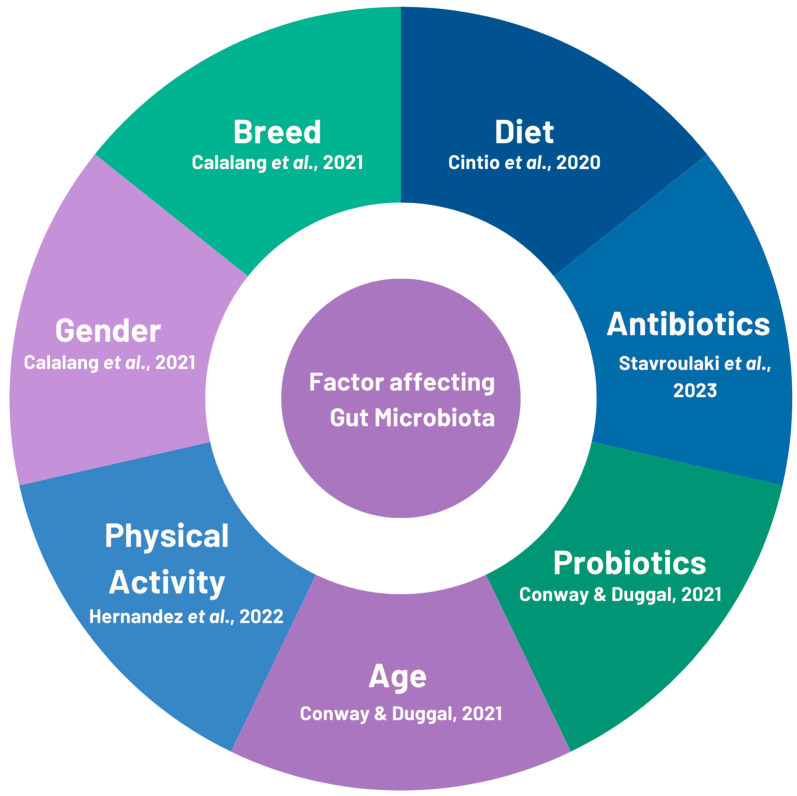
The composition of the gut microbiota can be substantially influenced by a variety of factors, such as age, diet, and the use of probiotics and medications, particularly antibiotics, as demonstrated by various animal models (Figure 1).
Figure 1.
Accountability of different factors affecting the gut microbiota in canines and felines. The information displayed in the figure was derived from previously published studies [23,24,26,27,28].
3.1. Age and Breed
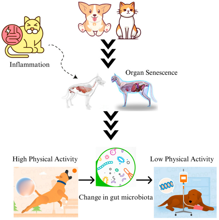
The influence of age, whether in humans or companion animals, emerges as a prominent determinant of the notable alterations observed in the composition of gut microbial communities, suggesting a discernible decline in microbial diversity [63]. The phenomenon of organ senescence, characterised by increased inflammatory reactions, engenders a persistent alteration in the composition and functionality of the gut microbiome, thereby exerting a discernible impact on the physical activity and pharmacological intake patterns of the organism in question (Figure 2) [64]. In dogs, after weaning, the presence of Fusobacteria becomes more prevalent, with Fusobacterium perfoetens being approximately twice as abundant in the 6–10-year-old group compared to the 0.5–1-year-old group (14.3% vs. 7.2%). While such an increase in abundance is positively associated with the age of the dogs [64,65], there is also a potential association between the increase in levels of Fusobacteria and the use of meat-based foods after the weaning stage in dogs [35,64].
Figure 2.
Companion animals may become less active due to gastrointestinal inflammation triggered by age- or diet-induced dysbiosis. Intestinal damage brought on by mucosal inflammation due to the changes in the gut microbiome might also result in decreased physical activity.
Similarly, in cats, between weeks 18 and 42, there were notable shifts in the average abundance of the four most common genera: Bifidobacterium, Lactobacillus, Prevotella, and Bacteroides [66,67]. The levels of Bacteroides and Prevotella showed a significant increase with age, while the levels of Bifidobacterium and Lactobacillus showed a significant decrease. By the time kittens reached 18 weeks of age, the microbiome was primarily composed of Lactobacillus (35%) and Bifidobacterium (20%). The increased presence of Lactobacillus and Bifidobacterium during the earlier time points (8–17 weeks) can be attributed to the impact of the milk-feeding/weaning period. By the time they reached 42 weeks of age, Bacteroides accounted for 16% of the population, followed by Prevotella at 14% and Megasphaera at 8.4%. The abundance of Megasphaera experienced a significant increase over time, rising from 0.1% and 0.2% at week 18 and week 30, respectively, to 8.4% at week 42 [66,67]. Studies have also demonstrated breed-specific differences in the canine intestinal microbiome. For example, Hooda et al. [68] compared three breeds: Maltese, Poodle, and Miniature Schnauzer, where the richness and abundance of Fusobacterium differed and the abundance of Firmicutes was lowest in Maltese dogs. On the contrary, Li et al. [69] showed the breed-specific differences using 16S rRNA gene sequencing techniques in the gut microbiota of Felinae and Ragdoll cats and proved that beneficial microbes like Enterococcus, Lactobacillus, Streptococcus, Roseburia, and Blautia, was significantly abundant in the Ragdoll group than in the Felinae group, suggesting its use in specific designing of probiotic.
3.2. Gender
There has been a significant increase in research indicating variations in gut microbiota between males and females, as observed in both animals (mostly in murine models) and humans [70,71]. For instance, a study conducted on non-obese diabetic (NOD)/ShiLtJ mice found that certain bacterial families such as Kineosporiaceae, Peptococcaceae, Porphyromonadaceae, Veillonellaceae, Lactobacillaceae, Peptostreptococcaceae, Bacteroidaceae, Cytophagaceae, and Enterobacteriaceae were more abundant in male mice compared to female mice [70]. Microbial dysbiosis caused by Bilateral ovariectomy has been reported in murine models [72,73], while in humans, bilateral ovariectomy has been linked to a higher presence of Clostridium bolteae [71]. There was a direct correlation between the level of non-ovarian systemic estrogens and the abundance of faecal Clostridia, which included non-Clostridiales and three genera in the Ruminococcaceae family [74]. In companion animal studies, it was found that the relative abundance (RA) of Firmicutes showed similarities between spayed females and castrated males, which was significantly higher in these groups compared to normal female and male dogs. In addition, there was a higher proportion of Firmicutes in male dogs compared to females in terms of their RA. On the other hand, male dogs had a lower RA of Bacteroidetes compared to females, but it was higher than that of castrated dogs (spayed females and castrated males). There was a higher RA of Fusobacterium in normal dogs (both female and male) compared to castrated dogs (spayed females and castrated males) [75].
3.3. Physical Activity
Research has shown a strong connection between physical activity and improved metabolic health. Human studies have indicated that physical activity can lead to a higher presence of beneficial gut microbes, such as Clostridiales [76]. Research has revealed a positive correlation between regular exercise and a higher presence of bacteria in the Erysipelotrichaceae family. Engaging in active transportation was found to have a positive impact on the levels of Phascolarctobacterium, while simultaneously reducing the levels of Clostridium. Engaging in active transportation for longer durations was found to be linked to a reduction in the presence of the Clostridiaceae family [76]. Several clinical trials have investigated the effects of exercise and dietary intervention on the canine gut microbiota [77,78]. The study by Kieler et al. did not find any evidence of exercise impacting the gut microbiota composition in a weight-loss programme that utilised a commercial high-protein, low-fat, and high-fibre dry diet [77]. In contrast, especially during exercise-related activity, the addition of glucosamine affects the composition of the microbiome in sled dogs [79]. When examining the taxonomic composition at the family level, it becomes evident that sled dogs who consume glucosamine experience a reduction in Lactobacillaceae and Anaerovoracaceae. In glucosamine-supplemented dogs, the abundance of Sellimonas, Eubacterium brachy, and Parvibacter was found to be reduced, especially after activity [79].
3.4. Antibiotics
Even though antibiotics were originally designed to combat specific pathogens in animals and humans, their molecular targets, such as the ribosome, RNA polymerase, and also cell wall, are highly conserved among different bacterial species. As a result, the use of antibiotics can affect both harmful and harmless bacteria, leading to disturbances in the ecological balance that plays a crucial role in various metabolic processes. Tackling bacterial infections has become more difficult, as different strains of bacteria are showing a growing resistance to antimicrobial treatments [80,81]. It is worth mentioning that the disruption of the gut community caused by antibiotic treatment allows the proliferation of harmful pathogens that can cause gastrointestinal diseases, including vancomycin-resistant Enterococcus, Salmonella spp., and drug-resistant Enterobacteriaceae, Clostridioides difficile [82].
Research conducted on both humans and dogs consistently shows that metabolic transformations due to antibiotic treatment are linked to noticeable reductions in the presence of key members from the Bacteroidetes, Firmicutes, and Actinobacteria, particularly Faecalibacterium [27,83,84], as they play a vital role in the gut microbiome’s metabolic processes. Studies on antibiotic treatment in murine models and humans showed that it often leads to a decline in their ability to ferment carbohydrates (leading to decreased SCFA production) and transform bile acids (resulting in more primary bile acids production) [85,86,87]. Using advanced-omic or quantitative polymerase chain reaction (q-PCR)-based techniques to investigate the effects of tylosin, metronidazole, or metronidazole in combination with enrofloxacin revealed a notable increase in the dysbiosis index and a lasting disturbance in bacterial abundances and a decrease in species diversity, even after discontinuation of the antibiotic exposure [88,89,90]. For example, after metronidazole treatment, there have been reports of a decrease in beneficial SCFAs-producing bacteria like Blautia spp. and Faecalibacterium spp. and an increase in potentially harmful bacteria like Escherichia spp. in dogs with CE [21]. The combination of amoxicillin and clavulanic acid also showed a more significant impact on the composition of faecal microbiota compared to amoxicillin alone, showing a decrease in SCFAs-producing Lactococcus spp. and Roseburia spp. [91]. Dogs receiving amoxicillin, in contrast to those receiving amoxicillin plus ribaxamase experienced changes in their gut microbiota, with increases in Fusobacteria, Firmicutes, and Proteobacteria, and decreases in Actinobacteria and Bacteroidetes [92]. The population of Enterobacteriaceae, Enterococcus spp., and Campylobacter spp. were found to increase, while Bacteroides spp. decreased following the administration of amoxicillin [93]. Dogs with CE treated with metronidazole experience changes in the composition of certain bacteria groups, such as Bacteroidetes, Firmicutes, Fusobacteria, and Actinobacteria [88]. Similarly, there were noticeable declines in the abundance of Bacteroidaceae, while conflicting findings were obtained for Clostridiaceae following tylosin treatment. Following tylosin therapy, certain health-related bacteria, such as Enterococcus spp. [57], exhibited an increase, whereas others, including Faecalibacterium spp., Blautia spp., and Turicibacter spp., exhibited a decline [90].
Changes in the faecal metabolome are observed when metronidazole or tylosin is administered [89,90]. In both healthy dogs and dogs with acute diarrhoea, there is an increase in primary bile acids, specifically chenodeoxycholic acid and cholic acid, while secondary bile acids, such as deoxycholic acid and lithocholic acid, decrease [89]. Similarly, in healthy dogs, the administration of tylosin has been shown to elevate the concentration of primary bile acids in faeces [90]. Furthermore, metronidazole treatment has been observed to result in reduced levels of faecal vitamin and antioxidant concentrations, as well as elevated levels of oxidative stress molecules, like ribonic acid and isothreonic acid, in healthy dogs [89].
Interestingly, kittens that had not been exposed to antimicrobials displayed a remarkable resistance to infection with enteropathogenic Escherichia coli, while after being infected with enteropathogenic E. coli and given a combination of amoxicillin–clavulanic acid and pradofloxacin, all the kittens showed symptoms. However, when they were also given a probiotic containing feline mucosa-associated microbiota, Enterococcus hirae, the severity of their gastrointestinal symptoms improved [94]. Cats treated with amoxicillin–clavulanic acid and doxycycline at 2 months of age continued to show a higher abundance of Proteobacteria members and a lower abundance of Firmicutes members up to 4–6 months of age [95]. Interestingly, in cats, reductions in Enterobacteriaceae, Veillonelaceae, Prevotellaceae, and Porphyromonadaceae were still observed even after 2 years of discontinuing clindamycin [96], and reductions in cholic acid and deoxycholic acid after one month and two years of the treatment, respectively. In cats, clindamycin had an impact on the levels of various metabolites associated with SCFAs, sphingolipids, amino acid metabolites (like tryptophan and indole-3-lactate), and antioxidant function [96,97]. The administration of amoxicillin–clavulanic acid in adult cats resulted in higher levels of Enterobacteriaceae and Enterococcus spp. in their faeces while reducing the presence of Collinsella spp. and Bifidobacterium spp. [98].
3.5. Diet
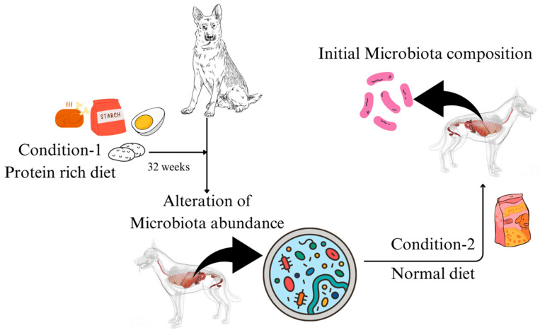
The type and composition of the diet that animal consumes act as a substrate for microbial growth, which leads to the production of SCFAs, secondary bile acids, amino acids, and fat-soluble vitamins released as microbial metabolites [99]. These metabolites, especially SCFAs, exert varied physiological responses in the host, for instance, reducing the inflammation in the intestine [100,101,102,103]. Additionally, the changes in the microbiota in the gut are a result of dietary micronutrients and have become an emerging area of current microbiome research. Molecules like fibre and carbohydrates (Table 3) are fermented by different bacterial species, altering the substrates in the gut and resulting in the growth of specific species and changes in the microbiome and metabolome. Studies conducted by [30], in which for 32 weeks, healthy dogs were fed purified amino acid (protein)-rich diets (Table 4) and digestible starch, which led to an alteration in the species composition, while upon returning to control diet, the microbiome species composition was found to be as to that initial (Figure 3). SCFA producers like Bacteroides, Prevotella, and Faecalibacterium were found to be increased in the weight loss diet (28.1% fibre) fed dogs group. However, the low-fat diet (8.6% fibre) group showed Faecalibacterium to be more dominant, suggesting that fibre content impacts the growth of the microorganism. The dietary composition exerts a significant impact on the modulation of gut microbiota growth, as exemplified by the observed correlation between the animal’s feeding regimen and the aforementioned microbial population dynamics.
Table 3.
Effect of fibre and the gut microbiome of domesticated cats and dogs [101].
| Impact on Dogs | ||||
|---|---|---|---|---|
| Diet Type | Technique | Results | Alterations in Abundance | References |
| Inulin-type fructans | 16S rRNA seq. | Firmicutes, Erysipelotrichaceae, and Turicibacteraceae | Increase | [104] |
| Beet pulp | 16S rRNA seq. | Erysipelotrichi and Fusobacteria | Decrease | [105] |
| Firmicutes and Clostridia | Increase | |||
| Yeast cell wall | 16S rRNA seq. | Bifidobacterium | Increase | [106] |
| Inulin | 16S rRNA seq. | Enterobacteriaceae | Decrease | [106] |
| Megamonas and Lactobacillus | Increase | |||
| Potato fibre | 16S rRNA seq. | Faecalibacterium, Lachnospira, faecal acetate, propionate and butyrate | Increase | [107,108] |
| Prevotella and Fusobacterium | Decrease | |||
| Soybean husk | qPCR | Clostridium cluster XI | Decrease | [109] |
| Total Lactobacilli, Faecalibacterium, Bacteroides-Prevotella-Porphyromonas, and Clostridium cluster XIVa | Increase | |||
| Impact on Cats | ||||
| FOS | qPCR | Bifidobacterium | Increase | [110] |
| 16S rRNA seq. | Actinobacteria | Increase | [111] | |
| GOS | qPCR | Bifidobacterium | Increase | [110] |
| Cellulose | 16S rRNA seq. | No changes | — | [111] |
| FOS and GOS | qPCR | Bifidobacterium, total SCFAs, butyrate, and valerate | Increase | [110] |
| FOS and inulin | 16S rRNA seq. | Veillonaceae | Increase | [112] |
| Gammaproteobacteria | Decrease | |||
| Inulin | 16S rRNA seq. | Bifidobacterium | Increase | [113] |
| Faecalibacterium and Fusobacterium | Decrease | |||
| Pectin | 16S rRNA seq. | Firmicutes | Increase | [111] |
| Wool hydrolysate | 16S rRNA seq. | No changes | — | [114] |
| Mixed insoluble fibres | 16S rRNA seq. | Blautia, Bacteroides, Turicibacter, acetic and propionic acids | Increase | [115] |
| Isobutyric, 2-methylbutyric, and isovaleric acids | Decrease | |||
| Inulin and cellulose | 16S rRNA seq. | Prevotella, Bifidobacterium, Lactobacillus, Megamonas, and unclassified Lachnospiraceae | Increase | [116] |
| Clostridium, Fusobacterium, and Eubacterium | Decrease | |||
FOS, fructooligosaccharides; qPCR, quantitative polymerase chain reaction; SCFAs, short-chain fatty acids; GOS, galactooligosaccharides; rRNA seq., ribosomal RNA sequencing. No changes (—).
Table 4.
The effect of high protein diets on the species diversity of gut microbiome in companion animals [101].
| Impact on Dogs | |||||
| Diet Type | Results | Feed Duration | Number of Individuals | References | |
| Bones and raw foods (BARF) | ↓ Bifidobacterium and Faecalibacterium; ↑ Fusobacteria, Escherichia coli, Streptococcus, and Clostridium |
4 weeks to 9 years | 27 | [117] | |
| Red meat | ↓ Faecalibacterium, Peptostreptococcus, Bacteroides, and Prevotella ↑ Fusobacterium, Lactobacillus, and Clostridium |
9 weeks | 7 | [35] | |
| Raw diet | ↑ Richness, evenness, Clostridium perfringens, Clostridium hiranonis, Dorea, and Fusobacterium varium | At least 1 year | 6 | [118] | |
| Kibble with boiled beef | ↓ Faecalibacterium prausnitzii ↑ Clostridium hiranonis, Dorea, Slackia, and unidentified Clostridiaceae |
1 week per combination | 11 | [119] | |
| Impact on Cats | |||||
| Diet Type | Results | Other Specifications | Feed Duration | Number of Individuals | References |
| High-protein low-carbohydrate dry food | ↓ Lactobacillus, Bifidobacterium, and Escherichia coli | Kitten, weaning diet | 8 weeks | 7 | [120] |
| ↓ Actinobacteria, Bifidobacterium, Dialister, Acidaminococcus, Megasphera, and Mitsuokella ↑ Fusobacteria, Clostridium, Faecalibacterium, Ruminococcus, Blautia, and Eubacterium |
Kitten, weaning diet | 8 weeks | 7 | [116] | |
| ↑ Species diversity; affected 194 metabolic pathways, including amino acid synthesis and metabolism | Kitten, weaning diet | 8 weeks | 6 | [121] | |
| Raw 1 to 3-day-old chicks | ↑ Peptococcus, Pseudobutyrivibrio, and unidentified Lachnospiraceae | Adult | 1.3 weeks | 5 | [122] |
| Raw | ↑ Clostridium, Fusobacterium, Eubacterium, and molar ratio of butyrate | Adult | 3 weeks | 12 | [123] |
| Raw plus plant fibre | ↓ Clostridium, Fusobacterium, and Eubacterium | Adult | 3 weeks | 12 | [123] |
| ↑ Prevotella | |||||
| Canned | ↓ Firmicutes, Bacteroides, Lactobacillus, and Streptococcus | Kitten, weaning diet | 9 weeks | 10 | [67] |
| ↑ Fusobacterium, Clostridium, unidentified Peptostreptococcaceae and Prevotellaceae | |||||
| ↓ Lactobacillus, Megasphera, and Olsenella | Adult | 5 weeks | 16 | [67] | |
| ↑ Species richness, Fusobacteria, Proteobacteria, Clostridium, Blautia, Bacteroides, and unidentified Peptostreptococcaceae | |||||
| ↓ Lactobacillus, Bifidobacterium, and Collinsella | Kitten, weaning diet | 9 weeks | 10 | [113] | |
| ↑ Bacteroides, Clostridium, Fusobacterium, genes involved in vitamin biosynthesis, metabolism, and transport | Kitten, weaning diet | ||||
Increase in abundance (↑); decrease in abundance (↓).
Figure 3.
Diet plays a crucial role in the gut microbiome of dogs. An experiment was performed where the dogs were fed the protein-rich diet for 32 weeks, and after that, they were shifted to a regular diet, leading to normalisation of the gut microbiome.
3.6. Probiotics
According to the World Health Organization (WHO), probiotics are described as a live microorganism that, upon administration in prescribed amounts, improves the health of the host [124]. The use of probiotics is generally recognised as valuable for maintaining and promoting gastrointestinal health in both farm animals and companion animals. The popularity of probiotic products for pets underscores the growing interest and awareness among pet owners regarding the potential benefits of probiotic supplementation for their animals’ well-being [62]. Furthermore, its use in companion animals such as dogs and cats has improved the gut microbiota composition, boosting the overall microbial balance to enhance immunologic responses, reduce intestinal inflammation, increase intestinal barrier function, and protect against colonisation by enteropathogens [125].
Bile acids possess detergent properties that can disrupt bacterial cell membranes, cause DNA damage, induce conformational changes in proteins, and chelate calcium and iron [126]. When exposed to bile acids, probiotic bacteria increase the production of certain proteins that help efflux of bile salts or protons. Additionally, they make changes to their overall metabolism in order to counteract the harmful effects of bile acid exposure [127]. Certain types of bacteria, like Lactobacillus and Bifidobacterium, can resist bile acids, which are linked to the activation of glycolysis [127]. Unconjugated bile acids have stronger antibacterial activity compared to conjugated bile acids. Probiotic bacteria such as Lactobacillus paracasei and Bifidobacterium longum exhibit greater resistance to bile (taurocholic acid and tauroursodeoxycholic acid exposure) [128]. Conjugated bile acid exposure for a brief period caused the activation of bacterial glycolysis, resulting in an increased growth rate of Bifidobacterium longum [128].
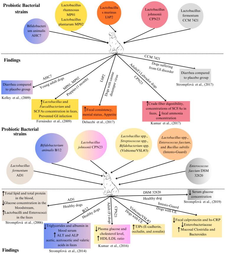
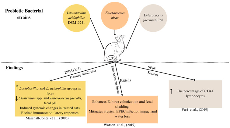
Most probiotics designed for humans and animals contain quantities of bifidobacteria and other lactic acid-producing bacteria [129]. A comprehensive list of bacterial strains utilised in both single-strain probiotics and multi-strain mixtures documented in various studies is presented in Table 5 (also in Figure 4 and Figure 5).
Figure 4.
Experiments showing the use of bacterial strains as probiotics and their corresponding finding in the dogs. AHC7: probiotic Bifidobacterium animalis strain AHC7 [111,130]; MP01: Lactobacillus rhamnosus MP01; MP02: Lactobacillus plantarum MP02 [131]; LbP2: probiotic Lactobacillus murinus LbP2 [132]; CPN23: Lactobacillus johnsonii CPN23 [133]; CCM 7421: probiotic Lactobacillus fermentum CCM 7421 [134]; GI: gastrointestinal; HDL: high-density lipoprotein; LDL: low-density lipoprotein; ALT: alanine transaminase; ALP: alkaline phosphatase; AD1: Lactobacillus fermentum AD1 [135]; B/12: Bifidobacterium animalis B/12 [136]; CE: chronic enteropathy; Visbiome/VSL#3: Lactobacillus plantarum DSM 24730, Streptococcus thermophilus DSM 24731, Bifidobacterium breve DSM 24732, Lactobacillus paracasei DSM 24733, Lactobacillus delbrueckii subsp. bulgaricus DSM 24734, Lactobacillus acidophilus DSM 24735, Bifidobacterium longum DSM 24736, and Bifidobacterium infantis DSM 24737 [54]. TJP: tight junction protein; hs-CRP: high-sensitivity C-reactive protein; DSM 32820: Enterococcus faecium DSM 32820 [137]. The upwards arrow (↑) represents “increase” and the downwards arrow (↓) represents “decrease”.
Figure 5.
Bacterial strains used as probiotics and their corresponding experimental finding in the cats. Cluster of differentiation 4 (CD4); atypical enteropathogenic Escherichia coli (EPEC) [94,138]. DSM13241: Lactobacillus acidophilus DSM13241 [139]; SF68: Enterococcus faecium SF68 [140]. The upwards arrow (↑) represents “increase” and the downwards arrow (↓) represents “decrease”.
Table 5.
Bacterial strains isolated from the canine and feline with probiotic properties and used for improving gut microbiome.
| Bacterial Strains | Amount | Source | Age/Conditions | Assessment | Findings Obtained | Reference |
|---|---|---|---|---|---|---|
| Bifidobacterium animalis AHC7 | 2 × 10¹⁰ CFU/day | Canine | Young adult dogs having acute diarrhoea | Managing acute diarrhoea |
|
[111,130] |
| Lactobacillus rhamnosus MP01, Lactobacillus plantarum MP02 | 109 CFU/day | Canine | Puppies (1 month) | Infection prevention in puppies |
|
[131] |
| Lactobacillus murinus LbP2 | 5 × 10⁹ CFU/day | Canine | Dogs with canine distemper virus (CDV)-associated diarrhoea | Mental and faecal status |
|
[132] |
| Lactobacillus johnsonii CPN23 | 2.3 × 10⁸ CFU/day | Canine | Female Labrador dogs (Adult) | Nutrient digestibility and faecal fermentative metabolites |
|
[133] |
| Lactobacillus fermentum CCM 7421 | 107–109 CFU/day | Canine | Dogs (having gastrointestinal disorder) | Composition of the faecal microbiome and blood samples |
|
[134] |
| Lactobacillus fermentum AD1 | 3 mL of 109 CFU/mL | Canine | Control healthy dogs | Composition of the faecal microbiome and blood samples |
|
[135] |
| Bifidobacterium animalis B/12 | 1 mL of 1.04 × 10⁹ CFU/mL | Canine | Control healthy dogs | Composition of the faecal microbiome and Blood samples |
|
[136] |
| Lactobacillus johnsonii CPN23 | 108 CFU/mL (0.1 mL/kg BW) | Canine | Female dogs (adult) | Assessment of blood sample profile |
|
[141] |
| Enterococcus faecium DSM 32820 | 109 CFU/day | Canine | Control healthy dogs | Blood sample profile |
|
[137] |
| Lactobacillus acidophilus DSM 13241 | 2 × 10⁸ CFU/day | Feline | Healthy adult cats | Improving intestinal health in cats |
|
[139] |
| Enterococcus hirae | 2.85–4.28 × 10⁸ CFU/day | Feline | Kittens | Preventing atypical Enteropathogenic E. coli (EPEC) in kittens |
|
[94,138] |
| Enterococcus faecium SF68 | 5 × 10⁹ CFU/day | Feline | Kittens | Enterococcus faecium strain SF68 supplementation on immune function |
|
[140] |
| Bacillus subtilis HH2 | 5 × 10⁹ CFU/day | Canine | Beagles with orally administered Enterotoxigenic Escherichia coli (ETEC) | Intestinal barrier integrity, faecal microbiota, and non-specific immunity |
|
[142,143] |
| Lactobacillus plantarum DSM 24730, Lactobacillus paracasei DSM 24733, Lactobacillus delbrueckii subsp. bulgaricus DSM 24734, Lactobacillus acidophilus DSM 24735, Streptococcus thermophilus DSM 24731, Bifidobacterium breve DSM 24732, Bifidobacterium longum DSM 24736, and Bifidobacterium infantis DSM 24737 | S. thermopilus 40.55%, Bifidobacteria 12.5%, Lactobacilli 13%, and other excipients 39.05% (112–225 × 109 CFU/10 kg) | Canine | Dogs with CE | Disease activity and mucosal microbiota changes and tight junction protein (TJP) expression |
|
[54] |
| Bifidobacterium bifidum, Enterococcus faecium and thermophilus, and Lactobacillus acidophilus, bulgaricus, casei, and lantarum | 5 × 10⁹ CFU/day | Feline | Healthy cats | Faecal microbiome and faecal metabolomics |
|
[97] |
| Lactobacillus acidophilus DSM 32241, Lactobacillus helveticus DSM 32242, Lactobacillus paracasei DSM 32243, Lactobacillus plantarum DSM 32244, and Lactobacillus brevis DSM 27961, Streptococcus thermophilus DSM 32245, Bifidobacterium lactis DSM 32246, Bifidobacterium lactis DSM 32247 | 400 billion cfu of lyophilised bacteria/day | Canine | Healthy dogs | Concentration of faecal immunoglobulin IgA, plasma IgG, and faecal microbiota composition |
|
[144] |
| Lactobacillus acidophilus, Lactobacillus casei, Enterococcus faecium, and Bacillus subtilis | 1 billion CFU/mL per 2.2 kg of body weight orally twice daily | Canine | Dogs with confirmed CE | Clinical signs, mucosal microbiota, and inflammatory indices |
|
[125] |
SCFAs, short-chain fatty acids; CE, chronic enteropathy; TJP: tight junction protein.
3.7. Faecal Microbiota Transplantation (FMT)
Faecal microbiota transplantation (FMT) is a recently developed therapeutic method that involves transferring faeces from a healthy donor to a recipient to have the potential to offer a diseased patient with a healthy microbiome. Research has demonstrated that FMT has shown greater effectiveness in managing dysbiosis with promising results than other treatments that modify the microbiome in humans [145] and in dogs with infectious and chronic GI diseases [146]. The faecal microbiota of dogs with diarrhoea treated with FMT as enema and dogs with IBD treated only with corticosteroids show a closer resemblance to the healthy canine faecal microbiota and with a decrease in cholic acid and the percentage of primary bile acids compared to dogs with diarrhoea and IBD treated with metronidazole [21,147]. While dogs suffering from diarrhoea caused by Clostridium perfringens toxin A did not respond to antimicrobial treatment, their diarrhoea was successfully resolved when they received FMT through an enema [24]. In a randomised clinical trial, the use of FMT resulted in faster clinical recovery and reduced hospitalisation time for puppies who survived acute hemorrhagic diarrhoea caused by canine parvovirus (CPV) [148]. Also, dogs with CE, when undergoing treatment with a single FMT enema, experienced a notable reduction in their canine IBD activity index (CIBDAI) score following the procedure [149].
4. Gut Microbiome and Diseases
4.1. Neurological Disorders/Disease
The gut–brain axis, or GBA, is a network of endocrinological, immunological, and neuronal mediators that interacts intricately between the gut and the brain [150]. The gut microbiota, the central nervous system (CNS), and the enteric nervous system (ENS) regulate the GBA [151]. Certain neuroactive substances generated in the gastrointestinal tract can pass through the blood–brain barrier (BBB) and the intestinal mucosal barrier to enter the CNS. These neuroactive molecules bring changes that lead to unfavourable conditions; for example, Escherichia coli and Pseudomonas can synthesise γ-aminobutyric acid (GABA), an inhibitory neurotransmitter that can cross the BBB. A decline in GABA levels can lead to the occurrence of epileptic seizures, as this inhibitory neurotransmitter (NT) plays a crucial role in regulating neuronal excitability in the mammalian brain. The disruption of GABAergic neurotransmission plays a significant role in the development and manifestation of various neurological disorders, such as epilepsy [152].
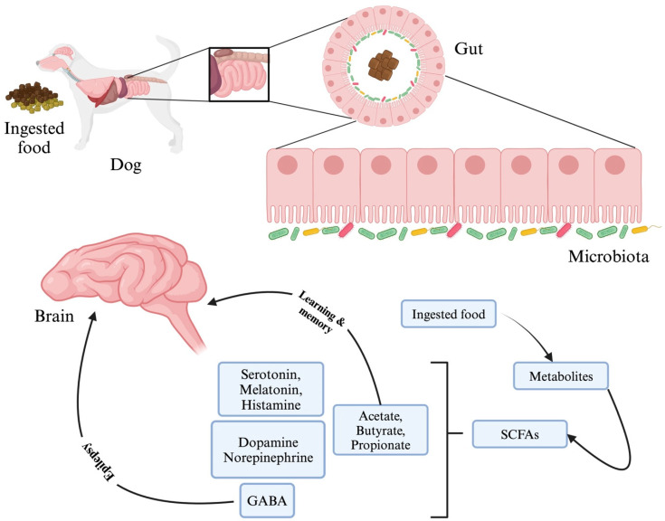
As mentioned previously, gut microbes also produce SCFA like propionate, butyrate, and acetate, which act as a source of energy for cell regeneration and mucous production (Figure 6). These SCFAs support intestinal epithelial cell regeneration, mucus formation, and integrity of the BBB. The intestinal fermentation of dietary fibres occurs anaerobically, yielding the highest concentration of these SCFAs. Butyrate serves as colonocytes’ main energy source and reduces inflammation in the intestines to preserve intestinal homeostasis. The generation of SCFAs by microbes is crucial in lowering gut pH and inhibiting potentially harmful bacteria growth. SCFAs have an impact on the CNS by interacting with the free fatty acid receptor (FFAR), a G protein-coupled receptor (GPR) on enteroendocrine cells, including GPR41 (FFAR3), GPR43 (FFAR2), GPR109A/HCAR (hydroxycarboxylic acid receptor)2, and GPR164 [153]. The interaction between SCFAs and these receptors leads to the secretion of various gut hormones and neurotransmitters (NTs), which in turn promotes indirect signaling to the brain via the systemic circulation or vagal pathways. Also, SCFAs play a crucial role in inhibiting the activity of histone deacetylase, which helps to promote the acetylation of lysine residues in nucleosomal histones and increase transcription [153]. Given the potential connection between histone acetylation and inflammation in the CNS, it is plausible that SCFAs may play a role in addressing disruptions to brain immunity.
Figure 6.
Schematic representation of the conversion of the digested food into metabolites like short-chain fatty acids (SCFAs) and its components like acetate, butyrate, and propionate, which have been shown to indirectly influence the learning and memory process by controlling the energy balance. Decreasing Gamma Amino Butyric Acid (GABA) levels have been shown to generate epileptic seizures. The dopamine–norepinephrine–epinephrine cycle stimulates hormonal and neuronal pathways, while norepinephrine, serotonin, melatonin, and histamine can function as both hormones and neurotransmitters and play a role in excitation/inhibition balance in the brain and epileptogenesis in addition to GABA. This figure was generated using BioRender (www.biorender.com; accessed on 3 May 2024).
The hypothesis is that gut microbiota is responsible for Aβ peptide accumulation in intestinal epithelial cells. It was reported by [154] that Aβ peptide and apoB were colocalised in the Golgi apparatus in low-fat and saturated-fat-fed mice. The amyloid β peptide (Aβ) plays a crucial role in initiating the progression of Alzheimer’s disease (AD) through the accumulation and aggregation process. This process can be triggered by either excessive production of Aβ or disruption in its clearance. The Aβ-like peptides are released from the gut microbiome, primarily from the Clostridiales order. Research conducted on a 5xFAD transgenic AD mice model revealed that the administration of prebiotic mannan oligosaccharide resulted in significant improvements that included a reduction in Aβ plaques, cognitive deficits, microglial activation, oxidative stress, and changes in the gut microbiome (GM). Research suggests that alterations in the brain mediated by GM may be influenced by SCFAs, as observed in studies where SCFA supplementation yielded similar results [155]. In another study, the effects of the prebiotic R13 on 5XFAD mice were investigated. The study revealed that this compound, acting as a tropomyosin receptor kinase B (TrkB) agonist, effectively inhibits the proinflammatory C/EBPB/AEP pathway in the gut. Additionally, it was observed that the presence of amyloid-positive signals in the gut was reduced as well [156]. Research has indicated that the KD may have potential benefits in AD [157]. Research conducted on murine models of AD has revealed promising results regarding the potential benefits of the KD, showing to decrease amyloid plaques, enhance memory, and alleviate neurodegeneration and neuroinflammation [158]. The KD can have a positive impact on the composition of gray matter and neurovascular function, boosting the presence of potentially beneficial gut microbiota (Akkermansia muciniphila and Lactobacillus) while decreasing the levels of potentially pro-inflammatory bacteria (Desulfovibrio and Turicibacter), potentially offering benefits for AD as reported in murine models [159]. A modified Mediterranean–ketogenic diet also exhibited the potential to impact the gut microbiota and SCFA production in individuals with mild cognitive impairment (MCI). Interestingly, these changes were found to be associated with the levels of amyloid measured in the cerebrospinal fluid (CSF) [160].
One of the most prevalent neurological conditions in both people and canines is epilepsy. Whilst studies completed in the UK suggested that 0.62–0.8% of dogs overall have epilepsy, the actual prevalence of the condition is unknown. The presence of gut microbiota (GM)-derived SCFAs can potentially influence the likelihood of experiencing seizures. This influence is believed to occur through the regulation of excitatory/inhibitory neurotransmitters, neuroinflammation, oxidative stress as well as psychosocial stress. When the ketogenic diet (KD) was introduced to epilepsy patients, it was found to have a positive impact on chemical messengers in the brain, such as GABA, agmatine, and monoamines. This led to a decrease in neuronal irritability and ultimately helped to reduce seizures in patients. In the CNS, there is an increase in the production of GABA, while the quantity of aspartate inhibitors decreases [161]. The decrease in aspartate levels caused by ketosis contributes to the activation of glutamate, which is then converted into glutamine. Neuron cells readily absorb glutamine and convert it into GABA, which has an inhibitory effect that helps to decrease oxidative stress [162]. It is worth mentioning that short-term feeding of the KD has also shown positive effects on shifting the metabolome towards a molecular signature that is anti-tumorigenic in dogs, possibly due to decreased abundance of Erysipelotrichales and Lactobacillales and increased abundance of Enterobacteriales, Fusobacteriales, Bifidobacteriales, Aeromonadales, and Selenomonadales [163].
Studies on the effects of the KD on seizures in murine models have yielded conflicting results, with some showing no impact, others indicating an increase in seizures, and some suggesting partial protection against seizures [164]. Several factors can influence the varying outcomes of rodent studies, including variations in experimental design, the age of the animals being tested, and the methods used to assess seizure threshold. Additionally, genetic differences between species can also play a role in determining susceptibility to seizures [165]. Several other studies report that KD helps prevent seizures by increasing the presence of important bacterial species such as Akkermansia muciniphila and Parabacteroides merdae that synergise to reduce the gammaglutamylation of amino acids, enhance the GABA/glutamate ratios in the hippocampus, and ultimately, protect against seizures [166]. As GABA has a role in the antiseizure effects of the KD [164], an altered diet can lead to uneven expression of GABA, which in turn can lead to a seizure.
Intestinal dysbiosis also leads to the severity of epilepsy since immune responses are elevated due to inflammation. Changes in the gut microbiota’s composition have been linked to gastrointestinal disorders in dogs [167], obesity [168], and, more recently, neurological conditions like meningoencephalomyelitis of unknown origin [169], as well as behavioral issues like aggression and phobic disorders [170]. Microbiota function in dogs with epilepsy remains the subject of relatively few investigations. In a study conducted by [171], the faecal samples collected from 13 pairs of dogs, each consisting of a drug-naive epileptic dog and a healthy dog kept on the same diet, were used to assess Lactobacillus populations in dogs with idiopathic epilepsy in comparison to healthy dogs. They failed to find any differences between the groups in the relative or absolute abundance of Lactobacillus species or large-scale microbial patterns. However, Firmicutes, Bacteroidetes, Proteobacteria, Fusobacteria, and Actinobacteria were the predominant bacterial populations in both healthy and epileptic dogs. In a study conducted by [172], the companion dogs (n = 29) were studied to understand changes in the composition of microbiota with aging by performing 16s rRNA sequencing from faecal samples, and it was found that higher age was related to lower Fusobacteria, higher cognitive performance was linked to lower Actinobacteria. Reports are stating the role of gut microbiota in the aggressive behavior of dogs, [170] identified Catenibacterium and Megamonas as major discriminates of aggressive behavior in dogs, later it was also found that Catenibacterium and Megamonasa are responsible for primary bile acid metabolism and abdominal pains in human [173]. Gut microbiota, thus, has multiple effects on the central nervous via the innervations from the enteral nervous system.
4.2. Cardiac Health
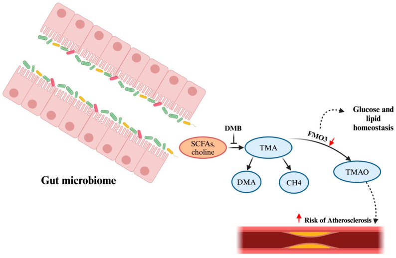
Dogs frequently exhibit congestive heart failure (CHF), which is characterised by acute respiratory distress brought on by pulmonary oedema, pleural effusion, or abdominal distension from ascites. Even when their heart disease is well controlled, dogs with congestive heart failure (CHF), especially those with right-sided congestive heart failure (RCHF), are frequently put to death because of severe cachexia and progressive inappetence [174]. A study conducted by Seo et al. [175] demonstrated that dogs with CHF had increased E. coli and uncharacterised species of Enterobacteriaceae. An increasing amount of research has shown that the gut microbiota and its metabolites play a role in the development and course of cardiovascular disease. In one study by Li et al. (2021), a potential relationship between the gut microbiome and myxomatous mitral valve disease (MMVD) in dogs was observed [176]. It was found that the gut dysbiosis index increases in proportion to the severity of MMVD and is inversely associated with Clostridium hiranonis, a key bile acid converter in the gut. Secondary bile acids are significant byproducts resulting from the fermentation of primary bile acids by gut microbes. The main secondary bile acids, deoxycholic acid (DCA) and lithocholic acid (LCA), have the ability to influence the composition of the microbial communities in the gut [177]. The study by Li et al. (2021) also revealed that secondary bile acids may promote the growth of beneficial bacteria but inhibit harmful species. For example, secondary BAs, such as DCA, enhance the growth of Fusobacterium, while LCA promotes Faecalibacterium growth. In contrast, DCA hinders the growth of E. coli [176]. These findings indicate a potential interplay between gut microbiota, gut microbiota-produced metabolites, and the pathophysiological progression of MMVD in dogs [176]. The oral microbiome can influence both these disorders and the composition of the gut microbiota. Even after traditional risk variables have been taken into account, there is still evidence of a favourable association between TMAO and the prediction of cardiovascular risk [178], while SCFAs, on the other hand, help to regulate blood pressure. A significant amount of choline, carnitine, and phosphatidylcholine are broken down into the precursor trimethylamine (TMA) by some gut bacteria (Figure 7). The downstream signaling and metabolism of TMA lead to the production of FMO, which is considered to be a marker for the treatment of cardiovascular disease. In acute myocardial infarction animal models, the gut microbiome, particularly the Tissierella Soehngenia genus, the Synergistetes phylum, the Spirochaetes phylum, the Lachnospiraceae family, and the Syntrophomonadaceae family, exhibits a greater trend. However, the research on the cardiovascular system is unexplored, and more mechanistic-based studies need to be accounted for to find the risk associated with heart failure.
Figure 7.
Schematic representation of gut microbiome converting ingested food into metabolites like short-chain fatty acids (SCFAs), choline, and carnitine, which is metabolised into TMA (Trimethylamine), and some part of it is converted into CH4 and DMA + formaldehyde. However, DMB is known to be a repressor of this conversion. TMA is further converted into FMO3 and TMAO after being transported to the liver via the portal vein. Any suppression or deletion in FMO3 leads to altered cholesterol uptake in the intestine and reverse transportation, which might increase the chances of atherosclerosis. This figure was generated using BioRender (www.biorender.com; accessed on 3 May 2024). Trimethylamine-N-oxide (TMAO). The upwards red arrow (↑) represents “increase” and the downwards red arrow (↓) represents “decrease”.
4.3. Chronic Inflammatory Enteropathies in Dogs and Cats
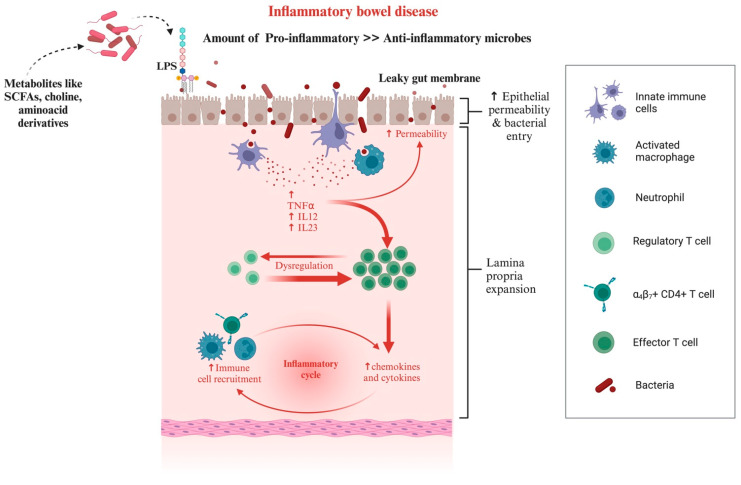
The intestinal microbiota of dogs and cats is linked to several primary GI disorders associated with dysbiosis. Canine chronic inflammatory enteropathy (CIE) is a common cause of chronic GI signs and histologic inflammation [179]. It is a multifactorial disorder where the interplay between intestinal immunity and environmental factors (diet, microbiota) initiates and drives chronic intestinal inflammation, causing vomiting, diarrhoea, alterations in appetite, and/or weight loss in affected dogs. Different microbiologic studies have shown an increase in intestinal Proteobacteria and a decreased abundance of Clostridiales associated with intestinal inflammation. Using FISH probes, one study discovered that Beagle dogs with persistent diarrhoea had considerably higher Bacteroides levels [180]. In a study conducted by Suchodolski et al. [181], wherein the changes in the faecal microbiome were studied, it was revealed that while there was a reduction in Faecalibacterium spp. and the phylum Fusobacteria during periods of clinically insignificant CIE, there was no significant difference during periods of active disease. In addition to providing the host with nutritional advantages, a balanced intestinal ecology primes and stimulates the immune system and helps defend against invasive intestinal pathogens. In animal model studies involving germ-free animals (rodents), there are morphological and immunological variations between germ-free and conventionally raised animals, demonstrating the effects of the resident microbiota on intestinal structure and function. In the GI tract, residing bacteria can release various compounds, including lipopolysaccharides (LPS), amyloid, and other immunogenic mixtures [125], into the surrounding intestinal environment [38,44,182,183,184]. Immune activation, either by the LPS secretion or an increase in the pro-inflammatory microbes compared to the anti-inflammatory microbes, shapes gut dysbiosis, further contributing to gut inflammation (Figure 8).
Figure 8.
Either ingestion or conversion to LPS or an increase in the ratio of pro-inflammatory to anti-inflammatory microbes can lead to a leaky gut membrane, which is characterised by an elevated response by the immune system. The release of chemokines, cytokines, and interleukins contributes to the progressing havoc leading to inflammatory bowel disease. This figure was generated using BioRender (www.biorender.com; accessed on 3 May 2024).
The etiology of chronic intestinal inflammation in canines may involve microbial imbalances, potentially serving as both a causative factor and a resultant outcome [38,185]. Dogs afflicted with CE have been observed to manifest a notable reduction in both faecal bacterial richness and diversity [21,102]. Moreover, it has been observed that both luminal and mucosal intestinal bacteria exhibit a shared dysbiotic profile, which is distinguished by a reduction in the abundance of Clostridium, Fusobacterium, and Bacteroides, alongside an elevation in the abundance of Enterobacteriaceae [125,186,187,188] in canine subjects presenting with persistent gastrointestinal symptom. The members of the Enterobacteriaceae family are regarded as pathogenic owing to their capacity to elicit innate immune reactions within the gastrointestinal tract [189]. The comparative analysis of microbial populations in diseased dogs and healthy control (HC) dogs revealed elevated levels of Bifidobacterium spp., Lactobacillus spp., Streptococcus spp., and potentially pathogenic Escherichia coli (E. coli) in the former group [102].
In another study, dogs subjected to a dietary intervention involving the administration of a supplement comprising a combination of four distinct strains of probiotic bacteria, prebiotics, and IgY exhibited notable alterations in their mucosal microbiota in comparison to dogs who received a hydrolysed diet (placebo) treatment [125]. Dogs that received supplementation exhibited elevated quantities of colonic mucosal Bacteroides spp. and Clostridium spp. while experiencing a reduction in the abundance of mucosal Enterobacteriaceae compared to dogs administered a placebo [125]. It was observed that there was an overrepresentation of mucosal Enterobacteriaceae in dogs with CE prior to treatment. However, a significant reduction in the abundance of these Enterobacteriaceae was observed in the colonic biopsies of canines that received the supplement [125]. Several other studies have consistently demonstrated a noteworthy decrease in the abundance of Clostridium spp. and Bacteroides spp. during the initial stages of these conditions [186,187,188]. Research findings indicate that these specific bacterial species have been identified to play a significant and advantageous role in producing SCFAs [40,41,102,190]. The SCFAs, including butyrate, serve as an energy source for colonocytes and play a significant role in upholding the integrity of the intestinal epithelial barrier [191].
4.4. Obesity
Pet populations are experiencing a concerning rise in obesity and overweight issues, with many contributing factors at play, such as sedentary lifestyles, high-calorie meals, genetic predispositions, and neutering. The significance of gut microbiota, specifically alterations in bacterial taxa and metabolite profiles, is underscored in relation to obesity [192] and associated diseases in cats and cardiac disease [175], diabetes mellitus [12], and orthopedic diseases [23] in dogs.
Multiple studies have found a reduction in bacterial diversity in the faecal microbiota of obese and overweight dogs compared to dogs with a healthy weight [193]. There was a higher presence of Actinobacteria and Roseburia in obese dogs [168]. It was found that the presence of the Actinobacteria class was more pronounced in obese dogs compared to dogs of normal weight. While the gut microbiota of lean dogs was primarily composed of microbes from Firmicutes (85% of the total population), the gut microbiota of obese dogs was mainly composed of bacteria from the phylum Proteobacteria, accounting for 76% of the total [194]. The presence of Gram-negative bacteria could potentially contribute to chronic low-grade inflammation in obese dogs by producing higher levels of intestinal LPS [195]. Overweight dogs, on the other hand, had a greater abundance of the Erysipelotrichi class, mainly due to differences in Eubacterium spp. [196]. There was a noticeable decrease in the abundance of Bifidobacteriales and a slight increase in the abundance of Aeromonadales in overweight dogs compared to obese dogs. Overweight dogs showed a higher prevalence of Prevotella copri and Clostridium compared to obese dogs. Previous studies have found that overweight dogs tend to have a greater presence of Fusobacteria, particularly Fusobacteria perfoetens, compared to normal dogs [193]. In contrast to overweight dogs, dogs with a healthy weight showed an elevated population of Erysipelotrichales, Erysipelotrichaceae, and Erysipelotrichi while having a reduced presence of Bifidobacteriales. Normal-weight and overweight dogs had a higher representation of the Blautia, Lachnospiraceae family, and Eubacterium biforme compared to obese dogs. Normal-weight dogs had a higher relative abundance of the Ruminococcus family compared to obese dogs [193,194]. A negatively correlated abundance of Megamonas and the weight-loss rate, a noticeable decrease in the presence of Ruminococcaceae, and lower levels of propionic and acetic acid in the faeces of dogs that experienced rapid weight loss, compared to dogs with slower weight loss, demonstrate that obese dogs have faecal bacteria that can produce propionic and acetic acids and may be less responsive to weight loss because they have a greater capacity to extract energy from the diet through SCFAs production [77]. Notably, prebiotics such as fructooligosaccharides have the potential to influence the composition of the gut microbiota positively. An effective treatment approach for obesity-related health problems in companion animals may involve the administration of short-chain fructo-oligosaccharides since it has been associated with enhanced microbial biodiversity and the synthesis of anti-inflammatory butyrate in obese dogs [77,104,168,192,193,194,196,197,198,199,200,201,202].
5. Future Prospects and Conclusions
This comprehensive review illuminates the intricate and multifaceted interplay between the gut microbiome and health in companion animals. Despite foundational characterisations, critical knowledge gaps persist regarding longitudinal microbial dynamics, functional host–microbe interactions, microbial biomarkers of health/disease states, standardised methodologies, environmental modulators, and transgenerational inheritance patterns. Addressing these gaps through rigorous research is pivotal for advancing microbiome-based diagnostics, therapeutics, and interventions to optimise health outcomes in companion animal species. Moreover, it is necessary to reevaluate the regular usage of antibiotics in companion animals experiencing GI symptoms, and other approaches that can improve the GI microbiome, such as FMT or advanced probiotics, should be investigated as alternatives. Though several studies evaluated the effects of probiotics like Bifidobacterium, Lactobacillus, and Enterococcus strains in dogs and cats, still more research is needed to understand their effects on the microbiome and their effectiveness in clinical settings.
Thus, in conclusion, gut health dependency can be attributed to parameters starting from microbiome modulation, nutrient uptake and utilisation, immunomodulatory effects, and use of strain-specific probiotics. Collectively, this review has provided a synthesised perspective on the evolving microbiome landscape in companion animals and its pivotal role in holistic health and disease management. Thus, more advanced studies should be envisaged for establishing a concrete link among the microbial species exhibiting synergistic effects in maintaining the health of companion animals.
Acknowledgments
The Publication Subvention Grants Program Award (for Open Access Journal Articles, Office of the Vice-President for Research, Iowa State University) to D.K.S. is acknowledged.
Supplementary Materials
The following supporting information can be downloaded at: https://www.mdpi.com/article/10.3390/microorganisms12091831/s1, Supplementary Figure S1. Worldwide preference for different companion animals (from aquaria population to big mammals). The information was accessed from worldpopulationreview.com (https://worldpopulationreview.com/country-rankings/pet-ownership-statistics-by-country; accessed on 10 August 2024). Supplementary Figure S2. The population of total companion animals and its estimates across the world. The information was accessed from worldpopulationreview.com (https://worldpopulationreview.com/country-rankings/pet-ownership-statistics-by-country; accessed on 10 August 2024). M: Million.
Conflicts of Interest
The authors declare no conflict of interest.
Funding Statement
The authors declare there was no external funding for the present review study.
Footnotes
Disclaimer/Publisher’s Note: The statements, opinions and data contained in all publications are solely those of the individual author(s) and contributor(s) and not of MDPI and/or the editor(s). MDPI and/or the editor(s) disclaim responsibility for any injury to people or property resulting from any ideas, methods, instructions or products referred to in the content.
References
- 1.Driscoll C.A., Macdonald D.W., O’Brien S.J. From Wild Animals to Domestic Pets, an Evolutionary View of Domestication. Proc. Natl. Acad. Sci. USA. 2009;106((Suppl. S1)):9971–9978. doi: 10.1073/pnas.0901586106. [DOI] [PMC free article] [PubMed] [Google Scholar]
- 2.Aydin N., Krueger J.I., Fischer J., Hahn D., Kastenmüller A., Frey D., Fischer P. “Man’s Best Friend:” How the Presence of a Dog Reduces Mental Distress after Social Exclusion. J. Exp. Soc. Psychol. 2012;48:446–449. doi: 10.1016/j.jesp.2011.09.011. [DOI] [Google Scholar]
- 3.Serpell J.A. Factors Influencing Human Attitudes to Animals and Their Welfare. Anim. Welf. 1994;13:S145–S151. doi: 10.1017/S0962728600014500. [DOI] [Google Scholar]
- 4.Harris Interactive: Harris Polls > Pets Aren’t Just Animals; They Are Members of the Family. [(accessed on 11 August 2024)]. Available online: https://www.harrisinteractives.com/NewsRoom/HarrisPolls/tabid/447/ctl/ReadCustomDefault/mid/1508/ArticleId/1076/Default.html.
- 5.Su Q., Liu Q. Factors Affecting Gut Microbiome in Daily Diet. Front. Nutr. 2021;8:644138. doi: 10.3389/fnut.2021.644138. [DOI] [PMC free article] [PubMed] [Google Scholar]
- 6.Turnbaugh P.J., Ley R.E., Hamady M., Fraser-Liggett C.M., Knight R., Gordon J.I. The Human Microbiome Project. Nature. 2007;449:804–810. doi: 10.1038/nature06244. [DOI] [PMC free article] [PubMed] [Google Scholar]
- 7.Sekirov I., Russell S.L., Antunes L.C.M., Finlay B.B. Gut Microbiota in Health and Disease. Physiol. Rev. 2010;90:859–904. doi: 10.1152/physrev.00045.2009. [DOI] [PubMed] [Google Scholar]
- 8.Dethlefsen L., McFall-Ngai M., Relman D.A. An Ecological and Evolutionary Perspective on Human–Microbe Mutualism and Disease. Nature. 2007;449:811–818. doi: 10.1038/nature06245. [DOI] [PMC free article] [PubMed] [Google Scholar]
- 9.Fetissov S.O. Role of the Gut Microbiota in Host Appetite Control: Bacterial Growth to Animal Feeding Behaviour. Nat. Rev. Endocrinol. 2017;13:11–25. doi: 10.1038/nrendo.2016.150. [DOI] [PubMed] [Google Scholar]
- 10.Fosgerau K., Hoffmann T. Peptide Therapeutics: Current Status and Future Directions. Drug Discov. Today. 2015;20:122–128. doi: 10.1016/j.drudis.2014.10.003. [DOI] [PubMed] [Google Scholar]
- 11.Jergens A.E., Guard B.C., Redfern A., Rossi G., Mochel J.P., Pilla R., Chandra L., Seo Y.-J., Steiner J.M., Lidbury J., et al. Microbiota-Related Changes in Unconjugated Fecal Bile Acids Are Associated with Naturally Occurring, Insulin-Dependent Diabetes Mellitus in Dogs. Front. Vet. Sci. 2019;6 doi: 10.3389/fvets.2019.00199. [DOI] [PMC free article] [PubMed] [Google Scholar]
- 12.Kieler I.N., Osto M., Hugentobler L., Puetz L., Gilbert M.T.P., Hansen T., Pedersen O., Reusch C.E., Zini E., Lutz T.A., et al. Diabetic Cats Have Decreased Gut Microbial Diversity and a Lack of Butyrate Producing Bacteria. Sci. Rep. 2019;9:4822. doi: 10.1038/s41598-019-41195-0. [DOI] [PMC free article] [PubMed] [Google Scholar]
- 13.Turnbaugh P.J., Hamady M., Yatsunenko T., Cantarel B.L., Duncan A., Ley R.E., Sogin M.L., Jones W.J., Roe B.A., Affourtit J.P., et al. A Core Gut Microbiome in Obese and Lean Twins. Nature. 2009;457:480–484. doi: 10.1038/nature07540. [DOI] [PMC free article] [PubMed] [Google Scholar]
- 14.Rosenbaum M., Knight R., Leibel R.L. The Gut Microbiota in Human Energy Homeostasis and Obesity. Trends Endocrinol. Metab. 2015;26:493–501. doi: 10.1016/j.tem.2015.07.002. [DOI] [PMC free article] [PubMed] [Google Scholar]
- 15.Lau W.L., Savoj J., Nakata M.B., Vaziri N.D. Altered Microbiome in Chronic Kidney Disease: Systemic Effects of Gut-Derived Uremic Toxins. Clin. Sci. 2018;132:509–522. doi: 10.1042/CS20171107. [DOI] [PubMed] [Google Scholar]
- 16.Hall J.A., Jackson M.I., Jewell D.E., Ephraim E. Chronic Kidney Disease in Cats Alters Response of the Plasma Metabolome and Fecal Microbiome to Dietary Fiber. PLoS ONE. 2020;15:e0235480. doi: 10.1371/journal.pone.0235480. [DOI] [PMC free article] [PubMed] [Google Scholar]
- 17.Penders J., Stobberingh E.E., van den Brandt P.A., Thijs C. The Role of the Intestinal Microbiota in the Development of Atopic Disorders. Allergy. 2007;62:1223–1236. doi: 10.1111/j.1398-9995.2007.01462.x. [DOI] [PubMed] [Google Scholar]
- 18.Kalenyak K., Isaiah A., Heilmann R.M., Suchodolski J.S., Burgener I.A. Comparison of the Intestinal Mucosal Microbiota in Dogs Diagnosed with Idiopathic Inflammatory Bowel Disease and Dogs with Food-Responsive Diarrhea before and after Treatment. FEMS Microbiol. Ecol. 2018;94:fix173. doi: 10.1093/femsec/fix173. [DOI] [PubMed] [Google Scholar]
- 19.Wang S., Martins R., Sullivan M.C., Friedman E.S., Misic A.M., El-Fahmawi A., De Martinis E.C.P., O’Brien K., Chen Y., Bradley C., et al. Diet-Induced Remission in Chronic Enteropathy Is Associated with Altered Microbial Community Structure and Synthesis of Secondary Bile Acids. Microbiome. 2019;7:126. doi: 10.1186/s40168-019-0740-4. [DOI] [PMC free article] [PubMed] [Google Scholar]
- 20.Silverman G.J., Azzouz D.F., Alekseyenko A.V. Systemic Lupus Erythematosus and Dysbiosis in the Microbiome: Cause or Effect or Both? Curr. Opin. Immunol. 2019;61:80–85. doi: 10.1016/j.coi.2019.08.007. [DOI] [PMC free article] [PubMed] [Google Scholar]
- 21.Minamoto Y., Otoni C.C., Steelman S.M., Büyükleblebici O., Steiner J.M., Jergens A.E., Suchodolski J.S. Alteration of the Fecal Microbiota and Serum Metabolite Profiles in Dogs with Idiopathic Inflammatory Bowel Disease. Gut Microbes. 2015;6:33–47. doi: 10.1080/19490976.2014.997612. [DOI] [PMC free article] [PubMed] [Google Scholar]
- 22.Hua T., Vemuri K., Pu M., Qu L., Han G.W., Wu Y., Zhao S., Shui W., Li S., Korde A., et al. Crystal Structure of the Human Cannabinoid Receptor CB1. Cell. 2016;167:750–762.e14. doi: 10.1016/j.cell.2016.10.004. [DOI] [PMC free article] [PubMed] [Google Scholar]
- 23.Cintio M., Scarsella E., Sgorlon S., Sandri M., Stefanon B. Gut Microbiome of Healthy and Arthritic Dogs. Vet. Sci. 2020;7:92. doi: 10.3390/vetsci7030092. [DOI] [PMC free article] [PubMed] [Google Scholar]
- 24.Stavroulaki E.M., Suchodolski J.S., Xenoulis P.G. Effects of Antimicrobials on the Gastrointestinal Microbiota of Dogs and Cats. Vet. J. 2023;291:105929. doi: 10.1016/j.tvjl.2022.105929. [DOI] [PubMed] [Google Scholar]
- 25.Sivamaruthi B.S., Kesika P., Chaiyasut C. Influence of Probiotic Supplementation on Health Status of the Dogs: A Review. Appl. Sci. 2021;11:11384. doi: 10.3390/app112311384. [DOI] [Google Scholar]
- 26.Conway J., A Duggal N. Ageing of the Gut Microbiome: Potential Influences on Immune Senescence and Inflammageing. Ageing Res. Rev. 2021;68:101323. doi: 10.1016/j.arr.2021.101323. [DOI] [PubMed] [Google Scholar]
- 27.Hernandez J., Rhimi S., Kriaa A., Mariaule V., Boudaya H., Drut A., Jablaoui A., Mkaouar H., Saidi A., Biourge V., et al. Domestic Environment and Gut Microbiota: Lessons from Pet Dogs. Microorganisms. 2022;10:949. doi: 10.3390/microorganisms10050949. [DOI] [PMC free article] [PubMed] [Google Scholar]
- 28.Calalang J., Cheung H., Lichimo K., So B. Identifying Breed, Dietary, and Reproductive Factors Affecting the Gut Microbiome of Dogs with Inflammatory Bowel Disease. Undergrad. J. Exp. Microbiol. Immunol. (UJEMI) 2021;26 [Google Scholar]
- 29.Ritchie L.E., Steiner J.M., Suchodolski J.S. Assessment of Microbial Diversity along the Feline Intestinal Tract Using 16S RRNA Gene Analysis. FEMS Microbiol. Ecol. 2008;66:590–598. doi: 10.1111/j.1574-6941.2008.00609.x. [DOI] [PubMed] [Google Scholar]
- 30.Allaway D., Haydock R., Lonsdale Z.N., Deusch O.D., O’Flynn C., Hughes K.R. Rapid Reconstitution of the Fecal Microbiome after Extended Diet-Induced Changes Indicates a Stable Gut Microbiome in Healthy Adult Dogs. Appl. Environ. Microbiol. 2020;86:e00562-20. doi: 10.1128/AEM.00562-20. [DOI] [PMC free article] [PubMed] [Google Scholar]
- 31.Suchodolski J.S., Camacho J., Steiner J.M. Analysis of Bacterial Diversity in the Canine Duodenum, Jejunum, Ileum, and Colon by Comparative 16S RRNA Gene Analysis. FEMS Microbiol. Ecol. 2008;66:567–578. doi: 10.1111/j.1574-6941.2008.00521.x. [DOI] [PubMed] [Google Scholar]
- 32.Vázquez-Baeza Y., Hyde E.R., Suchodolski J.S., Knight R. Dog and Human Inflammatory Bowel Disease Rely on Overlapping yet Distinct Dysbiosis Networks. Nat. Microbiol. 2016;1:16177. doi: 10.1038/nmicrobiol.2016.177. [DOI] [PubMed] [Google Scholar]
- 33.Castellarin M., Warren R.L., Freeman J.D., Dreolini L., Krzywinski M., Strauss J., Barnes R., Watson P., Allen-Vercoe E., Moore R.A., et al. Fusobacterium Nucleatum Infection Is Prevalent in Human Colorectal Carcinoma. Genome Res. 2012;22:299–306. doi: 10.1101/gr.126516.111. [DOI] [PMC free article] [PubMed] [Google Scholar]
- 34.Gevers D., Kugathasan S., Denson L.A., Vázquez-Baeza Y., Van Treuren W., Ren B., Schwager E., Knights D., Song S.J., Yassour M., et al. The Treatment-Naive Microbiome in New-Onset Crohn’s Disease. Cell Host Microbe. 2014;15:382–392. doi: 10.1016/j.chom.2014.02.005. [DOI] [PMC free article] [PubMed] [Google Scholar]
- 35.Bermingham E.N., Maclean P., Thomas D.G., Cave N.J., Young W. Key Bacterial Families (Clostridiaceae, Erysipelotrichaceae and Bacteroidaceae) Are Related to the Digestion of Protein and Energy in Dogs. PeerJ. 2017;2017:e3019. doi: 10.7717/peerj.3019. [DOI] [PMC free article] [PubMed] [Google Scholar]
- 36.Pilla R., Suchodolski J.S. The Role of the Canine Gut Microbiome and Metabolome in Health and Gastrointestinal Disease. Front. Vet. Sci. 2020;6 doi: 10.3389/fvets.2019.00498. [DOI] [PMC free article] [PubMed] [Google Scholar]
- 37.Kim J.Y., Lee J.W., Lee J.S., Jang D.S., Shim S.H. Inhibitory Effects of Compounds Isolated from Roots of Cynanchum Wilfordii on Oxidation and Glycation of Human Low-Density Lipoprotein (LDL) J. Funct. Foods. 2019;59:281–290. doi: 10.1016/j.jff.2019.05.045. [DOI] [Google Scholar]
- 38.Sahoo D.K., Heilmann R.M., Paital B., Patel A., Yadav V.K., Wong D., Jergens A.E. Oxidative Stress, Hormones, and Effects of Natural Antioxidants on Intestinal Inflammation in Inflammatory Bowel Disease. Front. Endocrinol. 2023;14:1217165. doi: 10.3389/fendo.2023.1217165. [DOI] [PMC free article] [PubMed] [Google Scholar]
- 39.Patani A., Balram D., Yadav V.K., Lian K.-Y., Patel A., Sahoo D.K. Harnessing the Power of Nutritional Antioxidants against Adrenal Hormone Imbalance-Associated Oxidative Stress. Front. Endocrinol. 2023;14:1271521. doi: 10.3389/fendo.2023.1271521. [DOI] [PMC free article] [PubMed] [Google Scholar]
- 40.Mishra B.P., Mishra J., Paital B., Rath P.K., Jena M.K., Reddy B.V.V., Pati P.K., Panda S.K., Sahoo D.K. Properties and Physiological Effects of Dietary Fiber-Enriched Meat Products: A Review. Front. Nutr. 2023;10:1275341. doi: 10.3389/fnut.2023.1275341. [DOI] [PMC free article] [PubMed] [Google Scholar]
- 41.Prajapati N., Patel J., Singh S., Yadav V.K., Joshi C., Patani A., Prajapati D., Sahoo D.K., Patel A. Postbiotic Production: Harnessing the Power of Microbial Metabolites for Health Applications. Front. Microbiol. 2023;14:1306192. doi: 10.3389/fmicb.2023.1306192. [DOI] [PMC free article] [PubMed] [Google Scholar]
- 42.Valdes A.M., Walter J., Segal E., Spector T.D. Role of the Gut Microbiota in Nutrition and Health. BMJ. 2018;361:36–44. doi: 10.1136/bmj.k2179. [DOI] [PMC free article] [PubMed] [Google Scholar]
- 43.Sahoo D.K., Heilmann R.M., Ackermann M., Parker V., Rudinsky A., Winston J.A., Bourgois-Mochel A., Iennarella C.A., Friedberg I., Allenspach K., et al. Tu1739 Micrornas as Potential Biomarkers for Diagnosis and Monitoring Chronic Inflammatory Enteropathy in Dogs. Gastroenterology. 2024;166:S-1401. doi: 10.1016/S0016-5085(24)03676-X. [DOI] [Google Scholar]
- 44.Sahoo D.K., Borcherding D.C., Chandra L., Jergens A.E., Atherly T., Bourgois-Mochel A., Ellinwood N.M., Snella E., Severin A.J., Martin M., et al. Differential Transcriptomic Profiles Following Stimulation with Lipopolysaccharide in Intestinal Organoids from Dogs with Inflammatory Bowel Disease and Intestinal Mast Cell Tumor. Cancers. 2022;14:3525. doi: 10.3390/cancers14143525. [DOI] [PMC free article] [PubMed] [Google Scholar]
- 45.Ziese A.-L., Suchodolski J.S. Impact of Changes in Gastrointestinal Microbiota in Canine and Feline Digestive Diseases. Vet. Clin. N. Am. Small Anim. Pract. 2021;51:155–169. doi: 10.1016/j.cvsm.2020.09.004. [DOI] [PubMed] [Google Scholar]
- 46.Wang M., Chen Y., Wang Y., Li Y., Zhang X., Zheng H., Ma F., Ma C., Lu B., Xie Z., et al. Beneficial Changes of Gut Microbiota and Metabolism in Weaned Rats with Lactobacillus Acidophilus NCFM and Bifidobacterium Lactis Bi-07 Supplementation. J. Funct. Foods. 2018;48:252–265. doi: 10.1016/j.jff.2018.07.008. [DOI] [Google Scholar]
- 47.Sauter S.N., Allenspach K., Gaschen F., Gröne A., Ontsouka E., Blum J.W. Cytokine Expression in an Ex Vivo Culture System of Duodenal Samples from Dogs with Chronic Enteropathies: Modulation by Probiotic Bacteria. Domest. Anim. Endocrinol. 2005;29:605–622. doi: 10.1016/j.domaniend.2005.04.006. [DOI] [PubMed] [Google Scholar]
- 48.Cosío-Carpintero K.E., Gutiérrez Olvera C., Márquez-Mota C.C., Ortega-Cerrilla M.E., Sánchez González M.G., Gutiérrez L. High Levels of Dietary Digestible Protein Transiently Promote Beneficial Bacteria in Adult Dog Feces. Vet. México OA. 2022;9 doi: 10.22201/fmvz.24486760e.2022.985. [DOI] [Google Scholar]
- 49.Janeczko S., Atwater D., Bogel E., Greiter-Wilke A., Gerold A., Baumgart M., Bender H., McDonough P.L., McDonough S.P., Goldstein R.E., et al. The Relationship of Mucosal Bacteria to Duodenal Histopathology, Cytokine MRNA, and Clinical Disease Activity in Cats with Inflammatory Bowel Disease. Vet. Microbiol. 2008;128:178–193. doi: 10.1016/j.vetmic.2007.10.014. [DOI] [PubMed] [Google Scholar]
- 50.Ranjbar M., Salehi R., Haghjooy Javanmard S., Rafiee L., Faraji H., Jafarpor S., Ferns G.A., Ghayour-Mobarhan M., Manian M., Nedaeinia R. The Dysbiosis Signature of Fusobacterium Nucleatum in Colorectal Cancer-Cause or Consequences? A Systematic Review. Cancer Cell Int. 2021;21:194. doi: 10.1186/s12935-021-01886-z. [DOI] [PMC free article] [PubMed] [Google Scholar]
- 51.Suchodolski J.S. Analysis of the Gut Microbiome in Dogs and Cats. Vet. Clin. Pathol. 2022;50((Suppl. S1)):6–17. doi: 10.1111/vcp.13031. [DOI] [PMC free article] [PubMed] [Google Scholar]
- 52.Tettamanti Boshier F.A., Srinivasan S., Lopez A., Hoffman N.G., Proll S., Fredricks D.N., Schiffer J.T. Complementing 16S RRNA Gene Amplicon Sequencing with Total Bacterial Load To Infer Absolute Species Concentrations in the Vaginal Microbiome. mSystems. 2020;5:2. doi: 10.1128/mSystems.00777-19. [DOI] [PMC free article] [PubMed] [Google Scholar]
- 53.Garraway K., Johannes C.M., Bryan A., Peauroi J., Rossi G., Zhang M., Wang C., Allenspach K., Jergens A.E. Relationship of the Mucosal Microbiota to Gastrointestinal Inflammation and Small Cell Intestinal Lymphoma in Cats. J. Vet. Intern. Med. 2018;32:1692–1702. doi: 10.1111/jvim.15291. [DOI] [PMC free article] [PubMed] [Google Scholar]
- 54.White R., Atherly T., Guard B., Rossi G., Wang C., Mosher C., Webb C., Hill S., Ackermann M., Sciabarra P., et al. Randomized, Controlled Trial Evaluating the Effect of Multi-Strain Probiotic on the Mucosal Microbiota in Canine Idiopathic Inflammatory Bowel Disease. Gut Microbes. 2017;8:451–466. doi: 10.1080/19490976.2017.1334754. [DOI] [PMC free article] [PubMed] [Google Scholar]
- 55.Garcia-Mazcorro J.F., Lanerie D.J., Dowd S.E., Paddock C.G., Grützner N., Steiner J.M., Ivanek R., Suchodolski J.S. Effect of a Multi-Species Synbiotic Formulation on Fecal Bacterial Microbiota of Healthy Cats and Dogs as Evaluated by Pyrosequencing. FEMS Microbiol. Ecol. 2011;78:542–554. doi: 10.1111/j.1574-6941.2011.01185.x. [DOI] [PubMed] [Google Scholar]
- 56.Hand D., Wallis C., Colyer A., Penn C.W. Pyrosequencing the Canine Faecal Microbiota: Breadth and Depth of Biodiversity. PLoS ONE. 2013;8:e53115. doi: 10.1371/journal.pone.0053115. [DOI] [PMC free article] [PubMed] [Google Scholar]
- 57.Suchodolski J.S., Dowd S.E., Westermarck E., Steiner J.M., Wolcott R.D., Spillmann T., Harmoinen J.A. The Effect of the Macrolide Antibiotic Tylosin on Microbial Diversity in the Canine Small Intestine as Demonstrated by Massive Parallel 16S RRNA Gene Sequencing. BMC Microbiol. 2009;9:210. doi: 10.1186/1471-2180-9-210. [DOI] [PMC free article] [PubMed] [Google Scholar]
- 58.Sturgeon A., Stull J.W., Costa M.C., Weese J.S. Metagenomic Analysis of the Canine Oral Cavity as Revealed by High-Throughput Pyrosequencing of the 16S RRNA Gene. Vet. Microbiol. 2013;162:891–898. doi: 10.1016/j.vetmic.2012.11.018. [DOI] [PubMed] [Google Scholar]
- 59.Sturgeon A., Pinder S.L., Costa M.C., Weese J.S. Characterization of the Oral Microbiota of Healthy Cats Using Next-Generation Sequencing. Vet. J. 2014;201:223–229. doi: 10.1016/j.tvjl.2014.01.024. [DOI] [PubMed] [Google Scholar]
- 60.Dorn E.S., Tress B., Suchodolski J.S., Nisar T., Ravindran P., Weber K., Hartmann K., Schulz B.S. Bacterial Microbiome in the Nose of Healthy Cats and in Cats with Nasal Disease. PLoS ONE. 2017;12:e0180299. doi: 10.1371/journal.pone.0180299. [DOI] [PMC free article] [PubMed] [Google Scholar]
- 61.Hutchins R.G., Bailey C.S., Jacob M.E., Harris T.L., Wood M.W., Saker K.E., Vaden S.L. The Effect of an Oral Probiotic Containing Lactobacillus, Bifidobacterium, and Bacillus Species on the Vaginal Microbiota of Spayed Female Dogs. J. Vet. Intern. Med. 2013;27:1368–1371. doi: 10.1111/jvim.12174. [DOI] [PubMed] [Google Scholar]
- 62.Hill C., Guarner F., Reid G., Gibson G.R., Merenstein D.J., Pot B., Morelli L., Canani R.B., Flint H.J., Salminen S., et al. The International Scientific Association for Probiotics and Prebiotics Consensus Statement on the Scope and Appropriate Use of the Term Probiotic. Nat. Rev. Gastroenterol. Hepatol. 2014;11:506–514. doi: 10.1038/nrgastro.2014.66. [DOI] [PubMed] [Google Scholar]
- 63.Bosco N., Noti M. The Aging Gut Microbiome and Its Impact on Host Immunity. Genes Immun. 2021;22:289–303. doi: 10.1038/s41435-021-00126-8. [DOI] [PMC free article] [PubMed] [Google Scholar]
- 64.You I., Kim M.J. Comparison of Gut Microbiota of 96 Healthy Dogs by Individual Traits: Breed, Age, and Body Condition Score. Animals. 2021;11:2432. doi: 10.3390/ani11082432. [DOI] [PMC free article] [PubMed] [Google Scholar]
- 65.Xu H., Huang W., Hou Q., Kwok L.Y., Laga W., Wang Y., Ma H., Sun Z., Zhang H. Oral Administration of Compound Probiotics Improved Canine Feed Intake, Weight Gain, Immunity and Intestinal Microbiota. Front. Immunol. 2019;10:666. doi: 10.3389/fimmu.2019.00666. [DOI] [PMC free article] [PubMed] [Google Scholar]
- 66.Deusch O., O’Flynn C., Colyer A., Swanson K.S., Allaway D., Morris P. A Longitudinal Study of the Feline Faecal Microbiome Identifies Changes into Early Adulthood Irrespective of Sexual Development. PLoS ONE. 2015;10:e0144881. doi: 10.1371/journal.pone.0144881. [DOI] [PMC free article] [PubMed] [Google Scholar]
- 67.Bermingham E.N., Young W., Butowski C.F., Moon C.D., Maclean P.H., Rosendale D., Cave N.J., Thomas D.G. The Fecal Microbiota in the Domestic Cat (Felis Catus) Is Influenced by Interactions between Age and Diet; A Five Year Longitudinal Study. Front. Microbiol. 2018;9:1231. doi: 10.3389/fmicb.2018.01231. [DOI] [PMC free article] [PubMed] [Google Scholar]
- 68.Hooda S., Minamoto Y., Suchodolski J.S., Swanson K.S. Current State of Knowledge: The Canine Gastrointestinal Microbiome. Anim. Health Res. Rev. 2012;13:78–88. doi: 10.1017/S1466252312000059. [DOI] [PubMed] [Google Scholar]
- 69.Li Z., Di D., Sun Q., Yao X., Wei J., Li B., Liu K., Shao D., Qiu Y., Liu H., et al. Comparative Analyses of the Gut Microbiota in Growing Ragdoll Cats and Felinae Cats. Animals. 2022;12:2467. doi: 10.3390/ani12182467. [DOI] [PMC free article] [PubMed] [Google Scholar]
- 70.Yurkovetskiy L., Burrows M., Khan A.A., Graham L., Volchkov P., Becker L., Antonopoulos D., Umesaki Y., Chervonsky A.V. Gender Bias in Autoimmunity Is Influenced by Microbiota. Immunity. 2013;39:400–412. doi: 10.1016/j.immuni.2013.08.013. [DOI] [PMC free article] [PubMed] [Google Scholar]
- 71.Sinha T., Vich Vila A., Garmaeva S., Jankipersadsing S.A., Imhann F., Collij V., Bonder M.J., Jiang X., Gurry T., Alm E.J., et al. Analysis of 1135 Gut Metagenomes Identifies Sex-Specific Resistome Profiles. Gut Microbes. 2019;10:358. doi: 10.1080/19490976.2018.1528822. [DOI] [PMC free article] [PubMed] [Google Scholar]
- 72.Org E., Mehrabian M., Parks B.W., Shipkova P., Liu X., Drake T.A., Lusis A.J. Sex Differences and Hormonal Effects on Gut Microbiota Composition in Mice. Gut Microbes. 2016;7:313. doi: 10.1080/19490976.2016.1203502. [DOI] [PMC free article] [PubMed] [Google Scholar]
- 73.Cox-York K.A., Sheflin A.M., Foster M.T., Gentile C.L., Kahl A., Koch L.G., Britton S.L., Weir T.L. Ovariectomy Results in Differential Shifts in Gut Microbiota in Low versus High Aerobic Capacity Rats. Physiol. Rep. 2015;3:e12488. doi: 10.14814/phy2.12488. [DOI] [PMC free article] [PubMed] [Google Scholar]
- 74.Flores R., Shi J., Fuhrman B., Xu X., Veenstra T.D., Gail M.H., Gajer P., Ravel J., Goedert J.J. Fecal Microbial Determinants of Fecal and Systemic Estrogens and Estrogen Metabolites: A Cross-Sectional Study. J. Transl. Med. 2012;10:253. doi: 10.1186/1479-5876-10-253. [DOI] [PMC free article] [PubMed] [Google Scholar]
- 75.Scarsella E., Stefanon B., Cintio M., Licastro D., Sgorlon S., Dal Monego S., Sandri M. Learning Machine Approach Reveals Microbial Signatures of Diet and Sex in Dog. PLoS ONE. 2020;15:e0237874. doi: 10.1371/journal.pone.0237874. [DOI] [PMC free article] [PubMed] [Google Scholar]
- 76.Holzhausen E.A., Malecki K.C., Sethi A.K., Gangnon R., Cadmus-Bertram L., Deblois C.L., Suen G., Safdar N., Peppard P.E. Assessing the Relationship between Physical Activity and the Gut Microbiome in a Large, Population-Based Sample of Wisconsin Adults. PLoS ONE. 2022;17:e0276684. doi: 10.1371/journal.pone.0276684. [DOI] [PMC free article] [PubMed] [Google Scholar]
- 77.Kieler I.N., Kamal S.S., Vitger A.D., Nielsen D.S., Lauridsen C., Bjornvad C.R. Gut Microbiota Composition May Relate to Weight Loss Rate in Obese Pet Dogs. Vet. Med. Sci. 2017;3:252. doi: 10.1002/vms3.80. [DOI] [PMC free article] [PubMed] [Google Scholar]
- 78.Coelho L.P., Kultima J.R., Costea P.I., Fournier C., Pan Y., Czarnecki-Maulden G., Hayward M.R., Forslund S.K., Schmidt T.S.B., Descombes P., et al. Similarity of the Dog and Human Gut Microbiomes in Gene Content and Response to Diet. Microbiome. 2018;6:72. doi: 10.1186/s40168-018-0450-3. [DOI] [PMC free article] [PubMed] [Google Scholar]
- 79.Wang D., Russel W.A., Macdonald K.M., De Leon V.M., Ay A., Belanger K.D. Analysis of the Gut Microbiome in Sled Dogs Reveals Glucosamine- and Activity-Related Effects on Gut Microbial Composition. Front. Vet. Sci. 2024;11:1272711. doi: 10.3389/fvets.2024.1272711. [DOI] [PMC free article] [PubMed] [Google Scholar]
- 80.Kushwaha S.O., Sahu S.K., Yadav V.K., Rathod M.C., Patel D., Sahoo D.K., Patel A. Bacteriophages as a Potential Substitute for Antibiotics: A Comprehensive Review. Cell Biochem. Funct. 2024;42:e4022. doi: 10.1002/cbf.4022. [DOI] [PubMed] [Google Scholar]
- 81.Turicea B., Sahoo D.K., Allbaugh R.A., Stinman C.C., Kubai M.A. Novel Treatment of Infectious Keratitis in Canine Corneas Using Ultraviolet C (UV-C) Light. Vet. Ophthalmol. 2024 doi: 10.1111/vop.13265. [DOI] [PubMed] [Google Scholar]
- 82.Anthony W.E., Burnham C.A.D., Dantas G., Kwon J.H. The Gut Microbiome as a Reservoir for Antimicrobial Resistance. J. Infect. Dis. 2021;223:S209–S213. doi: 10.1093/infdis/jiaa497. [DOI] [PMC free article] [PubMed] [Google Scholar]
- 83.Palleja A., Mikkelsen K.H., Forslund S.K., Kashani A., Allin K.H., Nielsen T., Hansen T.H., Liang S., Feng Q., Zhang C., et al. Recovery of Gut Microbiota of Healthy Adults Following Antibiotic Exposure. Nat. Microbiol. 2018;3:1255–1265. doi: 10.1038/s41564-018-0257-9. [DOI] [PubMed] [Google Scholar]
- 84.Fishbein S.R.S., Mahmud B., Dantas G. Antibiotic Perturbations to the Gut Microbiome. Nat. Rev. Microbiol. 2023;21:772–788. doi: 10.1038/s41579-023-00933-y. [DOI] [PubMed] [Google Scholar]
- 85.Zarrinpar A., Chaix A., Xu Z.Z., Chang M.W., Marotz C.A., Saghatelian A., Knight R., Panda S. Antibiotic-Induced Microbiome Depletion Alters Metabolic Homeostasis by Affecting Gut Signaling and Colonic Metabolism. Nat. Commun. 2018;9:2872. doi: 10.1038/s41467-018-05336-9. [DOI] [PMC free article] [PubMed] [Google Scholar]
- 86.Lloyd-Price J., Arze C., Ananthakrishnan A.N., Schirmer M., Avila-Pacheco J., Poon T.W., Andrews E., Ajami N.J., Bonham K.S., Brislawn C.J., et al. Multi-Omics of the Gut Microbial Ecosystem in Inflammatory Bowel Diseases. Nature. 2019;569:655–662. doi: 10.1038/s41586-019-1237-9. [DOI] [PMC free article] [PubMed] [Google Scholar]
- 87.Tsukuda N., Yahagi K., Hara T., Watanabe Y., Matsumoto H., Mori H., Higashi K., Tsuji H., Matsumoto S., Kurokawa K., et al. Key Bacterial Taxa and Metabolic Pathways Affecting Gut Short-Chain Fatty Acid Profiles in Early Life. ISME J. 2021;15:2574–2590. doi: 10.1038/s41396-021-00937-7. [DOI] [PMC free article] [PubMed] [Google Scholar]
- 88.Igarashi H., Maeda S., Ohno K., Horigome A., Odamaki T., Tsujimoto H. Effect of Oral Administration of Metronidazole or Prednisolone on Fecal Microbiota in Dogs. PLoS ONE. 2014;9:e107909. doi: 10.1371/journal.pone.0107909. [DOI] [PMC free article] [PubMed] [Google Scholar]
- 89.Pilla R., Gaschen F.P., Barr J.W., Olson E., Honneffer J., Guard B.C., Blake A.B., Villanueva D., Khattab M.R., AlShawaqfeh M.K., et al. Effects of Metronidazole on the Fecal Microbiome and Metabolome in Healthy Dogs. J. Vet. Intern. Med. 2020;34:1853–1866. doi: 10.1111/jvim.15871. [DOI] [PMC free article] [PubMed] [Google Scholar]
- 90.Manchester A.C., Webb C.B., Blake A.B., Sarwar F., Lidbury J.A., Steiner J.M., Suchodolski J.S. Long-Term Impact of Tylosin on Fecal Microbiota and Fecal Bile Acids of Healthy Dogs. J. Vet. Intern. Med. 2019;33:2605–2617. doi: 10.1111/jvim.15635. [DOI] [PMC free article] [PubMed] [Google Scholar]
- 91.Espinosa-Gongora C., Jessen L.R., Kieler I.N., Damborg P., Bjørnvad C.R., Gudeta D.D., Pires Dos Santos T., Sablier-Gallis F., Sayah-Jeanne S., Corbel T., et al. Impact of Oral Amoxicillin and Amoxicillin/Clavulanic Acid Treatment on Bacterial Diversity and β-Lactam Resistance in the Canine Faecal Microbiota. J. Antimicrob. Chemother. 2020;75:351–361. doi: 10.1093/jac/dkz458. [DOI] [PubMed] [Google Scholar]
- 92.Connelly S., Fanelli B., Hasan N.A., Colwell R.R., Kaleko M. Low Dose Oral Beta-Lactamase Protects the Gut Microbiome from Oral Beta-Lactam-Mediated Damage in Dogs. AIMS Public. Health. 2019;6:477. doi: 10.3934/publichealth.2019.4.477. [DOI] [PMC free article] [PubMed] [Google Scholar]
- 93.Grønvold A.M.R., L’Abée-Lund T.M., Sørum H., Skancke E., Yannarell A.C., MacKie R.I. Changes in Fecal Microbiota of Healthy Dogs Administered Amoxicillin. FEMS Microbiol. Ecol. 2009;71:313–326. doi: 10.1111/j.1574-6941.2009.00808.x. [DOI] [PubMed] [Google Scholar]
- 94.Watson V.E., Jacob M.E., Bruno-Bárcena J.M., Amirsultan S., Stauffer S.H., Píqueras V.O., Frias R., Gookin J.L. Influence of the Intestinal Microbiota on Disease Susceptibility in Kittens with Experimentally-Induced Carriage of Atypical Enteropathogenic Escherichia Coli. Vet. Microbiol. 2019;231:197–206. doi: 10.1016/j.vetmic.2019.03.020. [DOI] [PMC free article] [PubMed] [Google Scholar]
- 95.Stavroulaki E.M., Suchodolski J.S., Pilla R., Fosgate G.T., Sung C.H., Lidbury J.A., Steiner J.M., Xenoulis P.G. Short- and Long-Term Effects of Amoxicillin/Clavulanic Acid or Doxycycline on the Gastrointestinal Microbiome of Growing Cats. PLoS ONE. 2021;16:e0253031. doi: 10.1371/journal.pone.0253031. [DOI] [PMC free article] [PubMed] [Google Scholar]
- 96.Whittemore J.C., Stokes J.E., Laia N.L., Price J.M., Suchodolski J.S. Short and Long-Term Effects of a Synbiotic on Clinical Signs, the Fecal Microbiome, and Metabolomic Profiles in Healthy Research Cats Receiving Clindamycin: A Randomized, Controlled Trial. PeerJ. 2018;2018:e5130. doi: 10.7717/peerj.5130. [DOI] [PMC free article] [PubMed] [Google Scholar]
- 97.Whittemore J.C., Stokes J.E., Price J.M., Suchodolski J.S. Effects of a Synbiotic on the Fecal Microbiome and Metabolomic Profiles of Healthy Research Cats Administered Clindamycin: A Randomized, Controlled Trial. Gut Microbes. 2019;10:521–539. doi: 10.1080/19490976.2018.1560754. [DOI] [PMC free article] [PubMed] [Google Scholar]
- 98.Torres-Henderson C., Summers S., Suchodolski J., Lappin M.R. Effect of Enterococcus Faecium Strain SF68 on Gastrointestinal Signs and Fecal Microbiome in Cats Administered Amoxicillin-Clavulanate. Top. Companion Anim. Med. 2017;32:104–108. doi: 10.1053/j.tcam.2017.11.002. [DOI] [PubMed] [Google Scholar]
- 99.Clayton J.B., Al-Ghalith G.A., Long H.T., van Tuan B., Cabana F., Huang H., Vangay P., Ward T., van Minh V., Tam N.A., et al. Associations between Nutrition, Gut Microbiome, and Health in A Novel Nonhuman Primate Model. Sci. Rep. 2018;8:11159. doi: 10.1038/s41598-018-29277-x. [DOI] [PMC free article] [PubMed] [Google Scholar]
- 100.Duscha A., Gisevius B., Hirschberg S., Yissachar N., Stangl G.I., Dawin E., Bader V., Haase S., Kaisler J., David C., et al. Propionic Acid Shapes the Multiple Sclerosis Disease Course by an Immunomodulatory Mechanism. Cell. 2020;180:1067–1080.e16. doi: 10.1016/j.cell.2020.02.035. [DOI] [PubMed] [Google Scholar]
- 101.Pilla R., Suchodolski J.S. The Gut Microbiome of Dogs and Cats, and the Influence of Diet. Vet. Clin. N. Am. Small Anim. Pract. 2021;51:605–621. doi: 10.1016/j.cvsm.2021.01.002. [DOI] [PubMed] [Google Scholar]
- 102.Minamoto Y., Minamoto T., Isaiah A., Sattasathuchana P., Buono A., Rangachari V.R., McNeely I.H., Lidbury J., Steiner J.M., Suchodolski J.S. Fecal Short-Chain Fatty Acid Concentrations and Dysbiosis in Dogs with Chronic Enteropathy. J. Vet. Intern. Med. 2019;33:1608–1618. doi: 10.1111/jvim.15520. [DOI] [PMC free article] [PubMed] [Google Scholar]
- 103.Chaudhary A., Prajapati N., Prajapati A., Singh S., Joshi M., Prajapati D., Patani A., Sahoo D.K., Patel A. Postbiotic Emissaries: A Comprehensive Review on the Bioprospecting and Production of Bioactive Compounds by Enterococcus Species. Int. J. Food Sci. Technol. 2024 doi: 10.1111/ijfs.17431. [DOI] [Google Scholar]
- 104.Alexander C., Cross T.W.L., Devendran S., Neumer F., Theis S., Ridlon J.M., Suchodolski J.S., De Godoy M.R.C., Swanson K.S. Effects of Prebiotic Inulin-Type Fructans on Blood Metabolite and Hormone Concentrations and Faecal Microbiota and Metabolites in Overweight Dogs. Br. J. Nutr. 2018;120:711–720. doi: 10.1017/S0007114518001952. [DOI] [PubMed] [Google Scholar]
- 105.Middelbos I.S., Vester Boler B.M., Qu A., White B.A., Swanson K.S., Fahey G.C. Phylogenetic Characterization of Fecal Microbial Communities of Dogs Fed Diets with or without Supplemental Dietary Fiber Using 454 Pyrosequencing. PLoS ONE. 2010;5:e9768. doi: 10.1371/journal.pone.0009768. [DOI] [PMC free article] [PubMed] [Google Scholar]
- 106.Beloshapka A.N., Dowd S.E., Suchodolski J.S., Steiner J.M., Duclos L., Swanson K.S. Fecal Microbial Communities of Healthy Adult Dogs Fed Raw Meat-Based Diets with or without Inulin or Yeast Cell Wall Extracts as Assessed by 454 Pyrosequencing. FEMS Microbiol. Ecol. 2013;84:532–541. doi: 10.1111/1574-6941.12081. [DOI] [PubMed] [Google Scholar]
- 107.Panasevich M.R., Rossoni Serao M.C., de Godoy M.R.C., Swanson K.S., Guérin-Deremaux L., Lynch G.L., Wils D., Fahey G.C., Dilger R.N. Potato Fiber as a Dietary Fiber Source in Dog Foods. J. Anim. Sci. 2013;91:5344–5352. doi: 10.2527/jas.2013-6842. [DOI] [PubMed] [Google Scholar]
- 108.Panasevich M.R., Kerr K.R., Dilger R.N., Fahey G.C., Guérin-Deremaux L., Lynch G.L., Wils D., Suchodolski J.S., Steer J.M., Dowd S.E., et al. Modulation of the Faecal Microbiome of Healthy Adult Dogs by Inclusion of Potato Fibre in the Diet. Br. J. Nutr. 2015;113:125–133. doi: 10.1017/S0007114514003274. [DOI] [PubMed] [Google Scholar]
- 109.Jusakul A., Cutcutache I., Yong C.H., Lim J.Q., Huang M.N., Padmanabhan N., Nellore V., Kongpetch S., Ng A.W.T., Ng L.M. Whole-Genome and Epigenomic Landscapes of Etiologically Distinct Subtypes of Cholangiocarcinoma. Cancer Discov. 2017;7:1116–1135. doi: 10.1158/2159-8290.CD-17-0368. [DOI] [PMC free article] [PubMed] [Google Scholar]
- 110.Kanakupt K., Vester Boler B.M., Dunsford B.R., Fahey G.C. Effects of Short-Chain Fructooligosaccharides and Galactooligosaccharides, Individually and in Combination, on Nutrient Digestibility, Fecal Fermentative Metabolite Concentrations, and Large Bowel Microbial Ecology of Healthy Adults Cats. J. Anim. Sci. 2011;89:1376–1384. doi: 10.2527/jas.2010-3201. [DOI] [PubMed] [Google Scholar]
- 111.Barry Kiely M., Boileau T., Soon Park J., Minikhiem D., Kiely B., Boileau T., Park J.S. Clinical Benefits of Probiotic Canine-Derived Bifidobacterium Animalis Strain AHC7 in Dogs with Acute Idiopathic Diarrhea. Vet. J. 2009;10:121–130. [PubMed] [Google Scholar]
- 112.Garcia-Mazcorro J.F., Barcenas-Walls J.R., Suchodolski J.S., Steiner J.M. Molecular Assessment of the Fecal Microbiota in Healthy Cats and Dogs before and during Supplementation with Fructo-Oligosaccharides (FOS) and Inulin Using High-Throughput 454-Pyrosequencing. PeerJ. 2017;5:e3184. doi: 10.7717/peerj.3184. [DOI] [PMC free article] [PubMed] [Google Scholar]
- 113.Young W., Moon C.D., Thomas D.G., Cave N.J., Bermingham E.N. Pre- and Post-Weaning Diet Alters the Faecal Metagenome in the Cat with Differences in Vitamin and Carbohydrate Metabolism Gene Abundances. Sci. Rep. 2016;6:34668. doi: 10.1038/srep34668. [DOI] [PMC free article] [PubMed] [Google Scholar]
- 114.Deb-Choudhury S., Bermingham E.N., Young W., Barnett M.P.G., Knowles S.O., Harland D., Clerens S., Dyer J.M. The Effects of a Wool Hydrolysate on Short-Chain Fatty Acid Production and Fecal Microbial Composition in the Domestic Cat (Felis Catus) Food Funct. 2018;9:4107–4121. doi: 10.1039/C7FO02004J. [DOI] [PubMed] [Google Scholar]
- 115.Wernimont S., Fritsch D., Jackson M., Badri D., Cochrane C.-Y., Gross K. Specialized Dietary Fibers Alter Microbiome Composition & Promote Fermentative Metabolism in the Lower Gastrointestinal Tract of Healthy Adult Cats (P20-045-19) Curr. Dev. Nutr. 2019;3:nzz040.P20-045-19. doi: 10.1093/cdn/nzz040.P20-045-19. [DOI] [Google Scholar]
- 116.Hooda S., Vester Boler B.M., Kerr K.R., Dowd S.E., Swanson K.S. The Gut Microbiome of Kittens Is Affected by Dietary Protein:Carbohydrate Ratio and Associated with Blood Metabolite and Hormone Concentrations. Br. J. Nutr. 2013;109:1637–1646. doi: 10.1017/S0007114512003479. [DOI] [PubMed] [Google Scholar]
- 117.Schmidt M., Unterer S., Suchodolski J.S., Honneffer J.B., Guard B.C., Lidbury J.A., Steiner J.M., Fritz J., Kölle P. The Fecal Microbiome and Metabolome Differs between Dogs Fed Bones and Raw Food (BARF) Diets and Dogs Fed Commercial Diets. PLoS ONE. 2018;13:e0201279. doi: 10.1371/journal.pone.0201279. [DOI] [PMC free article] [PubMed] [Google Scholar]
- 118.Kim J., An J.-U., Kim W., Lee S., Cho S. Differences in the Gut Microbiota of Dogs (Canis Lupus Familiaris) Fed a Natural Diet or a Commercial Feed Revealed by the Illumina MiSeq Platform. Gut Pathog. 2017;9:68. doi: 10.1186/s13099-017-0218-5. [DOI] [PMC free article] [PubMed] [Google Scholar]
- 119.Herstad K.M.V., Gajardo K., Bakke A.M., Moe L., Ludvigsen J., Rudi K., Rud I., Sekelja M., Skancke E. A Diet Change from Dry Food to Beef Induces Reversible Changes on the Faecal Microbiota in Healthy, Adult Client-Owned Dogs. BMC Vet. Res. 2017;13:147. doi: 10.1186/s12917-017-1073-9. [DOI] [PMC free article] [PubMed] [Google Scholar]
- 120.Vester B.M., Dalsing B.L., Middelbos I.S., Apanavicius C.J., Lubbs D.C., Swanson K.S. Faecal Microbial Populations of Growing Kittens Fed High- or Moderate-Protein Diets. Arch. Anim. Nutr. 2009;63:254–265. doi: 10.1080/17450390902860000. [DOI] [Google Scholar]
- 121.Deusch O., O’Flynn C., Colyer A., Morris P., Allaway D., Jones P.G., Swanson K.S. Deep Illumina-Based Shotgun Sequencing Reveals Dietary Effects on the Structure and Function of the Fecal Microbiome of Growing Kittens. PLoS ONE. 2014;9:e101021. doi: 10.1371/journal.pone.0101021. [DOI] [PMC free article] [PubMed] [Google Scholar]
- 122.Kerr K.R., Dowd S.E., Swanson K.S. Faecal Microbiota of Domestic Cats Fed Raw Whole Chicks v. an Extruded Chicken-Based Diet. J. Nutr. Sci. 2014;3:e22. doi: 10.1017/jns.2014.21. [DOI] [PMC free article] [PubMed] [Google Scholar]
- 123.Butowski C.F., Thomas D.G., Young W., Cave N.J., McKenzie C.M., Rosendale D.I., Bermingham E.N. Addition of Plant Dietary Fibre to a Raw Red Meat High Protein, High Fat Diet, Alters the Faecal Bacteriome and Organic Acid Profiles of the Domestic Cat (Felis Catus) PLoS ONE. 2019;14:e0216072. doi: 10.1371/journal.pone.0216072. [DOI] [PMC free article] [PubMed] [Google Scholar]
- 124.Food: Guidelines for the Evaluation of Probiotics in Food—Google Scholar. [(accessed on 1 August 2024)]. Available online: https://scholar.google.com/scholar_lookup?title=Guidelines+for+the+Evaluation+of+Probiotics+in+Food&author=Food+and+Agriculture+Organization+%28FAO%29&publication_year=2002&inst=10763123895732025241.
- 125.Sahoo D.K., Allenspach K., Mochel J.P., Parker V., Rudinsky A.J., Winston J.A., Bourgois-Mochel A., Ackermann M., Heilmann R.M., Köller G., et al. Synbiotic-IgY Therapy Modulates the Mucosal Microbiome and Inflammatory Indices in Dogs with Chronic Inflammatory Enteropathy: A Randomized, Double-Blind, Placebo-Controlled Study. Vet. Sci. 2022;10:25. doi: 10.3390/vetsci10010025. [DOI] [PMC free article] [PubMed] [Google Scholar]
- 126.Urdaneta V., Casadesús J. Interactions between Bacteria and Bile Salts in the Gastrointestinal and Hepatobiliary Tracts. Front. Med. 2017;4:163. doi: 10.3389/fmed.2017.00163. [DOI] [PMC free article] [PubMed] [Google Scholar]
- 127.An C., Chon H., Ku W., Eom S., Seok M., Kim S., Lee J., Kim D., Lee S., Koo H., et al. Bile Acids: Major Regulator of the Gut Microbiome. Microorganisms. 2022;10:1792. doi: 10.3390/microorganisms10091792. [DOI] [PMC free article] [PubMed] [Google Scholar]
- 128.Tian Y., Gui W., Koo I., Smith P.B., Allman E.L., Nichols R.G., Rimal B., Cai J., Liu Q., Patterson A.D. The Microbiome Modulating Activity of Bile Acids. Gut Microbes. 2020;11:979. doi: 10.1080/19490976.2020.1732268. [DOI] [PMC free article] [PubMed] [Google Scholar]
- 129.Saxelin M., Tynkkynen S., Mattila-Sandholm T., de Vos W.M. Probiotic and Other Functional Microbes: From Markets to Mechanisms. Curr. Opin. Biotechnol. 2005;16:204–211. doi: 10.1016/j.copbio.2005.02.003. [DOI] [PubMed] [Google Scholar]
- 130.Schmitz S., Suchodolski J. Understanding the Canine Intestinal Microbiota and Its Modification by Pro-, Pre- and Synbiotics—What Is the Evidence? Vet. Med. Sci. 2016;2:71–94. doi: 10.1002/vms3.17. [DOI] [PMC free article] [PubMed] [Google Scholar]
- 131.Fernández L., Martínez R., Pérez M., Arroyo R., Rodríguez J.M. Characterization of Lactobacillus Rhamnosus MP01 and Lactobacillus Plantarum MP02 and Assessment of Their Potential for the Prevention of Gastrointestinal Infections in an Experimental Canine Model. Front. Microbiol. 2019;10:1117. doi: 10.3389/fmicb.2019.01117. [DOI] [PMC free article] [PubMed] [Google Scholar]
- 132.Delucchi L., Fraga M., Zunino P. Effect of the Probiotic Lactobacillus Murinus LbP2 on Clinical Parameters of Dogs with Distemper-Associated Diarrhea. Can. J. Vet. Res. 2017;81:118. [PMC free article] [PubMed] [Google Scholar]
- 133.Kumar S., Pattanaik A.K., Sharma S., Jadhav S.E., Dutta N., Kumar A. Probiotic Potential of a Lactobacillus Bacterium of Canine Faecal-Origin and Its Impact on Select Gut Health Indices and Immune Response of Dogs. Probiotics Antimicrob. Proteins. 2017;9:262–277. doi: 10.1007/s12602-017-9256-z. [DOI] [PubMed] [Google Scholar]
- 134.Strompfova V., Kubasova I., Laukova A. Health Benefits Observed after Probiotic Lactobacillus Fermentum CCM 7421 Application in Dogs. Appl. Microbiol. Biotechnol. 2017;101:6309–6320. doi: 10.1007/s00253-017-8425-z. [DOI] [PubMed] [Google Scholar]
- 135.Strompfová V., Marciňáková M., Simonová M., Bogovič-Matijašić B., Lauková A. Application of Potential Probiotic Lactobacillus Fermentum AD1 Strain in Healthy Dogs. Anaerobe. 2006;12:75–79. doi: 10.1016/j.anaerobe.2005.12.001. [DOI] [PubMed] [Google Scholar]
- 136.Strompfová V., Pogány Simonová M., Gancarčíková S., Mudroňová D., Farbáková J., Mad’ari A., Lauková A. Effect of Bifidobacterium Animalis B/12 Administration in Healthy Dogs. Anaerobe. 2014;28:37–43. doi: 10.1016/j.anaerobe.2014.05.001. [DOI] [PubMed] [Google Scholar]
- 137.Strompfová V., Kubašová I., Ščerbová J., Maďari A., Gancarčíková S., Mudroňová D., Miltko R., Belzecki G., Lauková A. Oral Administration of Bacteriocin-Producing and Non-Producing Strains of Enterococcus Faecium in Dogs. Appl. Microbiol. Biotechnol. 2019;103:4953–4965. doi: 10.1007/s00253-019-09847-3. [DOI] [PubMed] [Google Scholar]
- 138.Gookin J.L., Strong S.J., Bruno-Bárcena J.M., Stauffer S.H., Williams S., Wassack E., Azcarate-Peril M.A., Estrada M., Seguin A., Balzer J., et al. Randomized Placebo-Controlled Trial of Feline-Origin Enterococcus Hirae Probiotic Effects on Preventative Health and Fecal Microbiota Composition of Fostered Shelter Kittens. Front. Vet. Sci. 2022;9:923792. doi: 10.3389/fvets.2022.923792. [DOI] [PMC free article] [PubMed] [Google Scholar]
- 139.Marshall-Jones Z.V., Baillon M.-L.A., Croft J.M., Butterwick R.F. Effects of Lactobacillus Acidophilus DSM13241 as a Probiotic in Healthy Adult Cats. Am. J. Vet. Res. 2006;67:1005–1012. doi: 10.2460/ajvr.67.6.1005. [DOI] [PubMed] [Google Scholar]
- 140.Fusi E., Rizzi R., Polli M., Cannas S., Giardini A., Bruni N., Marelli S.P. Effects of Lactobacillus Acidophilus D2/CSL (CECT 4529) Supplementation on Healthy Cat Performance. Vet Rec Open. 2019;6:e000368. doi: 10.1136/vetreco-2019-000368. [DOI] [PMC free article] [PubMed] [Google Scholar]
- 141.Kumar A., Vandana, Singh M., Singh P.P., Singh S.K., Singh P.K., Pandey K.D. Isolation of Plant Growth Promoting Rhizobacteria and Their Impact on Growth and Curcumin Content in Curcuma longa L. Biocatal. Agric. Biotechnol. 2016;8:1–7. doi: 10.1016/j.bcab.2016.07.002. [DOI] [Google Scholar]
- 142.Lee D., Goh T.W., Kang M.G., Choi H.J., Yeo S.Y., Yang J., Huh C.S., Kim Y.Y., Kim Y. Perspectives and Advances in Probiotics and the Gut Microbiome in Companion Animals. J. Anim. Sci. Technol. 2022;64:197–217. doi: 10.5187/jast.2022.e8. [DOI] [PMC free article] [PubMed] [Google Scholar]
- 143.Yang J., Zhang X., Zhou Z., Li C., Luo R., Liu H., Fu H., Zhong Z., Shen L., Cao S., et al. Protective Effects of Bacillus Subtilis HH2 against Oral Enterotoxigenic Escherichia Coli in Beagles. Vet. Sci. 2023;10:432. doi: 10.3390/vetsci10070432. [DOI] [PMC free article] [PubMed] [Google Scholar]
- 144.Rossi G., Pengo G., Galosi L., Berardi S., Tambella A.M., Attili A.R., Gavazza A., Cerquetella M., Jergens A.E., Guard B.C., et al. Effects of the Probiotic Mixture Slab51® (SivoMixx®) as Food Supplement in Healthy Dogs: Evaluation of Fecal Microbiota, Clinical Parameters and Immune Function. Front. Vet. Sci. 2020;7:567174. doi: 10.3389/fvets.2020.00613. [DOI] [PMC free article] [PubMed] [Google Scholar]
- 145.Paramsothy S., Nielsen S., Kamm M.A., Deshpande N.P., Faith J.J., Clemente J.C., Paramsothy R., Walsh A.J., van den Bogaerde J., Samuel D., et al. Specific Bacteria and Metabolites Associated with Response to Fecal Microbiota Transplantation in Patients with Ulcerative Colitis. Gastroenterology. 2019;156:1440–1454.e2. doi: 10.1053/j.gastro.2018.12.001. [DOI] [PubMed] [Google Scholar]
- 146.Tuniyazi M., Hu X., Fu Y., Zhang N. Canine Fecal Microbiota Transplantation: Current Application and Possible Mechanisms. Vet. Sci. 2022;9:396. doi: 10.3390/vetsci9080396. [DOI] [PMC free article] [PubMed] [Google Scholar]
- 147.Chaitman J., Ziese A.L., Pilla R., Minamoto Y., Blake A.B., Guard B.C., Isaiah A., Lidbury J.A., Steiner J.M., Unterer S., et al. Fecal Microbial and Metabolic Profiles in Dogs with Acute Diarrhea Receiving Either Fecal Microbiota Transplantation or Oral Metronidazole. Front. Vet. Sci. 2020;7:527840. doi: 10.3389/fvets.2020.00192. [DOI] [PMC free article] [PubMed] [Google Scholar]
- 148.Pereira G.Q., Gomes L.A., Santos I.S., Alfieri A.F., Weese J.S., Costa M.C. Fecal Microbiota Transplantation in Puppies with Canine Parvovirus Infection. J. Vet. Intern. Med. 2018;32:707–711. doi: 10.1111/jvim.15072. [DOI] [PMC free article] [PubMed] [Google Scholar]
- 149.Niina A., Kibe R., Suzuki R., Yuchi Y., Teshima T., Matsumoto H., Kataoka Y., Koyama H. Improvement in Clinical Symptoms and Fecal Microbiome After Fecal Microbiota Transplantation in a Dog with Inflammatory Bowel Disease. Vet. Med. Res. Rep. 2019;10:197–201. doi: 10.2147/VMRR.S230862. [DOI] [PMC free article] [PubMed] [Google Scholar]
- 150.Ambrosini Y.M., Borcherding D., Kanthasamy A., Kim H.J., Willette A.A., Jergens A., Allenspach K., Mochel J.P. The Gut-Brain Axis in Neurodegenerative Diseases and Relevance of the Canine Model: A Review. Front. Aging Neurosci. 2019;11:130. doi: 10.3389/fnagi.2019.00130. [DOI] [PMC free article] [PubMed] [Google Scholar]
- 151.Grenham S., Clarke G., Cryan J.F., Dinan T.G. Brain-Gut-Microbe Communication in Health and Disease. Front. Physiol. 2011;2:94. doi: 10.3389/fphys.2011.00094. [DOI] [PMC free article] [PubMed] [Google Scholar]
- 152.Akyuz E., Polat A.K., Eroglu E., Kullu I., Angelopoulou E., Paudel Y.N. Revisiting the Role of Neurotransmitters in Epilepsy: An Updated Review. Life Sci. 2021;265:118826. doi: 10.1016/j.lfs.2020.118826. [DOI] [PubMed] [Google Scholar]
- 153.Liu P., Wang Y., Yang G., Zhang Q., Meng L., Xin Y., Jiang X. The Role of Short-Chain Fatty Acids in Intestinal Barrier Function, Inflammation, Oxidative Stress, and Colonic Carcinogenesis. Pharmacol. Res. 2021;165 doi: 10.1016/j.phrs.2021.105420. [DOI] [PubMed] [Google Scholar]
- 154.Galloway T.S., Saquing J.M., Knappe D.R.U., Barlaz M.A., Jonsson S., Björn A., Yamashita R., Ochi D., Watanuki Y., Moore C., et al. Transport and Release of Chemicals from Plastics to the Environment and to Wildlife. Philos. Trans. R. Soc. B Biol. Sci. 2009;364:2027–2045. doi: 10.1098/rstb.2008.0284. [DOI] [PMC free article] [PubMed] [Google Scholar]
- 155.Liu Q., Xi Y., Wang Q., Liu J., Li P., Meng X., Liu K., Chen W., Liu X., Liu Z. Mannan Oligosaccharide Attenuates Cognitive and Behavioral Disorders in the 5xFAD Alzheimer’s Disease Mouse Model via Regulating the Gut Microbiota-Brain Axis. Brain Behav. Immun. 2021;95:330–343. doi: 10.1016/j.bbi.2021.04.005. [DOI] [PubMed] [Google Scholar]
- 156.Chen C., Ahn E.H., Kang S.S., Liu X., Alam A., Ye K. Gut Dysbiosis Contributes to Amyloid Pathology, Associated with C/EBPβ/AEP Signaling Activation in Alzheimer’s Disease Mouse Model. Sci. Adv. 2020;6:eaba0466. doi: 10.1126/sciadv.aba0466. [DOI] [PMC free article] [PubMed] [Google Scholar]
- 157.Phillips M.C.L., Deprez L.M., Mortimer G.M.N., Murtagh D.K.J., McCoy S., Mylchreest R., Gilbertson L.J., Clark K.M., Simpson P.V., McManus E.J., et al. Randomized Crossover Trial of a Modified Ketogenic Diet in Alzheimer’s Disease. Alzheimers Res. Ther. 2021;13:51. doi: 10.1186/s13195-021-00783-x. [DOI] [PMC free article] [PubMed] [Google Scholar]
- 158.Xu Y., Jiang C., Wu J., Liu P., Deng X., Zhang Y., Peng B., Zhu Y. Ketogenic Diet Ameliorates Cognitive Impairment and Neuroinflammation in a Mouse Model of Alzheimer’s Disease. CNS Neurosci. Ther. 2022;28:580. doi: 10.1111/cns.13779. [DOI] [PMC free article] [PubMed] [Google Scholar]
- 159.Ma D., Wang A.C., Parikh I., Green S.J., Hoffman J.D., Chlipala G., Murphy M.P., Sokola B.S., Bauer B., Hartz A.M.S., et al. Ketogenic Diet Enhances Neurovascular Function with Altered Gut Microbiome in Young Healthy Mice. Sci. Rep. 2018;8:6670. doi: 10.1038/s41598-018-25190-5. [DOI] [PMC free article] [PubMed] [Google Scholar]
- 160.Nagpal R., Neth B.J., Wang S., Craft S., Yadav H. Modified Mediterranean-Ketogenic Diet Modulates Gut Microbiome and Short-Chain Fatty Acids in Association with Alzheimer’s Disease Markers in Subjects with Mild Cognitive Impairment. eBioMedicine. 2019;47:529. doi: 10.1016/j.ebiom.2019.08.032. [DOI] [PMC free article] [PubMed] [Google Scholar]
- 161.Barzegar M., Afghan M., Tarmahi V., Behtari M., Rahimi Khamaneh S., Raeisi S. Ketogenic Diet: Overview, Types, and Possible Anti-Seizure Mechanisms. Nutr. Neurosci. 2021;24:307–316. doi: 10.1080/1028415X.2019.1627769. [DOI] [PubMed] [Google Scholar]
- 162.Imdad K., Abualait T., Kanwal A., AlGhannam Z.T., Bashir S., Farrukh A., Khattak S.H., Albaradie R., Bashir S. The Metabolic Role of Ketogenic Diets in Treating Epilepsy. Nutrients. 2022;14:5074. doi: 10.3390/nu14235074. [DOI] [PMC free article] [PubMed] [Google Scholar]
- 163.Allenspach K., Borcherding D.C., Iennarella-Servantez C.A., Mosichuk A.P., Atherly T., Sahoo D.K., Kathrani A., Suchodolski J.S., Bourgois-Mochel A., Serao M.R., et al. Ketogenic Diets in Healthy Dogs Induce Gut and Serum Metabolome Changes Suggestive of Anti-tumourigenic Effects: A Model for Human Ketotherapy Trials. Clin. Transl. Med. 2022;12:e1047. doi: 10.1002/ctm2.1047. [DOI] [PMC free article] [PubMed] [Google Scholar]
- 164.Yudkoff M., Daikhin Y., Nissim I., Lazarow A., Nissim I. Ketogenic Diet, Amino Acid Metabolism, and Seizure Control. J. Neurosci. Res. 2001;66:931–940. doi: 10.1002/jnr.10083. [DOI] [PubMed] [Google Scholar]
- 165.Thavendiranathan P., Mendonca A., Dell C., Likhodii S.S., Musa K., Iracleous C., Cunnane S.C., Burnham W.M.I. The MCT Ketogenic Diet: Effects on Animal Seizure Models. Exp. Neurol. 2000;161:696–703. doi: 10.1006/exnr.1999.7298. [DOI] [PubMed] [Google Scholar]
- 166.Olson C.A., Vuong H.E., Yano J.M., Liang Q.Y., Nusbaum D.J., Hsiao E.Y. The Gut Microbiota Mediates the Anti-Seizure Effects of the Ketogenic Diet. Cell. 2018;173:1728. doi: 10.1016/j.cell.2018.04.027. [DOI] [PMC free article] [PubMed] [Google Scholar]
- 167.Hernández-Soberano C., Ruíz-Herrera L.F., Valencia-Cantero E. Endophytic Bacteria Arthrobacter Agilis UMCV2 and Bacillus Methylotrophicus M4-96 Stimulate Achene Germination, in Vitro Growth, and Greenhouse Yield of Strawberry (Fragaria × Ananassa) Sci. Hortic. 2020;261:109005. doi: 10.1016/j.scienta.2019.109005. [DOI] [Google Scholar]
- 168.Handl S., German A.J., Holden S.L., Dowd S.E., Steiner J.M., Heilmann R.M., Grant R.W., Swanson K.S., Suchodolski J.S. Faecal Microbiota in Lean and Obese Dogs. FEMS Microbiol. Ecol. 2013;84:332–343. doi: 10.1111/1574-6941.12067. [DOI] [PubMed] [Google Scholar]
- 169.Jeffery N.D., Barker A.K., Alcott C.J., Levine J.M., Meren I., Wengert J., Jergens A.E., Suchodolski J.S. The Association of Specific Constituents of the Fecal Microbiota with Immune-Mediated Brain Disease in Dogs. PLoS ONE. 2017;12:e0170589. doi: 10.1371/journal.pone.0170589. [DOI] [PMC free article] [PubMed] [Google Scholar]
- 170.Mondo E., Barone M., Soverini M., D’Amico F., Cocchi M., Petrulli C., Mattioli M., Marliani G., Candela M., Accorsi P.A. Gut Microbiome Structure and Adrenocortical Activity in Dogs with Aggressive and Phobic Behavioral Disorders. Heliyon. 2020;6:e03311. doi: 10.1016/j.heliyon.2020.e03311. [DOI] [PMC free article] [PubMed] [Google Scholar]
- 171.Muñana K.R., Nettifee J.A., Griffith E.H., Early P.J., Yoder N.C. Evaluation of a Collar-mounted Accelerometer for Detecting Seizure Activity in Dogs. J. Vet. Intern. Med. 2020;34:1239–1247. doi: 10.1111/jvim.15760. [DOI] [PMC free article] [PubMed] [Google Scholar]
- 172.Kubinyi E., Bel Rhali S., Sándor S., Szabó A., Felföldi T. Gut Microbiome Composition Is Associated with Age and Memory Performance in Pet Dogs. Animals. 2020;10:1488. doi: 10.3390/ani10091488. [DOI] [PMC free article] [PubMed] [Google Scholar]
- 173.Sakon H., Nagai F., Morotomi M., Tanaka R. Sutterella parvirubra sp. nov. and Megamonas funiformis sp. nov., Isolated from Human Faeces. Int. J. Syst. Evol. Microbiol. 2008;58:970–975. doi: 10.1099/ijs.0.65456-0. [DOI] [PubMed] [Google Scholar]
- 174.Oyama M.A., Rush J.E., O’Sullivan M.L., Williams R.M., Rozanski E.A., Petrie J.P., Sleeper M.M., Brown D.C. Perceptions and Priorities of Owners of Dogs with Heart Disease Regarding Quality versus Quantity of Life for Their Pets. J. Am. Vet. Med. Assoc. 2008;233:104–108. doi: 10.2460/javma.233.1.104. [DOI] [PubMed] [Google Scholar]
- 175.Seo J., Matthewman L., Xia D., Wilshaw J., Chang Y.M., Connolly D.J. The Gut Microbiome in Dogs with Congestive Heart Failure: A Pilot Study. Sci. Rep. 2020;10:13777. doi: 10.1038/s41598-020-70826-0. [DOI] [PMC free article] [PubMed] [Google Scholar]
- 176.Li Q., Larouche-Lebel É., Loughran K.A., Huh T.P., Suchodolski J.S., Oyama M.A. Gut Dysbiosis and Its Associations with Gut Microbiota-Derived Metabolites in Dogs with Myxomatous Mitral Valve Disease. mSystems. 2021;6:2. doi: 10.1128/msystems.00111-21. [DOI] [PMC free article] [PubMed] [Google Scholar]
- 177.Yang R., Qian L. Research on Gut Microbiota-Derived Secondary Bile Acids in Cancer. Integr. Cancer Ther. 2022;21:15347354221114100. doi: 10.1177/15347354221114100. [DOI] [PMC free article] [PubMed] [Google Scholar]
- 178.Tang W.H.W., Wang Z., Levison B.S., Koeth R.A., Britt E.B., Fu X., Wu Y., Hazen S.L. Intestinal Microbial Metabolism of Phosphatidylcholine and Cardiovascular Risk. N. Engl. J. Med. 2013;368:1575–1584. doi: 10.1056/NEJMoa1109400. [DOI] [PMC free article] [PubMed] [Google Scholar]
- 179.Fietz S.A., Kalusa M., Jergens A.E., Sahoo D.K., Stewart T., Heilmann R.M. Ultrastructural Changes in Chronic Inflammatory Enteropathies—A Comparison between Dogs and Humans. Front. Cell Dev. Biol. 2024;12:1379714. doi: 10.3389/fcell.2024.1379714. [DOI] [PMC free article] [PubMed] [Google Scholar]
- 180.Jia J., Frantz N., Khoo C., Gibson G.R., Rastall R.A., McCartney A.L. Investigation of the faecal microbiota associated with canine chronic diarrhoea. FEMS Microbiol. Ecol. 2010;71:304–312. doi: 10.1111/j.1574-6941.2009.00812.x. [DOI] [PubMed] [Google Scholar]
- 181.Suchodolski J.S., Markel M.E., Garcia-Mazcorro J.F., Unterer S., Heilmann R.M., Dowd S.E., Kachroo P., Ivanov I., Minamoto Y., Dillman E.M., et al. The Fecal Microbiome in Dogs with Acute Diarrhea and Idiopathic Inflammatory Bowel Disease. PLoS ONE. 2012;7:e51907. doi: 10.1371/journal.pone.0051907. [DOI] [PMC free article] [PubMed] [Google Scholar]
- 182.Oli M.W., Otoo H.N., Crowley P.J., Heim K.P., Nascimento M.M., Ramsook C.B., Lipke P.N., Brady L.J. Functional Amyloid Formation by Streptococcus Mutans. Microbiology. 2012;158:2903–2916. doi: 10.1099/mic.0.060855-0. [DOI] [PMC free article] [PubMed] [Google Scholar]
- 183.Núñez Sellés A.J., Vélez Castro H.T., Agüero-Agüero J., González-González J., Naddeo F., De Simone F., Rastrelli L. Isolation and Quantitative Analysis of Phenolic Antioxidants, Free Sugars, and Polyols from Mango (Mangifera indica L.) Stem Bark Aqueous Decoction Used in Cuba as a Nutritional Supplement. J. Agric. Food Chem. 2002;50:762–766. doi: 10.1021/jf011064b. [DOI] [PubMed] [Google Scholar]
- 184.Sahoo D.K., Wong D., Patani A., Paital B., Yadav V.K., Patel A., Jergens A.E. Exploring the Role of Antioxidants in Sepsis-Associated Oxidative Stress: A Comprehensive Review. Front. Cell. Infect. Microbiol. 2024;14:1348713. doi: 10.3389/fcimb.2024.1348713. [DOI] [PMC free article] [PubMed] [Google Scholar]
- 185.Carr A.P., Díaz-Regañón D., Gabriel V., Livania V., Liu D., Ahmed B.H., Lincoln A., Wickham H., Ralston A., Merodio M.M., et al. Changes of Enterocyte Morphology and Enterocyte: Goblet Cell Ratios in Dogs with Protein-Losing and Non-Protein-Losing Chronic Enteropathies. Vet. Sci. 2023;10:417. doi: 10.3390/vetsci10070417. [DOI] [PMC free article] [PubMed] [Google Scholar]
- 186.Simpson K.W., Dogan B., Rishniw M., Goldstein R.E., Klaessig S., McDonough P.L., German A.J., Yates R.M., Russell D.G., Johnson S.E., et al. Adherent and Invasive Escherichia Coli Is Associated with Granulomatous Colitis in Boxer Dogs. Infect. Immun. 2006;74:4778–4792. doi: 10.1128/IAI.00067-06. [DOI] [PMC free article] [PubMed] [Google Scholar]
- 187.Honneffer J.B., Minamoto Y., Suchodolski J.S. Microbiota Alterations in Acute and Chronic Gastrointestinal Inflammation of Cats and Dogs. World J. Gastroenterol. 2014;20:16489. doi: 10.3748/wjg.v20.i44.16489. [DOI] [PMC free article] [PubMed] [Google Scholar]
- 188.Cassmann E., White R., Atherly T., Wang C., Sun Y., Khoda S., Mosher C., Ackermann M., Jergens A. Alterations of the Ileal and Colonic Mucosal Microbiota in Canine Chronic Enteropathies. PLoS ONE. 2016;11:e0147321. doi: 10.1371/journal.pone.0147321. [DOI] [PMC free article] [PubMed] [Google Scholar]
- 189.Larsen J.M., Musavian H.S., Butt T.M., Ingvorsen C., Thysen A.H., Brix S. Chronic Obstructive Pulmonary Disease and Asthma-Associated Proteobacteria, but Not Commensal Prevotella Spp., Promote Toll-like Receptor 2-Independent Lung Inflammation and Pathology. Immunology. 2015;144:333–342. doi: 10.1111/imm.12376. [DOI] [PMC free article] [PubMed] [Google Scholar]
- 190.Arpaia N., Campbell C., Fan X., Dikiy S., Van Der Veeken J., Deroos P., Liu H., Cross J.R., Pfeffer K., Coffer P.J., et al. Metabolites Produced by Commensal Bacteria Promote Peripheral Regulatory T-Cell Generation. Nature. 2013;504:451–455. doi: 10.1038/nature12726. [DOI] [PMC free article] [PubMed] [Google Scholar]
- 191.Altomare A., Putignani L., Del Chierico F., Cocca S., Angeletti S., Ciccozzi M., Tripiciano C., Dalla Piccola B., Cicala M., Guarino M.P.L. Gut Mucosal-Associated Microbiota Better Discloses Inflammatory Bowel Disease Differential Patterns than Faecal Microbiota. Dig. Liver Dis. 2019;51:648–656. doi: 10.1016/j.dld.2018.11.021. [DOI] [PubMed] [Google Scholar]
- 192.Li Q., Lauber C.L., Czarnecki-Maulden G., Pan Y., Hannah S.S. Effects of the Dietary Protein and Carbohydrate Ratio on Gut Microbiomes in Dogs of Different Body Conditions. mBio. 2017;8:e01703-16. doi: 10.1128/mBio.01703-16. [DOI] [PMC free article] [PubMed] [Google Scholar]
- 193.Chun J.L., Ji S.Y., Lee S.D., Lee Y.K., Kim B., Kim K.H. Difference of Gut Microbiota Composition Based on the Body Condition in Dogs. J. Anim. Sci. Technol. 2020;62:239. doi: 10.5187/jast.2020.62.2.239. [DOI] [PMC free article] [PubMed] [Google Scholar]
- 194.Park H.J., Lee S.E., Kim H.B., Isaacson R.E., Seo K.W., Song K.H. Association of Obesity with Serum Leptin, Adiponectin, and Serotonin and Gut Microflora in Beagle Dogs. J. Vet. Intern. Med. 2015;29:43. doi: 10.1111/jvim.12455. [DOI] [PMC free article] [PubMed] [Google Scholar]
- 195.Moinard A., Payen C., Ouguerram K., André A., Hernandez J., Drut A., Biourge V.C., Suchodolski J.S., Flanagan J., Nguyen P., et al. Effects of High-Fat Diet at Two Energetic Levels on Fecal Microbiota, Colonic Barrier, and Metabolic Parameters in Dogs. Front. Vet. Sci. 2020;7:566282. doi: 10.3389/fvets.2020.566282. [DOI] [PMC free article] [PubMed] [Google Scholar]
- 196.Forster G.M., Stockman J., Noyes N., Heuberger A.L., Broeckling C.D., Bantle C.M., Ryan E.P. A Comparative Study of Serum Biochemistry, Metabolome and Microbiome Parameters of Clinically Healthy, Normal Weight, Overweight, and Obese Companion Dogs. Top. Companion Anim. Med. 2018;33:126–135. doi: 10.1053/j.tcam.2018.08.003. [DOI] [PubMed] [Google Scholar]
- 197.Grześkowiak Ł., Endo A., Beasley S., Salminen S. Microbiota and Probiotics in Canine and Feline Welfare. Anaerobe. 2015;34:14–23. doi: 10.1016/j.anaerobe.2015.04.002. [DOI] [PMC free article] [PubMed] [Google Scholar]
- 198.Osto M., Lutz T.A. Translational Value of Animal Models of Obesity—Focus on Dogs and Cats. Eur. J. Pharmacol. 2015;759:240–252. doi: 10.1016/j.ejphar.2015.03.036. [DOI] [PubMed] [Google Scholar]
- 199.Tal M., Weese J.S., Gomez D.E., Hesta M., Steiner J.M., Verbrugghe A. Bacterial Fecal Microbiota Is Only Minimally Affected by a Standardized Weight Loss Plan in Obese Cats. BMC Vet. Res. 2020;16:112. doi: 10.1186/s12917-020-02318-2. [DOI] [PMC free article] [PubMed] [Google Scholar]
- 200.Blake A.B., Suchodolski J.S. Importance of Gut Microbiota for the Health and Disease of Dogs and Cats. Anim. Front. 2016;6:37–42. doi: 10.2527/af.2016-0032. [DOI] [Google Scholar]
- 201.Apper E., Privet L., Taminiau B., Le Bourgot C., Svilar L., Martin J.-C., Diez M. Relationships between Gut Microbiota, Metabolome, Body Weight, and Glucose Homeostasis of Obese Dogs Fed with Diets Differing in Prebiotic and Protein Content. Microorganisms. 2020;8:513. doi: 10.3390/microorganisms8040513. [DOI] [PMC free article] [PubMed] [Google Scholar]
- 202.Alessandri G., Milani C., Mancabelli L., Longhi G., Anzalone R., Lugli G.A., Duranti S., Turroni F., Ossiprandi M.C., van Sinderen D., et al. Deciphering the Bifidobacterial Populations within the Canine and Feline Gut Microbiota. Appl. Environ. Microbiol. 2020;86:e02875-19. doi: 10.1128/AEM.02875-19. [DOI] [PMC free article] [PubMed] [Google Scholar]
Associated Data
This section collects any data citations, data availability statements, or supplementary materials included in this article.


