Abstract
Recombinant adeno-associated virus (rAAV) vectors stably transduce hepatocytes in experimental animals. Although the vector genomes are found both as extrachromosomes and as chromosomally integrated forms in hepatocytes, the relative proportion of each has not yet been clearly established. Using an in vivo assay based on the induction of hepatocellular regeneration via a surgical two-thirds partial hepatectomy, we have determined the proportion of integrated and extrachromosomal rAAV genomes in mouse livers and their relative contribution to stable gene expression in vivo. Plasma human coagulation factor IX (hF.IX) levels in mice originating from a chromosomally integrated hF.IX-expressing transposon vector remained unchanged with hepatectomy. This was in sharp contrast to what was observed when a surgical partial hepatectomy was performed in mice 6 weeks to 12 months after portal vein injection of a series of hF.IX-expressing rAAV vectors. At doses of 2.4 × 10¹¹ to 3.0 × 10¹¹ vector genomes per mouse (n = 12), hF.IX levels and the average number of stably transduced vector genomes per cell decreased by 92 and 86%, respectively, after hepatectomy. In a separate study, one of three mice injected with a higher dose of rAAV had a higher proportion (67%) of integrated genomes, the significance of which is not known. Nevertheless, in general, these results indicate that, in most cases, no more than ∼10% of stably transduced genomes integrated into host chromosomes in vivo. Additionally, the results demonstrate that extrachromosomal, not integrated, genomes are the major form of rAAV in the liver and are the primary source of rAAV-mediated gene expression. This small fraction of integrated genomes greatly decreases the potential risk of vector-related insertional mutagenesis associated with all integrating vectors but also raises uncertainties as to whether rAAV-mediated hepatic gene expression can persist lifelong after a single vector administration.
Adeno-associated virus (AAV) is a replication-defective human parvovirus with a single-stranded (ss) DNA genome of approximately 4.7 kb. Recombinant AAV (rAAV) vectors based on AAV type 2 are of great interest in the field of gene therapy for monogenic diseases because they can safely direct persistent transgene expression from transduced tissues in both experimental animals and human subjects (4, 10–13, 15, 18, 19, 26, 33–37, 41, 42, 44). Clinical trials using rAAV vectors are ongoing for the treatment of cystic fibrosis, hemophilia B, and limb girdle muscular dystrophy (11, 18, 39). Although rAAV vectors have been shown to result in therapeutic and long-term transgene expression when delivered directly to skeletal muscle and liver in vivo (4, 12, 13, 15, 26, 34–37, 41, 42), the mechanisms by which double-stranded (ds) rAAV vector genomes are stably maintained and persistently express transgenes in transduced livers have not been clearly delineated. Since rAAV vectors are capable of integration into the host chromosomes of mammalian cells in vitro (7, 9, 31, 32, 45), chromosomal integration represents one possible mechanism by which these vectors maintain persistent transgene expression. In the context of the muscle, however, in vivo integration has never been definitively established. Moreover, recent studies suggest that extrachromosomal ds circular rAAV genomes, which are often referred to as episomal circular genomes, are likely responsible for long-term transgene expression in this target tissue (8, 21, 46). However, we and others have shown that within the liver, rAAV vectors can integrate into chromosomal DNA in vivo, as determined by pulsed-field gel electrophoresis, fluorescent in situ hybridization (FISH), isolation of vector-cellular DNA junction fragments, and in vivo selection of hepatocytes with integrated rAAV vector genomes (5, 25, 27). Although extrachromosomal rAAV genomes have been detected in transduced mouse and dog livers (27, 37, 42; H. Nakai and M. A. Kay, unpublished results), the proportion of integrated and extrachromosomal genomes responsible for transgene expression has not been established. The plasmid rescue method used to demonstrate extrachromosomal circular rAAV genomes in transduced animal tissues (8, 27) does not allow an estimation of the relative abundance of the different rAAV genome forms (e.g., monomeric or concatemeric extrachromosomes or integrated forms) because not all can be rescued in bacteria. Southern blot analysis has been used to distinguish different rAAV forms, but it is not able to distinguish between integrated and extrachromosomal genomes when they form large concatemers.
To establish the proportion of extrachromosomal and integrated gene expression and determine the major source of transgene products, we stimulated hepatocyte division by a surgical two-thirds partial hepatectomy in rAAV-injected mice at a time when the transgene expression had reached a plateau. Liver cell mass is restored by one or two cell divisions from each remaining hepatocyte within weeks after this surgical procedure (17). Thus, surgical hepatectomy generates a condition under which extrachromosomal genomes will be diluted or lost with cell division, while integrated genomes should not be diluted, leading to complete restoration during hepatocyte repopulation.
MATERIALS AND METHODS
Construction of plasmids and rAAV vectors.
All rAAV vectors used in this study were based on AAV type 2 and produced by the triple-transfection method (22). Vectors were purified by two cycles of cesium chloride gradient ultracentrifugation followed by ultrafiltration-diafiltration, as previously described (3). The physical particle titers were determined by a quantitative dot blot assay (19). We used “p” and “AAV-” as a general system of nomenclature for our plasmids and rAAV vectors, respectively.
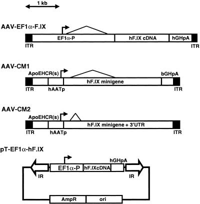
AAV-EF1α-F.IX, AAV-CM1, and AAV-CM2 were produced based on the plasmids pV4.1e-hF.IX (26), pAAV-CM1, and pAAV-CM2. To construct pAAV-CM1, the whole EF1α-hF.IX cassette (human elongation factor 1α [EF1α] gene enhancer-promoter-driven human coagulation factor IX [hF.IX] expression cassette) between two NotI sites that were located at the inner ends of both inverted terminal repeats (ITRs) was removed and replaced with another hF.IX expression cassette from pBS-ApoEHCR(s)-hAATp-hF.IXmg-bp (25), which contains the apolipoprotein E hepatic locus control region (HCR)–human α1-antitrypsin gene promoter [ApoEHCR(s)-hAATp]-driven hF.IX minigene (containing a 1.4-kb intron A from the hF.IX gene) with the bovine growth hormone gene polyadenylation signal. To construct pAAV-CM2, the hF.IX minigene and polyadenylation signal in pAAV-CM1 were replaced with the hF.IX minigene carrying an 0.3-kb intron A and 3′ untranslated region that included the hF.IX polyadenylation signal. AAV-EF1α-F.IX.PMT was produced using 293PMT cells that constitutively express the bacterial PaeR7 methyltransferase (28). All the adenine residues at the XhoI recognition sites in AAV-EF1α-F.IX.PMT vector were methylated by the PaeR7 methyltransferase (28, 29). Because our previous studies showed that most of the ds vector genomes in transduced livers were formed by annealing of complementary ss genomes (28), ds AAV-EF1α-F.IX.PMT vector genomes were fully adenine methylated at the XhoI sites and not cleaved with XhoI (which only cleaves fully unmethylated XhoI sites) unless the ds vector genomes fully replicated by passing through two or more cell cycles. Thus, methylation status at the XhoI site serves as a marker for vector genome replication.
Sleeping Beauty (SB)-based transposon plasmid pT-EF1α-hF.IX, carrying the EF1α enhancer-promoter-driven hF.IX expression cassette, and the pCMV-SB and pCMV-mSB transposase plasmids, encoding wild-type and nonfunctional mutant SB transposase genes, respectively, under the control of the human cytomegalovirus immediate-early gene enhancer-promoter, have been described elsewhere (14, 47).
All plasmids were amplified in either DH10B (Gibco BRL, Gaithersburg, Md.) or Sure (Stratagene, Cedar Creek, Tex.) bacteria.
Animal procedures.
Six- to eight-week-old female C57BL/6 mice were obtained from either Jackson Laboratory (Bar Harbor, Maine) or Taconic (Germantown, N.Y.). All animal procedures were performed according to the guidelines for animal care at Stanford University. Portal vein injection of rAAV vectors, hydrodynamics-based in vivo hepatocyte transfection of naked plasmid DNA by tail vein injection, and partial hepatectomy were performed as previously described (16, 20, 26, 47, 48). Blood samples were collected from the retro-orbital plexus. The summary of animal experiments is shown in Table 1.
TABLE 1.
Summary of the animal experiments
| Group | Vector | Dose/mouse | No. of mice | Time of PHxa | Analysis(-es) |
|---|---|---|---|---|---|
| 1 | AAV-EF1α-F.IX | 2.7 × 10¹¹ vg | 4 | Southern blotting | |
| 2 | pT-EF1α-hF.IX | 25 μg | 10 | 5 wk | hF.IX |
| pCMV-SB | 1 μg | ||||
| 3 | pT-EF1α-hF.IX | 25 μg | 10 | 5 wk | hF.IX |
| pCMV-mSB | 1 μg | ||||
| 4 | pT-EF1α-hF.IX | 25 μg | 5 | hF.IX | |
| 5 | AAV-CM1 | 3.0 × 10¹¹ vg | 4 | 12 mo | hF.IX, Southern blotting |
| 6 | AAV-CM2 | 3.0 × 10¹¹ vg | 4 | 12 mo | hF.IX, Southern blotting |
| 7 | AAV-EF1α-F.IX | 2.4 × 10¹¹ vg | 20 | 12 wk | hF.IX, Southern blotting |
| 8 | AAV-EF1α-F.IX.PMT | 2.4 × 10¹¹ vg | 3 | 6 wk, 12 mo | Southern blotting |
| 9 | AAV-EF1α-F.IX.PMT | 7.2 × 10¹¹ vg | 3 | 6 wk, 12 mo | Southern blotting |
PHx, partial hepatectomy.
Measurement of hF.IX in samples.
Enzyme-linked immunosorbent assay specific for hF.IX was employed for the measurement of hF.IX in mouse serum or plasma samples using affinity-purified plasma hF.IX (Calbiochem, La Jolla, Calif.) as a standard (40).
Southern blot analysis.
Total genomic DNA was extracted from livers and subjected to Southern blot analysis as previously described (27, 28). Briefly, 20 μg of total genomic liver DNAs was digested with a restriction enzyme or a combination of enzymes, separated on an 0.8% agarose gel, transferred to a nylon membrane (Duralon UV; Stratagene), and hybridized with 32P-labeled vector sequence-specific probes. Membranes were exposed to X-ray films at −80°C for 3 days to 2 weeks. The vector genome copy number standards (the number of ds vector genomes per diploid genomic equivalent) were 20 μg of naive mouse total liver DNAs mixed with the appropriate number of each vector plasmid. Band intensities were quantitated using a G710 calibrated imaging densitometer (Bio-Rad, Hercules, Calif.).
The relative abundance of replicated, XhoI-digestible (integrated) genomes after partial hepatectomy was compared with the amount of non-XhoI-digestible genomes at the time of hepatectomy (representing the sum of extrachromosomal and integrated genomes) in AAV-EF1α-F.IX.PMT-injected mice by quantitative Southern blot analysis.
RESULTS
Extrachromosomal ds circular monomers are maintained in mouse liver for over 1.5 years.
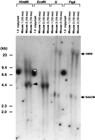
To estimate the abundance of extrachromosomal circular monomer rAAV vector genomes in mouse livers, we injected each of four mice with 2.7 × 10¹¹ particles of AAV-EF1α-F.IX (Fig. 1) via the portal vein (Table 1, group 1). Animals were sacrificed at 3, 10, and 19 months postinjection (n = 1, 1, and 2, respectively) and analyzed by Southern blotting. Our previous study showed the presence of extrachromosomal vector forms in mouse liver samples harvested 3 months postinjection (27). At one time, we thought that these extrachromosomal forms represented ss rAAV genomes that annealed during the isolation of tissue DNA and not bona fide molecular forms of the vector DNA (27). However, subsequent analyses demonstrated that these extrachromosomal bands primarily represented ds circular forms (as either supercoiled or relaxed) and/or linear ds monomer genomes (28) that persist in vivo. The relaxed circular monomer forms may be nicked supercoiled ds circular forms. In the samples from animals isolated 10 and 19 months postinjection, extrachromosomal forms were readily detectable by Southern blotting (Fig. 2). The persistence of extrachromosomal circular rAAV genomes in mouse liver detectable by Southern blotting has been recently reported by Song et al. (37). All of these data suggest that considerable amounts of extrachromosomal rAAV vector genomes can persist in mouse liver.
FIG. 1.
Map of rAAV vectors and plasmid: AAV-EF1α-F.IX, AAV-CM1, AAV-CM2, and pT-EF1α-hF.IX. The 1-kb scale is valid for the three vectors but not for the plasmid. EF1α-P, human elongation factor 1α gene enhancer-promoter; hGHpA, the human growth hormone gene polyadenylation signal; ApoEHCR(s), a shorter fragment of the HCR from the apolipoprotein E gene (ApoE) (25); hAATp, the human α1-antitrypsin gene promoter; hF.IX minigene, hF.IXcDNA containing 1.4-kb truncated intron A from the hF.IX gene; hF.IX minigene + 3′UTR, hF.IX cDNA containing 0.3-kb truncated intron A and 1.7-kb 3′ untranslated region (UTR) including the polyadenylation signal; IR, SB transposon inverted repeat; ori, plasmid origin of replication from pUC; Ampr, ampicillin resistance gene.
FIG. 2.
Southern blot analysis of liver DNA from three mice injected via the portal vein with AAV-EF1α-F.IX at a dose of 2.7 × 10¹¹ vg per animal. Twenty micrograms of total mouse liver DNA was digested with the enzymes indicated above the lanes or undigested (U), separated on an 0.8% agarose gel, blotted on a nylon membrane, and hybridized with a vector-specific PpuMI EF1α-hF.IX probe (the same as probe C in reference 27). The time of sacrifice is shown above each lane. The 1.0-copy/cell standard is 20 μg of naive mouse liver DNA mixed with the appropriate number of plasmid pV4.1e-hF.IX molecules. This control plasmid is linearized when cut with HindIII, EcoRI, or FspI, generating a 7.6-kb band. HindIII and EcoRI cut the vector genome once at the 3′ side and the center of the vector genome, respectively, while FspI does not cut within the vector genome. Head-to-tail and head-to-head molecules are denoted by closed and open arrowheads, respectively. The positions of high-molecular-weight (HMW) species and supercoiled ds circular monomers (SdsCM) of vector genomes are shown. Ethidium bromide staining of the gel showed no lane-to-lane variations of the amount of loaded DNA among digestions. Note that extrachromosomal forms of the vector genomes (SdsCM) are readily detectable. The Southern blot results for mice sacrificed 3 months postinjection were previously published (27).
Transgene expression from integrated plasmid vector genomes is not affected by partial hepatectomy.
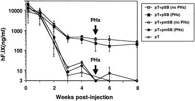
The Southern blot data shown here and our previous studies (25, 27) demonstrate the long-term presence of both integrated and extrachromosomal rAAV genomes. However, these earlier studies did not establish the relative proportion of integrated and extrachromosomal genomes or the contribution of each to transgene expression. In order to establish an assay that would enable us to distinguish between transgene expression from integrated genomes and that from extrachromosomal genomes, we first generated mice that contained an EF1α-hF.IX expression cassette integrated into the chromosomes of mouse hepatocytes, using the SB transposon system (47). These animals served as a control for chromosomal integration (Fig. 3; n = 10, group 2). Mice containing stably transfected hepatocytes were readily generated by injecting them via the tail vein with a plasmid containing an hF.IX transposon (pT-EF1α-hF.IX [Fig. 1]) together with a helper plasmid encoding the SB transposase (pCMV-SB). Importantly, the expression cassette contained within pT-EF1α-hF.IX was identical to one encoded by the rAAV vector AAV-EF1α-F.IX. Control groups received pT-EF1α-hF.IX either alone (n = 5, group 4) or in combination with pCMV-mSB (n = 10, group 3), which encodes a catalytically inactive mutant form of the SB transposase. Since pT-EF1α-hF.IX alone or in combination with pCMV-mSB did not integrate into chromosomes in hepatocytes in vivo, pT-EF1α-hF.IX DNA remained extrachromosomal and transgene expression was lost within 3 weeks in groups 3 and 4 (Fig. 3). In contrast, stable plasma hF.IX levels of approximately 300 ng/ml were achieved in mice 5 weeks postinjection with pT-EF1α-hF.IX and pCMV-SB (group 2). Thus, virtually all the hF.IX expression observed after 3 weeks came from integrated transposon genomes.
FIG. 3.
Effects of partial hepatectomy on integrated plasmid vector genomes in liver. The figure shows plasma hF.IX levels of mice injected with 25 μg of an SB-based transposon plasmid (pT-EF1α-hF.IX) via the tail vein together with 1 μg of a helper plasmid encoding active SB transposase (pCMV-SB) (group 2, n = 10) or inactive mutated SB (pCMV-mSB) (group 3, n = 10) or without any helper plasmid (group 4, n = 5). A two-thirds partial hepatectomy (PHx) was performed 5 weeks postinjection. pT, pT-EF1α-hF.IX; pSB, pCMV-SB; pmSB, pCMV-mSB. Vertical bars indicate standard deviations.
Five weeks postinjection, 10 mice (n = 5 each in groups 2 and 3) were partially hepatectomized. As shown in Fig. 3, plasma hF.IX levels did not change in group 2 regardless of whether they underwent a partial hepatectomy, indicating that integrated EF1α-hF.IX expression cassettes were not diluted and that transgene expression was not hampered by cell cycling. In addition, this also showed that 3 weeks was sufficient for a regenerating liver to completely restore its capacity to express the same levels of hF.IX as before hepatectomy. These results support partial hepatectomy as a useful and reasonable strategy to differentiate between the expression from integrated genomes and that from extrachromosomal vector genomes.
Loss of rAAV transgene expression and vector genomes in replicating hepatocytes.
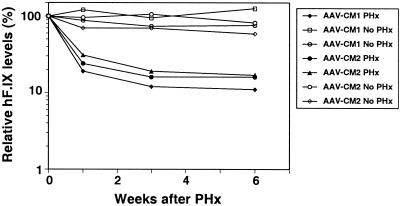
We previously reported that rAAV-injected mice that had not yet reached a steady-state level of transgene expression during the first 3 weeks postinjection had a falloff in rAAV genome copy number and transgene expression following partial hepatectomy (24). However, at this time, the rAAV genomes may not have fully integrated or formed stable structures. Therefore, we examined mice that were stably transduced by rAAV vectors (i.e., 6 or more weeks postinjection). For these studies, three hF.IX-expressing rAAV vectors were used. Eight mice were injected with either AAV-CM1 (Fig. 1) or AAV-CM2 (Fig. 1) (n = 4 each, groups 5 and 6, respectively) at a dose of 3.0 × 10¹¹ vector genomes (vg) per mouse, and two mice from each group were partially hepatectomized 12 months postinjection. The serum hF.IX levels at the time of partial hepatectomy were 8,411 ± 2,687 ng/ml (mean ± standard deviation) and 6,423 ± 922 ng/ml in AAV-CM1- and AAV-CM2-injected mice, respectively. We were unable to remove two-thirds of the liver in one mouse injected with AAV-CM1 because of severe adhesions in the peritoneal cavity. This animal was sacrificed at the time of surgery. All the mice were sacrificed 6 weeks after partial hepatectomy, and their livers were harvested to quantify the vector genomes in the liver. The serum hF.IX levels relative to those at hepatectomy are summarized in Fig. 4. The hF.IX levels dropped by 83 to 89% in hepatectomized mice but remained stable in all the nonhepatectomized mice. The numbers of vector genomes per cell before and after hepatectomy were 1.00 versus 0.06 in an AAV-CM1 injected mouse and 1.95 versus 0.63 and 2.18 versus 0.20 in AAV-CM2-injected mice (Table 2, groups 5 and 6).
FIG. 4.
rAAV-mediated gene expression in partially hepatectomized mice. The figure shows serum hF.IX levels after partial hepatectomy (PHx) performed a year after portal vein injection of 3.0 × 10¹¹ vg of AAV-CM1 or AAV-CM2. Results for control mice that were injected at the same time but did not receive partial hepatectomy are also shown. Each line represents an individual mouse. hF.IX levels are shown as percentages relative to their levels at the time of hepatectomy.
TABLE 2.
Summary of the decrease in hF.IX levels and vector copy numbers by partial hepatectomy (PHx)
| Group | Mouse no. | Vector dose (1011 vg/mouse) | Time of PHxa | Time (wk) of sacrifice after PHxa | hF.IX level (ng/ml)
|
% Decrease in hF.IX level | Vector copy no. (copies/cell)
|
% Decrease in copy no. | XhoI-digestible genomes (%)b | ||
|---|---|---|---|---|---|---|---|---|---|---|---|
| Pre-PHx | Post-PHx | Pre-PHx | Post-PHx | ||||||||
| 5 | 2 | 3.0 | 12 mo | 3 | 10,740 | 1,227 | 89 | 1.00 | 0.06 | 94 | |
| 6 | 1 | 3.0 | 12 mo | 3 | 6,759 | 1,115 | 84 | 1.95 | 0.63 | 68 | |
| 2 | 3.0 | 12 mo | 3 | 6,269 | 1,053 | 83 | 2.18 | 0.20 | 91 | ||
| 7 | 1 | 2.4 | 12 wk | 6 | 1,439 | 149 | 90 | 0.80 | NDc | NAd | |
| 2 | 2.4 | 12 wk | 6 | 1,356 | 67 | 95 | 0.94 | 0.12 | 87 | ||
| 3 | 2.4 | 12 wk | 6 | 1,344 | 97 | 93 | 0.62 | 0.06 | 90 | ||
| 4 | 2.4 | 12 wk | 6 | 1,599 | 57 | 96 | 1.18 | 0.12 | 90 | ||
| 5 | 2.4 | 12 wk | 6 | 1,332 | 7 | 99 | 1.76 | 0.08 | 95 | ||
| 6 | 2.4 | 12 wk | 6 | 1,216 | ND | NA | 0.92 | ND | NA | ||
| 7 | 2.4 | 12 wk | 6 | 1,666 | 50 | 97 | 0.90 | 0.14 | 84 | ||
| 8 | 2.4 | 12 wk | 6 | 1,678 | 36 | 98 | 2.08 | 0.08 | 96 | ||
| 9 | 2.4 | 12 wk | 6 | 1,079 | 68 | 94 | 0.44 | ND | NA | ||
| 10 | 2.4 | 12 wk | 6 | 1,120 | 106 | 90 | 0.60 | 0.22 | 63 | ||
| 8 | 1 | 2.4 | 6 wk | 1 | 1,013 | ND | NA | 0.85 | 0.16 | 81 | 0 |
| 2 | 2.4 | 6 wk | 1 | 1,063 | ND | NA | 0.46 | 0.31 | 33 | 8 | |
| 3 | 2.4 | 12 mo | 3 | ND | ND | NA | 0.50 | 0.28 | 44 | 20 | |
| 9 | 4 | 7.2 | 6 wk | 1 | 1,993 | ND | NA | 1.57 | 0.26 | 83 | 4 |
| 5 | 7.2 | 6 wk | 1 | 2,046 | ND | NA | 2.55 | 0.08 | 97 | 9 | |
| 6 | 7.2 | 12 mo | 3 | ND | ND | NA | 0.74 | 0.64 | 14 | 67 | |
hF.IX levels and vector copy numbers pre- and post-partial hepatectomy were determined at the time of partial hepatectomy and sacrifice, respectively.
The amount of XhoI-digestible genomes after partial hepatectomy relative to that of the total genomes before partial hepatectomy (percent).
ND, not done.
NA, not applicable.
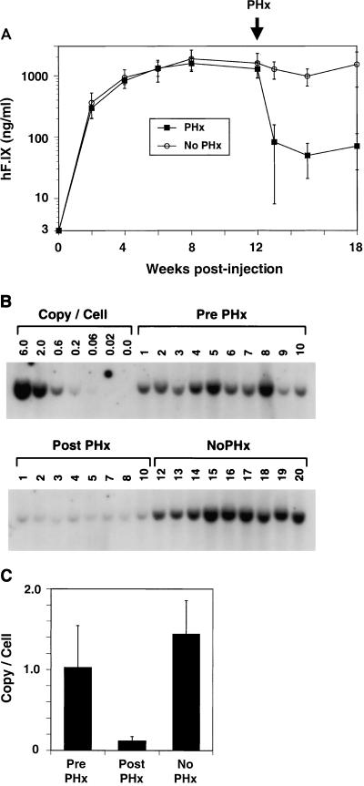
In a separate experiment, 20 mice were injected with 2.4 × 10¹¹ vg of AAV-EF1α-F.IX (group 7), and half of these animals were partially hepatectomized 12 weeks postinjection. Plasma hF.IX levels at the time of partial hepatectomy in the hepatectomized subgroup and levels in the nonhepatectomized subgroup were 1,383 ± 214 and 1,597 ± 690 ng/ml, respectively. As shown in Fig. 5A, the average hF.IX levels decreased by 95% over a period of 6 weeks after partial hepatectomy (from 1,383 ± 214 to 71 ± 42 ng/ml) (n = 9; one mouse was sacrificed before the end point of this study), while the hF.IX levels in the nonhepatectomized subgroup remained relatively stable during the same period (1,516 ± 872 ng/ml, corresponding to 6 weeks posthepatectomy; n = 10). Concordant with the plasma hF.IX levels, the average number of vector genomes per cell also fell after partial hepatectomy from 1.02 ± 0.52 copies per cell at the time of hepatectomy to 0.12 ± 0.03 copy per cell 6 weeks posthepatectomy (Fig. 5B and C).
FIG. 5.
rAAV-mediated gene expression and quantification of rAAV genomes in partially hepatectomized mice. (A) Plasma hF.IX levels of 20 mice injected with AAV-EF1α-F.IX at a dose of 2.4 × 10¹¹ vg per mouse. Half of the mice underwent partial hepatectomy (PHx) 12 weeks postinjection. Vertical bars indicate standard deviations. (B) Southern blot analysis of liver DNAs from the mice in panel A. All animals were sacrificed 18 weeks postinjection (6 weeks after partial hepatectomy for the partial hepatectomy group), and 20 μg of total genomic liver DNA was subjected to Southern blot analysis with BglII digestion and hybridized to a BglII F.IX probe (28). Copy number standards are indicated as 0.0 to 6.0 copies/cell. The numbers above each lane indicate individual mouse numbers. Ethidium bromide staining of the gel showed that all the lanes had the same amount of digested DNA (data not shown). Both blots were from the same membrane. (C) Comparison of rAAV vector genome copy numbers per cell before and after partial hepatectomy. The intensity of each band in panel B was determined by densitometry. A Student t test revealed a statistical difference between “Pre PHx” and “Post PHx” values (P < 0.0002). Vertical bars indicate standard deviations.
In each of these experiments, both reporter gene expression and the number of vector genomes decreased substantially after partial hepatectomy (summarized in Table 2). In fact, the relative hF.IX levels and vector copy numbers before and after partial hepatectomy were found to be (7.7 ± 5.2)% and (14.2 ± 11.4)%, respectively, collectively for animals in groups 5, 6, and 7. It is important to point out that these results may actually overestimate the number of integrated forms, since gene expression from some proportion of extrachromosomal forms may continue to persist in hepatocytes after partial hepatectomy. In support of this notion, we have determined that nonintegrated plasmid-derived gene expression was not completely eliminated but was reduced by 94 to 98% following a partial hepatectomy (data not shown). Nonetheless, these results suggest that most of the rAAV genomes and vector-based gene expression were extrachromosomally derived and were not from integrated rAAV genomes.
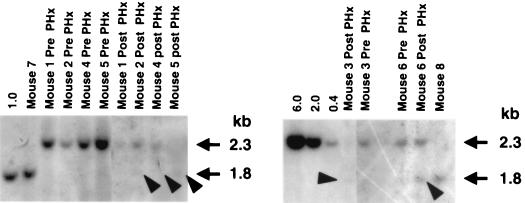
In order to confirm the presence of integrated genomes that can replicate during liver regeneration, we have developed a secondary assay that utilizes an rAAV vector whose genomes have been methylated at adenine residues of XhoI sites and are resistant to XhoI cleavage. Since loss of methylation and XhoI digestion will occur only with replication of the vector genomes, the amount of XhoI-sensitive (replicated) rAAV DNA present in vivo serves as a means to monitor replication of integrated rAAV DNA forms (28). In this study, six mice were injected with 2.4 × 10¹¹ (n = 3, group 8) or 7.2 × 10¹¹ (n = 3, group 9) vg of AAV-EF1α-F.IX.PMT vector via the portal vein. Twelve months postinjection, one animal receiving each dose was partially hepatectomized. The ds vector genomes in mouse livers harvested both at the time of partial hepatectomy and 3 weeks later were analyzed by Southern blotting for vector copy number and the presence of XhoI-digestible (replicated) genomes after digestion with BglII and in combination with XhoI, respectively. As summarized in Table 2, in a mouse that received 2.4 × 10¹¹ vg (mouse 3), the vector copy number decreased 44% after partial hepatectomy while 20% of the genomes were XhoI digestible. The decrease in vector copy number after hepatectomy was not as dramatic in this mouse as for others in the previous experiment. In contrast, a mouse that received 7.2 × 10¹¹ vg (mouse 6) showed only a 14% decrease in vector copy number after hepatectomy, and 67% of the genomes were digested with XhoI (Fig. 6).
FIG. 6.
The XhoI resistance assay of vector genomes in liver from mice injected with AAV-EF1α-F.IX.PMT vector at doses of 2.4 × 10¹¹ or 7.2 × 10¹¹ vg, before and after partial hepatectomy. Twenty micrograms of total liver DNA was digested with BglII and XhoI (except for the 0.4, 2.0, and 6.0 copy number standards, which were digested with only BglII) and subjected to Southern blot analysis with an XhoI F.IX probe (28). Copy number standards are 20 μg of naive mouse liver DNA mixed with the appropriate number of plasmid pV4.1e-hF.IX molecules followed by restriction enzyme digestion and shown above each lane as 1.0, 6.0, 2.0, and 0.4 copies/cell. The vector doses and the times of the analyses for mice 1 to 6 are summarized in Table 2. Mice 7 and 8 were injected with a nonmethylated AAV-EF1α-F.IX vector at a dose of 2.4 × 10¹¹ vg, and liver DNA was harvested 18 weeks postinjection (mouse 7) and at the end of this study (mouse 8). XhoI-resistant genomes migrate at the 2.3-kb position, while XhoI-digestible genomes migrate at the 1.8-kb position. Closed arrowheads indicate XhoI-digestible genomes. The vector copy numbers were determined by another Southern blotting with BglII digestion and a BglII F.IX probe (data not shown).
The four other mice (n = 2 in groups 8 and 9) were partially hepatectomized 6 weeks postinjection and showed a 33 to 97% drop in vector genomes 1 week after hepatectomy. None of these mice, including two mice injected with 7.2 × 10¹¹ vg (mice 4 and 5 in group 9), showed as high an integration efficiency as that observed for mouse 6. At this time, we do not know why only mouse 6 (in group 9) among 18 mice analyzed had such a high number of integrated genomes. Further analyses are required to fully elucidate the correlation between vector dose and integration efficiency in vivo.
DISCUSSION
There is strong evidence that some proportion of rAAV vectors integrate into chromosomes in hepatocytes in vivo (5, 23, 27); however, a series of studies including the present one have raised the importance of extrachromosomal genomes for persistent transgene expression. The relatively small number of integrated genomes clouds the possibility that a single administration of vector will persist lifelong in humans. Additional preclinical studies with large animals will be needed to help clarify this important issue.
After rAAV vectors enter the nuclei of hepatocytes, complementary ss rAAV genomes anneal to form transcriptionally active ds genomes. The ds forms may be converted into large concatemers via random end joining and/or integrate into chromosomes (28). Extrachromosomal ds vector genomes are present as linear and supercoiled circular monomers, as well as both circular and linear concatemers (8, 28, 37, 38). Among these extrachromosomal forms, ds circular monomers and concatemers seem to be the most important genomic structures contributing to persistent gene expression in muscle (8, 46). The fact that the relative fall in rAAV-mediated gene expression was similar in mice that underwent a partial hepatectomy 12 weeks after vector administration and those that underwent a partial hepatectomy 12 months after vector administration suggests that the relative number of extrachromosomal and integrated genomes did not substantially change over this period. However, the reason for the high proportion of integrated genomes in a mouse that received a higher dose of vector is not clear. Further studies are required to establish the kinetics of genome conversion in vivo.
We have previously suggested that the majority of rAAV genomes integrate as head-to-tail concatemers into chromosomes in mouse hepatocytes in vivo after portal vein injection of vector (23). This was based on finding rAAV FISH signals on sister chromatids of metaphase spreads from isolated hepatocytes, as well as a series of molecular analyses that included pulsed-field gel electrophoresis to look at the genome-sized DNA fragments containing rAAV genomes (23). While these previous studies provided us with the first evidence of in vivo integration, our further analyses elucidated that we had overlooked the presence of persistent extrachromosomal rAAV genomes in transduced hepatocytes (28). Our more recent studies of rAAV genome forms in transduced mouse liver in vivo demonstrated the presence of head-to-tail, head-to-head, and tail-to-tail high-molecular-weight rAAV concatemers (either integrated or extrachromosomal), as well as low-molecular-weight extrachromosomal genomes dominated by ds circular monomers. Recent evidence suggests that extrachromosomal FISH signals can associate with chromosomes in metaphase spreads (1). Thus, a certain proportion of DNAs in hepatocyte metaphases might have in fact represented extrachromosomal genomes that tightly associated with chromosomes, overestimating the integration efficiency in these previous studies. The present study clearly demonstrates that many rAAV genomes remain as extrachromosomes in the liver and that these extrachromosomal forms, rather than integrated genomes, are primarily responsible for persistent transgene expression from rAAV vectors.
The mechanistic reasons for the persistence of extrachromosomal rAAV genomes are not known. Gene expression from exogenous supercoiled circular plasmid vectors delivered into hepatocytes in vivo is generally transient, and when persistent, the expression levels are relatively low (2, 43, 49). Because both plasmids and ds circular rAAV genomes have similarity in their structures in that they are both supercoiled circular DNAs residing in nuclei, some unknown mechanism may operate for ds extrachromosomal rAAV genomes to persist and continue to express their transgene product. Although we do not yet understand why gene expression can persist in vivo from extrachromosomal rAAV genomes, there is some evidence that vector genome concatemerization and/or the presence of the AAV-ITR sequence itself may stabilize extrachromosomal genomes in nuclei (8, 30). Recently, Chen et al. reported that persistent, high-level transgene expression from plasmid-based vectors in mouse hepatocytes correlated with concatemerization of input linear plasmid vector genomes (6). On the other hand, Miao et al. have found that inclusion of the apolipoprotein E HCR together with a part of the hF.IX gene that included a portion of the first intron greatly augmented and stabilized hF.IX expression from extrachromosomal supercoiled monomer plasmid vectors transfected in mouse hepatocytes in vivo (25). The hypothesis was that a matrix attachment region included in the HCR and intron sequence may have facilitated interaction with the nuclear matrix, leading to persistent gene expression. Since AAV-CM1 and AAV-CM2 contained these elements and AAV-EF1α-F.IX carried an intron, it is also possible that inclusion of these stabilizing elements rather than concatemerization or the presence of AAV-ITR may have somehow influenced the persistence of vector genomes and rAAV-mediated gene expression. However, we have no evidence to support this assumption. Further analysis of extrachromosomal rAAV genomes in transduced hepatocytes should help unravel the mechanisms underlying persistent expression from rAAV genomes.
In summary, our current study establishes an important role for extrachromosomal rAAV vector genomes in maintaining persistent transgene expression in hepatocytes transduced by rAAV vectors. Our finding that the frequency of rAAV genome integration into hepatic chromosomes in vivo is actually quite low further reduces the theoretical risk of harmful side effects incurred during the use of rAAV and other integrating vector systems.
ACKNOWLEDGMENT
This work was supported by NIH grant HL64274.
REFERENCES
- 1.Baiker A, Maercker C, Piechaczek C, Schmidt S B, Bode J, Benham C, Lipps H J. Mitotic stability of an episomal vector containing a human scaffold/matrix-attached region is provided by association with nuclear matrix. Nat Cell Biol. 2000;2:182–184. doi: 10.1038/35004061. [DOI] [PubMed] [Google Scholar]
- 2.Budker V, Zhang G, Knechtle S, Wolff J A. Naked DNA delivered intraportally expresses efficiently in hepatocytes. Gene Ther. 1996;3:593–598. [PubMed] [Google Scholar]
- 3.Burton M, Nakai H, Colosi P, Cunningham J, Mitchell R, Couto L. Coexpression of factor VIII heavy and light chain adeno-associated viral vectors produces biologically active protein. Proc Natl Acad Sci USA. 1999;96:12725–12730. doi: 10.1073/pnas.96.22.12725. [DOI] [PMC free article] [PubMed] [Google Scholar]
- 4.Chao H, Samulski R, Bellinger D, Monahan P, Nichols T, Walsh C. Persistent expression of canine factor IX in hemophilia B canines. Gene Ther. 1999;6:1695–1704. doi: 10.1038/sj.gt.3301024. [DOI] [PubMed] [Google Scholar]
- 5.Chen S J, Tazelaar J, Moscioni A D, Wilson J M. In vivo selection of hepatocytes transduced with adeno-associated viral vectors. Mol Ther. 2000;1:414–422. doi: 10.1006/mthe.2000.0065. [DOI] [PubMed] [Google Scholar]
- 6.Chen Z-Y, Yant S, He C-Y, Meuse L, Shen S, Kay M A. Linear DNAs concatemerize in vivo and result in sustained transgene expression in mouse liver. Mol Ther. 2000;3:403–410. doi: 10.1006/mthe.2001.0278. [DOI] [PubMed] [Google Scholar]
- 7.Duan D, Fisher K J, Burda J F, Engelhardt J F. Structural and functional heterogeneity of integrated recombinant AAV genomes. Virus Res. 1997;48:41–56. doi: 10.1016/s0168-1702(96)01425-6. [DOI] [PubMed] [Google Scholar]
- 8.Duan D, Sharma P, Yang J, Yue Y, Dudus L, Zhang Y, Fisher K J, Engelhardt J F. Circular intermediates of recombinant adeno-associated virus have defined structural characteristics responsible for long-term episomal persistence in muscle tissue. J Virol. 1998;72:8568–8577. doi: 10.1128/jvi.72.11.8568-8577.1998. . (Erratum, 73:861, 1999.) [DOI] [PMC free article] [PubMed] [Google Scholar]
- 9.Fisher-Adams G, Wong K K, Jr, Podsakoff G, Forman S J, Chatterjee S. Integration of adeno-associated virus vectors in CD34+ human hematopoietic progenitor cells after transduction. Blood. 1996;88:492–504. [PubMed] [Google Scholar]
- 10.Flotte T R, Afione S A, Conrad C, McGrath S A, Solow R, Oka H, Zeitlin P L, Guggino W B, Carter B J. Stable in vivo expression of the cystic fibrosis transmembrane conductance regulator with an adeno-associated virus vector. Proc Natl Acad Sci USA. 1993;90:10613–10617. doi: 10.1073/pnas.90.22.10613. [DOI] [PMC free article] [PubMed] [Google Scholar]
- 11.Greelish J P, Su L T, Lankford E B, Burkman J M, Chen H, Konig S K, Mercier I M, Desjardins P R, Mitchell M A, Zheng X G, Leferovich J, Gao G P, Balice-Gordon R J, Wilson J M, Stedman H H. Stable restoration of the sarcoglycan complex in dystrophic muscle perfused with histamine and a recombinant adeno-associated viral vector. Nat Med. 1999;5:439–443. doi: 10.1038/7439. [DOI] [PubMed] [Google Scholar]
- 12.Herzog R W, Hagstrom J N, Kung S H, Tai S J, Wilson J M, Fisher K J, High K A. Stable gene transfer and expression of human blood coagulation factor IX after intramuscular injection of recombinant adeno-associated virus. Proc Natl Acad Sci USA. 1997;94:5804–5809. doi: 10.1073/pnas.94.11.5804. [DOI] [PMC free article] [PubMed] [Google Scholar]
- 13.Herzog R W, Yang E Y, Couto L B, Hagstrom J N, Elwell D, Fields P A, Burton M, Bellinger D A, Read M S, Brinkhous K M, Podsakoff G M, Nichols T C, Kurtzman G J, High K A. Long-term correction of canine hemophilia B by gene transfer of blood coagulation factor IX mediated by adeno-associated viral vector. Nat Med. 1999;5:56–63. doi: 10.1038/4743. [DOI] [PubMed] [Google Scholar]
- 14.Ivics Z, Hackett P B, Plasterk R H, Izsvak Z. Molecular reconstruction of Sleeping Beauty, a Tc1-like transposon from fish, and its transposition in human cells. Cell. 1997;91:501–510. doi: 10.1016/s0092-8674(00)80436-5. [DOI] [PubMed] [Google Scholar]
- 15.Kaplitt M G, Leone P, Samulski R J, Xiao X, Pfaff D W, O'Malley K L, During M J. Long-term gene expression and phenotypic correction using adeno-associated virus vectors in the mammalian brain. Nat Genet. 1994;8:148–154. doi: 10.1038/ng1094-148. [DOI] [PubMed] [Google Scholar]
- 16.Kay M A, Li Q, Liu T J, Leland F, Toman C, Finegold M, Woo S L. Hepatic gene therapy: persistent expression of human alpha 1-antitrypsin in mice after direct gene delivery in vivo. Hum Gene Ther. 1992;3:641–647. doi: 10.1089/hum.1992.3.6-641. [DOI] [PubMed] [Google Scholar]
- 17.Kay M A, Fausto N. Liver regeneration: prospects for therapy based on new technologies. Mol Med Today. 1997;3:108–115. doi: 10.1016/S1357-4310(96)10062-9. [DOI] [PubMed] [Google Scholar]
- 18.Kay M A, Manno C S, Ragni M V, Larson P J, Couto L B, McClelland A, Glader B, Chew A J, Tai S J, Herzog R W, Arruda V, Johnson F, Scallan C, Skarsgard E, Flake A W, High K A. Evidence for gene transfer and expression of factor IX in haemophilia B patients treated with an AAV vector. Nat Genet. 2000;24:257–261. doi: 10.1038/73464. [DOI] [PubMed] [Google Scholar]
- 19.Kessler P D, Podsakoff G M, Chen X, McQuiston S A, Colosi P C, Matelis L A, Kurtzman G J, Byrne B J. Gene delivery to skeletal muscle results in sustained expression and systemic delivery of a therapeutic protein. Proc Natl Acad Sci USA. 1996;93:14082–14087. doi: 10.1073/pnas.93.24.14082. [DOI] [PMC free article] [PubMed] [Google Scholar]
- 20.Liu F, Song Y, Liu D. Hydrodynamics-based transfection in animals by systemic administration of plasmid DNA. Gene Ther. 1999;6:1258–1266. doi: 10.1038/sj.gt.3300947. [DOI] [PubMed] [Google Scholar]
- 21.Malik A K, Monahan P E, Allen D L, Chen B G, Samulski R J, Kurachi K. Kinetics of recombinant adeno-associated virus-mediated gene transfer. J Virol. 2000;74:3555–3565. doi: 10.1128/jvi.74.8.3555-3565.2000. [DOI] [PMC free article] [PubMed] [Google Scholar]
- 22.Matsushita T, Elliger S, Elliger C, Podsakoff G, Villarreal L, Kurtzman G J, Iwaki Y, Colosi P. Adeno-associated virus vectors can be efficiently produced without helper virus. Gene Ther. 1998;5:938–945. doi: 10.1038/sj.gt.3300680. [DOI] [PubMed] [Google Scholar]
- 23.Miao C H, Snyder R O, Schowalter D B, Patijn G A, Donahue B, Winther B, Kay M A. The kinetics of rAAV integration in the liver. Nat Genet. 1998;19:13–15. doi: 10.1038/ng0598-13. [DOI] [PubMed] [Google Scholar]
- 24.Miao C H, Nakai H, Thompson A R, Storm T A, Chiu W, Snyder R O, Kay M A. Nonrandom transduction of recombinant adeno-associated viral vectors in mouse hepatocytes in vivo: cell cycling does not influence hepatocyte transduction. J Virol. 2000;74:3793–3803. doi: 10.1128/jvi.74.8.3793-3803.2000. [DOI] [PMC free article] [PubMed] [Google Scholar]
- 25.Miao C H, Ohashi K, Patijn G A, Meuse L, Ye X, Thompson A R, Kay M A. Inclusion of the hepatic locus control region, an intron, and untranslated region increases and stabilizes hepatic factor IX gene expression in vivo but not in vitro. Mol Ther. 2000;1:522–532. doi: 10.1006/mthe.2000.0075. [DOI] [PubMed] [Google Scholar]
- 26.Nakai H, Herzog R W, Hagstrom J N, Walter J, Kung S H, Yang E Y, Tai S J, Iwaki Y, Kurtzman G J, Fisher K J, Colosi P, Couto L B, High K A. Adeno-associated viral vector-mediated gene transfer of human blood coagulation factor IX into mouse liver. Blood. 1998;91:4600–4607. [PubMed] [Google Scholar]
- 27.Nakai H, Iwaki Y, Kay M A, Couto L B. Isolation of recombinant adeno-associated virus vector-cellular DNA junctions from mouse liver. J Virol. 1999;73:5438–5447. doi: 10.1128/jvi.73.7.5438-5447.1999. [DOI] [PMC free article] [PubMed] [Google Scholar]
- 28.Nakai H, Storm T A, Kay M A. Recruitment of single-stranded recombinant adeno-associated virus vector genomes and intermolecular recombination are responsible for stable transduction of liver in vivo. J Virol. 2000;74:9451–9463. doi: 10.1128/jvi.74.20.9451-9463.2000. [DOI] [PMC free article] [PubMed] [Google Scholar]
- 29.Nelson J E, Kay M A. Persistence of recombinant adenovirus in vivo is not dependent on vector DNA replication. J Virol. 1997;71:8902–8907. doi: 10.1128/jvi.71.11.8902-8907.1997. [DOI] [PMC free article] [PubMed] [Google Scholar]
- 30.Philip R, Brunette E, Kilinski L, Murugesh D, McNally M A, Ucar K, Rosenblatt J, Okarma T B, Lebkowski J S. Efficient and sustained gene expression in primary T lymphocytes and primary and cultured tumor cells mediated by adeno-associated virus plasmid DNA complexed to cationic liposomes. Mol Cell Biol. 1994;14:2411–2418. doi: 10.1128/mcb.14.4.2411. [DOI] [PMC free article] [PubMed] [Google Scholar]
- 31.Ponnazhagan S, Erikson D, Kearns W G, Zhou S Z, Nahreini P, Wang X S, Srivastava A. Lack of site-specific integration of the recombinant adeno-associated virus 2 genomes in human cells. Hum Gene Ther. 1997;8:275–284. doi: 10.1089/hum.1997.8.3-275. [DOI] [PubMed] [Google Scholar]
- 32.Rutledge E A, Russell D W. Adeno-associated virus vector integration junctions. J Virol. 1997;71:8429–8436. doi: 10.1128/jvi.71.11.8429-8436.1997. [DOI] [PMC free article] [PubMed] [Google Scholar]
- 33.Skorupa A F, Fisher K J, Wilson J M, Parente M K, Wolfe J H. Sustained production of beta-glucuronidase from localized sites after AAV vector gene transfer results in widespread distribution of enzyme and reversal of lysosomal storage lesions in a large volume of brain in mucopolysaccharidosis VII mice. Exp Neurol. 1999;160:17–27. doi: 10.1006/exnr.1999.7176. [DOI] [PubMed] [Google Scholar]
- 34.Snyder R O, Miao C H, Patijn G A, Spratt S K, Danos O, Nagy D, Gown A M, Winther B, Meuse L, Cohen L K, Thompson A R, Kay M A. Persistent and therapeutic concentrations of human factor IX in mice after hepatic gene transfer of recombinant AAV vectors. Nat Genet. 1997;16:270–276. doi: 10.1038/ng0797-270. [DOI] [PubMed] [Google Scholar]
- 35.Snyder R O, Spratt S K, Lagarde C, Bohl D, Kaspar B, Sloan B, Cohen L K, Danos O. Efficient and stable adeno-associated virus-mediated transduction in the skeletal muscle of adult immunocompetent mice. Hum Gene Ther. 1997;8:1891–1900. doi: 10.1089/hum.1997.8.16-1891. [DOI] [PubMed] [Google Scholar]
- 36.Snyder R O, Miao C, Meuse L, Tubb J, Donahue B A, Lin H F, Stafford D W, Patel S, Thompson A R, Nichols T, Read M S, Bellinger D A, Brinkhous K M, Kay M A. Correction of hemophilia B in canine and murine models using recombinant adeno-associated viral vectors. Nat Med. 1999;5:64–70. doi: 10.1038/4751. [DOI] [PubMed] [Google Scholar]
- 37.Song, S., J. Embury, P. J. Laipis, K. I. Berns, J. M. Crawford, and T. R. Flotte. Stable therapeutic serum levels of human alpha-1 antitrypsin (AAT) after portal vein injection of recombinant adeno-associated virus (rAAV) vectors. Gene Ther., in press. [DOI] [PubMed]
- 38.Vincent-Lacaze N, Snyder R O, Gluzman R, Bohl D, Lagarde C, Danos O. Structure of adeno-associated virus vector DNA following transduction of the skeletal muscle. J Virol. 1999;73:1949–1955. doi: 10.1128/jvi.73.3.1949-1955.1999. [DOI] [PMC free article] [PubMed] [Google Scholar]
- 39.Wagner J A, Messner A H, Moran M L, Daifuku R, Kouyama K, Desch J K, Manley S, Norbash A M, Conrad C K, Friborg S, Reynolds T, Guggino W B, Moss R B, Carter B J, Wine J J, Flotte T R, Gardner P. Safety and biological efficacy of an adeno-associated virus vector-cystic fibrosis transmembrane regulator (AAV-CFTR) in the cystic fibrosis maxillary sinus. Laryngoscope. 1999;109:266–274. doi: 10.1097/00005537-199902000-00017. [DOI] [PubMed] [Google Scholar]
- 40.Walter J, You Q, Hagstrom J N, Sands M, High K A. Successful expression of human factor IX following repeat administration of adenoviral vector in mice. Proc Natl Acad Sci USA. 1996;93:3056–3061. doi: 10.1073/pnas.93.7.3056. [DOI] [PMC free article] [PubMed] [Google Scholar]
- 41.Wang L, Nichols T C, Read M S, Bellinger D A, Verma I M. Sustained expression of therapeutic level of factor IX in hemophilia B dogs by AAV-mediated gene therapy in liver. Mol Ther. 2000;1:154–158. doi: 10.1006/mthe.2000.0031. [DOI] [PubMed] [Google Scholar]
- 42.Wang L, Takabe K, Bidlingmaier S M, Ill C R, Verma I M. Sustained correction of bleeding disorder in hemophilia B mice by gene therapy. Proc Natl Acad Sci USA. 1999;96:3906–3910. doi: 10.1073/pnas.96.7.3906. [DOI] [PMC free article] [PubMed] [Google Scholar]
- 43.Wu C H, Wilson J M, Wu G Y. Targeting genes: delivery and persistent expression of a foreign gene driven by mammalian regulatory elements in vivo. J Biol Chem. 1989;264:16985–16987. [PubMed] [Google Scholar]
- 44.Xiao X, Li J, Samulski R J. Efficient long-term gene transfer into muscle tissue of immunocompetent mice by adeno-associated virus vector. J Virol. 1996;70:8098–8108. doi: 10.1128/jvi.70.11.8098-8108.1996. [DOI] [PMC free article] [PubMed] [Google Scholar]
- 45.Yang C C, Xiao X, Zhu X, Ansardi D C, Epstein N D, Frey M R, Matera A G, Samulski R J. Cellular recombination pathways and viral terminal repeat hairpin structures are sufficient for adeno-associated virus integration in vivo and in vitro. J Virol. 1997;71:9231–9247. doi: 10.1128/jvi.71.12.9231-9247.1997. [DOI] [PMC free article] [PubMed] [Google Scholar]
- 46.Yang J, Zhou W, Zhang Y, Zidon T, Ritchie T, Engelhardt J F. Concatamerization of adeno-associated virus circular genomes occurs through intermolecular recombination. J Virol. 1999;73:9468–9477. doi: 10.1128/jvi.73.11.9468-9477.1999. [DOI] [PMC free article] [PubMed] [Google Scholar]
- 47.Yant S R, Meuse L, Chiu W, Ivics Z, Izsvak Z, Kay M A. Somatic integration and long-term transgene expression in normal and haemophilic mice using a DNA transposon system. Nat Genet. 2000;25:35–41. doi: 10.1038/75568. [DOI] [PubMed] [Google Scholar]
- 48.Zhang G, Budker V, Wolff J A. High levels of foreign gene expression in hepatocytes after tail vein injections of naked plasmid DNA. Hum Gene Ther. 1999;10:1735–1737. doi: 10.1089/10430349950017734. [DOI] [PubMed] [Google Scholar]
- 49.Zhang G, Vargo D, Budker V, Armstrong N, Knechtle S, Wolff J A. Expression of naked plasmid DNA injected into the afferent and efferent vessels of rodent and dog livers. Hum Gene Ther. 1997;8:1763–1772. doi: 10.1089/hum.1997.8.15-1763. [DOI] [PubMed] [Google Scholar]


