Abstract
Background
The relationship between the dynamic changes in insulin resistance (IR) and the prognosis of septic patients remains unclear. This study aims to investigate the correlation between the clinical subphenotype of IR represented by the triglyceride-glucose (TyG) index trajectory and the mortality rate among patients with sepsis.
Methods
In this retrospective cohort study, we utilized data from septic patients within the Medical Information Mart for Intensive Care (MIMIC)-IV database version 2.0 to construct trajectories of the TyG index over 72 h. Subsequently, we computed the similarity among various TyG index trajectories with the dynamic time warping (DTW) algorithm and utilized the hierarchical clustering (HC) algorithm to demarcate distinct cluster and identified subphenotypes according to the trajectory trend. Subsequently, we assessed the mortality risk between different subphenotypes using analyses such as survival analysis and validated the robustness of the results through propensity score matching (PSM) and various models.
Results
A total of 2350 patients were included in the study. Two trajectory trends: TyG index decreasing (n = 926) and TyG index increasing (n = 1424) were identified, which indicated corresponding to the clinical subphenotype of increased and alleviative IR respectively. The 28-day and in-hospital mortality for the increased IR group was 28.51% and 25.49% respectively. In comparison, patients in the alleviative IR group with a 28-day mortality of 23.54% and an in-hospital mortality of 21.60%. These subphenotypes exhibited distinct prognosis, time dependent Cox model showed the increased IR group with a higher 28-day mortality [hazard ratio (HR): 1.07, 95% confidence interval (CI): 1.02–1.12, P = 0.01] and in-hospital mortality [HR: 1.05, 95% CI: 1.00–1.11, P = 0.045] compared to the alleviative IR group. Sensitivity analyses with various models further validated the robustness of our findings.
Conclusion
Dynamic increase in the TyG index trajectory is associated with elevated mortality risk among patients with sepsis, which suggests that dynamic increased IR exacerbates the risk of poor outcomes in patients.
Supplementary Information
The online version contains supplementary material available at 10.1186/s12879-024-10005-y.
Keywords: Trajectory analysis, Triglyceride-glucose index, Dynamic data, Sepsis, Insulin resistance
Background
Sepsis continues to inflict a high mortality and disability rate within global intensive care units (ICU) [1]. It is defined as a life-threatening organ dysfunction caused by the host’s dysregulated response to infection [2]. This dysregulation encompasses not only immune and inflammatory responses and disruptions in metabolic reactions [3]. Notably, insulin resistance (IR) plays a pivotal role in the metabolic derangements observed in septic patients [4].
IR refers to the failure of a normal insulin response during synthetic metabolic processes. This aberrant process stands as a prominent manifestation of metabolic irregularities in septic patients [5]. The mechanisms behind this anomaly may involve mitochondrial damage secondary to sepsis, the translocation of glucose transporter type 4(GLUT4) [6], excessive sympathetic nervous system activation [7, 8], or the upregulation of counter-regulatory hormones [3]. Such metabolic aberrations in response may contribute to an escalated inflammatory response in septic patients, thereby increasing mortality rates [9]. Research has indeed confirmed a significant increase in IR among septic patients who succumbed to the condition [10]. However, the precise impact of the trajectory of IR on sepsis outcomes remains unclear.
The triglyceride-glucose (TyG) index has been validated as a reliable surrogate marker reflecting IR [11]. Consistency assessments have shown that TyG index possesses a similar efficacy in assessing IR compared to homeostasis model assessment-estimated insulin resistance (HOMA-IR) index [12]. Elevated TyG index levels have been linked to increased mortality rates in conditions such as coronary artery disease [13], stroke [14], myocardial infarction [15], and heart failure [16]. Moreover, the trajectory of TyG index is significantly correlated with the extent of atherosclerosis [17], stroke risk in hypertensive patients [18]. Furthermore, the TyG index has also been closely associated with the risk of mortality in critically ill patients [19]. Prior studies have addressed the correlation between the TyG index and sepsis mortality [20]. However, these studies have been confined solely to the measurement of a single instance of the TyG index, neglecting to consider the influence of dynamic TyG index trajectories representing changes in IR on the mortality of patients with sepsis. The relationship between the dynamic trajectory of TyG index and the mortality of patients with sepsis remains unclear.
Our study aimed to identified the clinical subphenotype in IR by the dynamic trajectory of TyG index, and investigate the relationship between the trajectory of TyG index and mortality rates in patients with sepsis within the first 72 h after ICU admission.
Methods
Study design and population
We conducted a retrospective observational study accordance with the REporting of studies Conducted using Observational Routinely-collected health Data (RECORD) statement with the Medical Information Mart for Intensive Care IV (MIMIC-IV) database version 2.0, which contains dynamic, granular data of individuals admitted to the ICU at Beth Israel Deaconess Medical Center between 2008 and 2019. Our team has obtained approved access to the MIMIC-IV database (ID 40974208). As the patient were deidentified, the Institutional Review Board at Beth Israel Deaconess Medical Center granted a waiver of informed consent (IRB #2001P001699).
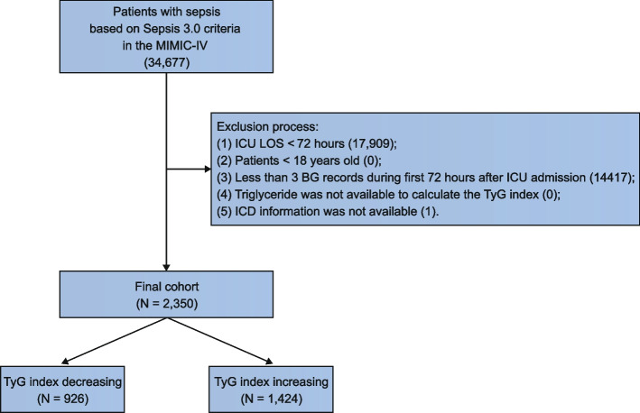
Patients with sepsis were identified with the sepsis 3.0 criteria. We excluded patients who were discharged from ICU within 72 h and the non-adult patients. To ensure that we could imputate the dynamic blood glucose (BG) records into 1-h resolution time series data with the Stineman interpolation algorithm, we excluded patients whose total BG records were less than 3 times within 72 h. In addition, patients whose triglyceride data was not available were also excluded to ensure that the TyG index could be calculated. In view of the triglycerides are not repeatedly measured within the first 3 days in most ICU clinical practice as triglycerides may not change significantly during this stage. The values of triglycerides used in this study encompass those recorded within 72 h after admission for the calculation of the TyG index. The detailed exclusion criteria in this retrospective observational study were set as follows: (1) ICU length of stay less than 72 h; (2) non-adult patients with age less than 18 years; (3) The total counts of BG were less than 3 during the first 72 h after ICU admission; (4) Triglyceride was not available to calculate TyG index; (5) International classification of diseases (ICD) information was not available (Fig. 1).
Fig. 1.
Study flowchart
Data extraction
All related data used in the study, including all BG and triglycerides records, study outcomes, demographic data, interventions, laboratory tests, vital signs, and scoring systems such as sequential organ failure assessment (SOFA) score and the simplified acute physiology score II (SAPS II) within the first 24 h of ICU admission were queried with structured query language (SQL) codes, which were developed and tested with DBeaver Community version 23.2.0 (https://dbeaver.io/download/), and executed with the DBI package version 1.1.3 (https://dbi.r-dbi.org/) to create the corresponding tables in the MIMIC-IV database and related variables in the R global environment for futher data analysis.
Exposure and outcomes
The TyG index is calculated using the formula:
We assessed overall IR during first 72 h after ICU admission with temporal TyG index trajectory. Specifically, we used the dynamic time warping (DTW) algorithm with the tslearn package version 0.6.2 to process the TyG index time series data (normalized by min–max), and calculated the similarity of the dynamic TyG index trend between each individual and other individuals, which formed a skew-symmetric matrix with similarity data. Then, we utilized an unsupervised machine learning algorithm hierarchical clustering (HC) provided by SciPy package version 1.11.4 to cluster the skew symmetric similarity matrix. The optimal number of clusters was determined by the average silhouette coefficient calculated with the scikit-learn package version 1.3.2. Generally, the higher the average silhouette level indicates the better clustering effect. We calculated the average silhouette coefficient of clusters 2–10, and selected the number of clusters corresponding to the highest average silhouette as the best clustering number. In addition, to ensure the stability of clustering, we also use NBclust package version 3.0.1 to calculate the best number of clusters of skew symmetric similarity matrix. The primary outcome was 28-day mortality and the secondary outcome was in-hospital mortality.
Covariates
In the current study, we defined a comprehensive set of 31 covariates based on a previous well-designed study [21] on sepsis, clustered into 5 distinct categories, served as potential confounders. These categories encompassed demographic and admission data (e.g., age, gender, weight, SAPS II, SOFA score, Charlson comorbidity index), therapeutic interventions [mechanical ventilation, sedative therapy, insulin therapy and vasopressor therapy], pre-existing comorbid conditions [e.g., heart failure (HF), hypertension, atrial fibrillation (AFIB), type 2 diabetes mellitus (T2DM), chronic renal disease, liver disease, chronic obstructive pulmonary disease (COPD), coronary artery disease (CAD), stroke, malignancy], vital signs [e.g., mean arterial pressure (MAP), temperature, heart rate], along with laboratory tests (e.g., white blood cell (WBC) count, hemoglobin, platelet count, potential of hydrogen (pH), partial pressure of oxygen (PO2), partial pressure of carbon dioxide (PCO2), lactate, creatinine). We employ the variance inflation factor (VIF) to assess the multicollinearity among covariates. Variables with a VIF > 5 are considered to have a strong correlation with the exposure factors and require further adjustment.
Statistical analysis
As the sample size of each group was less than 2000, we executed the Shapiro–Wilk normality test to evaluate the normal distribution of the data. F-test was applied to assess the equality of variances. Given the circumstances where the data exhibited normal distribution across groups and the homogeneity of variance test revealed no statistical difference, we conducted the t-test for continuous covariates. In contrast, if such conditions were not met, the Wilcoxon test was deemed suitable. The Chi-square test was utilized for categorical covariates, while Fisher’s exact test was used if the sample size for any cell was less than 10. Continuous variables were articulated as mean (standard deviation), while categorical variables were conveyed as numerical values (percentage). We used generalized additive model (GAM) to explore the relationship between TyG index and 28-day mortality [22].
We performed propensity score matching (PSM) and inverse probability of treatment weighting (IPTW) based on the propensity score to adjust for covariates, thereby fortifying the robustness of our results. The Matching package was utilized to generate a 1:1 matched cohort. The propensity score, generated with the logistic regression model, was used as the basis for further propensity score-based analysis. Whether the absolute values of standardized mean difference (SMD) of all covariates between groups exceeded the threshold of 0.1 was used to assess the balance of covariates. Multiple imputations (MI) were performed with the mice package [23] before PSM. The unadjusted log-rank test was employed with the survival package [24] to estimate the original cohort. When Kaplan–Meier curves cross, the log-rank test becomes unsuitable due to its assumption of a constant hazard ratio. In such instances, we will utilize a Time-Dependent Cox model for analysis, employing the tt function (defined as “tt = function (x, t, …) x * log(t + 20)”) for transformation.
To ensure the robustness of the results, we applied a series of models as sensitivity analyses for 28-day and in-hospital mortality. We employed the random forest algorithm to evaluate the impact of covariates on outcomes. This method calculates the importance of each feature variable in predicting the outcome and ranks them accordingly. Based on their significance, feature variables are categorized into three groups: “Confirmed,” “Tentative,” and “Rejected.” Variables labeled as “Confirmed” are considered the most influential and are incorporated into subsequent models for further analysis. The models included the McNemar’s test for matched cohort outcomes (PSM model) [21], the multivariable logistic model adjusted with all covariates, the multivariable logistic model adjusted with covariates selected by Boruta, the multivariable logistic model adjusted with all covariates using IPTW, and doubly robust estimation model [21]. Subgroup analyses were also performed by the level of covariates including age, gender, HF, hypertension, CAD and T2DM with jstable package version 1.1.2.
All statistical approaches were deployed with Python version 3.10.12 or R version 4.2.3. The threshold of statistical significance is established at P < 0.05.
Result
The tyg index trajectory and clinical subphenotype
We identified a total of 34,677 septic patients in the MIMIC-IV database based on the Sepsis 3.0 criteria. After excluding those with ICU stays less than 72 h, individuals under 18 years of age, patients with fewer than 3 blood glucose recordings within the initial 72 h of ICU admission, those with missing triglyceride data, and individuals lacking usable ICD information, the final cohort consisted of 2,350 patients (Fig. 1).
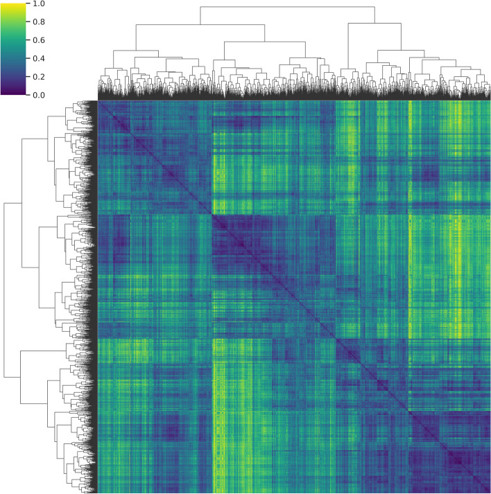
The DTW-derived skew-symmetric similarity matrix of TyG index trajectory was processed with processed by the unsupervised machine learning HC algorithm (Fig. 2). The highest silhouette score was detected at level of 2 clusters, indicating that the optimal number of clusters was 2, which was further confirmed by the NBclust package when processing the skew-symmetric similarity matrix.
Fig. 2.
Heatmap of similarity of the TyG index trajectory after hierarchical clustering. Each patient was depicted by horizontal and vertical axes. DTW-derived normalized pairwise patient similarity is represented by color intensity. The best number of clusters was 2 according to the silhouette score
Subsequently, we analyzed the dynamic trajectories of these two clusters. Patients in the TyG index increasing group (n = 1424) had a gradual rise in their TyG index from an initial value of approximately 9.10 to over 9.31 within the first 72 h, whereas patients in the TyG index decreasing group (n = 926) had a gradual decline from an initial value near 9.48 to approximately 9.03 (Fig. 3). Considering that the TyG index represents a dynamic change in the degree of IR, we identified these 2 clusters as clinical subphenotype of increased and alleviative IR respectively.
Fig. 3.
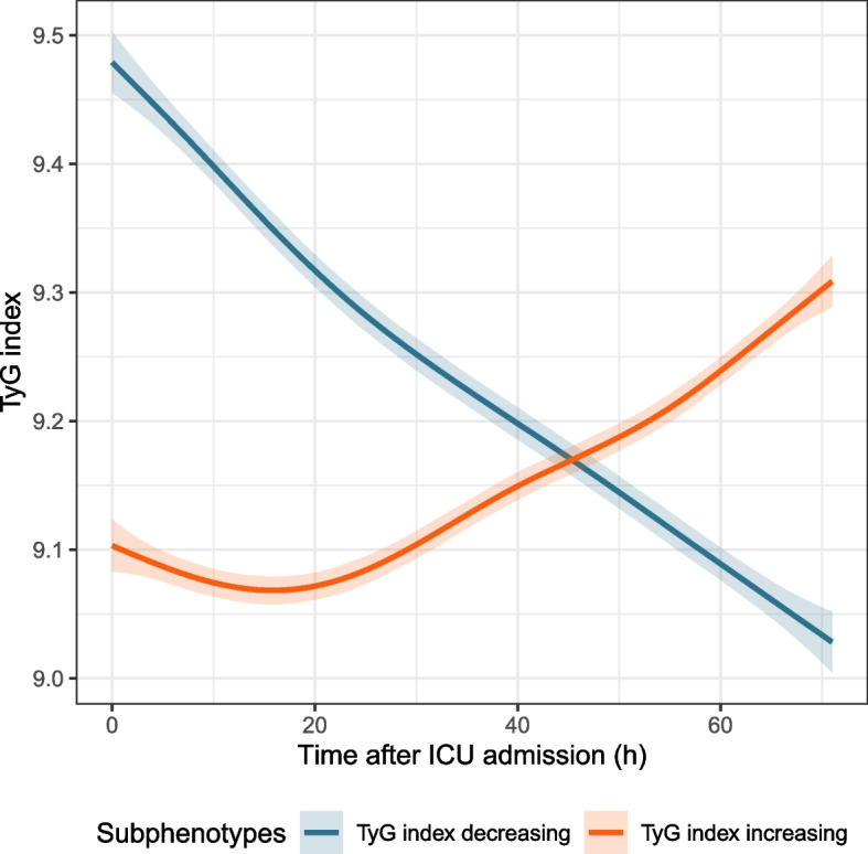
The TyG index trajectory revealed the increased and alleviative IR subphenotypes
The baseline characteristics of different trajectory subphenotypes
Table 1 presents the baseline data for the TyG index decreasing and increasing groups. Within the original cohort, both 2 groups exhibited differences in age, SOFA score, and Charlson comorbidity index. The decreasing group had a slightly lower mean age (60.55 ± 16.49) compared to the increasing group (62.77 ± 16.05). The baseline SOFA score was marginally lower in the increasing group (6.96 ± 4.14) compared to the decreasing group (7.49 ± 4.21). Concerning comorbidities, the incidence rates of hypertension, T2DM, renal disease, CAD, stroke, and other complications were lower in the decreasing group than in the increasing group. Regarding outcomes, the 28-day mortality rate was lower in the decreasing group (23.54%) compared to the increasing group (28.51%), and the in-hospital mortality rate was also lower in the decreasing group (21.60%) compared to the increasing group (25.49%). The detailed baseline data before PSM were provided in Additional File 1, Table S1.
Table 1.
Baseline characteristics before and after propensity score matching of two cohorts
| Before Matching | After Matching | |||||
|---|---|---|---|---|---|---|
| TyG index decreasing (N = 926) | TyG index increasing (N = 1424) | SMD | TyG index decreasing (N = 406) | TyG index increasing (N = 406) | SMD | |
| Age | 60.55 (16.49) | 62.77 (16.05) | 0.136 | 63.87 (15.09) | 59.97 (16.92) | 0.243 |
| Gender (Female) | 382 (41.25%) | 586 (41.15%) | 0.002 | 179 (44.09%) | 158 (38.92%) | 0.105 |
| Weight | 87.12 (25.69) | 86.82 (28.74) | 0.011 | 85.08 (25.88) | 86.80 (24.34) | 0.069 |
| SAPS II | 42.89 (15.08) | 42.28 (14.83) | 0.04 | 41.13 (13.76) | 42.37 (14.24) | 0.089 |
| SOFA score | 7.49 (4.21) | 6.96 (4.14) | 0.126 | 6.69 (4.11) | 7.45 (4.02) | 0.187 |
| Charlson comorbidity index | 4.93 (2.94) | 5.49 (3.08) | 0.186 | 5.49 (2.90) | 4.72 (2.92) | 0.265 |
| Mechanical ventilation (YES) | 668 (72.14%) | 1050 (73.74%) | 0.036 | 298 (73.40%) | 297 (73.15%) | 0.006 |
| Sedative therapy (YES) | 705 (76.13%) | 1068 (75.00%) | 0.026 | 304 (74.88%) | 312 (76.85%) | 0.046 |
| Insulin therapy (YES) | 390 (42.12%) | 487 (34.20%) | 0.164 | 121 (29.80%) | 157 (38.67%) | 0.188 |
| Vasopressor therapy (YES) | 489 (52.81%) | 632 (44.38%) | 0.169 | 177 (43.60%) | 223 (54.93%) | 0.228 |
| HF (YES) | 282 (30.45%) | 424 (29.78%) | 0.015 | 122 (30.05%) | 129 (31.77%) | 0.037 |
| Hypertension (YES) | 560 (60.48%) | 951 (66.78%) | 0.131 | 273 (67.24%) | 243 (59.85%) | 0.154 |
| AFIB (YES) | 121 (13.07%) | 171 (12.01%) | 0.032 | 53 (13.05%) | 47 (11.58%) | 0.045 |
| T2DM (YES) | 233 (25.16%) | 457 (32.09%) | 0.154 | 112 (27.59%) | 79 (19.46%) | 0.193 |
| Renal (YES) | 166 (17.93%) | 306 (21.49%) | 0.09 | 90 (22.17%) | 68 (16.75%) | 0.137 |
| Liver (YES) | 120 (12.96%) | 169 (11.87%) | 0.033 | 51 (12.56%) | 58 (14.29%) | 0.051 |
| COPD (YES) | 138 (14.90%) | 226 (15.87%) | 0.027 | 64 (15.76%) | 63 (15.52%) | 0.007 |
| CAD (YES) | 237 (25.59%) | 303 (21.28%) | 0.102 | 85 (20.94%) | 102 (25.12%) | 0.1 |
| Stroke (YES) | 198 (21.38%) | 435 (30.55%) | 0.21 | 122 (30.05%) | 66 (16.26%) | 0.331 |
| Malignancy (YES) | 165 (17.82%) | 252 (17.70%) | 0.003 | 71 (17.49%) | 69 (17.00%) | 0.013 |
| MAP | 86.40 (20.89) | 85.73 (19.24) | 0.034 | 85.01 (18.01) | 85.72 (20.85) | 0.036 |
| Temperature | 36.80 (0.99) | 36.88 (0.97) | 0.087 | 36.90 (0.85) | 36.75 (0.99) | 0.16 |
| Heart rate | 94.72 (21.37) | 92.64 (21.61) | 0.097 | 91.36 (20.20) | 95.05 (20.16) | 0.183 |
| WBC count | 14.98 (14.47) | 13.58 (11.76) | 0.106 | 13.27 (10.67) | 15.27 (13.54) | 0.164 |
| Hemoglobin | 10.99 (2.53) | 10.88 (2.48) | 0.045 | 10.75 (2.45) | 11.02 (2.51) | 0.111 |
| Platelet | 207.53 (120.49) | 199.73 (112.50) | 0.067 | 197.87 (115.46) | 212.51 (118.68) | 0.125 |
| pH | 7.33 (0.12) | 7.35 (0.11) | 0.177 | 7.36 (0.09) | 7.33 (0.11) | 0.298 |
| PO2 | 137.98 (96.11) | 139.70 (94.45) | 0.018 | 135.48 (82.64) | 131.97 (87.04) | 0.041 |
| PCO2 | 42.32 (13.87) | 43.14 (14.59) | 0.057 | 42.90 (12.87) | 42.14 (12.93) | 0.059 |
| Lactate | 2.80 (2.41) | 2.37 (2.34) | 0.183 | 2.02 (1.48) | 2.79 (2.01) | 0.437 |
| Creatinine | 1.67 (1.55) | 1.61 (1.61) | 0.037 | 1.50 (1.35) | 1.63 (1.53) | 0.088 |
| 28-day mortality (Death) | 218 (23.54%) | 406 (28.51%) | 0.113 | 94 (23.15%) | 124 (30.54%) | 0.167 |
| In-hospital mortality (Death) | 200 (21.60%) | 363 (25.49%) | 0.092 | 80 (19.70%) | 116 (28.57%) | 0.208 |
| Hospital LOS | 18.48 (15.84) | 18.22 (15.51) | 0.016 | 17.79 (14.68) | 18.79 (16.35) | 0.064 |
Values are presented as mean (standard deviation) for continuous variables and number (percentage) for categorical variables. Variables in bold have P -value < 0.05
To mitigate bias arising from baseline data imbalances, we performed PSM between the two groups. Table 1 illustrated the changes of patients between the two groups before and after PSM. Specifically, in the original cohort, the TyG index decreasing group comprised 926 patients, while the TyG index increasing group included 1,424 patients. After performing 1:1 propensity score matching, the number of patients in both groups was reduced to 406. Counts of variables with SMD greater than 0.1 significantly decreased between the groups compared to before matching, which indicated a better balance in baseline characteristics between the matched cohorts (Additional File 2, Table S2-S4, Additional File 1, Fig. S1).
Primary and secondary outcome
We first analyzed the association between the TyG index and 28-day mortality in septic patients. Using a GAM model, we found a nonlinear negative relationship between the TyG index and 28-day mortality in these patients (Additional file 2, Fig. S2).
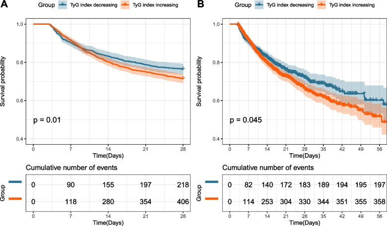
We analyzed the differences in 28-day and in-hospital mortality between the TyG index increasing and decreasing groups. Before PSM, the time dependent Cox model showed that the increasing group had a higher 28-day mortality risk compared to the decreasing group [hazard ratio (HR): 1.07, 95% confidence interval (CI): 1.02–1.12, P = 0.01]. A similar trend was observed for in-hospital mortality (HR: 1.05, 95% CI: 1.00–1.11, P = 0.045) (Additional File 1, Table S5, Table S6). As shown in Fig. 4, the increasing group exhibited significantly higher risks of 28-day (P = 0.01) and in-hospital mortality (P = 0.045) compared to the decreasing group in the original cohort.
Fig. 4.
K-M curve for 28-day (A) and in-hospital mortality (B) by time dependent Cox model
Sensitivity analyses and subgroup analyses
To validate the robustness of the outcomes, we constructed multiple models for sensitivity analyses. We employed the random forest algorithm for feature selection to assess the importance of variables on outcomes. As shown in Additional file 2, Fig. S3, a total of 13 variables were marked as “confirmed,” including SAPS-II, age, Charlson Comorbidity Index, SOFA score, temperature, WBC count, Lactate, liver disease, platelet, stroke, chronic renal disease, sedative therapy, and hemoglobin. These variables were incorporated into subsequent models for further analysis. Meanwhile, to evaluate the multicollinearity of the variables, we analyzed the VIF for all variables. As shown in Additional File 1, Tables S7-S8, the VIF values for all variables were below 5, indicating no need for further adjustments. The models for sensitivity analyses including the PSM model, multivariable logistic model adjusted with all covariates, multivariable logistic model adjusted with covariates selected by Boruta, multivariable logistic model adjusted with all covariates using IPTW, and doubly robust estimation with all covariates. As presented in Table 2, in all models, the increasing group demonstrated higher 28-day and in-hospital mortality relative to the decreasing group. Detailed data are provided in Additional File 1, Table S9-S18.
Table 2.
Primary and secondary outcome analyses with different models for cohort
| Models | P -value | Result |
|---|---|---|
| 28-day mortality | ||
| Propensity score matching model [OR (95% CI)]a | < 0.05 | 1.46 (1.07, 2.00) |
| Multivariable logistic model adjusted with all covariates [OR (95% CI)]a | < 0.05 | 1.28 (1.04, 1.57) |
| Multivariable logistic model adjusted with covariates selected by Boruta [OR (95% CI)]a | < 0.05 | 1.3 (1.06, 1.60) |
| Multivariable logistic model adjusted with all covariates using IPTW [OR (95% CI)]a | < 0.01 | 1.29 (1.11, 1.51) |
| Doubly robust estimation with all covariates [OR (95% CI)]a | < 0.05 | 1.29 (1.04, 1.61) |
| In-hospital mortality | ||
| Propensity score matching model [OR (95% CI)]a | < 0.01 | 1.61 (1.16, 2.23) |
| Multivariable logistic model adjusted with all covariates [OR (95% CI)]a | < 0.05 | 1.28 (1.03, 1.59) |
| Multivariable logistic model adjusted with covariates selected by Boruta [OR (95% CI)]a | < 0.05 | 1.3 (1.05, 1.60) |
| Multivariable logistic model adjusted with all covariates using IPTW [OR (95% CI)]a | < 0.01 | 1.28 (1.09, 1.50) |
| Doubly robust estimation with all covariates [OR (95% CI)]a | < 0.05 | 1.28 (1.02, 1.60) |
Statistical analyses of different models with P -value < 0.05 were displayed in bold
aOR Odds Ratio, CI Confidence Interval
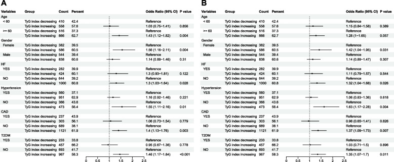
Moreover, we conducted a stratified analysis concerning the relationship between the TyG index increasing and decreasing groups for 28-day mortality, as well as in-hospital mortality. Similar findings persisted across the majority of subgroups (Fig. 5). The TyG index increasing group exhibited a significant association with elevated 28-day mortality (Fig. 5A) in subgroups consisting of females[odds ratio (OR): 1.56, 95% CI: 1.16–2.11, P = 0.004], aged ≥ 60 years (OR: 1.43, 95% CI: 1.12–1.82, P = 0.004), and those not afflicted with heart failure(OR: 1.3, 95CI: 1.03–1.64, P = 0.028), hypertension (OR: 1.55, 95% CI: 1.11–2.16, P = 0.01), CAD (OR: 1.4, 95% CI: 1.13–1.76, P = 0.003), or T2DM (OR: 1.46, 95% CI: 1.17–1.84, P = 0.001). Similar results were obtained in stratified analyses for the in-hospital mortality (Fig. 5B).
Fig. 5.
Forest plots of odds ratios for 28-day (A) and in-hospital mortality (B) in different subgroups
Discussion
Our study identified two dynamic TyG index trajectory subphenotypes in septic patients. Due to DTW’s capacity to accurately capture heterogeneous evolution within temporal sequences, it was utilized to determine how similar the trajectories of individual patients were to one another. And then HC was used to divided patients into similar trajectories together. This allowed for the identification of various clusters and the subsequent determination of trajectory differences. The subphenotypes were identified as the increased and alleviative IR groups, which were characterized by the TyG index increasing and decreasing within the first 72 h respectively. The increased IR group exhibited higher 28-day and in-hospital mortality than the decreasing group. Furthermore, sensitivity analyses further confirmed the robustness of our findings.
IR represents a significant manifestation of metabolic dysregulation in septic patients [4]. This metabolic abnormality has previously been connected to sympathetic nervous system activation [8], elevated plasma levels of counter-regulatory hormones [3], and GLUT4 translocation [6] in septic patients. The occurrence of IR is significantly correlated with adverse outcomes in septic patients and contributes to higher mortality rates [10]. With increased awareness and technological advancements, this metabolic abnormality has gained wider recognition and treatment. Clinical evaluation of the status of IR in septic patients is frequently insufficient, and the effect of IR trajectory on the mortality of septic patients has not been adequately assessed.
The TyG index has been considered an effective surrogate marker for assessing IR. Gold standard techniques like the euglycemic insulin clamp and intravenous glucose tolerance testing are pricy and difficult to perform, and HOMA-IR’s applicability is constrained by things like insulin therapy [25]. The TyG index has received a lot of attention as a useful tool for assessing IR in clinical settings because of its ease of use, low cost, and effectiveness that is comparable to HOMA-IR [26]. It has demonstrated its utility in non-diabetic patients and those receiving insulin therapy, where it may even outperform HOMA-IR [11, 27].
Previous research has focused on the predictive capabilities of the TyG index for cardiovascular diseases [27]. The TyG index has demonstrated good prognostic value in conditions such as coronary artery disease [28], heart failure [29], ischemic stroke [30], etc. Studies have shown that this risk prediction efficacy is unaffected by hypertriglyceridemia and diabetes [11]. For example, a nested case–control study involving 3,745 patients demonstrated an association between the TyG index and the risk of cardiovascular events (HR: 1.364, 95% CI: 1.100–1.691, P = 0.005) [31]. Another retrospective cohort study of 733 patients with ischemic stroke found a significant association between elevated TyG index and all-cause in-hospital mortality (HR: 1.371, 95% CI: 1.053–1.784, P = 0.013) [32]. Additionally, in acute coronary syndromes, a high TyG index is associated with higher mortality rates and a greater risk of major adverse cardiovascular events [15, 33]. The correlation of the TyG index with the incidence of organ dysfunction in cardiovascular disease patients has also been noted. The risk of acute kidney injury was significantly associated with an elevated TyG index, according to a retrospective cohort study of 1,393 patients with heart failure (HR: 1.57; 95% CI: 1.34–1.84; P = 0.001) [34].
Furthermore, the trajectory of the TyG index can serve as a predictor of cardiovascular diseases, demonstrating value in atherosclerosis and stroke. According to Yan et al.’s study, the brachial-ankle pulse wave velocity increased by 37.1 cm/s for every unit increase in the TyG index (95% CI: 23.7–50.6 cm/s, P < 0.001), with the highest TyG index trajectory group showing the fastest progression of arterial stiffness (OR 2.76; 95% CI: 1.40–7.54) [17]. Huang et al. analyzed 19,924 hypertensive patients and identified five different TyG index trajectories, with the elevated-increasing group having the highest risk of stroke (HR: 2.21, 95% CI: 1.49–3.28) [18]. These findings collectively demonstrate the TyG index’s strong predictive ability for both short-term and long-term risk in cardiovascular disease patients.
More recently, the TyG index has shown promise in predicting outcomes in critically ill patients. Liao et al. conducted a retrospective cohort study comprising 3,026 critically ill patients, revealing that the TyG index serves as an independent risk indicator for ICU mortality (HR: 1.72, 95% CI: 1.18–2.521.18–2.52, P = 0.005) [35]. Another study involving 639 critically ill patients with concomitant CKD and CAD demonstrated a significant association between elevated TyG index and one-year ICU mortality and in-hospital mortality [36]. Zheng et al.’s study investigated the association between the TyG index and mortality rates among septic patients. The research revealed a significant correlation between elevated TyG index and increased in-hospital mortality risk among patients with sepsis (OR 1.440; 95% CI 1.106–1.875; P = 0.00673) [20].
The studies mentioned above collectively affirm that TyG index, as an indicator reflective of IR status, demonstrates notable prognostic value. However, little attention has been devoted to exploring the impact of TyG index trajectory, or rather, the changes in IR, on outcomes in septic patients. Our research conveniently addresses this lacuna.
We explored the similarities of dynamic changes of the TyG index in different subjects, and identified two trajectory subphenotypes: the increased and alleviative IR groups, which stand for increasing and decreasing TyG index within the initial 72 h after ICU admission. As previously discussed, the TyG index serves as an alternative metric for IR, wherein its elevation implies a worsening of IR, and its decline suggests an amelioration of IR. Consequently, our findings can be interpreted as a significant correlation between the improvement in IR within the first 72 h of admission and a reduced mortality rate among septic patients. This outcome holds promising applications in clinical practice. By monitoring and calculating the TyG index and its alterations, we can dynamically assess a patient’s IR status, evaluate their metabolic anomalies, and thereby implement tailored preventive and therapeutic measures. In the stratified analysis, we observed that patients over 60 years old, without hypertension, heart failure, CAD, or T2DM had a higher risk of mortality when exhibiting an increasing trend in IR. This might be attributed to patients who were older or with HF, CAD, and T2DM inherently having a higher incidence of IR, thus underestimating the role of IR changes within these populations. Simultaneously, in the presence of an increasing IR trend, females faced an elevated risk of mortality. This association may be linked to the comparatively lower incidence of IR in females than males [37], thereby indicating a relatively diminished tolerance to the impacts of IR.
Our study confirmed that the elevation of the TyG index serves as a predictive factor for increased mortality risk in septic patients, elucidating that the worsening of IR may result in a heightened mortality risk among individuals afflicted by sepsis. Nevertheless, there are several limitations in our study. Firstly, it is imperative to acknowledge that this is a retrospective observational study, we have diligently employed a variety of rigorous and comprehensive statistical methodologies to mitigate bias and ensure the robustness of our findings. However, further prospective research is essential to validate the impact of TyG index variations on the clinical prognosis of septic patients. Secondly, due to limitations within the database, we were unable to confirm whether all blood glucose and lipid results were obtained in a fasting state. In most ICU settings, triglycerides are often not repeatedly measured within the first 3 days, making it challenging to calculate their dynamic change trajectories. While blood glucose measurements are relatively frequent in the ICU, exhibit significant dynamic changes, and can be used to compute dynamic trajectories. Therefore, in this study, the dynamic changes in the TyG index may be primarily driven by fluctuations in blood glucose. However, the TyG index is calculated from both blood glucose and triglyceride data, its trajectory meaningfully represents the dynamic changes in insulin resistance, as it incorporates the influence of triglycerides. Future research could consider increasing the measurement frequency of triglycerides to more precisely assess their dynamic change trajectories. Moreover, due to the presence of interventions such as insulin therapy and vasopressor medications, the trajectory changes may be influenced. Although we have adjusted potential variables into our model, there still might be latent influencing factors (such as insulin dosage and patient responses to insulin therapy). These factors need to be further controlled in detailed prospective studies. Thirdly, since the database only encompasses critically ill patients in the United States, conducting additional studies to verify whether these findings hold in other nations is imperative. Fourthly due to the need to calculate dynamic trajectories, we included only patients who stayed in the ICU for more than 72 h. Patients with severe conditions (e.g., those who died within 24 h) may have reached the endpoint of the study before their trajectories could be calculated, and patients with milder conditions (e.g., those under postoperative monitoring) often had too short a stay in the ICU to effectively assess “trajectory” changes during their ICU stay. The loss of these patients may impact the test’s effectiveness. Fifth, we utilized DTW and HC algorithm to determine the variation patterns of TyG index for patients, subsequently dividing them into two groups based on the results. This approach focuses on investigating the patterns of change for TyG index, namely, how these 2 different trends/subphenotypes in TyG index fluctuations affect prognosis of patients. However, when it comes to different individuals within the same subphenotype, for instance, the patients in IR decreasing subphenotype both exhibiting a decreasing trend in TyG index, we cannot fully quantify whether a decline from higher value to middle range is superior, inferior or equivalent to a decline from middle range to an even lower value. Similarly, it is difficult to judge which is more harmful among the individuals in the same trend of TyG index increasing. Precise estimations require further elaboration with larger sample sizes and higher-resolution data. Therefore, future prospective trials should consider the influence of these patients more thoroughly.
Conclusion
This study reveals that a dynamic increase in the triglyceride-glucose (TyG) index trajectory is significantly associated with higher mortality rates. Through analyzing the TyG index trajectory, we identified clinical subphenotypes reflecting changes in insulin resistance, offering new insights into patient prognosis. Notably, a sustained rise in the TyG index indicates worsening insulin resistance, which is closely linked to poorer clinical outcomes.
Supplementary information
Acknowledgements
We especially appreciate the MIMIC official team’s efforts to open-source the database and codes.
Abbreviations
- TyG
Triglyceride-glucose
- DTW
Dynamic time warping
- HC
Hierarchical clustering
- GLUT4
Glucose transporter type 4
- IR
Insulin resistance
- HOMA-IR
Homeostasis model assessment-estimated insulin resistance
- RECORD
REporting of studies Conducted using Observational Routinely-collected health Data
- MIMIC-IV
Medical Information Mart for Intensive Care IV
- BG
Blood glucose
- ICU
Intensive care unit
- ICD
International classification of diseases
- SOFA
Sequential organ failure assessment
- SAPS II
Simplified acute physiology score II
- SQL
Structured query language
- HF
Heart failure
- AFIB
Atrial fibrillation
- T2DM
Type 2 diabetes mellitus
- COPD
Chronic obstructive pulmonary disease
- CAD
Coronary artery disease
- MAP
Mean arterial pressure
- WBC
White blood cell
- pH
Potential of hydrogen
- PO2
Partial pressure of oxygen
- PCO2
Partial pressure of carbon dioxide
- GAM
Generalized additive model
- VIF
Variance inflation factor
- PSM
Propensity score matching
- IPTW
Inverse probability of treatment weighting
- SMD
Standardized mean difference
- MI
Multiple imputations
- HR
Hazard ratio
- CI
Confidence interval
- K-M
Kaplan–Meier
- OR
Odds ratio
Authors’ contributions
Yi-Le Ning, and Xiang-Hui Xu conceived and designed the study. Yi-Le Ning developed SQL, R, and Python codes for data analysis. Ce Sun and Xiang-Hui Xu verified the data and drafted the initial manuscript. Xiao-Li Niu and Yu Zhang contributed to the data interpretation. Yu Zhang, Ji-Hong Zhou and Ce Sun revised the manuscript and provided methodological guidance. All authors reviewed and approved the ultimate version of the submitted manuscript.
Funding
This study was supported by the Basic Research Projects Jointly Funding by Municipal Universities (Colleges) of Guangzhou Municipal Science and Technology Bureau (202201020325), Shenzhen Bao’an District High-quality Development Research Project (YNXM2024066, YNXM2024059), National Project for the Development of Key Specialties in Chinese Medicine (No. 900) and National Natural Science Foundation of China (No. 72404064).
Availability of data and materials
The MIMIC-IV database is publicly available on PhysioNet (https://www.physionet.org/). Concepts codes are available in the MIMIC Code Repository (https://github.com/MIT-LCP/mimic-code/).
Declarations
Ethics approval and consent to participate
Not applicable. This retrospective observational study is in accordance with the RECORD statement with the MIMIC-IV database version 2.0. The Beth Israel Deaconess Medical Center’s Institutional Review Board waived informed written consent because all patient data was deidentified (IRB #2001P001699). Because the study in the MIMIC-IV database was retrospective and observational, there was no requirement to acquire consent to participate. Our team can now access the MIMIC-IV database (Record ID 40974208).
Consent for publication
Not applicable. The need to obtain consent for publication was waived owing to the retrospective and observational nature of the study in the MIMIC-IV database.
Competing interests
The authors declare no competing interests.
Footnotes
Publisher’s Note
Springer Nature remains neutral with regard to jurisdictional claims in published maps and institutional affiliations.
Yi-Le Ning and Xiang-Hui Xu contributed equally to this work and shared first authorship.
Contributor Information
Yu Zhang, Email: zhangyu801026@qq.com.
Ji-Hong Zhou, Email: zhoujihong71@gzucm.edu.cn.
Ce Sun, Email: sunce.icu@gmail.com.
References
- 1.Evans L, Rhodes A, Alhazzani W, Antonelli M, Coopersmith CM, French C, Machado FR, McIntyre L, Ostermann M, Prescott HC, et al. Surviving sepsis campaign: international guidelines for management of sepsis and septic shock 2021. Intensive Care Med. 2021;47(11):1181–247. [DOI] [PMC free article] [PubMed] [Google Scholar]
- 2.Singer M, Deutschman CS, Seymour CW, Shankar-Hari M, Annane D, Bauer M, Bellomo R, Bernard GR, Chiche J-D, Coopersmith CM, et al. The Third International Consensus Definitions for Sepsis and Septic Shock (Sepsis-3). JAMA. 2016;315(8):801–10. [DOI] [PMC free article] [PubMed] [Google Scholar]
- 3.Marik PE, Raghavan M. Stress-hyperglycemia, insulin and immunomodulation in sepsis. Intensive Care Med. 2004;30(5):748–56. [DOI] [PubMed] [Google Scholar]
- 4.Rivas AM, Nugent K. Hyperglycemia, Insulin, and Insulin Resistance in Sepsis. Am J Med Sci. 2021;361(3):297–302. [DOI] [PubMed] [Google Scholar]
- 5.Wasyluk W, Zwolak A. Metabolic Alterations in Sepsis. J Clin Med. 2021;10(11):2412. [DOI] [PMC free article] [PubMed] [Google Scholar]
- 6.Preau S, Vodovar D, Jung B, Lancel S, Zafrani L, Flatres A, Oualha M, Voiriot G, Jouan Y, Joffre J, et al. Energetic dysfunction in sepsis: a narrative review. Ann Intensive Care. 2021;11(1):104. [DOI] [PMC free article] [PubMed] [Google Scholar]
- 7.Singer M, De Santis V, Vitale D, Jeffcoate W. Multiorgan failure is an adaptive, endocrine-mediated, metabolic response to overwhelming systemic inflammation. The Lancet. 2004;364(9433):545–8. [DOI] [PubMed] [Google Scholar]
- 8.Carlson GL, Little RA. Sympathetic nervous system and metabolism. In: Guarnieri G, Iscra F, editors. Metabolism and Artificial Nutrition in the Critically Ill. Milano: Springer Milan; 1999. p. 71–83. [Google Scholar]
- 9.Cheong HS, Chang Y, Kim Y, Joo E-J, Kwon M-J, Wild SH, Byrne CD, Ryu S. Glycaemic status, insulin resistance, and risk of infection-related mortality: a cohort study. Eur J Endocrinol. 2023;188(2):197–205. [DOI] [PubMed] [Google Scholar]
- 10.Khan MS, Gutch M, Kumar S, Kumar M. Insulin Resistance as a Prognostic Indicator in Severe Sepsis, Septic Shock and Multiorgan Dysfunction Syndrome. International Journal of Medicine and Public Health. 2020;10:47–50. [Google Scholar]
- 11.Simental-Mendía LE, Rodríguez-Morán M, Guerrero-Romero F. The Product of Fasting Glucose and Triglycerides As Surrogate for Identifying Insulin Resistance in Apparently Healthy Subjects. Metab Syndr Relat Disord. 2008;6(4):299–304. [DOI] [PubMed] [Google Scholar]
- 12.Vasques ACJ, Novaes FS. de Oliveira MdS, Matos Souza JR, Yamanaka A, Pareja JC, Tambascia MA, Saad MJA, Geloneze B: TyG index performs better than HOMA in a Brazilian population: A hyperglycemic clamp validated study. Diabetes Res Clin Pract. 2011;93(3):e98–100. [DOI] [PubMed] [Google Scholar]
- 13.Jin J-L, Sun D, Cao Y-X, Guo Y-L, Wu N-Q, Zhu C-G, Gao Y, Dong Q-T, Zhang H-W, Liu G, et al. Triglyceride glucose and haemoglobin glycation index for predicting outcomes in diabetes patients with new-onset, stable coronary artery disease: a nested case-control study. Ann Med. 2018;50(7):576–86. [DOI] [PubMed] [Google Scholar]
- 14.Shi W, Xing L, Jing L, Tian Y, Yan H, Sun Q, Dai D, Shi L, Liu S. Value of triglyceride-glucose index for the estimation of ischemic stroke risk: Insights from a general population. Nutr Metab Cardiovasc Dis. 2020;30(2):245–53. [DOI] [PubMed] [Google Scholar]
- 15.Luo E, Wang D, Yan G, Qiao Y, Liu B, Hou J, Tang C. High triglyceride–glucose index is associated with poor prognosis in patients with acute ST-elevation myocardial infarction after percutaneous coronary intervention. Cardiovasc Diabetol. 2019;18(1):150. [DOI] [PMC free article] [PubMed] [Google Scholar]
- 16.Khalaji A, Behnoush AH, Khanmohammadi S, Ghanbari Mardasi K, Sharifkashani S, Sahebkar A, Vinciguerra C, Cannavo A. Triglyceride-glucose index and heart failure: a systematic review and meta-analysis. Cardiovasc Diabetol. 2023;22(1):244. [DOI] [PMC free article] [PubMed] [Google Scholar]
- 17.Yan Y, Wang D, Sun Y, Ma Q, Wang K, Liao Y, Chen C, Jia H, Chu C, Zheng W, et al. Triglyceride-glucose index trajectory and arterial stiffness: results from Hanzhong Adolescent Hypertension Cohort Study. Cardiovasc Diabetol. 2022;21(1):33. [DOI] [PMC free article] [PubMed] [Google Scholar]
- 18.Huang Z, Ding X, Yue Q, Wang X, Chen Z, Cai Z, Li W, Cai Z, Chen G, Lan Y, et al. Triglyceride-glucose index trajectory and stroke incidence in patients with hypertension: a prospective cohort study. Cardiovasc Diabetol. 2022;21(1):141. [DOI] [PMC free article] [PubMed] [Google Scholar]
- 19.Lingli D, Yun Y, Kunling W, Cuining H, Dan W, Shan S. Association between TyG index and long-term mortality of critically ill patients: a retrospective study based on the MIMIC Database. BMJ Open. 2023;13(5): e065256. [DOI] [PMC free article] [PubMed] [Google Scholar]
- 20.Zheng R, Qian S, Shi Y, Lou C, Xu H, Pan J. Association between triglyceride-glucose index and in-hospital mortality in critically ill patients with sepsis: analysis of the MIMIC-IV database. Cardiovasc Diabetol. 2023;22(1):307. [DOI] [PMC free article] [PubMed] [Google Scholar]
- 21.Feng M, McSparron JI, Kien DT, Stone DJ, Roberts DH, Schwartzstein RM, Vieillard-Baron A, Celi LA. Transthoracic echocardiography and mortality in sepsis: analysis of the MIMIC-III database. Intensive Care Med. 2018;44(6):884–92. [DOI] [PubMed] [Google Scholar]
- 22.van den Boom W, Hoy M, Sankaran J, Liu M, Chahed H, Feng M, See KC. The Search for Optimal Oxygen Saturation Targets in Critically Ill Patients: Observational Data From Large ICU Databases. Chest. 2020;157(3):566–73. [DOI] [PubMed] [Google Scholar]
- 23.van Buuren S, Groothuis-Oudshoorn K. mice: Multivariate Imputation by Chained Equations in R. J Stat Softw. 2011;45(3):1–67. [Google Scholar]
- 24.Lin H, Zelterman D. Modeling Survival Data: Extending the Cox Model. Technometrics. 2002;44(1):85–6. [Google Scholar]
- 25.Minh HV, Tien HA, Sinh CT, Thang DC, Chen C-H, Tay JC, Siddique S, Wang T-D, Sogunuru GP, Chia Y-C, et al. Assessment of preferred methods to measure insulin resistance in Asian patients with hypertension. The Journal of Clinical Hypertension. 2021;23(3):529–37. [DOI] [PMC free article] [PubMed] [Google Scholar]
- 26.Placzkowska S, Pawlik-Sobecka L, Kokot I, Piwowar A. Indirect insulin resistance detection: Current clinical trends and laboratory limitations. Biomedical papers. 2019;163(3):187–99. [DOI] [PubMed] [Google Scholar]
- 27.Tao L-C. Xu J-n, Wang T-t, Hua F, Li J-J: Triglyceride-glucose index as a marker in cardiovascular diseases: landscape and limitations. Cardiovasc Diabetol. 2022;21(1):68. [DOI] [PMC free article] [PubMed] [Google Scholar]
- 28.da Silva A, Caldas APS, Hermsdorff HHM, Bersch-Ferreira ÂC, Torreglosa CR, Weber B, Bressan J. Triglyceride-glucose index is associated with symptomatic coronary artery disease in patients in secondary care. Cardiovasc Diabetol. 2019;18(1):89. [DOI] [PMC free article] [PubMed] [Google Scholar]
- 29.Zhou Q, Yang J, Tang H, Guo Z, Dong W, Wang Y, Meng X, Zhang K, Wang W, Shao C, et al. High triglyceride-glucose (TyG) index is associated with poor prognosis of heart failure with preserved ejection fraction. Cardiovasc Diabetol. 2023;22(1):263. [DOI] [PMC free article] [PubMed] [Google Scholar]
- 30.Wang F, Wang J, Han Y, Shi X, Xu X, Hou C, Gao J, Zhu S, Liu X. Triglyceride-glucose index and stroke recurrence in elderly patients with ischemic stroke. Front Endocrinol. 2022;13:1005614. [DOI] [PMC free article] [PubMed] [Google Scholar]
- 31.Jin J-L, Cao Y-X, Wu L-G, You X-D, Guo Y-L, Wu N-Q, Zhu C-G, Gao Y, Dong Q-T, Zhang H-W, et al. Triglyceride glucose index for predicting cardiovascular outcomes in patients with coronary artery disease. J Thorac Dis. 2018;10(11):6137–46. [DOI] [PMC free article] [PubMed] [Google Scholar]
- 32.Cai W, Xu J, Wu X, Chen Z, Zeng L, Song X, Zeng Y, Yu F. Association between triglyceride-glucose index and all-cause mortality in critically ill patients with ischemic stroke: analysis of the MIMIC-IV database. Cardiovasc Diabetol. 2023;22(1):138. [DOI] [PMC free article] [PubMed] [Google Scholar]
- 33.Hao Q, Yuanyuan Z, Lijuan C. The Prognostic Value of the Triglyceride Glucose Index in Patients With Acute Myocardial Infarction. J Cardiovasc Pharmacol Ther. 2023;28:10742484231181846. [DOI] [PubMed] [Google Scholar]
- 34.Yang Z, Gong H, Kan F, Ji N. Association between the triglyceride glucose (TyG) index and the risk of acute kidney injury in critically ill patients with heart failure: analysis of the MIMIC-IV database. Cardiovasc Diabetol. 2023;22(1):232. [DOI] [PMC free article] [PubMed] [Google Scholar]
- 35.Liao Y, Zhang R, Shi S, Zhao Y, He Y, Liao L, Lin X, Guo Q, Wang Y, Chen L, et al. Triglyceride-glucose index linked to all-cause mortality in critically ill patients: a cohort of 3026 patients. Cardiovasc Diabetol. 2022;21(1):128. [DOI] [PMC free article] [PubMed] [Google Scholar]
- 36.Ye Z, An S, Gao Y, Xie E, Zhao X, Guo Z, Li Y, Shen N, Zheng J. Association between the triglyceride glucose index and in-hospital and 1-year mortality in patients with chronic kidney disease and coronary artery disease in the intensive care unit. Cardiovasc Diabetol. 2023;22(1):110. [DOI] [PMC free article] [PubMed] [Google Scholar]
- 37.Li M, Chi X, Wang Y, Setrerrahmane S, Xie W, Xu H. Trends in insulin resistance: insights into mechanisms and therapeutic strategy. Signal Transduct Target Ther. 2022;7(1):216. [DOI] [PMC free article] [PubMed] [Google Scholar]
Associated Data
This section collects any data citations, data availability statements, or supplementary materials included in this article.
Supplementary Materials
Data Availability Statement
The MIMIC-IV database is publicly available on PhysioNet (https://www.physionet.org/). Concepts codes are available in the MIMIC Code Repository (https://github.com/MIT-LCP/mimic-code/).


