Abstract
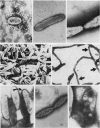
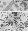
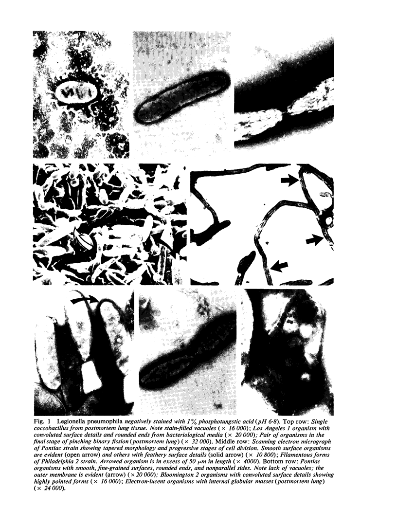
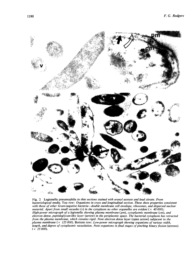
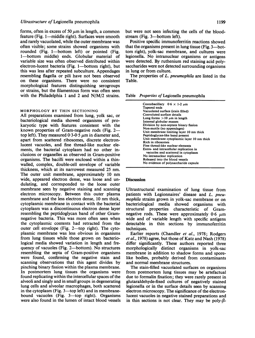
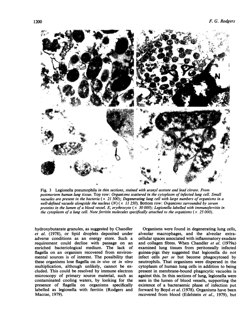
Eleven lung samples positive for Legionnaires' disease, 12 strains of Legionella pneumophila cultured on various bacteriological media, and one strain growth in the yolk sac of fertile hens' eggs were examined by negative staining, thin sectioning, and scanning electron microscopy. All organisms studied were ultrastructurally similar irrespective of strain, source, or method of cultivation, presenting mainly as short rods, 0.6 x 1.5 micrometer, with tapered ends, though long forms and filaments were also evident. In this they resembled typical Gram-negative organisms. Division was by non-septate binary fission, and the cell wall was composed of two triple-unit membranes with morphological evidence of a peptidoglycan layer. The bacterial cytoplasm was rich in ribosomes and nuclear elements and often contained vacuoles. No acid polysaccharides or bacterial appendages were detected surrounding the organisms. In lung tissue and yolk sac membranes, the organisms replicated within the cytoplasm of infected cells and in the intercellular spaces and were specifically identified in thin sections by immunoferritin techniques.
Full text
PDF
Images in this article
Selected References
These references are in PubMed. This may not be the complete list of references from this article.
- Boyd J. F., Buchanan W. M., MacLeod T. I., Dunn R. I., Weir W. P. Pathology of five Scottish deaths from pneumonic illnesses acquired in Spain due to Legionnaires' disease agent. J Clin Pathol. 1978 Sep;31(9):809–816. doi: 10.1136/jcp.31.9.809. [DOI] [PMC free article] [PubMed] [Google Scholar]
- Chandler F. W., Cole R. M., Hicklin M. D., Blackmon J. A., Callaway C. S. Ultrastructure of the Legionnaires' disease bacterium. A study using transmission electron microscopy. Ann Intern Med. 1979 Apr;90(4):642–647. doi: 10.7326/0003-4819-90-4-642. [DOI] [PubMed] [Google Scholar]
- Chandler F. W., Hicklin M. D., Blackmon J. A. Demonstration of the agent of Legionnaires' disease in tissue. N Engl J Med. 1977 Dec 1;297(22):1218–1220. doi: 10.1056/NEJM197712012972206. [DOI] [PubMed] [Google Scholar]
- Chandler F. W., McDade J. E., Hicklin M. D., Blackmon J. A., Thomason B. M., Ewing E. P., Jr Pathologic findings in guinea pigs inoculated intraperitoneally with the Legionnaires' disease bacterium. Ann Intern Med. 1979 Apr;90(4):671–675. doi: 10.7326/0003-4819-90-4-671. [DOI] [PubMed] [Google Scholar]
- Cherry W. B., Pittman B., Harris P. P., Hebert G. A., Thomason B. M., Thacker L., Weaver R. E. Detection of Legionnaires disease bacteria by direct immunofluorescent staining. J Clin Microbiol. 1978 Sep;8(3):329–338. doi: 10.1128/jcm.8.3.329-338.1978. [DOI] [PMC free article] [PubMed] [Google Scholar]
- Edelstein P. H., Meyer R. D., Finegold S. M. Isolation of Legionella pneumophila from blood. Lancet. 1979 Apr 7;1(8119):750–751. doi: 10.1016/s0140-6736(79)91207-8. [DOI] [PubMed] [Google Scholar]
- Flesher A. R., Ito S., Mansheim B. J., Kasper D. L. The cell envelope of the Legionnaires' disease bacterium. Morphologic and biochemical characteristics. Ann Intern Med. 1979 Apr;90(4):628–630. doi: 10.7326/0003-4819-90-4-628. [DOI] [PubMed] [Google Scholar]
- Fraser D. W., Tsai T. R., Orenstein W., Parkin W. E., Beecham H. J., Sharrar R. G., Harris J., Mallison G. F., Martin S. M., McDade J. E. Legionnaires' disease: description of an epidemic of pneumonia. N Engl J Med. 1977 Dec 1;297(22):1189–1197. doi: 10.1056/NEJM197712012972201. [DOI] [PubMed] [Google Scholar]
- Glavin F. L., Winn W. C., Jr, Craighead J. E. Ultrastructure of lung in Legionnaires' disease. Observations of three biopsies done during the Vermont epidemic. Ann Intern Med. 1979 Apr;90(4):555–559. doi: 10.7326/0003-4819-90-4-555. [DOI] [PubMed] [Google Scholar]
- Greaves P. G., Sharp G., Macrae A. D. Isolation of Legionella pneumophila. Lancet. 1979 Mar 10;1(8115):551–552. doi: 10.1016/s0140-6736(79)90969-3. [DOI] [PubMed] [Google Scholar]
- Katz S. M., Nash P. Legionnaires' disease: structural characteristics of the organism. Science. 1978 Feb 24;199(4331):896–897. doi: 10.1126/science.622573. [DOI] [PubMed] [Google Scholar]
- Keel J. A., Finnerty W. R., Feeley J. C. Fine structure of the Legionnaires' disease bacterium. In-vitro and in-vivo studies of four isolates. Ann Intern Med. 1979 Apr;90(4):652–655. doi: 10.7326/0003-4819-90-4-652. [DOI] [PubMed] [Google Scholar]
- Luft J. H. Ruthenium red and violet. II. Fine structural localization in animal tissues. Anat Rec. 1971 Nov;171(3):369–415. doi: 10.1002/ar.1091710303. [DOI] [PubMed] [Google Scholar]
- Rodgers F. G., Greaves P. W., Macrae A. D. Flagella and fimbriae on Legionella organisms. Lancet. 1979 Oct 6;2(8145):753–754. doi: 10.1016/s0140-6736(79)90691-3. [DOI] [PubMed] [Google Scholar]
- Rodgers F. G., Macrae A. D. Immunoferritin electronmicroscopy in legionellosis. Lancet. 1979 Apr 7;1(8119):786–786. doi: 10.1016/s0140-6736(79)91251-0. [DOI] [PubMed] [Google Scholar]
- Rodgers F. G., Macrae A. D., Lewis M. J. Electron microscopy of the organism of Legionnaires' disease. Nature. 1978 Apr 27;272(5656):825–826. doi: 10.1038/272825a0. [DOI] [PubMed] [Google Scholar]
- Rodgers F. G. Staining of Legionella pneumophila. Lancet. 1979 Mar 10;1(8115):551–551. doi: 10.1016/s0140-6736(79)90968-1. [DOI] [PubMed] [Google Scholar]
- Thomason B. M., Chandler F. W., Hollis D. G. Flagella on Legionnaires' disease bacteria: an interim report. Ann Intern Med. 1979 Aug;91(2):224–226. doi: 10.7326/0003-4819-91-2-224. [DOI] [PubMed] [Google Scholar]
- Thornley M. J., Glauert A. M. Fine structure and radiation resistance in Acinetobacter: studies on a resistant strain. J Cell Sci. 1968 Jun;3(2):273–294. doi: 10.1242/jcs.3.2.273. [DOI] [PubMed] [Google Scholar]


