Abstract
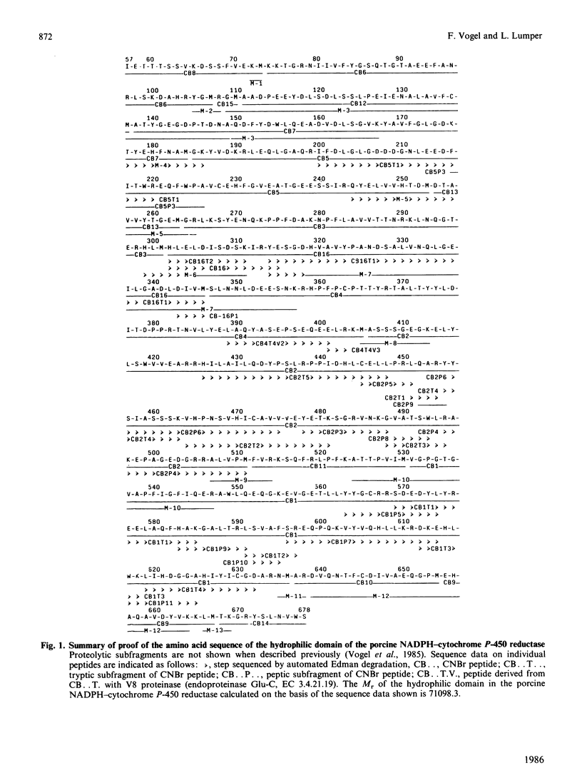
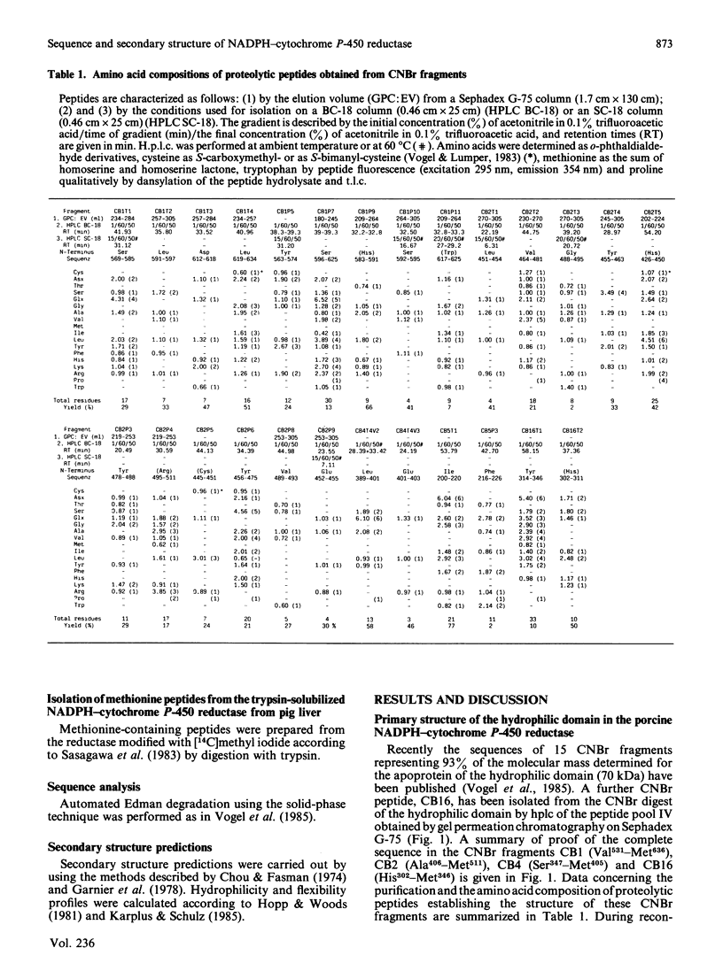
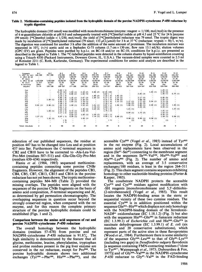
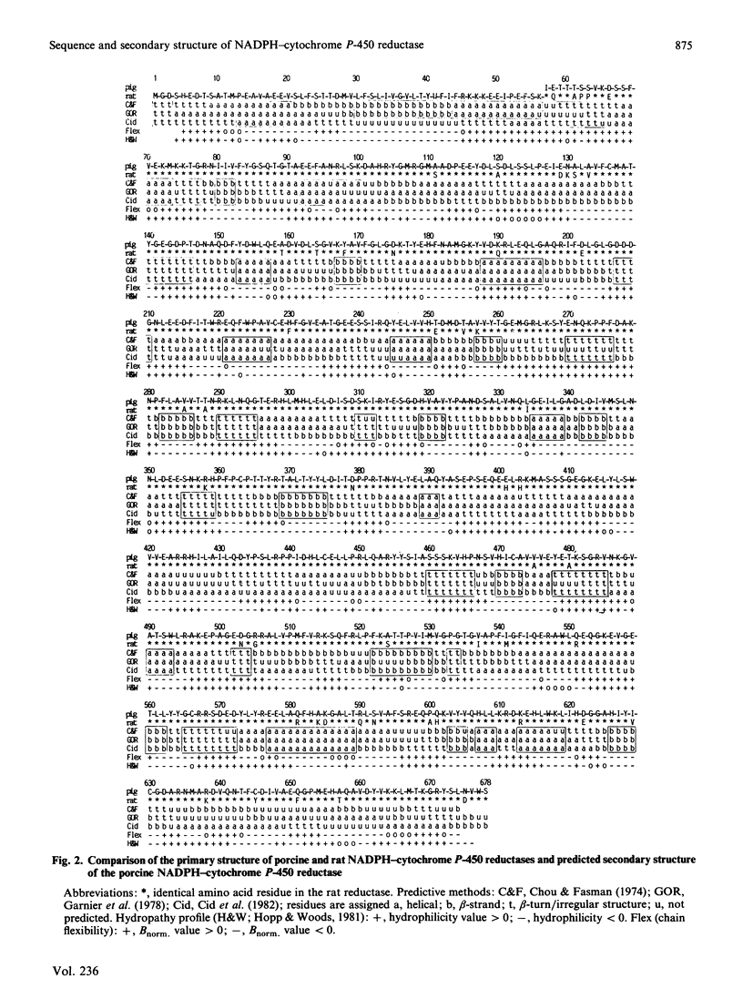
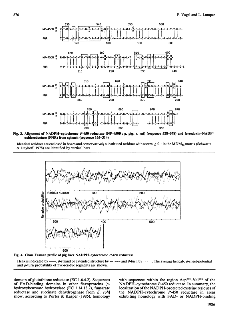
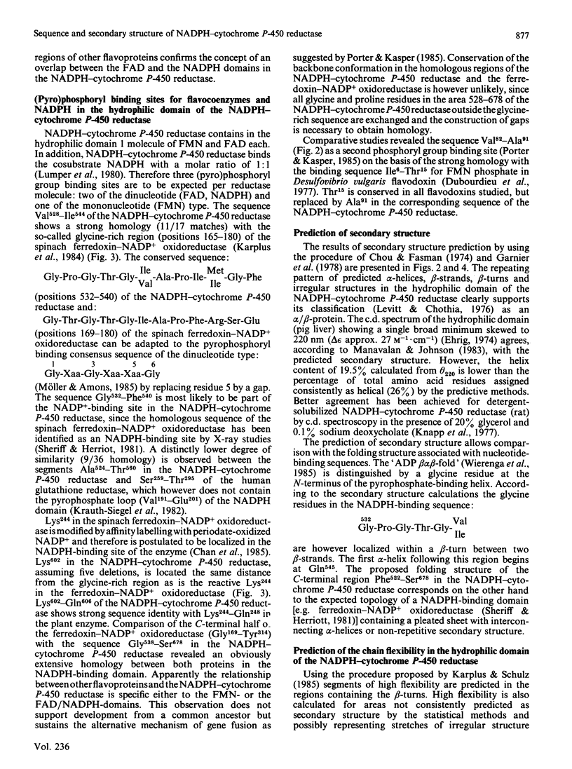
The 622-residue amino acid sequence of the hydrophilic domain in the porcine NADPH-cytochrome P-450 reductase (EC 1.6.2.4) is reported. The structural data required to complete the sequences published previously [Vogel, Kaiser, Witt & Lumper (1985) Biol. Chem. Hoppe-Seyler 366, 577-587] and to establish the primary structure of the porcine hydrophilic domain have been obtained by sequencing proteolytic subfragments derived from CNBr fragments and by characterizing the overlapping S-[14C]methylmethionine-containing peptides isolated from tryptic digests of the [14C]methyl-labelled hydrophilic domain. The hydrophilic domain displays 91.8% positional identity with that of the corresponding domain in the rat NADPH-cytochrome P-450 reductase. The region Val528-Ser678 in the NADPH-cytochrome P-450 reductase shows a significant homology to the sequence Ile165-Tyr314 in the spinach ferredoxin-NADP+ oxidoreductase. A model for the secondary structure of the hydrophilic domain has been derived by computer-assisted analysis of the amino acid sequence. Cys472 and Cys566 are protected against chemical modification in the NADP+ complex of the NADPH-cytochrome P-450 reductase.
Full text
PDF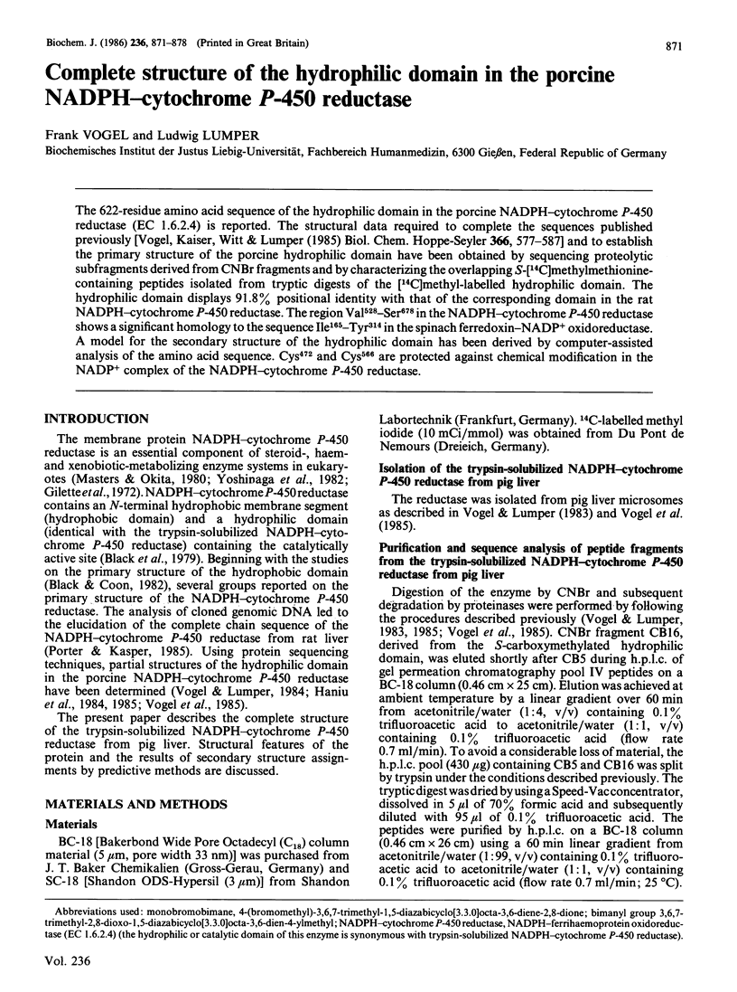
Selected References
These references are in PubMed. This may not be the complete list of references from this article.
- Black S. D., Coon M. J. Structural features of liver microsomal NADPH-cytochrome P-450 reductase. Hydrophobic domain, hydrophilic domain, and connecting region. J Biol Chem. 1982 May 25;257(10):5929–5938. [PubMed] [Google Scholar]
- Black S. D., French J. S., Williams C. H., Jr, Coon M. J. Role of a hydrophobic polypeptide in the N-terminal region of NADPH-cytochrome P-450 reductase in complex formation with P-450LM. Biochem Biophys Res Commun. 1979 Dec 28;91(4):1528–1535. doi: 10.1016/0006-291x(79)91238-5. [DOI] [PubMed] [Google Scholar]
- Chan R. L., Carrillo N., Vallejos R. H. Isolation and sequencing of an active-site peptide from spinach ferredoxin-NADP+ oxidoreductase after affinity labeling with periodate-oxidized NADP+. Arch Biochem Biophys. 1985 Jul;240(1):172–177. doi: 10.1016/0003-9861(85)90020-7. [DOI] [PubMed] [Google Scholar]
- Chou P. Y., Fasman G. D. Prediction of protein conformation. Biochemistry. 1974 Jan 15;13(2):222–245. doi: 10.1021/bi00699a002. [DOI] [PubMed] [Google Scholar]
- Dubourdieu M., Fox J. L. Amino acid sequence of Desulfovibrio vulgaris flavodoxin. J Biol Chem. 1977 Feb 25;252(4):1453–1463. [PubMed] [Google Scholar]
- Garnier J., Osguthorpe D. J., Robson B. Analysis of the accuracy and implications of simple methods for predicting the secondary structure of globular proteins. J Mol Biol. 1978 Mar 25;120(1):97–120. doi: 10.1016/0022-2836(78)90297-8. [DOI] [PubMed] [Google Scholar]
- Gillette J. R., Davis D. C., Sasame H. A. Cytochrome P-450 and its role in drug metabolism. Annu Rev Pharmacol. 1972;12:57–84. doi: 10.1146/annurev.pa.12.040172.000421. [DOI] [PubMed] [Google Scholar]
- Haniu M., Iyanagi T., Legesse K., Shively J. E. Structural analysis of NADPH-cytochrome P-450 reductase from porcine hepatic microsomes. Sequences of proteolytic fragments, cysteine-containing peptides, and a NADPH-protected cysteine peptide. J Biol Chem. 1984 Nov 25;259(22):13703–13711. [PubMed] [Google Scholar]
- Haniu M., Iyanagi T., Miller P., Shively J. E. Amino acid sequence of COOH-terminal 20K Da fragment from pig liver microsomal NADPH-cytochrome P-450 reductase. Biochem Biophys Res Commun. 1985 Feb 28;127(1):94–98. doi: 10.1016/s0006-291x(85)80130-3. [DOI] [PubMed] [Google Scholar]
- Hopp T. P., Woods K. R. Prediction of protein antigenic determinants from amino acid sequences. Proc Natl Acad Sci U S A. 1981 Jun;78(6):3824–3828. doi: 10.1073/pnas.78.6.3824. [DOI] [PMC free article] [PubMed] [Google Scholar]
- Karplus P. A., Walsh K. A., Herriott J. R. Amino acid sequence of spinach ferredoxin:NADP+ oxidoreductase. Biochemistry. 1984 Dec 18;23(26):6576–6583. doi: 10.1021/bi00321a046. [DOI] [PubMed] [Google Scholar]
- Knapp J. A., Dignam J. D., Strobel H. W. NADPH-cytochrome P-450 reductase. Circular dichroism and physical studies. J Biol Chem. 1977 Jan 25;252(2):437–443. [PubMed] [Google Scholar]
- Krauth-Siegel R. L., Blatterspiel R., Saleh M., Schiltz E., Schirmer R. H., Untucht-Grau R. Glutathione reductase from human erythrocytes. The sequences of the NADPH domain and of the interface domain. Eur J Biochem. 1982 Jan;121(2):259–267. doi: 10.1111/j.1432-1033.1982.tb05780.x. [DOI] [PubMed] [Google Scholar]
- Levitt M., Chothia C. Structural patterns in globular proteins. Nature. 1976 Jun 17;261(5561):552–558. doi: 10.1038/261552a0. [DOI] [PubMed] [Google Scholar]
- Lumper L., Busch F., Dzelić S., Henning J., Lazar T. Studies on the cosubstrate site of protease solubilized NADPH-cytochrome P450 reductase. Int J Pept Protein Res. 1980 Jul;16(1):83–96. doi: 10.1111/j.1399-3011.1980.tb02940.x. [DOI] [PubMed] [Google Scholar]
- Masters B. S., Okita R. T. The history, properties, and function of NADPH-cytochrome P-450 reductase. Pharmacol Ther. 1980;9(2):227–244. doi: 10.1016/s0163-7258(80)80020-9. [DOI] [PubMed] [Google Scholar]
- Möller W., Amons R. Phosphate-binding sequences in nucleotide-binding proteins. FEBS Lett. 1985 Jul 1;186(1):1–7. doi: 10.1016/0014-5793(85)81326-0. [DOI] [PubMed] [Google Scholar]
- Porter T. D., Kasper C. B. Coding nucleotide sequence of rat NADPH-cytochrome P-450 oxidoreductase cDNA and identification of flavin-binding domains. Proc Natl Acad Sci U S A. 1985 Feb;82(4):973–977. doi: 10.1073/pnas.82.4.973. [DOI] [PMC free article] [PubMed] [Google Scholar]
- Sasagawa T., Titani K., Walsh K. A. Selective isolation of methionine-containing peptides by hydrophobicity modulation. Anal Biochem. 1983 Feb 1;128(2):371–376. doi: 10.1016/0003-2697(83)90388-3. [DOI] [PubMed] [Google Scholar]
- Sheriff S., Herriott J. R. Structure of ferredoxin-NADP oxidoreductase and the location on the NADP binding site. Results at 3-7 A resolution. J Mol Biol. 1981 Jan 15;145(2):441–451. doi: 10.1016/0022-2836(81)90214-x. [DOI] [PubMed] [Google Scholar]
- Vogel F., Kaiser C., Witt I., Lumper L. NADPH-cytochrome P-450 reductase (pig liver). Studies on the sequence of the cyanogen bromide peptides from the catalytic domain and on the reactivity of the thiol groups. Biol Chem Hoppe Seyler. 1985 Jun;366(6):577–587. doi: 10.1515/bchm3.1985.366.1.577. [DOI] [PubMed] [Google Scholar]
- Vogel F., Lumper L. Fluorescence labelling of NADPH-cytochrome P-450 reductase with the monobromomethyl derivative of syn-9,10-dioxabimane. Biochem J. 1983 Oct 1;215(1):159–166. doi: 10.1042/bj2150159. [DOI] [PMC free article] [PubMed] [Google Scholar]
- Watenpaugh K. D., Sieker L. C., Jensen L. H. The binding of riboflavin-5'-phosphate in a flavoprotein: flavodoxin at 2.0-Angstrom resolution. Proc Natl Acad Sci U S A. 1973 Dec;70(12):3857–3860. doi: 10.1073/pnas.70.12.3857. [DOI] [PMC free article] [PubMed] [Google Scholar]
- Wood D., Darlison M. G., Wilde R. J., Guest J. R. Nucleotide sequence encoding the flavoprotein and hydrophobic subunits of the succinate dehydrogenase of Escherichia coli. Biochem J. 1984 Sep 1;222(2):519–534. doi: 10.1042/bj2220519. [DOI] [PMC free article] [PubMed] [Google Scholar]
- Yoshinaga T., Sassa S., Kappas A. The occurrence of molecular interactions among NADPH-cytochrome c reductase, heme oxygenase, and biliverdin reductase in heme degradation. J Biol Chem. 1982 Jul 10;257(13):7786–7793. [PubMed] [Google Scholar]


