Abstract
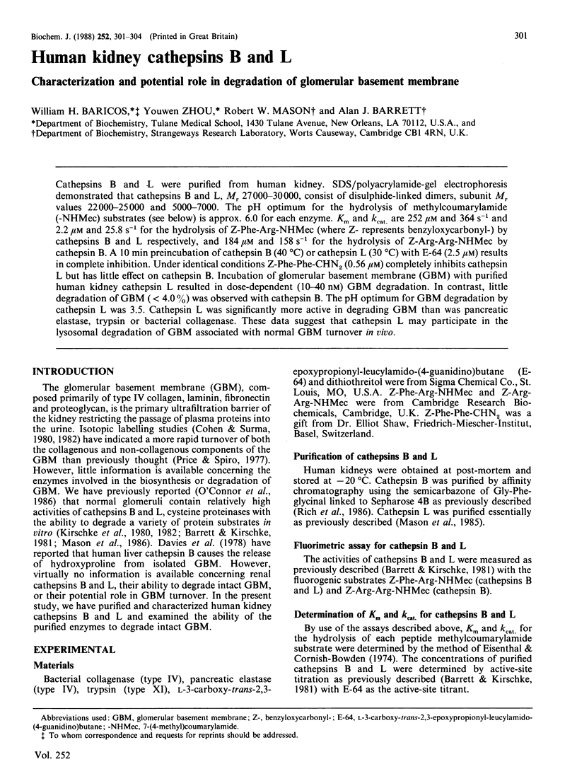
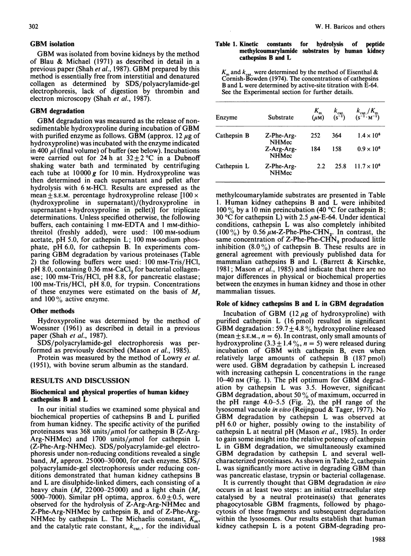
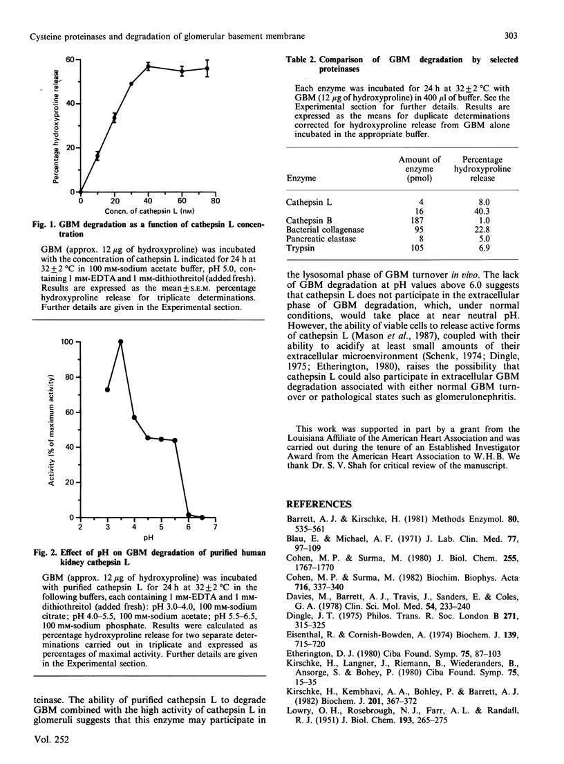
Cathepsins B and L were purified from human kidney. SDS/polyacrylamide-gel electrophoresis demonstrated that cathepsins B and L, Mr 27000-30000, consist of disulphide-linked dimers, subunit Mr values 22000-25000 and 5000-7000. The pH optimum for the hydrolysis of methylcoumarylamide (-NHMec) substrates (see below) is approx. 6.0 for each enzyme. Km and kcat. are 252 microM and 364s-1 and 2.2 microM and 25.8 s-1 for the hydrolysis of Z-Phe-Arg-NHMec (where Z- represents benzyloxycarbonyl-) by cathepsins B and L respectively, and 184 microM and 158 s-1 for the hydrolysis of Z-Arg-Arg-NHMec by cathepsin B. A 10 min preincubation of cathepsin B (40 degrees C) or cathepsin L (30 degrees C) with E-64 (2.5 microM) results in complete inhibition. Under identical conditions Z-Phe-Phe-CHN2 (0.56 microM) completely inhibits cathepsin L but has little effect on cathepsin B. Incubation of glomerular basement membrane (GBM) with purified human kidney cathepsin L resulted in dose-dependent (10-40 nM) GBM degradation. In contrast, little degradation of GBM (less than 4.0%) was observed with cathepsin B. The pH optimum for GBM degradation by cathepsin L was 3.5. Cathepsin L was significantly more active in degrading GBM than was pancreatic elastase, trypsin or bacterial collagenase. These data suggest that cathepsin L may participate in the lysosomal degradation of GBM associated with normal GBM turnover in vivo.
Full text
PDF


Selected References
These references are in PubMed. This may not be the complete list of references from this article.
- Barrett A. J., Kirschke H. Cathepsin B, Cathepsin H, and cathepsin L. Methods Enzymol. 1981;80(Pt 100):535–561. doi: 10.1016/s0076-6879(81)80043-2. [DOI] [PubMed] [Google Scholar]
- Blau E., Michael A. F. Rat glomerular basement membrane composition and metabolism in aminonucleoside nephrosis. J Lab Clin Med. 1971 Jan;77(1):97–109. [PubMed] [Google Scholar]
- Cohen M. P., Surma M. L. In vivo biosynthesis and turnover of 35S-labeled glomerular basement membrane. Biochim Biophys Acta. 1982 Jun 16;716(3):337–340. doi: 10.1016/0304-4165(82)90025-3. [DOI] [PubMed] [Google Scholar]
- Cohen M. P., Surma M. Renal glomerular basement membrane. In vivo biosynthesis and turnover in normal rats. J Biol Chem. 1980 Mar 10;255(5):1767–1770. [PubMed] [Google Scholar]
- Davies M., Barrett A. J., Travis J., Sanders E., Coles G. A. The degradation of human glomerular basement membrane with purified lysosomal proteinases: evidence for the pathogenic role of the polymorphonuclear leucocyte in glomerulonephritis. Clin Sci Mol Med. 1978 Mar;54(3):233–240. doi: 10.1042/cs0540233. [DOI] [PubMed] [Google Scholar]
- Eisenthal R., Cornish-Bowden A. The direct linear plot. A new graphical procedure for estimating enzyme kinetic parameters. Biochem J. 1974 Jun;139(3):715–720. doi: 10.1042/bj1390715. [DOI] [PMC free article] [PubMed] [Google Scholar]
- Etherington D. J. Proteinases in connective tissue breakdown. Ciba Found Symp. 1979;(75):87–103. doi: 10.1002/9780470720585.ch6. [DOI] [PubMed] [Google Scholar]
- Kirschke H., Kembhavi A. A., Bohley P., Barrett A. J. Action of rat liver cathepsin L on collagen and other substrates. Biochem J. 1982 Feb 1;201(2):367–372. doi: 10.1042/bj2010367. [DOI] [PMC free article] [PubMed] [Google Scholar]
- Kirschke H., Langner J., Riemann S., Wiederanders B., Ansorge S., Bohley P. Lysosomal cysteine proteinases. Ciba Found Symp. 1979;(75):15–35. doi: 10.1002/9780470720585.ch2. [DOI] [PubMed] [Google Scholar]
- LOWRY O. H., ROSEBROUGH N. J., FARR A. L., RANDALL R. J. Protein measurement with the Folin phenol reagent. J Biol Chem. 1951 Nov;193(1):265–275. [PubMed] [Google Scholar]
- Mason R. W., Gal S., Gottesman M. M. The identification of the major excreted protein (MEP) from a transformed mouse fibroblast cell line as a catalytically active precursor form of cathepsin L. Biochem J. 1987 Dec 1;248(2):449–454. doi: 10.1042/bj2480449. [DOI] [PMC free article] [PubMed] [Google Scholar]
- Mason R. W., Green G. D., Barrett A. J. Human liver cathepsin L. Biochem J. 1985 Feb 15;226(1):233–241. doi: 10.1042/bj2260233. [DOI] [PMC free article] [PubMed] [Google Scholar]
- Mason R. W., Johnson D. A., Barrett A. J., Chapman H. A. Elastinolytic activity of human cathepsin L. Biochem J. 1986 Feb 1;233(3):925–927. doi: 10.1042/bj2330925. [DOI] [PMC free article] [PubMed] [Google Scholar]
- Price R. G., Spiro R. G. Studies on the metabolism of the renal glomerular basement membrane. Turnover measurements in the rat with the use of radiolabeled amino acids. J Biol Chem. 1977 Dec 10;252(23):8597–8602. [PubMed] [Google Scholar]
- Reijngoud D. J., Tager J. M. The permeability properties of the lysosomal membrane. Biochim Biophys Acta. 1977 Nov 14;472(3-4):419–449. doi: 10.1016/0304-4157(77)90005-3. [DOI] [PubMed] [Google Scholar]
- Rich D. H., Brown M. A., Barrett A. J. Purification of cathepsin B by a new form of affinity chromatography. Biochem J. 1986 May 1;235(3):731–734. doi: 10.1042/bj2350731. [DOI] [PMC free article] [PubMed] [Google Scholar]
- Schenk R. K. 3. Ultrastruktur des Knochens (Referat) Verh Dtsch Ges Pathol. 1974;58:72–83. [PubMed] [Google Scholar]
- Shah S. V., Baricos W. H., Basci A. Degradation of human glomerular basement membrane by stimulated neutrophils. Activation of a metalloproteinase(s) by reactive oxygen metabolites. J Clin Invest. 1987 Jan;79(1):25–31. doi: 10.1172/JCI112790. [DOI] [PMC free article] [PubMed] [Google Scholar]
- WOESSNER J. F., Jr The determination of hydroxyproline in tissue and protein samples containing small proportions of this imino acid. Arch Biochem Biophys. 1961 May;93:440–447. doi: 10.1016/0003-9861(61)90291-0. [DOI] [PubMed] [Google Scholar]


