Abstract
Non-small cell lung cancer (NSCLC), representing 85% of lung cancer cases, is characterized by its heterogeneity and progression through distinct stages. This study applied Weighted Gene Co-expression Network Analysis (WGCNA) to explore the molecular mechanisms of NSCLC and identify potential therapeutic targets. Gene expression data from the GEO database were analyzed across four NSCLC stages (NSCLC1, NSCLC2, NSCLC3, and NSCLC4), with the NSCLC2 dataset selected as the reference for module preservation analysis. WGCNA identified eight highly preserved modules—Cyan, Yellow, Red, Dark Turquoise, Turquoise, White, Purple, and Royal Blue—across datasets, which were enriched in key pathways such as “Cell cycle” and “Pathways in cancer”, involving processes like cell division and inflammatory responses. Hub genes, including PLK1, CDK1, and EGFR, emerged as critical regulators of tumor proliferation and immune responses. Estrogen receptor ESR1 was also highlighted, correlating with improved survival outcomes, suggesting its potential as a prognostic marker. Signature-based drug repurposing analysis identified promising therapeutic candidates, including GW-5074, which inhibits RAF and disrupts the EGFR–RAS–RAF–MEK–ERK signaling cascade, and olomoucine, a CDK1 inhibitor. Additional candidates like pinocembrin, which reduces NSCLC cell invasion by modulating epithelial-mesenchymal transition, and citalopram, an SSRI with anti-carcinogenic properties, were also identified. These findings provide valuable insights into the molecular underpinnings of NSCLC and suggest new directions for therapeutic strategies through drug repurposing.
Keywords: non-small cell lung cancer (NSCLC), WGCNA, KEGG pathways, module preservation, drug repurposing, GW-5074, olomoucine, pinocembrin, citalopram, cell cycle regulation, protein binding, estrogen
1. Introduction
NSCLC, or non-small cell lung cancer, accounts for over 85% of all cases of lung cancer and is one of the most common types [1,2]. This form of cancer is known for its heterogeneous nature, distinguishing it from small cell lung cancer (SCLC) by its typically slower progression and varied presentation. NSCLC is broadly classified into several subtypes based on histological characteristics and the anatomical locations of tumor development. The most common type, adenocarcinoma, often occurs in the lungs’ outer regions and arises from the glandular cells that border the alveoli [3]. Squamous cell carcinoma is typically found in the core regions of the lungs and develops from the squamous cells that border the airways [4]. Large cell carcinoma, while less common, can appear in any lung area and is noted for its rapid growth and tendency to spread quickly [5]. The progression of NSCLC is categorized into four distinct stages, each reflecting the extent of disease spread and influencing treatment decisions. Stage I NSCLC is categorized into two sub-stages, 1A and 1B, primarily based on tumor size. At this stage, the cancer remains localized to the lungs and has not yet invaded nearby lymph nodes or other body parts. Stage II is separated into IIA and IIB substages, with subcategories that consider tumor size, location, and lymph node involvement. While the tumor has not spread to distant organs, tumors in this stage may be larger than those in stage I and may have begun to damage neighboring lymph nodes. In stage III, which is subdivided into IIIA, IIIB, and IIIC, the cancer’s extent is assessed by tumor size, location, and the degree of lymph node spread, often involving the lymph nodes in the mediastinum, the region between the lungs. Stage IV represents the most advanced form of NSCLC, characterized by metastasis to the lining of the lungs or other distant sites in the body [6].
Non-small cell lung cancer (NSCLC) is treated using a range of therapies, including surgery, chemotherapy, radiation therapy, targeted therapy, and immunotherapy, modified to the stage of cancer, the patient’s overall health, and specific mutations found in the tumor [7]. Surgery is frequently the preferred therapy for early-stage NSCLC (stages I–IIIA), with a target of completely removing the tumor. Chemotherapy is widely utilized in advanced stages (IIIB and IV), either as neoadjuvant therapy to decrease tumors before surgery or as adjuvant therapy to eradicate remaining cancer cells after surgery. Cisplatin, carboplatin, paclitaxel, docetaxel, gemcitabine, and pemetrexed are common chemotherapy medicines frequently used in combination to enhance efficacy [8]. Radiation therapy can be used to treat patients who are unable to undergo surgery, as well as to provide palliative care to alleviate symptoms caused by tumor growth. Targeted therapy is very effective in advanced NSCLC, as it focuses on specific genetic abnormalities in cancer cells. Key targeted therapies include RET inhibitors like selpercatinib for RET rearrangements, ALK inhibitors like alectinib and crizotinib for ALK gene alterations, and EGFR inhibitors like osimertinib and erlotinib for EGFR-mutated cancers. BRAF inhibitors like dabrafenib and MET inhibitors like capmatinib are also utilized to treat particular mutations [9,10]. Overall, the therapy landscape for NSCLC is diverse, with several modalities suited to particular patient profiles and tumor features.
This study employs Weighted Gene Co-expression Network Analysis (WGCNA) to gain a better understanding of the molecular pathways underlying non-small cell lung cancer (NSCLC) and to discover possible treatment targets. Using gene expression data analysis, WGCNA is a bioinformatics technique that may be used to discover gene groups with similar expression patterns and build networks of co-expressed genes. Through the association of these gene groups with clinical characteristics, WGCNA offers insights into the molecular mechanisms underpinning distinct phases of the disease [11]. Four datasets of NSCLC—NSCLC1 (GSE19804), NSCLC2 (GSE43580), NSCLC3 (GSE101929), and NSCLC4 (GSE19804)—were examined using data from the Gene Expression Omnibus (GEO). Gene modules that are consistent across various datasets were identified with the help of WGCNA, and their biological relevance was determined using functional annotation and pathway enrichment analysis. To identify significant hub genes in each module, protein–protein interaction networks were also built. This could aid in the discovery of possible drug candidates [12].
With this bioinformatics approach, the study aims to shed light on the genetic differences and similarities across various stages of NSCLC. This understanding could reveal important insights that may contribute to enhancing current treatment strategies or developing new therapeutic options for NSCLC patients.
2. Materials and Methods
2.1. Dataset Acquisition
Microarray gene expression datasets for use in WGCNA analysis were obtained from the Gene Expression Omnibus (NCBI GEO) of the National Center for Biotechnology Information (https://www.ncbi.nlm.nih.gov/geo/, accessed on 25 July 2024). These datasets included primary lung tumor samples from patients with different stages of non-small cell lung cancer (NSCLC)—Stages 1 (NSCLC1), 2 (NSCLC2), 3 (NSCLC3), and 4 (NSCLC4)—and contained expression data. To ensure consistency and minimize variability due to differences in hybridization settings, probe sequences, and other platform-specific characteristics, only datasets generated with the GPL570-HG-U133 Plus 2 Affymetrix Human Genome U133 Plus 2.0 Array were selected [13]. A total of 74 samples were obtained. An overview of the information and sample distribution for each of the four NSCLC stages can be seen in Table 1.
Table 1.
GEO datasets summary.
The Bioconductor affy package (Bioconductor v3.18, http://www.bioconductor.org, accessed on 25 July 2024) provides an extensive toolset for analyzing Affymetrix microarray data [14]. It includes functions for reading Affymetrix CEL files, preprocessing the data, and performing various analyses. The robust multi-array average (RMA) method was used to process all screened raw data, which involved background correction, quantile normalization, and log-2 transformation. To visually inspect the resulting data for irregularities, sample clustering dendrograms and a boxplot of expression values were created [15]. After removing control probes and applying variance filtering, the expression datasets were subset to include only common probes, focusing on lung tissue tumor expression samples while eliminating non-biological variation. The datasets were further refined by retaining only similar rows and removing data entries lacking gene symbols. Probe IDs were converted to gene symbols using the hgu133plus2.db database and the AnnotationDbi function. Finally, only probes present in all datasets were used, and samples without values after log-2 transformation were removed using the ‘goodSamplesGenesMs’ function from the WGCNA R package (R version 4.4.0).
2.2. Weighted Gene Co-Expression Network Analysis (WGCNA)
2.2.1. Estimating a Scale-Free Network
The ‘pickSoftThreshold’ function from the WGCNA R package was employed to assess the adherence of network data to a scale-free topology. This function generated a plot that evaluated the fit of the data to a scale-free topology model across a range of power levels (1 to 20). In a scale-free network, characterized by a power-law distribution, a minority of nodes (genes) have a large number of connections, while most have relatively few connections. The optimal power level (β) was identified as the lowest value at which the network begins to approximate a scale-free topology. This was determined by locating the point where further increases in power did not significantly improve the fit of the data to the scale-free model [16]. To validate the scale-free nature of the network, the relationship between the logarithm of the number of connections (log connectivity) and the logarithm of the probability of those connections (log connectivity probability) was analyzed. A linear trend in this plot would suggest that the network follows a scale-free distribution [17]. The plot of scale-free topology fit versus soft-thresholding power was examined to identify the point where the fit plateaus or reaches a high value. Additionally, the values of soft connectivity (k) were plotted to confirm the appropriateness of the selected power level (β).
2.2.2. Module Identification and Network Construction Using TOM
Pearson’s correlation was used to calculate the strength of the correlation between genes. Using the network type ‘signed’, this analysis considers the direction of the correlation (positive or negative). The Topological Overlap Measure (TOM) was used to evaluate the similarity of connection patterns between gene pairs within the network. Adjacency matrices were created to represent the strength of correlations between genes, which were adjusted using the chosen power level to emphasize strong correlations and downweight weak ones. The flashClust function was used to group genes into clusters based on their similarity, and the hclust function was employed to cut the dendrograms into distinct clusters (modules) of highly correlated genes. Hierarchical clustering was performed using the ‘average’ method, based on the distance matrix of the expression profiles. Different values (0 to 3) were tested using the cutreeHybrid function to determine the optimal level for splitting the dendrogram branches [18]. Consistent clusters over a range of deep split parameter values indicate successful detection of relevant gene modules.
2.2.3. Module Preservation Analysis
Gene modules from NSCLC1, NSCLC2, NSCLC3, and NSCLC4 were analyzed using the modulePreservation function from the WGCNA R package, configured with a ‘signed’ network type to account for the direction of gene correlations. To evaluate the stability and significance of module preservation, 100 random permutations were performed, and only modules containing at least 30 genes were included in the analysis. Module membership (kME), quantifying the correlation between each gene and its module’s overall expression profile, was calculated using the moduleEigengenes function, which generates a representative expression profile (eigengene) for each module. kME values for each gene were then computed based on their correlation with the module eigengene. This approach evaluates whether gene connection patterns observed under specific conditions, such as different disease states, are consistent across other conditions [19]. The stability and relevance of these gene modules were assessed by identifying key genes with strong correlations to the overall module expression profile.
2.3. Enrichment Analysis and KEGG Pathway
The Database for Annotation, Visualization, and Integrated Discovery (DAVID) was used for functional annotation clustering to assess the relevance of gene modules. The analysis incorporated three Gene Ontology (GO) categories: Biological Processes (BP), describing the roles and processes the genes are involved in; Cellular Components (CC), indicating the cellular locations of the genes or their products; and Molecular Functions (MF), detailing the activities of the gene products, such as binding or catalytic activity. The classification stringency was set to medium, grouping genes based on their functions with moderate strictness. Only GO terms with enrichment scores higher than 1.3 and an adjusted p-value of less than 0.05 were considered significant. Additionally, pathway enrichment analysis was conducted using the Kyoto Encyclopedia of Genes and Genomes (KEGG) database to identify pathways in which the genes are involved. Significant KEGG terms were pathways that were significantly enriched and clustered with the selected GO terms. These significant KEGG and GO terms helped to understand the broader biological functions and roles of the gene modules, providing insights into the activity locations of the genes, their functions, and the biological processes they participate in.
2.4. Protein–Protein Interaction and Hub Genes Identification
Protein–protein interaction (PPI) networks were generated for each of the highly preserved modules using Cytoscape v3.10.2. The networks were constructed by importing data from the public database STRING using the protein query as the data source, with a minimum cutoff score of 0.7 to ensure high confidence. After generating these networks in Cytoscape, the CytoHubba tool (v0.1.) was used to identify hub genes by evaluating their degree of centrality, which indicates the number of interactions a gene has in the network and its importance relative to other genes [20,21,22]. Hub genes were defined as the top five genes in each module with the highest number of interactions.
2.5. Signature-Based Drug Repurposing
The top 10 hub genes were first categorized as ‘upregulated’ or ‘downregulated’ using GEO2R (https://www.ncbi.nlm.nih.gov/geo/geo2r/, accessed on 25 July 2024) based on degree centrality. Following this classification, transcription profiles from the Molecular Signatures Database (MSigDB) and Connectivity Map (CMap) were used to identify drug candidates for repurposing via the Drug Repurposing Encyclopedia (DRE) [23,24]. The lists of upregulated and downregulated genes were submitted for drug repurposing analysis using molecular signature screening. Only drugs with clearly indicated mechanisms of action and false discovery rates (FDR) below 0.05 were included in the final analysis, while experimental drugs without specified mechanisms of action were excluded [15].
2.6. Language Refinement with AI Tools
In this study, AI tools, specifically OpenAI’s ChatGPT (GPT-4o mini) and Grammarly (v1.2.0.0), were used to assist in the revision of the manuscript for grammar correction and sentence construction. These tools were employed to improve the clarity and readability of the text after experimental content had been fully developed. No AI tools were used for data analysis, interpretation of results, or shaping conclusions. No research data or images provided here were generated or created with the assistance of any AI tools.
3. Results
3.1. Weighted Gene Co-Expression Network Analysis
3.1.1. Data Pre-Processing and Scale-Free Network Estimation
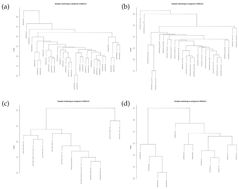
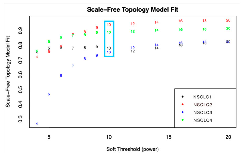
After preparing the data and filtering the genes, a total of 27,872 genes remained for further processing. The sample clustering dendrograms for each dataset, shown in Figure 1, revealed no clear outliers. Figure 2 depicts the variation of the scale-free topology fit index versus the soft-thresholding power (β) ranging from 1 to 20, illustrating the Scale-Free Topology Model Fit. The model starts to stabilize and form a linear trend when the power (β) reaches 10. Powers greater than 10 do not significantly improve the fit, as a power of 10 already provides a good scale-free structure. To construct biologically significant gene co-expression networks with minimal noise and information loss, it is crucial to approximate the ideal soft-thresholding power in WGCNA [25,26]. Generally, the soft-thresholding power decreases rapidly with increasing values, so selecting the lowest power that meets the scale-free topology standards is beneficial.
Figure 1.
Sample clustering dendrograms for (a) NSCLC1, (b) NSCLC2, (c) NSCLC3, (d) NSCLC4.
Figure 2.
The soft-thresholding power estimated from the four datasets was found using the network index plot and Scale-Free Topology Model Fit. The power at which the index began to plateau is indicated by the blue box.
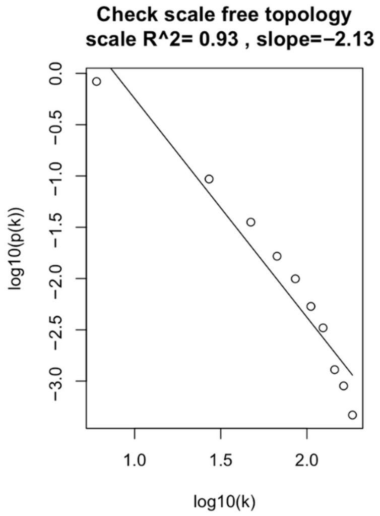
Figure 3 shows the log-log plot of the NSCLC2 dataset, illustrating the frequency of different connectivity values with a power value of 10. Among all datasets, the NSCLC2 dataset demonstrated the best results with an R2 value of 0.93. This high R2 value indicates strong gene connections and reliable results with clear patterns. Consequently, the NSCLC2 dataset was selected as the reference dataset for further analysis.
Figure 3.
The log-log plot of the approximated linear relationship for the NSCLC2 dataset at β = 10.
3.1.2. Identification of Modules Using TOM-Based Network Construction
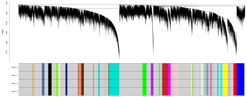
In TOM-based network construction and module identification, it is crucial to identify a reference dataset and project the module eigengenes of other datasets onto it. Module eigengenes, calculated as the first principal component of the expression data for all genes within a module, serve as summary statistics capturing the overall expression level of the module. Standardizing module definitions and improving biological interpretation are essential for facilitating cross-dataset analysis and meta-analyses [27]. By projecting the module eigengenes of different datasets onto a reference dataset (in this case, NSCLC2), cross-dataset comparisons and biological interpretations are enhanced. Consequently, NSCLC2 was chosen as the reference dataset for meta-analysis, and the identified modules are depicted in Figure 4.
Figure 4.
Dendrogram demonstrating module split sensitivity and gene clustering on TOM-based dissimilarity in NSCLC2. The different colored sections show the detected gene co-expression modules that match the area of the dendrogram.
In the NSCLC2 dataset analyzed using WGCNA, a total of 48 gene co-expression modules were discovered and categorized by color. The largest module, “grey”, contained 2500 genes, followed by “turquoise” with 1023 genes and “blue” with 744 genes. The “yellow” module had 440 genes, while the “brown” module included 575 genes. Other notable modules were “red” (321 genes), “black” (320 genes), and “green” (393 genes). Smaller modules, such as “floralwhite” (30 genes), “lightsteelblue1” (35 genes), and “ivory” (31 genes), were also identified. The diversity in module sizes reflects the impact of sample size and clustering resolution on network robustness. These modules represent groups of related genes with coordinated expression patterns across NSCLC stages, providing insights into the molecular processes and regulatory networks involved. Additionally, associating module eigengenes with external traits or clinical outcomes helps infer the biological significance of the modules and enhances the overall interpretation of the findings [28].
3.2. Module Preservation Analysis
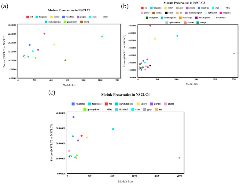
The preservation of gene co-expression network modules in non-small cell lung cancer stage 2 (NSCLC2) was evaluated across different stages of NSCLC (NSCLC1, NSCLC3, NSCLC4) using the highest Z-summary score for better preservation assessment. Only modules with a Z-score ≥ 10 across all datasets were considered significant [29]. Figure 5 illustrates the preservation of different modules across stages. Minor variations were observed between the preserved modules across datasets. Eight modules with high Z-scores were identified across all datasets: Red, Turquoise, Yellow, Royal Blue, Purple, Cyan, White, and Dark Turquoise. These modules are preserved across stages, indicating shared networks among different NSCLC stages. NSCLC3 exhibited the most significantly preserved modules compared to other stages, likely due to its transitional nature, where the cancer is more aggressive and may metastasize to nearby organs, potentially leading to NSCLC4 [24]. Eigengene-based connectivity (kME) was used to assess module membership and identify hub genes within co-expression modules. Genes with high kME values exhibit strong correlations with other genes within the same module and are ranked based on their connectivity. Top genes with the highest kME scores from each module were selected as promising biomarkers and therapeutic targets due to their high connectivity and association with disease progression and poor survival in NSCLC [27].
Figure 5.
Gene co-expression modules from the NSCLC2 network’s (a) NSCLC1, (b) NSCLC3, and (c) NSCLC4 datasets were subjected to a module preservation analysis. Highly maintained modules are those with a Z-score greater than or equal to 10.
3.3. Gene Ontology (GO) Terms and KEGG Pathway Analysis
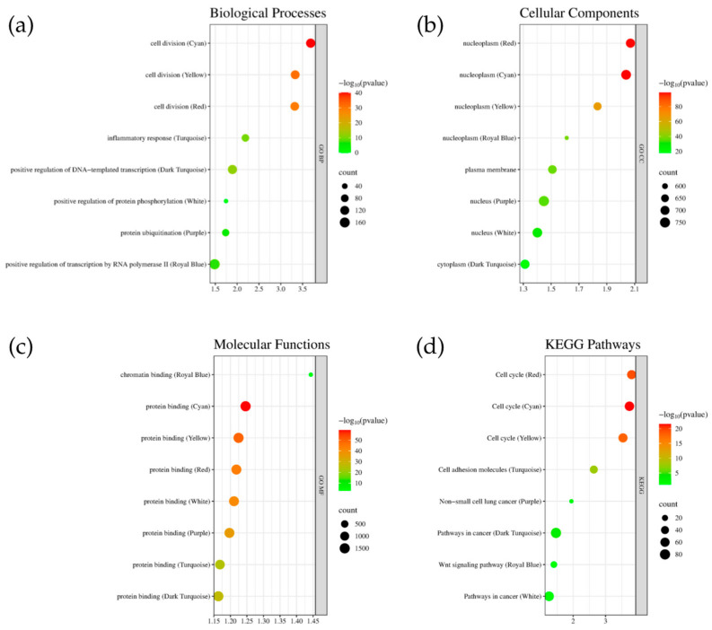
Figure 6 presents the GO terms and KEGG pathway analysis for the highly preserved genes in each module. The enrichment scores indicate significant connections to NSCLC progression. Specifically, the Cyan, Yellow, and Red modules are involved in cell division processes. The cellular components analysis shows that the Red, Cyan, and Yellow modules are all active in the nucleoplasm. Protein binding is prominent across all modules except Royal Blue. In the KEGG pathway analysis, the cell cycle is notably involved in the Red, Cyan, and Yellow modules. Additionally, the Purple pathway shows significant involvement in NSCLC, underscoring its relevance. The Dark Turquoise and White modules are associated with general ‘Pathways in Cancer’, with NSCLC being a specific type of lung cancer. Functional annotations were grouped using the DAVID webserver based on the top module.
Figure 6.
Top enriched terms for the Cyan, Yellow, Red, Turquoise, Dark Turquoise, White, Purple, and Royal Blue modules in terms of (a) biological processes, (b) cellular components, (c) molecular functions, and (d) KEGG pathways.
3.4. Protein–Protein Interaction Network and Key Hub Genes
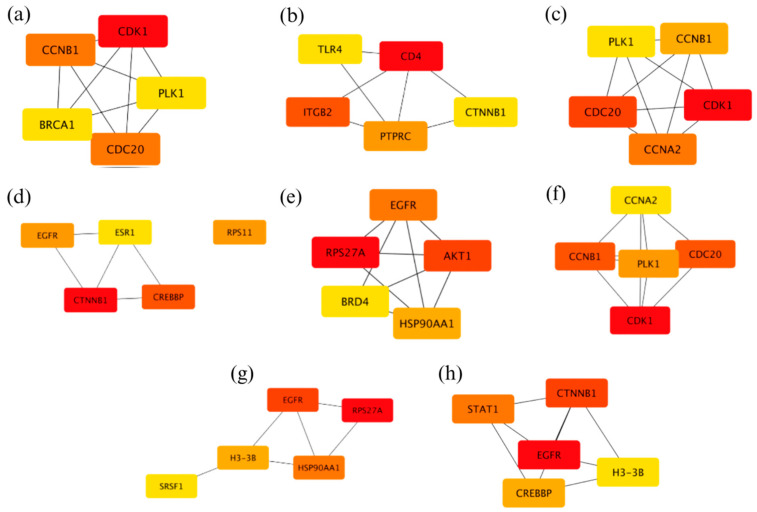
Using Cytoscape v3.10.2, the identified modules in gene co-expression networks were cross-referenced with PPI networks to validate and enhance the understanding of the underlying biological processes. A cutoff score of 0.7 was applied to ensure a high confidence level in the interactions [30]. The top five hub genes for each module were identified based on degree ranking using the Cytoscape CytoHubba plugin. Figure 7 displays these top five hub genes, with their rankings indicated by color intensity, where the brightest red represents the highest-ranking gene. After identifying the hub genes in the PPI network, the next step involved integrating these genes into broader signaling and regulatory networks. This integration helps elucidate their interactions with other pathways and their roles in the complex biological systems underlying NSCLC.
Figure 7.
Top five hub genes based on the PPI network of the modules (a) Red, (b) Turquoise, (c) Yellow, (d) Royal Blue, (e) Purple, (f) Cyan, (g) White, and (h) Dark Turquoise.
3.5. Signature-Based Drug Repurposing
Drugs that produce gene expression profiles inversely matched to the NSCLC hub gene signature were identified using DRE. These drugs hold potential for repurposing as they may counteract the distinctive expression patterns of hub genes associated with NSCLC. Table 2 presents a selection of the most promising drug candidates along with their relevant mechanisms of action. The false discovery rate (FDR) is a statistical metric that estimates the percentage of false positives among positive results, with lower FDR values indicating more reliable drug candidates [31]. In gene expression and drug development, a greater negative tau value often signifies a more pronounced and effective alteration in gene expression. However, tau values can have varying implications depending on the context [32]. The top-ranked drug candidates for upregulated expression include L-745870, pentolinium, GW-5074, pinocembrin, and bisphenol-A. For downregulated hub genes, the most promising therapeutic candidates are clopidogrel, RX-821002, FR-122047, olomoucine, and citalopram.
Table 2.
Top five drug candidates for the upregulated and downregulated hub genes for NSCLC.
| Expression | Genes | Drug | Mechanism | Tau | FDR |
|---|---|---|---|---|---|
| Upregulated | PLK1, EGFR, ESR1, CD4, CTNNB1, CREBBP, BRD4, AKT1 | L-745870 | Dopamine receptor antagonist | −99.8 | 0.000188 |
| Pentolinium | Cholinergic receptor antagonist | −99 | 0.00189 | ||
| Gw-5074 | Leucine-rich repeat kinase inhibitor, RAF inhibitor | −98.5 | 0.00481 | ||
| Pinocembrin | Cytochrome P450 inhibitor | −98 | 0.00208 | ||
| Bispherol-A | Synthetic estrogen | −97.5 | 0.000159 | ||
| Downregulated | CDK1, CDC20, RPS11, CCNA2, STAT1, SRSF1, BRCA1, CD20, CTNNB1, TLR4, ITGB2, PTPRC, RPS27A, HSP90AA1 | Clopidogrel | Purinergic receptor antagonist | −99.8 | 0.00396 |
| Rx-821002 | Adrenergic receptor antagonist | −99.8 | 0.00416 | ||
| Fr-122047 | Cyclooxygenase inhibitor | −99.6 | 0.00295 | ||
| Olomoucine | CDK indicator | −99.6 | 0.00596 | ||
| Citalopram | Selective serotonin reuptake inhibitor (SSRI) | −99.5 | 0.00596 |
4. Discussion
4.1. Gene Co-Expression Modules across the Datasets
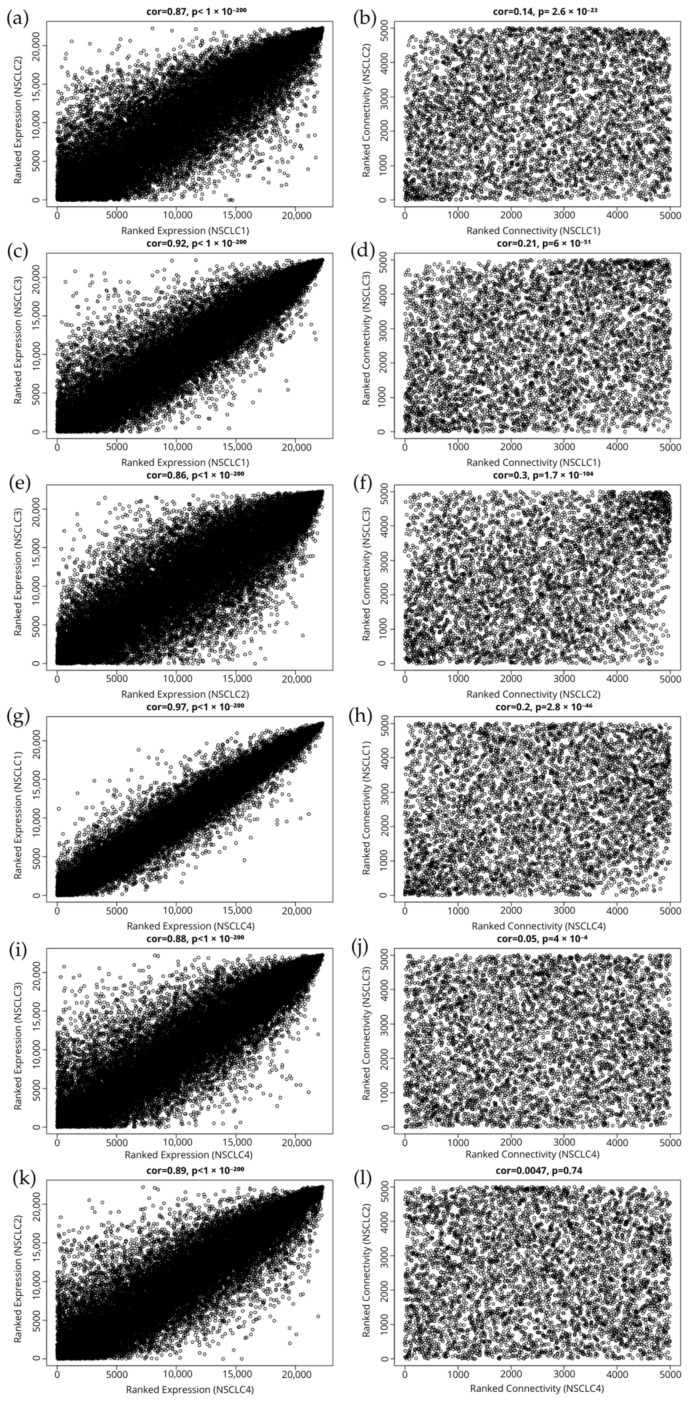
Weighted Gene Co-expression Network Analysis (WGCNA) is a powerful bioinformatics method for analyzing gene expression data, particularly in diseases such as non-small cell lung cancer (NSCLC). By identifying gene modules correlated with disease symptoms, WGCNA provides valuable insights into disease progression and potential treatment targets [33,34]. NSCLC is the most common type of lung cancer, accounting for approximately 85% of all lung cancer diagnoses [35]. The disease’s progression is often influenced by genetic abnormalities and changes in gene expression [36]. WGCNA constructs co-expression networks that reveal gene interactions and their impact on tumor activity. It also identifies pathways involved in disease networks by analyzing the interactions of highly preserved genes. This methodology supports the application of WGCNA to explore the disease network across various NSCLC stages, represented by the NSCLC1, NSCLC2, NSCLC3, and NSCLC4 datasets. NSCLC3, a transitional stage where the cancer is more aggressive and may spread to other organs [35], showed a higher number of preserved modules compared to other stages. This is reflected in Figure A1, which indicates a significant correlation (0.86) between NSCLC2 and NSCLC3, suggesting that despite representing different stages, their molecular characteristics overlap significantly. Enrichment analysis using DAVID revealed that the highly preserved modules Cyan, Yellow, and Red are predominantly involved in cell division, while the Turquoise module is associated with inflammatory responses (Figure 6a). The link between the Cyan, Yellow, and Red modules and cell division is particularly relevant for NSCLC, given the disease’s association with dysregulation of cell cycle processes. Research has shown that differentially expressed genes (DEGs) in NSCLC are significantly involved in various aspects of the cell cycle, including mitotic nuclear division and regulation [37,38]. The cellular component analysis (Figure 6b) highlights that modules like Dark Turquoise are primarily associated with nucleoplasm and cytoplasm. Alterations in nuclear body composition and function can disrupt gene expression regulation. For example, interactions between nuclear bodies and chromatin are crucial for maintaining transcriptional states. Highly transcribed genes often associate with proteins of the nuclear pore complex, facilitating RNA and protein transport between the nucleus and cytoplasm, which can impact cancer progression [39]. The cellular components of the nucleoplasm, particularly nuclear bodies, are integral to gene regulation and cellular responses, with significant implications for the pathophysiology of NSCLC. In general, proteins exhibit a wide range of binding capabilities, which are crucial for various biological functions. In terms of molecular function, all identified modules (Figure 6c) show significant involvement in protein binding. This interaction is essential for numerous biological processes, including signal transduction, enzymatic activity, and structural integrity within cells [40]. In NSCLC, protein binding is critical for tumor progression and metastasis. For example, proteins involved in EGFR signaling pathways are crucial for NSCLC development and maintenance. Ligand binding to EGFR can activate downstream signaling pathways that promote cell proliferation and survival, contributing to cancer progression [41]. A key limitation of this study is the small sample sizes for the NSCLC3 and NSCLC4 stages, which include only 11 and 12 samples, respectively. These limited numbers may affect the general application of the findings, as smaller samples can reduce statistical power and may not adequately represent the diversity within these stages. This limitation could impact the reliability of the identified gene modules and potential therapeutic targets, particularly regarding their applicability to a wider range of patients. While the results provide important insights into the molecular mechanisms of NSCLC progression, it is essential to exercise caution when applying these findings to the broader NSCLC population. To strengthen the conclusions drawn from this study, future research should focus on larger and more balanced sample sizes across all NSCLC stages. This approach will help validate and refine the findings, ultimately contributing to more effective treatment strategies for NSCLC.
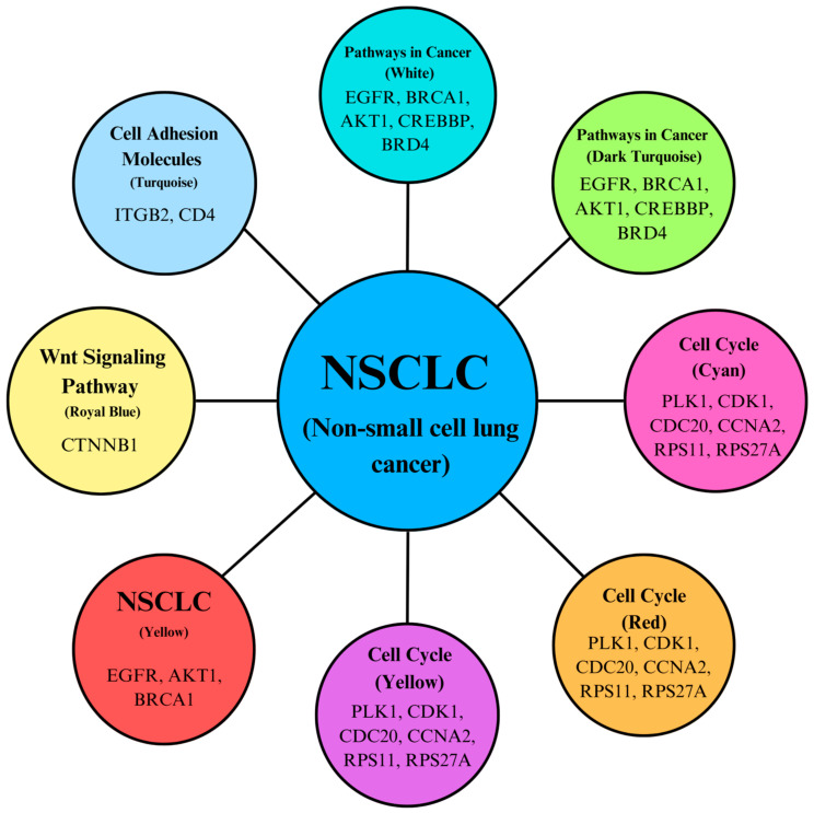
KEGG pathway analysis, as shown in Figure 8, highlights the hub genes associated with each pathway within the modules. The significant enrichment of the “Cell cycle” pathway underscores the dysregulation of cell cycle control mechanisms in NSCLC, which can lead to uncontrolled cell division. A study emphasizes the importance of cell cycle regulators, such as CDK1 and cyclins, in NSCLC, indicating that targeting these regulators can inhibit tumor growth and induce apoptosis in NSCLC cells [42]. The “Pathways in cancer” enrichment reflects the involvement of multiple signaling pathways in NSCLC tumorigenesis. Research by Cui et al. discusses the role of various oncogenes and tumor suppressor genes in NSCLC, including mutations in EGFR, K-RAS, and p53, which are critical in cancer pathways. These genetic alterations lead to uncontrolled cell proliferation and survival, highlighting their significance in NSCLC progression [43]. The “non-small cell lung cancer” pathway specifically addresses the molecular mechanisms underlying NSCLC, including key genetic alterations. The KEGG pathway analysis provides a comprehensive view of signaling pathways involved in NSCLC, emphasizing the roles of K-RAS mutations and EGFR overexpression in promoting cell proliferation and survival. This reinforces the relevance of these pathways in understanding NSCLC biology. Additionally, the “Cell adhesion molecules” pathway highlights the importance of cell–cell and cell–matrix interactions in NSCLC, which can impact tumor invasion and metastasis [42]. The study examines how alterations in cell adhesion molecules contribute to NSCLC metastasis, suggesting that targeting these interactions may help inhibit tumor spread and improve patient outcomes. This analysis suggests that there may be interactions, cross-talk, or amplification within these pathways that contribute to NSCLC progression.
Figure 8.
KEGG pathways interconnectivity based on the functions of key hub genes.
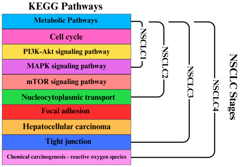
Additionally, the increased preservation of modules per stage is demonstrated by Figure 9, which displays the top KEGG pathways per module that could potentially act as significant point in the progression of NSCLC. Modules that reached the threshold of z ≥ 10 per stage were all considered in this figure. A study published in April 2024 demonstrated that targeted inhibition of the PI3K/AKT/mTOR pathway using specific compounds led to cell cycle arrest, apoptosis, and autophagy in NSCLC cells. This suggests that mTOR is a viable target for therapeutic intervention in NSCLC, providing a rationale for developing mTOR inhibitors as part of treatment strategies [44]. Studies have shown that alterations in nucleocytoplasmic transport mechanisms can lead to the aberrant localization of oncogenes and tumor suppressor proteins. For example, mutations in genes like p53 can disrupt normal transport processes, leading to enhanced cell proliferation and survival, which are hallmarks of NSCLC [45]. Research has identified specific genes within the focal adhesion pathway that are upregulated in NSCLC. A study found that genes such as VWF, CAV1, and ITGA8 were significantly enriched in the focal adhesion pathway and associated with poor prognosis in NSCLC patients [43]. This is also why it is present in the late stages of NSCLC, as shown in Figure 9. These findings suggest that focal adhesion signaling may play a critical role in the aggressiveness of NSCLC tumors. According to research, the PI3K/Akt/mTOR signaling pathway—which is essential for cell growth and survival—is one of the oncogenic pathways shared by both HCC and NSCLC. In both cancers, deviations in these pathways might result in the growth of tumors. According to a study, NSCLC and HCC pathway-related genes were found to be highly expressed in each other, indicating a possible molecular connection between the two cancers [43]. Tight junctions are crucial for maintaining epithelial integrity and regulating paracellular permeability. In NSCLC, alterations in tight junction proteins can lead to increased invasiveness and metastasis. A study indicated that the dysregulation of tight junctions contributes to the epithelial–mesenchymal transition (EMT), a process associated with enhanced migratory and invasive properties of cancer cells [46]. The pathway details how chemical carcinogens can induce oxidative stress, leading to DNA damage and mutations, which are critical in the initiation and progression of lung cancer. A study by Henkler et al. (2010) discusses the role of oxidative stress in carcinogenesis induced by metals and xenobiotics, emphasizing the importance of ROS in cancer development, including lung cancer [47]. These findings could lead to new research studies in NSCLC, including computational modeling, laboratory experiments, and clinical studies.
Figure 9.
Modules in KEGG pathways with increased preservation for each NSCLC stage (NSCLC1, NSCLC2, NSCLC3, NSCLC4).
4.2. Module Hub Genes and Their Functions
A number of hub genes play crucial roles in the pathophysiology and progression of non-small cell lung cancer (NSCLC), affecting various cellular processes. These genes can be categorized based on their functions in ribosomal protein synthesis, transcription, immune response, signal transduction, cell cycle regulation, and DNA damage repair. The hub genes associated with each module and their functions are listed in Table A1, derived from the GeneCards database (www.genecards.org, accessed 29 July 2024) [48].
4.2.1. Cell Cycle Regulation and Proliferation
The analysis of gene co-expression networks and module preservation in NSCLC datasets unveiled crucial insights into the disease’s pathophysiology. Weighted Gene Co-expression Network Analysis (WGCNA) identified several significant gene modules across different stages of NSCLC, with prominent modules related to cell cycle regulation and mitotic progression. These findings were further validated by KEGG pathway analysis, which highlighted the “Cell cycle” pathway as significantly enriched in modules such as Red, Cyan, and Yellow (Figure 8).
Polo-like kinase 1 (PLK1), identified as a hub gene in the context of mitotic regulation, was found to be a key player in the modules associated with cell cycle processes. Elevated PLK1 expression has been associated with poor prognosis and enhanced cell proliferation in NSCLC [49]. This is supported by the results observed in the module preservation analysis, where modules associated with cell division and mitosis showed high preservation across different stages of NSCLC (Figure 4 and Figure 5). Elevated PLK1 levels align with the observed enrichment in the “Cell cycle” pathway, reinforcing its role as a critical factor in NSCLC progression and a potential therapeutic target [50,51]. Targeting PLK1 with inhibitors, such as volasertib, has shown potential in improving NSCLC cells’ radiosensitivity [52,53]. Cyclin-dependent kinase 1 (CDK1) and Cell Division Cycle 20 (CDC20) were also identified as hub genes within modules associated with mitotic progression. Their dysregulation, particularly in the context of the G2/M transition, aligns with the results from the functional annotation clustering and pathway enrichment analyses. Modules Cyan, Yellow, and Red, which were highly preserved and enriched in cell cycle processes, underscore the critical role of these regulators in uncontrolled proliferation. The significant enrichment of the “Cell cycle” pathway across these modules highlights the importance of CDK1 and CDC20 in NSCLC, as their dysregulation contributes to tumor aggressiveness and resistance to treatment [54]. Cyclin A2 (CCNA2), involved in regulating the S phase and G2/M transition, was similarly identified as a crucial hub gene within the modules associated with cell cycle processes. The correlation between CCNA2’s role in cell cycle regulation and the observed enrichment in the “Cell cycle” pathway further supports the relevance of these findings in understanding NSCLC biology. Overall, the results from the module preservation analysis, KEGG pathway enrichment, and hub gene identification converge to highlight the central role of mitotic regulators and cell cycle processes in NSCLC. The significant preservation of these modules across datasets and their association with key biological pathways emphasize the potential of targeting these regulators, such as PLK1, CDK1, and CDC20, for therapeutic intervention in NSCLC.
While the study successfully identifies key pathways and genes associated with non-small cell lung cancer (NSCLC), a significant limitation is the absence of experimental functional validation to confirm the biological significance of these findings. Without such validation, the roles of the identified genes and pathways remain speculative. Functional studies, such as gene knockdown or overexpression experiments, are crucial for substantiating the biological relevance of these genes in NSCLC. For instance, research has demonstrated the impact of specific genes on tumor progression through gene knockdown experiments, revealing their roles in cell proliferation and apoptosis [55]. Similarly, overexpression studies have shown how certain mutations, such as those in the EGFR gene, can promote aggressive tumor behavior [56]. These experiments provide direct evidence of how these genes influence cancer-related processes. Future research should focus on conducting these functional studies to validate the computational predictions and deepen the understanding of the molecular mechanisms driving NSCLC. This validation is essential to move from theoretical insights to practical applications in developing targeted therapies, as confirmed roles of these genes will guide the development of more effective treatment strategies.
4.2.2. Signal Transduction
Non-small cell lung cancer (NSCLC) is frequently associated with mutations in EGFR, which affect up to 50% of East Asian populations and approximately 10% to 15% of cases in Western nations [57]. These mutations lead to uncontrolled cell growth and proliferation by activating downstream signaling pathways such as PI3K/AKT and MAPK [58]. The significant enrichment of the “Cell cycle” pathway observed in the study is closely linked to these signaling pathways. The PI3K/AKT/mTOR pathway is commonly altered in EGFR-mutant NSCLC. Research indicates that 67% of individuals with EGFR mutations exhibit basal activation of the AKT/mTOR axis [49]. Specific mutations within this pathway include: (1) PIK3CA mutations, which are present in 5–10% of EGFR-mutant NSCLC cases and confer resistance to EGFR TKIs, (2) loss of PTEN, which also results in resistance to EGFR inhibitors, and (3) AKT1 mutations, although less frequent, are notable for their impact on treatment resistance [59]. The identification of hub genes related to the PI3K/AKT/mTOR pathway supports these findings. AKT1, an essential effector in the PI3K pathway, was found to be a critical hub gene within modules associated with cell cycle regulation and mitotic progression. Its overactivation is linked to poor prognosis in NSCLC, aligning with the results that show its significant role in the enriched “Cell cycle” pathway [50]. Targeting AKT1 and other components of the PI3K pathway could potentially overcome EGFR TKI resistance, providing a valuable therapeutic strategy for NSCLC [60]. Moreover, BET inhibitors, which target BRD4 and disrupt its interaction with acetylated histones, have shown promising anti-tumor effects in preclinical NSCLC models [61]. The integration of these findings into the broader context of the study highlights the importance of targeting key signaling pathways and genetic alterations in NSCLC for effective treatment strategies.
4.2.3. Immune Response and Inflammation
The results from the gene co-expression network analysis and module preservation studies provide insights into the role of immune responses in NSCLC. Notably, the module preservation analysis identified several key modules related to immune function, including those associated with cell cycle regulation and mitotic progression. This association aligns with the preserved modules related to immune response identified in the analysis, suggesting a potential link between immune cell dynamics and the expression patterns of hub genes involved in cell cycle regulation.
The ability of CD4+ T cells to recognize neoantigens from mutations in oncogenes like HER2 and KRAS further supports their role in targeting tumor-specific antigens [62,63]. This capability complements the findings from KEGG pathway analysis, which highlighted significant enrichment in pathways related to cell cycle and mitotic regulation. The convergence of these results emphasizes the interplay between tumor cell proliferation and immune responses, underscoring the potential for therapeutic strategies that leverage CD4+ T cells to enhance anti-tumor immunity [64] while targeting key cell cycle regulators such as PLK1, CDK1, and CDC20. Thus, integrating immune response insights with molecular findings provides a comprehensive view of NSCLC progression and potential therapeutic approaches. A further limitation of this study is the absence of an analysis correlating the identified gene expression patterns with clinical data, such as patient survival rates or treatment responses. Integrating clinical data could substantially enhance the clinical relevance of the findings by establishing links between the molecular features identified and patient outcomes. Such an analysis would provide a more comprehensive understanding of the clinical implications of the identified genes and pathways, potentially guiding more personalized treatment strategies. Future studies should aim to incorporate clinical data to validate and expand upon the current findings, thereby offering deeper insights into the prognostic and therapeutic potential of the identified molecular targets.
4.2.4. The Influence of Estrogen on NSCLC Progression
It has been studied that estrogen receptors, in particular ESR1, contribute to the development of NSCLC. Research suggests that ESR1 could control a number of signaling pathways, such as those involved in cell division, migration, and invasion, which influence the behavior of tumors. For example, bioinformatics investigations have demonstrated a correlation between increased expression of genes implicated in crucial signaling pathways, like Wnt/β-Catenin and Notch, which are known to play important roles in the development of NSCLC, and higher ESR1 expression [65]. ESR1 expression levels were shown to classify patients into groups with different prognoses in a study that analyzed gene expression data from patients with non-small cell lung cancer. More specifically, compared to patients with lower expression levels, those with increased ESR1 expression showed superior overall survival rates [66]. This implies that ESR1 may function as a prognostic marker in non-small cell lung cancer in addition to influencing tumor biology. The estrogen pathway plays a major role in lung adenocarcinoma by influencing a number of cancer hallmarks. By inducing pro-inflammatory cytokines, attracting regulatory T cells, and generating an immunosuppressive tumor microenvironment, estrogen regulates immunological responses. These processes support the migration, metastasis, and survival of cancer cells [67].
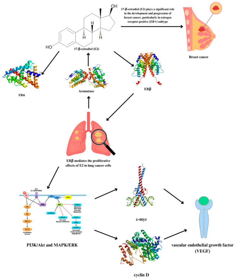
Interestingly, NSCLC cells can synthesize 17β-estradiol (E2) locally, similar to mechanisms observed in breast cancer. The two primary types of estrogen receptors via which E2 acts are ERα [68] and ERβ. ERα is expressed at lower levels in NSCLC, whereas ERβ is the more abundant protein. Research has demonstrated that ERβ mediates the proliferative effects of E2 in lung cancer cells, influencing the regulation of the cell cycle and promoting the formation of tumors via pathways such as PI3K/Akt and MAPK/ERK [69,70,71]. This receptor interaction increases the expression of genes associated with cell proliferation and survival, such as c-myc and cyclin D, and improves angiogenesis via vascular endothelial growth factor (VEGF) production [69,72].
On the other hand, aromatase, an enzyme that converts testosterone to estrogen, is substantially expressed in 44–86% of NSCLC tissues [72]. This is also linked to the biosynthesis of estrogen, particularly E2, which is a significant factor in the development and progression of breast cancer. Studies have shown that in postmenopausal breast cancer patients, the concentration of E2 in breast tissue can be significantly higher than in the plasma, suggesting that local estrogen production within tumors is a key factor in cancer progression [73,74]. A simple illustration of the process is shown in Figure 10.
Figure 10.
Local estrogen synthesis and estrogen receptor interaction in NSCLC and its potential metastasis to breast cancer.
4.3. Signature-Based Drug Repurposing
In the study of non-small cell lung cancer (NSCLC) progression, signature-based drug repurposing offers a promising approach by elucidating the complex interactions between various hub genes and their associated signaling pathways to identify potential therapeutic targets (Table 2). Preclinical studies have demonstrated that GW-5074 has antitumor benefits, particularly when combined with other RAF inhibitors such as sorafenib. This combination has shown synergistic effects, significantly increasing cell death in renal cell carcinoma cell lines compared to each drug alone [75]. GW-5074 inhibits RAF, disrupting the EGFR–RAS–RAF–MEK–ERK signaling cascade, which is crucial for cell proliferation and survival [76]. This suggests that GW-5074 could be effective against tumors with abnormal EGFR signaling due to mutations, such as those found in NSCLC. Additionally, one study discovered that GW5074 and sorafenib administration as part of a combination therapy considerably triggered necrotic death in various cancer cells in vivo and extended the survival of an animal disease model by dramatically suppressing primary and metastatic lesions. The study concluded that the co-administration of sorafenib with GW5074 showed anti-tumor effectiveness across a range of tumor types, in addition to a favorable safety profile [77]. Olomoucine may limit tumor growth by blocking both CDK1 and its downstream effects on CDC20, particularly in cancers with dysregulated pathways. In the context of non-small cell lung cancer (NSCLC), combining CDK inhibition with the resulting impact on CDC20 can be a strategic approach to limit the proliferation of cancer cells, potentially enhancing treatment effectiveness [78,79]. Pinocembrin treatment (50 μM) significantly reduced the number of migrating and invasive NSCLC cells by upregulating the protein level of E-cadherin and downregulating N-cadherin, vimentin and snail. This study shows that pinocembrin can prevent STAT3 from being activated, which is linked to EMT, and thereby reduce NSCLC cells’ ability to migrate and invade [80]. Citalopram is a member of the selective serotonin reuptake inhibitor (SSRI) class of antidepressants, which is used to treat depression [81]. Interestingly, a systematic review highlighted that antidepressants, including citalopram, have been associated with anti-carcinogenic effects through various mechanisms such as inducing apoptosis, inhibiting cell proliferation, and modifying immune responses. This review emphasizes the need for more clinical studies to better understand the role of antidepressants in cancer treatment, including NSCLC [82]. Overall, these potential drugs—GW-5074, olomoucine, pinocembrin, and citalopram—offer promising new avenues for treating NSCLC by targeting upregulated and downregulated hub genes identified in the study. By addressing the signaling pathways and genetic alterations associated with NSCLC, these repurposed drugs could contribute to more effective therapeutic strategies. These findings offer valuable insights into potential new targets for drug discovery and contribute to a deeper understanding of the underlying biology of NSCLC. While the study successfully identifies key hub genes and potential drug repurposing candidates for NSCLC, a significant limitation is the absence of experimental validation for these findings. Without in vitro or in vivo validation, the functional relevance and therapeutic potential of the identified genes and drugs remain unconfirmed. This limitation underscores the necessity for future experimental studies to validate the computational predictions and ensure their applicability in clinical settings. Experimental validation would provide critical insights into the mechanistic roles of these genes and the efficacy of the proposed drug candidates, thereby strengthening the overall conclusions of the study.
5. Conclusions
In conclusion, this study underscores the pivotal role of Weighted Gene Co-expression Network Analysis (WGCNA) in elucidating the progression of non-small cell lung cancer (NSCLC) and identifying potential therapeutic targets. The analysis reveals a substantial gene co-expression overlap between NSCLC3 and NSCLC2, the reference dataset, with a significant correlation suggesting shared molecular characteristics across different disease stages. This highlights the robustness of the findings and their applicability across NSCLC stages. Enrichment analysis identifies highly preserved modules—Cyan, Yellow, Red, and Turquoise—as predominantly involved in cell division and inflammatory responses, aligning with the dysregulation of cell cycle processes critical to NSCLC pathology. Key hub genes, such as PLK1, CDK1, and EGFR, emerge as crucial regulators of tumor proliferation, signal transduction, and immune response. The KEGG pathway analysis further emphasizes the significance of pathways like “Cell cycle”, “Pathways in cancer”, and “Cell adhesion molecules” in NSCLC progression, reinforcing the importance of these pathways in tumor development. The study also highlights the influence of estrogen receptors, particularly ESR1, on NSCLC biology. Elevated ESR1 expression correlates with improved survival outcomes, suggesting its utility as a prognostic marker. Additionally, the role of estrogen in regulating tumor biology through pathways such as PI3K/Akt and MAPK/ERK underscores its impact on cell proliferation and angiogenesis. The predominance of ERβ in NSCLC cells and its role in mediating estrogen effects further supports the need to consider hormonal factors in therapeutic strategies.
In terms of drug repurposing, the analysis identifies promising candidates such as GW-5074, which inhibits RAF and disrupts the EGFR–RAS–RAF–MEK–ERK signaling cascade, offering a potential strategy for targeting tumors with abnormal EGFR signaling. Preclinical studies have demonstrated that GW-5074 has antitumor benefits, particularly when combined with other RAF inhibitors such as sorafenib. This combination has shown synergistic effects, significantly increasing cell death in renal cell carcinoma cell lines compared to each drug alone. This suggests that GW-5074 could be effective against tumors with abnormal EGFR signaling due to mutations, such as those found in NSCLC. Additionally, one study discovered that GW-5074 and sorafenib administration as part of a combination therapy considerably triggered necrotic death in a variety of cancer cells in vivo and extended the survival of an animal disease model by dramatically suppressing primary and metastatic lesions. Additionally, olomoucine, by targeting CDK1 and CDC20, aligns with observed dysregulation in mitotic pathways, suggesting a strategic approach to limit cancer cell proliferation. In the context of NSCLC, combining CDK inhibition with the resulting impact on CDC20 can be a strategic approach to limit the proliferation of cancer cells, potentially enhancing treatment effectiveness. Furthermore, the study emphasizes the protective role of CD4+ T cells in NSCLC, with higher CD4+/total T cell ratios associated with improved clinical outcomes. The ability of CD4+ T cells to recognize tumor-specific antigens and their potential in immunotherapeutic strategies underscore the importance of integrating immune-based approaches into NSCLC treatment. Other potential drugs, such as pinocembrin, which reduces the number of migrating and invasive NSCLC cells by upregulating E-cadherin and downregulating N-cadherin, vimentin, and snail, show promise. This study shows that pinocembrin can prevent STAT3 from being activated, which is linked to EMT, and thereby reduce NSCLC cells’ ability to migrate and invade. Additionally, citalopram, a member of the selective serotonin reuptake inhibitor (SSRI) class of antidepressants, has been associated with anti-carcinogenic effects through various mechanisms such as inducing apoptosis, inhibiting cell proliferation, and modifying immune responses. This review emphasizes the need for more clinical studies to better understand the role of antidepressants in cancer treatment, including NSCLC.
Overall, this comprehensive analysis enhances the understanding of NSCLC biology and provides valuable directions for future research and clinical applications. The integration of gene co-expression networks, pathway enrichment, hormonal influences, and drug repurposing offers a robust framework for developing targeted and effective treatment strategies.
Acknowledgments
The authors would like to express their sincere gratitude to Nicholas Dale Talubo and Khy S. Suratos for helping with the data acquisition. They would also like to thank Mapua University for their support in the making of this research. Additionally, the authors acknowledge the use of OpenAI’s ChatGPT and Grammarly for improving the grammar and sentence structure during the preparation of this manuscript. These tools were used solely for language refinement and did not contribute to the scientific content or findings of the study.
Appendix A
Figure A1.
Ranked expression plots and ranked connectivity plots for dataset comparison (a,b) NSCLC1 vs. NSCLC2; (c,d) NSCLC1 vs. NSCLC3; (e,f) NSCLC2 vs. NSCLC3; (g,h) NSCLC4 vs. NSCLC1; (i,j) NSCLC4 vs. NSCLC3; and (k,l) NSCLC4 vs. NSCLC2.
Table A1.
Summary of hub genes and their protein functions from the GeneCards database.
| Gene | Protein | Function | Modules |
|---|---|---|---|
| PLK1 | Polo Like Kinase 1 | Because its depletion in cancer cells significantly reduced cell growth and caused apoptosis, cancer therapy targets this protein. | Red/Yellow/Cyan |
| EGFR | Epidermal Growth Factor Receptor | Lung cancer is linked to mutations in this gene. | Royal Blue/Purple/White/Dark Turquoise |
| ESR1 | Estrogen Receptor 1 | This gene encodes a receptor that is important in osteoporosis, endometrial cancer, and breast cancer. | Royal Blue |
| CD4 | CD4 Molecule | The protein stimulates or starts the early stage of T-cell activation and may be a key player in immune-mediated and viral disorders of the central nervous system that cause indirect neuronal damage. | Turquoise |
| CTNNB1 | Catenin β 1 | Colorectal cancer (CRC), pilomatrixoma (PTR), medulloblastoma (MDB), and ovarian cancer are all brought on by mutations in this gene. | Royal Blue/Dark Turquoise |
| CREBBP | CREB Binding Protein | This gene was first identified as a nuclear protein that binds to cAMP-response element-binding protein (CREB). Through the connection of chromatin remodeling to transcription factor recognition, it is now recognized to play important roles in embryonic development, growth control, and homeostasis. | Royal Blue/Dark Turquoise |
| BRD4 | Bromodomain Containing 4 | The chromosome 19 target of translocation t(15; 19)(q13; p13.1), which characterizes upper respiratory tract cancer in adolescents, has been linked to this gene. | Purple |
| CDK1 | Cyclin Dependent Kinase 1 | This protein’s phosphorylation and dephosphorylation play significant regulatory functions in the regulation of the cell cycle. | Red/Yellow/Cyan |
| CDC20 | Cell Division Cycle 20 | It appears that at various stages of the cell cycle, CDC20 functions as a regulatory protein through interactions with many other proteins. | Red/Yellow/Cyan |
| RPS11 | Ribosomal Protein S11 | This gene is distributed throughout the genome in several processed pseudogenes, as is common for genes encoding ribosomal proteins. | Royal Blue |
| CCNA2 | Cyclin A2 | This gene codes for a protein that is a member of the highly conserved cyclin family, which controls the cell cycle; adenocarcinoma and retinoblastoma are two diseases linked to CCNA2. | Yellow/Cyan |
| STAT1 | Signal Transducer and Activator of Transcription 1 | Receptor-associated kinases phosphorylate members of the STAT family in response to cytokines and growth factors. These members subsequently form homo- or heterodimers and translocate to the cell nucleus, where they function as transcription activators. | Dark Turquoise |
| SRSF1 | Serine And Arginine Rich Splicing Factor 1 | Depending on its contact partners and degree of phosphorylation, the encoded protein can either activate or suppress splicing. | White |
| BRCA1 | BRCA1 DNA Repair Associated | Hereditary breast and ovarian cancers are now identified by the presence of germline BRCA1 mutations. | Red |
| TLR4 | Toll Like Receptor 4 | The Toll-like receptor (TLR) family of proteins, which includes the protein produced by this gene, is essential for pathogen identification and the induction of innate immunity. | Turquoise |
| ITGB2 | Integrin Subunit β 2 | Integrins are essential proteins found on the cell surface that are involved in both cell adhesion and signaling mediated by the cell surface. | Turquoise |
| PTPRC | Protein Tyrosine Phosphatase Receptor Type C | Cell growth, differentiation, mitosis, and oncogenic transformation are just a few of the biological processes that PTPs are known to influence as signaling molecules. | Turquoise |
| RPS27A | Ribosomal Protein S27a | It is produced from a precursor protein that is either a single ubiquitin coupled to an unrelated protein or a polyubiquitin chain. | Royal Blue |
| HSP90AA1 | cluHeat Shock Protein 90 α Family Class A Member 1 | This gene codes for an inducible molecular chaperone protein, which is a homodimer. | Purple/White |
Table A2.
Top KEGG pathways from highly preserved modules (z > 10).
| Module | KEGG Term | Count | p-Value |
|---|---|---|---|
| Yellow | Metabolic pathways | 242 | 2.03 × 10⁻¹¹ |
| Pathways of neurodegeneration—multiple diseases | 78 | 1.14 × 10⁻⁴ | |
| Alzheimer disease | 72 | 2.45 × 10⁻⁶ | |
| Amyotrophic lateral sclerosis | 66 | 2.18 × 10⁻⁵ | |
| Cell cycle | 59 | 9.41 × 10⁻¹⁹ | |
| White | Pathways in cancer | 65 | 0.039962252 |
| Huntington disease | 41 | 0.038434334 | |
| Endocytosis | 39 | 0.003801247 | |
| Hepatocellular carcinoma | 32 | 3.90 × 10⁻⁴ | |
| Tight junction | 32 | 3.90 × 10⁻⁴ | |
| Purple | Metabolic pathways | 188 | 1.91 × 10⁻⁵ |
| Herpes simplex virus 1 infection | 84 | 2.43 × 10⁻⁷ | |
| Chemical carcinogenesis—reactive oxygen species | 31 | 0.0235166 | |
| Biosynthesis of cofactors | 29 | 2.67 × 10⁻⁴ | |
| Peroxisome | 25 | 2.02 × 10⁻⁷ | |
| Turquoise | Pathways in cancer | 85 | 2.89 × 10⁻⁶ |
| PI3K-Akt signaling pathway | 63 | 4.31 × 10⁻⁶ | |
| MAPK signaling pathway | 52 | 3.76 × 10⁻⁵ | |
| Rap1 signaling pathway | 45 | 6.60 × 10⁻⁷ | |
| Regulation of actin cytoskeleton | 44 | 1.50 × 10⁻⁵ | |
| Royal Blue | Endocytosis | 33 | 0.047955 |
| Cytoskeleton in muscle cells | 31 | 0.044336 | |
| Hepatocellular carcinoma | 24 | 0.04709 | |
| mTOR signaling pathway | 23 | 0.03917 | |
| Nucleocytoplasmic transport | 22 | 1.84 × 10⁻⁵ | |
| Red | Metabolic pathways | 202 | 2.29 × 10⁻⁶ |
| Pathways of neurodegeneration—multiple diseases | 62 | 0.015828 | |
| Cell cycle | 58 | 6.81 × 10⁻²⁰ | |
| Alzheimer disease | 54 | 0.006263 | |
| Amyotrophic lateral sclerosis | 51 | 0.008789 | |
| Cyan | Metabolic pathways | 240 | 4.52 × 10⁻⁹ |
| Pathways of neurodegeneration—multiple diseases | 84 | 1.69 × 10⁻⁵ | |
| Alzheimer disease | 77 | 3.46 × 10⁻⁷ | |
| Amyotrophic lateral sclerosis | 70 | 6.20 × 10⁻⁶ | |
| Cell cycle | 65 | 2.89 × 10⁻²² | |
| Dark Turquoise | Pathways in cancer | 80 | 3.49 × 10⁻⁴ |
| MAPK signaling pathway | 47 | 0.003029 | |
| Shigellosis | 40 | 0.004202 | |
| Focal adhesion | 39 | 1.49 × 10⁻⁴ | |
| Cytoskeleton in muscle cells | 39 | 0.002129 |
Author Contributions
Conceptualization, A.J.B.B., M.A.O. and L.L.T.; methodology, A.J.B.B., M.A.O. and L.L.T.; validation, M.A.O. and L.L.T.; formal analysis, A.J.B.B.; investigation, A.J.B.B. and M.A.O.; resources, A.J.B.B. and L.L.T.; data curation, A.J.B.B.; writing—original draft preparation, A.J.B.B.; writing—review and editing, A.J.B.B., M.A.O. and L.L.T.; visualization, A.J.B.B.; supervision, L.L.T. and P.-w.T.; funding acquisition, L.L.T. and P.-w.T. All authors have read and agreed to the published version of the manuscript.
Institutional Review Board Statement
Not applicable.
Informed Consent Statement
Not applicable.
Data Availability Statement
The gene microarray datasets used for the study are openly available in the NCBI Gene Expression Omnibus (GEO) database under the accession IDs GSE19804, GSE43580, and GSE101929 at https://www.ncbi.nlm.nih.gov/geo/ accessed on 25 July 2024.
Conflicts of Interest
The authors declare no conflicts of interest.
Funding Statement
This research received no external funding.
Footnotes
Disclaimer/Publisher’s Note: The statements, opinions and data contained in all publications are solely those of the individual author(s) and contributor(s) and not of MDPI and/or the editor(s). MDPI and/or the editor(s) disclaim responsibility for any injury to people or property resulting from any ideas, methods, instructions or products referred to in the content.
References
- 1.American Cancer Society What Is Lung Cancer? [(accessed on 5 July 2024)]. Available online: https://www.cancer.org/cancer/types/lung-cancer/about/what-is.html.
- 2.Siegel R.L., Miller K.D., Jemal A. Cancer statistics, 2016. CA Cancer J. Clin. 2016;66:7–30. doi: 10.3322/caac.21332. [DOI] [PubMed] [Google Scholar]
- 3.Sabbula B., Gasalberti D., Mukkamalla S. Squamous Cell Lung Cancer. StatPearls Publishing; Treasure Island, FL, USA: 2024. [PubMed] [Google Scholar]
- 4.Travis W.D., Brambilla E., Nicholson A.G., Yatabe Y., Austin J.H., Beasley M.B., Chirieac L.R., Dacic S., Duhig E., Flieder D.B., et al. The 2015 World Health Organization Classification of Lung Tumors: Impact of Genetic, Clinical and Radiologic Advances Since the 2004 Classification. J. Thorac. Oncol. 2015;10:1243–1260. doi: 10.1097/JTO.0000000000000630. [DOI] [PubMed] [Google Scholar]
- 5.Thomas T.H., Zimlich R. An Overview of Large Cell Lung Carcinoma (LCLC) Healthline; San Francisco, CA, USA: 2024. [Google Scholar]
- 6.American Lung Association Lung Cancer Staging. [(accessed on 5 July 2024)]. Available online: https://www.lung.org/lung-health-diseases/lung-disease-lookup/lung-cancer/symptoms-diagnosis/lung-cancer-staging.
- 7.NIH . Non-Small Cell Lung Cancer Treatment (PDQ®)–Health Professional Version. National Cancer Institute; Rockville, MD, USA: 2024. [Google Scholar]
- 8.Chen S., Qiao Y., Chen J., Li Y., Xie J., Cui P., Huang Z., Huang D., Gao Y., Hu Y., et al. Evolutions in the management of non-small cell lung cancer: A bibliometric study from the 100 most impactful articles in the field. Front. Oncol. 2022;12:939838. doi: 10.3389/fonc.2022.939838. [DOI] [PMC free article] [PubMed] [Google Scholar]
- 9.Ali A. Advances in Non-Small Cell Lung Cancer (NSCLC) Treatment—A Paradigm Shift in Oncology. Pharmaceuticals. 2024;17:246. doi: 10.3390/ph17020246. [DOI] [PMC free article] [PubMed] [Google Scholar]
- 10.Houda I., Dickhoff C., Uyl-de Groot C.A., Reguart N., Provencio M., Levy A., Dziadziuszko R., Pompili C., Di Maio M., Thomas M., et al. New systemic treatment paradigms in resectable non-small cell lung cancer and variations in patient access across Europe. Lancet Reg. Health Eur. 2024;38:100840. doi: 10.1016/j.lanepe.2024.100840. [DOI] [PMC free article] [PubMed] [Google Scholar]
- 11.Langfelder P., Horvath S. WGCNA: An R package for weighted correlation network analysis. BMC Bioinform. 2008;9:559. doi: 10.1186/1471-2105-9-559. [DOI] [PMC free article] [PubMed] [Google Scholar]
- 12.Basar M.A., Hosen M.F., Paul B.K., Hasan M.R., Shamim S.M., Bhuyian T. Identification of drug and protein-protein interaction network among stress and depression: A bioinformatics approach. Inform. Med. Unlocked. 2023;37:101174. doi: 10.1016/j.imu.2023.101174. [DOI] [Google Scholar]
- 13.Bhattacharya A., Bense R.D., Urzúa-Traslaviña C.G., de Vries E.G.E., van Vugt M.A.T.M., Fehrmann R.S.N. Transcriptional effects of copy number alterations in a large set of human cancers. Nat. Commun. 2020;11:715. doi: 10.1038/s41467-020-14605-5. [DOI] [PMC free article] [PubMed] [Google Scholar]
- 14.Rainer J., Sanchez-Cabo F., Stocker G., Sturn A., Trajanoski Z. CARMAweb: Comprehensive R- and bioconductor-based web service for microarray data analysis. Nucleic Acids. Res. 2006;34:W498–W503. doi: 10.1093/nar/gkl038. [DOI] [PMC free article] [PubMed] [Google Scholar]
- 15.Orda M.A., Fowler P.M.P.T., Tayo L.L. Modular Hub Genes in DNA Microarray Suggest Potential Signaling Pathway Interconnectivity in Various Glioma Grades. Biology. 2024;13:206. doi: 10.3390/biology13040206. [DOI] [PMC free article] [PubMed] [Google Scholar]
- 16.Hidalgo C.A., Barabasi A.-L. Scale-free networks. Scholarpedia. 2008;3:1716. doi: 10.4249/scholarpedia.1716. [DOI] [Google Scholar]
- 17.Wei H., Wang J., Wang T. An Improved B-A Model for Scale-free Network Evolution Model. J. Phys. Conf. Ser. 2020;1486:022034. doi: 10.1088/1742-6596/1486/2/022034. [DOI] [Google Scholar]
- 18.Langfelder P., Zhang B., Horvath S. Defining clusters from a hierarchical cluster tree: The Dynamic Tree Cut package for R. Bioinformatics. 2008;24:719–720. doi: 10.1093/bioinformatics/btm563. [DOI] [PubMed] [Google Scholar]
- 19.Fuller T.F., Ghazalpour A., Aten J.E., Drake T.A., Lusis A.J., Horvath S. Weighted gene coexpression network analysis strategies applied to mouse weight. Mamm. Genome. 2007;18:463–472. doi: 10.1007/s00335-007-9043-3. [DOI] [PMC free article] [PubMed] [Google Scholar]
- 20.Blake J.A., Harris M.A. The Gene Ontology (GO) Project: Structured Vocabularies for Molecular Biology and Their Application to Genome and Expression Analysis. Curr. Protoc. Bioinform. 2008;23:7.2.1–7.2.9. doi: 10.1002/0471250953.bi0702s23. [DOI] [PubMed] [Google Scholar]
- 21.Chin C.-H., Chen S.-H., Wu H.-H., Ho C.-W., Ko M.-T., Lin C.-Y. cytoHubba: Identifying hub objects and sub-networks from complex interactome. BMC Syst. Biol. 2014;8:S11. doi: 10.1186/1752-0509-8-S4-S11. [DOI] [PMC free article] [PubMed] [Google Scholar]
- 22.Li C.Y., Cai J.-H., Tsai J.J.P., Wang C.C.N. Identification of Hub Genes Associated with Development of Head and Neck Squamous Cell Carcinoma by Integrated Bioinformatics Analysis. Front. Oncol. 2020;10:681. doi: 10.3389/fonc.2020.00681. [DOI] [PMC free article] [PubMed] [Google Scholar]
- 23.Espinoza J.L., Shah N., Singh S., Nelson K.E., Dupont C.L. Applications of weighted association networks applied to compositional data in biology. Environ. Microbiol. 2020;22:3020–3038. doi: 10.1111/1462-2920.15091. [DOI] [PubMed] [Google Scholar]
- 24.Wexler A. Stages of Lung Cancer for Both NSCLC and SCLC. Medical News Today. Sep 1, 2023.
- 25.Zhou M., Guo R., Wang Y.-F., Yang W., Li R., Lu L. Application of Weighted Gene Coexpression Network Analysis to Identify Key Modules and Hub Genes in Systemic Juvenile Idiopathic Arthritis. Biomed. Res. Int. 2021;2021:9957569. doi: 10.1155/2021/9957569. [DOI] [PMC free article] [PubMed] [Google Scholar]
- 26.Manuel M.T.A., Tayo L.L. Navigating the Gene Co-Expression Network and Drug Repurposing Opportunities for Brain Disorders Associated with Neurocognitive Impairment. Brain. Sci. 2023;13:1564. doi: 10.3390/brainsci13111564. [DOI] [PMC free article] [PubMed] [Google Scholar]
- 27.Mo Z., Jiang X., Zhang Y., Zhai M., Hu L., Xuan J. Weighted Gene Co-Expression Network Analysis Reveals Key Pathways and Hub Genes Associated with Successful Grafting in Pecan (Carya illinoinensis) Forests. 2023;14:835. doi: 10.3390/f14040835. [DOI] [Google Scholar]
- 28.Garcia J.P.T., Tayo L.L. Theoretical Studies of DNA Microarray Present Potential Molecular and Cellular Interconnectivity of Signaling Pathways in Immune System Dysregulation. Genes. 2024;15:393. doi: 10.3390/genes15040393. [DOI] [PMC free article] [PubMed] [Google Scholar]
- 29.Mailem R.C., Tayo L.L. Drug Repurposing Using Gene Co-Expression and Module Preservation Analysis in Acute Respiratory Distress Syndrome (ARDS), Systemic Inflammatory Response Syndrome (SIRS), Sepsis, and COVID-19. Biology. 2022;11:1827. doi: 10.3390/biology11121827. [DOI] [PMC free article] [PubMed] [Google Scholar]
- 30.Kosoglu K., Aydin Z., Tuncbag N., Gursoy A., Keskin O. Structural coverage of the human interactome. Brief. Bioinform. 2023;25:bbad496. doi: 10.1093/bib/bbad496. [DOI] [PMC free article] [PubMed] [Google Scholar]
- 31.Alka O., Shanthamoorthy P., Witting M., Kleigrewe K., Kohlbacher O., Röst H.L. DIAMetAlyzer allows automated false-discovery rate-controlled analysis for data-independent acquisition in metabolomics. Nat. Commun. 2022;13:1347. doi: 10.1038/s41467-022-29006-z. [DOI] [PMC free article] [PubMed] [Google Scholar]
- 32.Stein-O’Brien G.L., Palaganas R., Meyer E.M., Redding-Ochoa J., Pletnikova O., Guo H., Bell W.R., Troncoso J.C., Huganir R.L., Morris M. Transcriptional Signatures of Hippocampal Tau Pathology in Primary Age-Related Tauopathy and Alzheimer’s Disease. medRxiv. 2023 doi: 10.1101/2023.09.12.23295440. [DOI] [Google Scholar]
- 33.Darzi M., Gorgin S., Majidzadeh-A K., Esmaeili R. Gene co-expression network analysis reveals immune cell infiltration as a favorable prognostic marker in non-uterine leiomyosarcoma. Sci. Rep. 2021;11:2339. doi: 10.1038/s41598-021-81952-8. [DOI] [PMC free article] [PubMed] [Google Scholar]
- 34.Luo X., Feng L., Xu W., Bai X., Wu M. Weighted gene co-expression network analysis of hub genes in lung adenocarcinoma. Evol. Bioinform. Online. 2021;17:11769343211009898. doi: 10.1177/11769343211009898. [DOI] [PMC free article] [PubMed] [Google Scholar]
- 35.Petrella F., Rizzo S., Attili I., Passaro A., Zilli T., Martucci F., Bonomo L., Del Grande F., Casiraghi M., De Marinis F., et al. Stage III Non-Small-Cell Lung Cancer: An Overview of Treatment Options. Curr. Oncol. 2023;30:3160–3175. doi: 10.3390/curroncol30030239. [DOI] [PMC free article] [PubMed] [Google Scholar]
- 36.Thandra K.C., Barsouk A., Saginala K., Aluru J.S., Barsouk A. Epidemiology of lung cancer. Contemp. Oncol. 2021;25:45–52. doi: 10.5114/wo.2021.103829. [DOI] [PMC free article] [PubMed] [Google Scholar]
- 37.Yin Y., Li D., He M., Wang J. The identification of hub biomarkers and pathways in lung cancer and prognostic evaluation. Transl. Cancer Res. 2022;11:2622–2635. doi: 10.21037/tcr-22-245. [DOI] [PMC free article] [PubMed] [Google Scholar]
- 38.Erkin Ö.C., Cömertpay B., Göv E. Integrative Analysis for Identification of Therapeutic Targets and Prognostic Signatures in Non-Small Cell Lung Cancer. Bioinform. Biol. Insights. 2022;16:11779322221088796. doi: 10.1177/11779322221088796. [DOI] [PMC free article] [PubMed] [Google Scholar]
- 39.Van de Vosse D.W., Wan Y., Wozniak R.W., Aitchison J.D. Role of the nuclear envelope in genome organization and gene expression. Wiley Interdiscip. Rev. Syst. Biol. Med. 2011;3:147–166. doi: 10.1002/wsbm.101. [DOI] [PMC free article] [PubMed] [Google Scholar]
- 40.Johnson A., Lewis J., Alberts B. Molecular Biology of the Cell. 4th ed. NIH; Bethesda, MD, USA: 2002. [Google Scholar]
- 41.Cao L., Coventry B., Goreshnik I., Huang B., Sheffler W., Park J.S., Jude K.M., Marković I., Kadam R.U., Verschueren K.H., et al. Design of protein-binding proteins from the target structure alone. Nature. 2022;605:551–560. doi: 10.1038/s41586-022-04654-9. [DOI] [PMC free article] [PubMed] [Google Scholar]
- 42.Ye X., Gao Q., Wu J., Zhou L., Tao M. Identification of significant genes in non-small cell lung cancer by bioinformatics analyses. Transl. Cancer Res. 2020;9:4330–4340. doi: 10.21037/tcr-19-2596. [DOI] [PMC free article] [PubMed] [Google Scholar]
- 43.Cui S., Huang F., Yu F., Jian S., Zhang L., Liu X., Chen S., Zhou Y., Xiao L. Identification of genes and pathways leading to poor prognosis of non-small cell lung cancer using integrated bioinformatics analysis. Transl. Cancer Res. 2022;11:710–724. doi: 10.21037/tcr-21-1986. [DOI] [PMC free article] [PubMed] [Google Scholar]
- 44.Li X.-Q., Cheng X.-J., Wu J., Wu K.-F., Liu T. Targeted inhibition of the PI3K/AKT/mTOR pathway by (+)-anthrabenzoxocinone induces cell cycle arrest, apoptosis, and autophagy in non-small cell lung cancer. Cell. Mol. Biol. Lett. 2024;29:58. doi: 10.1186/s11658-024-00578-6. [DOI] [PMC free article] [PubMed] [Google Scholar]
- 45.Genome.jp Non-Small Cell Lung Cancer—Homo Sapiens (Human) [(accessed on 5 July 2024)]. Available online: https://www.genome.jp/pathway/hsa05223.
- 46.Jin X., Liu X., Zhang Z., Guan Y., Xv R., Li J. Identification of key pathways and genes in lung carcinogenesis. Oncol. Lett. 2018;16:4185–4192. doi: 10.3892/ol.2018.9203. [DOI] [PMC free article] [PubMed] [Google Scholar]
- 47.Henkler F., Brinkmann J., Luch A. The Role of Oxidative Stress in Carcinogenesis Induced by Metals and Xenobiotics. Cancers. 2010;2:376–396. doi: 10.3390/cancers2020376. [DOI] [PMC free article] [PubMed] [Google Scholar]
- 48.Stelzer G., Rosen N., Plaschkes I., Zimmerman S., Twik M., Fishilevich S., Stein T.I., Nudel R., Lieder I., Mazor Y., et al. The GeneCards Suite: From Gene Data Mining to Disease Genome Sequence Analyses. Curr. Protoc. Bioinform. 2016;54:1–30. doi: 10.1002/cpbi.5. [DOI] [PubMed] [Google Scholar]
- 49.Colicino E.G., Hehnly H. Regulating a key mitotic regulator, polo-like kinase 1 (PLK1) Cytoskeleton. 2018;75:481–494. doi: 10.1002/cm.21504. [DOI] [PMC free article] [PubMed] [Google Scholar]
- 50.Lee S.-Y., Jang C., Lee K.-A. Polo-like kinases (plks), a key regulator of cell cycle and new potential target for cancer therapy. Dev. Reprod. 2014;18:65–71. doi: 10.12717/DR.2014.18.1.065. [DOI] [PMC free article] [PubMed] [Google Scholar]
- 51.Kim T. Recent Progress on the Localization of PLK1 to the Kinetochore and Its Role in Mitosis. Int. J. Mol. Sci. 2022;23:5252. doi: 10.3390/ijms23095252. [DOI] [PMC free article] [PubMed] [Google Scholar]
- 52.Van den Bossche J., Domen A., Peeters M., Deben C., De Pauw I., Jacobs J., De Bruycker S., Specenier P., Pauwels P., Vermorken J.B., et al. Radiosensitization of Non-Small Cell Lung Cancer Cells by the Plk1 Inhibitor Volasertib Is Dependent on the p53 Status. Cancers. 2019;11:1893. doi: 10.3390/cancers11121893. [DOI] [PMC free article] [PubMed] [Google Scholar]
- 53.Liu J., Lu K.-H., Liu Z.-L., Sun M., De W., Wang Z.-X. MicroRNA-100 is a potential molecular marker of non-small cell lung cancer and functions as a tumor suppressor by targeting polo-like kinase 1. BMC Cancer. 2012;12:519. doi: 10.1186/1471-2407-12-519. [DOI] [PMC free article] [PubMed] [Google Scholar]
- 54.Otto T., Sicinski P. Cell cycle proteins as promising targets in cancer therapy. Nat. Rev. Cancer. 2017;17:93–115. doi: 10.1038/nrc.2016.138. [DOI] [PMC free article] [PubMed] [Google Scholar]
- 55.Chen K., Chen C., Tseng Y., Chang Y., Chang M. GCIP functions as a tumor suppressor in non-small cell lung cancer by suppressing Id1-mediated tumor promotion. Oncotarget. 2014;5:5017–5028. doi: 10.18632/oncotarget.2075. [DOI] [PMC free article] [PubMed] [Google Scholar]
- 56.Zeng Y., Guo T., Zhou Y., Zhao Y., Chu L., Chu X., Yang X., Ni J., Zhu Z. Clinical outcomes of advanced non-small cell lung cancer patients harboring distinct subtypes of EGFR mutations and receiving first-line tyrosine kinase inhibitors: Brain metastasis and de novo T790M matters. BMC Cancer. 2022;22:198. doi: 10.1186/s12885-022-09245-5. [DOI] [PMC free article] [PubMed] [Google Scholar]
- 57.Farnsworth D.A., Chen Y.T., de Rappard Yuswack G., Lockwood W.W. Emerging Molecular Dependencies of Mutant EGFR-Driven Non-Small Cell Lung Cancer. Cells. 2021;10:3553. doi: 10.3390/cells10123553. [DOI] [PMC free article] [PubMed] [Google Scholar]
- 58.Rosell R., Cardona A.F., Arrieta O., Aguilar A., Ito M., Pedraz C., Codony-Servat J., Santarpia M. Coregulation of pathways in lung cancer patients with EGFR mutation: Therapeutic opportunities. Br. J. Cancer. 2021;125:1602–1611. doi: 10.1038/s41416-021-01519-2. [DOI] [PMC free article] [PubMed] [Google Scholar]
- 59.Sanaei M.-J., Razi S., Pourbagheri-Sigaroodi A., Bashash D. The PI3K/Akt/mTOR pathway in lung cancer; oncogenic alterations, therapeutic opportunities, challenges, and a glance at the application of nanoparticles. Transl. Oncol. 2022;18:101364. doi: 10.1016/j.tranon.2022.101364. [DOI] [PMC free article] [PubMed] [Google Scholar]
- 60.Yip P.Y. Phosphatidylinositol 3-kinase-AKT-mammalian target of rapamycin (PI3K-Akt-mTOR) signaling pathway in non-small cell lung cancer. Transl. Lung Cancer Res. 2015;4:165. doi: 10.3978/j.issn.2218-6751.2015.01.04. [DOI] [PMC free article] [PubMed] [Google Scholar]
- 61.Wang Z.-Q., Zhang Z.C., Wu Y.Y., Pi Y.N., Lou S.H., Liu T.B., Lou G., Yang C. Bromodomain and extraterminal (BET) proteins: Biological functions, diseases, and targeted therapy. Signal. Transduct. Target. Ther. 2023;8:420. doi: 10.1038/s41392-023-01647-6. [DOI] [PMC free article] [PubMed] [Google Scholar]
- 62.Yang X., Li Q., Zeng T. Peripheral CD4+ T cells correlate with response and survival in patients with advanced non-small cell lung cancer receiving chemo-immunotherapy. Front. Immunol. 2024;15:1364507. doi: 10.3389/fimmu.2024.1364507. [DOI] [PMC free article] [PubMed] [Google Scholar]
- 63.Tay R.E., Richardson E.K., Toh H.C. Revisiting the role of CD4+ T cells in cancer immunotherapy—New insights into old paradigms. Cancer Gene. Ther. 2021;28:5–17. doi: 10.1038/s41417-020-0183-x. [DOI] [PMC free article] [PubMed] [Google Scholar]
- 64.Veatch J.R., Jesernig B.L., Kargl J., Fitzgibbon M., Lee S.M., Baik C., Martins R., Houghton A.M., Riddell S.R. Endogenous CD4+ T Cells Recognize Neoantigens in Lung Cancer Patients, Including Recurrent Oncogenic KRAS and ERBB2 (Her2) Driver Mutations. Cancer Immunol. Res. 2019;7:910–922. doi: 10.1158/2326-6066.CIR-18-0402. [DOI] [PMC free article] [PubMed] [Google Scholar]
- 65.Gao X., Cai Y., Wang Z., He W., Cao S., Xu R., Chen H. Estrogen receptors promote NSCLC progression by modulating the membrane receptor signaling network: A systems biology perspective. J. Transl. Med. 2019;17:308. doi: 10.1186/s12967-019-2056-3. [DOI] [PMC free article] [PubMed] [Google Scholar]
- 66.Aresti U., Carrera S., Iruarrizaga E., Fuente N., Marrodan I., de Lobera A.R., Munoz A., Buque A., Condori E., Ugalde I., et al. Estrogen Receptor 1 Gene Expression and Its Combination with Estrogen Receptor 2 or Aromatase Expression Predicts Survival in Non-Small Cell Lung Cancer. PLoS ONE. 2014;9:e109659. doi: 10.1371/journal.pone.0109659. [DOI] [PMC free article] [PubMed] [Google Scholar]
- 67.Rodriguez-Lara V., Avila-Costa M.R. An Overview of Lung Cancer in Women and the Impact of Estrogen in Lung Carcinogenesis and Lung Cancer Treatment. Front. Med. 2021;8:600121. doi: 10.3389/fmed.2021.600121. [DOI] [PMC free article] [PubMed] [Google Scholar]
- 68.Pasamba E.C., Orda M.A., Villanueva B.H.A., Tsai P.-W., Tayo L.L. Transcriptomic Analysis of Hub Genes Reveals Associated Inflammatory Pathways in Estrogen-Dependent Gynecological Diseases. Biology. 2024;13:397. doi: 10.3390/biology13060397. [DOI] [PMC free article] [PubMed] [Google Scholar]
- 69.Smida T., Bruno T.C., Stabile L.P. Influence of Estrogen on the NSCLC Microenvironment: A Comprehensive Picture and Clinical Implications. Front. Oncol. 2020;10:137. doi: 10.3389/fonc.2020.00137. [DOI] [PMC free article] [PubMed] [Google Scholar]
- 70.Musial C., Zaucha R., Kuban-Jankowska A., Konieczna L., Belka M., Marino Gammazza A., Baczek T., Cappello F., Wozniak M., Gorska-Ponikowska M. Plausible Role of Estrogens in Pathogenesis, Progression and Therapy of Lung Cancer. Int. J. Environ. Res. Public Health. 2021;18:648. doi: 10.3390/ijerph18020648. [DOI] [PMC free article] [PubMed] [Google Scholar]
- 71.Chakraborty S., Ganti A.K., Marr A., Batra S.K. Lung cancer in women: Role of estrogens. Expert Rev. Respir. Med. 2010;4:509–518. doi: 10.1586/ers.10.50. [DOI] [PMC free article] [PubMed] [Google Scholar]
- 72.Rodriguez-Lara V., Hernandez-Martinez J.-M., Arrieta O. Influence of estrogen in non-small cell lung cancer and its clinical implications. J. Thorac. Dis. 2018;10:482–497. doi: 10.21037/jtd.2017.12.61. [DOI] [PMC free article] [PubMed] [Google Scholar]
- 73.Chumsri S., Howes T., Bao T., Sabnis G., Brodie A. Aromatase, aromatase inhibitors, and breast cancer. J. Steroid Biochem. Mol. Biol. 2011;125:13–22. doi: 10.1016/j.jsbmb.2011.02.001. [DOI] [PMC free article] [PubMed] [Google Scholar]
- 74.Khan S.I., Zhao J., Khan I.A., Walker L.A., Dasmahapatra A.K. Potential utility of natural products as regulators of breast cancer-associated aromatase promoters. Reprod. Biol. Endocrinol. 2011;9:91. doi: 10.1186/1477-7827-9-91. [DOI] [PMC free article] [PubMed] [Google Scholar]
- 75.Tsai Y.-T., Chuang M.J., Tang S.H., Wu S.T., Chen Y.C., Sun G.H., Hsiao P.W., Huang S.M., Lee H.J., Yu C.P., et al. Novel Cancer Therapeutics with Allosteric Modulation of the Mitochondrial C-Raf–DAPK Complex by Raf Inhibitor Combination Therapy. Cancer Res. 2015;75:3568–3582. doi: 10.1158/0008-5472.CAN-14-3264. [DOI] [PubMed] [Google Scholar]
- 76.Singh A.K., Sonawane P., Kumar A., Singh H., Naumovich V., Pathak P., Grishina M., Khalilullah H., Jaremko M., Emwas A.H., et al. Challenges and Opportunities in the Crusade of BRAF Inhibitors: From 2002 to 2022. ACS Omega. 2023;8:27819–27844. doi: 10.1021/acsomega.3c00332. [DOI] [PMC free article] [PubMed] [Google Scholar]
- 77.Kao C.-C., Ho C.L., Yang M.H., Tsai Y.T., Liu S.Y., Chang P.Y., Wu Y.-Y., Chen J.-H., Huang T.-C., Yehn R.-H., et al. Phase I Targeted Combination Trial of Sorafenib and GW5074 in Patients with Advanced Refractory Solid Tumors. J. Clin. Med. 2022;11:2183. doi: 10.3390/jcm11082183. [DOI] [PMC free article] [PubMed] [Google Scholar]
- 78.Bailon-Moscoso N., Cevallos-Solorzano G., Romero-Benavides J.C., Orellana M.I.R. Natural Compounds as Modulators of Cell Cycle Arrest: Application for Anticancer Chemotherapies. Curr. Genom. 2017;18:106–131. doi: 10.2174/1389202917666160808125645. [DOI] [PMC free article] [PubMed] [Google Scholar]
- 79.Chan Y.W., Ma H.T., Wong W., Ho C.C., On K.F., Poon R.Y.C. CDK1 inhibitors antagonize the immediate apoptosis triggered by spindle disruption but promote apoptosis following the subsequent rereplication and abnormal mitosis. Cell Cycle. 2008;7:1449–1461. doi: 10.4161/cc.7.10.5880. [DOI] [PubMed] [Google Scholar]
- 80.Zhang J., Xue Z., Wang B., Wen J., Wang Y. Pinocembrin inhibits migration and invasion of nonsmall cell lung cancer cells by inhibiting STAT3 signaling. STEMedicine. 2023;4:e173. doi: 10.37175/stemedicine.v4i2.173. [DOI] [Google Scholar]
- 81.National Library of Medicine (US) Citalopram. MedlinePlus. [(accessed on 25 July 2024)]; Available online: https://medlineplus.gov/druginfo/meds/a699001.html.
- 82.Zheng Y., Chang X., Huang Y., He D. The application of antidepressant drugs in cancer treatment. Biomed. Pharmacother. 2023;157:113985. doi: 10.1016/j.biopha.2022.113985. [DOI] [PubMed] [Google Scholar]
Associated Data
This section collects any data citations, data availability statements, or supplementary materials included in this article.
Data Availability Statement
The gene microarray datasets used for the study are openly available in the NCBI Gene Expression Omnibus (GEO) database under the accession IDs GSE19804, GSE43580, and GSE101929 at https://www.ncbi.nlm.nih.gov/geo/ accessed on 25 July 2024.


